Report Primary Tuberculosis of the Cheek: A Common Disease ...
3
www.mjms.usm.my © Penerbit Universiti Sains Malaysia, 2014 For permission, please email:[email protected] Case Report Submitted: 30 Oct 2012 Accepted: 5 Jan 2013 Primary Tuberculosis of the Cheek: A Common Disease with a Rare Presentation Neena Chaudhary 1 , Deepak K Gupta 1 , Santosha Ram Choudhary 1 , Leelavathi dawson 2 1 Department of Otorhinolaryngology, VMMC and Safdarjung Hospital, New Delhi, 110029, India 2 Department of Pathology, VMMC and Safdarjung Hospital, New Delhi, 110029, India Abstract Tuberculosis of the extra-oral region is uncommon and is rarely primary. Extra-oral involvement of the cheek in the absence of tuberculosis elsewhere in the body is rare. To the best of our knowledge, we report here the first case of primary tuberculosis of the cheek in a 31-year- old male presenting as a nodular swelling of the cheek. Previous reported cases of extra-oral involvement of the cheek involved either fistula or sinus of the cheek. Excisional biopsy for tissue diagnosis and bacterial examination with culture should be performed for an early diagnosis as a delay in treatment can lead to devastating consequences. Keywords: cheek, extraoral, tuberculosis, primary Introduction Tuberculosis is a chronic granulomatous disease, which in humans is mainly caused by Mycobacterium tuberculosis, Mycobacterium bovis and atypical mycobacteria. The disease can affect various parts of the body, but oral involvement is rare. It is estimated that only 0.05–5% of total tuberculosis cases may present with oral manifestations (1). The descending order of incidence of tubercular involvement of the oral region is as follows: the tongue, soft palate, uvula, gingivae, lips, and salivary gland (2). Primary tuberculosis of the cheek in the absence of the disease elsewhere in the body is very rare. Studies reporting extra-oral involvement of the cheek in the past have involved fistula (2), sinus (3) or ulceration of the cheek (4). To the best of our knowledge, we report the first case of primary tuberculosis of the cheek presenting as a nodular swelling of the cheek. Case Report A 31-year-old male presented to the Department of Otorhinolaryngology for evaluation of swelling over the left maxillary region for the past three months. The patient denied any history of trauma, tooth extraction, nasal bleeding or nasal obstruction. His personal medical history and family history (negative for tuberculosis) were unremarkable. Clinical examination revealed a swelling in the left cheek that gradually increased to 4 × 4 cm. The swelling was situated just lateral to the nose over the left maxilla and did not obliterate the nasomaxillary groove. It was firm-to-hard and non-tender and had an ill-defined margin, without a raised temperature. The skin over the swelling was normal, and there were no scars, sinus, fistula or ulceration over the palate, alveolus or gingiva- buccal sulcus. The left eye appeared to be slightly pushed from below, and there was no periorbital oedema, headache, photophobia, chemosis or cranial nerve palsies. Nasal examination and diagnostic nasal endoscopy were within normal limits, without any evidence of swelling or ulceration. There was no palpable cervical or axillary lymphadenopathy. There was no history of any chemotherapy or prolonged drug intake/ abuse. Blood investigations revealed hemoglobin 11.4 gm%, a total leukocyte count of 10 000 and an elevated erythrocyte sedimentation rate (60 mm/h) (Westergren). The patient tested negative for human immunodeficiency virus 1 and 2. The Mantoux test was positive, with a wheal of 28 mm. The sputum for acid-fast bacilli test was negative, and a chest radiograph (posteroanterior view) was normal (Figure 1). Fine-needle aspiration cytology of the swelling showed epithelioid cell granulomas, with giant 66 Malays J Med Sci. Jan-Feb 2014; 21(1): 66-68
Transcript of Report Primary Tuberculosis of the Cheek: A Common Disease ...
www.mjms.usm.my © Penerbit Universiti Sains Malaysia, 2014 For permission, please email:[email protected]
Case Report
Submitted: 30 Oct 2012Accepted: 5 Jan 2013
Primary Tuberculosis of the Cheek: A Common Disease with a Rare Presentation
Neena Chaudhary1, Deepak K Gupta1, Santosha Ram Choudhary1, Leelavathi dawson2
1 DepartmentofOtorhinolaryngology,VMMCandSafdarjungHospital, NewDelhi,110029,India
2 DepartmentofPathology,VMMCandSafdarjungHospital,NewDelhi,110029,India
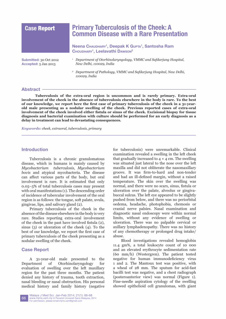
Abstract Tuberculosis of the extra-oral region is uncommon and is rarely primary. Extra-oralinvolvementofthecheekintheabsenceoftuberculosiselsewhereinthebodyisrare.Tothebestofourknowledge,wereportherethefirstcaseofprimarytuberculosisofthecheekina31-year-old male presenting as a nodular swelling of the cheek. Previous reported cases of extra-oralinvolvementofthecheekinvolvedeitherfistulaorsinusofthecheek.Excisionalbiopsyfortissuediagnosisandbacterialexaminationwithcultureshouldbeperformedforanearlydiagnosisasadelayintreatmentcanleadtodevastatingconsequences.
Keywords:cheek,extraoral,tuberculosis,primary
Introduction
Tuberculosis is a chronic granulomatous disease, which in humans is mainly caused by Mycobacterium tuberculosis, Mycobacteriumbovis and atypical mycobacteria. The disease can affect various parts of the body, but oral involvement is rare. It is estimated that only 0.05–5% of total tuberculosis cases may present with oral manifestations (1). The descending order of incidence of tubercular involvement of the oral region is as follows: the tongue, soft palate, uvula, gingivae, lips, and salivary gland (2). Primary tuberculosis of the cheek in the absence of the disease elsewhere in the body is very rare. Studies reporting extra-oral involvement of the cheek in the past have involved fistula (2), sinus (3) or ulceration of the cheek (4). To the best of our knowledge, we report the first case of primary tuberculosis of the cheek presenting as a nodular swelling of the cheek.
Case Report
A 31-year-old male presented to the Department of Otorhinolaryngology for evaluation of swelling over the left maxillary region for the past three months. The patient denied any history of trauma, tooth extraction, nasal bleeding or nasal obstruction. His personal medical history and family history (negative
for tuberculosis) were unremarkable. Clinical examination revealed a swelling in the left cheek that gradually increased to 4 × 4 cm. The swelling was situated just lateral to the nose over the left maxilla and did not obliterate the nasomaxillary groove. It was firm-to-hard and non-tender and had an ill-defined margin, without a raised temperature. The skin over the swelling was normal, and there were no scars, sinus, fistula or ulceration over the palate, alveolus or gingiva-buccal sulcus. The left eye appeared to be slightly pushed from below, and there was no periorbital oedema, headache, photophobia, chemosis or cranial nerve palsies. Nasal examination and diagnostic nasal endoscopy were within normal limits, without any evidence of swelling or ulceration. There was no palpable cervical or axillary lymphadenopathy. There was no history of any chemotherapy or prolonged drug intake/abuse. Blood investigations revealed hemoglobin 11.4 gm%, a total leukocyte count of 10 000 and an elevated erythrocyte sedimentation rate (60 mm/h) (Westergren). The patient tested negative for human immunodeficiency virus 1 and 2. The Mantoux test was positive, with a wheal of 28 mm. The sputum for acid-fast bacilli test was negative, and a chest radiograph (posteroanterior view) was normal (Figure 1). Fine-needle aspiration cytology of the swelling showed epithelioid cell granulomas, with giant
66Malays J Med Sci. Jan-Feb 2014; 21(1): 66-68

Case Report | Primary tuberculosis of cheek
www.mjms.usm.my 67
cells, suggestive of a granulomatous lesion. A contrast-enhanced computed tomography of the neck (Figure 2) revealed a soft tissue lesion anterior to the left maxillary sinus, without any bony extensions. In light of the findings described above, an excisional biopsy under general anaesthesia was planned. A sub-labial incision revealed an encapsulated mass anterior to the anterolateral wall of the maxilla. The entire mass was dissected all around and totally excised. An infra-orbital bundle was engulfed in the mass and had to be sacrificed. The immediate post-operative period was uneventful, and the patient was discharged. Histopathological analysis of the excisional biopsy from the cheek showed lymphoplasmocytic infiltrate and multiple necrotising caseating epithelioid cell granulomas, with Langhans giant cells diagnostic of tuberculosis (Figure 3). In BACTEC culture media, acid-fast bacilli growth was present after 10 days, and the bacteria were sensitive to all first-line anti-tubercular drugs. Anti-tubercular treatment started with a four-drug regime: Isoniazid (500 mg), Rifampicin (450 mg), Ethambutol (1200 mg), and Pyrazinamide (1500 mg) three times a week for the first two months. Thereafter, two drugs, isoniazid (600 mg) and rifampicin (450 mg) were taken three times a week for four months. During the follow-up period of one year, neither swelling nor ulceration was noted at the surgical site.
Discussion
Tuberculosis of the cheek is uncommon and is, in most cases, secondary to tuberculosis elsewhere in the body. Primary tuberculosis is rare and is caused by infection withM. tuberculosis. It is characterised by the formation of a primary complex in the lungs because the bacterium has a particular affinity for the lungs. Primary tuberculosis is considered as an initial infection of any part of the body. Secondary tuberculosis is the result of reinfection with the tubercular bacillus or reactivation of dormant endogenous bacilli. Secondary tuberculosis of the cheek is accompanied by lesions in the lung, lymph nodes or any other organ system of the body that can be diagnosed by usual signs and symptoms. However, the diagnosis of primary tuberculosis of the cheek poses a difficulty for clinicians. The tongue is the most common site for tuberculosis in the oral cavity (5). Other sites of infection in the oral cavity include the gingivae, floor of the mouth, palate, lips and buccal mucosa (4). Common sites for tuberculosis in the extra-oral region include the
Figure 1: Chest X-ray of the patient showing bilaterally normal lung parenchyma, trachea and mediastinum, without any fibrocavitory lesion.
jaw bone and salivary glands. Tuberculosis mainly occurs at these sites as a result of deep extension of gingival lesions or infected post-extraction tooth sockets through either haematogenous or lymphatic spread (6). In our case, we did not find any evidence of tuberculosis elsewhere in the body. The sputum for acid-fast bacilli test and chest x-ray PA view were normal, and there were no signs and symptoms of systemic tuberculosis. Hathiram et al. (4) reported a case that presented as an exophytic growth of the cheek that was diagnosed as carcinoma but confirmed as tuberculoma of the cheek by punch biopsy.
Figure2:Enhanced axial computed tomography (CT) image showing a mildly enhanced, well-defined soft tissue density lesion anterior to the left maxillary sinus (red arrow), without any evidence of involvement of underlying bone.

68 www.mjms.usm.my
Malays J Med Sci. Jan-Feb 2014; 21(1): 66-68
The pathogenesis of primary tuberculosis of the cheek remains unclear. Usually, the microorganism requires disruption of the oral mucosa to become pathogenic. Kumar et al. (7) reported that the habit of brushing the teeth with twigs of neem (Azadirachtaindica) in rural India sometimes causes trauma to the palate and thus predisposes the wound to seeding with M. tuberculosis. Our patient also comes from a rural part of India where this practice exists, and this may have caused the mucosal breech. The commonest manifestation of oral tuberculosis is an ulcerative lesion of the mucosa, but it may present in a variety of other forms such as nodules, tuberculoma and peri-apical granulomas (8). The extra-oral involvement of the cheek is very uncommon. In our case, it involved a nodular swelling of the cheek. In the absence of any systemic signs and symptoms of tuberculosis, as in our case, it is advisable to perform an excisional biopsy for tissue diagnosis and bacteriological examination for a definitive diagnosis. Any delay in treatment can lead to devastating consequences.
Acknowledgement
There is neither conflict of interest between authors nor financial or material support from any firm, funding organization or sponsor in design and conduct of the study; collection, management, analysis, and interpretation of the data; and preparation, review, or approval of the manuscript.
Figure 3: Biopsy from the cheek showing multiple caseating epithelioid cell granulomas, with Langhans giant cells (encircled) (hematoxylin and eosin staining, 20× magnification).
Conflict of Interest
None.
Funds
None.
Authors’ Contributions
Conception and design: NC, DKGDrafting of the article: NC, DKG, LD, SRCCritical revision of the article for the important intellectual content: NC, DKG, LDCollection and assembly of data: SRC
Correspondence
Dr Deepak K GuptaMBBS, MCh (Banaras Hindu University)Department of otorhinolaryngologyVMMC and Safdarjung HospitalNew Delhi, 110029India Tel: +91112 7490 397Email: [email protected]
References
1. Ito FA, de Andrade CR, Vargas PA, Jorge J, Lopes MA. Primary tuberculosis of oral cavity. Oral Dis.2005;11(1):50–53. doi: 10.1111/j.1601-0825.2004.01 055.x.
2. Purohit SD, Mathur BB, Gupta PR, Agarwal KC, Hathi HH. Tuberculous fistula of cheek. Report of a case. OralSurgOralMedOralPath. 1985;60(1):41–42.
3. Gillbe GV. A tuberculous sinus of the cheek. BrJOralSurg.1968;5(3):203–205.
4. Hathiram BT, Grewal DS, Irani DK, Tankwal PM, Patnakar M. Tuberculoma of cheek: a case report. JLaryngolOtol.1997;111(9):872–873.
5. Gupta A, Shinde KJ, Bhardwaj I. Primary lingual tuberculosis- A case report. J Laryngol Otol.1998;112(1):86–87.
6. Ebenezer J, Samuel R, Mathew GC, Koshy S, Chacko RK, Jesudason MV. Primary oral tuberculosis: report of two cases.IndianJDentRes.2006;17(1):41–44.
7. Kumar V, Singh AP, Meher R, Raj A. Primary tuberculosis of oral cavity: a rare entity revisited. IndianJPediatr. 2011;78(3):354–356. doi: 10.1007/s12098-010-0266-z.
8. Shafer WG, Hine MK, Levy MB. A textbook of oralpathology.4th ed. Philadelphia (US): WB Saunders Co; 1983. p. 340–440.