Reparative Reading, Post-structuralist Hermeneutics and T. S. Eliot's ...

ANNALS OFSURGERYVOL. 122 SEPTEMBER, 1945 No. 3
REPARATIVE SURGERY OF COMPOUND BATTLE FRACTURESIN THE MEDITERRANEAN THEATER OF OPERATIONS
LT. COL. O$cAR P. HAMPTON, JR., M.C., A.U.S.ORTHOPEDIC CONSULTANT, M.T.O., U.S.A.
EXTREMITY SURGERY of the war wounded is divided into three phases':i. Initial: The primary excisional surgery performed in Army Field
and Evacuation Hospitals as soon after wounding as possible (usually 8-24hours) directed at the saving of life and limb and the prevention of infection.
2. Reparative: Performed in fixed hospitals at the Base directed at woundhealing, anatomic and functional restoration of the extremity and rehabili-tation or safe transport to the Zone of Interior in a lag-period of treatmentafter all that is necessary in the overseas Theater to minimize permanentdisability has been achieved.
3. Reconstructive: Performed in the hospitals of the Zone of Interiordirected at correction of residual defects and deformities resulting from thewar wound.
Reparative surgery is largely dependent upon adequate initial surgery,2including bold incision, excision of dead and devitalized tissue, good drainageof the wound depths and dead space, an occlusive dressing, and adequateimmobilization. It is facilitated by a short line of evacuation which permitstransferring of the patient within a few days after wounding to a BaseHospital that is functioning close behind the combat zone. When theseconditions are ideally fulfilled, surgical repair of open wounds is a logicaland successful procedure in wound management. If the initial surgery hasbeen inadequate, additional excisional surgery anticipating staged repair isusually necessary to prevent or cut short wound sepsis. Successful repara-tive surgery may make reconstructive surgery unnecessary or may returnthe patient to the Zone of Interior in a condition that will permit earlierand complete reconstructive surgery with enhanced chances of success.
CONCEPT' OF REPARATIVE SURGERY
Since the early days of this Theater in North Africa, repeated observa-tion has established that wounds with obviously retained devitalized tissuebecame septic and drained pus profusely. Large hematomata in undraineddead space often decomposed into pus. Systemic or local chemotherapy,or their combination, did not prevent wound sepsis in the presence of deadtissue. Once wound sepsis had developed, there was continuing local
289

OSCAR P. HAMPTON, JR. Annals of Surg9e
necrosis of living tissue and a vicious circle was established. Large granu-lating areas exuding plasma were observed to develop and harbor surfaceinfection. These septic wounds, however, seldom manifested the cardinalsigns of inflammation. Conversely, wounds which were free of devitalizedtissue on their admission to Base Hospitals were clean grossly and free ofsepsis. As healing by granulation occurred, many developed surfaceinfection.
Following these observations, reparative surgery began with the successfulsecondary closure of clinically clean soft-part wounds, even though culturestaken preoperatively demonstrated the presence of an aerobic and anerobicbacterial flora."1 3' 4, 9 A wound free of devitalized tissue sutured at its primarydressing, four or five days after initial surgery, without dead space, hematomain the depths, or excessive tension, and supported by a good pressure dressingand adequate splinting healed regardless of the bacterial flora. Successful.suture depended upon a proper clinical appraisal of the wound, atraumatictechnic, and surgical limitations imposed by the character of the defect. Theclinical observations supported by the bacteriologic studies of Lyons andRustigian3 on war wounds which demonstrated that clean wounds andwounds with established sepsis may have comparable bacterial flora, haveled to the following concept:
Wound sepsis becomes established as a result of the septic decompositionof devitalized tissue, including hematoma in dead space, rather than fronithe action of bacteria on living tissue. The devitalized tissue serves as apabulum3 for wound pathogens. If the pabulum is not present and is notcreated by surgery, and if living tissue is protected from invasive infectionby an effective antibacterial agent, the bacterial flora of an open wound maybe disregarded, wound sepsis need not be feared, and any indicated reparativeprocedure may be performed under established surgical principles with theanticipation of good wound healing.
Battle-incurred compound fractures demand special considerations whencompared with those resulting from traffic and other accidents. Battlefractures are always compounded from "without-in" by missiles which havepassed through clothing, often soaked with the grime and mud of the battle-field. The great majority are caused by high explosive shell fragmentsresulting in extensive muscle and bone damage. Clothing, wood, metallicforeign bodies, cement and mud are frequently buried in the depths of thewound far removed from their point of entry. In spite of excellent fieldservice in this Theater for evacuation of the wounded from the battlefieldto a hospital equipped for surgery, the time-interval between woundingand initial surgery usually exceeds I2 and often 24 hours. Accordingly, thewounds are not merely contaminated but are heavily infected with bacteria.The fractures are usually severely comminuted, often with bone loss. Theseinjuries require long incisions, often multiple, for adequate exposure -of thedlevitalized tissue and foreign material and to permit adequate excisionalsurgery. The large, often irregular, or multiple wounds made by surgeon
290

Volume 122 COMPOUND BATTLE FRACTURESNumber 3
and missile must remain unsutured following initial surgery. They, together'vith muscle andl bone loss incident to the injury and the suirgery, present apicture and a problem peculiar to military surgery.
Reparative surgery recognizes that complete excision of the devitalizedtissue in compound battle fractures is usually impossible or impracticable.At initial surgery, completely detached bone fragments- are deemed to beavascular tissue and potential sequestra and, therefore, they are removedtogether with the devitalized soft tissue. Fragments with complete or partialperiosteal attachment are preserved projected towards union of the fracture.Muscle, fascia, tendon, and periosteum attached to the fragments and thedenuded cortex of bone constitute questionable devitalized tissue whichprobably remains in every fracture. Blood clot may form in an undrainedarea particularly in the dead space of the unreduced fracture or the defectcreated by the necessary muscle excision. The wounds of these injurieswhich have had adequate initial surgery have been observed in many instancesto be draining profusely on admission to the Base Hospital. Unless woundsepsis became established, the profuse drainage ceased after several days.The discharge has been attributed to the spontaneous sequesfration of theresiduum of devitalized tissue and has been termed "the products of injurynecrosis."'6 However, it is recognized that the residual devitalized soft tissue,partially denuded fragments, or dead space with a contaminated blood clotmay be a nidus of infection with wound pathogens,3 leading to abscess forma-tion with continuing necrosis of living tissue within the wound. These aresome of the factors that create specialized problems in the reparativemanagement of compound fractures.
Every method of treating compound fractures seeks to obtain bony unionwith minimum deformity, a healed wound and maximum function of theextremity. During the year 1943 and early I944, in the North AfricanTheater of Operations, compound battle fractures were treated by a modifiedOrr method,6 consisting of an open wound, infrequent occlusive dressingsand traction or plaster immobilization. Wound healing by granulation andthe resultant scar formation were accepted as necessary undesirables. Incertain instances poor fracture results, malunions, or inevitable nonunionswere accepted rather than risk "a stirring-up" of the wound by an open reduc-tion. Wound sepsis with continuing local necrosis of living tissues becameestablished in many cases, particularly in the exposed fracture sites of sub-cutaneous bones. Wounds with gross retained dead tissue were oftenmanaged by a "hands-off" policy which anticipated the spontaneous seques-tration of the dead tissue rather than a delayed surgical excision. Theunreduced fracture which called for repeated manipulations or adjustmentsof position in traction was particularly vulnerable to sepsis. A properappraisal of the problem demonstrated the need for improvement which couldl)e achieved only by a changing approach.
With a background of a year's experience, study and observation in the'rheater of Operations, reparative surgery of compound fractures was
291

OSCAR P. HAMPTON, JR. Annals of SurgerySeptember, 1945
visualized and partially planned during the late months of I943. Followingin the wake of successful reparative management of soft-part wounds, itwas initiated during the first quarter of I944, catalyzed by the availability ofpenicillin therapy.* During the memorable days of Cassino and Anzio, itdeveloped into a plan of management based upon continuing pooled experiencesof the Theater surgeons.
In this Theater certain previously planned favorable operations factorsobtained:
i. Experienced Forward Hospitals with standardized principles of ex-cisional surgery and transportation splinting.
2. Short chain of evacuation, ambulance and train (Cassino), and airevacuation (Anzio) from Forward to Base Hospital predisposing to safeearly transfer of the wounded.
3. Experienced Base Hospitals functioning close behind the combat zone.4. A bed status in the Base that permitted the patients to be held for
reparative surgery and rehabilitation or transfer to the Zone of Interior.5. An Army blood bank supplying low titer-o blood to and augmenting
that drawn in Forward Hospitals and unit banks in each Base Hospitalsupplying type specific blood.
Such was the prologue for reparative surgery of compound fractures atthe "Fall of Rome."
The reparative surgical program for compound fractures has as itsobjectives: i. Minimum wound sepsis. 2. Improved fracture reduction andstabilization. 3. More rapid wound healing, with minimum scar formation.4. Maximum functional restoration of the extremity.
These objectives are approached by a plan of management based upon:(a) Blood replacement. (b) Chemotherapy. (c) Surgery. Good surgeryis the keystone of the program, with blood replacement and chemotherapyas adjuncts.
BLOOD REPLACEMENT
In spite of what is considered to be an adequate use of blood replacementtherapy in the forward area to combat shock, traumatic and operative, patientswith compound fractures of the long bones have consistently shown anemiaon admission to the Base. Tables I and II show the hematocrit readingsobtained in two groups of battle casualties on admission to OrthopedicSections of two General Hospitals. The tables are separated to allowcolumn 4 of Table I to be presented as evidence of the blood loss sustainedby a patient with a battle-incurred fracture of the femur. It will be notedthat 50 per cent of this group had hematocrits under 30.
* The counsel and active participation of Major Champ Lyons, M.C., of the staff ofSuirgical Consultant, M.A.T.O.U.S.A., was invaluable in the development of the program.
292

Volume 122 COMPOUND BATTLE FRACTURESNumber S
TABLE 1(7) No. Cases ofHematocrit No. Cases Percentage F.C.C. FemurUnder 30 .... ;........ 33 24% 1931-35 .24 17%7 936-40 .56 40% 8Over 40 ... '25 18% 2
138 100% 38
TABLE I I (8)Hematocrit No. Cases PercentageUnder 30 ........................ 37 22%30-36 .......................... 44 26%37-42 .......................... 5432Over42 .......................... 31 19FO
166 100%
In order to correct the secondary anemia, type-specific cross-matchedwhole blood is given preoperatively in an effort to obtain an hematocrit readingof 40, or better, in all compound fractures on which any major reparativeprocedure is contemplated. Preoperative blood requirements are calculatedon the basis of 500 cc. for each three to four points deficit of the hematocrit.There is no proof that this therapy is necessary but it is accepted a priorithat the wounded man with an hematocrit of 40 is in better condition towithstand a long anesthesia and operative procedure than if his anemia isuncorrected. Additional blood to compensate for operative loss is frequentlygiven during the operation and postoperatively if anemia is reestablished.Repeated observations by many surgeons that the patients tolerated welland "looked good" after the surgery is sufficient to justify this use of bloodreplacement therapy. It has not been possible to compile confirmatoryevidence, but blood therapy is believed to aid in the prevention of chronicsepsis, and in wound healing.
CHEMOTHERAPY
Penicillin has been accepted as the most powerful available antibacterialagent to which the bacterial flora, aerobe and anerobe, of war wounds havebeen proven sensitive: It is recognized that penicillin will protect livingtissue against invasive infection but it is also recognized that penicillin will notsterilize dead, devitalized or avascular tissue which, inadvisably or of neces-sity, remains in the wound, nor will it prevent the septic decomposition ofa contaminated blood clot which collects in unobliterated or undrained deadspace or neutralize locally necrotizing enzymes in undrained pus.9' 10 There-fore, penicillin is used for the protection of the living tissue from the invasiveaction of bacteria accepted as present in the residuum of devitalized tissueremaining in compound fractures. The agent will not sterilize that residuum,therefore, surgical measures are necessary for its management. Therapy iscontinued until wound surgery has been completed, wound healing has beensufficiently obtained and the residuum of devitalized tissue has sequestratedand drained off or has absorbed. Penicillin therapy is used to provide anincreased margin of safety in the performance of the indicated surgery.
Penicillin is used routinely and no advantage can be seen in attemptingthe surgery without it. While there is no proof that it is a necessity, and
293

OSCAR P. HAMPTON, JR. AnnalsIfSurgery
althouglh successful reparative procedures on compound fractures without ithave been reported, there are two cases on record in which gas gangreneand death followed reparative operations upon compound fractures withoutthe use of the agent. No deaths or serious untoward results from sepsishave been reported in similar cases receiving penicillin therapy as an adjunctto the surgery.
Systemic administration of penicillin, 25,000 units intramuscularly evervthree hours is the basic therapy. Local instillation into joints, I,OOO unitsper cc. is supplemental. Otherwise no local therapy is used in extremitysurgery.
Patients with compound fractures as a rule are receiving penicillinitlherapy in the Evacuation or Field Hospital when they are transferred tothe Base. Therapy is reinstituted on admission to the Base Hospital andcontinued until five to ten days after the last traumatizing surgery (wlhiclmay produce more devitalized tissue) until as outlined above, the woundhias sufficiently healed and contaminated devitalized tissue is no longerin evidence.
SURGERY
The surgery is aggressive rather than passive. Wounds are explored toinsure the adequacy of the initial surgery, fractures may be fixed internallyand soft-part wounds may be sutured. But the success of the programdepends upon the quality of surgical judgment and technic. Every caserequires a decision as to the anesthetic; the extent of further excisionalsurgery; whether to use some form of internal fixation; the extent ofclosure of the compounding wound possibly aided by relaxing incisions orflaps; whether, where and how to drain; and the postoperative method ofobtaining or maintaining reduction.
Five to ten days will have elapsed since initial surgery before the patientwith a compound fracture is ready for reparative surgery. With adequateblood replacement, continuing penicillin therapy and good roentgenogramsmade in the Base Hospital, he is anesthetized in an operating room preparedfor any indicated surgery, be it excisional or reparative. There, the Evacua-tion Hospital encasement and dressing are removed, the extremity'preparedand draped and the wound inspected. A pneumatic tourniquet is frequentlyused not only to provide a '"dry" operative field but to minimize blood losson the table.
Wound Revision: The entire wound including the fracture site is exposedby gentle retraction and explored to insure the adequacy of the initial surgery.Incisions are enlarged if necessary to facilitate exposure. Any remainingforeign material, accessible foreign bo(lies, totally detached bone fragmentsor devitalized soft tissue are remuoved. Old blood clot is cleaned out. Meansby which dead space may be obliterated or drained are considered. Furtherexcisional surgery is not infrequently indicated. Failure to perform wound
294
Volumie 122Number 3 COMPOUND BATTLE FRACTURES
revision soon after admission to the base is believed to account for manypoor results seen in the past. Muscle tissue which appeared viable at initialsurgery and, therefore, was not removed may have necrosed in the interim.\Vhen the remaining devitalized tissue of dirty wounds was not excised,wound sepsis with continuing local necrosis of living tissues was often estab-lished. Late wound exploration in cases of established sepsis has frequentlyrevealed foreign material, or totally detached indriven fragments of corticalbone. Their removal plus proper reparative surgery was followed bysubsidence of wound sepsis (Cases I, 2 and I5). Reduction to the minimumof residual devitalized tissue is the most important step towards the mini-mizing of sepsis and is the keystone of the plan of management. Whensepsis intervenes, reparative measures are doomed to failure, delayed ornonunion may follow, and wound healing will be postponed or prevented.
Fracture Management: The thorough wound visualization of reparativesurgery affords the advantages of open reduction of fractures. Interveningsoft parts are removed. Fragment ends caught in muscle are released..Rotated and twisted fragments are aligned. Complete appraisal of the problemat hand by direct vision as well as by roentgenogram is valuable in deter-mining the means of obtaining and maintaining fracture reduction. Thebest possible fracture reduction is the objective of fracture management.In addition to the anticipated favorable anatomic result, stabilized fracturereduction eliminates the dead space of an unreduced fracture, and avoidstraumatizing multiple manipulations or adjustments of traction in delayedefforts to effect reduction, thereby minimizing the chances of sepsis. In aneffort to achieve the maximum fracture reduction, internal fixation is some-times used under the following principles:
Internal Fixation: Internal fixation is by no means an objective of theprogram, and it is usually neither advisable nor possible because of severecomminution. However, the program permits the use of internal fixationwith the limitations outlined below when it is indicated to maintain fracturereduction. Eighteen and eight molybdenum steel is relatively inert in thetissues and is not considered per se detrimental to wound healing. The fixa-tion may be plating, multiple screws or wire loops.
The rigid stabilization of the fracture in reduction by a plate or multiplescrews offers certain advantages (Cases 3, 4, 8, II, I2):
i. Anatomic opposition and alignment anticipating faster bony unionwith no deformity.
2. The dead space and traumatizing manipulations outlined above areavoided.
3. Handling of the extremity for necessary subsequent wound care isfacilitated.
4. Early joint motion and muscle exercise anticipating a more rapidreturn to function may be permitted.
5. The management of concurrent injuries which preclude traction andrequire repeated trips to the operating room is facilitated. -
295

OSCAR P. HAMPTON, JR. Aealsmof Surgery
However, the use of internal fixation is limited by three factors otherthan comminution:
Ix The desire to minimize intrawound trauma, e.g., retractor pull, vesselligatures-which creates additional devitalized tissue.
2. Interference with the covering of all exposed bone cortex with vascularsoft parts (to be discussed under closure) (Case 5).
3. The desire to avoid periosteal stripping with its danger of massivesequestration which may be necessary to permit the application of a boneplate (Case 6).
When periosteum is stripped from bone, the outer cortex will die.11A basis for this statement is the experimental observation in dogs that theperiosteal blood supply nourishes the outer third of the cortex of shafts oflong bones.12 If there is no sepsis, the dying bone is replaced by newbone as one process. But if sepsis is present reattachment of periosteum orother soft parts is prevented and the outer cortex becomes a sequestrum.Therefore periosteal stripping which deprives, the outer cortex of bone of itsnourishment is an important consideration in surgery in a known "infected"field. Practically, if the wound is appraised clean and the other factors arefavorable, especially the availability of vascular soft parts, as in the arm orthigh, there is less hesitancy in stripping sufficient periosteum to permit theindicated surgery but if it is appraised "dirty"* or doubtful, the stripping isrestricted or avoided.
Whlere the factors that might restrict its use are not unfavorable andthe fracture permits, rigid internal fixation is frequently employed in orderto gain the advantages of a well-reduced and stabilized fracture. Fixationthrough the compounding wound is at times practical but has the disadvan-tages of retraumatizing tissue and placing the metal on bone usually devoidof periosteum and at the bottom of dead space created by excision of devital-ized muscle. Therefore, for plating, a separate standard approach to thefracture which permits covering of the bone and metal by periosteum andvascular soft parts is advisable (Case 4).
Every refinement in the technic of internal fixation is considered impor-tant. There must be intimate contact of the fragments; plate should besufficiently long (Murray13 recommends that the length of the plate be fivetimes the diameter of the bone at the fracture); drill holes should be onlyslightly larger than the shaft of the screw, less the threads, preferably atright angles to the bone and wobbling of the drill or a drill bit at an angleshould be avoided to prevent scoring of the drill hole. (Electrically drivendrills require extreme caution to prevent burning of the bone); screws
*The term "dirty wound," as a contrast to clean wound is in common usage inthis Theater and is herein used to describe the wound which is visualized to containgross, unexcised devitalized tissue, is discharging pus, often foul-smelling, from thedepths of the wound, or is covered by a gray, slimy purulent exudate. Cardinal signs ofinflammation are not necessarily present in the dirty wound. When they are present,the wound is said to present "invasive infection."186
296
Volume 122Number 8 COMPOUND BATTLE FRACTURES
should be held "true," inserted by a steady hand and be long enough toprotrude through the opposite cortex. Oblique screws across the fracture ina plane at or near go degrees from that of the plate will increase the rigidity.
In actual practice when internal fixation is deemed indicated multiplescrew (two or more) fixation is frequently used (Cases 7, 8 and i i). Manyfractures by their obliquity lend themselves to it, little or no additionalperiosteal stripping is required to permit placement of the screws, andintrawound trauma is not excessive. If the fracture does not permit rigidfixation because of comminution, one or more wire loops may be used to holdmajor fragments in approximation. These can usually be placed withoutadditional periosteal stripping, a factor of particular importance in a woundwith recognized established sepsis. In comminuted fractures with segmentalbone loss, wire loops permit approximation of the major fragments (Cases9 and io).
Bony union is a prime consideration in any fracture and contact of thefragments greatly enhances the chances of union. Therefore the shorteningof an extremity to overcome segmental loss and obtain contact of fragmentsis often a justifiable indicated procedure that is permitted by reparative frac-ture surgery. Nerve trunk or muscle group deficits associated with a fracturemay indicate the deliberate removal of attached bone fragments and shorteningof the extremity, thereby permitting restoration of continuity of all thesevered major structures projected towards the maximum functional restora-tion of the extremity instead of a good fracture result as determined by theroentgenogram.
Wound Closure and Drainage: Wound closure is premised upon adequateinitial surgery resulting in a clinically clean wound requiring little or nowound revision or traumatizing surgery and upon the feasibility of obliteratingor draining dependently the residual dead space. The lag-period betweeninitial and reparative surgery permits drainage of the products of injurynecrosis. If initial surgery has been inadequate, resulting in a clinicallydirty wound requiring extensive excisional surgery at wound revision, woundclosure must be staged until after an additional lag-period for open drainage.
The hazards of an open wound in a compound fracture are the sequestra-tion and sloughing of exposed bone cortex, tendon and fascia, plus reinfectionat dressings and slow wound healing by granulation. The advantage of anopen wound is continuing drainage from the depths of the wound untilhealing by granulation has sealed-off the fracture site. The gaping woundforms a natural channel for drainage. However, when it is not dependentand sepsis intervenes, there may be pocketing, puddling or pooling of pus inthe fracture site or adjacent fascial planes with continuing local necrosis ofthe collagenous tissues.
Reparative surgery of compound fractures recognizes and attempts toovercome by wound closure the hazards of the open wound but also recognizesthe advisability of a means of egress for the possible septic breakdown of anyresidual devitalized tissue not yet separated and of a contaminated hematoma
297

OSCAR P. HAMPTON, JR. Annals of SurgerySeptember, 1945
in unobliterated dead space. In the uninfected field, e.g., the simple fractureor following a clean surgical operation, body processes will absorb devitalizedtissue and blood clot. In the infected* field the same absorption might occurbut the complete closure of wounds of compound fractures is justified onlywhen the pabulum for wound sepsis is nil. A deep abscess about the fracturesite underneath a sutured or healed epithelial bridge may produce irreparabledamage. Therefore, an increased margin of safety can be obtained byproviding drainage, dependent if possible, utilizing wounds or counterincisionsas indicated. Drains are inserted so as not to cause tissue necrosis and areremoved between the third and tenth day depending upon the drainage indica-tions before rigid sinus formation occurs.
The problem of closure of the compounding wound is approached withthe major objective of covering exposed bone cortex, tendon and fascia withhlealthy soft parts and the minor objective of reducing skin defects to a sizethat is compatible with adequate drainage. The sliding or rotation of flaps isoften employed to gain these objectives (Cases ii and 13). The hazard ofperiosteal stripping finds its antithesis in the value of covering bone exposedby trauma. Soft parts must adhere to the bony cortex to permit its "revascu-larization," whereby the dying bone may be absorbed and replaced by newliving bone. Otherwise sequestration is inevitable (Case I5). Therefore,wound closure is designed to obviate the hazards of exposed bone cortex thesalvage of which is probably the most important attainment of reparativesurgery of compound fractures (Cases io, II, 13 and 14).
When soft-part masses fall over and protect structures that are vulnerableto exposure, e.g., the muscles of the thigh over the femur, the major hazardof the open wound is removed and surgical closure is of less importance.The open wound may be the optimum method for free drainage and isutilized when closure, complete or partial, affords no definite advantages.The closure of a small wound compounding a fracture of the femur isinconsequential as the soft parts will be healed before the bone unites.2The open wound is particularly advantageous for drainage following trauma-tizing surgery, e.g., extensive wound revision for dirty wounds or difficultinternal fixations (Cases 3 and 4). In such cases, skin suture is avoidedor staged. However, skin defects usually may be reduced and still permitadequate drainage. When the wound is clean and requires no traumatizingsurgery, and when dead space is at a minimum, skin may be sutured com-pletely or with a small drain of dry fine-mesh gauze or soft rubber tissueemerging through the most dependent portion of the wound or a counter-incision (Cases 8, 9 and io). When two wounds compound the fracture,one may be closed completely and the other (usually the more dependent)left open or partially closed, with or without drainage material. Surgicallimitations, i.e., tension, dead space or difficult dependent drainage as inanterior wounds over fractures of the tibia, may preclude wound suture and
* Infected, herein, denotes only the recognized presence of a bacterial flora capableof establishing wound sepsis in the presence of dead tissue.
298

Volume 122Numb,elr 3 COMPOUND BATTLE FRACTURES
the wound may require loose packing anticipating healing from the bottomiiby granulation (Orr method) (Cases 7, 8 and I4), but in many of these,partial wound closure may be employed to cover exposed cortex of bone.Partial wound closure in reducing the magnitude of compounding woundsfacilitates the sealing-off of the fracture site by healing processes rather thanattempting the immediate conversion of the compound to a simple fractureand, therefore, it is frequently employed to reduce the size of defects of thecompounding wounds. The reduction to a minimum of skin defects mini-mizes scar, promotes earlier wound healing and leads to improved functionalresults.
Postoperative Managemtent: Immediate adequate reduction and stabiliza-tion of the fracture is essential to reduce dead space, prevent the continuingtrauma of fragment ends, and provide wound rest to promote wound healingIn many cases sheet wadding and plaster encasements for immobilization otthe fracture in reduiction provide pressure dressings for the control of deadspace and wound edema. When skeletal traction is the method of choice forpostoperative fracture management, the wounds are supported by bulkydressings and elastic bandages. Variations of, and adjuncts to, skeletaltraction methods are frequently employed in obtaining and maintainingfracture reduction, e.g., Army leg splint, "Navy" traction, two-wire traction14(Plate i). Anesthesia is often continued until the completion of the tractionset-up on the ward permitting immediate manual reduction verified roentgeno-logically. By this plan, reduction in traction is quickly obtained, and it ismaintained by the skeletal traction. Fractures fixed internally are alsoimmobilized externally by plaster or skeletal traction. In the postoperativemanagement of internally stabilized fractures of the femur, skeletal tractionaffords added protection and permits adequate wound care, early knee motionand physiotherapy.
The case reports and illustrations which follow are presented to illustratethe details of the principles of reparative surgery as applied to compoundfractures. Each case demonstrates the application or omission of one ormore of the principles covered in the manuscript. While the majority ofthe cases in the group illustrate results to be anticipated by reparative surgery,cases illustrating certain pitfalls that occurred during the formative stage ofthe program are included to emphasize certain conclusions.
Internal fixation has been used in nine cases, herein reported, includingtwo cases of wire approximations of major fragments. The predominanceof internal fixation in these reports should not be interpreted to mean thatthe method is employed in the majority of cases, for such is far from true.The group of cases included illustrate the indications for the,method andconcurrently, other principles. Skeletal traction (Plate i-a-e) is the usualmethod of obtaining and maintaining fracture reduction when traction isnecessary.
Penicillin therapy, unless otherwise stated, and blood replacement therapywere used in each case according to the plan outlined in the manuscript.
299

OSCAR P. HAMPTON, JR. Annals of SurgerySeptember, 1945
PLATE IA. Fracture of the femoral shaft in the midthird in balanced suspension skeletal traction utilizing
the Army leg splint with the Pierson attachment and a Kirschner wire through the tibial tubercle. Theleg splint.-Pierson method is used in the majority of cases.
B. Fracture of the upper third of the femur in balanced skeletal traction, utilizing the "Navy"method. It is an excellenit method for upper third fractures, with high thigh or posterior wounds.
C. A ward of fractures of the upper third of the femur treated in balanced skeletal traction,employing the "90-90-90" method (the hip, knee and ankle joint position). After a few weeks in this)osition during which posterior wound management and fracture reduction is effected, the leg splint-Pierson method is substituted.
300

pLA:E I
.
B
C301
r< r:
.,:.- el..
OSCAR P. HAMPTON, JR.
I'lAfE I N 1
Annals of SurgerySeptember, 1945
302
Volume 1nNumber 8 I
COMPOUND BATTLE FRACTURES
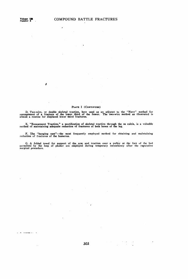
PLATE I (CONTINUED)
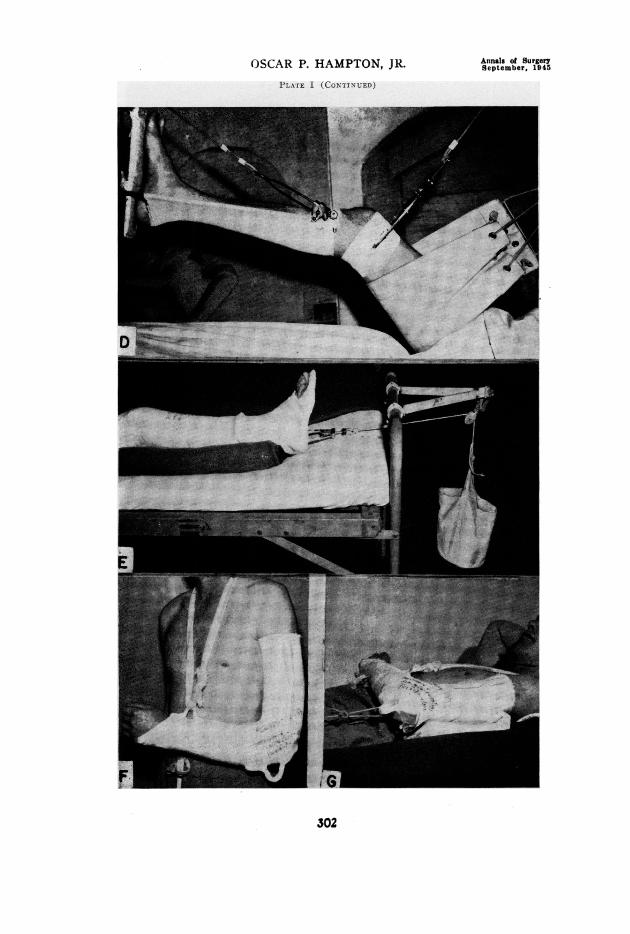
D. Two-wire, or double skeletal traction, here used as an adjunct to the "Navy" method forinanagement of a fracture of the lower third of the femur. The two-wire method as illustrated isalmost a routine for displaced lower third fractures.
E. "Encasement Traction," a modification of skeletal traction through the os calcis, is a valuablemethod of maintaining adequate reduction of fractures of both bones of the leg.
F. The "hanging cast"-the most frequently employed method for obtaining and maintaillingre(luction of fractures of the humerus.
G. A folded towel for support of the arm and traction over a pulley at the foot of the bedlermitted by the loop of plastcr are employed during temporary recumbency after the reparativesurgical procedure.
303

OSCAR P. HAMPTON, JR. Annals of SurgerySeptember, 1945;
> C4~~~~~~~~~~C
.. ., ...3s i a aC's c
E.CU 0B ' o
o 0o
0..
0
>' .~Y O
0 CU
In CIS
be
o C
co.co
U; a
_ Y sO~~~~~h
304

Volume 122 COMPOUND BATTLE FRACTURESNumber 3
_ C ~~~~~~~~~a*0
004
co
Io 0
0) -
wb_w w _ ~~~~~~~~~~~~~~~~~o =1ui 6.;~~~~~~~~~~~~~~~~~
0 t
0 0O
4- 0
.0
04-
beQ
. ~~~~~~~~~~~~~~~~~~~~~~C z
'-.o
!~~ ~ ~~~~~~~~~_beg
4)a
co.A
co
.- I sss a a a~~~~~~~~~~~~_ , . . > a @ @~~~~~~~~~~
_ E X o 3~~~~~~~~~~~~~~~~~~~.
_ ID a J~~~~~~~~~~~~~~w _ Se. . S_) sw__ e v o "~~~~~~rtslk9'.i!~~~~~~~~~~~~~~~~~~~~~~~~~~~~~~~~~~~~~~~~~~~~~~~~~~~~~~~~~~b4
L'.tE n X E~~~~~~~~~~~~~_ | _ s . a~~~~~~~~~~~~~~~~~~~~~~~~~~~~~~a~~ds|\.
305

l
OSCAR P. HAMPTON, JR.
PLATE III
cto
Annals of SurgertSeptember, 1945
i.
.
b
.'-, 5
I
AC
c
pa
PLATE III.-Case 4: a. Compounding wound.b. Posterolateral approach.c. The fracture ieduction.d. Internal fixation.e. Closure of the operative wound, with drainage. The artist has failed to depict comniinutioi
atid an obliquity in the fracture.306

Cu
. P0
X>
d~~~~~~~~~0 oQ
0
rA4.
0 -bv
te~~~~~~~~~~~~~~~~~~~~~~~~~~~~U|co<ua
> _
tv CZ >
CZ
-iAm ~~4) v0C
-o
w~~~~~~~~~~~~~~~~~~~~~~~~~~~~~~~ -U,}-^ C
03Cu
0..
CZ
CZ
0I
307

OSCAR P. HAMPTON, JR.
l
4.
308
V'olumtie 122Number 3 COMPOUND BATTLE FRACTURES
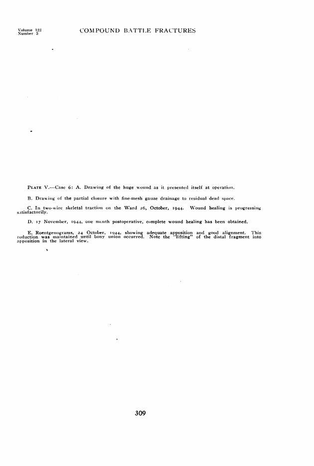
PLATE V.-Case 6: A. Drawinig of the huge wound as it pr-esenited itself at operatioln.
B. Drawinig of the partial closture with fine-mesh gauze drainiage to residual dead space.
C. In two-wire skeletal tractiotn on the Ward 26, October, I944. Wound healing is p)rogressingsatisfactorily.
D. 17 Novemiiber, 1944, onie niuiith postoperative, complete wound healing has beeni obtained.
E. Roentgenograms, 24 October, 1944, showing adequate apposition and good alignment. Thisr-eduction was mainitained until boniy uniion occurred. Note the "lifting" of the distal fragmenit itntoapplosition in the lateral view.
309

OSCAR P. HAMPTON, JR
PLATE VIA
~ ~~ ~ ~~~~~~~~~~~~~.
. : Y { 4~~~~~~~~~:0 . _ :. . :\~~~~_ _ .:: .~~~~~~~~~~.'./1<2W2 4,....
B
PLATE VI.-Case 7: A. I5 July, 1944. The former defect which has filled with granula-tions, without sinus to bone.
B. TS Jully, 1944. The healed operative incision for the plating of the fibula.
310
Annals of SurgerySeptember, 1945

PLAATE VII
A.
.. :..:.... :....
*-N,-.::..
_'S^e0S::50_... :_ ...<.
II
I
.} e:::?_, :s
Ti{-.i F 4; ::
i. ;:
I t . ;
-p-
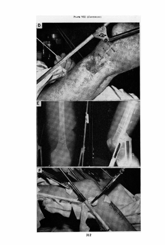
PLATE VII.-Case 8: A. Compounding wounds of the right thigh at reparative surgery,g April, 1944.
B. The internal fixation by four screws. Minimal periosteal stripping was required.C. Partial closure and loose packing of the dead space, with dependent drainage through a
separate incision in the posterolateral fascial plane.311
I! I.M.
PLAtI VII (CoNTIxN7Et)'
P , '-V.
"i,;'" '...SF
NE
A
.I*.s
312
q,l

COMPOUND BATTLE FRACTURES
PLATE VII (CONCLUDED)
D. Sutured and drainage wounds are firmly healed and the dead space has filled withgranulations, without sinus to bone. 6 July,. 1944.
E. Roentgenograms of right femur made postoperative.F. Left lower extremity in two-wire traction showing the healed anterior thigh wound.
The posterior wound was also healed. 6 July, I944.G. Roentgenograms of left femur in two-wire traction.
H. The patient fully ambulatory with all wounds healed and go degrees knee flexion(excellent for lower third battle fractures) in early I945.
313
Volume 122Number 3

OSCAR P. HAMPTON, JR. Annals of SurgerySeptember, 1945
PLATE VIII
PLATE VIII.--Case q): A. Lateral wotiundi in] oelratingroom 30 October, 1944.
B. Medial wounid ini operating room *Zo ( )tle. o 944.Note jarge blood clots in the wouid.
C. Sutured lateral wounid at reparative surgery ;o Octobler,1944.
D. Grafted nmelial wvound 30 October, 1944.
E. A. P. and lateral views of fracture in plaster. Approuxi-mately niie-itnch bionie deficit hias been overtcomIIe liv tie wireloop fixation.
314
o...^::.* :;. .=ih,<* if . .f',':: S
r it . i... ,> S.:..

Volume 122 COMPOUND BATTLE FRACTURESNumber 3
PLATE IX
A
B
PLA-rE IX.-Case I0: A. The united fracture of the humerus in July asshown roentgenologically. The humerus was shortened about one and one-half inches at reparative surgery to obtain contact of fragments.
B. The healed grafted area over the humerus. The graft had been per-formed through a large window in the spica, hence, the. raw area on the chesthad not been grafted.
315

OSCAR P. HAMPTON, JR.PLATE X
Aninals of SurgerySeptember. 194-7.
A
:.;., ,:. .,^.
, .iN 9w o:^. : : .* . ....... ...... .. . . :*:v. ..:: :.:..
:X..: ..... .. ... .. ..........._.X .e,. _
_.^ a__ _
_
_l _
B
C
PLATE X.-Case IxI: A. 15 July, 1944, four weeks after reparativesurgery. Healed sutured wound over the tibia and the granulating relaxingincision. The latter might have been split-skin grafted.
B. The healed lateral wound through which drainage was establishedfor a few days.
C. Roentgenograms made pre- and postoperative. The upper screwmissed the drill hole in the distal cortex.
316

Volume 122 COMPOUND BATTLE FRACTURESNumber 3
0
.0 0Hprt Z / ~~~~~~~~~~~~~~~~~~~, 0)o0
4'
0.04). .. 0.o
0
bo.o
: - ct v @~~4* ~~~~~~~~~~~~o 0 Y Y\_~~~~~~~~~~~~~~~~~~~~~~~~~~~~~~~~~~~~~~~~~~~c C eY
_i~~~~~~~~~~~~~~~~~~~~~~4 Cd bO
} S W edY:3.
_r Gv~~~~~~~~~~~~~~~~~~~~~~~~~~~~~~~r
... e~~~~~~~~~~~~~~~~~~~~~~~~~~~~~~~~~~4
317

OSCAR P. HAMPTON, JR. Annals of SurgetsY.September. 194.5
PL-T,E Xi I
A
(.
PLATE XII.-Case I3: A. Roentgenograms made at the Base Hospital.B. Sutured compounding wounid and skin-grafted relaxing incision of reparative surgery.
C. The healed wound and 95% take on skin graft, two weeks after reparative surgery. Sounidwound healing followed shortly. The patient returned to duty in this Theater.
318

COMPOUND BATTLE FRACTURESP '.'viF XIIT
A
*4h..-v. AA _
... . . .
~~~~~~~~
PLATE XIII.-Case 14: A. Roentgenograms made x6 March, 1944.B. The leg wound in the operating room just prior to reparative surgery.Note the pneumatic tourniquet. Blood loss from upper tibial fractures is usually
severe.
C. The healed sutured projections of the wound and the clean fracturecavity on 23 March, 1944. Sutures were removed, the cavity loosely filled withdry fine-mesh gauze and a plaster encasement applied, anticipating no wounddisturbance for several weeks.
319
Volume 122Number 3

OSCAR P. HAMPTON, JR. AnnaTs of gurgeit1September, 1945
ID
0
oa
g.- I,)
tI00
u au
co co
! * o.C
(U 3.,£l.0q
co co
w 0
._.~
coo o
bO
94' .-
C0.0,w *onSCO .nnA
X
V.
4' 0ceN
(U 4.b(U'.
c.oco
>
c4j-a.04
320

COMPOUND BATTLE FRACTURES
PLATE XIV (CONTINUIED)
.'
vei-efseogstruvr
hol forjdmi~~~~~~~~~~~~~~~~~
K
E. Sequestra removed from the femur (left) and tibia (right), at operation on6 April, 11944.
F. Drawings of the reparative surgery on the femur.
321
Volum. 122Number S

OSCAR P. HAMPTON, JR. September. 1945
ILLUSTRATIVE CASE REPORTS*Case I.-Diagnosis: Penetrating wound of left knee joint, with fracture of medial
femoral condyle, incomplete.Wounded: July I, 1944, 2100 hours, by high explosive shell fragment which em-
bedded in the medial femoral condyle through the articular surface.Initial Surgery: July 2, o400 hours, time-interval, seven hours. Through a two-inch
arthrotomy incision, the foreign body wts removed, the joint cleaned and the synoviaand capsule closed. Penicillin was instilled into the joint and given systemically. Aplaster encasement was used for immobilization.
Reparative Surgery: Soon after admission to the Base, wound and joint sepsis wasfound to be established. Maggots crawled from the joint. The joint was reexploredand a piece of khaki cloth was found buried in the defect in the femoral condyle. Removalof it and devitalized cartilage followed by lavage, joint closure, local and systemicpenicillin and immobilization in a hip spica, produced a subsidence of the infection. Alate follow-up, April 5, I945, revealed go degrees of painless motion at the knee and allwounds healed.
COMMENT: Incomplete initial surgery allowed foreign material to remain.Established sepsis indicated a surgical approach. At wound revision, thekhaki cloth and remaining devitalized cartilage were removed, permittingindicated reparative surgery. The completion of excisional surgery soon afteradmission to the Base Hospital is the keystone of the plan of management.
Case 2.-Diagnosis: i. F. C. C. left tibia, upper half. 2. F. C. C. of right femur,upper third (not here considered).
Wounded: February i8, i944, o8oo hours, by high explosive shell fragments whichpenetrated the left leg fracturing the tibia.
Initial Surgery: February i8, I944, I030 hours. Time-interval, 2.5 hours. Debride-ment, vaselined gauze dressing and a plaster encasement.
Reparative Surgery: The primary encasement was changed soon after admission andagain on March I3, i944, when it was noted that the drainage was purulent and foul-smelling. An incipient osteomyelitis was thought to be present. On March 30, I944,the wound and fracture were explored. Several dead unattached indriven fragmentsof bone were removed. The wound was loosely filled with fine-mesh gauze and an encase-ment applied. On May I, I944, at change of encasement, the wound was clean, therewas no foul drainage, and there was clinical evidence of bony stability.
COMMENT: Totally detached bone fragments are devitalized tissue thatshould be removed at initial surgery. Wound revision as the primary stepin reparative surgery insures the adequacy of initial surgery. If this fracturesite had been explored on admission to the Base, a septic tibia might havebeen prevented.
Case 3.-Diagnosis: F. C. C. left femur and patella.Wounded: February i6, I944, I045 hours, by a high explosive shell fragment at
Anzio, Italy.Initial Surgery: February i6, 1944, 2320 hours. Time-interval, I235 hours. All wounds
debrided and metallic foreign body removed from the left knee, I.5 hip spica applied.. Hewas evacuated to the Base on February ig, I944, by L. S. T.
*The reported cases were managed by the staffs of Forward and Base Hospitalsof the Mediterranean Theater of Operations. It is regretted that because of insufficientinformation adequate acknowledgment cannot be given to the surgeons who producedthe splendid results. The photographs and artist's drawings were produced by detach-ments of the Museum and Medical Arts Service.
322

Volme 122Number 3 COMPOUND BATTLE FRACTURES
Reparative Surgery: On February 22, two days after admission to the Base Hos-pital, exposure of the wound revealed incomplete initial surgery, requiring further ex-cisional surgery. The wound was left open. Skeletal traction was instituted.
On March 14, 2I days after wound revision, he appeared sick and washed-out, andhad a continuous low grade fever. The fracture site was visible through a gaping lateralwound. Skeletal traction had failed to obtain fracture reduction. Roentgenogramsshowed the fracture in distraction, and the seat of a gas abscess. Three thousand cubiccentimeters of blood had been administered in the Base Hospital. Operation March i5,1944: An abscess in the posterolateral plane of thigh, pocketing in the proximal portion,was incised and drained. Totally loose bone fragments were removed, and the fracturewas fixed in reduction by a bone plate. After excision of the old wound edge andgranulation tissue, the exposed bone, including the fracture site, was covered by partialwound closure. The fracture site and fascial plane were adequately drained by theremaining gaping incision. Two thousand cubic centimeters of blood were given on theday of surgery and penicillin therapy in adequate dosage was instituted. On March 21,I944, six days later, the wound was found to be clean and was sutured over a Penrosedrain emerging at its proximal most dependent portion.
Postope-ative course was not eventful. The drain was removed on the seventh post-operative day. A minimal amount of purulent drainage continued intermittently forseveral weeks. On May 2, 1944, there was no drainage site. A small sequestrum wassuggested by late roentgenograms. The patient was evacuated to the Zone of Interiorin a plaster encasement in mid-May.
In the Z. of I., solid bony union in anatomic alignment and wound healing followed.There was some absorption about one screw, therefore, the metal was removed. Thewound of this procedure healed per primam.
COMMENT: This septic fracture developed following inadequate initial sur-gery. The fracture site was the seat of dead space and gas abscess formation.Drainage of the septic process by a lateral wound had been inadequate and apocket of pus had formed in the posterior proximal thigh. At reparativesurgery, sequestra were removed and the dead space of an unreduced fracturewas obliterated. The fracture was stabilized in reduction and sufficient woundclosure was done to cover all exposed bone. The wide-open posterior woundprovided dependent drainage for the residual dead space which was furtherreduced by the staged closure six days later.
By reparative surgery, sepsis was controlled, the unreduced fracture wasstabilized, and bone and wound healing were obtained.
Case 4.-Diagnosis: F. C. C. of femur.Wounded: March IO, I944, i5oo hours, by a high explosive shell fragment which
penetrated the left thigh medially, fracturing the femur in the midthird.Initial Surgery: March IO, 1944, I900 hours. Time-interval, four hours. Debridement
and removal of foreign bodies, loose fine-mesh gauze drain and dressing and plasterencasement.
Reparative Surgery: On the igth of March, nine days after wounding, and twodays after admission to the Base Hospital, the fracture was approached through aposterolateral incision, passing between the vastus lateralis and the biceps femoris, andstabilized in reduction by a bone plate. An additional screw was inserted through thecompounding medial wound. The compounding wound was closed without drainage.The operative approach was closed over a soft Penrose drain. The extremity was placedin skeletal traction in a Thomas splint and Pierson attachment. The drain and sutureswere removed on the tenth postoperative day. Healing was excellent, and the drainagearea was dry on April 13, I944. Beginning about April i, i944, active and passive knee
323
OSCAR P. HAMPTON, JR. Aials ofetm rger
motion were permitted and quadriceps exercises were encouraged. In mid-April a I.5hip spica was applied for transportation to the Zone of Interior. The fracture went onto union and the wound remained healed. The range of knee motion by early I945 waspractically normal. In March, 1945, he returned to duty in a motor pool at a largeGeneral Hospital.
COMMENT: A standard anatomic plane approach was used, which per-miiitted the bone exposed by surgery to be covered by healthy soft parts andalso permitted (lependent drainage. The fracture was anatomically reducedand stabilized, which permitted the necessary handling of the extremity forthe removal of drain and sutures. The procedure permitted early knee jointmotion and quadriceps exercises. The patient was evacuated to the Zone ofInterior approximately one month after wounding. Treatment of the fractureby skeletal traction would probably have given adequate reduction but jointexercises would have been delayed and approximately three months hospital-ization would have been required in a busy Theater of Operations.
Case 5.-Diagnosis: F. C. C. right tibia and fibula.Wounded: March 27, 1944, 05oo hours, by high explosive shell fragments pene-
trating right leg (also injuries of other extremities) fracturing the tibia and fibula inthe midthird.
Initial Surgery: March 27, I944, o0oo hours. Time-interval, four hours. Allwounds debrided, foreign bodies removed, sulfa crystals, vaselined gauze dressing andplaster encasement.
Reparative Surgery: On admission, March 31, I944, his hematocrit was 22.Twenty-four hundred cubic centimeters of blood were given over a three-day period.At operation, April 3, I944, anterior wounds over a fracture were connected. Anunsuccessful effort was made to fix the fracture by multiple screws. Then periosteumover a long middle fragment was stripped and a long plate was applied anteromedially,stabilizing the fracture. Two posteromedial wounds were connected to form a relaxingincision, allowing closure of the operative wound. However, the latter failed to healcompletely. The center of the incision opened exposing one inch of plate. There was noevidence of wound sepsis but simply failure of healing due to mechanical factors. Athis last plaster change in this Theater in mid-May, the wound was clean but about .75-inchof plate was exposed. Following removal of the metal and several sequestra, in the Zoneof Interior, at which time the fracture was firmly united, the wound healed and functionof the extremity was resumed.
COMMENT: In retrospect, the fractured tibia might have been adequatelystabilized by plating the fibula or treated by encasement traction, therebyavoiding periosteal stripping and the placing of metal at a point where itinterfered with closure of soft parts over bone. The anteromedial surfaceof the tibia is not a good location for the plate if there is any question ofwound healing. The wounds in this case determined that the location of theincision was over the site of the metal.
Case 6.-Diagnosis: F. C. C. femur, junction M/3 and L/3.Wounded: September 28, I944, by small arms fire.Initial Sur.qery: October 1, I944. Time-interval, 6o hours. Extensive excision
of devitalized muscle with established sepsis was necessary in the posterolateral thighthrough a huge, jagged wound. Vaselined gauze dressing and 1.5 hip spica.
Reparative Surgery: On October I7, 1944 (delayed for tactical reasons), the encase-ment was removed, a K-wire was inserted in the tibial tubercle and the extremity placed
324
Volume 122 COMPOUND BATTLE FRACTURESNumber 3
in the o-90-9o operative position. The contour and location of the fracture plus thedelay in definitive fracture management might have been considered an indication forrigid internal fixation. However, wound exploration revealed no exposed bone andextensive periosteal stripping would have been necessary for fixation. Therefore, skeletaltraction was selected, using the tibial wire for traction and a supracondylar wire for lift.The huge wound was partially closed with good drainage. A snug pressure dressingwas applied.
COMMENT: The disadvantages of internal fixation in compound fracturesoutweighed the advantages, therefore, it was not employed. To have ex-tensively stripped periosteum would have recompounded the fracture, riskingbone sequestration. The result justified the judgment.
Case 7.-Dsagnosis: F. C. C. of tibia and fibula, left.Wounded: May p24, I944, by high explosive shell fragment which penetrated the
medial surface of the left leg midthird fracturing both bones.Initial Surgery: Not recorded but apparently routine.Reparative Surgery: At reparative surgery, May 26, i944, several totally loose
fragments of a badly comminuted tibia were removed through the compounding wound.The transverse fracture of the fibula was plated through a separate operative approach,thereby stabilizing the fractured tibia in adequate reduction. The operative wound wassutured but muscle and skin loss precluded suture of the compounding wound. It wasfilled with fine-mesh gauze and an encasement applied for O,r treatment. The suturedwound healed and the medial wound granulated to complete healing before evacuation tothe Zone of Interior in mid-July.
The wound remained healed but union of the fracture was delayed (bone loss).Because wound healing had been achieved early, reinforcing bone grafting was carriedout as soon as the indication could be determined. The fracture is now solidly united infull length and alignment.
COMMENT: Wound revision as the primary step of reparative surgeryof compound fractures revealed totally loose bone fragments. Fibula platingconverted for practical purposes the fracture of both bones of the leg into afracture of only the tibia. The character of the defect of the wound of injuryprecluded closure. Therefore, the Orr method was employed with goodresults. The excellent reparative surgery permitted early and completereconstructive surgery.
Case 8.-Diagnosis: F. C. C. of the femur, bilateral.Wounded: March 26, I944, 0300 hours, by machine gun bullets perforating each
thigh, fracturing each femur about the junction of the middle and lower thirds.Initial Surgery: March 26, 0930 hours. Time-interval, 6.5 hours. The wounds
of entry and exit were incised and the bullet tracks debrided of the devitalized tissue.The wounds of the left thigh were not extensive, but there was severe muscle damageof the right thigh which created a loss of continuity of the vastus lateralis muscle. Con-siderable muscle was necessarily excised.
Reparative Surgery: On April 9, 1944, three days after admission to the BaseHospital, and after 1,500 cc. of blood replacement, reparative surgery was carried outon both femurs. The right femur was reduced and stabilized by multiple screw fixation,through the compounding wound, enlarged by an incision distally. The size of thedefect was reduced by as much closure ;'s possible, which placed soft parts over themetal and most of the exposed bone. The remaining cavity was loosely filled withfine-mesh gauze. Tn addition, dependent drainage was established through the postero-
325
OSCAR P. HAMPTON, JR. Annals of SurgerySeptember. 1945
lateral plane. The compounding wounds on the left were sutured and dependent drainagewas established. Both drains were removed on the eighth postoperative day.
Both extremities were placed in skeletal traction-that on the right to protect theinternal fixation, permit early joint motion and provide access to the wounds for necessarydressings-that on the left, for definitive fracture reduction. The sutured portion ofthe right thigh wound healed and the defect slowly filled with granulations. It wasnecessary to use two-wire skeletal traction on the left femur but union in excellentreduction was obtained and the wounds healed. A late follow-up observation on March23, 1945, reveals the fracture firmly united, all wounds healed and about go degreesof motion in each knee.
COMMENT: Multiple screw fixation of the right femur produced anatomicreduction and alignment with minimal periosteal stripping. The partialclosure covered practically all exposed bone, but it was necessary to resortto a method of loose packing and infrequent dressings to permit granulationsto fill the defect. The dependent drainage established on the right wasconsidered important, but that on the left might have been omitted. In fact,the surgeon performing the operation stated that his drain did not reach thefracture site.
Case 9.-Diagnosis: F. C. C. humerus with bone loss.Wounded: October 2I, 1944, I200 hours, by high explosive shell fragment per-
forating arm and fracturing the humerus.Initial Surgery: October 2I, I944. Excision of devitalized tissue. Four centimeters
of humerus were missing; the brachial artery, median and ulnar nerves were exposedand found intact; the radial nerve was severed. Because of danger of injury to theartery by the sharp fragment ends, a wire loop was used to overcome the 4 cm. gapand hold the fragments in approximation. The wounds were dressed and a Velpeauplaster utilized as transportation splinting.
Reparative Surgery: On October 30, I944, the fracture site was inspected, thewounds cleaned of blood clots and a few tags of muscle excised. The lateral wound wasclosed. The medial wound was grafted. A slip of fine-mesh gauze extended through thegrafted medial wound to dead space about fracture site. A pressure dressing wasapplied and a shoulder spica used for immobilization. On November 17, I944, at changeof plaster, the lateral wound was solidly healed, a 75 per cent take of the graft wasseen. The fracture site appeared to have sealed-off, and there was no openilng to bone.
COMMENT: This case illustrates an excellent use of internal fixation atinitial surgery and justifies a policy of permitting the procedure in ForwardHospitals on definite indications usually to protect vessels or nerves. If thewire loop had not been used in the Evacuation Hospital, it would have beenplaced in the Base to overcome the bone deficit. By closure and graft, plus apartial open wound with fine-mesh gauze for drainage the skin defects wereminimized and the compound fracture soon became sealed-off.
Case zo.-Diagnosis: F. C. C. right humerus.Summary: The patient was wounded on April 8, 1944 by a high explosive shell
fragment which produced a massive soft-tissue injury of the right arm and a com-minuted fracture of the humerus. A radial palsy was present. Initial surgei y wasthe routine. At reparative surgery in the Base, several totally loose bone fragmentswere removed, producing a one-inch segmental bone defect, which was overcome bythe use of a wire loop to hold the major fragments in contact. The radial nerve wasvisualized intact The muscles of the arm were sutured over the exposed bone with
326

Volume 122 COMPOUND BATTLE FRACTURESNumbier 3
fine-mesh gauze drainage from the fracture site and a shoulder spica applied. Aftera granulating bed had formed, the skin defect was covered by a split-skin graft onMay 20, I944. Complete healing of the compounding wound was obtained and thefracture united prior to evacuation to the Zone of Interior in July. There was partialrecovery of radial power.
COMMENT: At reparative surgery, potential sequestra were removedand bony contact predisposing to union was obtained by wire loop internalfixation. Muscle closure over bone obtained the major objective of woundclosure. Fine-mesh gauze drainage was used for a few days. Delayed skingrafting completed the reparative surgery.
Case II.-Diagnosis: F. C. C. of tibia and fibula.Wounded: June 8, I944, by high explosive shell fragments penetrating the left leg,
fracturing the tibia and fibula.Initial Surgery: Not recorded but presumably routine.Reparative Surgery: June 17, 1944, four days after admission to Base Hospital,
exposure revealed two clean wounds, one anteromedial exposing the fracture site, theother posterolateral. The tibia was stabilized in reduction by multiple screw fixationthrough the anteromedial incision, which was then sutured after a posteromedial relaxingincision. Drainage was established through the posterolateral injury wound. Thesutured wound healed and the two posterior wounds were almost healed, with no sinusforms tion, when he was evacuated to the Zone of Interior in mid-July.
In the Zone of Interior the fracture united in anatomic alignment and the woundsremained healed. He is now on duty in a General Hospital.
COMMENT: Multiple screw fixation permitted stabilization of the fracturein anatomic reduction without additional periosteal stripping and withoutexcessive intrawound trauma. The relaxing incision permitted the slidingof a skin flap and closure of the anterior wound without tension over theexposed bone, thereby attaining a mawr objective of reparative surgery ofcompound fractures.
Case 12.-Diagnosis: F. C. C. femur.Wounded: October 26, I944, I200 hours, by high explosive shell fragment, which
fractured the left femur in its midthird and produced an extensive soft-tissue wound.Initial Surgery: October 26, 2200 hours. Time-interval, io hours. Excision of
devitalized tissue; vaselined gauze dressing and Tobruk splint.Reparative Surgery: November I, 1944, six days after wounding, and three days
after admission to the Base Hospital, during which time 1,500 cc. of whole blood weregiven, the femur was stabilized by multiple screw fixation, and the large gaping woundwas partially closed. The wound was dressed with fine-mesh gauze and the extremityplaced in skeletal traction. November I5, 1944, the sutured wounds were healed. Thefracture site had sealed-off so the remaining defect was skin grafted.
COMMENT: Multiple screw fixation stabilized the fracture in anatomicreduction and permitted handling of extremity for subsequent managementof the extensive soft-part wound. Partial skin closure reduced the size ofthe defect. This is an excellent example of reparative surgery of a severebattle compound fracture. Reduction in skeletal traction is difficult to main-tain when the surrounding soft tissue loss is extensive.
Case I3.-.Diagnosis: F. C. C. right tibia lower third.Summiary: The patient was wounded by a fragment following a land-mine explosion
which penetrated the anteromedial surface of the right leg, fracturing the tibia. At327

OSCAR P. HAMPTON, JR. Annals of SurgerySeptember, 1945
initial surgery, the wound of entry was debrided and a foreign body removed. Atreparative surgery in the Base Hospital a long posteromedial relaxing incision permittedclosure of the compounding wound covering the exposed fracture site. The defect createdby the relaxing incision was covered by split-skin graft. Complete wound healingwas obtained.
COMMENT: The exposed fracture site was covered with soft-parts facili-tating their revascularization, preventing contamination at changes of plasterand providing healthy skin over the bone.
Case 14.-Diagnosis: F. C. C. tibia, upper third, right.Wounded: March 8, 1944, I745 hours, by high explosive shell fragment which
perforated the proximal leg, comminuting the tibia.Initial surgery: M,rch 8, 1944, 2130 hours. Time-interval, 3.75 hours. The wounds
of entry and exit were connected to provide exposure for debridement and arrest of severehemorrhage from the cancellous bone which required tight packing.
Reparative Surgery: March i6, 1944, the day after admission to the Base Hospital,the fracture site was cleansed, the irregular wound was sutured so as to cover, as bestas possible, the exposed bony cortex, tips of denuded fragments remaining exposed wereronguered away, the wound was dressed with fine-mesh gauze and an encasement applied.The patient remained on his side, facilitating dependent drainage. At change of encase-ment a week later the wound was clean and "dry" and healing of the closed woundspermitted removal of the sutures. The cavitv remaining was loosely filled with fine-mesh gauze and an encasement applied. The latter was changed in late April, at whichtime the wound was clean and partially filled by healthy granulations. No bony cortexwas exposed. He was then evacuated to the Zone of Interior.
COMMENT: The partial wound closure covered, protected and aided inpreserving the exposed cortical bone and reduced the size of the wound defect.Initial surgery was excellent, so no further excisional surgery was necessary.The character of the defect includingethe loss of tissue and residual deadspace dictated the Orr method of treatment.
Case I5.-Diagnosis: (i) F. C. C. left femur, midthird. (2) F. C. C. left tibia,upper third.
Wounded: January i8, 1944, 1500 hours, by high explosive shell fragments pene-trating left leg and thigh anteriorly, fracturing the tibia and femur.
Initial Surgery: January i8, 1944: Debridement of all wounds, removal of foreignbodies, application of a I.5 hip spica.
Early Base Care: January 30, 1944, I2 days after wounding, the wounds weredressed, and femoral skeletal traction instituted, with a boot encasement on the leg andfoot. The anterolateral wound compounding the femur was extensive, with muscle lossexposing the femoral fragments for several inches. The fracture of the femur unitedin good position but there was massive sequestration of portions of the major fragmentsas well as minor comminuted pieces. The thigh and leg wounds continued to drainwith no signs of healing but the patient was not toxemic.
Reparative Surgery: April 6, I944, 2.5 months after injury, at operation, thesequestra -f the femur were removed, and dependent drainage was established in additionto the open anterior wound. At exploration of the tibia, sequestra of indriven corticalbone in the marrow cavity were removed. Both compounding wounds were looselyfilled with dry fine-mesh gauze, and a plaster encasement applied. Twelve days later,at a change of encasement, both wounds appeared clean. The patient remained afebrile,and was evacuated to the Zone of Interior about May I, i944.
In the Zone of Interior all wounds were healed by September, T944. Both fractureswere sufficiently solid by December to permit weight-bearing.
328

Volume 122Number 3 COMPOUND BATTLE FRACTURES
COMMENT: There was massive sequestration of bone not covered by softparts, including part of the major fragments. Wound sepsis of both thethigh and leg wounds persisted because of sequestra. Early reparative surgeryby wound closure over exposed bone might have prevented the sequestrationof the femoral fragments and by adequate wound revision might haveprevented the septic tibial wound. Following delayed reparative surgery,the processes were controlled, and wound and fracture healing were achieved.
DIscussION.-The plan of management provides indicated surgery in aneffort to achieve the best possible anatomic and functional result in the leastpracticable period of time. The old concept that surgery in a known infectedfield would .result in failure and possible serious complications is ignored.The success attained varies with the accuracy of surgical judgment and theskill of operative technic. Wound revision is conceived as a meticulouscompletion of excisional surgery to remove tissue that may harbor infectionrather than a meddlesome and traumatizing procedure. Clean, well-drainedwounds require little or no revision. Fracture management permits thesurgeon to "know" the fracture. The adjustment of fragments under directvision may be an important step towards obtaining maximum fracturereduction.
Internal fixation of battle fractures is admittedly a controversial subject.It is utilized when its advantages outweigh the disadvantages and is employedfrequently at the primary operation of reparative surgery in fractures aboutjoints to permit anatomic replacement of articular surfaces, e.g., condyles offemur or humerus; in fractures of long bones deep in muscle tissue, a situa-tion which favors early reattachment of soft parts, e.g., the femoral shaft andtipper radius; in those fractures which experience teaches are difficult to holdin reduction by other means, e.g., olecranon, associated massive soft tissueloss (Case I2), and in fractures with segmental bone loss to achieve contactof fragments in an effort to prevent nonunion. It' is to be avoided whenthe disadvantages predominate, e.g., the tibia, where periosteal stripping ishazardous because the overlying skin is not a sufficiently vascular soft partand where metal may interfere with even skin closure. The surgeon whofinds many indications for internal fixation in the management of simplefractures will find many indications in battle fractures, but he must ever bemindful of the hazards of the method. He who uses it as a last resort insimple fractures will use it sparingly in battle fractures. An accurateappraisal of the possibilities of stabilizing the fracture by plating or multiplescrews is essential. If the fracture remains unfixed after the metal is placed.the procedure is doomed. as motion at the fracture site will produce absorp-tion about the screws. Experience verifies this conception.
When the indications and advantages are not clear-cut. it is preferableto perform wound closure and attempt fracture reduction by manipulationor tractioni. If these are unsuccessful, a planned open reduction and internalfixation may be carried out later, perhaps after wound healing. The importantpoint is that poor anatomic results are no longer accepted for fear of
329

OSCAR P. HAMPTON, JR. Annal of SurgerySeptember, 1945
lighting-up infection if they can be prevented by surgical measures per-formed under good principles.
Reparative surgery has established delayed closure over fractures as alogical and surgically sound procedure. Wound closure is conceived primarilyto salvage the denuded bone, protect the exposed fracture site and preventsepsis; secondarily, it attempts to speed wound healing by the surgicalapproximation of tissues, thereby minimizing the resultant scar. It does sounder the conception that a wide open wound is not essential for adequatedrainage if excisional surgery is complete and dead space held to a minimum;that drainage is preferably dependent and that it may be adequately providedin many instances by fine-mesh gauze or rubber wicks emerging throughsutured wounds or counterincisions. The theoretic objection that drains tofracture sites are conducive to sinus formation has not been substantiated inthis experience. Wound healing, while affected by several factors, is anatural cellular growth15 provided the wound does not contain dead tissue,strangulating ligatures, dead space, etc. Wound closure, in an effort toachieve rapid wound healing, is practiced to the extent to which these qualify-ing factors may be surgically obviated.
Clinically clean cases on admission to the Base Hospital lend themselvesto the full program, with anticipated good results. Of even greater impor-tance, a surgical approach is established for the clinically dirty wounds andfor wounds with established sepsis, groups which always were the majorproblems in war surgery. By judicious application of the surgical principlesof reparative surgery, i.e., excision of dead tissue, obliteration or dependentdrainage of dead space, pressure dressings, adequate reduction and immobili-zation of fractures and staged closures, these problem cases may be convertedinto clean cases, reparative procedures instituted and the objectives of theprogram achieved.A thesis of this treatise is the restoration (or preservation) of the periosteal
blood supply of the cortex of bone to prevent its sequestration. Indeed, themajor problem of the management of battle fractures is the denuded cortexof bone which will surely sequestrate unless it is rapidly revascularized. Inthe presence of sequestrating bone, wound healing and fracture union areretarded or prevented. If all denuded bone in a battle fracture could beexcised, wound healing would come easy, but the price in deformity isprohibitive. Therefore, the problem is the restoration of vitality to denudedbone while at the same time obtaining and maintaining fracture reductionprojected towards bony union and the functional restoration of the extremity.The principles of reparative surgery of compound fractures are designed tosolve that problem.
APPRAISAL OF RESULTS
In a Theater of Operations, statistical results on compound fracturescannot be compiled. End-results are not seen as many cases are evacuated tothe Zone of Interior before wound or fracture healing is complete. Multiple
330
Volume 12Number 3 COMPOUND BATTLE FRACTURES
observers in many hospitals compiling tables of results would only confusethe issues. Therefore, conclusions of experienced overseas War Surgeonsbased upon continuing study and observation must serve to evaluate theover-all program. The consensus of opinion on the reparative program forcompound fractures is summarized as follows:
I. Septic patients are few. No deaths, amputations- or serious sequelaeresulting from overzealous reparative surgery have been reported. Thisrefutes the old impression that surgery in an infected field would establish ageneralized sepsis.
2. Wound sepsis has been minimized. When it is established followingreparative surgery, wound revision is again employed excising or drainingthe pabulum anticipating staged closure if surgically feasible, rather thanawait sequestration of the devitalized tissue and risk further local necrosisof living tissues.
3. Fracture reductions are greatly improved as inadequate reduction isnot tolerated if it can be improved by nonoperative or operative procedures.Segmental bone deficits forecasting nonunion are rarely accepted.
4. Internal fixation of fractures particularly around joints has restoredjoint congruity and permitted early joint motion and muscle exercise pointingtowards improved functional results.
5. Complete wound healing following suture has been obtained in manycases. In others, the fracture site was rapidly closed-off resulting in, forpractical purposes, a simple fracture with skin defects to heal by granulationaided by split-skin grafting. In many cases prolonged drainage from thedepths of the wound has been inevitable with any form of treatment, e.g.,badly comminuted fractures with many partially detached fragments andwith associated dead space. Drainage will persist until the denuded bone hasbeen revitalized or becomes a sequestrum and removed. As sequestrationoccurs, sinus formation develops and persists. If there is free egress forthe drainage, continuing local necrosis is nil or at a minimum. Where thesinus is to sequestra that could not be prevented surgically, they must beaccepted as a result of the injury. Here, again, the failure of wound healingresults from retained dead tissue, the sequestrum, rather than from theinvasive action of bacteria per se. When the degree of wound healingobtained has not been that anticipated, the result has been attributed to errorsin judgment as to what was surgically feasible or to errors in technic. Underthe plan of management scar formation, with its effects on future function.has been minimized.
It is regretted that figures are not available on the end-results of fractureand wound healing obtained when metallic internal fixation was used. It isstressed that reduction of the fracture, not the use of internal fixation isthe objective. Observation within the Theater and reports from the Zoneof Interior indicate that, in a substantial majority, the fractures have nnitedand the wounds have healed. Persistent sinus formation, possibly to metal,possibly to sequestra, is anticipated in a certain percentage of cases. If
331
OSCAR P. HAMPTON, JR. kimofery
union of the fracture in good position occurs and the wound heals afterremoval of the metal and sequestra in the Zone of Interior, as is anticipatedin this group, the result will be considered satisfactory. Any nonunions shouldbe evaluated against the probabilities of nonunion had internal fixation notbeen used. The over-all results must be evaluated in the light of the problemat hand for which the surgeon chooses internal fixation as a part of reparativesurgery. Through arrangements approved by the Surgeon-General and theSurgeon, Mediterranean Theater of Operations, a detailed follow-up studyon this group of cases is under way, and will be the subject of a later report.A comparative appraisal of the reparative program with methods of
management previously employed is deemed unessential. It is sufficient thatveteran war surgeons who have observed and studied the development andresults (as seen in this Theater) of reparative surgery of compound fracturesare satisfied that the objectives of the program have been "surrounded andisolated" if not "taken." Further experience with continuing reevaluation ofresults will undoubtedly produce modifications in the surgical management ofthese war wounded. Blood and penicillin, the surgical adjuncts, havecontributed greatly, possibly their maximum, to the success of the program.However, improved results may be anticipated with perfection of surgery forwhich there is no substitute in the management of the wounds of war.
BIBLIOGRAPHYChurchill, Edward D.: The Surgical Management of the Wounded in the Mediter-
ranean Theater at the Time of the Fall of Rome. ANNALS OF SURGERY, 120, No. 3,268, September, I944.
2 Circular Letter No. 26: Wound Management. Surgeon, MATOUSA, April I9, 1944.: Lyons, C., and Rustigian, R.: To be published.4 Wilson, H., and Threadgill, F. D.: Secondary Suture of War Wounds. Army Medical
Bulletin, November, I944.MacLennan, J. D.: Anaerobic Infections of War Wounds in the Middle East. Lancet,
2, 63, July, 1943.$ Hampton, 0. P., Jr., and Parker, J. M.: Observations on Battle Fractures of the
Extremities. Surgery, I5, No. 6, 869, June, 144.7 Obletz, Ben: To be published.8 Edwards, Joe C.: A Report.9 Lyons, C.: Penicillin Therapy of Surgical Infections in the U. S. Army: A Report.
J. A. M. A., 123, 1007, 1943.10 Circular Letter No. 36: Penicillin Therapy. Surgeon, MATOUSA, July 1, I944.11 Boyd's Surgical Pathology, page 666, W. B. Saunders Co., i942.12 Johnson, R. W., Jr.: Physiological Study of Blood Supply of Diaphysis. Jour. Bone
and Joint Surg., 9, 153, January, 1927.13 Murray, Clay R.: The Detailed Operative Technique for Open Reduction and Internal
Fixation of Fractures of the Long Bones. Jour. Bone and Joint Surg., 26, No. 2,307, April, I944.
14 Modlin, John J.: Double Skeletal Traction in Battle Fractures of the Lower Femur.In press.
16 Ledbetter, S. L., Jr.: Wound Healing and the Closure of Wounds. J. M. A., Stateof Alabama, 12, 46, August, I942.
1' Churchill, Edward D.: A Classification of Wound Infection. Unpublished but quotedby A. 0. Whipple in Surg. Gvnec. and Obst., So, No. 3, 263, March T, T945.
332