Repair of Wounds of the Mucosa in the Rectum of the...
12
Repair of Wounds of the Mucosa in the Rectum of the Cat by R. M. H. MCMINN and F. R. JOHNSON 1 From the Department of Anatomy, University of Sheffield WITH THREE PLATES INTRODUCTION INVESTIGATORS of wound healing in the alimentary canal have carried out their experiments mainly in the gastro-duodenal region, in view of the impor- tance of the problem of peptic ulceration in man. Little attention has been paid to repair processes at lower levels of the intestinal tract; histological investiga- tions on this subject in the large intestine appear to have been carried out only by O'Connor (1954, 1956) and by Lumb & Protheroe (1955). Sircus (1956) studied the ulceration that ensued in portions of colon that had been implanted into the stomach wall in dogs, but his interest lay in the mechanism of ulcer production rather than ulcer healing, and Truelove's (1957) biopsies from patients with ulcerative colitis were used largely to assess a method of treatment and not to investigate repair processes. The present series of experiments was carried out in order to study the repair of mucosal lesions in the rectum of the cat. Routine histological studies were supplemented by histochemical observations on both the normal and regene- rating tissues. MATERIALS AND METHODS All experiments were performed on healthy adult cats, a total of 34 animals being used. Under nembutal anaesthesia, a piece of mucous membrane 0-5-1-0 sq. cm. in size was removed per anum from the rectum, at a site on the dorsal wall 2 cm. proximal to the muco-cutaneous junction. The mucosa in this region is very mobile, a factor which allowed it to be picked up with forceps prior to cutting off the required area with scissors. The lesion so created can be con- veniently referred to as an 'artificial ulcer'. The animals were allowed to survive after operation for periods ranging from 24 hours to 6 months. Following death by coal-gas poisoning, the site of the lesion, together with an 1 Authors' addresses: Dr. R. M. H. McMinn, Department of Anatomy, The University, Sheffield 10, U.K.; Dr. F. R. Johnson, Department of Anatomy, London Hospital Medical College, Turner Street, London E.C. 1, U.K. [J. Embryol. exp. Morph. Vol. 6, Part 3, pp. 509-517, September 1958]
Transcript of Repair of Wounds of the Mucosa in the Rectum of the...
Repair of Wounds of the Mucosa in the Rectumof the Cat
by R. M. H. MCMINN and F. R. J O H N S O N 1
From the Department of Anatomy, University of Sheffield
WITH THREE PLATES
INTRODUCTION
INVESTIGATORS of wound healing in the alimentary canal have carried outtheir experiments mainly in the gastro-duodenal region, in view of the impor-tance of the problem of peptic ulceration in man. Little attention has been paidto repair processes at lower levels of the intestinal tract; histological investiga-tions on this subject in the large intestine appear to have been carried out onlyby O'Connor (1954, 1956) and by Lumb & Protheroe (1955). Sircus (1956)studied the ulceration that ensued in portions of colon that had been implantedinto the stomach wall in dogs, but his interest lay in the mechanism of ulcerproduction rather than ulcer healing, and Truelove's (1957) biopsies frompatients with ulcerative colitis were used largely to assess a method of treatmentand not to investigate repair processes.
The present series of experiments was carried out in order to study the repairof mucosal lesions in the rectum of the cat. Routine histological studies weresupplemented by histochemical observations on both the normal and regene-rating tissues.
MATERIALS AND METHODS
All experiments were performed on healthy adult cats, a total of 34 animalsbeing used. Under nembutal anaesthesia, a piece of mucous membrane 0-5-1-0sq. cm. in size was removed per anum from the rectum, at a site on the dorsalwall 2 cm. proximal to the muco-cutaneous junction. The mucosa in this regionis very mobile, a factor which allowed it to be picked up with forceps prior tocutting off the required area with scissors. The lesion so created can be con-veniently referred to as an 'artificial ulcer'. The animals were allowed to surviveafter operation for periods ranging from 24 hours to 6 months.
Following death by coal-gas poisoning, the site of the lesion, together with an
1 Authors' addresses: Dr. R. M. H. McMinn, Department of Anatomy, The University,Sheffield 10, U.K.; Dr. F. R. Johnson, Department of Anatomy, London Hospital MedicalCollege, Turner Street, London E.C. 1, U.K.[J. Embryol. exp. Morph. Vol. 6, Part 3, pp. 509-517, September 1958]

510 R. M. H. McMINN AND F. R. JOHNSON
area of normal surrounding tissue, was removed and fixed in ice-cold 80 percent, alcohol. Some specimens were fixed in Gendre's fluid after freeze-substitu-tion. After embedding in paraffin wax, serial sections were cut at a thickness of8 fx, and every twentieth mounted for routine staining with haematoxylin andeosin. These were examined, and from regions of particular interest serial sec-tions were mounted for further study. The following techniques were used:
(1) Iron haematoxylin and picrofuchsin (van Gieson), for the demonstrationof connective tissue.
(2) The Gomori cobalt sulphide technique for alkaline phosphatase, with themodification of Kabat & Furth (1941). Incubation times varying from a fewminutes to 24 hours were used.
(3) The periodic acid-Schiff (PAS) technique for the demonstration of poly-saccharides (Hotchkiss, 1948). Diastase-labile material, following digestion withsaliva, was identified as glycogen.
RESULTS
Histological study of specimens from animals killed 24 hours after operationshowed that at the ulcer site the whole thickness of the mucosa, including themuscularis mucosae, had been removed. The floor of the ulcer consisted of theinner layer of the muscularis externa, with some overlying submucosa and avariable amount of blood and fibrin clot. The epithelium and lamina propriaat the ulcer margins had fallen towards the ulcer floor, thus covering up the cutends of the muscularis mucosae, which may have assisted the process by retract-ing. In some specimens a few epithelial cells were found to be overlying the edgeof the ulcer floor.
By the end of 2 days epithelial cells, in continuity with those of the undis-turbed mucosa at the wound margins, were found covering the periphery of theulcer (Plate 1, fig. A). Many of the cells were sufficiently flattened to give theappearance of a squamous epithelium (Plate 1, fig. E). During the next few daysthe spread or migration of epithelial cells towards the ulcer centre continued,but by the fourth day the very flat type of cell was no longer seen; all werebecoming cuboidal or low columnar (Plate 1, fig. B). Among the many serialsections studied from ulcers in the first few days after operation, four sectionsonly were found in which a single mitotic figure was seen among the spreadingcells (Plate 1, fig. F). The apparent absence of goblet cells in the migrating epi-thelium was notable.
In the floor of the wound there was evidence of proliferative activity inconnective tissue-cells from the second day onwards. This was not confined toregions near the surface of the wound; mitoses in connective tissue-cells werealso found in the septa between bundles of muscle fibres in that part of themuscularis externa that lay below the ulcer area (Plate 1, fig. C). Beneath thespreading epithelium, and in unepithelialized areas in the centre of the ulcer,typical granulation tissue was appearing, as evidenced by the presence of young

WOUND REPAIR IN THE RECTUM 511
fibroblasts with well-marked cytoplasmic processes (Plate 1, fig. B), and budsof capillary blood-vessels.
There was considerable variation in the subsequent course of the healingprocess from one cat to another, particularly in regard to the amount of granu-lation tissue formed in the ulcer floor. Where the amount of such tissue wasrelatively small, ulcers examined at the end of one week after operation re-vealed that the epithelium had migrated for about 1,000 /* towards the centreof the lesion, and that in some places near the original ulcer margin it dippeddown into the underlying granulation tissue, forming shallow epithelializeddepressions (Plate 2, fig. 1). All the epithelial cells were now tall columnar inform (Plate 1, fig. G), and although some goblet cells were seen they were notnumerous. Between the second and fourth weeks such lesions had becomecompletely epithelialized, and showed over the entire wound area numerousshallow depressions (Plate 1, fig. D) whose epithelium contained occasionalmitotic figures (Plate 1, fig. H) and some goblet cells (Plate 2, fig. J). Thesegoblets were not as numerous as in normal crypt epithelium. The granulationtissue had become organized and mitotic figures in connective tissue-cells werealmost absent.
In wounds that showed an exuberance of granulation tissue, shallow epi-thelialized depressions were not seen, and deep crypts, with many goblet cells,abutted against the mound of granulation tissue, the surface of which was notcovered by epithelium (Plate 3, fig. O).
At the end of 3 and 6 months the whole ulcer area was covered with a mucosathat closely resembled the normal, but the muscularis mucosae was absentfrom the region of the wound and its cut edges remained to give some indica-tion of the original margin of the lesion. Some specimens exhibited a rathershallow mucosa in which the crypts were a little less deep and less closelypacked than normal (Plate 2, fig. K). Others showed strands of fibrous tissuethat passed obliquely from the submucosa into the mucosa between groups ofcrypts (Plate 3, fig. P).
Histochemical reactions
The epithelium in the undisturbed mucosa gave a strong reaction for alkalinephosphatase at the striated borders of the cells (Plate 2, fig. L). The reactioncould be seen with incubation periods of only a few minutes, and was presenteven in the deepest parts of the crypts, although it was less intense here than atmore superficial levels. Specimens examined within the first few days afteroperation showed that the migrating epithelial cells gave a completely negativereaction for phosphatase (Plate 2, fig. L). At the end of the first week the reac-tion in the new epithelium overlying the ulcer floor was still negative, even inthe cells that lined the shallow depressions near the ulcer margins (Plate 2,fig. M). However, by the end of the third week a reaction of normal intensity

512 R. M. H. M c M I N N AND F. R. J O H N S O N
was seen in all epithelial cells (Plate 2, fig. N), and this degree of phosphataseactivity was present at all subsequent stages of healing.
In the developing and maturing granulation tissue of the ulcer floor, therewas a complete absence of phosphatase at all stages of the healing processexamined, including those early stages at which proliferative activity of connec-tive tissue-cells was observed (Plate 2, fig. M). Even after prolonged incubationperiods of 24 hours this new tissue was still negative.
With the PAS technique, the epithelium in the normal rectum showed thepresence of PAS-positive material in the position of the striated border (Plate 3,fig. Q). As with alkaline phosphatase, the reaction was also present in cells inthe deepest parts of the crypts, though of diminished intensity. The materialwas not diastase-labile following digestion with saliva and hence was not glyco-gen, nor could the presence of glycogen be demonstrated in any other part ofnormal epithelial cells. The spreading epithelium that was seen in ulcers a fewdays old showed little or no PAS-positive material at the free borders of mostof the cells (Plate 3, fig. R); the absence of PAS-positive material was not ascomplete as the absence of phosphatase. After 7 days a strong reaction hadreturned to the position of the striated border (Plate 3, fig. S), and as in thenormal rectum the material resisted digestion with saliva. At the same time,a number of small granules, intranuclear in position and diastase-labile, wererevealed in the tall columnar cells of this spreading epithelium (Plate 3, figs.T, U). Some of the cells lining the shallow depressions near the ulcer margincontained these glycogen granules (Plate 3, fig. V). It was not possible to demon-strate glycogen at any later stages, but the PAS-positive border was seen in allepithelial cells throughout the remainder of the healing process. Although anumber of workers, e.g. Culling (1957) and Hale (1957), have recommendedfixation in Gendre's fluid for the preservation of glycogen, the use of ice-cold 80 per cent, alcohol in the present circumstances appeared to be equallyefficacious.
DISCUSSION
The results indicate that an effective restoration of mucous membrane occursat the site of a mucosal lesion in the rectum, but the muscularis mucosae showsno evidence of restoration. The absence of regeneration in this component ofthe alimentary wall was noted during wound healing in the cat's oesophagus(McMinn & Johnson, 1958) and small intestine (McMinn & Mitchell, 1954), andhas been repeatedly observed both in other animals and in man (Ivy, Grossman,& Bachrach, 1952). The migration of epithelium from normal mucosa at theulcer margins and the accumulation of granulation tissue in the floor of theulcer are phenomena that are typical of wound healing, but there are a numberof features of interest that call for some discussion.

WOUND REPAIR IN THE RECTUM 513
The problem of mucosal restoration
Concerning the problem of whether the mucosa is made good by the forma-tion of new crypts, or by a rearrangement of old crypts as a result of contractionexerted by the fibrous tissue that develops in the floor of the lesion, it is pertinentto compare the results of the present work with the observations made byO'Connor (1956) on mucosal repair in the large intestine of the mouse. He con-cluded that the gap in the mucosa was filled by fibrous tissue that contracted andso approximated the cut edges of the mucosa. In the rectum of the mouse, theoutgrowth of epithelium from the wound margins never exceeded 600 fx, and hefound no evidence of new gland formation.
From the present work on the cat it could be suggested that the conditionsseen in Plate 2, figs. O and P are comparable with the findings in the mouse. Onthe other hand, those in Plate 1, fig. D and Plate 2, figs. I-K might be cited asevidence of new crypt formation. It could still be argued that the shallow depres-sions seen for example in Plate 2, fig. J are merely old crypts that have beenflattened and pulled towards the ulcer centre by the newly forming fibrous tissue.However, the relative absence of goblet cells, abundant in normal crypts, doesnot support this contention. Furthermore, the histochemical findings offerevidence that new crypt formation does occur, at least in some specimens, forthe following reasons. It has been seen that during the first week 'new' epithe-lium, i.e. epithelium that grows out from the ulcer margins over the peripheryof the floor, gives a negative reaction for alkaline phosphatase, in contrast tothe epithelium of undisturbed mucosa that is always strongly positive. By theend of 7 days new epithelium still gives a negative reaction for phosphatase, butmay now contain glycogen granules that are never present in undisturbedmucosa. Thus the absence of phosphatase and the presence of glycogen appearto be criteria of 'new' epithelial cells. Such criteria are present in cells liningshallow depressions near the wound margins, and since 'old' crypt epitheliumalways gives a positive reaction for phosphatase and never contains glycogen,the present histochemical findings support the concept of new crypt formation.It is not possible to assess the extent to which fibrous tissue contraction may beassisting the closure of the defect. The fact that after the end of 2-3 weeks thehistochemical reactions of the epithelium had returned to normal renders thesetests of no value in determining whether new crypt formation occurred at laterstages.
It should be noted that in those specimens in which there are no shallowdepressions near the ulcer margins, the granulation tissue is profuse and hasreached the level of the tops of the normal crypts, or has even exceeded it, as inPlate 3, fig. O. Where such depressions are present and regarded as the pre-cursors of new crypts, the granulation tissue is much less exuberant (Plate 2,fig. J). It seems possible that an end result, such as is illustrated in Plate 3, fig. P,where there are fibrous tissue strands between the crypts, may occur when

514 R. M. H. McMINN AND F. R. JOHNSON
granulation tissue is abundant and subsequently contracts; the shallower type ofmucosa illustrated in Plate 2, fig. K may be a later development of the patternof healing seen in Plate 2, fig. J, which with the histochemical evidence suggestsnew crypt formation. While either of these two postulated courses of events mayoccur, the factors that determine which of them takes place in any particularwound are unknown.
At higher levels of the alimentary tract it would appear that new gland forma-tion is the rule during the repair of mucosal lesions. In ileal wounds in the catMcMinn & Mitchell (1954) noted the new formation of crypts and villi withoutany excessive fibrosis, while Florey & Harding (1935) found that new crypt andvillous formation took place in healing duodenal lesions. In stomach wounds ofthe cat and other animals, a number of investigators have described the forma-tion of new glands (Longmire, Beal, Lipmann, & Bishop, 1952; Williams, 1953;Myhre, 1956). Since O'Connor (1956) found no evidence of new crypt formationat any stage of healing in the rectum of the mouse, it would be interesting toknow the nature of the healing process in the stomach and small intestine of thisanimal.
It should be noted that Lumb & Protheroe (1955), who studied repair follow-ing surgical trauma in the human rectum, considered that outgrowing epitheliumwas unlikely to form new crypts. The pattern of mucosal healing in the upperpart of the human alimentary tract (e.g. stomach and duodenum) has led to thebelief that new gland formation does occur (Ivy, Grossman, & Bachrach, 1952),findings that are similar to those in the experimental animal at comparable sites.
Histochemistry of the epithelium
The accumulation of glycogen granules, in epithelium wherein glycogen is notnormally detectable histochemically, may be comparable with the increase thatoccurs in other regenerating epithelia, e.g. in the skin and oesophagus (Bradfield,1951; McMinn & Johnson, 1958), although the amount seen in the rectal cells issmall compared with that found in the other epithelia mentioned. The epitheliumof the crypts of both small and large intestines normally displays a considerableamount of proliferative activity, but the newly formed cells that result from suchmitoses never contain glycogen, so that mere youth of cells would not appear tobe a factor concerned in glycogen accumulation. It seems more likely that anutritional disturbance occurs due to an abnormal environment.
Mitoses in migrating epithelium
No mitotic counts were carried out in the present study, but there did notappear to be any grossly recognizable increase in mitotic activity in epitheliumat the wound margins, whereas most other epithelia do show such an increasewhen regenerating. However, on the basis of studies with colchicine, it has beenshown (McMinn & Mitchell, 1954; McMinn, 1954) that in the ileum there is nosignificant increase in mitotic activity at the margin of a healing wound, and that

WOUND REPAIR IN THE RECTUM 515
the presence of a gap in continuity does not serve as a stimulus to increased cell-division in the epithelium of that organ. It may be that the rectum is similar inthis respect. The presence of mitotic figures in the migrating epithelium in therectum is a phenomenon that has not previously been described in epitheliumof the small or large intestines, but the finding during the present investigationof four mitoses in layers of spreading cells indicates that such cells are capable ofdividing. Although the absence of proliferation in spreading cells has been heldto be one of the characteristic features of wound healing in epithelia (Arey, 1936),the present authors have found many mitotic figures in the migrating cells ofregenerating urinary bladder and gall-bladder epithelia (Johnson & McMinn,1955; McMinn & Johnson, 1955,1956,1957). In the light of this evidence, somerevision of existing concepts becomes necessary.
Alkaline phosphatase of granulation tissueA number of workers (e.g. Fell & Danielli, 1943) have provided evidence sug-
gesting that alkaline phosphatase is concerned with the elaboration of fibrillarprotein. The complete absence of phosphatase in the maturing granulation tissueof rectal wounds in the cat does not support this hypothesis, but is in keepingwith the findings of the present authors during wound healing in other hollowviscera in this animal. The matter is fully discussed elsewhere (Johnson &McMinn, 1958).
SUMMARY
1. Wound healing in the rectum of the cat has been studied following thecreation of artificial ulcers by removing small areas of mucosa.
2. During the first few days of the repair process epithelial cells migratedfrom the wound margins over the floor of the ulcer, in which granulation tissuebegan to accumulate.
3. Mitotic figures were occasionally found in migrating epithelium.4. The striated borders of normal epithelial cells gave a strong reaction for
alkaline phosphatase and were PAS-positive. These reactions were not presentduring migration in the early stages of repair. Glycogen granules, not normallydetectable histochemically in this epithelium, were found in migrating cells atthe end of the first week.
5. The pattern of healing at later stages showed considerable variation,according to the amount of granulation tissue formed. Where the amount of suchtissue was relatively small, increasing epithelialization occurred, with evidenceof new crypt formation; where granulation tissue was exuberant, the margins ofthe original lesion may have been drawn together by the contraction of matur-ing connective tissue.
6. Alkaline phosphatase was absent from the developing and maturing con-nective tissue.

516 R. M. H. M c M I N N AND F. R. JOHNSON
ACKNOWLEDGEMENTS
The authors wish to thank Professor Francis Da vies for his helpful criticismduring the preparation of this paper. They also thank Mr. J. H. Kugler for thepreparation of photomicrographs, and Mr. J. H. Morrill and Miss C. J. Crock-ford for technical assistance.
R E F E R E N C E S
AREY, L. B. (1936). Wound healing. Physiol. Rev. 16, 327-406.BRADFIELD, J. R. G. (1951). Glycogen of vertebrate epidermis. Nature, London, 167, 40-41.CULLING, C. F. A. (1957). Handbook of Histopathological Technique (including Museum Tech-
nique). London: Butterworth & Co. Ltd.FELL, H. B., & DANIELLI, J. F. (1943). The enzymes of healing wounds. I. The distribution of
alkaline phosphomonoesterase in experimental wounds and burns in the rat. Brit. J. exp.Path. 24, 196-203.
FLOREY, H. W., & HARDING, H. E. (1935). The healing of artificial defects of the duodenal mucosa.J. Path. Bact. 40,211-18.
HALE, A. J. (1957). The histochemistry of polysaccharides. G. H. Bourne & J. F. Danielli, ed..International Review of Cytology, 6, 193-263. New York: Academic Press Inc.
HOTCHKISS, R. D. (1948). A microchemical reaction resulting in the staining of polysaccharidestructures in fixed tissue preparations. Arch. Biochem. 16, 131-41.
IVY, A. C, GROSSMAN, M. I., & BACHRACH, A. L. (1952). Peptic Ulcer. London: J. & A.Churchill Ltd.
JOHNSON, F. R., & MCMINN, R. M. H. (1955). The behaviour of implantation grafts of bladdermucosa. J. Anat. London, 89, 450-6.
(1958). The association of alkaline phosphatase with fibrogenesis. / . Anat. Lond. (inthe press).
KABAT, E. A., & FURTH, J. (1941). A histochemical study of the distribution of alkaline phospha-tase in various normal and neoplastic tissues. Amer. J. Path. 17, 303-18.
LONGMIRE, W. P., BEAL, J. M., LIPPMAN, H. N., & BISHOP, R. C. (1952). Studies on the regenera-tion of gastric mucosa in the experimental animal. Surgery, 32, 384-94.
LUMB, G., & PROTHEROE, R. H. B. (1955). Biopsy of the rectum in ulcerative colitis. Lancet, 2,1208-15.
MCMINN, R. M. H. (1954). The rate of renewal of intestinal epithelium in the cat. / . Anat.London, 88, 527-32.& JOHNSON, F. R. (1955). The repair of artificial ulcers in the urinary bladder of the cat.
Brit. J. Surg. 43, 99-103.(1956). Mitosis in migrating epithelial cells. Nature, London, 178, 212.(1957). Wound healing in the gall-bladder of the cat. Brit. J. Surg. 45, 76-80.(1958). Epithelial regeneration and wound healing in the oesophagus of the cat. J.
Embryol exp. Morph. d, 288-96.& MITCHELL, J. E. (1954). The formation of villi following artificial lesions of the mucosa in
the small intestine of the cat. / . Anat. London, 88, 99-107.MYHRE, E. (1956). Regeneration of the fundic mucosa in rats. Effect of estrone and of castration.
Arch. Path. Chicago, 62, 30-36.O'CONNOR, R. J. (1954). The healing of localised areas of necrosis in the colon of the mouse.
Brit. J. exp. Path. 35, 545-50.(1956). Mucosal repair in the rectum. Brit. J. Surg. 44, 93-97
SIRCUS, W. (1956). The resistance to digestion of the stomach-implanted dog's colon. Brit. J. Surg.43, 429-35.
TRUELOVE, S. C. (1957). Treatment of ulcerative colitis with local hydrocortisone hemisuccinatesodium. Brit. med. J. 1, 1437-43.
WILLIAMS, A. W. (1953). Observations on the healing of experimental gastric ulcers in smalllaboratory animals. Brit. J. Surg. 41, 319-26.

J. Embryol. exp. Morph. Vol. 6, Part 3
- • * • • .
R. M. H. McMINN and F. R. JOHNSON
Plate 1

J. Embryol. exp. Morph. Vol. 6, Part 3
R. M. H. McMINN and F. R. JOHNSONPlate 2

J. Embryol. exp. Morph. Vol. 6, Part 3
\
R. M. H. McMINN and F. R. JOHNSONPlate 3
WOUND REPAIR IN THE RECTUM 517
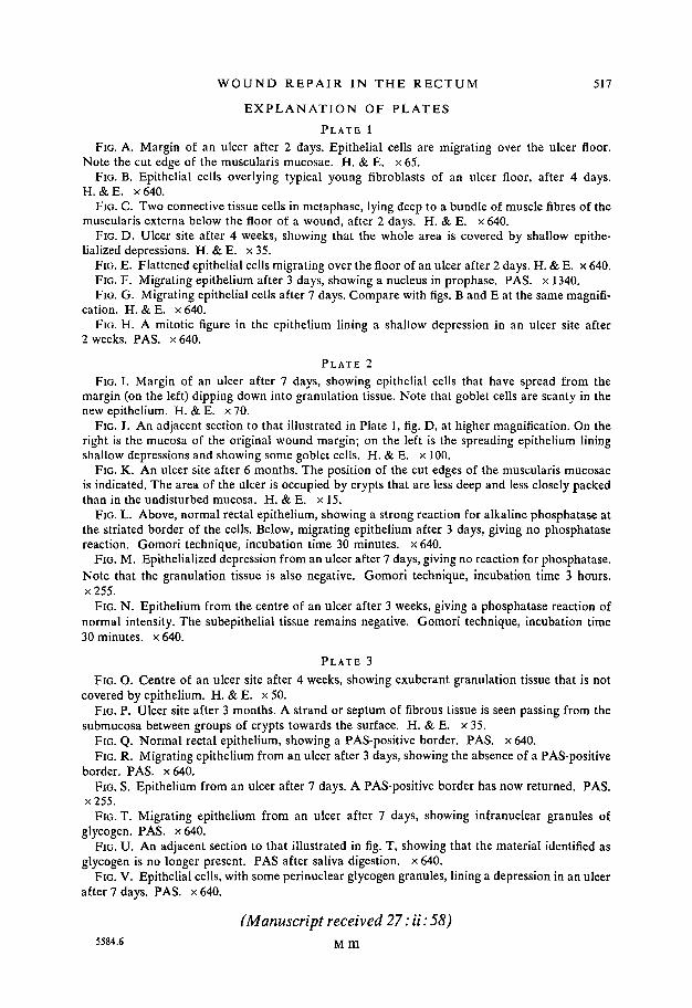
E X P L A N A T I O N OF PLATES
P L A T E 1
FIG. A. Margin of an ulcer after 2 days. Epithelial cells are migrating over the ulcer floor.Note the cut edge of the muscularis mucosae. H. & E. x 65.
FIG. B. Epithelial cells overlying typical young fibroblasts of an ulcer floor, after 4 days.H. &E. x640.
FIG. C. TWO connective tissue cells in metaphase, lying deep to a bundle of muscle fibres of themuscularis externa below the floor of a wound, after 2 days. H. & E. x 640.
FIG. D. Ulcer site after 4 weeks, showing that the whole area is covered by shallow epithe-lialized depressions. H. & E. x 35.
FIG. E. Flattened epithelial cells migrating over the floor of an ulcer after 2 days. H. & E. x 640.FIG. F. Migrating epithelium after 3 days, showing a nucleus in prophase. PAS. x 1340.FIG. G. Migrating epithelial cells after 7 days. Compare with figs. B and E at the same magnifi-
cation. H. &E. x640.FIG. H. A mitotic figure in the epithelium lining a shallow depression in an ulcer site after
2 weeks. PAS. x 640.
PLATE 2
FIG. I. Margin of an ulcer after 7 days, showing epithelial cells that have spread from themargin (on the left) dipping down into granulation tissue. Note that goblet cells are scanty in thenew epithelium. H. & E. x 70.
FIG. J. An adjacent section to that illustrated in Plate 1, fig. D, at higher magnification. On theright is the mucosa of the original wound margin; on the left is the spreading epithelium liningshallow depressions and showing some goblet cells. H. & E. x 100.
FIG. K. An ulcer site after 6 months. The position of the cut edges of the muscularis mucosaeis indicated. The area of the ulcer is occupied by crypts that are less deep and less closely packedthan in the undisturbed mucosa. H. & E. x 15.
FIG. L. Above, normal rectal epithelium, showing a strong reaction for alkaline phosphatase atthe striated border of the cells. Below, migrating epithelium after 3 days, giving no phosphatasereaction. Gomori technique, incubation time 30 minutes, x 640.
FIG. M. Epithelialized depression from an ulcer after 7 days, giving no reaction for phosphatase.Note that the granulation tissue is also negative. Gomori technique, incubation time 3 hours.x255.
FIG. N. Epithelium from the centre of an ulcer after 3 weeks, giving a phosphatase reaction ofnormal intensity. The subepithelial tissue remains negative. Gomori technique, incubation time30 minutes, x 640.
PLATE 3
FIG. O. Centre of an ulcer site after 4 weeks, showing exuberant granulation tissue that is notcovered by epithelium. H. & E. x 50.
FIG. P. Ulcer site after 3 months. A strand or septum of fibrous tissue is seen passing from thesubmucosa between groups of crypts towards the surface. H. & E. x 35.
FIG. Q. Normal rectal epithelium, showing a PAS-positive border. PAS. x 640.FIG. R. Migrating epithelium from an ulcer after 3 days, showing the absence of a PAS-positive
border. PAS. x640.FIG. S. Epithelium from an ulcer after 7 days. A PAS-positive border has now returned. PAS.
x255.FIG. T. Migrating epithelium from an ulcer after 7 days, showing infranuclear granules of
glycogen. PAS. x640.FIG. U. An adjacent section to that illustrated in fig. T, showing that the material identified as
glycogen is no longer present. PAS after saliva digestion, x 640.FIG. V. Epithelial cells, with some perinuclear glycogen granules, lining a depression in an ulcer
after 7 days. PAS. x 640.
(Manuscript received 27: ii: 58)5584.6 M m