
Renal Function and Urine Production in the Compromised Fetus
16
8 Renal Function and Urine Production in the Compromised Fetus Mats Fagerquist North Elfsborg County Hospital, Trollhattan Sweden 1. Introduction Fetal urine production begins in the first trimester. Autopsy findings of filled urinary bladders in human fetuses have been reported from 11 gestational weeks (Abramovich, 1968). In 1970, the first report on ultrasound investigations of the fetal urinary bladder was published (Garrett et al., 1970). Three years later, a method for estimating fetal urine production was introduced (Campbell et al., 1973). In this paper, a short summary of amniotic fluid turnover is presented. Moreover, fetal renal development, artery flow velocity and urine production in normal and compromised fetuses are dealt with. The gradual development of the 2D ultrasound technique for estimating of the fetal urine production rate (HFUPR) is described. In addition, some confounding factors are mentioned. Finally, two important clinical questions, which must be taken into consideration when utilising the HFUPR for fetal surveillance, are identified. 2. Amniotic fluid The volume of amniotic fluid increases during pregnancy (Queenan et al., 1972). In general, the secretion of liquid by the kidneys and from the fetal lungs and oro-nasal cavity is balanced by the removal of equal amounts of liquid (Flack et al., 1995). The main clearance pathway is the swallowing of fluid by the fetus. Additionally, albeit to a lesser degree, fluid passes from the amniotic lumen via the surfaces of the placenta and umbilical cord into the fetal blood circulation (the intramembranous pathway) and into the mother’s circulation (the transmembranous pathway) via the uterine wall through the surface of the amniotic sac outside the placental border. 2.1 Abnormal amounts of amniotic fluid Oligohydramnios (reduced volume < 300 mL) is found in 3-5% (Hansmann, 1985; Volante et al., 2004). Rupture of the membranes is the most common cause of oligohydramnios. A reduction in amniotic fluid volume is of particular concern when it occurs in conjunction with structural fetal anomalies, fetal growth restriction, kidney abnormalities, postdate pregnancies and maternal disease. In these high-risk conditions, it is associated with a poor perinatal outcome (Camanni et al., 2009; Hill et al., 1983). Early onset of oligohydramnios www.intechopen.com
Transcript of Renal Function and Urine Production in the Compromised Fetus

8
Renal Function and Urine Production in the Compromised Fetus
Mats Fagerquist North Elfsborg County Hospital, Trollhattan
Sweden
1. Introduction
Fetal urine production begins in the first trimester. Autopsy findings of filled urinary
bladders in human fetuses have been reported from 11 gestational weeks (Abramovich,
1968). In 1970, the first report on ultrasound investigations of the fetal urinary bladder was
published (Garrett et al., 1970). Three years later, a method for estimating fetal urine
production was introduced (Campbell et al., 1973).
In this paper, a short summary of amniotic fluid turnover is presented. Moreover, fetal renal
development, artery flow velocity and urine production in normal and compromised fetuses
are dealt with. The gradual development of the 2D ultrasound technique for estimating of
the fetal urine production rate (HFUPR) is described. In addition, some confounding factors
are mentioned. Finally, two important clinical questions, which must be taken into
consideration when utilising the HFUPR for fetal surveillance, are identified.
2. Amniotic fluid
The volume of amniotic fluid increases during pregnancy (Queenan et al., 1972). In general,
the secretion of liquid by the kidneys and from the fetal lungs and oro-nasal cavity is
balanced by the removal of equal amounts of liquid (Flack et al., 1995). The main clearance
pathway is the swallowing of fluid by the fetus. Additionally, albeit to a lesser degree, fluid
passes from the amniotic lumen via the surfaces of the placenta and umbilical cord into the
fetal blood circulation (the intramembranous pathway) and into the mother’s circulation
(the transmembranous pathway) via the uterine wall through the surface of the amniotic sac
outside the placental border.
2.1 Abnormal amounts of amniotic fluid
Oligohydramnios (reduced volume < 300 mL) is found in 3-5% (Hansmann, 1985; Volante et al., 2004). Rupture of the membranes is the most common cause of oligohydramnios. A reduction in amniotic fluid volume is of particular concern when it occurs in conjunction with structural fetal anomalies, fetal growth restriction, kidney abnormalities, postdate pregnancies and maternal disease. In these high-risk conditions, it is associated with a poor perinatal outcome (Camanni et al., 2009; Hill et al., 1983). Early onset of oligohydramnios
www.intechopen.com

From Preconception to Postpartum
134
adversely affects fetal lung development, resulting in pulmonary hypoplasia, which might lead to death from severe respiratory insufficiency (Nicolini et al., 1989). However, numerous factors complicate the ultrasonographic diagnosis of oligohydramnios. They include the lack of a complete and detailed understanding of the physiology of the dynamics of oligohydramnios. For example, in 40%, the oligohydramnios occurs without any high-risk conditions and the current available data support the expectant non-interventional management of these cases complicated by isolated oligohydramnios (Sherer, 2002).
Data: Gilbert WM och Brace RA. Amniotic fluid volume and normal flows to and from the amniotic cavity. Semin Perinatol. 1993; 17: 150-157
Fig. 1. The amniotic fluid turnover at term. Half the secreted liquid from the fetal lungs and oro-nasal cavity reaches the amniotic sack and the other half is swallowed. The clearance pathways are denoted in italics.
Polyhydramnios (increased volume > 2,000 mL) is found in 1-3% (Volante et al., 2004). The
underlying cause of excessive amniotic fluid volume is obvious in some clinical conditions
in some clinical conditions and in cases of an minor an minor increase in amniotic fluid
volume, the perinatal outcome is good. However, maternal kidney disease, diabetes type 2
and fetal conditions, such as chromosomal abnormalities, most commonly trisomy 21,
followed by trisomy 18 and trisomy 13, might be causes (Hill et al., 1987). Moreover,
polyhydramnios can be the result of oesophageal atresia and defects in the fetal CNS (Barkin
et al., 1987; Kimble et al., 1998).
www.intechopen.com

Renal Function and Urine Production in the Compromised Fetus
135
In cases with abnormal amniotic fluid volume, prenatal ultrasonography has been recommended for the evaluation of fetal anatomy and growth, swallowing patterns, blood flow velocity in different vessels and repeated estimation of the amount of fluid.
3. Fetal urine production
During the filling phase, the increasing volume of the urinary bladder can be observed, documented and assessed by ultrasound scans.
Fig. 2. This figure shows an appropriate longitudinal bladder image. The 2D ultrasound image on the ultrasound screen was documented on a CD and the volume was calculated in a computer.
The Hourly Fetal Urine Production Rate (HFUPR) can be estimated by regression analysis of calculated bladder volumes documented at different time points within one filling phase (Campbell et al., 1973; Fagerquist et al., 2001; Groome et al., 1991; Nicolaides et al., 1990; Rabinowitz et al., 1989; van Otterlo et al., 1977; Wladimiroff and Campbell, 1974), or by the difference between the maximum and minimum volumes divided by the time interval (Deutinger et al., 1987; Shin et al., 1987; Takeuchi et al., 1994).
www.intechopen.com

From Preconception to Postpartum
136
Fig. 3. The Hourly Fetal Urine Production Rate (HFUPR) estimation was based on the increase in bladder volume during a filling phase and extrapolated to a time span of one hour.
The filling and emptying dynamics of the fetal urinary bladder have been investigated in detail. The mean time for the bladder-filling phase was 25 minutes (range 7-43 minutes) and it was not significantly influenced by gestational age (Rabinowitz et al., 1989).
Gestational age (weeks) Maximum volumes (mL) HFUPR (mL/hour)
20 1 5 21 2 6 22 2 7 23 3 8 24 4 9 25 5 10 26 6 11 27 7 13 28 9 14 29 10 16 30 11 18 31 13 20 32 14 22 33 16 25 34 18 27 35 20 30 36 22 33 37 24 37 38 27 41 39 30 46 40 32 51
(Rabinowitz R, Peters MT, Vyas S, Campbell S, Nicolaides KH. Measurement of fetal urine production in normal pregnancy by real-time ultrasonography. Am J Obstet Gynecol 1989;161(5):1264-6)
Table 1. The maximum bladder volumes before emptying and HFUPR at different gestational ages were calculated by the author according to formulas in the reference article.
www.intechopen.com

Renal Function and Urine Production in the Compromised Fetus
137
3.1 Urine production in the intra-uterine growth-restricted fetus
When comparing Intra-Uterine Growth-Restricted (IUGR) fetuses with Appropriate weight for Gestational Age (AGA) fetuses at the same gestational age, the HFUPR was significantly lower for IUGR fetuses (Nicolaides et al., 1990; Takeuchi et al., 1994; van Otterlo et al., 1977; Wladimiroff and Campbell, 1974). However, there were no significant differences when the IUGR fetuses were compared with controls of corresponding body weights but with lower gestational ages (Wladimiroff and Campbell, 1974). It was assumed that the reduced urine production rate for IUGR fetuses reflected renal hypoplasia, due to growth retardation. Although different investigations have presented various normal values, the HFUPR in IUGR fetuses compared with fetuses of normal size (AGA) has generally been reported to be lower (Nicolaides et al., 1990; Takeuchi et al., 1994; van Otterlo et al., 1977).
4. Fetal kidneys
In a human 2D ultrasound study comprising IUGR and AGA fetuses, the volume of fetal kidneys, as well as the urine production rate, was estimated (Deutinger et al., 1987). In IUGR
fetuses, both the volume of the kidneys and the HFUPR were significantly reduced when compared with the AGA fetuses. In agreement with this study, the growth of fetal kidneys
was significantly slower in Small for Gestational Age (SGA) vs. AGA fetuses when it came to the anterio-posterior diameter and transverse circumference of the kidneys (Konje et al.,
1997). This divergence was most marked after 26 weeks of gestation.
The fetal kidneys gradually increase in volume with gestational age (Hansmann, 1985).
Renal weight as an autopsy finding is, however, often compromised and associated with a coefficient of variation as large as 50%, due to oedema and passive venous engorgement.
Renal functional capacity depends on the number of nephrons, but no known relationship exists between renal weight and the number of glomeruli (Hinchliffe et al., 1991). For many
years, estimates of glomerular numbers have therefore been derived using a variety of methods (Bendtsen and Nyengaard, 1989). Unfortunately, these methods have been shown
to have some degrees of bias. However, a new stereological dissector technique permits the direct, unbiased estimation of glomerular numbers (Hinchliffe et al., 1992). This new
dissector method was used to estimate the number of nephrons in fetuses. The number was 15,000 per kidney in human fetuses at 15 gestational weeks and between 740,000 and
1,060,000 at term.
The total number of nephrons was estimated in a comparative investigation of six IUGR
stillbirths of known gestational age with controls comprising eleven stillbirths with a birth weight greater than the 10th percentile (prenatal period) and eight liveborn IUGR infants,
who died within a year of birth, with a control group of seven appropriately grown infants who also died within a year of birth (postnatal period). The number of nephrons for five of
the six IUGR stillborn children and all the growth-retarded children who died within one year was significantly reduced compared with the controls (Hinchliffe et al., 1992).
Moreover, in animal models, growth restriction has been associated with a reduced number of nephrons (Bauer et al., 2002; Bauer et al., 2003). It has been suggested that the mechanism
underlying the reduced number of nephrons in IUGR fetuses is increased apoptosis due to changes in the levels of apoptosis-related proteins (Pham et al., 2003).
www.intechopen.com

From Preconception to Postpartum
138
4.1 Renal artery flow velocity and urine production in fetuses with hypoxemia
The HFUPR was determined by 2D ultrasound immediately before cordocentesis for blood
gas analysis in 27 Small for Gestational Age (SGA) and 101 AGA fetuses (Nicolaides et al.,
1990). The HFUPR was reduced in the group of SGA fetuses in comparison with AGA
fetuses. Furthermore, the reduction in urine production for the SGA fetuses was correlated
with the degree of fetal hypoxemia, while the degree of fetal hypoxemia did not correlate
with the degree of fetal smallness.
Several studies demonstrate associations between increased impedance in the fetal renal
arteries and factors suggestive of compromised fetal conditions and, in some studies, also
reduced urine production rates (Mikovic et al., 2003; Miura, 1991; Stigter et al., 2001; Vyas
et al., 1989). In one study, the renal artery flow-velocity wave forms were examined in
normal and hypoxemic human fetuses (Vyas et al., 1989). The Pulsatility Index (PI), which
is peak systolic velocity minus end diastolic velocity over mean velocity, was higher in
SGA than in AGA fetuses. Furthermore, using cordocentesis in the SGA fetuses, a
significant, direct correlation was found between blood oxygen deficit and increased renal
artery PI (Vyas et al., 1989). Moreover, in a study of 35 IUGR fetuses, the PI in the fetal
renal arteries was significantly increased (Mikovic et al., 2003). In studies of fetal urine
production, it was demonstrated that the PI in the renal artery was higher in IUGR than in
AGA fetuses and that it displayed a negative correlation with the urine production rate
and the amniotic fluid volume (Miura, 1991). In spite of varying results regarding the PI
in fetal renal arteries (Silver et al., 2003; Stigter et al., 2001), the data suggest that, in fetal
hypoxemia, there is a redistribution of blood flow, with a decrease in renal blood
perfusion and a decrease in HFUPR. These findings may be important, as it would be of
great clinical interest to detect whether or not a particular fetus with growth restriction is
further compromised.
5. Confounding factors
A highly significant diurnal rhythm was observed in an on-line, computerised study of
sheep (Brace and Moore, 1991). In that study, the urine flow rate was measured
continuously over a period of days. In 8/9 animals, the peak flow rate occurred at around 9
pm, while it occurred at 9.30 am in the remaining sheep fetus. The maximum urine flow was
28 ± 5% above the 24-hour mean. Although this significant diurnal variation was
demonstrated in an animal model, it is important not to disregard a possible diurnal
variation in human fetal urine production as well.
The urine production rate was estimated two hours before and two hours after maternal
breakfast in 25 AGA and 15 IUGR fetuses. After breakfast, the HFUPR increased in AGA
fetuses but did not change in IUGR fetuses (Yasuhi et al., 1996). The PI of the fetal renal
artery was significantly reduced after maternal food ingestion in uncomplicated pregnancies
(Yasuhi et al., 1997). To our knowledge, no conflicting reports have been published and, to
avoid the confounding variation due to diurnal variation and maternal meal ingestion, it is
recommended that the estimation of fetal urine production should be performed under
standardised conditions. Also, maternal water ingestion might influence the fetal urine
production rate (Flack et al., 1995; Oosterhof et al., 2000).
www.intechopen.com

Renal Function and Urine Production in the Compromised Fetus
139
6. Methods for estimating the volume of the fetal urinary bladder
Calculated bladder volumes cannot be validated in living human fetuses, as the true fetal urinary bladder volumes are not known. The reliability of the estimated bladder dimensions, on the other hand, has been evaluated using the 2D ultrasound technique (Fagerquist et al., 2001; Fagerquist et al., 2003; Fagerquist et al., 2002). The calculation of the measurement error was based on the variability in repeated estimations of identical bladder volumes. Standard deviation (SD) was used because the variation was normally distributed. Furthermore, there was a linear relationship between bladder volume and the measurement error, which has been thoroughly documented in three previous studies (Fagerquist et al., 2001; Fagerquist et al., 2003; Fagerquist et al., 2002). This is a prerequisite for using a linear regression function (Skrepnek, 2005).
Fig. 4. The SD was calculated when estimating the bladder volume of 120 fetuses. Different methods were used and this gave rise to 222 relationships between SD and bladder volume. The maximum and minimum bladder volumes were 80.5 mL and 0.1 mL respectively. The distribution of the SDs supports a linear relationship (correlation coefficient 0.36).
www.intechopen.com
From Preconception to Postpartum
140
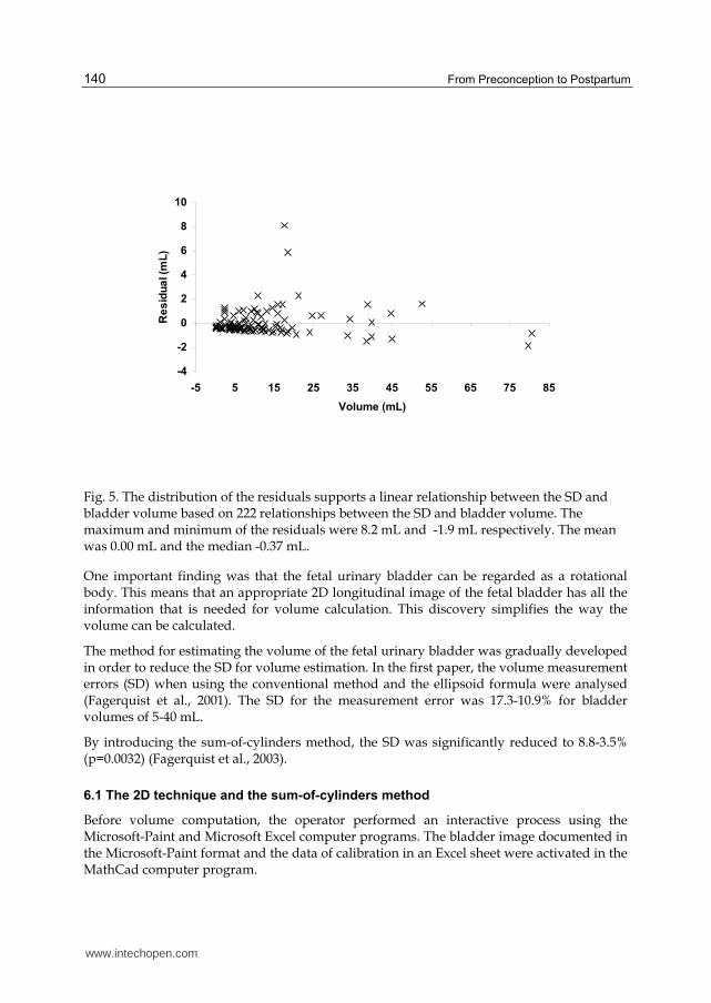
Fig. 5. The distribution of the residuals supports a linear relationship between the SD and bladder volume based on 222 relationships between the SD and bladder volume. The maximum and minimum of the residuals were 8.2 mL and -1.9 mL respectively. The mean was 0.00 mL and the median -0.37 mL.
One important finding was that the fetal urinary bladder can be regarded as a rotational body. This means that an appropriate 2D longitudinal image of the fetal bladder has all the information that is needed for volume calculation. This discovery simplifies the way the volume can be calculated.
The method for estimating the volume of the fetal urinary bladder was gradually developed in order to reduce the SD for volume estimation. In the first paper, the volume measurement errors (SD) when using the conventional method and the ellipsoid formula were analysed (Fagerquist et al., 2001). The SD for the measurement error was 17.3-10.9% for bladder volumes of 5-40 mL.
By introducing the sum-of-cylinders method, the SD was significantly reduced to 8.8-3.5% (p=0.0032) (Fagerquist et al., 2003).
6.1 The 2D technique and the sum-of-cylinders method
Before volume computation, the operator performed an interactive process using the Microsoft-Paint and Microsoft Excel computer programs. The bladder image documented in the Microsoft-Paint format and the data of calibration in an Excel sheet were activated in the MathCad computer program.
www.intechopen.com

Renal Function and Urine Production in the Compromised Fetus
141
Fig. 6. The image that was going to be used for volume computation according to the sum-of-cylinders method was created in an interactive process. Firstly, the operator traced the bladder borders with a red digital marker on the computer screen using the Microsoft-Paint computer program.
The software determines the co-ordinates for the bladder boundary pixels. The image was analysed by the software and each column of pixels was scanned 1) from left to right on the screen, moving from top of the image to bottom in each column to identify the red marked top pixel of the bladder border and 2) from right to left on the screen, moving from bottom of the image to top in each column to identify the red marked bottom pixel of the bladder border. The length of each vertical strip (from top to bottom red pixels in each column) perpendicular to the long axis was computed. The length of each strip was regarded as the diameter of a cylinder, one pixel high. The total bladder volume was calculated by adding all the cylinders.
www.intechopen.com

From Preconception to Postpartum
142
Fig. 7. Using the MathCad computer programs, the vertical distances between marked pixels included in the bladder border was estimated. The bladder image was then electronically subdivided in vertical cylinders and the sum of these cylinders with the height of one pixel equals the bladder volume.
6.2 The 3D ultrasound technique
The 3D ultrasound technique and integrated software, such as the “Virtual Organ
Computer-aided AnaLysis” system (VOCALTM), are already available for volume estimation
and measurements in the in vitro setting and are both reliable and valid (Raine-Fenning et
al., 2003). Moreover, this technique has been applied to the fetal urinary bladder (Lee et al.,
2007; Touboul et al., 2008). Unfortunately, the 3D technique is prone to the same types of
problem encountered in 2D ultrasound imaging, plus others unique to volume acquisition
and visualisation (Nelson et al., 2000). When selecting the initial bladder image, the operator
can avoid shadows from the fetal pelvis. However, according to the VOCAL system, the
subsequent process for volume estimation is automatic and disturbing shadows are not
avoided. Furthermore, in this program, only 40 electronic points are available for the
contour marking, which is another disadvantage.
www.intechopen.com

Renal Function and Urine Production in the Compromised Fetus
143
On the other hand, when using the 2D ultrasound technique, the operator can avoid disturbing shadows by selecting an appropriate longitudinal bladder image. The bladder is defined by the operator who electronically marks the pixels, which are included in the bladder contour. Typically, this corresponds to 200-300 pixels in the boundary. In this way, the technical limit for optimal precision, one pixel, is reached; this is the smallest unit of display resolution.
7. The measurement error when estimating the HFUPR
When estimating the HFUPR, the measurement error is made when assessing the volume of
the bladder, and that SD has been estimated for the 2D technique and the sum-of-cylinders
method. There are some other factors that influence the HFUPR measurement error, the
magnitude of the HFUPR and the number and time points of bladder image capture. To
date, no information relating to the SD for bladder volume estimation by 3D ultrasound,
which is a prerequisite for the subsequent analysis of estimation accuracy, is available.
When utilising the HFUPR for fetal surveillance, it is necessary to know whether the estimated
HUFPR is pathologically low, i.e. below the 5th percentile point. It is therefore necessary to
answer the question: 1) What is the risk of false readings at the 5th percentile point, for
example, even though the true HFUPR is at a higher percentile point? Furthermore, it is
necessary to answer the question: 2) How much of an observed HFUPR change (for example,
during daily controls) can be explained exclusively by measurement error? The implication of
the volume measurement error was demonstrated in detail in a publication publication
concerning the 2D technique and the sum-of-cylinders method. (Fagerquist et al., 2010).
8. Conclusion
The data suggest that, in fetal hypoxemia, there might be a redistribution of blood flow,
with a reduction in both renal perfusion and fetal urine production rate, which can be
estimated by ultrasound. These findings may be important, as it would be of great clinical
interest to determine whether or not a particular fetus with growth restriction is further
compromised. To utilise the HFUPR, for fetal surveillance, a program is available for
estimating the risk of false readings at a low percentile point, even though the true HFUPR
is at a higher percentile point, and the degree to which an observed HFUPR change can be
explained exclusively by measurement error.
9. Acknowledgments
I would like to thank Ingemar Kjellmer, Professor at the Department of Pediatrics,
Sahlgrenska University Hospital, Gothenburg, for his enthusiasm and encouragement,
Anders Odén, Adjunct Professor of Biostatistics, Chalmers University of Technology,
Gothenburg, for his patience, and Ulf Fagerquist, Tech Lic, my brother and friend, who
created the computer programs, which gave us the opportunity to approach the true volume
of the fetal urinary bladder.
Moreover, I would like to express my sincere thanks to Hans Steyskal, Professor and
Mathematics Consultant, Concord, MA, USA, for his advice, and Sture G. Blomberg, MD,
PhD, for his valuable criticism and support.
www.intechopen.com

From Preconception to Postpartum
144
10. References
Abramovich DR. 1968. The volume of amniotic fluid in early pregnancy. J Obstet Gynaecol Br Commonw 75(7):728-731.
Barkin SZ, Pretorius DH, Beckett MK, Manchester DK, Nelson TR, Manco-Johnson ML. 1987. Severe polyhydramnios: incidence of anomalies. AJR Am J Roentgenol 148(1):155-159.
Bauer R, Walter B, Bauer K, Klupsch R, Patt S, Zwiener U. 2002. Intrauterine growth restriction reduces nephron number and renal excretory function in newborn piglets. Acta Physiol Scand 176(2):83-90.
Bauer R, Walter B, Brust P, Fuchtner F, Zwiener U. 2003. Impact of asymmetric intrauterine growth restriction on organ function in newborn piglets. Eur J Obstet Gynecol Reprod Biol 110 Suppl 1:S40-49.
Bendtsen TF, Nyengaard JR. 1989. Unbiased estimation of particle number using sections--an historical perspective with special reference to the stereology of glomeruli. J Microsc 153 (Pt 1):93-102.
Brace RA, Moore TR. 1991. Diurnal rhythms in fetal urine flow, vascular pressures, and heart rate in sheep. Am J Physiol 261(4 Pt 2):R1015-1021.
Camanni D, Zaccara A, Capitanucci ML, Brizzi C, Mobili L, Giorlandino C, Mosiello G, De Gennaro M. 2009. Acute oligohydramnios: antenatal expression of VURD syndrome? Fetal Diagn Ther 26(4):185-188.
Campbell S, Wladimiroff JW, Dewhurst CJ. 1973. The antenatal measurement of fetal urine production. J Obstet Gynaecol Br Commonw 80(8):680-686.
Deutinger J, Bartl W, Pfersmann C, Neumark J, Bernaschek G. 1987. Fetal kidney volume and urine production in cases of fetal growth retardation. J Perinat Med 15(3):307-315.
Fagerquist M, Fagerquist U, Oden A, Blomberg SG. 2001. Fetal urine production and accuracy when estimating fetal urinary bladder volume. Ultrasound Obstet Gynecol 17(2):132-139.
Fagerquist M, Fagerquist U, Oden A, Blomberg SG. 2003. Estimation of fetal urinary bladder volume using the sum-of-cylinders method vs. the ellipsoid formula. Ultrasound Obstet Gynecol 22(1):67-73.
Fagerquist M, Fagerquist U, Steyskal H, Oden A, Blomberg SG. 2002. Accuracy in estimating fetal urinary bladder volume using a modified ultrasound technique. Ultrasound Obstet Gynecol 19(4):371-379.
Fagerquist MA, Fagerquist UO, Oden A, Blomberg SG, Mattsson LA. 2010. Derivations that enable the testing of fetal urine production as a method of fetal surveillance. Arch Gynecol. Obstet. 282(5): 481-6.
Flack NJ, Sepulveda W, Bower S, Fisk NM. 1995. Acute maternal hydration in third-trimester oligohydramnios: effects on amniotic fluid volume, uteroplacental perfusion, and fetal blood flow and urine output. Am J Obstet Gynecol 173(4):1186-1191.
Garrett WJ, Grunwald G, Robinson DE. 1970. Prenatal diagnosis of fetal polycystic kidney by ultrasound. Aust N Z J Obstet Gynaecol 10(1):7-9.
Groome LJ, Owen J, Neely CL, Hauth JC. 1991. Oligohydramnios: antepartum fetal urine production and intrapartum fetal distress. Am J Obstet Gynecol 165(4 Pt 1):1077-1080.
www.intechopen.com

Renal Function and Urine Production in the Compromised Fetus
145
Hansmann M. 1985. Ultrasound Diagnosis in Obstetrics and Gynecology. Springer-Verlag Berlin.
Hill LM, Breckle R, Thomas ML, Fries JK. 1987. Polyhydramnios: ultrasonically detected prevalence and neonatal outcome. Obstet Gynecol 69(1):21-25.
Hill LM, Breckle R, Wolfgram KR, O'Brien PC. 1983. Oligohydramnios: ultrasonically detected incidence and subsequent fetal outcome. Am J Obstet Gynecol 147(4):407-410.
Hinchliffe SA, Lynch MR, Sargent PH, Howard CV, Van Velzen D. 1992. The effect of intrauterine growth retardation on the development of renal nephrons. Br J Obstet Gynaecol 99(4):296-301.
Hinchliffe SA, Sargent PH, Howard CV, Chan YF, van Velzen D. 1991. Human intrauterine renal growth expressed in absolute number of glomeruli assessed by the disector method and Cavalieri principle. Lab Invest 64(6):777-784.
Kimble RM, Harding JE, Kolbe A. 1998. Does gut atresia cause polyhydramnios? Pediatr Surg Int 13(2-3):115-117.
Konje JC, Okaro CI, Bell SC, de Chazal R, Taylor DJ. 1997. A cross-sectional study of changes in fetal renal size with gestation in appropriate- and small-for-gestational-age fetuses. Ultrasound Obstet Gynecol 10(1):22-26.
Lee SM, Park SK, Shim SS, Jun JK, Park JS, Syn HC. 2007. Measurement of fetal urine production by three-dimensional ultrasonography in normal pregnancy. Ultrasound Obstet Gynecol 30(3):281-286.
Mikovic Z, Mandic V, Djukic M, Egic A, Filimonovic D, Cerovic N, Popovac M. 2003. [Longitudinal analysis of arterial Doppler parameters in growth retarded fetuses]. Srp Arh Celok Lek 131(1-2):21-25.
Miura H. 1991. [Evaluation of fetal renal arterial blood flow waveforms with pulsed Doppler flowmetry and the correlation to estimated fetal body weight, fetal urine production rate and amniotic fluid volume]. Nippon Sanka Fujinka Gakkai Zasshi 43(12):1647-1652.
Nelson TR, Pretorius DH, Hull A, Riccabona M, Sklansky MS, James G. 2000. Sources and impact of artifacts on clinical three-dimensional ultrasound imaging. Ultrasound Obstet Gynecol 16(4):374-383.
Nicolaides KH, Peters MT, Vyas S, Rabinowitz R, Rosen DJ, Campbell S. 1990. Relation of rate of urine production to oxygen tension in small-for-gestational-age fetuses. Am J Obstet Gynecol 162(2):387-391.
Nicolini U, Fisk NM, Rodeck CH, Talbert DG, Wigglesworth JS. 1989. Low amniotic pressure in oligohydramnios--is this the cause of pulmonary hypoplasia? Am J Obstet Gynecol 161(5):1098-1101.
Oosterhof H, Haak MC, Aarnoudse JG. 2000. Acute maternal rehydration increases the urine production rate in the near-term human fetus. Am J Obstet Gynecol 183(1):226-229.
Pham TD, MacLennan NK, Chiu CT, Laksana GS, Hsu JL, Lane RH. 2003. Uteroplacental insufficiency increases apoptosis and alters p53 gene methylation in the full-term IUGR rat kidney. Am J Physiol Regul Integr Comp Physiol 285(5):R962-970.
Queenan JT, Thompson W, Whitfield CR, Shah SI. 1972. Amniotic fluid volumes in normal pregnancies. Am J Obstet Gynecol 114(1):34-38.
www.intechopen.com

From Preconception to Postpartum
146
Rabinowitz R, Peters MT, Vyas S, Campbell S, Nicolaides KH. 1989. Measurement of fetal urine production in normal pregnancy by real-time ultrasonography. Am J Obstet Gynecol 161(5):1264-1266.
Raine-Fenning NJ, Clewes JS, Kendall NR, Bunkheila AK, Campbell BK, Johnson IR. 2003. The interobserver reliability and validity of volume calculation from three-dimensional ultrasound datasets in the in vitro setting. Ultrasound Obstet Gynecol 21(3):283-291.
Sherer DM. 2002. A review of amniotic fluid dynamics and the enigma of isolated oligohydramnios. Am J Perinatol 19(5):253-266.
Shin T, Koyanagi T, Hara K, Kubota S, Nakano H. 1987. Development of urine production and urination in the human fetus assessed by real-time ultrasound. Asia Oceania J Obstet Gynaecol 13(4):473-479.
Silver LE, Decamps PJ, Korst LM, Platt LD, Castro LC. 2003. Intrauterine growth restriction is accompanied by decreased renal volume in the human fetus. Am J Obstet Gynecol 188(5):1320-1325.
Skrepnek GH. 2005. Regression Methods in the Empiric Analysis of Health Care Data. J Manag Care Pharm 11(3):240-251.
Stigter RH, Mulder EJ, Bruinse HW, Visser GH. 2001. Doppler studies on the fetal renal artery in the severely growth-restricted fetus. Ultrasound Obstet Gynecol 18(2):141-145.
Takeuchi H, Koyanagi T, Yoshizato T, Takashima T, Satoh S, Nakano H. 1994. Fetal urine production at different gestational ages: correlation to various compromised fetuses in utero. Early Hum Dev 40(1):1-11.
Touboul C, Boulvain M, Picone O, Levaillant JM, Frydman R, Senat MV. 2008. Normal fetal urine production rate estimated with 3-dimensional ultrasonography using the rotational technique (virtual organ computer-aided analysis). Am J Obstet Gynecol.
van Otterlo LC, Wladimiroff JW, Wallenburg HC. 1977. Relationship between fetal urine production and amniotic fluid volume in normal pregnancy and pregnancy complicated by diabetes. Br J Obstet Gynaecol 84(3):205-209.
Wladimiroff JW, Campbell S. 1974. Fetal urine-production rates in normal and complicated pregnancy. Lancet 1(7849):151-154.
Volante E, Gramellini D, Moretti S, Kaihura C, Bevilacqua G. 2004. Alteration of the amniotic fluid and neonatal outcome. Acta Biomed Ateneo Parmense 75 Suppl 1:71-75.
Vyas S, Nicolaides KH, Campbell S. 1989. Renal artery flow-velocity waveforms in normal and hypoxemic fetuses. Am J Obstet Gynecol 161(1):168-172.
Yasuhi I, Hirai M, Ishimaru T, Yamabe T. 1996. Change in fetal urine production rate in growth-restricted fetuses after maternal meal ingestion. Obstet Gynecol 88(5):833-837.
Yasuhi I, Hirai M, Oka S, Nakajima H, Ishimaru T. 1997. Effect of maternal meal ingestion on fetal renal artery resistance. Obstet Gynecol 90(3):340-343.
www.intechopen.com

From Preconception to PostpartumEdited by Dr. Stavros Sifakis
ISBN 978-953-51-0353-0Hard cover, 314 pagesPublisher InTechPublished online 23, March, 2012Published in print edition March, 2012
InTech EuropeUniversity Campus STeP Ri Slavka Krautzeka 83/A 51000 Rijeka, Croatia Phone: +385 (51) 770 447 Fax: +385 (51) 686 166www.intechopen.com
InTech ChinaUnit 405, Office Block, Hotel Equatorial Shanghai No.65, Yan An Road (West), Shanghai, 200040, China
Phone: +86-21-62489820 Fax: +86-21-62489821
Obstetrics is evolving rapidly and finds itself today at the forefront of numerous developments. Providingselected updates on contemporary issues of basic research and clinical practice, as well as dealing withpreconception, pregnancy, labor and postpartum, the present book guides the reader through the tough andcomplex decisions in the clinical management. Furthermore, it deepens the scientific understanding in thepathogenetic mechanisms implicated in pregnancy and motivates further research by providing evidence ofthe current knowledge and future perspectives in this field. Written by an international panel of distinguishedauthors who have produced stimulating articles, the multidisciplinary readers will find this book a valuable toolin the understanding of the maternal, placental and fetal interactions which are crucial for a successfulpregnancy outcome.
How to referenceIn order to correctly reference this scholarly work, feel free to copy and paste the following:
Mats Fagerquist (2012). Renal Function and Urine Production in the Compromised Fetus, From Preconceptionto Postpartum, Dr. Stavros Sifakis (Ed.), ISBN: 978-953-51-0353-0, InTech, Available from:http://www.intechopen.com/books/from-preconception-to-postpartum/renal-function-and-urine-production-in-the-compromised-fetus

© 2012 The Author(s). Licensee IntechOpen. This is an open access articledistributed under the terms of the Creative Commons Attribution 3.0License, which permits unrestricted use, distribution, and reproduction inany medium, provided the original work is properly cited.