Renal Analysis Clinical Pathology. Basic Kidney Structure Recall the urinary system from A&P. The...
23
Renal Analysis Clinical Pathology
-
Upload
maximilian-campbell -
Category
Documents
-
view
214 -
download
1
Transcript of Renal Analysis Clinical Pathology. Basic Kidney Structure Recall the urinary system from A&P. The...
Renal Analysis
Clinical Pathology
Basic Kidney Structure
• Recall the urinary system from A&P.• The glomerulus produces an ultrafiltrate of
plasma which is ultimately voided in the urine.
• Concentration and dilution of urine allows for a 10kg dog to produced 60 L of glomerular filtrate/day but less than 1% is eliminated as urine.
• Reabsorption allows for water to be attracted by solutes (Na, Cl, urea) in the medullary interstitium from the distal and collecting tubules.

Kidneys
• Kidneys are highly susceptible to injury from drugs or toxins.
• 20-25% of cardiac output each minute reaches the kidneys delivering large amounts of any drug in circulation to the kidneys.
• Nephrotoxic drugs include:• Acetaminonophen• Gentamicin• Arsenic

Definitions
• Azotemia• An increase in nitrogen containing
waste products such as urea and creatinine in the blood.
• Uremia• Sydrome characterized by a
constellation of clinical signs caused by the retention of wastes normally excreted by the kidneys.
• An azotemic patient is not always uremic, however a uremic patient is always azotemic


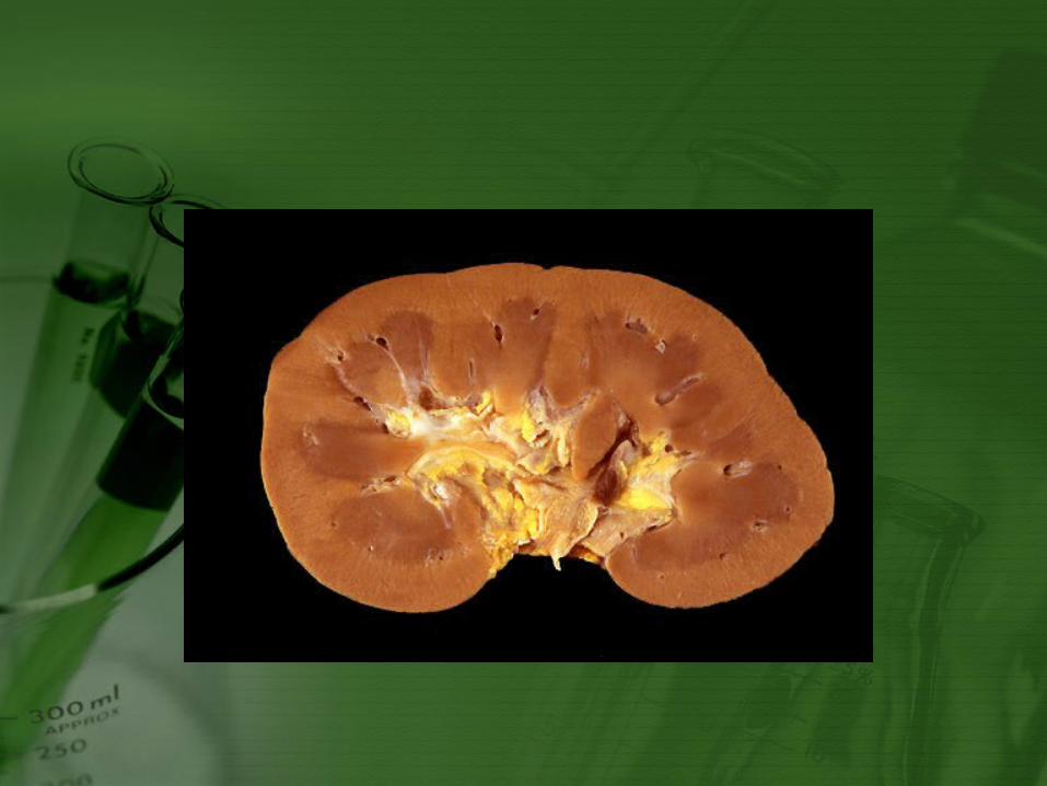
Normal Renal Cells

Kidney Assays
• Both urine and blood are used to evaluate kidney function.
• Functions of the Kidneys:• Conserve water and electrolytes• Regulate hydrogen ions to maintain body pH• Conserve nutrients as glucose and proteins• Produce renin- enzyme to control blood
pressure. • Remove end products of nitrogen metabolism:
• Urea-nitrogenous compound that is product of amino acid breakdown in the liver.
• Creatine• Allantoin

• Kidney Functions continued:• Produce Erythropoietin• Produce Prostaglandins which:
• stimulate contractility of uterine and other smooth muscle
• lower BP• regulate stomach acid secretion• regulate body temperature• platelet aggregation• control inflammation• activate Vitamin D.

Blood Urea Nitrogen (BUN)
• BUN levels used to evaluate kidney function based on ability of kidney to remove nitrogenous waste (urea) from blood.
• Not sensitive- approximate 75% of kidney tissue must be nonfunctional for elevated levels.
• Used as a screen for renal disease in any ill patient, especially those vomiting, wt. loss, chronic nonregenerative anemia, pu/pd, anuria/oliguria, chronic UTI, or dehydration.
BUN Continued
• Normal animals BUN is filtered out of plasma by renal glomeruli and some excreted in urine.
• Normal BUN for dog is 10-30 mg/dl. • Handling:
• Hemolysis has little effect• High protein diet can increase BUN due to
increased breakdown of Amino Acids • 18 hour fast is recommended• Contamination with urease-producing bacteria
(staph aureus, proteus, klebsiella) may result in decomposition of urea-prevent by analysis within several hours of collection or refrigerated sample.
• Can store for 8 hours at 20˚ C

Creatinine
• Metabolite of creatine• Creatine stores energy in muscles in the form of
phosphocreatine.• Creatinine is formed by the decomposition of
creatine.• Creatinine in the blood is filtered through the
glomeruli and eliminated in urine• Can also be found in sweat, feces, and
vomitus.• Not very accurate indicator of kidney function
(same as BUN)• Normal creatinine is 1-2 mg/dl• Not affected by a high-protein diet

• Increased BUN and normal to low Creatinine
• Early prenatal azotemia
• High protein diet• GI hemorrhage• Tetracycline or
corticosteroid administration
• Fever• Severe muscle trauma• Decreased muscle
mass
• Increased Creatinine and normal to low BUN
• Hepatic insufficiency• PU/PD• Low-protein diet• Myositis/muscle
trauma• Cooked meat diet

Phosphorus
• Levels may increase in patients with decreases in GFR (Glomerular filtration rate) similar to BUN and Creatinine concentrations.
• Controlling the patients with hyperphospatemia is important in patients with chronic renal failure to combat renal secondary hyperparathyroidism

Sodium Sulfanilate and PSP Clearance
• Sodium Sulfanilate• Older veterinary test that is no longer used but
was easily performed.• Replaced by endogenous creatinin clearance
test.• Na sulfanilateremoved only glomerular filtration
in dogs
• Phenosulfonpthalein (PSP)• Excreted by renal tubules• Measures renal blood flow• Requires 2/3 of nephrons to be nonfunctional
Endogenous Creatinine Clearance
• A natural tracer of glomerular filtration• Measures blood creatine and accurate, timed urine collection
via catherization, total volume of urine produced is measured in 24 hours.
• Clearance= Urine volume (in mL) x urine creatinine (mg/dl) divided by time (min) x serum creatinine x weight
• Renal disease is the major cause of decreased creatinine clearance although decreased cardiac output may also be responsible.
• Normal values:• Dogs: 2.4-5.0 ml/min/kg• Cats: 1.9 to 5.0 ml/min/kg

Modified Water-Deprivation Test (Vasopressin Response)
• Use in patients with pu/pd• Vasopressin or Antidiuretic hormone (ADH) from
neurohypophysis signals kidneys to retain water• Targets the renal collecting duct
• Give ADH to test for• Failing kidney function• Psychogenic polydipsia• Nephrogenic diabetes insipidus
• If kidney does not respond to ADH• Neurogenic diabetes insipidus
• If ADH is not released• Hyperadrenocorticism

Water Deprivation Test
• Trick is to safely dehydrate the patient until a stimulus for endogenous ADH release (5% of body weight loss).• Monitor patient by weight loss, clinical
signs of dehydration, and urine specific gravity
• Continued diuresis and dilute urine indicate a lack of endogenous ADH or unresponsive nephrons
• In dogs with kedney failure, unresponsiveness precedes azotemia

Water Deprivation Test Contraindications
• Dehydration- will risk hypovolemia and shock.
• Azotemia- already attests to kidney dysfunction.
• Diabetes insipidus- dangerous and useless

Urine Protein/Creatinine Ratio
• Quantiative assessment of renal proteinuria is of diagnostic significance in renal disease.
• Based on concept that tubular concentration of urine increases both urinary protein & creatinine equally.
• Canine: 5-10 ml urine by cystocentesis and protein and creatinine concentrations are determined• Normal: less than 1• Nephrotic syndrome: ratio is greater than 1• Severe glomerulonephritis: ratio is greater than
10

Tests of Glomerular Function (GFR)
• Assessed by clearance of radioactive isotopes
• Primarily used in research due to complexity

Functional Clearance of Electrolytes (FC)
• Na, K, P, and Cl is increased with renal damage
• Tests allow for differentiation between prerenal problems and renal failure
• Measure electrolyte in plasma and urine and creatinine in both• If results are normal, issue is prerenal• If increases the is renal