remodeling in the rat coronary arteries
20
1 Chronic administration of quercetin induces biomechanical and pharmacological 1 remodeling in the rat coronary arteries 2 Anna Monori-Kiss 1* , Fanni Kiss 1 , Jose Martin Restifo 1,2 , Emil Monos 1 , Gyorgy Laszlo 3 Nadasy 3 4 5 1. Institute of Clinical Experimental Research, Semmelweis University, Budapest, 6 Hungary 7 2. University of Palermo, Faculty of Pharmacy, Palermo, Italy 8 3. Department of Physiology, Semmelweis University, Budapest, Hungary 9 Short title: Quercetin-induced remodeling of coronary arterioles 10 * Corresponding author: Anna Monori-Kiss Institute of Clinical Experimental Research, Semmelweis University, Budapest, Hungary H-1094 Budapest, Tűzoltó Str 37-47., Hungary, Room 3.316 Phone: + (36 1) 2100290/60316 Mobil: + (36 30) 6456536 Fax: + (36 1) 3343162 11 Email: [email protected] (A M-K) 12
Transcript of remodeling in the rat coronary arteries
1
Chronic administration of quercetin induces biomechanical and pharmacological 1
remodeling in the rat coronary arteries 2
Anna Monori-Kiss1*, Fanni Kiss1, Jose Martin Restifo1,2, Emil Monos1, Gyorgy Laszlo 3
Nadasy3 4
5
1. Institute of Clinical Experimental Research, Semmelweis University, Budapest, 6
Hungary 7
2. University of Palermo, Faculty of Pharmacy, Palermo, Italy 8
3. Department of Physiology, Semmelweis University, Budapest, Hungary 9
Short title: Quercetin-induced remodeling of coronary arterioles 10
* Corresponding author: Anna Monori-Kiss
Institute of Clinical Experimental Research, Semmelweis University, Budapest, Hungary
H-1094 Budapest, Tűzoltó Str 37-47., Hungary, Room 3.316
Phone: + (36 1) 2100290/60316
Mobil: + (36 30) 6456536
Fax: + (36 1) 3343162 11
Email: [email protected] (A M-K) 12
Zdenka.Stadnikova
Pre-press
2
Summary 1
Acute dilation brought about by the dietary flavonoid quercetin in coronary arterioles has 2
been described earlier, but no information is available on its chronic effects. Male Wistar rats 3
(body weight about 190 g) were divided to two groups: the quercetin-treated group (n=22) 4
had quercetin supplementation of approximately 30 mg/kg/day, whereas the control group 5
(n=20) had none. After eight weeks of treatment, intramural coronary arterioles with identical 6
passive diameters (178±14 and 171±9 µm) were prepared and their biomechanics and 7
pharmacological reactivities were tested using pressure arteriography ex vivo. The 8
spontaneous tone of quercetin-treated arteries was higher (16.5±1.9% vs. 12.9±0.9%), which 9
resulted in a reduced lumen size (144±9 μm vs. 167±12 μm), thicker vascular wall (22.6±1.8 10
μm vs. 17.4±1.6 μm) and decreased tangential wall stress (16.8±1.1 kPa vs. 20.5±1.6 kPa) in 11
supplemented animals (in spontaneous tone at 50 mmHg, p<0.01 in all these comparisons). 12
Elevated basal NO release resulted in increased endothelial dilation in quercetin-treated 13
animals, especially at higher intraluminal pressures (10.8±2.5% vs. 5.7±1.3% at 70 mmHg, 14
p<0.01). We found remodeling of the geometry of coronary arterioles to ensure higher 15
dilatory reserve and nitrogen monoxide production, as well as lowered elastic stress of the 16
vessel wall. 17
Keywords: quercetin, vascular remodeling, coronary circulation, arterioles, endothelial nitric 18
oxide synthase 19
Abbreviations. nKR: normal Krebs-Ringer solution, Ca2+free: Ca2+-free Krebs-Ringer 20
solution, Ach: acetylcholine, BK: bradykinin, L-NAME: nitro-L-arginine methyl ester, NO: 21
nitric oxide, eNOS: endothelial nitric oxide synthase, ROS: reactive oxygen species 22

3
Introduction 1
Dietary polyphenols are present in a mixed human diet in remarkable amounts, around 2
1 g/day. They are represented by diverse molecules, quercetin being one of the most frequent 3
components among them (28-42 mg/day) (Edwards et al. 2007, Scalbert and Williamson 4
2000). This amount has cardioprotective, antihypertensive (Larson et al. 2012), antioxidant 5
(Galisteo et al. 2004) and antilipemic (Lee et al. 2011) effects. Proper functioning of coronary 6
resistance arteries is a key condition to supply the myocardium with oxygen and nutrients. 7
Although the acute dilating effect of quercetin on major and resistance-sized vessels including 8
coronary resistance arteries (Ibarra et al. 2003, Monori-Kiss et al. 2014) has been proven 9
earlier, much less is known about the chronic effects of quercetin on resistance arteries. No 10
publication deals with coronary resistance arteries in this respect, despite the accepted view 11
that quercetin is a potent preventive and therapeutic substance for several forms of 12
cardiovascular disease, including cardiac hypertrophy (Yan et al. 2013, Han et al. 2009). 13
Targeted chronic remodeling studies on large arteries have shown decreased neointima 14
formation and decreased collagen deposition in the abdominal aorta (Huang et al. 2009) as 15
well as decreased collagen I and III expression in myocardial tissue (Yan et al. 2013). These 16
results raise the possibility but do not prove that chronic quercetin supplementation might also 17
favorably affect the biomechanical properties of resistance-sized arteries. Whereas there are 18
several differences between human and rodent metabolism, oral administration of quercetin to 19
rodents seems to be a good model for polyphenol-rich food in humans (Kawai et al. 2009). 20
The aim of this study was to examine the long-term effects of a realistic dose of the 21
flavonoid quercetin on the segmental remodeling of biomechanical and pharmacological 22
properties of coronary arterioles compared to those from untreated normal control rats kept in 23
parallel. It is hypothesized that long-term quercetin-treatment enhances basal NO-mediated 24
dilation, limits dilation to norepinephrine and improves adaptive function of smooth muscle. 25

4
Materials and Methods 1
1. Animal treatment and preparation of the segments 2
All procedures conformed to the Guide for the Care and Use of Laboratory Animals 3
(Guide for the care and use of the laboratory animals, 8th edition, ELAR/NRC 2011), the legal 4
and institutional guidelines for animal care and were approved by the Animal Care Committee 5
of the Semmelweis University and Hungarian authorities (22.1/2960/003/2009). Male Wistar 6
rats at the age of approximately 2 months (180-200 g body weight) were randomly distributed 7
to two groups. All animals had the same rat chow ad libitum (S8106-S011 SM, Ssniff 8
Spezialdiaten). The quercetin-treated group (n=22) had this standard rat chow and a 9
suspension of quercetin, 0.3 g/liter suspended in tap water, to drink ad libitum. After 10
sterilization in autoclave the chow is almost quercetin-free (Yoo et al. 2012). Based on an 11
average of 100 ml pro kg body weight water consumption (Wade et al. 2002), this treatment 12
means approximately 30 mg/ kg body weight of quercetin supplementation pro day. 13
Considering the higher metabolic rate per kg body weight of rats, this dose is comparable with 14
5 mg/kg/day human dose (Reagan-Shaw et al. 2008). A suspension of this concentration was 15
prepared of quercetin hydrate (IUPAC name of quercetin: 2-(3,4-dihydroxyphenyl)-3,5,7-16
trihydroxy-4H-chromen-4-one, purity ≥95% measured by HPLC analysis performed by 17
manufacturer) without any excipient. Suspensions proved to be stable. To prevent oxidation, 18
the suspension was freshly prepared on every second day. The control group (n=20) was kept 19
in parallel (at the same temperature and room) on the same common chow but provided with 20
tap water (vehicle of the suspension) without any supplementation. The animals’ weight, 21
turgor of the skin and behavior were checked twice a week. 22
After 8 weeks of treatment, rats were anesthetized with pentobarbital (Nembutal, 23
Ceva, 45 mg/kg body weight i.p.). The heart was removed, its weight was measured, and was 24

5
next put in cold oxygenized Krebs-Ringer solution (composition in mmol/liter: NaCl 119, 1
KCl 4.7, NaH2PO4 1.2, MgSO4 1.17, NaHCO3 24, CaCl2 2.5, glucose 5.5, and EDTA 0.034). 2
A small intramural coronary arteriole with an outer diameter between 150-200 µm was 3
prepared in situ from a terminal branch of the left anterior descendent coronary artery, as 4
described earlier (Nadasy et al. 2001). The excised vessel segments had more than 2.0 mm 5
length to maintain the physiological cylindrical shape. They were cannulated at both ends 6
using microcannulas with outer diameters around 130 µm, and mounted in a glass-bottomed 7
organ bath (Experimetria LTD), then axially extended by 10 %, to simulate the in vivo axial 8
extension ratio. Artery segments were pressurized intraluminally by servo-controlled pumps 9
(Living Systems). The bath was thermostated at 37 oC, and bubbled with a gas mixture of 5% 10
CO2, 20% O2 and 75% of N2, keeping the pH at 7.4. During incubation, continuous 11
superfusion was ensured at a rate of 2.8 ml/min, whereas the bath volume was 12.0 ml. The 12
organ bath was positioned on the stage of an inverted microscope (Leica), where pictures of 13
the arteries were taken by a digital camera (Leica DFC 320). Pictures were analyzed offline 14
(Leica Qwin), where inner and outer diameters were measured. Calibration was made using a 15
micrometer etalon (Wild). All chemicals (acetylcholine chloride, L-norepinephrine 16
hydrochloride, nitro-L-arginine methyl ester hydrochloride (L-NAME), bradykinin acetate 17
(purity of all chemicals over 98%) and quercetin hydrate (purity over 95%)) were purchased 18
from Sigma-Aldrich. 19
2. Ex vivo protocols 20
To study the biomechanical properties of coronary resistance artery segments, two 21
protocols were used. In the first series of experiments, arteries from 12 quercetin-treated 22
animals and 10 control rats were taken and incubated in nKR solution at 50 mmHg 23
intraluminal pressure for 30 minutes. Arteries develop spontaneous tone under these 24
conditions. A pressure diameter curve was next determined by raising the pressure from 10 to 25

6
100 mmHg in 10 mmHg steps with a 3-minute incubation at each pressure. After a 10-minute 1
rest the original diameter was restored, and we added norepinephrine to the bath (final 2
concentration 10 µmol/liter), incubated the vessel for 10 minutes and pressure diameter 3
curves were repeatedly recorded. Without washout, we added 10 µmol/liter acetylcholine, 4
incubated for 10 minutes, and repeated the pressure diameter curve. 100 µmol/liter L-NAME 5
was next added to block NO synthesis. After 20 minutes of incubation, a pressure diameter 6
curve was taken again. To test reproducibility, a washout with nKR solution was made, 7
followed by incubation for 20 minutes. Vessels with spontaneous tone differing from the 8
original by more than 5% at this point were rejected. Finally, the superfusion was changed to 9
Ca2+-free Krebs-Ringer solution (composition in mmol/liter: NaCl 92, KCl 4.7, NaH2PO4 10
1.18, MgCl2 20, MgSO4 1.17, NaHCO3 24, glucose 5.5, EGTA 2, and EDTA 0.025), and after 11
20 minutes of incubation the passive pressure diameter curve was recorded. 12
In the second series, we investigated the properties of artery segments from 10 animals 13
from both groups. The protocol started with a 30-minute incubation at 50 mmHg in nKR 14
solution, and a pressure diameter curve was next taken as described earlier. After a 10-minute 15
rest, bradykinin was added to the bath in a concentration of 1 µmol/liter, and arterioles were 16
incubated for 10 minutes before the pressure diameter curve was repeated. Bradykinin was 17
then washed out, the original tone was checked, and L-NAME was added in 100 µmol/liter 18
concentration. The incubation time was 20 minutes, similarly to the first protocol, and 19
pressure diameter curves were taken again. Reproducibility was tested by measuring 20
spontaneous tone in nKR solution at 50 mmHg; the superfusion was next changed to Ca2+-21
free Krebs-Ringer, and the passive curve was recorded. 22
3. Calculation formulas 23

7
Di (actual) is the inner diameter in µm at the actual pressure and in solution. Di (passive) is the inner 1
diameter (µm) measured in Ca2+-free Krebs-Ringer solution at the given pressure. 2
Actual diameters were normalized for the passive conditions at each pressure level (% of 3
Ca2+-free= (Di actual*100)/Di passive). Spontaneous tone was calculated as percentage of Ca2+-4
free diameter (spontaneous tone (%) = 100-inner diameter (% of Ca2+-free)). Calculation of 5
wall stress was based on the Laplace-Frank equation (σ = (Pt*ri)/h), where Pt is the transmural 6
pressure (in this case, the intraluminal pressure), ri is the inner radius in µm, and h is wall 7
thickness in µm. Calculation of incremental elastic modulus was based on Cox’s formula 8
(Cox 1979) (Einc (kPa) = [(2ri2*ro)*ΔP]/[(ro
2-ri2)*(r2o-r1o)]), where ri is the inner radius in µm 9
at lower pressure, ro is the outer radius in µm at lower pressure, ΔP is the increase in pressure 10
(in this case ΔP = 10 mmHg = 1,33 kPa), r2o is the outer radius at higher pressure and r1o is the 11
outer radius at lower pressure. 12
4. Statistical analysis and data presentation 13
Data recording and calculations were made with Microsoft Excel. Statistical analysis was 14
performed with GraphPad Prism 6. Values are expressed as mean, with the standard error of 15
mean included. Statistical comparisons were made using one or two-way ANOVA with 16
Bonferrroni post-hoc test, and linear regression. Statistically significant differences were 17
accepted with p values less than 0.05. 18
Results 19
Both groups were healthy, the behavior, alertness, movement, hair or body position of 20
the animals did not show any difference. The average body weight at the beginning of the 21
treatment was 194±4 g. After 8 weeks, no difference was found either in body weights (507±9 22
g, vs. 504±10 g) or heart weights (1.38±0.14 g vs. 1.35±0.18 g) between the control and 23
quercetin-supplemented groups, respectively. 24

8
As described in Methods, coronary resistance arteries with close to identical outer 1
diameters during preparation have been selected. Fig.1.A shows that there was no difference 2
in fully relaxed inner diameters measured in Ca2+-free solutions (passive diameter). The 3
passive incremental elastic moduli did not differ either (Fig 1.B). However, inner diameters in 4
oxygenized 37 oC warm Krebs-Ringer solution were significantly smaller in quercetin-treated 5
animals (Fig 2.A) as a result of elevated spontaneous myogenic tone, characteristic for 6
intramural coronary arterioles (Fig 2.B, p<0.01). This also resulted in a thicker vascular wall 7
under active conditions (Fig. 2.C). Under passive conditions, wall thicknesses and relaxed 8
diameters were practically identical (Fig 2.D, p<0.01). In turn, this increase in vessel wall 9
thickness resulted in decreased tangential wall stress in quercetin-treated animals under active 10
conditions (Fig 2.E, p<0.01). Applying linear regression we found significant difference 11
between slopes (95% confidence intervals: control 0.3896 to 0.4821, quercetin treated 0.3163 12
to 0.3865, p=0.004). 13
We found a slight but significant reduction in vasodilatory response to 10 µmol/liter 14
norepinephrine in the range of 10 to 80 mmHg (p=0.01, Fig 3) in quercetin-treated animals. 15
Dilation rather than constriction is the typical response to norepinephrine in resistance-sized 16
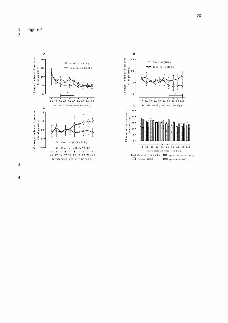
coronary arterioles (Ming et al. 1997). At the same time, acetylcholine (10 µmol/liter) and 17
bradykinin (1 µmol/liter) induced dilation have also been somewhat reduced (Fig 4.A and Fig 18
4.B), However, administration of L-NAME (100 μmol/liter) induced much more forceful 19
contractions in quercetin-supplemented animals, revealing that basal NO-dependent 20
vasodilation was much higher in them (between 60-100 mmHg, p<0.03, Fig 4. C). Fig. 4.D. 21
reveals that while there is no difference in maximum endothelial dilation capacity, a higher 22
part of this capacity is used under basal conditions in quercetin-supplemented animals’ 23
vessels. 24
Discussion 25

9
In this study we identified the effects of a 30 mg/kg/day supplementary dose of 1
quercetin compared to those of the routine quercetin intake with standard rat chow, on the 2
passive and active biomechanical properties, and on some of the pharmacological 3
responsiveness of intramural coronary arterioles of the rat. Passive properties such as wall 4
thickness and passive elasticity did not change due to quercetin supplementation for eight 5
weeks in comparison with the control. However, characteristic remodeling of the active 6
biomechanical and the pharmacological properties could be observed. Spontaneous tone 7
increased and caused reduced lumen and increased vascular wall thickness in spontaneously 8
contracted arteries, ensuring a higher dilatation reserve for these arteries. In parallel with this, 9
a significantly elevated basal endothelial dilation of the arteries from quercetin-supplemented 10
animals was found. 11
In an earlier report, quercetin supplementation resulted in improvement of wall 12
elasticity in abdominal aortas denuded by a balloon catheter (Huang et al. 2009). No change 13
in passive segmental geometry or passive elastic properties was found in our experiment on 14
coronary arterioles. This can be explained by the use of a lower amount of quercetin in our 15
studies. We applied a 30 mg/kg daily dose in contrast to the evidently pharmacological doses 16
of 100 mg/kg and 200 mg/kg used by the above cited authors. This dose of 30 mg/kg/day in 17
rats is thought to be comparable with 5 mg/kg/day in humans (Reagan-Shaw et al. 2008), a 18
dose commonly advised for human nutrition studies (McAnulty et al. 2013, Perez et al. 2014). 19
Furthermore, we must not forget that our studies were made in otherwise healthy vessels, not 20
on pathologic large artery specimens. 21
Increased myogenic tone is one of our key observations. It can be the result of altered 22
calcium homeostasis in smooth muscle cells by quercetin acting as an activator of L-type Ca2+ 23
channels (Saponara et al. 2002), and also having a biphasic effect on Ca2+ ATP-ase 24
(McKenna et al. 1996). The tone elevation we observed may have two consequences. First, in 25
10
situ lumen size decreases. That means that despite the identity of passive vessel 1
characteristics, the position in the coronary network of the quercetin-treated artery segments 2
we prepared was different from that of the control artery segments with the same passive 3
diameter. For example, at 70 mmHg intraluminal pressure the inner diameter of treated 4
arteries was 147±10 μm, whereas that of the untreated ones was 172±13 μm (p<0.05 with 5
Bonferroni post hoc test). This difference means that upon full relaxation (supposing other 6
parameters are unchanged) there is a 75.2% elevation in flow in the control, and a 101.6% 7
elevation in blood flow in quercetin-treated arteries (computation based on the Poiseuille 8
law). We can declare that the quercetin-treated arteries had a much higher dilatation reserve 9
for coronary vasomotion. The second difference concerns in situ wall thickness. At 70 mmHg 10
intraluminal pressure arteries in spontaneous contraction had a wall thickness of 16.4±1.6 μm, 11
whereas in quercetin–treated arteries 22.3±2.0 μm wall thickness was observed. These values 12
correspond to tangential wall stresses of 30.9±2.5 kPa in control vessels, and to 24.5±1.8 kPa 13
in quercetin-treated arteries at 70 mmHg intraluminal pressure under spontaneous myogenic 14
tone (p<0.05). There is good reason to assume that quercetin-treated arteries function at much 15
lower wall stresses than control ones in vivo. 16
Type β2 adrenergic receptors prevail on smooth muscle cells of resistance-sized 17
coronary arteries; the direct effect of norepinephrine on these vessels is relaxation (Ming et al. 18
1997). In our experiments, both control and quercetin-treated arteries are relaxed by 19
norepinephrine, the latter group producing less extensive relaxation. This is in good 20
agreement with recent observations, according to which quercetin, its glucosides and its 3-21
glucuronide metabolite inhibit the activity of the enzyme adenylyl cyclase (Yamazaki et al. 22
2014, Pavan et al. 2015). After oral administration quercetin is metabolized to sulphates and 23
glucuronides by the liver both in humans (Ishizawa et al. 2011) and in the rat (Omar et al. 24
2014). A β-glucuronidase can cleave quercetin from the metabolite in the vessel wall (Perez-25

11
Vizcaino et al. 2012), thus these compounds can affect adenylyl cyclase, causing limited 1
relaxation. Interestingly, recent studies indicate the haemodynamic effect of 3-(3-2
hydroxyphenyl)propionic acid, produced by the human colon microflora from quercetin 3
(Najmanová et al. 2016). 4
One important observation of our experiments was that chronic supplementation of 5
quercetin enhanced NO-mediated dilation as shown by the L-NAME contractions, especially 6
at higher intraluminal pressures. Because quercetin supplementation does not increase eNOS 7
expression in healthy rodents (Takahashi et al. 2015, Wan et al. 2009), this could be the 8
consequence of two mechanisms. First, quercetin induces rapid phosphorylation of eNOS at 9
serine 1179, which in turn increases the activity of the enzyme (Li et al. 2012). Another 10
mechanism can be an enhanced Ca2+-entry into endothelial cells and consequent elevated NO 11
production involving large conductance Ca2+-activated K+ channels (BK(Ca) channels) 12
(Kuhlmann et al. 2005). As it is summarized in Fig.4D, the higher basal NO release of 13
supplemented arteries (L-NAME effect) could not be further increased by eNOS activators 14
(Ach, BK), and this explains the reduced effect of these substances shown in Fig.4.A and 15
Fig.4.B. Direct measurements on smooth muscle cell calcium homeostasis and endothelial 16
expression of eNOS were not made. These conclusions are based on literature, because of 17
available data in published studies. 18
Some limitation of our study seems to be the precise dosage of quercetin. Taking into 19
consideration of the long animal treatment period, we choose the safer dosage by drinking 20
water instead of gavage. In both groups, 3 animals were kept in a cage to minimize non-21
specific stress. With standardizing the environment, we ensured standard and average water 22
consumption during treatment, which provided a fairly standardized quercetin intake. 23

12
In conclusion, chronic administration of quercetin to rats in dietetically real amounts 1
induces a structural and functional remodeling of resistance coronary artery segments, 2
including reduced the elastic stress of the vessel wall, increased dilatory reserve, and 3
augmented NO-mediated endothelial dilation. Quercetin-treatment results in a substantially 4
higher (30%) spontaneous tone. The enhanced basal NO-mediated dilation and higher 5
spontaneous tone together may provide a new balance point of vasodilatory and 6
vasoconstrictor mechanisms. Although polyphenols are not vitamins (Vickery et al. 1950), 7
long-term quercetin intake may result in wider adaptation range and lower elastic stress for 8
coronary arteries. 9
Acknowledgement 10
Expert technical assistance of Ms Ildiko Orevecz is appreciated. The study was supported by 11
Hungarian National grant OTKA TO 32019 and K-112964, by the Hungarian Hypertension 12
Society and the Hungarian Kidney Foundation. The Hungarian Ministry of Human Resources 13
provided a PhD stipend for the first author (A. M.-K.) 14
Conflict of interest 15
No conflicts of interest, financial or otherwise, are declared by the authors. 16

13
References 1
COX R H: Comparison of arterial wall mechanics in normotensive and spontaneously 2 hypertensive rats. Am J Physiol 237: H159-167, 1979. 3 EDWARDS R L, LYON T, LITWIN S E, RABOVSKY A, SYMONS J D, JALILI T: 4 Quercetin reduces blood pressure in hypertensive subjects. J Nutr 137: 2405-2411, 2007. 5 GALISTEO M, GARCIA-SAURA M F, JIMENEZ R, VILLAR I C, ZARZUELO A, 6 VARGAS F, DUARTE J: Effects of chronic quercetin treatment on antioxidant defence 7 system and oxidative status of deoxycorticosterone acetate-salt-hypertensive rats. Mol Cell 8 Biochem 259: 91-99, 2004. 9 HAN J J, HAO J, KIM C H, HONG J S, AHN H Y, LEE Y S: Quercetin prevents cardiac 10 hypertrophy induced by pressure overload in rats. J Vet Med Sci 71: 737-743, 2009. 11 HUANG B F, WANG W, FU Y C, ZHOU X H, WANG X: The effect of quercetin on 12 neointima formation in a rat artery balloon injury model. Pathol Res Pract 205: 515-523, 13 2009. 14 IBARRA M, MORENO L, VERA R, COGOLLUDO A, DUARTE J, TAMARGO J, PEREZ-15 VIZCAINO F: Effects of the flavonoid quercetin and its methylated metabolite isorhamnetin 16 in isolated arteries from spontaneously hypertensive rats. Planta Med 69: 995-1000, 2003. 17 ISHIZAWA K, YOSHIZUMI M, KAWAI Y, TERAO J, KIHIRA Y, IKEDA Y, TOMITA S, 18 MINAKUCHI K, TSUCHIYA K, TAMAKI T: Pharmacology in health food: metabolism of 19 quercetin in vivo and its protective effect against arteriosclerosis. J Pharmacol Sci 115: 466-20 470, 2011. 21 KAWAI Y, SAITO S, NISHIKAWA T, ISHISAKA A, MUROTA K, TERAO J: Different 22 profiles of quercetin metabolites in rat plasma: comparison of two administration methods. 23 Biosci Biotechnol Biochem 73: 517-523, 2009. 24 KUHLMANN C R, SCHAEFER C A, KOSOK C, ABDALLAH Y, WALTHER S, 25 LUDDERS D W, NEUMANN T, TILLMANNS H, SCHAFER C, PIPER H M, ERDOGAN 26 A: Quercetin-induced induction of the NO/cGMP pathway depends on Ca2+-activated K+ 27 channel-induced hyperpolarization-mediated Ca2+-entry into cultured human endothelial cells. 28 Planta Med 71: 520-524, 2005. 29 LARSON A, WITMAN M A, GUO Y, IVES S, RICHARDSON R S, BRUNO R S, JALILI 30 T, SYMONS J D: Acute, quercetin-induced reductions in blood pressure in hypertensive 31 individuals are not secondary to lower plasma angiotensin-converting enzyme activity or 32 endothelin-1: nitric oxide. Nutr Res 32: 557-564, 2012. 33 LEE K-H, PARK E, LEE H-J, KIM M-O, CHA Y-J, KIM J-M, LEE H, SHIN M-J: Effects of 34 daily quercetin-rich supplementation on cardiometabolic risks in male smokers. Nutr Res 35 Pract 5: 28-33, 2011. 36 LI P G, SUN L, HAN X, LING S, GAN W T, XU J W: Quercetin induces rapid eNOS 37 phosphorylation and vasodilation by an Akt-independent and PKA-dependent mechanism. 38 Pharmacology 89: 220-228, 2012. 39 MCANULTY L S, MILLER L E, HOSICK P A, UTTER A C, QUINDRY J C, MCANULTY 40 S R: Effect of resveratrol and quercetin supplementation on redox status and inflammation 41 after exercise. Appl Physiol Nutr Metab 38: 760-765, 2013. 42 MCKENNA E, SMITH J S, COLL K E, MAZACK E K, MAYER E J, ANTANAVAGE J, 43 WIEDMANN R T, JOHNSON R G, JR.: Dissociation of phospholamban regulation of 44 cardiac sarcoplasmic reticulum Ca2+ATPase by quercetin. J Biol Chem 271: 24517-24525, 45 1996. 46 MING Z, PARENT R, LAVALLEE M: Beta 2-adrenergic dilation of resistance coronary 47 vessels involves KATP channels and nitric oxide in conscious dogs. Circulation 95: 1568-48 1576, 1997. 49

14
MONORI-KISS A, MONOS E, NÁDASY G L: Quantitative Analysis of Vasodilatory Action 1 of Quercetin on Intramural Coronary Resistance Arteries of the Rat In Vitro. PLoS One 9: 2 e105587, 2014. 3 NADASY G L, SZEKERES M, DEZSI L, VARBIRO S, SZEKACS B, MONOS E: 4 Preparation of intramural small coronary artery and arteriole segments and resistance artery 5 networks from the rat heart for microarteriography and for in situ perfusion video mapping. 6 Microvasc Res 61: 282-286, 2001. 7 NAJMANOVÁ I, POUROVÁ J, VOPRŠALOVÁ M, PILAŘOVÁ V, SEMECKÝ V, 8 NOVÁKOVÁ L, MLADĚNKA P: Flavonoid metabolite 3-(3-hydroxyphenyl)propionic acid 9 formed by human microflora decreases arterial blood pressure in rats. Mol Nutr Food Res 60: 10 981-991, 2016. 11 OMAR K, GRANT M H, HENDERSON C, WATSON D G: The complex degradation and 12 metabolism of quercetin in rat hepatocyte incubations. Xenobiotica 44: 1074-1082, 2014. 13 PAVAN B, CAPUZZO A, FORLANI G: Quercetin and quercetin-3-O-glucoside interact with 14 different components of the cAMP signaling cascade in human retinal pigment epithelial 15 cells. Life Sci 121: 166-173, 2015. 16 PEREZ-VIZCAINO F, DUARTE J, SANTOS-BUELGA C: The flavonoid paradox: 17 conjugation and deconjugation as key steps for the biological activity of flavonoids. J Sci 18 Food Agric 92: 1822-1825, 2012. 19 PEREZ A, GONZALEZ-MANZANO S, JIMENEZ R, PEREZ-ABUD R, HARO J M, 20 OSUNA A, SANTOS-BUELGA C, DUARTE J, PEREZ-VIZCAINO F: The flavonoid 21 quercetin induces acute vasodilator effects in healthy volunteers: correlation with beta-22 glucuronidase activity. Pharmacol Res 89: 11-18, 2014. 23 REAGAN-SHAW S, NIHAL M, AHMAD N: Dose translation from animal to human studies 24 revisited. FASEB J 22: 659-661, 2008. 25 SAPONARA S, SGARAGLI G, FUSI F: Quercetin as a novel activator of L-type Ca2+ 26 channels in rat tail artery smooth muscle cells. Br J Pharmacol 135: 1819-1827, 2002. 27 SCALBERT A, WILLIAMSON G: Dietary intake and bioavailability of polyphenols. J Nutr 28 130: 2073s-2085s, 2000. 29 TAKAHASHI A, INOUE H, MISHIMA K, IDE F, NAKAYAMA R, HASAKA A, RYO K, 30 ITO Y, SAKURAI T, HASEGAWA Y, SAITO I: Evaluation of the Effects of Quercetin on 31 Damaged Salivary Secretion. PLoS One 10: e0116008, 2015. 32 VICKERY H B, NELSON E M, ALMQUIST H J, ELVEHJEM C A: Term "vitamin P" 33 recommended to be discontinued. Science 112: 628, 1950. 34 WADE C E, MILLER M M, BAER L A, MORAN M M, STEELE M K, STEIN T P: Body 35 mass, energy intake, and water consumption of rats and humans during space flight. Nutrition 36 18: 829-836, 2002. 37 WAN L L, XIA J, YE D, LIU J, CHEN J, WANG G: Effects of quercetin on gene and protein 38 expression of NOX and NOS after myocardial ischemia and reperfusion in rabbit. Cardiovasc 39 Ther 27: 28-33, 2009. 40 YAMAZAKI S, MIYOSHI N, KAWABATA K, YASUDA M, SHIMOI K: Quercetin-3-O-41 glucuronide inhibits noradrenaline-promoted invasion of MDA-MB-231 human breast cancer 42 cells by blocking beta(2)-adrenergic signaling. Arch Biochem Biophys 557: 18-27, 2014. 43 YAN L, ZHANG J D, WANG B, LV Y J, JIANG H, LIU G L, QIAO Y, REN M, GUO X F: 44 Quercetin inhibits left ventricular hypertrophy in spontaneously hypertensive rats and inhibits 45 angiotensin II-induced H9C2 cells hypertrophy by enhancing PPAR-γ expression and 46 suppressing AP-1 activity. PLoS One 8: e72548, 2013. 47 YOO J, KIM Y, YOO S-H, INGLETT G E, LEE S: Reduction of rutin loss in buckwheat 48 noodles and their physicochemical characterisation. Food Chem 132: 2107-2111, 2012. 49
50
15
Legends of figures 1
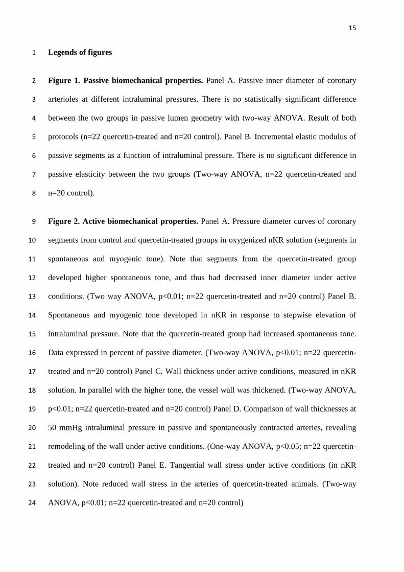
Figure 1. Passive biomechanical properties. Panel A. Passive inner diameter of coronary 2
arterioles at different intraluminal pressures. There is no statistically significant difference 3
between the two groups in passive lumen geometry with two-way ANOVA. Result of both 4
protocols (n=22 quercetin-treated and n=20 control). Panel B. Incremental elastic modulus of 5
passive segments as a function of intraluminal pressure. There is no significant difference in 6
passive elasticity between the two groups (Two-way ANOVA, n=22 quercetin-treated and 7
n=20 control). 8
Figure 2. Active biomechanical properties. Panel A. Pressure diameter curves of coronary 9
segments from control and quercetin-treated groups in oxygenized nKR solution (segments in 10
spontaneous and myogenic tone). Note that segments from the quercetin-treated group 11
developed higher spontaneous tone, and thus had decreased inner diameter under active 12
conditions. (Two way ANOVA, p<0.01; n=22 quercetin-treated and n=20 control) Panel B. 13
Spontaneous and myogenic tone developed in nKR in response to stepwise elevation of 14
intraluminal pressure. Note that the quercetin-treated group had increased spontaneous tone. 15
Data expressed in percent of passive diameter. (Two-way ANOVA, p<0.01; n=22 quercetin-16
treated and n=20 control) Panel C. Wall thickness under active conditions, measured in nKR 17
solution. In parallel with the higher tone, the vessel wall was thickened. (Two-way ANOVA, 18
p<0.01; n=22 quercetin-treated and n=20 control) Panel D. Comparison of wall thicknesses at 19
50 mmHg intraluminal pressure in passive and spontaneously contracted arteries, revealing 20
remodeling of the wall under active conditions. (One-way ANOVA, p<0.05; n=22 quercetin-21
treated and n=20 control) Panel E. Tangential wall stress under active conditions (in nKR 22
solution). Note reduced wall stress in the arteries of quercetin-treated animals. (Two-way 23
ANOVA, p<0.01; n=22 quercetin-treated and n=20 control) 24

16
Figure 3. Dilation induced by 10 μmol/liter norepinephrine (as compared to segments in 1
myogenic tone in nKR solution). See the reduced beta adrenergic dilation of quercetin-2
treated segments. Statistically significant between 10-80 mmHg intraluminal pressure. (Two-3
way ANOVA p<0.01) 4
Figure 4. NO-mediated dilation. Panel A. Vasodilatation of spontaneously contracted 5
segments induced by 10 μmol/liter acetylcholine. Note reduced acetylcholine dilation in the 6
pressure range of 30-60 mmHg. (Two-way ANOVA p<0.01) Panel B. Vasodilatation of 7
spontaneously contracted segments induced by 1 μmol/liter bradykinin. Note reduced 8
bradykinin-stimulated dilation in the pressure range of 70-100 mmHg. (Two-way ANOVA 9
p<0.01) Panel C. Additional vasoconstriction induced in spontaneously contracted segments 10
by application of the NO synthase blocker L-NAME (100 μmol/liter). Note higher level of 11
basal NO dilation of quercetin-treated segments. Significant between 60-100 mmHg 12
intraluminal pressure. (Two-way ANOVA, p<0.05; n=10 quercetin treated, n=10 control, data 13
from second series of experiment) Panel D.: Sum of basal and bradykinin-induced endothelial 14
vasodilation. Basal NO-mediated dilatation is measured with application of L-NAME (bars 15
with pattern). In control vessels this dilator effect is decreasing as a function of increasing 16
intraluminal pressure, while in quercetin-treated vessels basal NO-mediated dilation is 17
constant. Bradykinin induced endothelial vasodilation (bars without pattern) is a reserve of 18
NO-mediated vasodilation. Note that maximum NO-induced vasodilation (sum of basal and 19
induced NO-mediated dilation) did not differ between the two groups, whereas quercetin-20
treated segments showed higher basal NO dilation activity using up a higher portion of that 21
maximum capacity under basal conditions. 22
23

17
Figure 1 1
2

18
Figure 2 1
2
3

19
Figure 3 1 2
1 0 2 0 3 0 4 0 5 0 6 0 7 0 8 0 9 0 1 0 0
0
5
1 0
1 5C o n tro l
Q u e rc e t in
*
In tra lu m in a l p re s s u re (m m H g )
Ch
an
ges
in
in
ner
dia
met
er(%
of
pa
ssiv
e)
3
4
5
20
Figure 4 1 2
1 0 2 0 3 0 4 0 5 0 6 0 7 0 8 0 9 0 1 0 0
0
5
1 0
1 5
2 0
C o n tro l (A c h )
Q u e rc e t in (A c h )
A
*
In tra lu m in a l p re s s u re (m m H g )
Ch
an
ges
in
in
ner
dia
met
er(%
of
pa
ssiv
e)
1 0 2 0 3 0 4 0 5 0 6 0 7 0 8 0 9 0 1 0 0
0
5
1 0
1 5C o n tro l (B K )
Q u e rc e t in (B K )
*
B
In tra lu m in a l p re s s u re (m m H g )
Ch
an
ges
in
in
ner
dia
met
er(%
of
pa
ssiv
e)
1 0 2 0 3 0 4 0 5 0 6 0 7 0 8 0 9 0 1 0 0
-2 0
-1 5
-1 0
-5
0
Q u e rc e t in (L -N A M E )
C o n tro l (L -N A M E )
C
*
In tra lu m in a l p re s s u re (m m H g )
Ch
an
ges
in
in
ner
dia
met
er(%
of
pa
ssiv
e)
1 0 2 0 3 0 4 0 5 0 6 0 7 0 8 0 9 0 1 0 0
0
5
1 0
1 5
2 0
2 5
C o n tro l (L -N A M E ) Q u e rc e t in (L -N A M E )
C o n tro l (B K ) Q u e rc e t in (B K )
D
In tra lu m in a l p re s s u re (m m H g )
Ch
an
ges
in
in
ner
dia
met
er(%
of
pa
ssiv
e)
3
4