Relative Motion Splint: Active Motion After Extensor …...by encouraging full finger flexion in...
8
SURGICAL TECHNIQUE Relative Motion Splint: Active Motion After Extensor Tendon Injury and Repair Wyndell H. Merritt, MD The relative motion splint was initially developed to facilitate postoperative rehabilitation after repair of extensor tendon injuries at the dorsum of the hand and forearm. It has subsequently been used for rehabilitation of sagittal band injuries and after repair of closed attrition extensor tendon ruptures in rheumatoid arthritis. This is much less awkward than other braces and can readily be worn during normal past-time and work activities. This so-called immediate controlled active motion splinting protocol has also more recently been applied to both operative and nonsurgical rehabilitation for boutonniere deformity. (J Hand Surg Am. 2014;39(6):1187e1194. Copyright Ó 2014 by the American Society for Surgery of the Hand. All rights reserved.) Key words Tendon therapy, extensor tendon, laceration, boutonniere, sagittal band. E XTENSOR TENDON INJURIES HAVE traditionally been treated by either 4 to 6 weeks’ immobilization, with possible loss of flexion owing to joint stiffness, or dynamic splinting that may (or may not) provide passive gliding of the injured tendon but re- quires wearing an awkward device. 1 More recently, early active “short arc motion” is proposed for ex- tensor tendon injuries of the finger, but this requires careful monitoring and intensive therapy with active motion only under therapist supervision during the first 3 weeks, and immobilization in a “palmar resting splint” followed by a home program that requires a compliant patient to do home active motion exercises. 2 Relative motion splinting, also known as imme- diate controlled active motion splinting, 3 encourages immediate full active function of the hand, unre- stricted other than 15 to 20 less extension or flexion of the metacarpophalangeal (MCP) joint, depending on the injury. We have used this patient-friendly management technique in long extensor lacerations for over 30 years, 4 and then more recently for boutonniere deformity, sagittal band rupture, 5 and side-to-side tendon repair after rheumatoid extensor tendon rup- tures in caput ulnae syndrome, preserving full inter- phalangeal joint motion and minimizing the need for intensive hand therapy. RATIONALE The relative motion concept is simple. When an injured tendon is placed in 15 to 20 less relative motion than adjacent tendons from a shared muscle (extensor or flexor), it will experience markedly less force than adjacent tendons, 6 regardless of the position from full extension to full flexion (minus the 15 to 20 difference at the MCP joint). With long extensor repair in zones IV to VII, all extensors function like the single unit extensor digitorum communis muscle. Independent extensors recover equally well as the common extensors when placed in 15 to 20 greater MCP extension than the adjacent digits (Fig. 1). This concept is also effective in reducing tension across sagittal band ruptures and side-to-side tendon transfers in caput ulnae syndrome. We call this the relative motion extensor splint. 7 In boutonniere deformity, the lumbricals have 4 times the excursion of the more powerful interossei, From the Department of Plastic Surgery, University of Virginia, Charlottesville, VA. Received for publication March 10, 2014; accepted in revised form March 16, 2014. The author expresses appreciation to Dr. Don Lalonde for use of his video of dramatic repair and splinting in a boutonniere deformity patient whose extensor hood had been ground off. No benefits in any form have been received or will be received related directly or indirectly to the subject of this article. Corresponding author: Wyndell H. Merritt, MD, Department of Plastic Surgery, Uni- versity of Virginia, 7660 E. Parham Road, MOB I, Suite 200, Henrico, VA 23294; e-mail: [email protected]. 0363-5023/14/3906-0030$36.00/0 http://dx.doi.org/10.1016/j.jhsa.2014.03.015 Ó 2014 ASSH r Published by Elsevier, Inc. All rights reserved. r 1187 Surgical Technique
Transcript of Relative Motion Splint: Active Motion After Extensor …...by encouraging full finger flexion in...
From the Department of Plastic Surgery, University of Virginia, Ch
Received for publication March 10, 2014; accepted in revised for
The author expresses appreciation to Dr. Don Lalonde for use of hisand splinting in a boutonniere deformity patient whose extensor ho
No benefits in any form have been received or will be receiindirectly to the subject of this article.
Corresponding author: Wyndell H. Merritt, MD, Departmentversity of Virginia, 7660 E. Parham Road, MOB I, Suite 200, [email protected].
0363-5023/14/3906-0030$36.00/0http://dx.doi.org/10.1016/j.jhsa.2014.03.015
SURGICAL TECHNIQUE
Relative Motion Splint: Active Motion After
Extensor Tendon Injury and Repair
Wyndell H. Merritt, MD
echnique
The relative motion splint was initially developed to facilitate postoperative rehabilitation afterrepair of extensor tendon injuries at the dorsumof the hand and forearm. It has subsequently beenused for rehabilitation of sagittal band injuries and after repair of closed attrition extensor tendonruptures in rheumatoid arthritis. This is much less awkward than other braces and can readily beworn during normal past-time and work activities. This so-called immediate controlled activemotion splinting protocol has also more recently been applied to both operative and nonsurgicalrehabilitation for boutonniere deformity. (J Hand Surg Am. 2014;39(6):1187e1194. Copyright� 2014 by the American Society for Surgery of the Hand. All rights reserved.)Key words Tendon therapy, extensor tendon, laceration, boutonniere, sagittal band.
calT
Surgi
E XTENSOR TENDON INJURIES HAVE traditionally beentreated by either 4 to 6 weeks’ immobilization,with possible loss of flexion owing to joint
stiffness, or dynamic splinting that may (or may not)provide passive gliding of the injured tendon but re-quires wearing an awkward device.1 More recently,early active “short arc motion” is proposed for ex-tensor tendon injuries of the finger, but this requirescareful monitoring and intensive therapy with activemotion only under therapist supervision during thefirst 3 weeks, and immobilization in a “palmar restingsplint” followed by a home program that requires acompliant patient to do home activemotion exercises.2
Relative motion splinting, also known as imme-diate controlled active motion splinting,3 encouragesimmediate full active function of the hand, unre-stricted other than 15� to 20� less extension or flexion
arlottesville, VA.
m March 16, 2014.
video of dramatic repairod had been ground off.
ved related directly or
of Plastic Surgery, Uni-rico, VA 23294; e-mail:
� 2
of the metacarpophalangeal (MCP) joint, dependingon the injury. We have used this patient-friendlymanagement technique in long extensor lacerations forover 30 years,4 and then more recently for boutonnieredeformity, sagittal band rupture,5 and side-to-sidetendon repair after rheumatoid extensor tendon rup-tures in caput ulnae syndrome, preserving full inter-phalangeal joint motion and minimizing the need forintensive hand therapy.
RATIONALEThe relative motion concept is simple. When aninjured tendon is placed in 15� to 20� less relativemotion than adjacent tendons from a shared muscle(extensor or flexor), it will experience markedly lessforce than adjacent tendons,6 regardless of the positionfrom full extension to full flexion (minus the 15� to20� difference at the MCP joint). With long extensorrepair in zones IV to VII, all extensors function likethe single unit extensor digitorum communis muscle.Independent extensors recover equally well as thecommon extensors when placed in 15� to 20� greaterMCP extension than the adjacent digits (Fig. 1). Thisconcept is also effective in reducing tension acrosssagittal band ruptures and side-to-side tendon transfersin caput ulnae syndrome. We call this the relativemotion extensor splint.7
In boutonniere deformity, the lumbricals have 4times the excursion of the more powerful interossei,
014 ASSH r Published by Elsevier, Inc. All rights reserved. r 1187
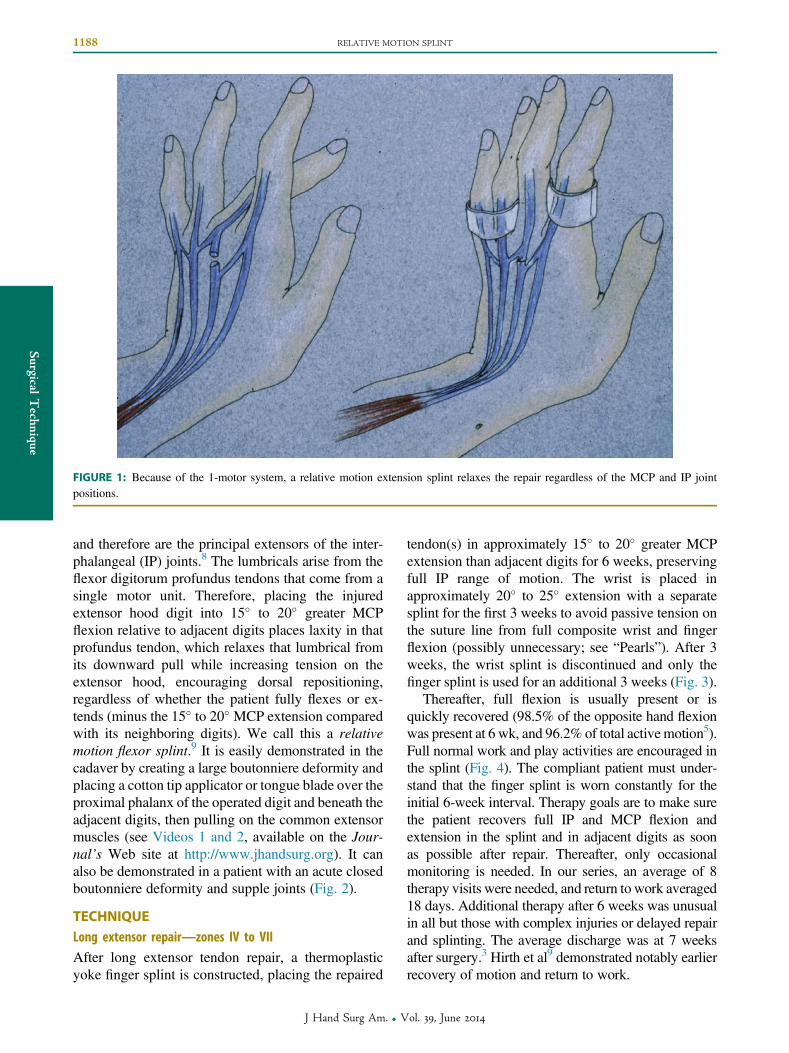
FIGURE 1: Because of the 1-motor system, a relative motion extension splint relaxes the repair regardless of the MCP and IP jointpositions.
1188 RELATIVE MOTION SPLINT
Surgical
Tech
nique
and therefore are the principal extensors of the inter-phalangeal (IP) joints.8 The lumbricals arise from theflexor digitorum profundus tendons that come from asingle motor unit. Therefore, placing the injuredextensor hood digit into 15� to 20� greater MCPflexion relative to adjacent digits places laxity in thatprofundus tendon, which relaxes that lumbrical fromits downward pull while increasing tension on theextensor hood, encouraging dorsal repositioning,regardless of whether the patient fully flexes or ex-tends (minus the 15� to 20� MCP extension comparedwith its neighboring digits). We call this a relativemotion flexor splint.9 It is easily demonstrated in thecadaver by creating a large boutonniere deformity andplacing a cotton tip applicator or tongue blade over theproximal phalanx of the operated digit and beneath theadjacent digits, then pulling on the common extensormuscles (see Videos 1 and 2, available on the Jour-nal’s Web site at http://www.jhandsurg.org). It canalso be demonstrated in a patient with an acute closedboutonniere deformity and supple joints (Fig. 2).
TECHNIQUELong extensor repair—zones IV to VII
After long extensor tendon repair, a thermoplasticyoke finger splint is constructed, placing the repaired
J Hand Surg Am. r V
tendon(s) in approximately 15� to 20� greater MCPextension than adjacent digits for 6 weeks, preservingfull IP range of motion. The wrist is placed inapproximately 20� to 25� extension with a separatesplint for the first 3 weeks to avoid passive tension onthe suture line from full composite wrist and fingerflexion (possibly unnecessary; see “Pearls”). After 3weeks, the wrist splint is discontinued and only thefinger splint is used for an additional 3 weeks (Fig. 3).
Thereafter, full flexion is usually present or isquickly recovered (98.5% of the opposite hand flexionwas present at 6 wk, and 96.2% of total activemotion5).Full normal work and play activities are encouraged inthe splint (Fig. 4). The compliant patient must under-stand that the finger splint is worn constantly for theinitial 6-week interval. Therapy goals are to make surethe patient recovers full IP and MCP flexion andextension in the splint and in adjacent digits as soonas possible after repair. Thereafter, only occasionalmonitoring is needed. In our series, an average of 8therapy visits were needed, and return to work averaged18 days. Additional therapy after 6 weeks was unusualin all but those with complex injuries or delayed repairand splinting. The average discharge was at 7 weeksafter surgery.3 Hirth et al9 demonstrated notably earlierrecovery of motion and return to work.
ol. 39, June 2014

FIGURE 2: A Acute boutonniere deformity. B The deformity is easily corrected by simply pushing the proximal phalanx into greaterMCP flexion. C Boutonniere splint for the little finger, with no wrist component for 6 weeks.
FIGURE 3: Immediate active motion after long finger extensor repair: A wrist splinted 3 weeks; B finger splinted 6 weeks.
RELATIVE MOTION SPLINT 1189
Surgical
Technique
Sagittal band rupture—acute and chronic
Patients with acute sagittal band rupture, less than 2or 3 weeks after injury, can usually centralize thetendon when placed in extension and will experience
J Hand Surg Am. r V
pain relief soon after relative motion extensor splinting,even when flexing in the splint. These patients aresplinted in a similar fashion to long extensor repairs,without the wrist splint component. Success is evident
ol. 39, June 2014
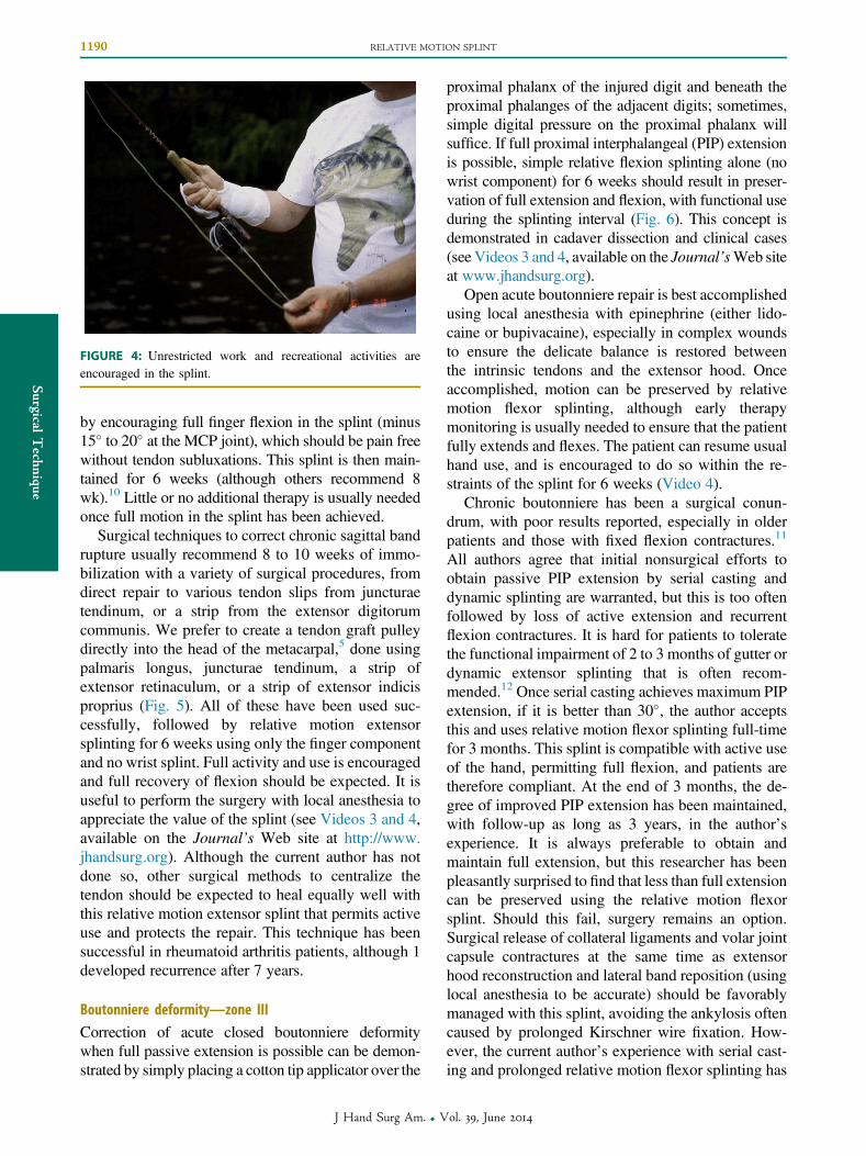
FIGURE 4: Unrestricted work and recreational activities areencouraged in the splint.
1190 RELATIVE MOTION SPLINT
Surgical
Tech
nique
by encouraging full finger flexion in the splint (minus15� to 20� at the MCP joint), which should be pain freewithout tendon subluxations. This splint is then main-tained for 6 weeks (although others recommend 8wk).10 Little or no additional therapy is usually neededonce full motion in the splint has been achieved.
Surgical techniques to correct chronic sagittal bandrupture usually recommend 8 to 10 weeks of immo-bilization with a variety of surgical procedures, fromdirect repair to various tendon slips from juncturaetendinum, or a strip from the extensor digitorumcommunis. We prefer to create a tendon graft pulleydirectly into the head of the metacarpal,5 done usingpalmaris longus, juncturae tendinum, a strip ofextensor retinaculum, or a strip of extensor indicisproprius (Fig. 5). All of these have been used suc-cessfully, followed by relative motion extensorsplinting for 6 weeks using only the finger componentand no wrist splint. Full activity and use is encouragedand full recovery of flexion should be expected. It isuseful to perform the surgery with local anesthesia toappreciate the value of the splint (see Videos 3 and 4,available on the Journal’s Web site at http://www.jhandsurg.org). Although the current author has notdone so, other surgical methods to centralize thetendon should be expected to heal equally well withthis relative motion extensor splint that permits activeuse and protects the repair. This technique has beensuccessful in rheumatoid arthritis patients, although 1developed recurrence after 7 years.
Boutonniere deformity—zone III
Correction of acute closed boutonniere deformitywhen full passive extension is possible can be demon-strated by simply placing a cotton tip applicator over the
J Hand Surg Am. r V
proximal phalanx of the injured digit and beneath theproximal phalanges of the adjacent digits; sometimes,simple digital pressure on the proximal phalanx willsuffice. If full proximal interphalangeal (PIP) extensionis possible, simple relative flexion splinting alone (nowrist component) for 6 weeks should result in preser-vation of full extension and flexion, with functional useduring the splinting interval (Fig. 6). This concept isdemonstrated in cadaver dissection and clinical cases(see Videos 3 and 4, available on the Journal’sWeb siteat www.jhandsurg.org).
Open acute boutonniere repair is best accomplishedusing local anesthesia with epinephrine (either lido-caine or bupivacaine), especially in complex woundsto ensure the delicate balance is restored betweenthe intrinsic tendons and the extensor hood. Onceaccomplished, motion can be preserved by relativemotion flexor splinting, although early therapymonitoring is usually needed to ensure that the patientfully extends and flexes. The patient can resume usualhand use, and is encouraged to do so within the re-straints of the splint for 6 weeks (Video 4).
Chronic boutonniere has been a surgical conun-drum, with poor results reported, especially in olderpatients and those with fixed flexion contractures.11
All authors agree that initial nonsurgical efforts toobtain passive PIP extension by serial casting anddynamic splinting are warranted, but this is too oftenfollowed by loss of active extension and recurrentflexion contractures. It is hard for patients to toleratethe functional impairment of 2 to 3 months of gutter ordynamic extensor splinting that is often recom-mended.12 Once serial casting achieves maximum PIPextension, if it is better than 30�, the author acceptsthis and uses relative motion flexor splinting full-timefor 3 months. This splint is compatible with active useof the hand, permitting full flexion, and patients aretherefore compliant. At the end of 3 months, the de-gree of improved PIP extension has been maintained,with follow-up as long as 3 years, in the author’sexperience. It is always preferable to obtain andmaintain full extension, but this researcher has beenpleasantly surprised to find that less than full extensioncan be preserved using the relative motion flexorsplint. Should this fail, surgery remains an option.Surgical release of collateral ligaments and volar jointcapsule contractures at the same time as extensorhood reconstruction and lateral band reposition (usinglocal anesthesia to be accurate) should be favorablymanaged with this splint, avoiding the ankylosis oftencaused by prolonged Kirschner wire fixation. How-ever, the current author’s experience with serial cast-ing and prolonged relative motion flexor splinting has
ol. 39, June 2014
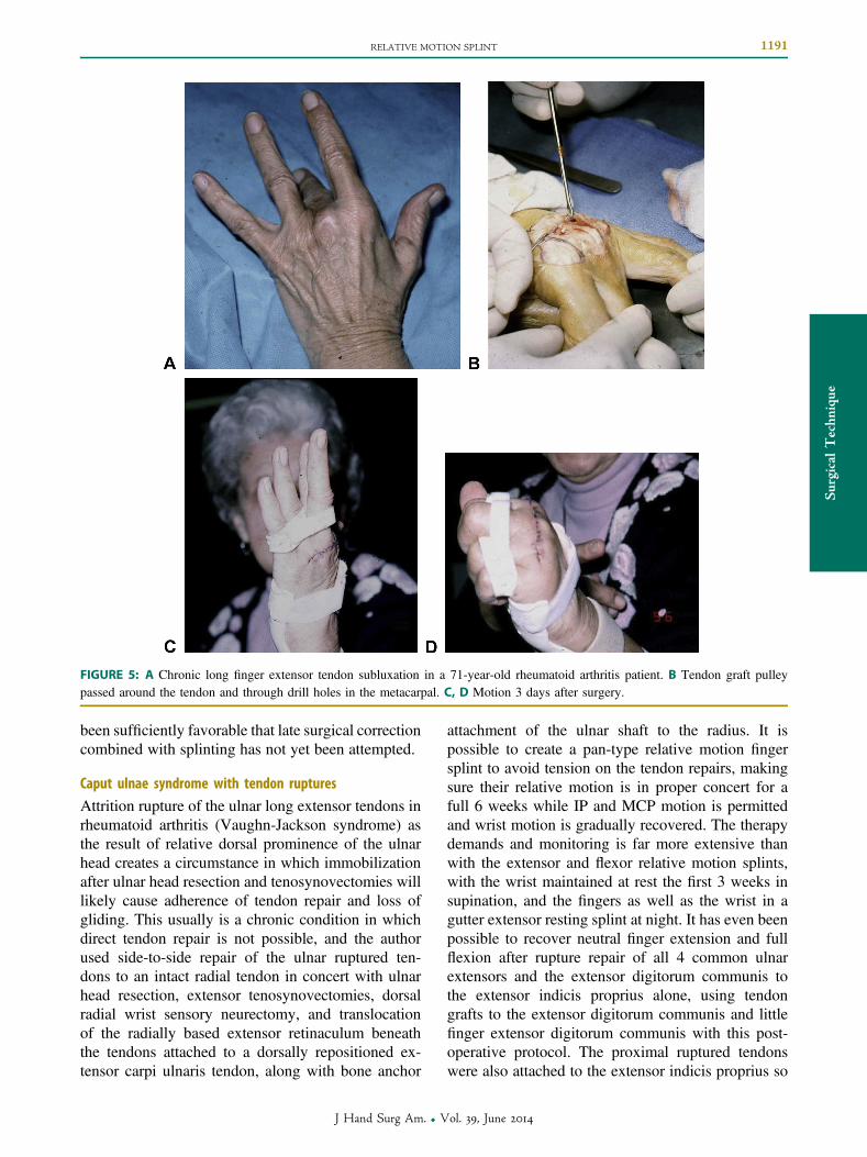
FIGURE 5: A Chronic long finger extensor tendon subluxation in a 71-year-old rheumatoid arthritis patient. B Tendon graft pulleypassed around the tendon and through drill holes in the metacarpal. C, D Motion 3 days after surgery.
RELATIVE MOTION SPLINT 1191
Surgical
Technique
been sufficiently favorable that late surgical correctioncombined with splinting has not yet been attempted.
Caput ulnae syndrome with tendon ruptures
Attrition rupture of the ulnar long extensor tendons inrheumatoid arthritis (Vaughn-Jackson syndrome) asthe result of relative dorsal prominence of the ulnarhead creates a circumstance in which immobilizationafter ulnar head resection and tenosynovectomies willlikely cause adherence of tendon repair and loss ofgliding. This usually is a chronic condition in whichdirect tendon repair is not possible, and the authorused side-to-side repair of the ulnar ruptured ten-dons to an intact radial tendon in concert with ulnarhead resection, extensor tenosynovectomies, dorsalradial wrist sensory neurectomy, and translocationof the radially based extensor retinaculum beneaththe tendons attached to a dorsally repositioned ex-tensor carpi ulnaris tendon, along with bone anchor
J Hand Surg Am. r V
attachment of the ulnar shaft to the radius. It ispossible to create a pan-type relative motion fingersplint to avoid tension on the tendon repairs, makingsure their relative motion is in proper concert for afull 6 weeks while IP and MCP motion is permittedand wrist motion is gradually recovered. The therapydemands and monitoring is far more extensive thanwith the extensor and flexor relative motion splints,with the wrist maintained at rest the first 3 weeks insupination, and the fingers as well as the wrist in agutter extensor resting splint at night. It has even beenpossible to recover neutral finger extension and fullflexion after rupture repair of all 4 common ulnarextensors and the extensor digitorum communis tothe extensor indicis proprius alone, using tendongrafts to the extensor digitorum communis and littlefinger extensor digitorum communis with this post-operative protocol. The proximal ruptured tendonswere also attached to the extensor indicis proprius so
ol. 39, June 2014
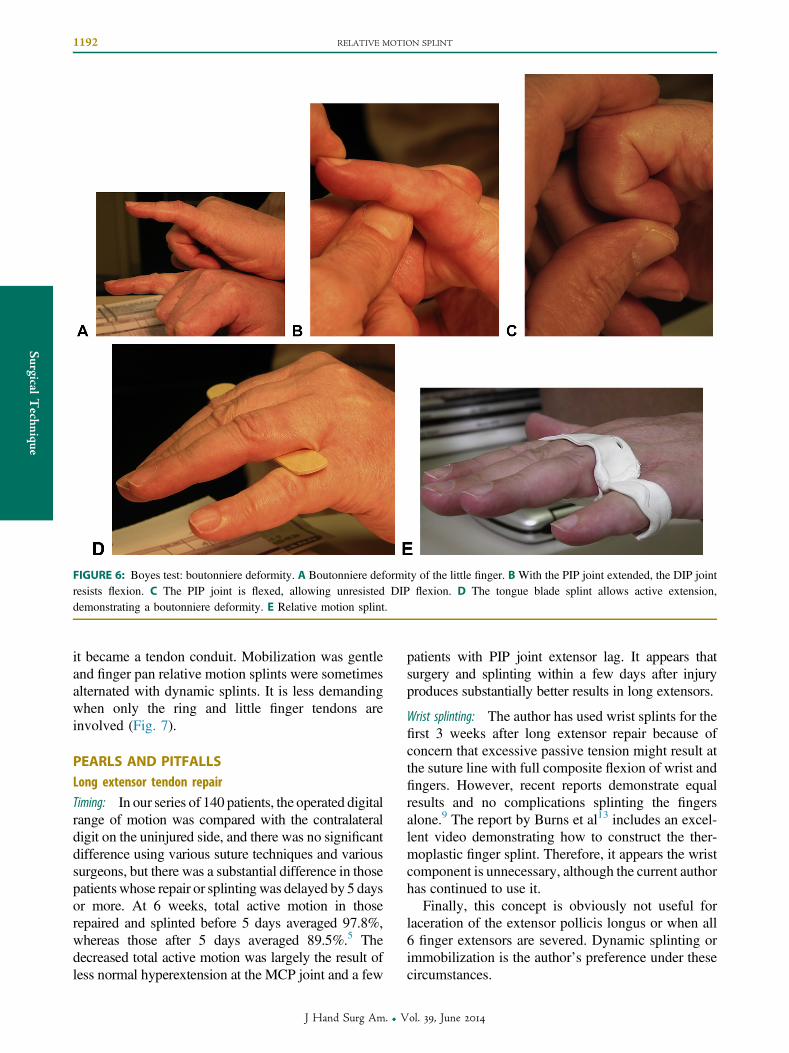
FIGURE 6: Boyes test: boutonniere deformity. A Boutonniere deformity of the little finger. B With the PIP joint extended, the DIP jointresists flexion. C The PIP joint is flexed, allowing unresisted DIP flexion. D The tongue blade splint allows active extension,demonstrating a boutonniere deformity. E Relative motion splint.
1192 RELATIVE MOTION SPLINT
Surgical
Tech
nique
it became a tendon conduit. Mobilization was gentleand finger pan relative motion splints were sometimesalternated with dynamic splints. It is less demandingwhen only the ring and little finger tendons areinvolved (Fig. 7).
PEARLS AND PITFALLSLong extensor tendon repair
Timing: In our series of 140 patients, the operated digitalrange of motion was compared with the contralateraldigit on the uninjured side, and there was no significantdifference using various suture techniques and varioussurgeons, but there was a substantial difference in thosepatientswhose repair or splintingwas delayed by 5 daysor more. At 6 weeks, total active motion in thoserepaired and splinted before 5 days averaged 97.8%,whereas those after 5 days averaged 89.5%.5 Thedecreased total active motion was largely the result ofless normal hyperextension at the MCP joint and a few
J Hand Surg Am. r V
patients with PIP joint extensor lag. It appears thatsurgery and splinting within a few days after injuryproduces substantially better results in long extensors.
Wrist splinting: The author has used wrist splints for thefirst 3 weeks after long extensor repair because ofconcern that excessive passive tension might result atthe suture line with full composite flexion of wrist andfingers. However, recent reports demonstrate equalresults and no complications splinting the fingersalone.9 The report by Burns et al13 includes an excel-lent video demonstrating how to construct the ther-moplastic finger splint. Therefore, it appears the wristcomponent is unnecessary, although the current authorhas continued to use it.
Finally, this concept is obviously not useful forlaceration of the extensor pollicis longus or when all6 finger extensors are severed. Dynamic splinting orimmobilization is the author’s preference under thesecircumstances.
ol. 39, June 2014
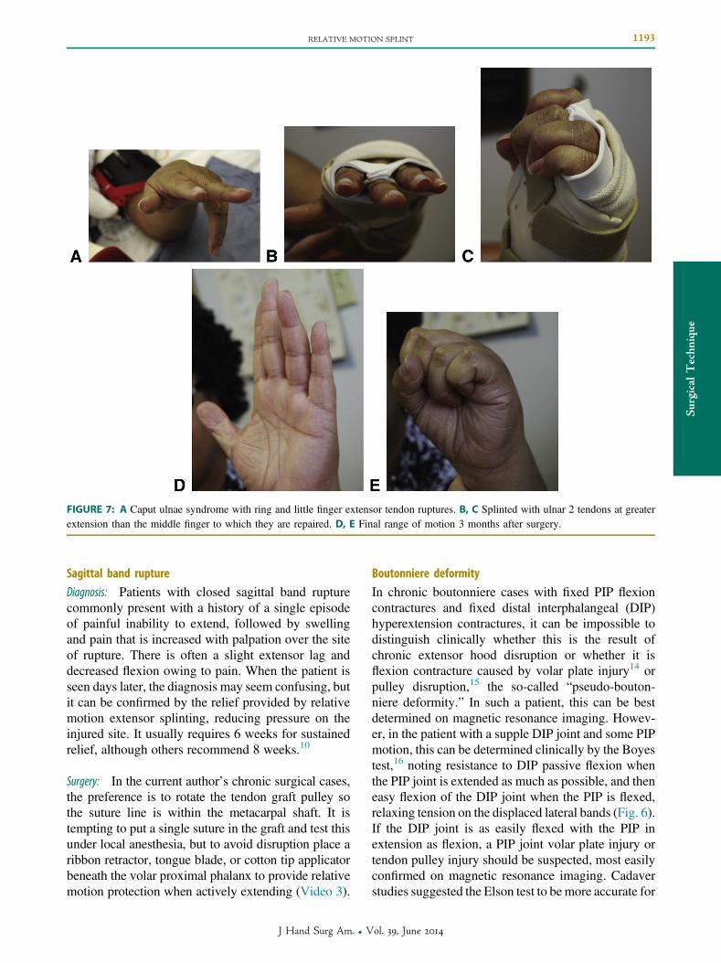
FIGURE 7: A Caput ulnae syndrome with ring and little finger extensor tendon ruptures. B, C Splinted with ulnar 2 tendons at greaterextension than the middle finger to which they are repaired. D, E Final range of motion 3 months after surgery.
RELATIVE MOTION SPLINT 1193
Surgical
Technique
Sagittal band rupture
Diagnosis: Patients with closed sagittal band rupturecommonly present with a history of a single episodeof painful inability to extend, followed by swellingand pain that is increased with palpation over the siteof rupture. There is often a slight extensor lag anddecreased flexion owing to pain. When the patient isseen days later, the diagnosis may seem confusing, butit can be confirmed by the relief provided by relativemotion extensor splinting, reducing pressure on theinjured site. It usually requires 6 weeks for sustainedrelief, although others recommend 8 weeks.10
Surgery: In the current author’s chronic surgical cases,the preference is to rotate the tendon graft pulley sothe suture line is within the metacarpal shaft. It istempting to put a single suture in the graft and test thisunder local anesthesia, but to avoid disruption place aribbon retractor, tongue blade, or cotton tip applicatorbeneath the volar proximal phalanx to provide relativemotion protection when actively extending (Video 3).
J Hand Surg Am. r V
Boutonniere deformity
In chronic boutonniere cases with fixed PIP flexioncontractures and fixed distal interphalangeal (DIP)hyperextension contractures, it can be impossible todistinguish clinically whether this is the result ofchronic extensor hood disruption or whether it isflexion contracture caused by volar plate injury14 orpulley disruption,15 the so-called “pseudo-bouton-niere deformity.” In such a patient, this can be bestdetermined on magnetic resonance imaging. Howev-er, in the patient with a supple DIP joint and some PIPmotion, this can be determined clinically by the Boyestest,16 noting resistance to DIP passive flexion whenthe PIP joint is extended as much as possible, and theneasy flexion of the DIP joint when the PIP is flexed,relaxing tension on the displaced lateral bands (Fig. 6).If the DIP joint is as easily flexed with the PIP inextension as flexion, a PIP joint volar plate injury ortendon pulley injury should be suspected, most easilyconfirmed on magnetic resonance imaging. Cadaverstudies suggested the Elson test to bemore accurate for
ol. 39, June 2014

1194 RELATIVE MOTION SPLINT
Surgical
Tech
nique
acute boutonniere injury and the Boyes test to beinaccurate.17 However, the cadaver does not developinflammatory remodeling and lateral band fixationin a volar displaced position as does the patient withchronic boutonniere. In the author’s experience, theestablished boutonniere is most easily identified by theBoyes test.17 Patients with pseudoboutonniere defor-mity will also need serial casting, and then prolongeddynamic extension splinting, although some haveadvocated volar plate13 and pulley14 surgery.
Caput ulnae syndrome
It is imperative to repair and maintain the extensors inthe proper sequence, with each ulnar digit in slightlymore flexion. Whereas the other extensor conditionsdiscussed generally require less hand therapy thanconventional methods, these patients require intenseindividualized hand therapy management. If the pa-tient regains MCP and IP flexion early, care must betaken to avoid tight grip with wrist flexion because ofpossible rupture. More frequently, patients must beassisted to gain flexion, gliding the repaired extensorsover the surgical field after tenosynovectomies andbone resection. Thereafter, patients may developextensor lag, so they must be rested in full MCP and IPextension, and sometimes assisted by dynamic exten-sion. The retinaculum is usually repositioned beneaththe extensor tendons because most of these patientshave poor wrist extension before surgery, and extensorbowstringing is not a likely problem. However, in somecases, a strip of dorsal retinaculum is reconstructed ifgood extension was present preoperatively. Fingerextension to neutral is all that usually can be expected,unless only the little finger tendons were ruptured.
DISCUSSIONThe relative motion concept to permit early activemotion has many varied applications whenever mul-tiple tendons are available from a common muscle.Relative motion flexor splinting can be used to pro-tect flexor tendon repair if early full flexion is foundafter 3 weeks and the patient wants to engage in fullvigorous activity; and it can assist recovery of fullPIP extension in patients who are stiff after chronictrigger finger release or other conditions that maycause tight intrinsics. In its best role, relative motionsplinting is used in combination with local anes-thesia and epinephrine (for example, with intrinsic
J Hand Surg Am. r V
tendon transfer to restore IP extension or in complexboutonniere repair), which provides an opportunity toverify the success of repair as well as the protectionafforded by the splint. Its greatest advantages are thesimplicity of concept and ease of use for patients.
REFERENCES
1. Strauch RJ. Extensor tendon injury. In: Wolfe SWW, Hotchkiss RN,Pederson WC, Kozin SH, eds. Green’s Operative Hand Surgery. 6th ed.Philadelphia, PA: Elsevier Churchill Livingstone; 2011:166e167.
2. McAuliffe JA. Early active short arc motion following central sliprepair. J Hand Surg Am. 2011;36(1):143e146.
3. Howell J, Merritt WH, Robinson S. Achieving immediate activemotion following zone 4-7 extensor tendon repair. J Hand Ther.2005;18(2):182e190.
4. Merritt WH. Complications of hand surgery. In: Greenfield LJ, ed.Complications in Surgery Trauma. Philadelphia, PA: Lippincott;1984:852e885.
5. Merritt WH, Howell J, Tune R, Saunders S, Hardy M. Achievingimmediate active motion by using relative motion splinting after longextensor tendon repair and sagittal band ruptures with tendon sub-luxation. Operative Techniques in Plastic and Reconstructive Sur-gery. 2000;7(1):1e38.
6. Sharma J, Lian N, Owen J, Wayne J, Isaacs J. Analysis of relativemotion splint in the treatment of zone VI extensor tendon injuries.J Hand Surg Am. 2006;31(7):1118e1122.
7. Merritt WH. What’s in a name? “ICAM” versus “relative motion”splints: what should we call these immediate active motion splints?AAHS Hand Surgery Quarterly. 2012;Fall:8.
8. Brand P. Mechanics of individual muscles at individual joints. In:Brand P, ed. Clinical Mechanics of the Hand. St. Louis, MO:CV Mosby; 1985:288e289.
9. Hirth MJ, Bennett K, Mah E, et al. Early return to work and improvedrange of motion with modified relative motion splinting: a retro-spective comparison with immobilization splinting for zones V andVI extensor tendon repairs. Hand Ther. 2011;16(4):86e94.
10. Catalano L, Gupta S, Ragland R, Glickel S, Johnson C, Barron O.Closed treatment of nonrheumatoid extensor tendon dislocations at themetacarpophalangeal joint. J Hand Surg Am. 2006;31(2):242e245.
11. Steichen J, Strickland J, Call W, Powell S. Results of surgicaltreatment of chronic boutonniere deformity: an analysis of prognosticfactors. In: Strickland J, Steichen J, eds. Difficult Problems in HandSurgery. St Louis, MO: CV Mosby; 1982:62e69.
12. Strauch RJ. Extensor tendon injury. In: Wolfe SWW, Hotchkiss RN,Pederson WC, Kozin SH, eds. Green’s Operative Hand Surgery. 6thed. Philadelphia, PA: Elsevier Churchill Livingstone; 2011:178.
13. Burns MC, Derby B, Neumeister MW. Wyndell Merritt imme-diate controlled active motion (ICAM) protocol following exten-sor tendon repairs in zone IV-VII: review of literature, orthosisdesign and case study—a multimedia article. Hand (N Y). 2013;8(1):17e22.
14. McCue F, Honner R, Gieck J, Andrews J, Hakala M. A pseudo-boutonniere deformity. Hand (N Y). 1975;7(2):166e170.
15. Bowers W, Kuzma G, Bynum D. Closed traumatic rupture of fingerflexor pulleys. J Hand Surg Am. 1994;19(5):782e787.
16. Boyes JH. Bunnel’s Surgery of the Hand. 5th ed. Philadelphia, PA:JB Lippincott; 1970:441.
17. Rubin J, Bozentka DJ, Bora FW. Diagnosis of closed central slipinjuries. J Hand Surg Br. 1996;21(5):614e616.
ol. 39, June 2014