Relationship of the Parietal Foramen and ... - scielo.cl
12
553 Int. J. Morphol., 27(2):553-564, 2009. Relationship of the Parietal Foramen and Complexity of the Human Sagittal Suture Relación del Foramen Parietal y la Complejidad de la Sutura Sagital Humana Robert W. Mann; Jiro Manabe & John E. Byrd MANN, R. W.; MANABE, J. & BYRD, J. E. Relationship of the parietal foramen and complexity of the human sagittal suture. Int. J. Morphol., 27(2):553-564, 2009. SUMMARY: The purpose of this paper is to report on the relationship between the parietal foramen and complexity of the human sagittal suture. Examination of 110 Japanese human skulls (males=67, females=43) with at least one parietal foramen revealed that the sagittal suture in the area of the Obelion was the simplest portion (i.e., fewest interdigitations and shortest length) of the suture (paired t- test, P<0.0005), when compared to the outstretched suture length of three established sections: 1. Parietal foramen section (P); 2. Anterior to section P (B); and 3. Posterior to section P (L). Sutural complexity was also compared between individuals with unilateral foramen (n=48) and bilateral foramina (n=62) to see if there was a statistically significant difference. The results revealed a slight difference in section P (ANOVA Bonferroni, P<0.05), denoting that the sagittal suture at the Obelion in individuals with unilateral parietal foramen is more complex than in individuals with bilateral foramina. While no difference in sex was noted, this simplicity in part likely reflects redirected bone stresses around a circular opening resulting in reduced tensile stresses and increased compressive stresses adjacent to the parietal foramen. This phenomenon warrants additional research and has implications for bone biomechanics and development of the cranial sutures. KEY WORDS: Sagittal suture; Complexity; Parietal foramen; Age. INTRODUCTION Study of the human cranial sutures has focused on such diverse topics as fractal analysis (Hartwig 1991; Yu et al., 2003; Skrzat & Walocha, 2003a, 2003b, 2004; Gorski & Skrzat, 2006), indicators of age (Parsons & Box, 1905; Todd & Lyon, 1924, 1925a,b,c; Retzlaff et al., 1979; Baker, 1984; Meindl & Lovejoy, 1985; Lynnerup & Jacobsen, 2003), frequency and location of accessory ossicles (Berry & Berry, 1967; El-Najjar & Dawson, 1977; Sanchez-Lara et al., 2007), cranial dimensions (Skrzat et al ., 2004) and sutural complexity as indicators of bone stresses (Anton et al., 1992; Kanisius & Luke, 1994; Nicolay & Vaders, 2006). Few studies or reports, however, have focused on the frequency, location and variation of parietal foramina, and even fewer on the possible relationship between parietal foramina and complexity of the human sagittal suture (Topinard, 1878; Currarino, 1976). Although the biomechanical explanation for this causal relationship is outside the scope of this study, the present work reveals both a visual and statistically significant relationship between the complexity of the sagittal suture and proximity of the parietal foramen. The authors hypothesize that the parietal foramen, a normally occurring feature in human skulls, acts to alter and redirect compressive and tensile stresses resulting in a relatively simple suture. Development of parietal bone. The parietal bone develops from a single intramembranous center near the eminence, beginning in the eighth fetal week and radiates outward in a sunburst pattern (Hoheisel, 1930; Currarino; Fazekas & Kosa, 1978 cited in Steele & Bramblett, 1988). The four sides of this approximately square shaped bone, which forms the sides and roof of the skull, join the frontal bone anteriorly along the coronal suture, the temporal and sphenoid bones inferiorly at the squamosal suture, the occipital bone posteriorly along the lambdoidal suture, and the opposing parietal bone superiorly at the sagittal suture (Gray, 1910). The concave endocranial surface of the two parietal bones contains deep grooves for the meningeal vessels, a shallow sulcus along the midline for the superior sagittal sinus, several Joint POW/MIA Accounting Command, 310 Worchester Ave., Hickam AFB, Hawaii, 96853-5530, USA.
Transcript of Relationship of the Parietal Foramen and ... - scielo.cl
553
Int. J. Morphol.,27(2):553-564, 2009.
Relationship of the Parietal Foramen and Complexityof the Human Sagittal Suture
Relación del Foramen Parietal y la Complejidad de la Sutura Sagital Humana
Robert W. Mann; Jiro Manabe & John E. Byrd
MANN, R. W.; MANABE, J. & BYRD, J. E. Relationship of the parietal foramen and complexity of the human sagittal suture. Int. J.Morphol., 27(2):553-564, 2009.
SUMMARY: The purpose of this paper is to report on the relationship between the parietal foramen and complexity of the humansagittal suture. Examination of 110 Japanese human skulls (males=67, females=43) with at least one parietal foramen revealed that thesagittal suture in the area of the Obelion was the simplest portion (i.e., fewest interdigitations and shortest length) of the suture (paired t-test, P<0.0005), when compared to the outstretched suture length of three established sections: 1. Parietal foramen section (P); 2. Anteriorto section P (B); and 3. Posterior to section P (L). Sutural complexity was also compared between individuals with unilateral foramen(n=48) and bilateral foramina (n=62) to see if there was a statistically significant difference. The results revealed a slight difference insection P (ANOVA Bonferroni, P<0.05), denoting that the sagittal suture at the Obelion in individuals with unilateral parietal foramen ismore complex than in individuals with bilateral foramina. While no difference in sex was noted, this simplicity in part likely reflectsredirected bone stresses around a circular opening resulting in reduced tensile stresses and increased compressive stresses adjacent to theparietal foramen. This phenomenon warrants additional research and has implications for bone biomechanics and development of thecranial sutures.
KEY WORDS: Sagittal suture; Complexity; Parietal foramen; Age.
INTRODUCTION
Study of the human cranial sutures has focused onsuch diverse topics as fractal analysis (Hartwig 1991; Yu etal., 2003; Skrzat & Walocha, 2003a, 2003b, 2004; Gorski &Skrzat, 2006), indicators of age (Parsons & Box, 1905; Todd& Lyon, 1924, 1925a,b,c; Retzlaff et al., 1979; Baker, 1984;Meindl & Lovejoy, 1985; Lynnerup & Jacobsen, 2003),frequency and location of accessory ossicles (Berry & Berry,1967; El-Najjar & Dawson, 1977; Sanchez-Lara et al., 2007),cranial dimensions (Skrzat et al., 2004) and suturalcomplexity as indicators of bone stresses (Anton et al., 1992;Kanisius & Luke, 1994; Nicolay & Vaders, 2006). Fewstudies or reports, however, have focused on the frequency,location and variation of parietal foramina, and even feweron the possible relationship between parietal foramina andcomplexity of the human sagittal suture (Topinard, 1878;Currarino, 1976). Although the biomechanical explanationfor this causal relationship is outside the scope of this study,the present work reveals both a visual and statisticallysignificant relationship between the complexity of the sagittal
suture and proximity of the parietal foramen. The authorshypothesize that the parietal foramen, a normally occurringfeature in human skulls, acts to alter and redirect compressiveand tensile stresses resulting in a relatively simple suture.
Development of parietal bone. The parietal bone developsfrom a single intramembranous center near the eminence,beginning in the eighth fetal week and radiates outward in asunburst pattern (Hoheisel, 1930; Currarino; Fazekas &Kosa, 1978 cited in Steele & Bramblett, 1988). The foursides of this approximately square shaped bone, which formsthe sides and roof of the skull, join the frontal bone anteriorlyalong the coronal suture, the temporal and sphenoid bonesinferiorly at the squamosal suture, the occipital boneposteriorly along the lambdoidal suture, and the opposingparietal bone superiorly at the sagittal suture (Gray, 1910).The concave endocranial surface of the two parietal bonescontains deep grooves for the meningeal vessels, a shallowsulcus along the midline for the superior sagittal sinus, several
Joint POW/MIA Accounting Command, 310 Worchester Ave., Hickam AFB, Hawaii, 96853-5530, USA.
554
cluster-shaped depressions and pits resembling clusters ofgrapes for the arachnoid granulations.
Slowed or delayed ossification in the posterior one-third of the parietal bone at the Obelion results in a slit orv-shaped notch, sometimes referred to as the subsagittalsuture of Pozzi (Breathnach, 1965), fonticuius obelicus(Pernkopf, 1963), pars obelica (Scheuer & Black, 2004),or sagittal or third fontanelle, that widens the sagittal sutureat that level (Currarino). Obelion, so named for itsresemblance to the Greek symbol obelos ÷ refers to thatportion of the sagittal suture between the parietal foramina;the dots of obelos represent the parietal foramina (Jamieson,1937). Ossification and closure of the third fontanelle,which usually occurs in the fifth fetal month, often resultsin vestiges and normal anatomical variants known asobeliac bones, enlarged parietal foramina, parietal fissure,and one or more parietal foramina (Currarino). The roleof the parietal foramen, other than transmitting an emissaryvein or artery to the superior sagittal sinus, is still underinvestigation but may simply be the result of delayed orincomplete ossification as just described (Wu et al., 2000).In addition, when a parietal foramen is unusually large asthe result of slowed ossification, the area around the fora-men may be thinner and flatter (Boyd, 1930).
Simplification of the sagittal suture near the parietalforamen, a condition occasionally reported in the clinical,anatomical and anthropological literature (Topinard;Currarino; Hauser & DeStefano, 1989; Scheuer & Black),therefore, could be explained by slowed or delayedossification. Koskinen et al. (1976), in contrast, suggestthat rapid growth of the parietal bone would result in anincrease in interdigitations and an overlapping of the suture.If this feature prevails as a uniform type, the complexityof the sagittal suture near the parietal foramen results inexcess bone development, and the prematurely fusingsuture with abnormal fibroblast growth factor (FGF) mightlead to precocious osteogenic differentiation andcraniofacial malformation (Sarkar et al., 2001; Moore etal., 2002). Thus, ossification of the parietal bone might beone of the crucial variables in understanding the basicpattern of the sagittal suture and its simplicity near theparietal foramen.
Vault suture morphology, whether complex or sim-ple, is a feature that physical anthropologists and otherssometimes use as an indicator of ancestry when examininghuman skeletal remains. For example, highly complexsutures are often viewed as a Mongoloid (Asian) trait andsimple sutures as Caucasoid (White) or Negroid (Black)(Rhine, 1990; Burns, 1999; Byers, 2008). The degree ofsuture complexity and its relationship to ancestry, therefore,
has a medicolegal significance and may play a role inestablishing the identity of unknown remains.
Although subject to a host of variables and notnecessarily an accurate indicator of age (Hershkovitz etal., 1997), fusion of the vault sutures (Todd & Lyon, 1924,1925a, b, c; Meindel & Lovejoy) and maxillary sutures(Mann et al., 1987, 1991) has often been used as a methodto estimate age at death. For example, Persson et al. (1978)advocated that the fusion of sutures is related to vascular,hormonal, genetic and biomechanical factors and theObelion is believed to be the first region of the sagittalsuture to begin closure (Topinard; Currarino). The presentauthors were unable to find additional information orresearch explaining why the sagittal suture starts to closeat the Obelion first, despite suture closure being quite va-riable in individuals. Closure of the sagittal suture involvesthree fontanelles: 1. the anterior fontanelle located wherethe frontal bone and parietal bones meet, 2. the posteriorfontanelle where the occipital and parietal bones meet, and3. the third or sagittal fontanelle which is present in 50-80% of perinatal skulls, leading to the formation of one ormore parietal foramina postnatally. The third fontanelleusually closes within the first two years postnatally (96%of cases) (Scheuer & Black). The timing of bony unionand closure of this fontanelle might be related to complexityand morphology of the sagittal suture.
The reported frequency of the parietal foramenvaries from 50 to 80% of individuals in different populationgroups (Scott, 1893; Boyd; Wysocki et al., 2006; Yoshiokaet al., 2006) and has been shown to vary little in its number,location, size, and shape. As for position, the parietal fo-ramen is reported as being located in the posterior one-fifth (Topinard) or one-third of the parietal bone; in otherwords, its location is approximately 2cm anterior to theLambda in newborns and 2-5cm anterior in adults(Currarino), or an average distance of 83mm from the Inion(Yoshioka et al.).
The size of the parietal foramen has been reportedas between 1.8-2.0mm (Wysocki et al.; Yoshioka et al.),although Boyd stated that the average is under 0.5mm anda size greater than 1.5 mm is rare. Unexpectedly, Wysockiet al. found that the average size of parietal foramina inPolish females was twice as large as in males (3.0mm infemales and 1.5mm in males), ranging from 0.38-16.8mm,suggesting sexual dimorphism in parietal ossification. Boydhowever, found no difference in age or sex. Circular, oval,or slit-like enlarged parietal foramina measuring severalcentimeters in diameter or length, however, have beenreported in the literature (O’Rahilly & Twohig, 1952;Mann, 1990; Keats, 1992).
MANN, R. W.; MANABE, J. & BYRD, J. E. Relationship of the parietal foramen and complexity of the human sagittal suture. Int. J. Morphol., 27(2):553-564, 2009.

555
MATERIAL AND METHOD
Statistical analysis was conducted to test if the simplerform (i.e., complexity) of the sagittal suture anterior and pos-terior to the parietal foramen was consistent. The sample ofskulls with an open or visible sagittal suture consisted of 79Japanese males and 58 Japanese females (n=137) selectedfrom the skeletal collection at the School of Medicine, Chi-ba University and the University Museum, the Universityof Tokyo in Japan (Fig. 1). A piece of clear plastic was tapedover the outer surface of the skull and a black Sharpie® Ul-tra Fine permanent marking pen was used to trace the lengthof the sagittal suture and mark the location of one or moreparietal foramina. These records served as the basis forfurther measurement.
To determine if the suture is less complex (i.e., fewestinterdigitations and shortest length) in the area of the parietalforamina, samples with one or more parietal foramina wereselected and the number of foramina in the left and rightparietal bones noted. Skulls with a relatively complex sagittalsuture were visually examined to determine if there was anymorphological difference beyond the suture in those withone or more parietal foramina compared to skulls withoutforamina (closed or absent).
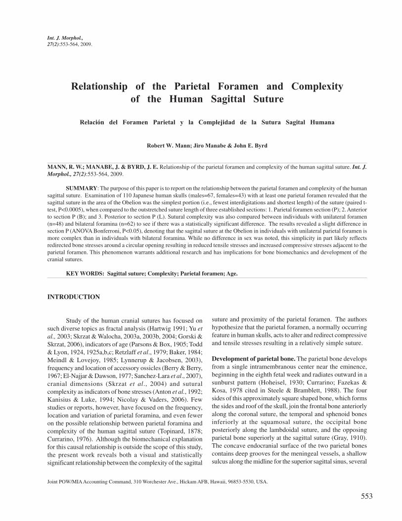
For each sagittal suture, a straight line was drawnfrom Bregma to Lambda. Next, three sections of equal length(15mm long each) were established as follows: (P) =centered on Obelion; (B) = anterior to section (P) andObelion; and (L) = posterior to section (P) and Obelion (Fig.2). This process was performed by manually annotating aphotocopied image, which was then scanned at a highresolution and saved in TIF format. The digitally saved imagewas then opened with the software ImageJ, offered by theNational Institutes of Health (NIH) and available at: http://rsb.info.nih.gov/ij/ (Rasband, 1997-2000), and the“outstretched” suture length of the three sections wasmeasured. Measurement was taken along the suture lines, toinclude lines along ossicles or other isolated suture lines.Where the suture was interrupted, no measurement was taken.Thus, obliterated lines made no contribution to the data. Eachoutstretched suture in the three sections P, B, and L wasmeasured to compare the complexity among the threesections and to see if there is any difference between thesexes.
To test the hypothesis that the sagittal suture issimplest (i.e. shortest outstretched length) at or around theparietal foramen, the authors compared the outstretchedsuture length of the three sections, B, P, and L and computedthe frequency at which each section was simplest on each
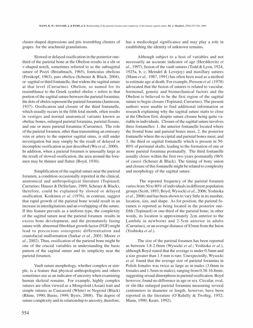
skull. Furthermore, the mean value of the combined sectionsB and L (described: section [B+L], calculated as: mean value= [section B + section L]/2), was compared with section P inorder to assess if the suture around parietal foramen is leastcomplex than its adjacent anterior and posterior portion. Itmust be noted that suture obliteration was observed morefrequently in section P (13 instances of an interruption inthe suture line), but the combination of sections B and Lexhibited a similar number of instances of interruption (11instances). A paired t-test examined any statisticallysignificant difference in the outstretched length betweensection P and section B+L, and the eta squared statistic wasused to reveal how large the difference was. This wasfollowed by an ANOVA which tested the null hypothesisthat there were no significant differences among the threesections. The ANOVA was followed up with pairwisecomparisons of the three sections utilizing the Bonferonicriterion for significance. A correlation analysis utilizingSpearman’s rho was performed to investigate the strengthof relationship between lengths of the sections.
Fig. 1. Sagittal suture with bilateral parietal foramina (left). Thesuture was traced and the location of the foramina was recorded onthe plastic overlay. Sagittal suture without parietal foramen (right).Despite the absence of a parietal foramen, the suture is simplest inthe area (the Obelion) where one or more parietal foramina areusually located (square).
MANN, R. W.; MANABE, J. & BYRD, J. E. Relationship of the parietal foramen and complexity of the human sagittal suture. Int. J. Morphol., 27(2):553-564, 2009.
556
These tests were followed by an ANOVA and aGeneralized Linear Model test to determine if the presenceof multiple foramina affects the complexity of the suturein the vicinity of Obelion. First, a comparison of thecomplexity of the raw total length of section P betweenindividuals with unilateral and bilateral foramina was madeand examined (note: individuals without foramen were
excluded due to technical difficulty). Second, rather thanuse the raw total length of section P, the proportion of thethree sections accountable to P was used as follows: propP= P/(B+P+L). Another variable, “foramen” indicatedwhether the cranium had one or two foramina. The thirdvariable included in the model was “complexity”, whichwas calculated as the total sagittal suture length (followingthe actual contour) divided by the parietal chord.Complexity was included in the model to account for anysampling error in reference to the overall complexityexhibited by the suture. The GLM model was: propP =foramen * complexity, and the Type III sum of squareswere used as the basis for the F tests. Statistical calculationswere done in SPSS 15.0.
RESULTS
Sixty-seven (85%) of 79 male Japanese skulls and43 of 58 (74%) Japanese female skulls possessed foramina(see Table I). This result is similar to what other scientistshave reported (50-80%; c.f. Boyd; Scott; Wysocki et al.;Yoshioka et al.). The location of the parietal foramen wasinvestigated utilizing skulls with one or more foramina.The curved distance from the Lambda to the foramen wasmeasured, which resulted in an average distance and rangeof 3.8cm and 2-5cm, respectively. Exceptions to this ave-rage were found in two skulls where foramina exceeded 2-3mm above 5cm, giving a frequency distribution of: 2-2.5cm (4.6%), 2.6-3cm (4%), 3.1-3.5cm (17.3%), 3.6-4cm(34.6%), 4.1-4.5cm (32.6%), and more than 4.6cm (6.6%).Although some skulls did not exhibit a foramen, thereappeared to be a portion of least complexity occurring 2-5cm anterior to the Lambda, where the parietal foramenlikely would have been located (the Obelion) (Fig. 1).
Our results, just as reported by Currarino, indicatethat the suture was simplest at the level nearest to theparietal foramen in the majority of individuals, renderingthe section P the least complex of the three sections (seeFig. 3A, 3B and 4). Of 67 male skulls with one or moreparietal foramina, section P was the least complex amongthe three sections in 60 (or 89%) of individuals. Section Pwas least complex in 38 (88%) of 43 female skulls. Theaverage suture length of section P was similar in malesand females (21.7mm and 21.3mm). Of all the sections, Bwas the most complex section (average suture length,53.0mm in males and 48.1mm in females) followed bysection L (average suture length, 33.3mm in males and34.6mm in females) (Fig. 3A and 4). Figure 3B showsproportional comparison for each section and reveals thateach section is more likely segregated.
Fig. 2. Method for measuring suture complexity between thesection at the parietal foramen (P) and the anterior (B), andposterior (L) sections. Each section measures 15mm in length.First, a line is drawn between Bregma and Lambda, then a15mm section nearest the parietal foramen is set (if theforamina are located bilaterally, a middle point between theanterior and posterior levels of the foramina becomes thehalfway point of the section. The anterior (B) and posterior(L) sections are obtained after the position of the parietal fo-ramen (P) is established.
MANN, R. W.; MANABE, J. & BYRD, J. E. Relationship of the parietal foramen and complexity of the human sagittal suture. Int. J. Morphol., 27(2):553-564, 2009.

557
Fig. 3. A (top): Length of suture in each section: B, P and L (fromleft) Unit is mm. B (middle): Proportion of the sections of B, P andL (from left). Unit is percentage (value (%) = section length dividedby total length of the three sections). C (bottom): Correlationbetween proportion of section B and proportion of section L. Of 67male skulls with one or more parietal foramina, section P in 60individuals (89%) was the least complex among three sections inthe following order: P, L, and B, and 38 (88%) of 43 female skulls;the average suture length of section P in males was 21.7mm and21.3mm in females. Of all these sections, section B was the mostcomplex section (average suture length, 53.0mm in males and48.1mm in females) followed by section L (average suture length,33.3mm in males and 34.6mm in females).
MANN, R. W.; MANABE, J. & BYRD, J. E. Relationship of the parietal foramen and complexity of the human sagittal suture. Int. J. Morphol., 27(2):553-564, 2009.

558
Sex (n) Foramen OnSuture (n/%)
Unilateral ForamenLeft (n/%)
Unilateral ForamenRight (n/%)
BilateralForamina (n/%)
No Foramen(n/%)
Male (79) 0/0 12/15 16/20 39/49 12/15
Female (58) 1/2 9/15 10/17 23/39 15/25
Table I. Frequency of parietal foramen in the sample.
T-test (P to B+L)Section N Mean (mm) S D
t P
P 110 23.131 7.238
B & L 110 45.508 16.151-17.159 <0.001
Table II. Comparison of complexity between section P and its anterior and posterior sections.
Section P was compared with the mean of sections B and L (as B +L / 2). T-test reveals a significant difference between section P and the other twosections.
Table III. Complexity of the three sections of the sagittal suture in the vicinity of Obelion.
ANOVA that tests the null hypothesis that there are no differences in the complexity of the three sections of the sagittal suture in the vicinity of the Obelion.All pairwise comparisons indicated significant difference at the 0.05 level using the Bonferroni criterion.
Source Type III Sum of Squares d.f. Mean Square F P
Corrected Model 0.064 2 0.032 19.430 0.000
Intercept 0.685 1 0.685 416.951 0.000
Foramen 0.001 1 0.001 0.418 0.519
Complexity 0.063 1 0.063 38.547 0.000
Error 0.166 101 0.002
Total 4.847 104
Table IV. Suture complexity difference at section P between individuals with unilateral and bilateral foramina.
A (top): Suture complexity comparing the raw total length of section P between individuals with unilateral and bilateral foramina. B (bottom): The modeltested was: propP = foramen * complexity, and was intended to provide a basis for evaluating the effects of multiple foramina while controlling for theeffect of the overall complexity of the suture. Variable propP was calculated as P / (P+B+L). Complexity was calculated as the total sagittal suture lengthdivided by the parietal chord. Simple statistics with an F-test are given in A and GLM test results in B.
MANN, R. W.; MANABE, J. & BYRD, J. E. Relationship of the parietal foramen and complexity of the human sagittal suture. Int. J. Morphol., 27(2):553-564, 2009.
One-way ANOVAParietal Foramen N Mean (mm) S D
F P
Unilateral 48 24.671 8.213
Bilateral 62 21.939 6.1913.959 <0.05
Section N Minimum (mm) Maximum (mm) Mean (mm) S D
B 110 16.54 117.81 53.08 20.80
P 110 10.18 49.22 23.13 7.24
L 110 10.85 91.45 37.93 16.45
Source Sum of Squares d.f. Mean Square F P
Between groups 49348.63 2 24674.31 97.95 0.000
Within groups 82376.54 327 251.92
Total 131725.16 329
A
B
559
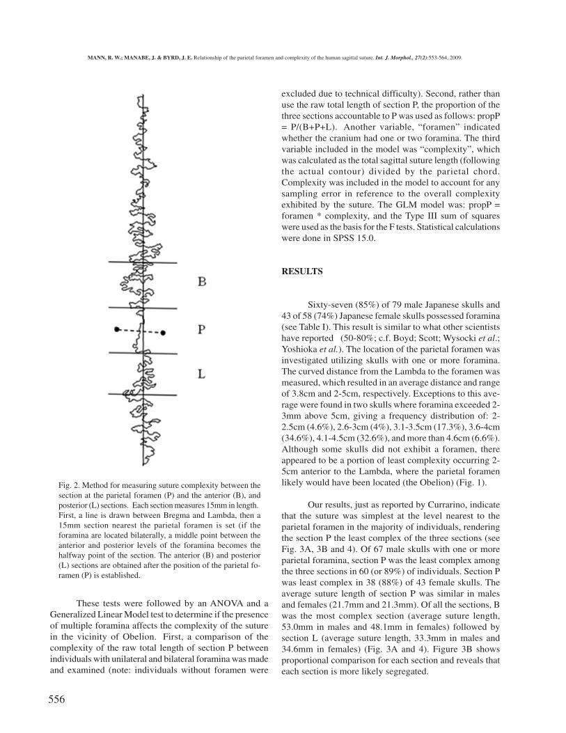
Fig. 5. Diagram of possible tensile activity (arrows) in relation to sutural complexity with parietal foramen/foramina. Shaded circle reveals a possible area of reduced tensile stresses.
Fig. 4. Distribution of each sectional outstretched suture length. “Section B+L” is the meanvalue: value=(section B + section L)/2. Notably, length of section P is clustered around 25mm.
MANN, R. W.; MANABE, J. & BYRD, J. E. Relationship of the parietal foramen and complexity of the human sagittal suture. Int. J. Morphol., 27(2):553-564, 2009.
560
Using the total sample of 110 skulls, a paired t-testwas conducted to examine if there was a statisticallysignificant difference in sectional length between section Pand section B+L (mean value), without discriminating bysex. The results indicate that there is a significant differencebetween section P and section B+L (paired t-test, t=-17.159,P<0.0005) (Table II). Eta squared was also computed in thisregard and reveals relatively larger effect (0.73). The ANOVAtest found that the three sections are significantly different(F=97.947, P<0.0001) and all are different from one anotherat the P<0.0001 level (Table III). The order of complexityfrom most to least complex was: B, L and P.
A relatively high correlation was seen in proportionB (value = length of B/total length of the three sections)versus proportion L [Spearman’s rho, r=-0.79, n=110,P<0.0005] (Fig. 3C), and there was no significantcorrelation between proportion P and other two sections.This finding might reflect that suture complexity shares acertain pattern between section B and section L, while therewas no correlation with section P because it wasconsistently less complex, regardless of the complexity ofsection B and/or L.
Exceptions to these findings were found in the skullsof seven males and five females. Two males and one femaleshowed relatively consistent complexity throughout theparietal chord; however, section P proved to be morecomplex than sections B or L. This varying complexitymight be the result of the rate of development of the parietalbone, combined with irregular or differing tensile andcompressive stresses exerted by the temporalis muscle.Also, less complexity adjacent to Obelion was seen in threemales and two females. In another two males and twofemales, section P was slightly longer because the suturewas simple in both sections P and L. While the role of themusculature on sutural complexity is outside the scope ofthis study, the areas of the sagittal suture that lie closest tothe parietal foramen are consistently simpler than thoseareas closer to Bregma and Lambda.
The suture samples grouped by unilateral foramenand bilateral foramina were compared utilizing the ANOVAand the GLM test. In the ANOVA comparing the total rawlength of section P between these two groups, there was asignificant difference (F=3.959, P<0.05), indicating that thesagittal suture in individuals with unilateral parietal fora-men are slightly more complex in this section than individualswith bilateral foramina [unilateral (Mean=24.67(mm),SD=8.21), bilateral (Mean=21.93, SD=6.19)] (Table 4A).However, results in the GLM indicate that the number offoramina does not significantly affect the complexity of thesuture (Table 4B).
DISCUSSION
Examination revealed that in 89.5% and 88.3% (malesand females, respectively) of Japanese human skulls, the areaof the sagittal suture closest to the parietal foramen was theleast complex and shortest segment compared to the sectionsimmediately anterior and posterior to the Obelion. The relativesimplicity of the sagittal suture at the Obelion is likely the resultof many variables, including redirected stresses associated withmastication and other physical activities, as well as its proximityto the lambdoidal suture and muscular activity associated withthe nuchal crest. Perhaps not surprisingly, the sagittal suture atthe Obelion in individuals with a unilateral foramen shows aslight tendency to be more complex than in individuals withbilateral foramina. Test results indicate, however, that thispattern could be a result of sampling error. Were the patternreal, this increased complexity in a unilateral trait versus a bi-lateral trait could possibly reflect increased tension in theformer, resulting in greater complexity (Fig. 5).
Numerous animal studies, for example, have shownthat the morphology of a suture is affected by biomechanicalstresses associated with chewing and biting (Hubbard et al.,1971; Behrents et al., 1978; Smith & Hylander, 1985; Hylanderet al., 1991; Huang et al., 1994; Herring & Teng, 2000; Markeyet al., 2006; Herring & Mucci, 2007 and Markey & Marshall,2007), which results in a combination of tensile, compressionand shear forces along different parts of the same suture. Alaqeelet al. (2006), for example, reported that mechanical forces andstresses associated with a variety of activities results in shearstresses of the frontal and parietal bones along the coronalsuture, compressive forces in the parietal bones near the vertex,and tensile forces in the posterior portions of the parietal bonesin the area of the parietal foramen. Generally speaking and inthe words of Enlow (1990), sutures are “tension-adapted growthsites,” such that tensile forces result in a widening of suturemargins and bone development.
Experimental in vivo studies of animals, however, haveprovided contrasting results. Markey et al. (2006), for example,surgically implanted strain gauges into the sutures of fish(Polypterus endlicherii) to study the relationship between sutureform and function during feeding. These researchers foundthat the interfrontal and frontoparietal sutures that are normallyloaded in tension were less interdigitated in cross section thanthe interparietal suture that was simultaneously undergoingcompression. Markey and colleagues found that interdigitated(complex) sutures resulted from compression and lessinterdigitated (less complex, simpler) sutures were associatedwith tension. Additional animal studies in fish (Markey &Marshall, 2007), pigs (Herring & Mucci, 1991; Herring & Teng,2000), and goats (Jaslow & Biewener, 1995) reported similar
MANN, R. W.; MANABE, J. & BYRD, J. E. Relationship of the parietal foramen and complexity of the human sagittal suture. Int. J. Morphol., 27(2):553-564, 2009.

561
findings, indicating that increased interdigitation is associatedwith compression and decreased interdigitation with tension.
Mechanical forces exerted, absorbed and redirected inthe areas surrounding the parietal foramina might result in anincrease in tensile forces in certain areas and a decrease incompressive forces in other areas of the parietal bone near theLambda, resulting in reduced complexity along the sagittalsuture. While perhaps not a true analogy, the circular openingin a rotunda designed architecturally to strengthen a structure,might reflect a similar condition in bone, such that the opening(foramen) alters or redirects the weight and stresses exerted onthe structure. It is also possible that the presence of emissaryvessels filling the parietal foramina alters the stresses in thearea of the Obelion.
ACKNOWLEDGEMENTS
This research was supported in part by anappointment of the second author (Jiro Manabe) to thePostgraduate Research Participation Program at the JointPOW/MIA Accounting Command/Central IdentificationLaboratory (JPAC/CIL) administered by the Oak RidgeInstitute for Science and Education through an interagencyagreement between the U.S. Department of Energy andJPAC/CIL. We thank Dr Kazuhiro Sakaue at Departmentof Anthropology, National Museum of Nature and Science,Dr Yoshiharu Matsuno at School of Medicine, ChibaUniversity and Dr Gen Suwa at the University Museum,the University of Tokyo for their cooperation.
MANN, R. W.; MANABE, J. & BYRD, J. E. Relación del foramen parietal y la complejidad de la sutura sagital humana. Int. J.Morphol., 27(2):553-564, 2009.
RESUMEN: El propósito de este trabajo es informar sobre la relación entre el foramen parietal y la complejidad de la suturasagital en humanos. Se examinaron 110 cráneos humanos de individuos japoneses (hombres = 67, mujeres = 43) con al menos unforamen parietal, revelando que la sutura sagital en el área del obelion fue la parte más simple (es decir, menos interdigitaciones y menorlongitud) de la sutura (vinculado la prueba t, p <0,0005). Cuando se comparó la extensión de la longitud de la sutura se establecieron tressecciones: 1. Sección foramen parietal (P); 2. Anterior a la sección P (B), y 3. Posterior a la sección P (L). La complejidad de la suturatambién fue comparada entre los individuos con foramen unilateral (n = 48) y forámenes bilaterales (n = 62) para ver si había unadiferencia estadísticamente significativa. Los resultados revelaron una ligera diferencia en la sección P (ANOVA Bonferroni, P <0,05),que indica que la sutura sagital a nivel del obelion en los individuos con foramen parietal unilateral es más compleja que en los individuoscon forámenes bilaterales. Si bien no hubo diferencia según sexo, esta simplicidad en parte, probablemente refleja la redirección de lasfuerzas del hueso alrededor de una abertura circular, lo que reduce la resistencia a la tracción y aumenta la fuerza de compresiónadyacente al foramen parietal. Este fenómeno justifica la investigación adicional y tiene implicaciones para el desarrollo óseo y biomecánicade las suturas craneales.
PALABRAS CLAVE: Sutura sagital; Complejidad; Foramen parietal; Edad.
REFERENCES
Alaqeel, S. M.; Hinton, R. J. & Opperman, L. A. Cellularresponse to force application at craniofacial sutures.Orthod. Craniofac. Res., 9(3):111-22, 2006.
Anton, S. C.; Jaslow, C. R. & Swartz, S. M. Suturalcomplexity in artificially deformed human (Homosapiens). Crania. J. Morphol., 214(3):321-32, 1992.
Baker, R. K. The relationship of cranial suture closure andage analyzed in a modern multi-racial sample of malesand females. MA thesis, California State University,Fullerton, 1984.
Behrents, R. G.; Carlson, D. S. & Abdelnour, T. In vivoanalysis of bone strain about the sagittal suture in macacamulatta during masticatory movements. J. Dent. Res.,57(9-10):904-8, 1978.
Berry, A. C. & Berry, R. J. Epigenetic variation in the humancranium. J. Anat., 101(2):361-79, 1967.
Boyd, G. I. The emissary foramina of the cranium in manand the anthropoids. J. Anat., 65(1):108-21, 1930.
Breathnach, A. S. Frazer’s anatomy of the human skeleton.6. Ed. London, J&A Churchill Ltd, 1965.
Burns, K. R. Forensic anthropology training manual. NewJersey, Prentice Hall, 1999.
Byers, S. N. Introduction to forensic anthropology. Boston,Allyn & Bacon, 2008.
Currarino, G. Normal variants and congenital anomalies in theregion of the obelion. Am. J. Roentgenol., 127(3):487-94, 1976.
MANN, R. W.; MANABE, J. & BYRD, J. E. Relationship of the parietal foramen and complexity of the human sagittal suture. Int. J. Morphol., 27(2):553-564, 2009.

562
El-Najjar, M. & Dawson, G. L. The effect of artificial cranialdeformation on the incidence of wormian bones in thelambdoidal suture. Am. J. Phys. Anthropol., 46(1):155-60, 1977.
Enlow, D. H. Handbook of facial growth. 3. Ed. Philadelphia,PA, WB Saunders Co, 1990.
Górski, A. Z. & Skrzat, J. Error estimation of the fractaldimension measurements of cranial sutures. J. Anat.,208(3):353-9, 2006.
Gray, H. Anatomy: descriptive and applied (ed. Spitzka EA),18. Ed. Philadelphia, PA, Lea & Febiger, 1910.
Hartwig, W. C. Fractal analysis of sagittal suture morphology.J. Morphol., 210(3):289-98, 1991.
Hauser, G. & DeStefano, G. F. Epigenetic variants of thehuman skull. Stuttgart, Schweizerbart, 1989.
Herring, S. W. & Mucci, R. J. In vivo strain in cranial sutures:the zygomatic arch. J. Morphol., 207(3):225-39, 1991.
Herring, S. W. & Teng, S. Strain in the braincases and itssutures during function. Am. J. Phys. Anthropol.,112(4):575-93, 2000.
Hershkovitz, I.; Latimer, B.; Dutour, O.; Jellema, L. M.;Wish-Baratz, S.; Rothschild, C. & Rothschild, B. M. Theelusive petroexoccipital articulation. Am. J. Phys.Anthropol., 103(3):365-73, 1997.
Hoheisel, W. F. An anomalous Indian occiput. Anat. Rec.,45(2):129-35, 1930.
Huang, X.; Zhang, G. & Herring, S. W. Age changes inmastication in the pig. Comp. Biochem. Physiol.,107(4):647-54, 1994.
Hubbard, R. P.; Melvin, J. W. & Barodawala, I. T. Flexureof cranial sutures. J. Biomech., 4(6):491-6, 1971.
Hylander, W. L.; Picq, P. G. & Johnson, K. R. Masticatory-stress hypotheses and the supraorbital region of primates.Am. J. Phys. Anthropol., 86(1):1-36, 1991.
Jamieson, E. B. Dixon’s manual of human osteology. 2. Ed.London, Humphrey Milford, 1937.
Jaslow, C. R. & Biewener, A. A. Strain patterns in thehorncores, cranial bones and sutures of goats (Caprahircus) during impact loading. J. Zool., 235:193-210, 1995.
Kanisius, P. H. & Luke, D. A. Is the complexity of the humansagittal suture related to the size of the temporal muscle?Int. J. Anthropol., 9(4):265-72, 1994.
Keats, T. E. Atlas of normal roentgen variants that maysimulate disease. 5. Ed. St. Louis, Mosby Year Book,1992.
Koskinen, L.; Isotupa, K. & Koski, K. A note on craniofacialsutural growth. Am. J. Phys. Anthropol., 45(3):511-6,1976.
Lynnerup, N. & Jacobsen, J. C. B. Brief communication:age and fractal dimensions of human sagittal and coronalsutures. Am. J. Phys. Anthropol., 121(4):332-6, 2003.
Mann, R. W. Enlarged parietal foramina and craniosynostosisin an American Indian child. Am. J. Roentgenol.,154(3):658, 1990.
Mann, R. W.; Symes, S. A. & Bass, W. M. Maxillary sutureobliteration: aging the human skeleton based on intactor fragmentary maxilla. J. Forensic Sci., 32(1):148-57,1987.
Mann, R. W.; Jantz, R. L.; Bass, W. M. & Willey, P. Maxillarysuture obliteration: a visual method for estimating agein the human skeleton. J. Forensic Sci., 36(3):781-91,1991.
Markey, M. J.; Main, R. P. & Marshall, C. R. In vivo cranialsuture function and suture morphology in the extant fishPolypterus: implications for inferring skull function inliving and fossil fish. J. Exp. Biol., 209(Pt 11):2085-101,2006.
Markey, M. J. & Marshall, C. R. Terrestrial-style feeding ina very early aquatic tetrapod is supported by evidencefrom experimental analysis of suture morphology. Proc.Natl. Acad. Sci U. S. A., 104(17):7134-8, 2007.
Meindl, R. S. & Lovejoy, C. O. Ectocranial suture closure: arevised method for the determination of skeletal age atdeath based on the lateral-anterior sutures. Am. J. Phys.Anthropol., 68(1):57-66, 1985.
Moore, R.; Ferretti, P.; Copp, A. & Thorogood, P. Blockingendogenous FGF-2 activity prevents cranial osteogenesis.Dev. Biol., 243(1):99-114, 2002.
Nicolay, C. W. & Vaders, M. J. Cranial suture complexity inwhite-tailed deer (Odocoileus virginianus). J. Morphol.,267(7):841-9, 2006.
MANN, R. W.; MANABE, J. & BYRD, J. E. Relationship of the parietal foramen and complexity of the human sagittal suture. Int. J. Morphol., 27(2):553-564, 2009.
563
O’Rahilly, R. & Twohig, M. J. Foramina parietaliapermagna. Am. J. Roentgenol., 67(4):551-61, 1952.
Parsons, F. G. & Box, C. R. The relation of the cranial suturesto age. JRAI, 35:3038, 1905.
Pernkopf, E. Atlas of topographical and applied humananatomy in 2 volumes. (ed. Ferner, H). Philadelphia, PA,WB Saunders, 1963.
Persson, M.; Magnusson, B. C. & Thilander, B. Suturalclosure in rabbit and man: a morphological andhistochemical study. J. Anat., 125(2):313-21, 1978.
Rasband, W. S. ImageJ, [online] U. S. National Institutes ofHealth, Bethesda, Maryland, USA. Available at: http://rsb.info.nih.gov/ij/ [accessed 28 August 2008], 1997-2008.
Retzlaff, E. W.; Upledger, J. E.; Mitchell, F. L. Jr. & Walsh,J. Aging of cranial sutures in humans. Anat. Rec.,193:663, 1979.
Rhine, S. Non-metric skull racing. In Gill, G. W. & Rhine,S., editors. Skeletal attribution of race: methods forforensic anthropology. Albuquerque, Maxwell Museumof Anthropology, 1990. pp.9-20.
Sanchez-Lara, P. A.; Graham, J. M. Jr.; Hing, A. V.; Lee, J.& Cunningham, M. The morphogenesis of wormianbones: a study of craniosynostosis and purposeful cranialdeformation. Am. J. Med. Genet. A., 143A(24):3243-51,2007.
Sarkar, S.; Petiot, A.; Copp, A.; Ferretti, P. & Thorogood, P.FGF2 promotes skeletogenic differentiation of cranialneural crest cells. Development, 128(11):2143-52, 2001.
Scheuer, L. & Black, S. The juvenile skeleton. Amsterdam,Elsevier Academic Press, 2004.
Scott, J. H. Contribution to the osteology of the aboriginesof New Zealand and the Chatham Islands. Trans. NewZealand Inst., 26:1-64, 1893.
Skrzat, J. & Walocha, J. Application of fractal dimension inevaluation of cranial suture complexity. HarFA [Online],1:39-41. Available at: http://www.fch.vutbr.cz/lectures/imagesci/ [accessed 23 October 2008], 2003a.
Skrzat, J. & Walocha, J. Fractal dimensions of the sagittal(interparietal) sutures in humans. Folia. Morphol.,62(2):119-22, 2003b.
Skrzat, J. & Walocha, J. Cranial sutures as the fractalcoastlines. HarFA [Online], 1:66-9. Available at: http://www.fch.vutbr.cz/lectures/imagesci/ [accessed 23October 2008], 2004.
Skrzat, J.; Brzozowska, I. & Walocha, J. A preliminary studyon the relationship between the complexity of the sagittalsuture and cranial dimensions. Folia. Morphol., 63(1):43-6, 2004.
Smith, K. K. & Hylander, W. L. Strain gauge measurementof mesokinetic movement in the lizard Varanusexanthematicus. J. Exp. Biol., 114:53-70, 1985.
Steele, G. D. & Bramblett, C. A. The anatomy and biologyof the human skeleton. Texas, Texas A & M UniversityPress, 1988.
Todd, T. W. & Lyon, D. W. Endocranial suture closure, itsprogress and age relationship: part I adult males of thewhile stock. Am. J. Phys. Anthropol., 7(3):325-84, 1924.
Todd, T. W. & Lyon, D. W. Cranial suture closure, its progressand age relationship: part II ectocranial closure in adultmales of the white stock. Am. J. Phys. Anthropol.,8(1):23-45, 1925a.
Todd, T. W. & Lyon, D. W. Cranial suture closure, its progressand age relationship: part III endocranial closure in adultmales of Negro stock. Am. J. Phys. Anthropol., 8(1):47-71, 1925b.
Todd, T. W. & Lyon, D. W. Cranial suture closure, its progressand age relationship: part IV ectocranial closure in adultmales of Negro stock. Am. J. Phys. Anthropol., 8(2):149-68, 1925c.
Topinard, P. Anthropology. London, Chapman and Hall,1878.
Wu, Y. Q.; Badano, J. L.; McCaskill, C.; Vogel, H.; Potocki,L. & Shaffer, L. G. Haploinsufficiency of ALX4 as apotential cause of parietal foramina in the 11p11.2contiguous gene-deletion syndrome. Am. J. Hum. Genet.,67(5):1327-32, 2000.
Wysocki, J.; Reymond, J.; Skarzynski, H. & Wrbel, B. Thesize of selected human skull foramina in relation to skullcapacity. Folia. Morphol., 65(4):301-8, 2006.
Yoshioka, N.; Rhoton, A. L. & Abe, H. Scalp to meningealarterial anastomosis in the parietal foramen.Neurosurgery, 58(1):123-6, 2006.
MANN, R. W.; MANABE, J. & BYRD, J. E. Relationship of the parietal foramen and complexity of the human sagittal suture. Int. J. Morphol., 27(2):553-564, 2009.

564
Yu, J. C.; Wright, R.; Williamson, M. A.; Braselton, J. P. III.& Abell, M. L. A fractal analysis of human cranialsutures. Cleft Palate Craniofac., 40(4):409-15, 2003.
Correspondence to:Jiro ManabeJPAC/Central Identification Laboratory310 Worchester Ave. Hickam AFBHI 96853-5530, USA. Tel: 808-448-1768Fax: 808-448 1982 Email: [email protected] Received: 15-04-2009Accepted: 16-05-2009
MANN, R. W.; MANABE, J. & BYRD, J. E. Relationship of the parietal foramen and complexity of the human sagittal suture. Int. J. Morphol., 27(2):553-564, 2009.