Rehabilitation of the severely atrophic maxilla using ... · Purpose: To illustrate the workflow...
14
Int J Oral Implantol 2019;12(3):359–372 359 ORIGINAL ARTICLE KEY WORDS bone resorption, edentulous patient, maxillary advancement, maxillary atrophy, zygomatic implant placement ABSTRACT Purpose: To illustrate the workflow for simultaneous LeFort I maxillary advancement and zygo- matic implant (ZI) placement. Materials and methods: Three consecutive patients referred for the rehabilitation of the severely atrophic maxilla were treated with simultaneous LeFort I maxillary advancement and ZI place- ment. An evaluation of the treatment protocol was carried out to validate the proposed work- flow: indications, treatment planning, surgical splint manufacturing, surgical procedure and pros- thetic loading. Results: Maxillary reposition was carried out according to the previous virtual planning. Conse- quently, in all cases extrasinusal or sinus slot paths were used, proper emergence of the implant platform fully surrounded by alveolar bone was ensured, and full-arch rehabilitation supported by ZI was performed. A straight facial profile was achieved postoperatively in all cases and no surgical complications were noted. No resorption of maxillary distal bone was evident at the end of the first year of follow-up. However, a mean relapse of −4.3 mm (−10.06%) was evidenced for maxillary downward movement, and conversely, an extra-forward maxillary movement was observed (mean +1.4 mm, +82.8%) in all cases. Conclusions: Besides restoring oral function and aesthetics, this technique avoids donor site mor- bidity, decreases surgical time, and shortens the overall rehabilitation period. Conflict of interest statement: The authors have no financial interests to declare regarding the contents of this article. Federico Hernández-Alfaro, Gian Maria Michele Ragucci, Irene Méndez-Manjón, Maria Giralt-Hernando, Raquel Guijarro-Martínez, Pelayo Sicilia-Blanco, Natalia Ventura-Martínez, Adaia Valls-Ontañón Rehabilitation of the severely atrophic maxilla using LeFort I maxillary advancement and simultaneous zygoma implant placement: Proof of concept Introduction Resorption of the maxillary alveolar process after tooth loss leads to a three-dimensional (3D) atro- phy that is worsened by the pneumatisation of the maxillary sinuses. Rehabilitation of these cases is a great challenge both from a surgical and prosthetic perspective. To date, several strategies have been described for bone regeneration prior to conventional im- plant placement, namely LeFort I osteotomy with downward and forward repositioning of the max- illa alone or in combination with interpositional bone grafting, distraction osteogenesis, inlay grafting to the maxillary antrum and nasal floor, and onlay grafts of several origins 1-3 . All of these
Transcript of Rehabilitation of the severely atrophic maxilla using ... · Purpose: To illustrate the workflow...

Int J Oral Implantol 2019;12(3):359–372 359
ORIGINAL ARTICLE
KEY WORDS bone resorption, edentulous patient, maxillary advancement, maxillary atrophy, zygomatic implant placement
ABSTRACTPurpose: To illustrate the workflow for simultaneous LeFort I maxillary advancement and zygo-matic implant (ZI) placement.Materials and methods: Three consecutive patients referred for the rehabilitation of the severely atrophic maxilla were treated with simultaneous LeFort I maxillary advancement and ZI place-ment. An evaluation of the treatment protocol was carried out to validate the proposed work-flow: indications, treatment planning, surgical splint manufacturing, surgical procedure and pros-thetic loading.Results: Maxillary reposition was carried out according to the previous virtual planning. Conse-quently, in all cases extrasinusal or sinus slot paths were used, proper emergence of the implant platform fully surrounded by alveolar bone was ensured, and full-arch rehabilitation supported by ZI was performed. A straight facial profile was achieved postoperatively in all cases and no surgical complications were noted. No resorption of maxillary distal bone was evident at the end of the first year of follow-up. However, a mean relapse of −4.3 mm (−10.06%) was evidenced for maxillary downward movement, and conversely, an extra-forward maxillary movement was observed (mean +1.4 mm, +82.8%) in all cases.Conclusions: Besides restoring oral function and aesthetics, this technique avoids donor site mor-bidity, decreases surgical time, and shortens the overall rehabilitation period.
Conflict of interest statement: The authors have no financial interests to declare regarding the contents of this article.
Federico Hernández-Alfaro, Gian Maria Michele Ragucci, Irene Méndez-Manjón, Maria Giralt-Hernando, Raquel Guijarro-Martínez, Pelayo Sicilia-Blanco, Natalia Ventura-Martínez, Adaia Valls-Ontañón
Rehabilitation of the severely atrophic maxilla using LeFort I maxillary advancement and simultaneous zygoma implant placement: Proof of concept
Introduction
Resorption of the maxillary alveolar process after tooth loss leads to a three-dimensional (3D) atro-phy that is worsened by the pneumatisation of the maxillary sinuses. Rehabilitation of these cases is a great challenge both from a surgical and prosthetic perspective.
To date, several strategies have been described for bone regeneration prior to conventional im-plant placement, namely LeFort I osteotomy with downward and forward repositioning of the max-illa alone or in combination with interpositional bone grafting, distraction osteogenesis, inlay grafting to the maxillary antrum and nasal floor, and onlay grafts of several origins1-3. All of these

Int J Oral Implantol 2019;12(3):359–372
Hernández-Alfaro et al Maxillary advancement and zygoma implant placement
360
protocols involve various surgical procedures with high surgical morbidity and a complex, long pros-thetic rehabilitation process.
Since the introduction of zygomatic implant (ZI) by Brånemark4, several authors have defended this approach as an effective solution to fully rehabili-tate the severely atrophic maxilla5. Additionally, ZI offers the possibility of immediate loading, thereby guaranteeing a shorter provisionalisation process. The original procedure defi ned by Brånemark4
consisted of the insertion of a 35 to 55 mm-long implant anchored in the zygomatic bone follow-ing an intrasinusal trajectory, which required fen-estration of the maxillary sinus to allow the implant platform to emerge in the palatal cortex of the al-veolar crest. However, it is well acknowledged that the palatal emergence of the ZI renders prosthetic re habilitation uncomfortable for both the clinician and patient, due to compromised cleaning and dic-tion, respectively6. Moreover, it has been suggested that intrasinusal installation of the ZI may lead to sinus-related complications5,7. Thus, several techni-cal modifi cations such as sinus slot and extrasinus techniques have been proposed7-9. Furthermore, currently there is consensus in choosing a prosthet-ically-guided position, and placing the implant plat-form as centred as possible in the residual crest of
the alveolar ridge and completely surrounded by bone, with the implant being extrasinusal in most cases7-9.
To conduct the ZI with a well-designed pros-thesis is a non-grafting option for the edentulous patient with maxillary vertical and transversal skel-etal discrepancy and mild sagittal atrophy. How-ever, severe sagittal discrepancy continues to be an unsolvable problem if onlay grafts are not placed in the premaxilla. In this context, Nocini et al10
proposed the LeFort I maxillary advancement with simultaneous ZI placement without bone grafting. The aim of this paper is to support simultaneous LeFort I maxillary advancement and ZI placement and add certain technical modifi cations, such as using the distal maxillary segment as a ‘pedicled onlay bone graft’. For this purpose, the workfl ow followed in three cases is described in depth, cov-ering the indications, treatment planning, surgical splint manufacturing, surgical procedure, prosthetic loading, and follow-up.
Materials and methods
Three consecutive patients were referred for the rehabilitation of the severely atrophic maxilla11.
a
cb
Fig 1a-i Preop-erative facial and intraoral pictures and panoramic radiographs of each patient.

Hernández-Alfaro et al Maxillary advancement and zygoma implant placement
Int J Oral Implantol 2019;12(3):359–372 361
g
ih
All the patients were treated with simultaneous LeFort I maxillary advancement and ZI placement. An evaluation of the treatment protocol followed in all the three cases was carried out to validate the proposed workfl ow.
Patient selection and diagnostic work-upThe inclusion criteria were as follows: patients who presented a 3D severe maxillary hypoplasia and concave facial profi le, who required full maxillary arch dental rehabilitation (Fig 1). Patients who did not agree with the surgical or implant treatment were excluded. The Helsinki Declaration guidelines
d
fe
Int J Oral Implantol 2019;12(3):359–372
Hernández-Alfaro et al Maxillary advancement and zygoma implant placement
362
on medical protocol and ethics were followed at all treatment phases. A specific written informed consent was obtained.
The diagnostic work-up consisted of physical intraoral and facial examination with intraoral and facial photographic records, cone beam computed tomography (CBCT) examination (i-CAT, Imaging Sciences International, Hatfield, USA), and the use of plaster dental casts.
Full denture wax try-in and bite registration
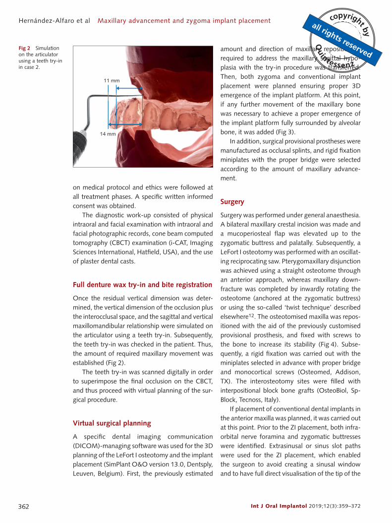
Once the residual vertical dimension was deter-mined, the vertical dimension of the occlusion plus the interocclusal space, and the sagittal and vertical maxillomandibular relationship were simulated on the articulator using a teeth try-in. Subsequently, the teeth try-in was checked in the patient. Thus, the amount of required maxillary movement was established (Fig 2).
The teeth try-in was scanned digitally in order to superimpose the final occlusion on the CBCT, and thus proceed with virtual planning of the sur-gical procedure.
Virtual surgical planning
A specific dental imaging communication (DICOM)-managing software was used for the 3D planning of the LeFort I osteotomy and the implant placement (SimPlant O&O version 13.0, Dentsply, Leuven, Belgium). First, the previously estimated
amount and direction of maxillary repositioning required to address the maxillary sagittal hypo-plasia with the try-in procedure was transferred. Then, both zygoma and conventional implant placement were planned ensuring proper 3D emergence of the implant platform. At this point, if any further movement of the maxillary bone was necessary to achieve a proper emergence of the implant platform fully surrounded by alveolar bone, it was added (Fig 3).
In addition, surgical provisional prostheses were manufactured as occlusal splints, and rigid fixation miniplates with the proper bridge were selected according to the amount of maxillary advance-ment.
Surgery
Surgery was performed under general anaesthesia. A bilateral maxillary crestal incision was made and a mucoperiosteal flap was elevated up to the zygomatic buttress and palatally. Subsequently, a LeFort I osteotomy was performed with an oscillat-ing reciprocating saw. Pterygomaxillary disjunction was achieved using a straight osteotome through an anterior approach, whereas maxillary down-fracture was completed by inwardly rotating the osteotome (anchored at the zygomatic buttress) or using the so-called ‘twist technique’ described elsewhere12. The osteotomised maxilla was repos-itioned with the aid of the previously customised provisional prosthesis, and fixed with screws to the bone to increase its stability (Fig 4). Subse-quently, a rigid fixation was carried out with the miniplates selected in advance with proper bridge and monocortical screws (Osteomed, Addison, TX). The interosteotomy sites were filled with interpositional block bone grafts (OsteoBiol, Sp-Block, Tecnoss, Italy).
If placement of conventional dental implants in the anterior maxilla was planned, it was carried out at this point. Prior to the ZI placement, both infra-orbital nerve foramina and zygomatic buttresses were identified. Extrasinusal or sinus slot paths were used for the ZI placement, which enabled the surgeon to avoid creating a sinusal window and to have full direct visualisation of the tip of the
Fig 2 Simulation on the articulator using a teeth try-in in case 2.
11 mm
14 mm

Hernández-Alfaro et al Maxillary advancement and zygoma implant placement
Int J Oral Implantol 2019;12(3):359–372 363
Fig 3a-f Virtual surgical planning of each patient. (a and b) Case 1; (c and d) Case 2; (e and f) Case 3.
a b
c d
e f
Fig 4 Maxillary reposition using a customised provisional pros-thesis fixed with a screw to the bone (case 3).

Int J Oral Implantol 2019;12(3):359–372
Hernández-Alfaro et al Maxillary advancement and zygoma implant placement
364
implant drill at all times, to ensure a proper ZI tra-jectory. The parallelism between the implant heads was checked bilaterally. Then, a gentle dissection of both buccal fat pads was performed, and these were repositioned over the most lateral aspects of the maxilla, thereby achieving a good coverage of the extrasinus path of the ZI with soft tissue thickening and mucosal reinforcement especially at the uppermost buccal sulcus area (Fig 5). Abut-ment screws were placed on each implant and the mucoperiosteal flaps were readapted and sutured back into position with resorbable sutures (Vicryl 4.0, Ethicon, Sommerville, NJ, USA).
A closed-circuit cold-water mask (17ºC) was worn during the first postoperative day. The
following drugs were prescribed during the first 10 postoperative days: 500/125 mg amoxicillin/clavulanic acid antibiotics (every 8 hours); 25 mg dexketoprofen anti-inflammatory (every 8 hours); and 575 mg metamizole analgesic (every 8 hours).
Prosthetic loading
Impressions of both arches and a bite registration were obtained immediately after the surgery to manufacture a provisional fixed prosthesis. The maxillomandibular relationship was determined based on the preoperative teeth try-in, which was used to simulate and plan the surgery. The teeth try-in was adapted to create the space for the
Fig 5a-e Intra-operative pictures of each case showing maxillary reposition, implants placement and the Bichat’s fat pad flap used. (a) Case 1; (b to d) Case 2; (e) Case 3.
a b
c d
e

Hernández-Alfaro et al Maxillary advancement and zygoma implant placement
Int J Oral Implantol 2019;12(3):359–372 365
transepithelial abutments to ensure correct fi tting in the maxilla. Then, the bite registration was per-formed. The provisional prosthesis was delivered 24 hours after the surgery (Fig 6).
Postoperative evaluation
Eventual complications and postsurgical stability were evaluated at the end of the fi rst week, fi rst month, and at 6 months and 1 year follow-up appointments (Fig 7); and two control CBCT scans were performed at the end of the fi rst month (T1) and the end of the fi rst year (T2) of the follow-up.
To evaluate the stability of maxillary advance-ment and an eventual resorption of the distal maxillary segment used as a ‘pedicled onlay bone
graft’, the two postoperative CBCT exams (i-CAT, Vision-Q Version 1.8.0.5) obtained at two specifi c time points (T1 and T2) were carried out. CBCTs were obtained in the DICOM format and pro-cessed with a specifi c third-party software (Dol-phin 3D Orthognathic Surgery Planning Software, version 11.8, in a Pentium 4 Processor 3.8 GZ, W/SP5 Windows XP Professional, 120 GB memory, 2 GB RAM). A 3D volume was created with the hard tissue reconstruction for the T1 and T2 data-bases. The 3D superimposition and dimensional comparisons were performed by means of surface matching between different datasets (Fig 8).
To evaluate the postsurgical stability the follow-ing linear measurements were conducted at the maxillary midline, in all three spatial planes:
a b
Fig 6a-b Provi-sional prosthesis loaded 24 hours after surgery in (a) case 2 and (b) case 3.
cb
a
Fig 7a-i Post-operative facial and intraoral pictures and panoramic radiographs of each patient.

Int J Oral Implantol 2019;12(3):359–372
Hernández-Alfaro et al Maxillary advancement and zygoma implant placement
366
Fig 7d-i Post-operative facial and intraoral pictures and panoramic radiographs of each patient.
fe
d
ih
g
• Sagittal plane: projected distance from A-point to nasion perpendicular (A-Nper).
• Transverse plane: distance between both greater palatine foramina (PFR-PFL).
• Vertical plane: perpendicular distance from A-point to the Frankfort horizontal plane through nasion (A-FHN).
In addition, the following 3D maxillary measure-ments were taken to assess the maxillary resorp-tion:• Sagittal plane: distance between the posterior
nasal spine (PNS) and A-point (PNS-A).• Transverse plane: distance between both
greater palatine foramens (PFR-PFL).

Hernández-Alfaro et al Maxillary advancement and zygoma implant placement
Int J Oral Implantol 2019;12(3):359–372 367
Fig 8a-f Superim-position of the two postoperative CBCT images of each patient in order to evaluate implant stability, maxillary movement relapse and maxillary resorption. White represents the CBCT taken at the end of the first month of follow-up (T1); and green is the CBCT taken 1 year after the procedure (T2): (a and b) Case 1; (c and d) Case 2; (e and f) Case 3..
e f
a b
c d
• Vertical plane: length of the anterior cortical of the incisive fossa (IF).
Statistical analysis
Statistical analysis was carried out using SPSS for Windows (version 15.0.1, SPSS, Chicago, IL).
Descriptive statistics were used for the quantita-tive analysis. Each patient’s percentage variation in maxillary surgical movements (relapse) was cal-culated as follows: [1-year postoperative A-point position × 100/1-month postoperative A-point position].

Int J Oral Implantol 2019;12(3):359–372
Hernández-Alfaro et al Maxillary advancement and zygoma implant placement
368
ResultsThe three clinical cases presented here are summa-rised in Table 1 and illustrated in Fig 1. The sample under study comprised two male patients and a female patient with a median age of 50.7 years of age (from 45 to 56 years). Apart from their need for complex oral rehabilitation, all the patients complained of a concave facial profile and, as planned, a straight facial profile was achieved post-operatively in all cases. Regarding surgery, extrasi-nusal or sinus slot paths were used in all cases, and proper emergence of the implant platform fully surrounded by alveolar bone was ensured. Two protocols for full-arch rehabilitation supported by the ZI were used depending on whether con-ventional implants could be placed in the anterior
region of the maxilla or not. Two cases were solved using two posterior ZI in combination with four conventional fixtures in the anterior maxilla, and the remaining case through four ZI (quadruple ZIs protocol).
The postoperative pictures and follow-up records are shown in Fig 7 and Table 2, respect-ively. No surgical complications, such as infraorbital nerve (V2) damage and orbital, infratemporal fossa or intracranial involvement, were noted. The immediate postoperative courses were unevent-ful. Two conventional implants placed in the an-terior maxilla in case 2 failed. However, the pros-thetic loading was carried out successfully with the remaining two ZI and two conventional implants in the anterior maxilla.
Table 1 Descriptive analysis of each patient and surgery performed in each case
Case Age (y)
Gender Initial situation Initial/final facial profile
Maxillary reposition
Number of maxillary implants
ZI: position (length, mm)(Brand)
1 45 M Partially edentulous + periodontal disease
Concave/straight Advancement + downwards
2 ZI + 4 anterior conventional implants
#16: 45 mm#26: 50 mm(Nobel Biocare, Gothenburg, Sweden)
2 56 M Fully edentulous Concave/straight Advancement + downwards
2 ZI + 4 anterior conventional implants
#16: 52.5 mm#26: 52.5 mm(Neodent, Curitiba, Brazil)
3 51 F Fully edentulous Concave/straight Advancement + posteriorly downwards
4 ZI #13: 50 mm#16: 45 mm#23: 52.5 mm#26: 50 mm(Neodent, Curitiba, Brazil)
ZI, zygomatic implant.
Table 2 Prosthetic and loading aspects and follow-up for each case
Case Loading protocol Provisional prosthesis
Permanent prosthesis Follow-up (months)
Number ZI failure
ZI Survival rate
1 Immediate Acrylic resin Fixed white and pink aesthetics with titanium framework
45 0 100%
2 Immediate Acrylic resin Fixed white and pink aesthetics with titanium framework
18 0 100%
3 Immediate Acrylic resin Fixed white and pink aesthetics with titanium framework
12 0 100%
ZI, zygomatic implant.

Hernández-Alfaro et al Maxillary advancement and zygoma implant placement
Int J Oral Implantol 2019;12(3):359–372 369
Eventual complications and the surgical stabil-ity were assessed through clinical and radiological controls (Fig 8 and Table 3). Regarding the implant stability, apart from the above-mentioned failure of 2 implants, they were well positioned and osseoin-tegrated, no peri-implant mucositis and mucosal dehiscence were observed, and no sinus pathology was detected. On the other hand, with regards to maxillary bone reposition, no resorption of maxil-lary distal bone was evident at the end of the first year of follow-up. However, a mean relapse of –4.3 mm (–10.06%) was evidenced for maxillary downward movement, and conversely, an extra-forward maxillary movement was observed in all cases (mean +1.4 mm, + 82.8%) (Table 3).
The patients’ and surgeon’s degree of satis-faction with the aesthetic and functional results was excellent: an attractive smile and more youth-ful appearance were achieved while restoring an appropriate occlusion (Fig 7).
Discussion
So far, the ZI is considered a predictable treatment option for patients with an atrophic maxilla. How-ever, in the most severe cases of maxillary bone deficiency, additional bone grafting procedures are required in order to facilitate an ideal 3D implant placement in terms of implant survival rate, pros-thetic oral rehabilitation and biological complica-tions13.
Since the first description of ZI by Brånemark4 for a full rehabilitation of the severely atrophic maxilla, several technical modifications have been reported in order to avoid its two main drawbacks: sinus and palatal emergence-related problems7-9.
Postsurgical sinus disease has been reported as one of the major biological complications of the ZI technique13. Hence, the sinus slot and extrasi-nus techniques7-9 have been proposed. In the
Table 3 Measurements to evaluate maxillary reposition stability and distal maxillary segment resorption
Case Presurgery 1-month follow-up
A-Nper A-FHN PFR-PFL PNS-A IF A-Nper A-FHN PFR-PFL PNS-A IF
1 –5.3 51.2 25.4 48.0 15.3 9.5 52.5 25.4 48.1 15.1
2 –3.9 57.8 26.0 50.5 14.0 11.9 60.9 26.0 50.3 14.1
3 –1.2 46.0 24.5 50.1 13.5 10.8 47.9 24.5 50.5 13.8
1-year follow-up Surgical movement Relapse Maxillary resorp-tion
Case A-Nper A-FHN PFR-PFL PNS-A IF Sagittal Vertical Trans-versal
Sagittal Vertical Trans-versal
1 7.8 54.8 25.4 47.9 15.3 1.3 14.8 0 +2.3 +176.9%
–1.7–11.4%
0 None
2 10.5 62.8 26.0 50.5 14.0 3.1 15.8 0 +1.9 +61.2%
–1.4 –8.8%
0 None
3 9.6 48.1 24.5 50.5 13.7 1.9 12.0 0 +0.2 +10.5%
–1.2 –10%
0 None
A-Nper, A-point to nasion perpendicular; A-FHN, A-point to the Frankfort horizontal plane through nasion; PFR-PFL, transverse plane (distance between both greater palatine foramina); PNS-A, sagittal plane (distance between the posterior nasal spine [PNS] and A-point); IF, vertical plane (length of the anterior cortical of the incisive fossa).

Int J Oral Implantol 2019;12(3):359–372
Hernández-Alfaro et al Maxillary advancement and zygoma implant placement
370
present case series, an extrasinusal placement of the ZI was carried out. In this sense, and unlike the Nocini et al10 technique, the window across the upper aspect of the maxillary buttress to support correct intrasinus ZI path was avoided, thereby reducing the operative time and sinus morbidity8.
On the other hand, achieving a proper emer-gence of the implant platform, that is, as centred as possible in the residual crest of the alveolar ridge and completely surrounded by bone, is a major challenge when placing a ZI7-9. In cases where the lateral maxillary wall is grossly concave or a severe maxillary atrophy exists, the platform and the threads of the implant may be subperiosteal and not supported by bone. This may lead to drawbacks in terms of peri-implant mucositis and mucosal dehiscence, in spite of implant ‘coverage manoeuvres’ such as advancement flaps, buccal fat pad or AlloDerm placement14. In this context, the lack of transversal maxillary bone support can be solved with a concomitant LeFort I maxillary advancement, where the backmost (and widest) area of the maxilla is brought forward.
As highlighted in the present protocol, com-puter-assisted virtual planning and prefabricated surgical splints were essential tools to ensure the proper implant emergence and corresponding maxillary advancement reposition15. During the computer-assisted preoperative planning of the above-presented cases, the software evidenced that the ZI did not contact any maxillary support-ing surface transversally, suggesting the need for a bone graft. Nevertheless, it could be verified that the maxillary advancement by means of a LeFort I osteotomy could be used as a bone graft-sub-stitute to address the lack of transversal alveolar bone support, thus, improving the transversal ZI platform emergence and the final stability. This is the main difference from the methodology pro-posed by Nocini et al10, who reports an implant platform emergence in the palatal cortical bone of the alveolar crest following the intrasinus ZI path.
Besides addressing transversal maxillary defi-ciency, severe maxillary atrophy requires a holistic 3D approach. Vertical defects can be easily man-aged prosthetically with full arch metal-resin (clas-sically known as hybrid prosthesis), metal-ceramic
or zirconia prostheses, restoring the pink and white aesthetics. In cases where labial support is needed, an overdenture is indicated to restore the aesthetics while allowing correct hygiene. None-theless, focusing specifically on the reconstruc-tion of severe anterior maxillary defects, and thus, sagittal maxillary atrophy, there are few nongraft-ing options. The placement of four ZI (quadruple ZIs approach16) with a well-designed prosthesis, represents a reliable treatment approach for the reconstruction of anterior maxillary defects with mild sagittal atrophy13. However, the lack of any maxillary supporting bone in severe sagittal atro-phies represents an unsolvable problem without premaxilla onlay grafts, or the above-mentioned simultaneous LeFort I maxillary advancement tech-nique10. Accordingly, the distal maxillary segment moved forward was used as a ‘pedicled onlay bone graft’. Since autologous corticocancellous bone blocks from a donor site are the gold standard grafting option because the maxilla offers only small amounts of mainly cancellous autografts, this is an exceptional situation17. Moreover, this source has as advantages the proximity of donor and recipient sites, convenient surgical access, and low morbidity18.
There are two protocols for full-arch rehabilita-tion supported by the ZI: using four ZI (quadruple ZIs16); or two posterior ZIs in combination with at least two conventional fixtures in the anterior max-illa. Although the ZI has been reported to have a high survival rate, of 96.7% to 100%13,14,19, while standard implants in the anterior atrophic maxilla (either with or without bone grafting) have shown a relatively high failure rate, of 8% to 27%19, there is still limited evidence to demonstrate a better survival rate of one technique over the other when considering ZI protocols13.
When the quadruple ZIs protocol is used, the implant platform emergence is usually located at the level of the lateral incisor or canine and second premolar or first molar, for the anterior and pos-terior ZI, respectively. Computer-guided ‘flap-less’ surgery using stereolithographic templates is not recommended for ZI placement, because direct visualisation of the path of the drills is impera-tive to avoid potential major complications such as

Hernández-Alfaro et al Maxillary advancement and zygoma implant placement
Int J Oral Implantol 2019;12(3):359–372 371
infraorabital nerve (V2) damage, and involvement of the orbit, infratemporal fossa or cranial base. Thus, the use of surgical splints for the ZI installa-tion is only recommended for marking the location of implant emergence, if necessary.
As the distal maxillary segment was used as a ‘pedicled onlay bone graft’, its eventual resorption rate was evaluated. Fortunately, maxillary bone resorption could be ruled out since both postoper-ative CBCTs, performed 1 month and 1 year after the procedure, matched perfectly on superimposi-tion. However, a long-term follow-up evidenced a light vertical relapse of –4.3 mm (–10.06%) for the maxillary downward movement. Conversely, an extra-forward movement in the sagittal plane was observed, probably due to the occlusal stabilisa-tion effect of the prosthetic loading. In spite of the vertical relapse, the implant emergence was not modified, nor were more implant threads exposed. Nevertheless, further studies using longer follow-up periods and randomised controlled trials are required, to assess the long-term effect of this technique on the 3D stability of the maxillary bone atrophy and ZI survival rate.
Finally, the precise virtual planning and malar bone anchorage granted primary implant stabil-ity and therefore, immediate provisional prosthesis loading was achieved. This, in turn, shortens the edentulous period of the patient20.
Conclusions
Simultaneous maxillary advancement and ZI place-ment is an excellent surgical option for selected cases of severe maxillary atrophy. Besides achiev-ing successful functional and aesthetic oral rehabil-itation, this technique provides a high implant sur-vival rate, avoids donor site morbidity, decreases surgical time, and shortens the overall rehabilita-tion period.
References1. Keller EE, Van Roekel NB, Desjardins RP, Tolman DE.
Prosthetic-surgical reconstruction of the severely resorbed maxilla with iliac bone grafting and tissue–integrat-ed prostheses. Int J Oral Maxillofac Implants 1987;2: 155–165.
2. Sailer HF. A new method of inserting endosseous implants in totally atrophic maxillae. J Craniomaxillofac Surg 1989;17:299–305.
3. Boyne PJ, James RA. Grafting of the maxillary sinus floor with autogenous marrow and bone. J Oral Surg 1980;38:613–616.
4. Brånemark PI. ‘Surgery and fixture installation’ in zygo-maticus fixture clinical procedures. First ed. 1998; Goteborg, Sweden.
5. Brånemark PI, Gröndahl K, Ohrnell LO, et al. Zygoma fixture in the management of advanced atrophy of the maxilla: technique and long-term results. Scand J Plast Reconstr Surg Hand Surg 2004;38:70–85.
6. Bothur S, Garsten M. Initial speech problems in patients treated with multiple zygomatic implants. Int J Oral Maxil-lofac Implants 2010;25:379–384.
7. Stella JP, Warner MR. Sinus slot technique for simplifica-tion and improved orientation of zygomaticus dental implants: a technical note. Int J Oral Maxillofac Implants 2000;15:889–893.
8. Aparicio C, Ouazzani W, Aparicio A, et al. Extrasinus zygomatic implants: three-year experience from a new surgical approach for patients with pronounced buccal concavities in the edentulous maxilla. Clin Implant Dent Relat Res 2010;12:55–61.
9. Maló P, Nobre Mde A, Lopes I. A new approach to re-habilitate the severely atrophic maxilla using extramaxil-lary anchored implants in immediate function: a pilot study. J Prosthet Dent. 2008;100:354–366.
10. Nocini PF, D’Agostino A, Chiarini L, Trevisiol L, Procacci P. Simultaneous Le Fort I osteotomy and zygomatic implants placement with delayed prosthetic rehabilitation. J Crani-ofac Surg 2014;25:1021–1024.
11. Cawood JI, Howell RA. A classification of the edentulous jaws. Int J Oral Maxillofac Surg 1988;17:232–236.
12. Hernández-Alfaro F, Guijarro-Martínez R. ‘Twist tech-nique’ for pterygomaxillary dysjunction in minimally invasive Le Fort I osteotomy. J Oral Maxillofac Surg 2013;71:389–392.
13. Wang F, Monje A, Lin GH, et al. Reliability of four zygo-matic implant-supported prostheses for the rehabilitation of the atrophic maxilla: a systematic review. Int J Oral Maxillofac Implants 2015;30:293–298.
14. Bedrossian E, Bedrossian EA. Prevention and the manage-ment of complications using the zygoma implant: a review and clinical experiences. Int J Oral Maxillofac Implants 2018;33:e135–e145.
15. Bertos Quílez J, Guijarro-Martínez R, Aboul-Hosn Cente-nero S, Hernández-Alfaro F. Virtual quad zygoma implant placement using cone beam computed tomography: sufficiency of malar bone volume, intraosseous implant length, and relationship to the sinus according to the degree of alveolar bone atrophy. Int J Oral Maxillofac Surg 2018;47:252–261.
16. Bothur S, Jonsson G, Sandahl L. Modified technique using multiple zygomatic implants in reconstruction of the atrophic maxilla: a technical note. Int J Oral Maxillofac Implants. 2003;18:902–904.
17. Khoury F, Hanser T. Mandibular bone block harvesting from the retromolar region: a 10-year prospective clinical study. Int J Oral Maxillofac Implants 2015;30:688–697.

Int J Oral Implantol 2019;12(3):359–372
Hernández-Alfaro et al Maxillary advancement and zygoma implant placement
372
18. Nkenke E, Neukam FW. Autogenous bone harvesting and grafting in advanced jaw resorption: morbidity, resorption and implant survival. Eur J Oral Implantol 2014;7(Supp 2): S203–217.
19. Chrcanovic BR, Abreu MH. Survival and complications of zygomatic implants: a systematic review. Oral Maxillofac Surg 2013;17:81–93.
Federico Hernández-Alfaro, MD, DDS, PhD, FEBOMSMaxillofacial Institute, Teknon Medi-cal Centre Barcelona, Barcelona, Spain; Department of Oral and Maxillofacial Surgery, Universitat Internacional de Cata-lunya, Sant Cugat del Vallès, Barcelona, Spain
Gian Maria Michele Ragucci, DDSDepartment of Oral and Maxillofacial Surgery, Universitat Internacional de Cata-lunya, Sant Cugat del Vallès, Barcelona, Spain
Irene Méndez-Manjón, DDS, PhDMaxillofacial Institute, Teknon Medi-cal Centre Barcelona, Barcelona, Spain; Department of Oral and Maxillofacial Surgery, Universitat Internacional de Cata-lunya, Sant Cugat del Vallès, Barcelona, Spain
Maria Giralt-Hernando, DDSDepartment of Oral and Maxillofacial Surgery, Universitat Internacional de Cata-lunya, Sant Cugat del Vallès, Barcelona, Spain
Raquel Guijarro-Martínez, MD, DDS, PhD, FEBOMSMaxillofacial Institute, Teknon Medi-cal Centre Barcelona, Barcelona, Spain; Department of Oral and Maxillofacial Surgery, Universitat Internacional de Cata-lunya, Sant Cugat del Vallès, Barcelona, Spain
Pelayo Sicilia-Blanco, DDSDepartment of Oral and Maxillofacial Surgery, Universitat Internacional de Cata-lunya, Sant Cugat del Vallès, Barcelona, Spain
Natalia Ventura-Martínez, MD, DDS, PhD, FEBOMSMaxillofacial Institute, Teknon Medical Centre Barcelona, Barcelona, Spain
Adaia Valls-Ontañón, MD, DDS, PhD, FEBOMSMaxillofacial Institute, Teknon Medi-cal Centre Barcelona, Barcelona, Spain; Department of Oral and Maxillofacial Surgery, Universitat Internacional de Catalunya, Sant Cugat del Vallès, Barce-lona, Spain
Federico Hernández-Alfaro
Correspondence to: Dr Adaia Valls-Ontañón, Maxillofacial Institute, Teknon Medical Centre, Carrer de Vilana, 12, 08022 Barcelona, Spain. E-mail: [email protected]
20. Peñarrocha M, García B, Martí E, Boronat A. Rehabilitation of severely atrophic maxillae with fixed implant-supported prostheses using zygomatic implants placed using the sinus slot technique: clinical report on a series of 21 patients. Int J Oral Maxillofac Implants. 2007;22:645–650.