Regulationofmesenchymalstromalcellsthrough fine tuning of ... ·...
13
Regulation of mesenchymal stromal cells through fine tuning of canonical Wnt signaling Jin-A Kim a , Hyun-Kyung Choi a , Tae-Min Kim b , Sun-Hee Leem c , Il-Hoan Oh a , ⁎ a Catholic High-Performance Cell Therapy Center & Department of Medical Life Science, Republic of Korea b Center for Cancer Evolution, Medical Research Center, The Catholic University of Korea, Republic of Korea c Dept. of Biological Science, Dong-A University, Busan, Republic of Korea Received 21 July 2014; received in revised form 23 February 2015; accepted 25 February 2015 Available online 18 March 2015 Abstract Mesenchymal stromal cells (MSCs) have been extensively utilized for various cell therapeutic trials, but the signals regulating their stromal function remain largely unclear. Here, we show that canonical Wnt signals distinctively regulate MSCs in a biphasic manner depending on signal intensity, i.e., MSCs exhibit proliferation and progenitor self-renewal under low Wnt/β-catenin signaling, whereas they exhibit enhanced osteogenic differentiation with priming to osteoblast-like lineages under high Wnt/β-catenin signaling. Moreover, low or high levels of β-catenin in MSCs distinctly regulated the hematopoietic support of MSCs to promote proliferation or the undifferentiated state of hematopoietic progenitors, respectively. A gene expression study demonstrated that different intracellular levels of β-catenin in MSCs induce distinct transcriptomic changes in subsets of genes belonging to different gene function categories. Different β-catenin levels also induced differences in intracellular levels of the β-catenin co-factors, Tcf-1 and Lef-1. Moreover, nano-scale mass spectrometry of proteins that co-precipitated with β-catenin revealed distinctive spectra of proteins selectively interacting with β-catenin at specific expression levels. Together, these results show that Wnt/ β-catenin signals can coax distinct transcription milieu to induce different transcription profiles in MSCs depending on the signal intensity and that fine-tuning of the canonical Wnt signaling intensity can regulate the phase-specific functionality of MSCs. © 2015 The Authors. Published by Elsevier B.V. This is an open access article under the CC BY-NC-ND license (http://creativecommons.org/licenses/by-nc-nd/4.0/). Introduction Mesenchymal stromal cells (MSCs) are non-hematopoietic adherent cell populations derived from stroma of bone marrow (BM), adipose tissue, or placental tissue (Keating, 2006; Pittenger et al., 1999). These MSCs are being utilized for therapeutic trials, facilitating the establishment of a regenerative microenvironment for hematopoietic stem cells (HSCs), neuronal stem cells, and other tissue-specific stem cells (Frenette et al., 2013; Murphy et al., 2013; Satija et al., 2009). MSCs exert therapeutic effects via paracrine secretion of various growth factors that can stimulate regeneration of injured tissues (Caplan and Correa, 2011). In addition, MSCs exert niche effects on endogenous stem cells through cell–cell interactions, as typically exemplified by the BM stem cell niche in the endosteal and peri-vascular regions (Oh and Humphries, 2012; Oh and Kwon, 2010). ⁎ Corresponding author at: Catholic High-Performance Cell Therapy Center, The Catholic University of Korea, 505, Banpo-Dong, Seocho-Ku, Seoul 137-701, Republic of Korea. Fax: +82 2 591 3994. E-mail address: [email protected] (I.-H. Oh). http://dx.doi.org/10.1016/j.scr.2015.02.007 1873-5061/© 2015 The Authors. Published by Elsevier B.V. This is an open access article under the CC BY-NC-ND license (http://creativecommons.org/licenses/by-nc-nd/4.0/). Available online at www.sciencedirect.com ScienceDirect www.elsevier.com/locate/scr Stem Cell Research (2015) 14, 356–368
Transcript of Regulationofmesenchymalstromalcellsthrough fine tuning of ... ·...

Ava i l ab l e on l i ne a t www.sc i enced i r ec t . com
ScienceDirect
www.e l sev i e r . com / l oca te / s c r
Stem Cell Research (2015) 14, 356–368
Regulation ofmesenchymal stromal cells throughfine tuning of canonical Wnt signaling
Jin-A Kima, Hyun-Kyung Choi a, Tae-Min Kimb, Sun-Hee Leemc, Il-Hoan Oha,⁎a Catholic High-Performance Cell Therapy Center & Department of Medical Life Science, Republic of Koreab Center for Cancer Evolution, Medical Research Center, The Catholic University of Korea, Republic of Koreac Dept. of Biological Science, Dong-A University, Busan, Republic of Korea
Received 21 July 2014; received in revised form 23 February 2015; accepted 25 February 2015Available online 18 March 2015
AbstractMesenchymal stromal cells (MSCs) have beenextensively utilized for various cell therapeutic trials, but the signals regulating their stromalfunction remain largely unclear. Here, we show that canonical Wnt signals distinctively regulate MSCs in a biphasicmanner depending onsignal intensity, i.e., MSCs exhibit proliferation and progenitor self-renewal under low Wnt/β-catenin signaling, whereas they exhibitenhanced osteogenic differentiation with priming to osteoblast-like lineages under high Wnt/β-catenin signaling. Moreover, low orhigh levels of β-catenin in MSCs distinctly regulated the hematopoietic support of MSCs to promote proliferation or theundifferentiated state of hematopoietic progenitors, respectively. A gene expression study demonstrated that differentintracellular levels of β-catenin in MSCs induce distinct transcriptomic changes in subsets of genes belonging to different genefunction categories. Different β-catenin levels also induced differences in intracellular levels of the β-catenin co-factors,Tcf-1 and Lef-1. Moreover, nano-scale mass spectrometry of proteins that co-precipitated with β-catenin revealed distinctivespectra of proteins selectively interacting with β-catenin at specific expression levels. Together, these results show that Wnt/β-catenin signals can coax distinct transcription milieu to induce different transcription profiles in MSCs depending on the signalintensity and that fine-tuning of the canonical Wnt signaling intensity can regulate the phase-specific functionality of MSCs.
© 2015 The Authors. Published by Elsevier B.V. This is an open access article under the CC BY-NC-NDlicense (http://creativecommons.org/licenses/by-nc-nd/4.0/).Introduction
Mesenchymal stromal cells (MSCs) are non-hematopoieticadherent cell populations derived from stroma of bonemarrow (BM), adipose tissue, or placental tissue (Keating,2006; Pittenger et al., 1999). These MSCs are being utilized
⁎ Corresponding author at: Catholic High-Performance Cell TherapyCenter, The Catholic University of Korea, 505, Banpo-Dong, Seocho-Ku,Seoul 137-701, Republic of Korea. Fax: +82 2 591 3994.
E-mail address: [email protected] (I.-H. Oh).
http://dx.doi.org/10.1016/j.scr.2015.02.0071873-5061/© 2015 The Authors. Published by Elsevier B.V. This(http://creativecommons.org/licenses/by-nc-nd/4.0/).
for therapeutic trials, facilitating the establishment of aregenerative microenvironment for hematopoietic stemcells (HSCs), neuronal stem cells, and other tissue-specificstem cells (Frenette et al., 2013; Murphy et al., 2013; Satijaet al., 2009). MSCs exert therapeutic effects via paracrinesecretion of various growth factors that can stimulateregeneration of injured tissues (Caplan and Correa, 2011).In addition, MSCs exert niche effects on endogenous stemcells through cell–cell interactions, as typically exemplifiedby the BM stem cell niche in the endosteal and peri-vascularregions (Oh and Humphries, 2012; Oh and Kwon, 2010).
is an open access article under the CC BY-NC-ND license

357Regulation of mesenchymal stromal cells through fine tuning of canonical Wnt signaling
Accordingly, the signals controlling the biological proper-ties of MSCs exert regulatory influences on tissue regenerativeactivity. For example, adipogenic differentiation of MSCs inBM suppresses the regeneration of HSCs in myeloablated hosts(Naveiras et al., 2009), whereas cells of the osteoblasticlineage support hematopoietic activity (Calvi et al., 2003;Zhang et al., 2003). In addition, the differentiation status ofMSCs also influences the niche activity, i.e., recent studieshave shown that among the heterogeneous subpopulation ofMSCs in BM, primitive subsets of MSCs including prx-1(+) MSCs(Greenbaum et al., 2013) and nestin (+) MSCs (Mendez-Ferreret al., 2010) play a major role in regulating HSC self-renewalin the perivascular niche of the BM. Accordingly, extensivestudies are under way to identify the signals and molecularmechanisms that control the proliferation/self-renewaland differentiation of MSCs under various physiological andpathological conditions.
Wnts are secreted glycoproteins associated with the cellsurface or extracellular matrix that influence diverse biolog-ical processes, including embryonic induction and cell fatespecification. In the canonical Wnt signaling pathway, Wntbinds to seven-pass transmembrane Frizzled (Fz) familyreceptors and the single-pass co-receptors LRP 5, 6 (LDL-receptor-related protein 5,6), to induce β-catenin stabiliza-tion. Subsequently, the stabilized β-catenin translocates tothe nucleus and forms a complex with the DNA-bindingtranscription factors TCF/LEF to activate a Wnt-controlledgene expression program (Kikuchi et al., 2006).
Several studies have shown that canonical wnt signalingplays a critical role in regulating cell fate decisions of MSCs,but variable biological effects on MSCs have been reported,i.e., up-regulation of canonical wnt signaling stimulated theproliferation of MSCs, inhibited their osteogenic differentia-tion, or promoted their osteogenic commitment (Boland et al.,2004; Cook et al., 2014; Gaur et al., 2005; Kawai et al., 2007;Takada et al., 2009), indicating that multiple, complex effectsare mediated by canonical wnt signals.
In addition to the proliferation and differentiation of MSCs,canonical wnt signaling has been implicated in the regulationof the stromal activity of MSCs. For example, we and othersrecently showed that activation of Wnt/β-catenin signals inMSCs enhances the self-renewal of HSCs by triggering cross-talk of Wnt-Notch signals in the stem cell niche (Kim et al.,2009; Oh, 2010), and intra-femoral injection of β-catenin-stabilized MSCs promotes regeneration of HSCs (Ahn et al.,2010). Similarly, β-catenin expression in the BM stromawas shown to be necessary for maintenance of long-termhematopoietic cells (Kim et al., 2009; Nemeth et al., 2009). Incontrast, myeloproliferative diseases developed in transgenicmice with constitutive activation of β-catenin in bonemarrowosteoblastic cells (Kode et al., 2014), indicating that canonicalWnt signals in MSCs should be fine-tuned for their coordinatedstromal function.
However, the regulatory mode of Wnt/β-catenin signalsfor MSCs, especially with regard to signaling intensity,remains unclear. Therefore, in this study, to preciselycharacterize the biological effects of canonical wnt signalingin MSCs, we investigated the possibility that MSCs aredistinctively regulated by fine-tuning of canonical Wntsignaling. We show that β-catenin can cause distincttransactivation in MSCs depending on its level of accumu-lation, resulting in distinct behavior of MSCs. Our study
reveals the fine-tuning of Wnt/β-catenin signaling for coordi-nation of MSC function.
Materials and methods
Animals and cells
C57BL/6J-Ly 5.2 (BL6) from Jackson Laboratory (Bar Harbor,ME, USA) were used in the experiments under the approvalfrom the Animal Experiment Board of the Catholic Universityof Korea. Enrichment of hematopoietic progenitors by 5-fluorouracil (5-FU BMCs) was performed as described (Kim etal., 2009). MSCs were isolated and passage cultured untilbecome negative for CD45 as described (Kim et al., 2009) andcharacterized by surface phenotype (Supplemental Fig. 1).
Construction of expression plasmids, retrovirusproduction, and transduction of MSCs
A stable form of the β-catenin gene (S37A) (Kim et al., 2009)was cloned into two types of retroviral vector, MPG(MSCV-PGK-GFP) or QGCX2 (CMV-GFP-IRES). MSCs weretransduced with each retroviral vector with multiplicity ofinfection (MOI) of 30, then sorted for transduced (GFP+) cellsas described (Hong et al., 2014).
Reporter assay
The TOPFLASH and FOPFLASH reporter constructs containingeight TCF/LEF binding sites or mutated binding sites (BiecheleandMoon, 2008)were transfected into cells alongwith effectorplasmids. Luciferase activity was measured by the LuciferaseAssay System (Promega Corp., Madison, WI) according to themanufacturer's instructions. Transfection efficiency was nor-malized for β-galactosidase activity. To measure osteocalcinpromoter activity, p6OSE2-luc containing six copies of theRunx2 binding site in the osteocalcin promoter regionwas used(Jung et al., 2009).
Cell proliferation and BrdU incorporation assay
Cell proliferation assay (4 days) for transduced (GFP+) MSCsand their BrdU incorporation (12 h) were analyzed as previ-ously described (Jung et al., 2009), visualized by secondaryantibody conjugated with Alexa-593 (Molecular Probes,Eugene, OR), then mounted with DAPI (Molecular Probes) foranalysis.
Colony formation and osteogenic and adipogenicdifferentiation of MSCs
Osteogenic and adipogenic differentiation of MSCs wasperformed as previously described (Jung et al., 2009),followed by Alizarin Red S (Sigma, St Louis, MO) or Oil RedO (Sigma) staining, respectively. The osteogenic mineraliza-tion or adipogenic lipid droplets were eluted and quantita-tively measured by spectrophotometry at 570 and 520 nm,respectively. For colony formation, MSCs were maintainedfor 6 days under low (10 ng/ml) and high (100 ng/ml)concentrations of recombinant Wnt3a ligand and plated at

358 J.-A. Kim et al.
a density of 1000 cells per 100-mm dish. After incubation for14 days, colonies (N50 cells) were fixed and stained withcrystal violet (Sigma).
In-vivo ossification of MSCs in subrenal capsule
MSCs (1 × 10⁵ each) were preconditioned (24 h), resuspendedin Matrigel (with 100 ng/ml rhBMP-2) and inoculated under-neath the renal capsule of 11 week old mice as previouslydescribed (Chan et al., 2009). After 2 weeks, the kidneys weredissected and fixed and paraffin sections were stained witheither H&E or Alizarin Red stains. For immunohistochemistrystaining of GFP, sections were stained with anti-GFP anti-body (Abcam, Cambridge, UK), visualized with DAB substratekit (BD Pharmingen), and followed by counterstaining withhematoxylin.
Measurement of β-catenin protein levels in thefresh bone marrow stromal cells
Fresh bone marrow cells were stained with antibodies againstCD45, CD31, Ter119 (BD Pharmingen), and Leptin Receptor(Sigma). Stained cells were fixed and permeabilized withFixation or permeabilization buffer (eBioscience), and thenstained with anti-β-catenin antibody and anti-rabbit Cy5(Bethyl Laboratories, Montgomery, USA) as described (Kim etal., 2009).
Nuclear extract and Western blot
For Western blot, MSCs were lysed in 2× Laemmli buffer,boiled for 5 min and cleared before electrophoresis. Nuclearextraction of MSCs was performed using NE-PER nuclear andcytoplasmic extraction reagents (Thermo). The membraneswere probed with antibodies against HA (Sigma), active β-catenin (Merck Millipore, Billerica, MA), β-actin (Millipore),LEF1, TCF1 (Cell Signaling), TCF3, and TCF-4 (Santa Cruz).
Co-culture of hematopoietic cells andMSCs expressingβ-catenin
MSCs transduced with each retroviral vector (GFP+) wereirradiated (1500 cGy) and co-cultured with 5-FU BMCs for5 days in the presence of growth factors as described (Kim etal., 2009). The undifferentiated phenotype of co-culturedhematopoietic cells was analyzed by as previously described(Kim et al., 2009).
Microarray analysis
A gene expression study using microarrays was performedas previously described (Hong et al., 2014). Briefly, biotin-labeled cRNA samples were prepared using the IlluminaTotalPrep RNA Amplification kit (Ambion, Austin, TX) andhybridized to a version 2 of the Illumina Mouse-6 BeadChip(48 K) according to the manufacturer's instructions (Illumina,San Diego, CA). Array data were analyzed using IlluminaBeadStudio software. Average linkage hierarchical clusteranalysis was performed with a 1-Pearson correlation as asimilarity metric using the GeneCluster/TreeView program
(http://rana.lbl.gov/EisenSoftware.htm). In total, 134 uniquegenes were selected by t-test (p b .005) with false discoveryrate (FDR b 0.1) statistical confidence; the expression ofthese genes was increased by three-fold or more in MPG-β-catenin or QG-β-catenin compared with MPG or QG. TheGene Ontology program (http://david.abcc.ncifcrf.gov/) wasthen used to categorize genes in subgroups based on theirbiologic function.
Co-immunoprecipitation of β-catenin
Co-immunoprecipitation of proteins was performed as previ-ously described (Antrobus and Borner, 2011). Briefly, cellswere lysed and incubated with mouse anti-immunoglobulin Gand anti-HA antibodies (Sigma), added with protein G plus/protein A-agarose beads (Calbiochem, Darmstadt, Germany),then precipitated, and subjected to proteomic analysis.
Nano-scale liquid chromatography/mass spectrometryof proteins co-precipitated with β-catenin
NanoLC–MS/MS analysis was performed on an Agilent 1100Series Nano-LC and LTQ-mass spectrometer (Thermo Electron,San Jose, CA) as previously described (Gaspari and Cuda,2011). Mass spectra were acquired using data-dependentacquisition with full mass scan (400–1800 m/z) followed byMS/MS scans. Each MS/MS scan acquiredwas an average of onemicroscan on the LTQ.
Database searching for proteomic analysis
The RAW files were converted and then searched usingMascot Server 2.2 (Matrix Science, Boston, MA). The datawere searched against the NCBI human database (v.110704).Within the Mascot search, trypsin was selected as theenzyme, and one missed cleavage was allowed per peptide.All spectral count values were reported across samples, andonly the proteins that included at least a top-ranked peptideand were reported in all samples were considered in thespectral counting analysis.
Statistical analysis
Data are expressed as the mean ± SEM. The significanceof differences between groups was analyzed using Student'st test. p b 0.05 was considered to be statistically significant(* p b 0.05, ** p b 0.01, *** p b 0.001, **** p b 0.0001).
Results
Establishment of bone marrow MSCs expressing highand low levels of active β-catenin protein
To establish MSCs that express high and low levels ofβ-catenin, we employed two types of retroviral vectorsencoding the active form of β-catenin (S37A), where theexpression of the coding gene was driven by the viral LTR(MPG-β-catenin) or CMV promoter (QG-β-catenin) (Figs. 1A,B). When each retroviral vector was transduced into 293Tcells or primary BM-derived MSCS along with the TOPFLASH

359Regulation of mesenchymal stromal cells through fine tuning of canonical Wnt signaling
reporter, the QG-β-catenin vector exhibited markedly highertransactivation of the target reporter gene than the MPG-β-catenin vector in both types of cells (108- and 38-fold higherin 293T cells and MSCs, respectively) (Fig. 1C). Moreover, BM-derived MSCs transduced with QG-β-catenin exhibited a muchhigher level of β-catenin protein expression compared tothe levels in MSCs transduced with MGP-β-catenin (Fig. 1D).However, MSCs transduced with MPG-β-catenin exhibited onlymodestly increased levels of transactivation and protein levelscompared to basal levels in control group MSCs (Fig. 1C right,Fig. 3D). These results show that these two types of MSCscan be employed as a model for MSCs expressing high (MSC-QG-β-catenin; H-BC/MSC) or low (MPG-β-catenin; L-BC/MSC)levels of β-catenin.
Only a low level of active β-catenin accumulationpromotes the proliferation and self-renewal ofcolony-forming mesenchymal progenitors
We first compared the morphology and proliferation of MSCsexpressing high and low levels of β-catenin during in-vitroculture. As shown in Fig. 2A, MSCs expressing high levels ofβ-catenin (H-BC/MSCs) exhibited a larger size than thoseexpressing low levels of β-catenin (L-BC/MSC), as deter-mined by flow cytometry plots. In addition, the L-BC/MSCsexhibited higher proliferative activity during in vitro culturethan the control MSCs, whereas H-BC/MSCs exhibitedsignificantly lower proliferation than the control (Fig. 2B).
Figure 1 Establishment of MSCswith different intracellular levels ofof β-catenin replacing serine S37 into Ala. TCF/LEF, T-cell factor-1/hemagglutinin-tag. (B): Retroviral constructs encoding green fluoresce(C): Comparisons of transactivation potential of β-catenin in 293Transactivation was assayed by luciferase activity in each transducetransactivation of reporter genes by β-catenin was determined by theshown (3 replicates, n = 9). (*: p b 0.05, **; p b 0.01). (D): Expression ovector. Shown are the representative Western blots using antibodies aactive β-catenin protein relative to β-actin compared to control MSCs
Similarly, BrdU uptake was increased in L-BC/MSCs butdecreased in H-BC/MSCs (Fig. 2C), indicating that MSC pro-liferation is promoted predominantly under low levels ofβ-catenin accumulation. Consistent with this finding, thec-myc expression level was increased in L-BC/MSCs butdecreased in H-BC/MSCs compared to control MSCs (Fig. 2D).Together, these results show that β-catenin exerts differenteffects on the proliferation of MSCs depending on its level ofexpression in MSCs.
Next, we examined the effects on the colony formation(CFU-F) of MSCs by plating equivalent numbers of MSCs ineach group. The colony formation of L-BC/MSCs was en-hanced compared to that of control MSCs, whereas that ofH-BC/MSCs was decreased (Fig. 3A). In addition, the coloniesthat formed from L-BC/MSCs were larger than those fromH-BC/MSCs (Fig. 3B), reflecting the higher proliferativeactivity of MSCs during the colonization process. However,no difference was observed for the survival and senescenceof MSCs expressing high or low levels of β-catenin (Supple-mental Fig. 2). Moreover, L-BC/MSCs exhibited higherexpression of genes associated with the primitive state ofMSCs, such as Oct-4, Nanog, Sox-2, and Nestin (Greco et al.,2007; Tsai et al., 2012), than H-BC/MSCs (Fig. 3C). Theseresults show that only low-level accumulation of β-catenincan support the maintenance and self-renewal of MSCprogenitors that are capable of forming colonies.
To further examine these effects, we examined whetherdifferences in the concentration of Wnt ligands could inducesimilar differences in MSCs by treating non-transduced MSCs
activeβ-catenin. (A): Schematic representation of the active formlymphoid enhancing factor-1 binding site; HA, human influenzance protein (GFP) alongwith β-catenin under different promoters.T cells (left) or primary bone marrow-derived MSCs (right).d cell after normalization against β-galactosidase activity. Thefolds of TOP/ FOP ratio relative to that of control. Mean ± SEM isf β-catenin proteins in MSCs transduced by each type of retroviralgainst indicated proteins (left) and their folds expression levels of(right) with SE (3 exp. n = 3). (*: p b 0.05, **; p b 0.01).

Figure 2 Distinct effects of different levels of β-catenin on MSC proliferation. (A): Representative flow cytometry plots for MSCswith low and high expression of β-catenin. (B,C): Proliferation of MSCs with low and high expression of β-catenin was compared withcontrol transduced MSCs (B) and % of BrdU+ cells among DAPI+ was compared for each group of MSCs (C). Means ± SEM are shown(3 experiments, n = 9, **; p b 0.01). (D): Expression of c-myc in MSCs with low and high β-catenin. Shown are the representativeRT-PCR plot (upper) and fold expression level normalized to GAPDH levels relative to the control (lower).
360 J.-A. Kim et al.
with high (100 ng/ml) and low (10 ng/ml) concentrations ofWnt3a ligand. As shown in Figs. 3D and E, high and lowconcentrations of Wnt3a ligand produced similar differencesin the protein levels of β-catenin and transactivation of theTOPFLASH reporter (Figs. 3D and E), which produced similarincreases and decreases in CFU-F numbers, respectively(Fig. 3F), reproducing the effects of expressing high and lowlevels of β-catenin MSCs. Next, we examined the relationbetween β-catenin levels and CFU-Fs by comparing the levelsin fresh bonemarrow stromal cells. As shown, the stromal cellsexpressing leptin receptor, the primitive subsets of MSCS thatwas shown to be highly enriched with CFU-F (Zhou et al.,2014), exhibited lower level of β-catenin protein than leptinreceptor (−) cells, thus showing similar correlation betweenhigher CFU-Fs and lower level β-catenin levels under in-vivoconditions (Fig. 3G).
These results together show that the strength of Wnt/β-catenin signaling in MSCs exerts distinct effects on theproliferation and self-renewal of MSC progenitors, pointing toa dose-specific response of MSCs to Wnt/β-catenin signals.
Differentiation of MSCs is regulated by β-catenindistinctly from colonization and proliferation
We examined the effects of β-catenin levels on differenti-ation of MSCs towards osteogenic or adipogenic lineages. Asshown in Fig. 4A, osteogenic differentiation was increased inL-BC/MSCs compared to control MSCs, and differentiation
was further increased with high levels of β-catenin (H-BC/MSC). In contrast, adipogenic differentiation of MSCs wasdecreased in L-BC/MSCs compared to control MSCs, and thisinhibition of adipogenic differentiation was also furtherdecreased under high accumulation of β-catenin (H-BC/MSCs) (Fig. 4B), indicating that the differentiation of MSCs isregulated by Wnt/β-catenin signals in a manner dependenton β-catenin levels.
Interestingly, β-catenin accumulation in proliferating MSCscaused priming of MSCs towards osteogenic lineages and awayfrom adipogenic lineages, i.e., β-catenin-expressing MSCs (atboth low and high levels) already had higher expression ofosteogenic genes (ALP and OC) with a higher percentage ofALP(+) cells and lower expression of adipogenic genes (PPAR-γand αP2) than control MSCs even before the induction ofdifferentiation (Fig. 4C, 4D). Similarly, MSCs treatedwith a highconcentration of Wnt3a ligand exhibited higher osteocalcinpromoter activity during the proliferation phase (Fig. 4E). Inaddition, MSCs that had been treated with a high concentra-tion of Wnt3a ligand exhibited enhanced mineral depositionwhen induced to differentiate (Fig. 4F), indicating that Wnt/β-catenin signals prime MSCs towards osteogenic lineages in amanner proportional to the intensity of the signals.
Moreover, when the MSCs expressing high and low levelsof β-catenin were inoculated into subrenal capsule forin-vivo ossification (Chan et al., 2009), L-BC/MSCs exhibitedincreased levels of osteogenic mineralization than controlMSCs, which was further increased in the H-BC/MSCs(Fig. 4G), indicating that the in-vitro findings on effects of

Figure 3 Distinct effects of signal intensity of Wnt/β-catenin on self-renewal of mesenchymal progenitors. (A): Comparisons of CFU-Fin MSCs with high and low expression of β-catenin. The number of colonies from 1000 cells of each group of MSCs is shown (mean ± SEM, 3experiments, n = 9). **; p b 0.01. (B): Representative morphology of colonies stained with crystal violet (scale bar = 50 μm). (C): Effectsofβ-catenin level in MSCs on expression of pluripotency-related genes. Expression of each indicated genewas analyzed by RT-PCR in MSCstransduced with each retroviral vector encoding β-catenin. (D–F): Concentration effects of recombinant Wnt3a ligand on MSCs. (D) Theaccumulation of β-catenin under each dose of Wnt3a ligand was analyzed by Western blot (E). Transactivation of reporters in MSCstreated with low (10 ng/ml) or high (100 ng/ml) concentrations of recombinant Wnt3a ligand was analyzed by relative folds of TOP/FOPratios after normalization against β-galactosidase activity. (F) MSCs maintained for 6 days under low and high concentrationsof recombinant Wnt3a ligand were plated for colony formation. The mean number of CFU-Fs from 1000 MSC cells is shown with the SEM(3 replicates, n = 3 each). *; p b 0.05, **; p b 0.01. (G): Comparisons of β-catenin protein levels in subsets of fresh BM stromal cells. FreshBM cells were analyzed by intracellular staining for β-catenin protein. Shown are the representative flow cytometry plot for LeptinReceptor (+) or (−) cells among MSCs (CD45/31/Ter119-) (upper) and levels of β-catenin protein in each subset of MSCs represented byspecific mean fluorescent intensity (Δ M.F.I.) normalized by those of isotype control (n = 3) (lower).
361Regulation of mesenchymal stromal cells through fine tuning of canonical Wnt signaling
β-catenin levels are similarly reproduced during in-vivodifferentiation of MSCs.
Thus, in contrast to the proliferation/self-renewal of MSCsobserved under low levels of Wnt/β-catenin, a high level ofWnt/β-catenin drives MSC differentiation from adipogenic toosteogenic lineages, indicating that two distinct aspects ofMSC function aremodulated by the intensity of Wnt/β-cateninsignal.
Distinct niche activity of MSCs according toβ-catenin stabilization level
Because previous studies have shown that β-catenin stabili-zation in MSCs stimulates HSC self-renewal and maintenance
(Kim et al., 2009; Nemeth et al., 2009), we next examinedwhether this hematopoiesis-supporting function of MSCs isdistinctively regulated by the strength of Wnt/β-cateninsignaling in MSCs. To examine the possibility, L-BC/MSCs orH-BC/MSCs were used as a feeder layer and co-cultured withhematopoietic progenitors (5-FU treated BM cells) for 5 days.When the proliferation of hematopoietic cells (CD45+) wasanalyzed during co-culture with MSCs, L-BC/MSCs supportedthe expansion of hematopoietic cells to a level comparable tothat in control MSCs, but H-BC/MSCs induced significantlylower proliferation of the hematopoietic cells. In contrast, thefrequency of phenotypically defined HSCs (Lin-Sca-1+c-kit+; LSK)was significantly higher in the co-culture on H-BC/MSCs than inthe co-culture on L-BC/MSCs or control/MSCs (Fig. 5B). Thus,whereas the net expansion of undifferentiated hematopoietic

Figure 4 Concentration effects of β-catenin on the osteogenic and adipogenic differentiation of MSCs. (A, B): Effects of β-cateninlevels on osteogenic and adipogenic differentiation. Each group MSC was subjected to osteogenic or adipogenic differentiation andstained with Alizarin-Red S or Oil Red O, respectively. Representative staining profiles (left, 100×) and spectrophotometricquantification of osteogenic (A) and adipogenic differentiation (B) are shown (mean ± SEM, from 3 experiments, n = 6). **; p b 0.01.***; p b 0.001, ****; p b 0.0001. (C): Expression of osteogenic and adipogenic genes. Representative plots for RT-PCR analysis ofosteogenic genes (upper) and adipogenic genes (lower) in each group of MSCs before (D + 0) and 5 days after differentiation induction(D + 5) are shown. (D): ALP activity in MSCs expressing β-catenin. Representative images (100×) and % of ALP(+) cells are shown(mean ± SEM, from 3 experiments, n = 9). *; p b 0.05, **; p b 0.01. (E): Concentration effects of Wnt3a ligand on osteocalcinpromoter activity in MSCs. MSCs pre-treated with the indicated concentrations of recombinant Wnt3a were transfected withosteocalcin promoter reporters (6OSE2-luc). The mean ± SEM of the fold transactivation of osteocalcin reporters in each type oftreated MSC is shown relative to that in BSA-treated MSCs (3 experiments, n = 9). **; p b 0.01. (F): Concentration effects of Wnt3aligand on osteogenic differentiation. MSCs pre-treated with each indicated concentration of Wnt3a ligand were subjected toosteogenic differentiation. Quantification of mineral deposition 5 days after induction of differentiation is shown (mean ± SEM valuesfrom 3 experiments, n = 6). **; p b 0.01. (G): Effect of β-catenin dose on the in-vivo osteogenic differentiation. Each indicated MSCgroup was inoculated into subrenal capsules in a mixture of Matrigel and kidneys were analyzed 2 weeks after injection.Representative images are shown for gross morphology, fluorescence (GFP), hematoxylin & eosin staining, immunohistochemicalstaining for GFP and Alizarin-red staining for mineralization. NC, group transplanted only with Matrigel without MSCs. The whitearrows indicate bonny area in the kidney, and the black arrows indicate mineralized nodules with alizarin red staining. Notecomparable level of transplanted (GFP+) cells for each injected MSC group.
362 J.-A. Kim et al.

363Regulation of mesenchymal stromal cells through fine tuning of canonical Wnt signaling
cells (LSK) was similarly increased in both types ofβ-catenin-stabilized MSCs compared to the control group(Fig. 5C), high and low levels of β-catenin promoted distinctmodes of self-renewal, predominantly supporting the mainte-nance of the undifferentiated state or the proliferation of thecells, respectively.
To explore the molecular basis for this distinction inhematopoietic support, we compared these two types ofMSCs for expression of microenvironmental cross-talk mole-cules such as Notch ligand, CXCL-12 (Greenbaum et al.,2013), and Ang-1 (Arai et al., 2004). As shown in Fig. 5D,MSCs expressing high and low levels of β-catenin exhibitedcomparable levels of expression of Ang-1 (Arai et al., 2004)and CXCL-12 (Greenbaum et al., 2013). In contrast, adifference was observed for molecules involved in Notchsignaling such as Jagged-1 or Mfap2 (microfibril-associatedglycoprotein 2, proliferin-2) (Choong et al., 2003); thesemolecules were expressed at a higher level in L-BC/MSCsthan in H-BC/MSCs. Together, these results show thatdifferent levels of β-catenin in MSCs can provide distinct
Figure 5 Distinct hematopoietic supporting activity of MSCs accoron each indicated MSC group for 5 days and analyzed for expansionhematopoietic cells compared to input cells (mean ± SEM values fromSca-1+c-kit+) cells in hematopoietic cells (CD45+) after coculturep b 0.05 **; p b 0.01. (C): Net expansion of LSK cells after co-culturinput LSK cell number are shown (mean ± SEM values from 3 experegulatory factors in each group of MSCs. Expression of indicated ge
stromal functions for hematopoietic progenitor cells bypresenting distinct spectra of niche cross-talk signals.
Transcriptional reprogramming of MSCs with differ-ent amounts of β-catenin accumulation
We examined the gene expression profiles of MSCs with highand low levels of β-catenin using a microarray. We firstselected 1000 genes with highly variable expression amongthe groups and performed hierarchical clustering (Fig. 6A),which clearly segregated the control MSCs from MSCswith β-catenin stabilization. The segregation of expressionprofiles was also apparent between MSCs with high and lowlevels of β-catenin accumulation, which displayed differen-tial gene expression in subsets of genes (arrows in Fig. 6A).However, the level of relative expression (β-catenin versuscontrols) measured for each gene was positively correlated(r = 0.416; Fig. 6B) between MSCs with high and low levels ofβ-catenin accumulation. These findings suggest that low and
ding to the β-catenin level in MSCs. 5-FU BMCs were co-culturedof hematopoietic progenitors. (A): Fold change of total CD45+
3 experiments, n = 3). **; p b 0.01. (B): Percentage of LSK (Lin−
for 5 days (mean ± SEM values from 3 experiments, n = 3). *;e. Fold changes in the total number of LSK cells relative to theriments, n = 3). *; p b 0.05. (D): Expression of hematopoieticnes was analyzed by RT-PCR using GAPDH as an internal control.
364 J.-A. Kim et al.
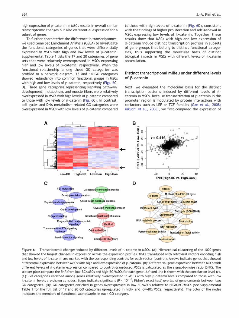
high expression of β-catenin in MSCs results in overall similartranscriptomic changes but also differential expression for asubset of genes.
To further characterize the difference in transcriptomes,we used Gene Set Enrichment Analysis (GSEA) to investigatethe functional categories of genes that were differentiallyexpressed in MSCs with high and low levels of β-catenin.Supplemental Table 1 lists the 17 and 20 categories of genesets that were relatively overexpressed in MSCs expressinghigh and low levels of β-catenin, respectively. When thefunctional relationship among these GO categories wasprofiled in a network diagram, 15 and 14 GO categoriesshowed redundancy into common functional groups in MSCswith high and low levels of β-catenin, respectively (Figs. 6C,D). Three gene categories representing signaling pathway/development, metabolism, and muscle fibers were relativelyoverexpressed in MSCs with high levels of β-catenin comparedto those with low levels of β-catenin (Fig. 6C). In contrast,cell cycle- and DNA metabolism-related GO categories wereoverexpressed in MSCs with low levels of β-catenin compared
Figure 6 Transcriptomic changes induced by different levels of βthat showed the largest changes in expression across the expressionand low levels of β-catenin are marked with the corresponding contrdifferential expression between MSCs with high and low expression odifferent levels of β-catenin expression compared to control-transdscatter plots compare the SNR from low-BC/MSCs and high-BC/MSCs f(C): GO categories enriched among genes relatively overexpressedβ-catenin levels are shown as nodes. Edges indicate significant (P b
GO categories. (D): GO categories enriched in genes overexpressedTable 1 for the full list of 17 and 20 GO categories upregulated inindicates the members of functional subnetworks in each GO catego
to those with high levels of β-catenin (Fig. 6D), consistentwith the findings of higher proliferation and self-renewal inMSCs expressing low levels of β-catenin. Together, theseresults show that MSCs with high and low expression ofβ-catenin induce distinct transcription profiles in subsetsof gene groups that belong to distinct functional catego-ries, thus supporting the molecular basis of distinctbiological impacts in MSCs with different levels of β-cateninaccumulation.
Distinct transcriptional milieu under different levelsof β-catenin
Next, we evaluated the molecular basis for the distincttranscription patterns induced by different levels of β-catenin in MSCs. Because transactivation of β-catenin in thepromoter region is modulated by protein interactions withco-factors such as LEF or TCF families (Gan et al., 2008;Kikuchi et al., 2006), we first compared the expression of
-catenin in MSCs. (A): Hierarchical clustering of the 1000 genesprofiles. MSCs transduced with retroviral vectors encoding highols for each vector (control). Arrows indicate genes that showedf β-catenin. (B): Differential gene expression between MSCs withuced MSCs is calculated as the signal-to-noise ratio (SNR). Theor each gene. A fitted line is shown with the correlation level (r).in MSCs with high β-catenin levels compared to those with low10−10; Fisher's exact test) overlap of gene contents between twoin low-BC/MSCs relative to HIGH-BC/MSCs (see Supplementalhigh- and low-BC/MSCs, respectively). The color of the nodesry.

365Regulation of mesenchymal stromal cells through fine tuning of canonical Wnt signaling
these co-factors in each type of MSC. As shown, MSCs with lowexpression of β-catenin exhibited prominently higher levelsof LEF-1 and TCF-1, whereas MSCs with high expression ofβ-catenin exhibited profound suppression of LEF-1 withmodest differences in the levels of TCF3 and TCF4 (Figs. 7A,B). These results suggest that different intracellular levels ofβ-catenin could result in distinct transcriptional complexes atthe promoter to generate different transcription profiles.
To further explore the possibility of variation in thetranscriptional milieu under different intracellular levels ofβ-catenin, we examined the cellular proteins that interactwith β-catenin in each condition. We performed a proteomicanalysis of intracellular proteins that co-precipitated withβ-catenin using nano-scale liquid chromatography/massspectrometry (Nano LC–MS/MS) (Gaspari and Cuda, 2011).As shown in Supplemental Table 2, extensive differenceswere seen in the spectra of proteins that co-precipitatedwith each level of β-catenin, suggesting that β-catenininteracts with different intracellular proteins depending onits intracellular accumulation levels.
Taken together, these results show that the transcriptionmilieu varies according to the intracellular concentration ofβ-catenin, which thus serves as a distinct transactivation
Figure 7 Changes in the expression of co-factors for β-catenin witlevels of β-catenin were analyzed for expression of co-factors forco-factor compared to those in control MSCs were analyzed by real-t(mean ± SEM values from 3 experiments, n = 3). *; p b 0.05 **; p b
MSC expressing high and low levels of β-catenin. (C): Hypotheticsignaling in MSCs. Differences in the intensity of Wnt/β-catenin signai.e., TCF-1 is dominantly up-regulated in Low-BC, but Lef-1 is profouintracellular proteins (marked with blue and red hexagon) interactunder high and low levels of β-catenin to induce distinct transcriptioMSCs under these two conditions. * L1: LEF-1, T1: TCF-1, T3: TCF-3levels in the cells, and hexagons denote intracellular proteins intera
factor that can induce distinct transcription profiles in MSCsand thereby produce distinct functional outcomes.
Discussion
Recent studies have focused on signals that regulate prolifer-ation/differentiation and stromal function under variousphysiological and pathological conditions. Of these regulatorysignals, canonical Wnt signals have been implicated in theregulation of MSCs either by controlling the expression ofcross-talk molecules such as Jagged-1 or dll-1 (Kim et al.,2009) or by controlling MSC differentiation into osteogenic oradipogenic lineages (Ling et al., 2009; Takada et al., 2009).Moreover, constitutive stabilization of β-catenin in bonemarrow stromal cells was associated with development ofmyeloproliferative disease (Kode et al., 2014), indicating thatcanonical Wnt signaling in stromal cells should be fine-tunedfor coordinated microenvironmental regulation of hematopoi-etic activity.
However, little is known regarding the precise biologicalimpact of the signal intensity of canonical Wnt signalingin MSCs. Moreover, the effects of Wnt signaling on the
h respect to β-catenin levels. (A): MSCs expressing high and lowβ-catenin transactivation. Relative expression levels of each
ime quantitative RT-PCR (RQ-PCR) after normalization to GAPDH0.01. (B): Western blot for co-factors in the nuclear extracts ofal model for concentration-specific effects of Wnt/β-cateninling induce differences in the transcription milieu for β-catenin,ndly down-regulated in High-BC. In addition, distinct spectra ofwith β-catenin. Thus, different protein complexes are formedn profiles and therefore generate distinct biological functions in, T4: TCF-4. The size of each factor denotes relative expressioncting with β-catenin.

366 J.-A. Kim et al.
differentiation of MSCs is controversial (Boland et al., 2004;Cook et al., 2014; Gaur et al., 2005; Kawai et al., 2007),raising the possibility that the regulatory effects of canonicalWnt signals in MSCs could be more complex than previouslythought.
Therefore, to investigate the possibility that the biologicalcharacteristics of MSCs are regulated by fine-tuning of Wntsignaling strength, we established MSCs with high and lowexpression of β-catenin by transduction of the cells with twodifferent retroviral vectors. From this approach, we foundthat two distinct aspects of MSC function were controlled bythe intensity of Wnt/β-catenin signals, i.e., proliferation andself-renewal of MSCs were promoted only under low levelsof Wnt/β-catenin, whereas osteogenic differentiation waspromoted in a manner proportional to the intensity of the Wntsignals.
Moreover, MSCs proliferating under low Wnt/β-cateninexhibited higher expression of pluripotency genes such asOct-4, Sox-2, and Nanog (Greco et al., 2007; Tsai et al.,2012), indicative of a more primitive state, whereas thecells exhibited more committed osteoblast-like propertiesunder high Wnt/β-catenin.
Interestingly, studies have shown that the characteristicsof primitive MSCs derived from embryonic or inducedpluripotent stem cells as well as MSCs from fetal tissueexhibit higher levels of pluripotent gene expression, higherCFU-F and proliferation together with higher osteogenic, butnot adipogenic differentiation potential compared to adulttissue-derived MSCs (Boyd et al., 2009; Chen et al., 2012)(Guillot et al., 2007; Zhang et al., 2009), which was similarlyobserved among MSCs with early and late passages duringserial subculture (Vacanti et al., 2005). Thus, taken thesefindings together, MSCs under low Wnt/β-catenin levelsmimic the primitive MSCs sharing exhibiting commonfunctional characteristics with those MSCs.
Of note, differences in the accumulation levels ofβ-catenin in MSCs also resulted in distinct stimulatoryeffects on hematopoietic progenitor cells, i.e., whereasMSCs with low expression of β-catenin better supported theproliferation of hematopoietic cells than MSCs with highexpression of β-catenin, maintenance of the undifferentiat-ed state was better supported by MSCs with high levels ofβ-catenin.
Together, these findings suggest that the MSCs exhibitdistinct biological responses to physiological conditions andexert distinct influences on the tissue regenerative processdepending on the strength of Wnt/β-catenin signaling intheir microenvironment. Although further studies on thephysiological significance of such regulation of niche activityare warranted, our findings point to the importance offine-tuning Wnt/β-catenin signaling for coordination ofregulatory effects in MSCs.
At present, the mechanisms underlying the concentration-specific effects of Wnt/β-catenin signaling remain unclear.However, our study shows that β-catenin induces distincttranscriptomic changes depending on the level of β-cateninaccumulation, especially for subsets of genes in distinctfunctional groups of gene ontology. In particular, low levelsof β-catenin in MSCs resulted in up-regulation of genesinvolved in cell cycle and DNA metabolism, whereas highlevels of β-catenin caused up-regulation of genes involvedin signaling pathway/development and metabolism. Thus,
β-catenin can serve as a distinct transactivation moleculedepending on its level of accumulation in the cells to inducedifferent transcriptomic changes in the cells.
Although the molecular basis for the distinct trans-activation of β-catenin remains unclear, our study raises thepossibility that the transcriptional milieu at the targetpromoters could vary according to the level of β-cateninaccumulation. Supporting this possibility, MSCs expressingdifferent levels of β-catenin exhibited different intracellularexpression of co-factors, such that high and low levels ofβ-catenin resulted in different levels of LEF-1 and TCF-1.Thus, it is possible that different levels of β-catenin result inthe formation of a distinct transcription complex at the targetpromoters, thus resulting in different components of thetranscription milieu. Moreover, nano-scale LC/mass spectro-photometry showed that β-catenin interacts with differentintracellular proteins, as evidenced by the distinct spectrumof co-precipitated proteins specific to each concentration ofβ-catenin.
Therefore, out study shows that different intracellularlevels of β-catenin can affect the transcription milieu, andthus β-catenin may serve as a distinct transactivationmolecule to produce distinct biological effects in MSCs(schematically shown in Fig. 7C).
Of note, our finding of distinct transactivation withdifferent intracellular levels of β-catenin could provide amechanism for broader spectra of stem cell regulation byWnt signals, as several studies have shown that theregulatory effects of Wnt signals on HSCs are dependent onthe intensity of the signals (Luis et al., 2011). Similarly,differentiation of ES cells towards specific lineages wasinfluenced by the strength of Wnt signals (Kielman et al.,2002; Reya and Clevers, 2005), providing a common schemefor regulation by Wnt signals. In this sense, it is plausiblethat the current paradigm of concentration-specific regula-tion by Wnt/β-catenin signaling is conserved over a broadextent of tissue regeneration and that various types of stemcells and their niche function can be regulated through finetuning of Wnt/β-catenin signaling. Further studies arenecessary for a more complete understanding of theregulation of various tissue regenerative processes by Wnt/β-catenin signaling.
Nevertheless, our study shows that different intracellularlevels of β-catenin in MSCs may result in distinct biologicalresponses of MSCs and that β-catenin serves as a distincttransactivation molecule that regulates MSC function ac-cording to Wnt signaling intensity. Our study provides anovel insight into the importance of fine tuning of Wnt/β-catenin signals for coordination of various regenerativebiological functions.
Supplementary data to this article can be found online athttp://dx.doi.org/10.1016/j.scr.2015.02.007.
Author contributions
JAK., HKC.: performed the experiments and generated thedata.
SHL., TMK.: performed gene expression analysis and bio-informatics study.
IHO.: designed the current study, supervised the study,and wrote the manuscript.

367Regulation of mesenchymal stromal cells through fine tuning of canonical Wnt signaling
Acknowledgments
The proteomics analysis using nano liquid chromatography/mass spectrometry (Nano LC–MS/MS) was performed by theYonsei Proteome Research Center (www.proteomix.org),Korea. This research was supported by the National ResearchFoundation of Korea (NRF) funded by the Ministry of Science,ICT & Future Planning (No. 2011-0019352) and a grant of theKorea Health Technology R&D Project, Ministry of Health &Welfare, Republic of Korea (A120262).
References
Ahn, J.Y., Park, G., Shim, J.S., Lee, J.W., Oh, I.H., 2010.Intramarrow injection of beta-catenin-activated, but notnaive mesenchymal stromal cells stimulates self-renewal ofhematopoietic stem cells in bone marrow. Exp. Mol. Med. 42,122–131.
Antrobus, R., Borner, G.H., 2011. Improved elution conditions fornative co-immunoprecipitation. PLoS ONE 6, e18218.
Arai, F., Hirao, A., Ohmura, M., Sato, H., Matsuoka, S., Takubo, K.,Ito, K., Koh, G.Y., Suda, T., 2004. Tie2/angiopoietin-1 signalingregulates hematopoietic stem cell quiescence in the bonemarrow niche. Cell 118, 149–161.
Biechele, T.L., Moon, R.T., 2008. Assaying beta-catenin/TCFtranscription with beta-catenin/TCF transcription-based report-er constructs. Methods Mol. Biol. (Clifton, N. J.) 468, 99–110.
Boland, G.M., Perkins, G., Hall, D.J., Tuan, R.S., 2004. Wnt 3a pro-motes proliferation and suppresses osteogenic differentiation ofadult human mesenchymal stem cells. J. Cell. Biochem. 93,1210–1230.
Boyd, N.L., Robbins, K.R., Dhara, S.K., West, F.D., Stice, S.L., 2009.Human embryonic stem cell-derived mesoderm-like epitheliumtransitions to mesenchymal progenitor cells. Tissue Eng. A 15,1897–1907.
Calvi, L.M., Adams, G.B., Weibrecht, K.W., Weber, J.M., Olson, D.P.,Knight, M.C., Martin, R.P., Schipani, E., Divieti, P., Bringhurst,F.R., et al., 2003. Osteoblastic cells regulate the haematopoieticstem cell niche. Nature 425, 841–846.
Caplan, A.I., Correa, D., 2011. The MSC: an injury drugstore. CellStem Cell 9, 11–15.
Chan, C.K., Chen, C.C., Luppen, C.A., Kim, J.B., DeBoer, A.T., Wei,K., Helms, J.A., Kuo, C.J., Kraft, D.L., Weissman, I.L., 2009.Endochondral ossification is required for haematopoietic stem-cell niche formation. Nature 457, 490–494.
Chen, Y.S., Pelekanos, R.A., Ellis, R.L., Horne, R., Wolvetang, E.J.,Fisk, N.M., 2012. Small molecule mesengenic induction of humaninduced pluripotent stem cells to generate mesenchymal stem/stromal cells. Stem Cells Transl. Med. 1, 83–95.
Choong, M.L., Tan, A.C., Luo, B., Lodish, H.F., 2003. A novel role forproliferin-2 in the ex vivo expansion of hematopoietic stem cells.FEBS Lett. 550, 155–162.
Cook, D.A., Fellgett, S.W., Pownall, M.E., O'Shea, P.J., Genever, P.G.,2014. Wnt-dependent osteogenic commitment of bone marrowstromal cells using a novel GSK3beta inhibitor. Stem Cell Res. 12,415–427.
Frenette, P.S., Pinho, S., Lucas, D., Scheiermann, C., 2013.Mesenchymal stem cell: keystone of the hematopoietic stemcell niche and a stepping-stone for regenerative medicine. Annu.Rev. Immunol. 31, 285–316.
Gan, X.Q., Wang, J.Y., Xi, Y., Wu, Z.L., Li, Y.P., Li, L., 2008.Nuclear Dvl, c-Jun, beta-catenin, and TCF form a complexleading to stabilization of beta-catenin–TCF interaction. J. CellBiol. 180, 1087–1100.
Gaspari, M., Cuda, G., 2011. Nano LC–MS/MS: a robust setup forproteomic analysis. MethodsMol. Biol. (Clifton, N. J.) 790, 115–126.
Gaur, T., Lengner, C.J., Hovhannisyan, H., Bhat, R.A., Bodine, P.V.,Komm, B.S., Javed, A., van Wijnen, A.J., Stein, J.L., Stein, G.S.,et al., 2005. Canonical WNT signaling promotes osteogenesis bydirectly stimulating Runx2 gene expression. J. Biol. Chem. 280,33132–33140.
Greco, S.J., Liu, K., Rameshwar, P., 2007. Functional similaritiesamong genes regulated by OCT4 in human mesenchymal andembryonic stem cells. Stem Cells (Dayt., Ohio) 25, 3143–3154.
Greenbaum, A., Hsu, Y.M., Day, R.B., Schuettpelz, L.G., Christopher,M.J., Borgerding, J.N., Nagasawa, T., Link, D.C., 2013. CXCL12 inearly mesenchymal progenitors is required for haematopoieticstem-cell maintenance. Nature 495, 227–230.
Guillot, P.V., Gotherstrom, C., Chan, J., Kurata, H., Fisk, N.M.,2007. Human first-trimester fetal MSC express pluripotencymarkers and grow faster and have longer telomeres than adultMSC. Stem Cells (Dayt., Ohio) 25, 646–654.
Hong, S.H., Yang, S.J., Kim, T.M., Shim, J.S., Lee, H.S., Lee, G.Y.,Park, B.B., Nam, S.W., Ryoo, Z.Y., Oh, I.H., 2014. Molecularintegration of HoxB4 and STAT3 for self-renewal of hematopoi-etic stem cells: a model of molecular convergence for stemness.Stem Cells (Dayt. Ohio) 32, 1313–1322.
Jung, J., Moon, N., Ahn, J.Y., Oh, E.J., Kim, M., Cho, C.S., Shin,J.C., Oh, I.H., 2009. Mesenchymal stromal cells expanded inhuman allogenic cord blood serum display higher self-renewaland enhanced osteogenic potential. Stem Cells Dev. 18,559–571.
Kawai, M., Mushiake, S., Bessho, K., Murakami, M., Namba, N.,Kokubu, C., Michigami, T., Ozono, K., 2007. Wnt/Lrp/beta-catenin signaling suppresses adipogenesis by inhibiting mutualactivation of PPARgamma and C/EBPalpha. Biochem. Biophys.Res. Commun. 363, 276–282.
Keating, A., 2006. Mesenchymal stromal cells. Curr. Opin. Hematol.13, 419–425.
Kielman, M.F., Rindapaa, M., Gaspar, C., van Poppel, N., Breukel,C., van Leeuwen, S., Taketo, M.M., Roberts, S., Smits, R.,Fodde, R., 2002. Apc modulates embryonic stem-cell differen-tiation by controlling the dosage of beta-catenin signaling. Nat.Genet. 32, 594–605.
Kikuchi, A., Kishida, S., Yamamoto, H., 2006. Regulation of Wntsignaling by protein–protein interaction and post-translationalmodifications. Exp. Mol. Med. 38, 1–10.
Kim, J.A., Kang, Y.J., Park, G., Kim, M., Park, Y.O., Kim, H., Leem,S.H., Chu, I.S., Lee, J.S., Jho, E.H., et al., 2009. Identificationof a stroma-mediated Wnt/beta-catenin signal promoting self-renewal of hematopoietic stem cells in the stem cell niche. StemCells (Dayt., Ohio) 27, 1318–1329.
Kode, A., Manavalan, J.S., Mosialou, I., Bhagat, G., Rathinam, C.V.,Luo, N., Khiabanian, H., Lee, A., Murty, V.V., Friedman, R., etal., 2014. Leukaemogenesis induced by an activating beta-catenin mutation in osteoblasts. Nature 506, 240–244.
Ling, L., Nurcombe, V., Cool, S.M., 2009. Wnt signaling controls thefate of mesenchymal stem cells. Gene 433, 1–7.
Luis, T.C., Naber, B.A., Roozen, P.P., Brugman, M.H., de Haas, E.F.,Ghazvini, M., Fibbe, W.E., van Dongen, J.J., Fodde, R., Staal,F.J., 2011. Canonical wnt signaling regulates hematopoiesis in adosage-dependent fashion. Cell Stem Cell 9, 345–356.
Mendez-Ferrer, S., Michurina, T.V., Ferraro, F., Mazloom, A.R.,Macarthur, B.D., Lira, S.A., Scadden, D.T., Ma'ayan, A., Enikolopov,G.N., Frenette, P.S., 2010. Mesenchymal and haematopoietic stemcells form a unique bone marrow niche. Nature 466, 829–834.
Murphy, M.B., Moncivais, K., Caplan, A.I., 2013. Mesenchymal stemcells: environmentally responsive therapeutics for regenerativemedicine. Exp. Mol. Med. 45, e54.
Naveiras, O., Nardi, V., Wenzel, P.L., Hauschka, P.V., Fahey, F.,Daley, G.Q., 2009. Bone-marrow adipocytes as negative regulatorsof the haematopoietic microenvironment. Nature 460, 259–263.
Nemeth, M.J., Mak, K.K., Yang, Y., Bodine, D.M., 2009. Beta-catenin expression in the bone marrow microenvironment is

368 J.-A. Kim et al.
required for long-term maintenance of primitive hematopoieticcells. Stem Cells (Dayt., Ohio) 27, 1109–1119.
Oh, I.H., 2010. Microenvironmental targeting of Wnt/beta-cateninsignals for hematopoietic stem cell regulation. Expert. Opin.Biol. Ther. 10, 1315–1329.
Oh, I.H., Humphries, R.K., 2012. Concise review: multidimensionalregulation of the hematopoietic stem cell state. Stem Cells(Dayt., Ohio) 30, 82–88.
Oh, I.H., Kwon, K.R., 2010. Concise review: multiple niches forhematopoietic stem cell regulations. Stem Cells (Dayt., Ohio)28, 1243–1249.
Pittenger, M.F., Mackay, A.M., Beck, S.C., Jaiswal, R.K., Douglas,R., Mosca, J.D., Moorman, M.A., Simonetti, D.W., Craig, S.,Marshak, D.R., 1999. Multilineage potential of adult humanmesenchymal stem cells. Science 284, 143–147.
Reya, T., Clevers, H., 2005. Wnt signalling in stem cells and cancer.Nature 434, 843–850.
Satija, N.K., Singh, V.K., Verma, Y.K., Gupta, P., Sharma, S., Afrin,F., Sharma, M., Sharma, P., Tripathi, R.P., Gurudutta, G.U.,2009. Mesenchymal stem cell-based therapy: a new paradigm inregenerative medicine. J. Cell. Mol. Med. 13, 4385–4402.
Takada, I., Kouzmenko, A.P., Kato, S., 2009. Wnt and PPARgammasignaling in osteoblastogenesis and adipogenesis. Nat. Rev.Rheumatol. 5, 442–447.
Tsai, C.C., Su, P.F., Huang, Y.F., Yew, T.L., Hung, S.C., 2012. Oct4and Nanog directly regulate Dnmt1 to maintain self-renewal andundifferentiated state in mesenchymal stem cells. Mol. Cell 47,169–182.
Vacanti, V., Kong, E., Suzuki, G., Sato, K., Canty, J.M., Lee, T.,2005. Phenotypic changes of adult porcine mesenchymal stemcells induced by prolonged passaging in culture. J. Cell. Physiol.205, 194–201.
Zhang, J., Niu, C., Ye, L., Huang, H., He, X., Tong, W.G., Ross, J.,Haug, J., Johnson, T., Feng, J.Q., et al., 2003. Identification ofthe haematopoietic stem cell niche and control of the niche size.Nature 425, 836–841.
Zhang, Z.Y., Teoh, S.H., Chong, M.S., Schantz, J.T., Fisk, N.M.,Choolani, M.A., Chan, J., 2009. Superior osteogenic capacity forbone tissue engineering of fetal compared with perinatal andadult mesenchymal stem cells. Stem Cells (Dayt., Ohio) 27,126–137.
Zhou, B.O., Yue, R., Murphy, M.M., Peyer, J.G., Morrison, S.J.,2014. Leptin-receptor-expressing mesenchymal stromal cellsrepresent the main source of bone formed by adult bonemarrow. Cell Stem Cell 15, 154–168.