Regulation of microbial populations by coral surface … stress (e.g. bleaching), disease and...
14
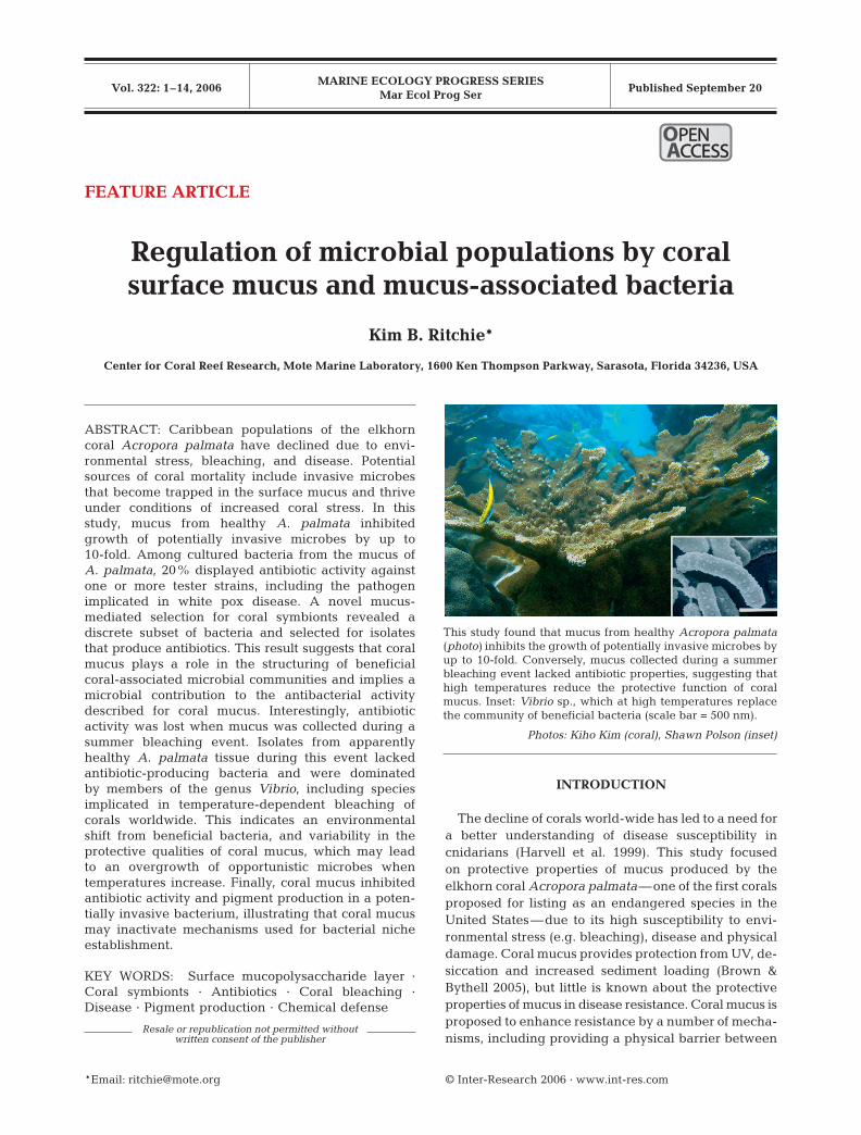
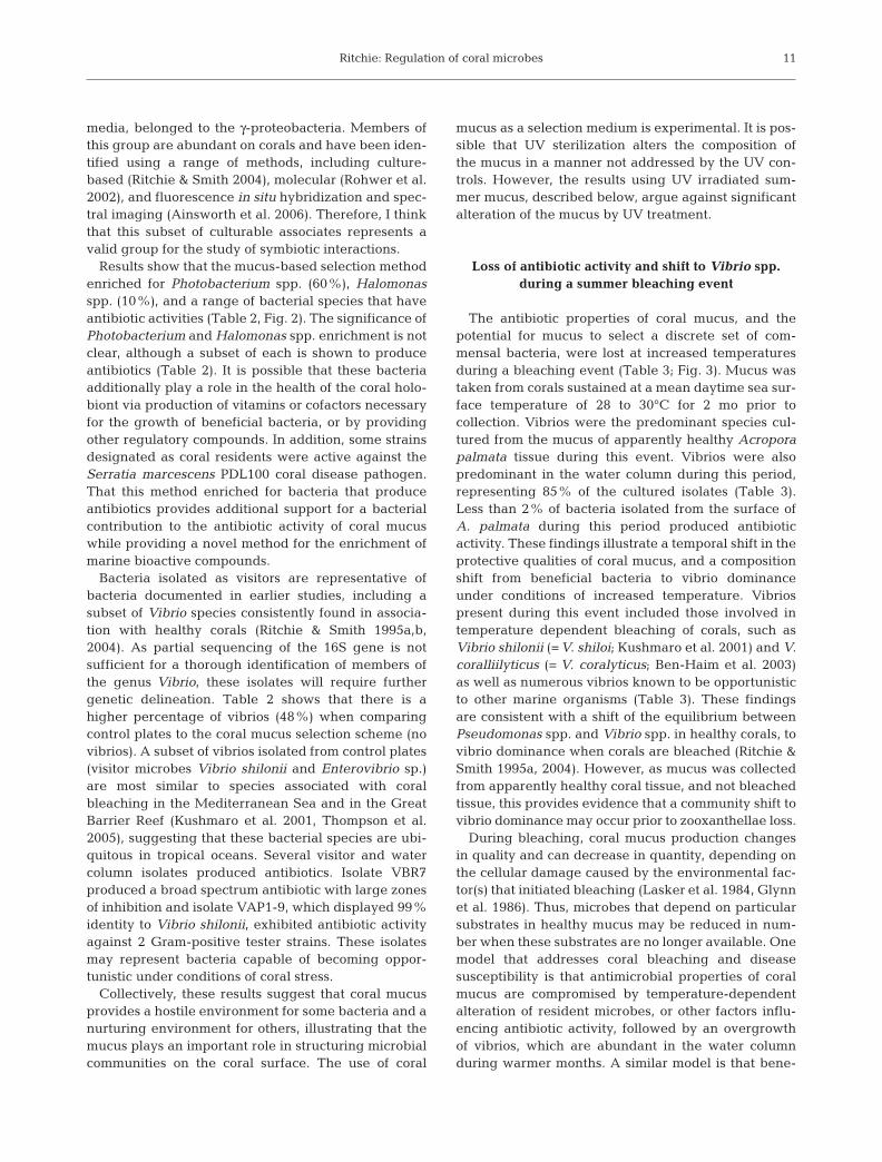
MARINE ECOLOGY PROGRESS SERIES Mar Ecol Prog Ser Vol. 322: 1–14, 2006 Published September 20 INTRODUCTION The decline of corals world-wide has led to a need for a better understanding of disease susceptibility in cnidarians (Harvell et al. 1999). This study focused on protective properties of mucus produced by the elkhorn coral Acropora palmata — one of the first corals proposed for listing as an endangered species in the United States — due to its high susceptibility to envi- ronmental stress (e.g. bleaching), disease and physical damage. Coral mucus provides protection from UV, de- siccation and increased sediment loading (Brown & Bythell 2005), but little is known about the protective properties of mucus in disease resistance. Coral mucus is proposed to enhance resistance by a number of mecha- nisms, including providing a physical barrier between © Inter-Research 2006 · www.int-res.com *Email: [email protected] FEATURE ARTICLE Regulation of microbial populations by coral surface mucus and mucus-associated bacteria Kim B. Ritchie* Center for Coral Reef Research, Mote Marine Laboratory, 1600 Ken Thompson Parkway, Sarasota, Florida 34236, USA ABSTRACT: Caribbean populations of the elkhorn coral Acropora palmata have declined due to envi- ronmental stress, bleaching, and disease. Potential sources of coral mortality include invasive microbes that become trapped in the surface mucus and thrive under conditions of increased coral stress. In this study, mucus from healthy A. palmata inhibited growth of potentially invasive microbes by up to 10-fold. Among cultured bacteria from the mucus of A. palmata, 20% displayed antibiotic activity against one or more tester strains, including the pathogen implicated in white pox disease. A novel mucus- mediated selection for coral symbionts revealed a discrete subset of bacteria and selected for isolates that produce antibiotics. This result suggests that coral mucus plays a role in the structuring of beneficial coral-associated microbial communities and implies a microbial contribution to the antibacterial activity described for coral mucus. Interestingly, antibiotic activity was lost when mucus was collected during a summer bleaching event. Isolates from apparently healthy A. palmata tissue during this event lacked antibiotic-producing bacteria and were dominated by members of the genus Vibrio, including species implicated in temperature-dependent bleaching of corals worldwide. This indicates an environmental shift from beneficial bacteria, and variability in the protective qualities of coral mucus, which may lead to an overgrowth of opportunistic microbes when temperatures increase. Finally, coral mucus inhibited antibiotic activity and pigment production in a poten- tially invasive bacterium, illustrating that coral mucus may inactivate mechanisms used for bacterial niche establishment. KEY WORDS: Surface mucopolysaccharide layer · Coral symbionts · Antibiotics · Coral bleaching · Disease · Pigment production · Chemical defense Resale or republication not permitted without written consent of the publisher This study found that mucus from healthy Acropora palmata ( photo) inhibits the growth of potentially invasive microbes by up to 10-fold. Conversely, mucus collected during a summer bleaching event lacked antibiotic properties, suggesting that high temperatures reduce the protective function of coral mucus. Inset: Vibrio sp., which at high temperatures replace the community of beneficial bacteria (scale bar = 500 nm). Photos: Kiho Kim (coral), Shawn Polson (inset) OPEN PEN ACCESS CCESS
-
Upload
nguyenlien -
Category
Documents
-
view
217 -
download
0
Transcript of Regulation of microbial populations by coral surface … stress (e.g. bleaching), disease and...
MARINE ECOLOGY PROGRESS SERIESMar Ecol Prog Ser
Vol. 322: 1–14, 2006 Published September 20
INTRODUCTION
The decline of corals world-wide has led to a need fora better understanding of disease susceptibility incnidarians (Harvell et al. 1999). This study focusedon protective properties of mucus produced by theelkhorn coral Acropora palmata—one of the first coralsproposed for listing as an endangered species in theUnited States—due to its high susceptibility to envi-ronmental stress (e.g. bleaching), disease and physicaldamage. Coral mucus provides protection from UV, de-siccation and increased sediment loading (Brown &Bythell 2005), but little is known about the protectiveproperties of mucus in disease resistance. Coral mucus isproposed to enhance resistance by a number of mecha-nisms, including providing a physical barrier between
© Inter-Research 2006 · www.int-res.com*Email: [email protected]
FEATURE ARTICLE
Regulation of microbial populations by coralsurface mucus and mucus-associated bacteria
Kim B. Ritchie*
Center for Coral Reef Research, Mote Marine Laboratory, 1600 Ken Thompson Parkway, Sarasota, Florida 34236, USA
ABSTRACT: Caribbean populations of the elkhorncoral Acropora palmata have declined due to envi-ronmental stress, bleaching, and disease. Potentialsources of coral mortality include invasive microbesthat become trapped in the surface mucus and thriveunder conditions of increased coral stress. In thisstudy, mucus from healthy A. palmata inhibitedgrowth of potentially invasive microbes by up to10-fold. Among cultured bacteria from the mucus ofA. palmata, 20% displayed antibiotic activity againstone or more tester strains, including the pathogenimplicated in white pox disease. A novel mucus-mediated selection for coral symbionts revealed adiscrete subset of bacteria and selected for isolatesthat produce antibiotics. This result suggests that coralmucus plays a role in the structuring of beneficialcoral-associated microbial communities and implies amicrobial contribution to the antibacterial activitydescribed for coral mucus. Interestingly, antibioticactivity was lost when mucus was collected during asummer bleaching event. Isolates from apparentlyhealthy A. palmata tissue during this event lackedantibiotic-producing bacteria and were dominatedby members of the genus Vibrio, including speciesimplicated in temperature-dependent bleaching ofcorals worldwide. This indicates an environmentalshift from beneficial bacteria, and variability in theprotective qualities of coral mucus, which may leadto an overgrowth of opportunistic microbes whentemperatures increase. Finally, coral mucus inhibitedantibiotic activity and pigment production in a poten-tially invasive bacterium, illustrating that coral mucusmay inactivate mechanisms used for bacterial nicheestablishment.
KEY WORDS: Surface mucopolysaccharide layer ·Coral symbionts · Antibiotics · Coral bleaching ·Disease · Pigment production · Chemical defense
Resale or republication not permitted without written consent of the publisher
This study found that mucus from healthy Acropora palmata(photo) inhibits the growth of potentially invasive microbes byup to 10-fold. Conversely, mucus collected during a summerbleaching event lacked antibiotic properties, suggesting thathigh temperatures reduce the protective function of coralmucus. Inset: Vibrio sp., which at high temperatures replacethe community of beneficial bacteria (scale bar = 500 nm).
Photos: Kiho Kim (coral), Shawn Polson (inset)
OPENPEN ACCESSCCESS

Mar Ecol Prog Ser 322: 1–14, 2006
the coral and the environment, mucociliary transport ofmicrobes for removal via ingestion by the coralhost, sloughing to avoid colonization by invasive mi-crobes, and acting as a medium for secreted allelochem-icals with antimicrobial properties (reviewed in Brown &Bythell 2005). Extracts from soft corals and gorgoniancorals have antimicrobial properties (Burkholder & Burk-holder 1958, Kim 1994, Slattery et al. 1995, 1997, Kelmanet al. 1998, 2006). Koh (1997) described antimicrobialcompounds present in numerous Pacific scleractiniancoral species and recent work by Geffen & Rosenberg(2005) shows that the Red Sea scleractinian coral Pocillo-pora damicornis exhibits antimicrobial activity. How-ever, the origin of these allelochemicals is unknown.
Corals harbor a diverse array of bacterial associates(reviewed in Brown & Bythell 2005), some of which arethought to be species-specific (Ritchie & Smith 1997,Rohwer et al. 2002). To date, very little is known of themetabolic capabilities of these bacteria, their functionon the coral surface, and their potential benefit to thecoral, zooxanthellae, or both. Bacterial symbionts havebeen shown to be responsible for the production of sec-ondary metabolites previously attributed to the hostorganism (Yasumoto et al. 1986, Elyakov et al. 1991).Some marine macroorganisms combat microbial foul-ing by producing compounds that inhibit bacterialgrowth or attachment, while others rely on microbialproduction of defense compounds (Gil-Turnes et al.1989, Holmstrom et al. 1992, Armstrong et al. 2001).Commensal relationships between bacteria in multi-species biofilms can play a role in determining the spa-tial distribution of microbial populations within thebiofilm (Neilson et al. 2000, Rao et al. 2005). Castillo etal. (2001) determined that 30% of bacteria isolatedfrom coral species have antibiotic capabilities. Theroles played by coral mucus and mucus-associated bac-teria in determining the compositions of coral-associ-ated microbial communities are currently unknown.
This study addressed antibiotic activity associated withAcropora palmata mucus and mucus-associated bacte-ria. Here, antibiotic refers to a substance that selectivelykills or inhibits the growth of a microorganism. In addi-tion, an experimental approach was used to investigatethe potential of coral mucus as a selection medium forcoral symbionts. For the purpose of this study, a symbiontis defined as a bacterium that benefits from properties ofthe coral mucus while providing a benefit to the coral inreturn (a mutualistic relationship).
MATERIALS AND METHODS
Bacterial strains. To isolate culturable bacteria, Acro-pora palmata mucus samples were diluted in sterileseawater and plated onto glycerol artificial seawater
agar (GASWA; Smith & Hayasaka 1982), followed byincubation at 24°C. Microorganisms exhibiting aunique colony or cellular morphology (as compared toother colonies on a single plate) were subcultured topurification under the same growth conditions. Forease of performing medium through-put antibioticassays, 96-well microtiter plate libraries of culturablebacterial isolates were generated. Each library con-sisted of 96 individually purified coral bacterial isolatesstored frozen at –80°C in liquid GASWA medium con-taining 30% glycerol. Culturable bacterial librarieswere archived for further antibiotic screening andspecies identification. Strain VBR 7 was isolated fromA. palmata mucus in a screen for putative invasivemicrobes (described below). Other sources of poten-tially invasive microbes used in this study includedcanal water from Big Pine canal (Sands, 24° 40.31’ N,81° 20.40’ W), dust from Mali, Africa (12° 37.22’ N,7° 59.40’ E; provided by V. Garrison, USGS), and waterfrom Looe Key Reef (24° 33.78’ N, 81° 24.05’ W). Testerstrains used for antibiotic assays are described below.
Sampling. Samples were taken from 12 Acroporapalmata colonies at various locations at Looe KeyReef, in the Florida Keys (24° 32.76’ N, 81° 24.21’ Wand 24° 32.75’ N, 81° 24.35’ W), between January andDecember of 2005 and were used to generate cultur-able bacterial libraries for antibiotic production assays.Samples for coral mucus selection experiments weretaken from 3 A. palmata colonies in April 2005 (meanwater temperature of 24°C, sustained at 22 to 25°C for2 mo prior to sampling) and 3 A. palmata colonies inSeptember of 2005 (mean water temperature of 30°C,sustained at 28 to 30°C for 2 mo prior to sampling).September samples were taken during a bleachingevent in which all A. palmata colonies observed wereaffected by hurricane damage, bleaching or disease.During September sampling, mucus from apparentlyhealthy areas of A. palmata colonies was collected.These were areas that appeared healthy, but that wereadjacent to entirely bleached areas, or areas withsloughed tissue, in each colony sampled.
To collect samples under field conditions, 30 ml ofthe surface mucopolysaccharide layer from appar-ently healthy Acropora palmata were harvestedfrom the skyward-facing, sun-exposed portion of thecolony, 5 to 10 cm from the actively growing edge.Mucus from each colony was consistently collectedover an area of 5 × 5 cm after gentle agitation with asterile syringe. Agitation encourages sloughing of theviscous mucus matrix for ease of syringe aspiration,while reducing aspiration of water. Samples weremaintained at 24°C and processed within 2 h of col-lection. Water samples were taken by briefly openinga sterile 15 ml conical tube 1 m from the coral colonyprior to mucus sampling. Water samples were used
2

Ritchie: Regulation of coral microbes
in both mucus challenge experiments and bacterialcomposition comparisons.
Inhibition of potentially invasive microbes. Sources ofpotentially invasive microbes were challenged withmucus collected from A. palmata. Potential sources of in-vasive microbes were chosen based on previous implica-tions to disease causation (Patterson et al. 2002, Garrisonet al. 2003). These sources included Florida Keys canalwater, African dust, and water column microbes. Mucus-treated media were used to test growth inhibition byplating out 400 µl of undiluted coral mucus onto GASWAmedium and allowing it to dry for 10 min. Mucus treatedplates were sterilized via UV irradiation by placing un-covered Petri plates face downward onto a M-26 UVPtransilluminator for 10 min at 302 nm wavelength. UVirradiation was chosen for sterilization, as mucus is tooviscous for filter sterilization and heat/pressure steriliza-tion would greatly alter components, including antibioticproperties, of coral mucus. UV irradiated mucus-treatedplates that were left un-inoculated were used to controlfor complete UV killing in each experiment. To addresspotential UV alteration of media, inoculates from Africandust and canal water were used in control compar-isons measuring growth on UV irradiated versus non-irradiated GASWA medium.
Seawater was tested on mucus-treated GASWAmedium to address growth inhibition of marine bacte-ria present in the water column. In order to addresscoral resistance to potential human-derived waterquality contaminants, such as Serratia marscecens orEscherichia coli, mucus-treated Luria Bertani (LB) agarwas used when testing Florida Keys canal water-associated microbes. Dust from Mali, Africa (collectedby V. Garrison) was tested on GASWA medium toaddress inhibition of microbes from dust events withthe potential to remain viable in sea water. Both seawater and canal water were concentrated by centrifu-gation, resuspended in 1/5 volume of supernatant, andinoculated onto each media treatment; 50 µg of dustwere added to 1 ml of sterile seawater, serially dilutedand plated onto GASWA mucus-treated and GASWAcontrol plates.
In addition to potential sources of invasive microbes,both Gram-positive and Gram-negative tester strainswere used in mucus inhibition assays to address therange of bacterial inhibition by Acropora palmatamucus. Exponentially growing cultures of each testerstrain were serially diluted and assayed as describedabove. Tester strains included Bacillus subtilis (ATCC6633 Km resistant), Staphylococcus aureus (MRSA,ATCC 43300), Salmonella typhimurium (ATCC 6994),and the Serratia marsescens white pox isolate, PDL100(ATCC BAA-632).
Individual experiments were repeated 4 times.Experimental and control plates were incubated at
24°C for 48 h. Dilutions containing between 200 and400 colonies on control media (GASWA + UV treat-ment) were used in comparison with the correspond-ing tester plates (GASWA + Mucus + UV treatment).Colony count, mean and SD were recorded for eachexperiment. To derive measures of fold-inhibition, theexperimental mean was divided into the control meanfor each experiment.
Antibacterial production assays. Acropora palmatamucus-associated bacteria were used in a primaryscreen to test for the production of anti-bacterialcompounds against a range of tester strains, including:Serratia marcescens PDL100, a pathogen implicated inwhite pox disease of A. palmata (ATCC BAA-632; Pat-terson et al. 2002), methicillin-resistant Staphylococcusaureus (MRSA; ATCC 43300), methicillin-sensitiveS. aureus (MSSA; ATCC 29213), vancomycin-resistantEnterococcus (VRE; Microgenomics), Bacillus subtilis(ATCC 6633 Km resistant), Salmonella typhimurium(ATCC 6994), Enterococcus faecalis (ATCC 29212),Shigella (Microgenomics),Escherichia coli O157 (Micro-genomics), and Agrobacterium tumefaciens (Micro-genomics). Culturable bacterial libraries, stored in96-well plate format, were inoculated onto rectangularPetri plates containing GASWA solid medium andgrown for 2 d at 24°C. Growth of coral bacteria wasfollowed by UV irradiation for 15 to 20 min to inhibitcross contamination from coral-derived bacteria dur-ing antibiotic testing. Overnight cultures of testerstrains were grown in LB, Tryptic Soy Broth (TSB) orGASWA liquid medium, as appropriate. Strain-specificsoft agar medium (0.8%) was melted and cooled to42°C; 8 ml of soft agar was inoculated with individuallog-phase tester strains and poured over UV irradiatedlibrary plates. Plates were incubated for 2 d at 30°C.Growth inhibition resulted in a clear zone of inhibition(non-growth of the overlayed tester strain) around acoral-derived bacterial colony that produced an activeantibacterial compound. Zones of inhibition were mea-sured using calipers (0.02 mm) and scored as distance(in mm) from the outside of the clearing zone to theouter edge of the coral-derived bacterial colony tested.Antibiotic spectra of library isolates were used tofurther dereplicate marine isolates.
Selection for coral bacterial symbionts. To selectfor antibiotic-producing coral bacteria, a culture-based approach was designed to take advantage ofthe antibiotic properties associated with Acroporapalmata mucus. This method was hypothesized toeliminate bacteria that are inhibited by mucus-asso-ciated antibiotic activity (trapped microbes that maybecome invasive), while selecting for individualsresistant to antibiotic properties of mucus (potentiallybeneficial coral bacteria, including antibiotic pro-ducers).
3

Mar Ecol Prog Ser 322: 1–14, 2006
This assay was carried out as described above formucus inhibitory assays. However, instead of challeng-ing mucus-treated plates with sources of potentiallyinvasive microbes, coral mucus dilutions from the samecoral colony used to prepare plates were inoculatedonto GASWA mucus-treated or GASWA control plates.Colonies were counted and colony forming units(CFUs) per ml estimated for each dilution to representfold-inhibition. Individual colonies were picked frommucus treated plates to represent potential coral bac-terial symbionts (designated as ‘residents’). Isolatesfrom the control plates were selected as putative tran-sient bacteria, or bacteria that are potentially invasiveunder the right conditions (‘visitors’). Water columnisolates were purified after growth on GASWA mediumfor comparison to residents and visitors pools.
Mucus inhibition of antibiotic properties in a poten-tially invasive bacterium. Mucus used in challengeexperiments with a potentially invasive microbe (VBR7)was taken from Acropora palmata (April and Septem-ber 2005, Florida Keys), Pseudopterogorgia americana(April 2005, Florida Keys) and Montastraea faveolata(April 2005, Florida Keys and Flower Garden Banks).From each coral source, 400 µl of undiluted coralmucus were plated onto GASWA solid media, allowedto dry for 10 min, and UV irradiated for 10 min toprevent outgrowth of mucus-associated microbes. Tworesulting colony morphologies of VBR7 were darkpurple (after growth on GASWA control medium) andwhite (after growth on each mucus-treated medium).The 2 morphologies of the VBR 7 strain were patchedonto replica GASWA plates and assayed for antibioticactivity as described above.
Bacterial identification. For species identification ofculturable coral isolates, DNA extraction was per-formed on each strain via a chemical lysis protocoldetailed in Weidner et al. (1996). PCR amplificationwas carried out on genomic DNA with oligonucleotideforward primer R1n, corresponding to position 22 to 41of the Escherichia coli 16S rRNA gene, and reverseprimer U2 corresponding to complementary position1085 to 1066 (Weidner et al. 1996). PCR products wereelectrophoresed on a 1% agarose gel, and verifiedusing the AlphaImager 3300. A ~1100 bp fragment waspurified from the PCR reactions using the Qiagen PCRpurification kit. PCR products were directly sequencedvia BigDye™ terminator cycling and automated se-quencing (Macrogen) using R1n and U2 for forwardand reverse strand synthesis (Weidner et al. 1996).Consensus sequences from forward and reverse strandswere generated and GenBank BLAST searches wereperformed in order to demonstrate percentage iden-tity to known bacteria (Altschul et al. 1997). DNAsequences were deposited into GenBank. Accessionnumbers are provided in Tables 2 & 3.
RESULTS
Antibiotic properties of Acropora palmata mucus
UV irradiation for 10 min at a wavelength of 302 nm in-hibited the growth of all mucus-associated microbes forover 2 wk (data not shown). UV irradiation did not signif-icantly affect CFUs per ml of associated microbes com-pared to non-irradiated control plates (data not shown).
Acropora palmata mucus collected in April 2005 in-hibited growth of microbes from Florida Keys canalwater 10-fold, and water column and African dust mi-crobes roughly 4-fold (Table 1). In addition, A. palmatamucus inhibited the growth of both Gram-positive andGram-negative tester strains, including Bacillus subtilis(8-fold), Staphylococcus aureus (5-fold), Salmonellatyphimurium (4-fold), and the Serratia marcescenswhite pox isolate PDL100 (2-fold; Table 1).
In contrast, mucus collected from apparently healthyareas of Acropora palmata in September 2005, duringa bleaching event, had no significant inhibitory effectsagainst the Serratia marcescens isolate PDL100, ormicrobes from Florida Keys canal water, African dust,or the water column (Table 1).
Antibiotic production by coral associated bacteria
Initial antibiotic testing of libraries containing 776culturable bacteria strains collected from Acroporapalmata throughout 2004 revealed that 155 isolates(20%) inhibited the growth of 1 or more tester strainsand 62 (8%) of these isolates showed antibacterialactivity against the Serratia marcescens isolate, PDL100(data not shown).
Selection for coral bacterial symbionts
April 2005 sampling
Selection of putative coral symbionts from mucusdilutions plated onto mucus-treated media consistentlyresulted in 50- to 80-fold growth inhibition as com-pared to GASWA control medium (Fig. 1). Frommucus-treated plates, 96 potential symbionts wereselected, and designated as putative coral ‘residents’;95 bacteria were isolated from control plates anddesignated as putative ‘visitors’. For comparison, 50water column bacteria were isolated from GASWAcontrol medium. Of the 96 resident bacteria tested forantibiotic production in this experiment, 39 (41%) dis-played antimicrobial activity against 1 or more testerstrains. Of the 95 visitor bacteria tested for antibioticproduction, 15 (16%) produced antibiotic activity
4

Ritchie: Regulation of coral microbes
against 1 or more tester strain. Bacterial numbers werecondensed in group pools by dereplication based onantibiotic spectrum and by loss upon repeated subcul-turings. This reduced the residents pool to 30 viablestrains, the visitors pool to 31 viable strains, and thewater column pool to 25 viable strains. Genetic analy-sis based on partial sequencing of the 16S rDNA genefurther dereplicated pools to 17, 17, and 12 unique iso-lates, from the residents, the visitors, and the water-column pools, respectively. Isolates that appearedgenetically identical based on 16S sequencing andcolony morphology, but displayed different antibioticspectra, were counted as unique. Genetic identities,antibiotic spectra, and GenBank accession numbersof this subset of isolates are shown in Table 2. Fig. 2illustrates isolates that are unique within the residents,visitors, and water column pools.
September 2005 sampling
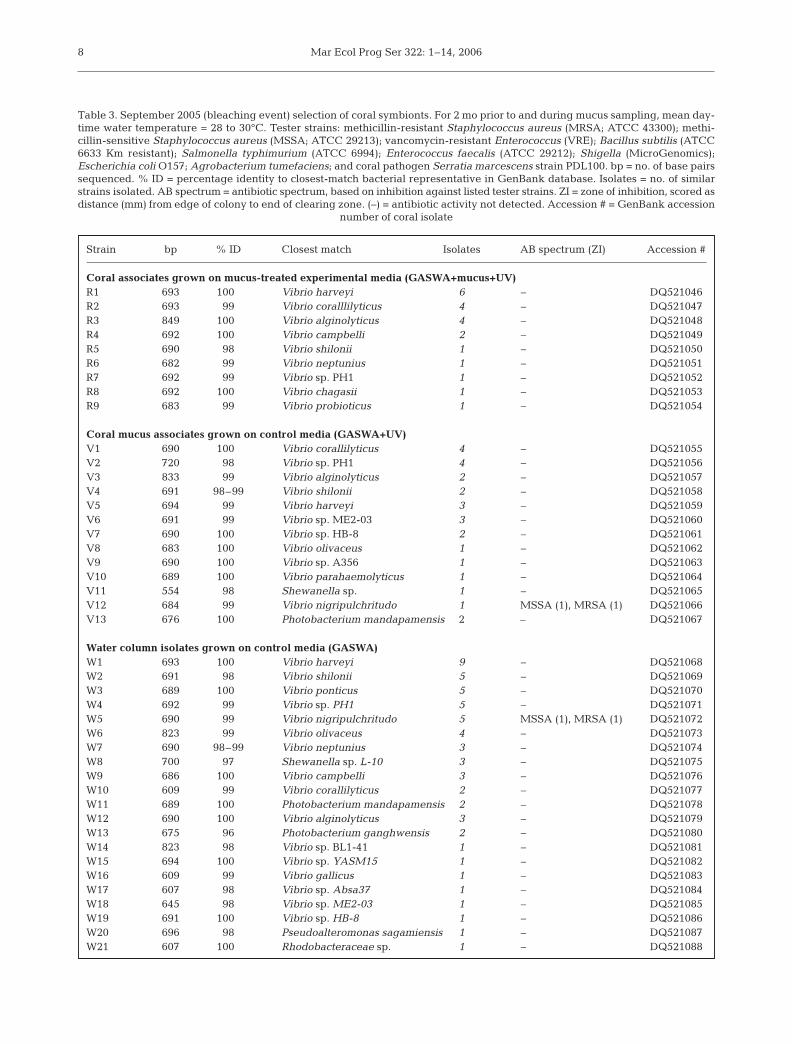
Selection of putative coral symbionts from appar-ently healthy areas of Acropora palmata during a latesummer bleaching event resulted in no growth inhibi-tion on mucus-treated media as compared to GASWAcontrol media (Table 1). Nevertheless, for the purposeof comparison to spring 2005 selections, 21 isolateswere chosen from mucus treated plates and were de-signated as putative coral residents. From the controlplates, 27 bacteria were isolated and designated asputative visitors. For comparison, 59 water column iso-lates were chosen from GASWA control plates. Resultsshow that A. palmata mucus collected during the Sep-tember 2005 bleaching event lost the ability to select adiscrete set of isolates (Table 3; Fig. 3). Genetic analy-sis based on partial sequencing of the 16S rRNA gene
5
Table 1. Acropora palmata mucus inhibition assays. Inhibition of tester strains and potential sources of invasive microbes testedby plating mucus (M) collected at two different times of the year (with differing mean daily water temperatures) onto glycerol ar-tificial seawater (GASWA) medium (G), followed by UV irradiation to inhibit out-growth of mucus-associated microbes. Variousdilutions or concentrations of inoculum were plated onto each treatment medium (see ‘Materials & methods’ for further details);dilutions producing between 200 and 400 colonies on control plates (G+UV) were compared to corresponding tester plates(G+M+UV). Numbers based on mean (±SD) of 4 plates for each experiment. Fold inhibition estimated by dividing experimental
mean into control mean. (–) = no data available for a particular source
Inoculum April 2005 September 2005No. of colonies Fold No. of colonies Fold
G+UV G+M+UV inhibition G+UV G+M+UV inhibition(ctrl) (expt) (ctrl) (expt)
Bacillus subtilis 395 (±22) 51 (±11) 7.8 – – –Staphylococcus aureus 245 (±40) 46 (±10) 5.4 – – –Salmonella typhimurium 217 (±34) 55 (±12) 3.9 – – –Serratia marcescens 193 (±33) 91 (±13) 2.1 233 (±26) 277 (±25) –0.8Water column 305 (±29) 76 (±7) 4.0 188 (±27) 231 (±14) –0.8Canal water 269 (±24) 27 (±9) 10.0 328 (±28) 274 (±20) 1.2African dust 278 (±51) 65 (±9) 4.3 206 (±24) 191 (±22) 1.1
Fig. 1. Selection scheme. Elkhorncoral Acropora palmata mucus dilu-tions (1:100) inoculated onto glycerolartificial seawater (GASWA) solidmedium treated with coral mucus(left) or GASWA control medium(right). Bacterial growth was inhib-ited 50- to 80-fold on GASWA me-dium treated with mucus collected
in spring 2005

Mar Ecol Prog Ser 322: 1–14, 20066
Table 2. Partial list of bacterial strains isolated from April 2005 selection for coral symbionts. For 2 mo prior to and during mucussampling, mean daytime water temperature = 22 to 25°C. Not all isolates initially tested for antibiotic activity are included, due toloss of viability during repeated sub-culturing and/or frozen storage. Tester strains: methicillin-resistant Staphylococcus aureus(MRSA; ATCC 43300); methicillin-sensitive Staphylococcus aureus (MSSA; ATCC 29213); vancomycin-resistant Enterococcus(VRE); Bacillus subtilis (ATCC 6633 Km resistant); Salmonella typhimurium (ATCC 6994); Enterococcus faecalis (ATCC 29212);Shigella (MicroGenomics); Escherichia coli O157; Agrobacterium tumefaciens; and coral pathogen Serratia marcescens strainPDL100. Residents: coral associates grown on mucus-treated experimental medium (GASWA+mucus+UV) as potential sym-bionts. Visitors: coral associates grown on control medium (GASWA+UV) as transient and/or potentially invasive microbes. Watercolumn bacteria: water column isolates grown on control medium (GASWA). bp = no. of base pairs sequenced. % ID = percentageidentity to closest-match bacterial representative in GenBank database. Isolates = no. of similar strains isolated. AB spectrum =antibiotic spectrum, based on inhibition against listed tester strains. ZI = zone of inhibition, scored as distance (mm) from edgeof colony to end of clearing zone. (–) = antibiotic activity not detected. Accession # = GenBank accession number of coral isolate
Strain bp % ID Closest match Isolates AB spectrum (ZI) Accession #
ResidentsR1ma 802 1000 Bacillus megaterium 1 B. subtilis (5) DQ530511R1mb 830 99 Agrobacterium tumefaciens 2 – DQ530512R2te 744 99 Photobacterium phosphoreum 2 – DQ530513R1m1 801 99 Photobacterium damselae 1 – DQ530514R1m2 682 99 Peligiobacter variabilis 1 – DQ530515R1m3 807 99 Photobacterium sp. YS27-3 2 – DQ530516R1t2 700 98 Alteromonas sp. 1 – DQ530517R1t3 789 99 Halomonas meridiana 1 – DQ530518R1t4 690 1000 Halomonas meridiana 2 B. subtilis (1.5) DQ530519R1t6 682 1000 Photobacterium mandapumensis 2 B. subtilis (5) DQ530520R1t9 770 99 Exiguonobacterium sp 1 S. marscesens (1.5) DQ530521R1m5 804 1000 Photobacterium mandapumensis 4 – DQ530522R2m1 825 99 Uncultured Alteromonas 1 S. marscesens (1.5) DQ530523R2m2 805 99 Photobacterium leiognathis 1 S. marscesens (1.5) DQ530524R2t1 831 99 Photobacterium damselae 3 S. marscesens (1.5) DQ530525R2t2 816 99 Photobacterium sp. YS27-3 2 S. marscesens (1.5) DQ530526R2t3 833 1000 Photobacterium sp YS27-3 3 B. subtilis (1.5), S. marscesens (1) DQ530527
VisitorsV1mt1 869 99 Staphylococcus sp. 2 – DQ530528V2mt2 811 99 Arctic sea ice bacterium 2 – DQ530529V2mt3 866 99 Agrobacterium sp. 2 – DQ530530VBR1 738 1000 Vibrio fortis 2 – DQ530531VBR2 720 1000 Vibrio shilonii 2 – DQ530532VBR5 806 99 Enterovibrio coralii 3 – DQ530533VBR6 787 98 Vibrio harveyii 5 – DQ530534VBR7 676 1000 Pseudoalteromonadaceae bacterium 1 B. subtilis (8), MRSA (6), DQ530535
MSSA (6), VRE (5), E. faecalis (5), S. typhimurium (7), Shigella (7), A. tumefaciens (6), E. coli (4.5), S. marscesens (4)
VBR8 835 1000 Pseudoalteromonadaceae bacterium 1 – DQ530536VBR10 807 1000 Vibrio olivaceus 3 – DQ530537VBR12 654 99 Vibrio nigripulchritudo 1 MRSA (1), MSSA (1) DQ530538VBR16 730 99 Pseudoalteromonas sp. 1 B. subtilis DQ530539VBR19 797 1000 Pseudovibrio/Alpha proteobacterium 1 MRSA (4) DQ530540VBR22 798 1000 Alpha proteobacterium Z143-1 2 B. subtilis (2.5), MRSA (6), DQ530541
MSSA (7), S. typhimurium (2),Shigella (5), A. tumefaciens (4)
VBR27 648 96 Vibrio hollisae 1 – DQ530542VAP1-8 726 99 Vibrio harveyii 1 B. subtilis (1), S. typhimurium (4) DQ530543VAP1-9 718 99 Vibrio shilonii 1 B. subtilis (1), S. typhimurium (4) DQ530544
Water column bacteriaW1 868 99 Staphylococcus saprophyticus 4 – DQ530545W2 745 99 Staphylococcus aureus 2 – DQ530546W3 837 99 Agrobacterium sp. 3 – DQ530547W4 652 99 Vibrio nigripulchritudo 3 – DQ530548W5 680 1000 Vibrio harveyii 2 – DQ530549W6 787 99 Alpha proteobacterium 3 – DQ530550W7 875 99 Pseudoalteromonas sp. 1 B. subtilis (3) DQ530551W8 573 99 Pseudoalteromonas sp. 2 – DQ530552W9 689 1000 Alpha proteobacterium 1 – DQ530553W10 401 99 Vibrio alginolyticus 1 – DQ530554W11 717 99 Kocuria sp. 1 – DQ530555W12 613 98 Photobacterium eurosenbergii 2 – DQ530556

Ritchie: Regulation of coral microbes
placed 100% of resident, 92% (22 of 24) of visitor and86% (49 of 58) of water-column isolates within thegenus Vibrio. No resident isolates displayed antibioticactivity. Only one isolate, most similar to V. nigri-pulchritudo and common to both the visitor pool andseawater, had antibiotic activity (Table 3).
Coral mucus regulates metabolic activities in acoral-associated bacterium
One visitor isolate (VBR7), from Acropora palmatamucus produced a dark purple, non-diffusible, pig-ment on control plates, but lost the capability toproduce the pigment when grown on A. palmatamucus-treated media (Fig. 4A). VBR7 also produced abroad-spectrum antibiotic (Table 2). Partial sequenc-ing of the 16S rRNA gene identified this bacterium as99% identical to Pseudoalteromonaceae bacterium(Table 2). Antibiotic tests were performed on thisisolate during pigment production and after pigmentloss. Fig. 4B shows isolate VBR7 producing pigment oncontrol media (left side, each panel) and after loss ofpigment upon growth on coral mucus (right side, each
panel). Each replica panel was overlaid with a differ-ent tester strain. The antibacterial compound associ-ated with VBR7 was active against all Gram-positiveand Gram-negative tester strains, with zones of inhibi-tion ranging from 3 to 8 mm. This indicated the pres-ence of a readily diffusible, broad-spectrum antibiotic.Loss of pigment and antibiotic activity in VBR7 wasalso demonstrated when VBR7 was grown in the pres-ence of mucus isolated from the gorgonian coral Pseu-dopterogorgia americana (collected from the FloridaKeys), and the star boulder coral Montastraea faveo-lata (collected from both the Florida Keys and FlowerGarden Banks).
DISCUSSION
While coral health appears to be declining worldwide, the Caribbean elkhorn coral Acropora palmatahas suffered the greatest losses, to the extent that it hasrecently been listed as a threatened species under theUS Endangered Species Act. A. palmata appears par-ticularly sensitive to stress and disease, although littleis known of its physiological response to stressors
7
ResidentsVisitorsPhotobacterium mandapumensis
Photobacterium leignathis*Photobacterium phosphoreum Photobacterium sp. YS27-3*Halomonas meridianaExiguobacterium sp.*Peligiobacter variabilisBacillus megateriumAlteromonas sp.*Photobacterium damselae*
Vibrio shiloniiEnterovibrio coraliiPseudomonas luteoviolaceaPseudoalteromonadaceae bacterium*Vibrio hollisaeVibrio fortisVibrio olivaceus
Photobacteriumdamselae
Agrobacterium tumefaciens Staphylococcus sp.
Vibrio nigripulchritudoVibrio harveyi Alpha proteobacterium Arctic sea ice bacterium
Water column
Kocuria sp.Vibrio chagassiPhotobacterium sp.Pseudoalteromonas sp.Vibrio alginolyiticusStaphylococcus aureusStaphylococcus saprophyticusPhotobacterium eurosenbergii
Fig. 2. Mucus selection scheme to enrich for coral symbionts (April 2005). Coral mucus sampled when mean daytime watertemperature = 24°C. Residents (top left): representative bacteria selected on mucus-treated medium (GASWA + mucus + UV) asputative coral symbionts. Visitors (top right): selected on control GASWA medium. Water column (bottom): selected from thewater column on control medium. Overlap = bacteria common to different treatments and sources. Bold: bacteria producingan antimicrobial compound (residents, 41% total; visitors, 16% total; not all representatives shown). *Bacteria producing
antibiotic activity against Serratia marcescens strain PDL110
Mar Ecol Prog Ser 322: 1–14, 20068
Table 3. September 2005 (bleaching event) selection of coral symbionts. For 2 mo prior to and during mucus sampling, mean day-time water temperature = 28 to 30°C. Tester strains: methicillin-resistant Staphylococcus aureus (MRSA; ATCC 43300); methi-cillin-sensitive Staphylococcus aureus (MSSA; ATCC 29213); vancomycin-resistant Enterococcus (VRE); Bacillus subtilis (ATCC6633 Km resistant); Salmonella typhimurium (ATCC 6994); Enterococcus faecalis (ATCC 29212); Shigella (MicroGenomics);Escherichia coli O157; Agrobacterium tumefaciens; and coral pathogen Serratia marcescens strain PDL100. bp = no. of base pairssequenced. % ID = percentage identity to closest-match bacterial representative in GenBank database. Isolates = no. of similarstrains isolated. AB spectrum = antibiotic spectrum, based on inhibition against listed tester strains. ZI = zone of inhibition, scored asdistance (mm) from edge of colony to end of clearing zone. (–) = antibiotic activity not detected. Accession # = GenBank accession
number of coral isolate
Strain bp % ID Closest match Isolates AB spectrum (ZI) Accession #
Coral associates grown on mucus-treated experimental media (GASWA+mucus+UV)R1 693 1000 Vibrio harveyi 6 – DQ521046R2 693 99 Vibrio coralllilyticus 4 – DQ521047R3 849 1000 Vibrio alginolyticus 4 – DQ521048R4 692 1000 Vibrio campbelli 2 – DQ521049R5 690 98 Vibrio shilonii 1 – DQ521050R6 682 99 Vibrio neptunius 1 – DQ521051R7 692 99 Vibrio sp. PH1 1 – DQ521052R8 692 1000 Vibrio chagasii 1 – DQ521053R9 683 99 Vibrio probioticus 1 – DQ521054
Coral mucus associates grown on control media (GASWA+UV)V1 690 1000 Vibrio corallilyticus 4 – DQ521055V2 720 98 Vibrio sp. PH1 4 – DQ521056V3 833 99 Vibrio alginolyticus 2 – DQ521057V4 691 98–99 Vibrio shilonii 2 – DQ521058V5 694 99 Vibrio harveyi 3 – DQ521059V6 691 99 Vibrio sp. ME2-03 3 – DQ521060V7 690 1000 Vibrio sp. HB-8 2 – DQ521061V8 683 1000 Vibrio olivaceus 1 – DQ521062V9 690 1000 Vibrio sp. A356 1 – DQ521063V10 689 1000 Vibrio parahaemolyticus 1 – DQ521064V11 554 98 Shewanella sp. 1 – DQ521065V12 684 99 Vibrio nigripulchritudo 1 MSSA (1), MRSA (1) DQ521066V13 676 1000 Photobacterium mandapamensis 2 – DQ521067
Water column isolates grown on control media (GASWA)W1 693 1000 Vibrio harveyi 9 – DQ521068W2 691 98 Vibrio shilonii 5 – DQ521069W3 689 1000 Vibrio ponticus 5 – DQ521070W4 692 99 Vibrio sp. PH1 5 – DQ521071W5 690 99 Vibrio nigripulchritudo 5 MSSA (1), MRSA (1) DQ521072W6 823 99 Vibrio olivaceus 4 – DQ521073W7 690 98–99 Vibrio neptunius 3 – DQ521074W8 700 97 Shewanella sp. L-10 3 – DQ521075W9 686 1000 Vibrio campbelli 3 – DQ521076W10 609 99 Vibrio corallilyticus 2 – DQ521077W11 689 1000 Photobacterium mandapamensis 2 – DQ521078W12 690 1000 Vibrio alginolyticus 3 – DQ521079W13 675 96 Photobacterium ganghwensis 2 – DQ521080W14 823 98 Vibrio sp. BL1-41 1 – DQ521081W15 694 1000 Vibrio sp. YASM15 1 – DQ521082W16 609 99 Vibrio gallicus 1 – DQ521083W17 607 98 Vibrio sp. Absa37 1 – DQ521084W18 645 98 Vibrio sp. ME2-03 1 – DQ521085W19 691 1000 Vibrio sp. HB-8 1 – DQ521086W20 696 98 Pseudoalteromonas sagamiensis 1 – DQ521087W21 607 1000 Rhodobacteraceae sp. 1 – DQ521088

Ritchie: Regulation of coral microbes
(Antonius 1977, 1981, Gladfelter et al. 1977, Bruckner2002, Precht 2004, Sutherland & Ritchie 2004). Somesuggested sources of coral decline include increasedsea surface temperatures, coastal degradation, pollu-tion, diseases, ecosystem imbalance caused by anthro-pogenic influences, and the synergistic effect of
multiple stressors (Harvell et al. 2002, Rosenberg &Ben-Haim 2002, Sutherland et al. 2004). One possibleexplanation for an increased incidence of coral dis-eases is stress-induced susceptibility to opportunisticmicrobes trapped in the coral mucus. Numeroussources of pathogenic microbes have been suggested
9
Mucus-treated medium Control medium
Vibri chagasiiVibrio probioticus
Vibrio spp.Vibrio parahaemolyticus
Vibrio campbeliiVibrio coralliilyticusVibrio alginolyticusVibrio shiloniiVibrio harveyiVibrio sp. PH1
Photobacterium mandapamensisShewanella sp.Vibrio nigripulchritudoVibrio olivaceusVibrio sp. ME2-03
Vibrio neptunius
Vibrio spp.Vibrio ponticusVibrio pectenicidaVibrio gallicusPhotobacterium sp.RhodobacteraceaePseudoalteromonas sagamiensis
Water column
Fig. 3. Mucus selection scheme (September, 2005). Coral mucus sampled when mean daytime water temperature = 30°C. Repre-sentative bacteria selected on mucus-treated medium (GASWA + mucus + UV; top left); control medium (GASWA + UV; topright); or water column bacteria isolated on control medium (GASWA; bottom). Note that applied selection scheme falls apart(cf. Fig. 2), with loss of diversity in all treatments and sources tested; high incidence of Vibrio spp., including numerous vibriosimplicated in coral bleaching and opportunistic diseases of marine organisms. Overlap = bacteria common to different treatments
and sources. Bold = bacteria producing an antibacterial compound
Fig. 4. Coral mucus inactivates metabolic properties of potentially invasive bacterium. (A) Putative ‘visitor’ bacterium VBR7 iso-lated from elkhorn coral Acropora palmata mucus produced a deep purple pigment when grown on GASWA control medium(right) but not when grown on medium treated with mucus collected from A. palmata (left), scleractinian coral Montastrea fave-olata, or gorgonian coral Pseudopterogorgia americana (not shown). (B) Each panel shows antibiotic activity of replicas of isolateVBR7 before (control medium; left side, each panel) and after pigment loss on mucus treated medium (right side, each panel).Tester strains used were Bacillus subtilis, methicillin-resistant Staphylococcus aureus (MRSA), methicillin-sensitive S. aureus
(MSSA), and Shigella. Arrow: MRSA growth inhibition zone produced by VBR7
Zone ofinhibition

Mar Ecol Prog Ser 322: 1–14, 2006
as possible threats to the health of corals in the FloridaKeys. These include canal water, which is a source ofhuman enteric bacteria such as Serratia marscesens(Patterson et al. 2002); African dust, hundreds of mil-lions of tons of which are transported to the Caribbeaneach year along with associated bacteria, fungi andviruses (Garrison et al. 2003); and water columnmicrobes that may become opportunistic under condi-tions of increased temperature and nutrient load.
Acropora palmata mucus as a biochemical defense
This study shows that mucus collected from Acrop-ora palmata has antibiotic activity against (1) Gram-positive and Gram-negative bacteria, (2) a number ofpotentially invasive microbes (including microbes fromFlorida Keys canal water, African dust, and surround-ing sea water), and (3) a pathogen implicated in whitepox disease of A. palmata. This result suggests thathealthy A. palmata employ a biochemical mechanismfor disease resistance that may act as a primary de-fense against pathogens. In contrast, mucus collectedfrom A. palmata during a period of increased watertemperature did not show significant antibiotic activityagainst the same suite of sources and tester strains,suggesting that the protective mechanism employedby A. palmata is lost when temperatures increase. Thisobservation suggests a mechanism to the hypothesisthat increased temperatures lower coral resistance,resulting in an increased susceptibility to disease.
Beneficial coral bacteria
Both culture-dependent and independent methodshave been used to understand microbial communitieson the surface of corals (reviewed in Brown & Bythell2005). Both methods show that corals favor specificpopulations of bacterial associates that are predictedto be mutualistic with the coral host (Ritchie & Smith1997, 2004, Rohwer et al. 2002). While culture-basedmethods are biased toward the small percentage ofmicrobial associates that are able to be cultivated inthe laboratory, culture-independent studies based on16S rDNA are limited in their prediction of compoundproduction and metabolic potential of coral-associatedbacteria. This study did not attempt to address totalmicrobial community structure of Acropora palmata,but focused on a subset of marine bacteria culturedunder specific growth conditions to investigate apotential bacterial contribution to the antibiotic activityseen in coral mucus.
Initial testing of culturable bacteria collected fromA. palmata mucus throughout the course of 2004
illustrated that roughly 20% displayed antibacterialactivities. This result suggests a potential role for coralbacterial associates in the production of mucus-associated antibiotic activity. In situ antibiotic produc-tion by associated bacteria is a means of securing aniche by controlling microbial populations competingfor the same resources (Neilson et al. 2000, Rao etal. 2005). The level of antibiotic contribution by thesebacteria in situ remains to be determined.
Selection for coral symbionts
Coral mucus traps particles and microbes that passby in the water column (Wild et al. 2004). Therefore,attempts at understanding the importance of coral-associated microbial communities may be misleadingdue to the fact that coral mucus is indiscriminate, re-taining microbes from a fluctuating water mass thatmay not be present in the water column at the time ofcollection. These dynamic fluctuations include coastalupwelling, countless local and regional influences, andlocal flora and fauna. Thus, many microbes trapped incoral mucus are less likely to be true ‘residents,’ ormutualists, but rather ‘visitors’ consisting of transientcommensal microorganisms that do no good or harm,or organisms that can potentially become opportunisticunder the right conditions.
The phylogenetic approaches used in most com-prehensive analyses of unculturable coral microbesprovide information on the identity of microbes pre-sent, but very little information relative to microbialinteractions, or information necessary for the elucida-tion of true coral residents (microbes beneficial to thecoral host, zooxanthellae, or other resident microbes).Here, a culture-based approach was developed toaddress this challenge by using sterile coral mucus as aselection medium for coral symbionts: bacteria thatbenefit from nutrients available on the coral surfacewhile providing a benefit to the coral in return. Thisapproach is based on the hypothesis that true coralsymbionts will be resistant to the antibiotic propertiesof Acropora palmata mucus, while many trapped bac-teria may be sensitive to the bacteriocidal, or bacterio-static, properties in coral mucus.
This selection scheme was first applied using Acro-pora palmata mucus collected in spring 2005 undertemperatures that were sustained at 22 to 24°C for2 mo prior to sampling. Bacteria isolated on mucus-treated plates as putative symbionts were designatedas coral ‘residents,’ while bacteria isolated from controlmedia were designated as potential ‘visitors,’ or tran-sient bacteria trapped from the water column. Fig. 2illustrates that the vast majority of bacteria cultured,using either mucus-treated selection media or control
10
Ritchie: Regulation of coral microbes
media, belonged to the γ-proteobacteria. Members ofthis group are abundant on corals and have been iden-tified using a range of methods, including culture-based (Ritchie & Smith 2004), molecular (Rohwer et al.2002), and fluorescence in situ hybridization and spec-tral imaging (Ainsworth et al. 2006). Therefore, I thinkthat this subset of culturable associates represents avalid group for the study of symbiotic interactions.
Results show that the mucus-based selection methodenriched for Photobacterium spp. (60%), Halomonasspp. (10%), and a range of bacterial species that haveantibiotic activities (Table 2, Fig. 2). The significance ofPhotobacterium and Halomonas spp. enrichment is notclear, although a subset of each is shown to produceantibiotics (Table 2). It is possible that these bacteriaadditionally play a role in the health of the coral holo-biont via production of vitamins or cofactors necessaryfor the growth of beneficial bacteria, or by providingother regulatory compounds. In addition, some strainsdesignated as coral residents were active against theSerratia marcescens PDL100 coral disease pathogen.That this method enriched for bacteria that produceantibiotics provides additional support for a bacterialcontribution to the antibiotic activity of coral mucuswhile providing a novel method for the enrichment ofmarine bioactive compounds.
Bacteria isolated as visitors are representative ofbacteria documented in earlier studies, including asubset of Vibrio species consistently found in associa-tion with healthy corals (Ritchie & Smith 1995a,b,2004). As partial sequencing of the 16S gene is notsufficient for a thorough identification of members ofthe genus Vibrio, these isolates will require furthergenetic delineation. Table 2 shows that there is ahigher percentage of vibrios (48%) when comparingcontrol plates to the coral mucus selection scheme (novibrios). A subset of vibrios isolated from control plates(visitor microbes Vibrio shilonii and Enterovibrio sp.)are most similar to species associated with coralbleaching in the Mediterranean Sea and in the GreatBarrier Reef (Kushmaro et al. 2001, Thompson et al.2005), suggesting that these bacterial species are ubi-quitous in tropical oceans. Several visitor and watercolumn isolates produced antibiotics. Isolate VBR7produced a broad spectrum antibiotic with large zonesof inhibition and isolate VAP1-9, which displayed 99%identity to Vibrio shilonii, exhibited antibiotic activityagainst 2 Gram-positive tester strains. These isolatesmay represent bacteria capable of becoming oppor-tunistic under conditions of coral stress.
Collectively, these results suggest that coral mucusprovides a hostile environment for some bacteria and anurturing environment for others, illustrating that themucus plays an important role in structuring microbialcommunities on the coral surface. The use of coral
mucus as a selection medium is experimental. It is pos-sible that UV sterilization alters the composition ofthe mucus in a manner not addressed by the UV con-trols. However, the results using UV irradiated sum-mer mucus, described below, argue against significantalteration of the mucus by UV treatment.
Loss of antibiotic activity and shift to Vibrio spp.during a summer bleaching event
The antibiotic properties of coral mucus, and thepotential for mucus to select a discrete set of com-mensal bacteria, were lost at increased temperaturesduring a bleaching event (Table 3; Fig. 3). Mucus wastaken from corals sustained at a mean daytime sea sur-face temperature of 28 to 30°C for 2 mo prior tocollection. Vibrios were the predominant species cul-tured from the mucus of apparently healthy Acroporapalmata tissue during this event. Vibrios were alsopredominant in the water column during this period,representing 85% of the cultured isolates (Table 3).Less than 2% of bacteria isolated from the surface ofA. palmata during this period produced antibioticactivity. These findings illustrate a temporal shift in theprotective qualities of coral mucus, and a compositionshift from beneficial bacteria to vibrio dominanceunder conditions of increased temperature. Vibriospresent during this event included those involved intemperature dependent bleaching of corals, such asVibrio shilonii (=V. shiloi; Kushmaro et al. 2001) and V.coralliilyticus (= V. coralyticus; Ben-Haim et al. 2003)as well as numerous vibrios known to be opportunisticto other marine organisms (Table 3). These findingsare consistent with a shift of the equilibrium betweenPseudomonas spp. and Vibrio spp. in healthy corals, tovibrio dominance when corals are bleached (Ritchie &Smith 1995a, 2004). However, as mucus was collectedfrom apparently healthy coral tissue, and not bleachedtissue, this provides evidence that a community shift tovibrio dominance may occur prior to zooxanthellae loss.
During bleaching, coral mucus production changesin quality and can decrease in quantity, depending onthe cellular damage caused by the environmental fac-tor(s) that initiated bleaching (Lasker et al. 1984, Glynnet al. 1986). Thus, microbes that depend on particularsubstrates in healthy mucus may be reduced in num-ber when these substrates are no longer available. Onemodel that addresses coral bleaching and diseasesusceptibility is that antimicrobial properties of coralmucus are compromised by temperature-dependentalteration of resident microbes, or other factors influ-encing antibiotic activity, followed by an overgrowthof vibrios, which are abundant in the water columnduring warmer months. A similar model is that bene-
11

Mar Ecol Prog Ser 322: 1–14, 2006
ficial microbes are simply out-competed by vibrios,many of which thrive under conditions of increasedtemperature (Lipp et al. 2002, Rosenberg & Ben-Haim2002, Thompson et al. 2005). Both models (Fig. 5) aresupported by observations in this study and others.Kuntz et al. (2005) reported that growth stimulation ofbacterial communities present on corals may directlyresult in coral mortality. In addition, it has been shownthat 4 vibrio strains, inoculated as a group, were able tocause signs of yellow band disease when temperatureswere increased (Cervino et al. 2004).
Another possible mechanism that supports bothmodels is cooperation among similar bacteria (Foster2005). This theory is distinct from a one-pathogenmodel for coral bleaching by Vibrio species (Kushmaroet al. 2001, Ben-Haim et al. 2003), in that it is based ona bacterial version of kin-selection theory, where simi-lar bacteria are able to co-operate to secure a nichebecause they share like genes (Foster 2005). This mayalso apply to vibrios associated with corals. In this sce-nario, vibrios may compete for space on the coral sur-face, reducing relatedness among beneficial surfacebacteria by sharing similar gene products involved insecuring a niche (such as feeding enzymes and viru-lence factors, among others). The result is the swamp-ing of resident beneficial microbes, the initiation ofcoral bleaching and, perhaps ultimately, an increase indisease susceptibility.
Regardless of the precise ordering of events, duringtimes of increased sea surface temperature, corals are
more susceptible to disease (Rosenberg & Ben-Haim2002), which is demonstrated in this study to be corre-lated with a loss of a protective function provided inthe coral mucus and an increase in the number ofvibrios, both in the water column and on the surface ofapparently healthy coral tissue. These observationsmay offer further clarification for the increase in coraldisease incidence that occurs following bleachingevents (Causey 2001, Harvell et al. 2002, Weil et al.2006).
Undocumented function of coral mucus
While studying the interaction of Acropora palmatamucus with potentially invasive microbes, it was notedthat one visitor isolate (VBR7), a Pseudoalteromona-ceae bacterium isolated from A. palmata mucus, pro-duced a dark purple, non-diffusible pigment on controlplates, but lost the ability to produce the pigment whengrown on A. palmata mucus-treated media (Fig. 4a).This isolate was initially shown to produce a power-ful, broad-spectrum antibiotic (Table 2). As pigmentproduction is often correlated with antibiotic activity(Ritchie, pers. obs.), antibiotic tests were performed onthis isolate both during pigment production and afterpigment loss. Results showed that VBR7 produced abroad-spectrum, readily diffusible antibiotic whengrown on control media, during pigment expression.Conversely, antibiotic activity was absent when thepigment was lost in this strain. Loss of pigmentproduction and antibiotic activity in VBR7 was alsodemonstrated when VBR7 was grown in the presenceof A. palmata mucus collected during summer months,mucus isolated from the gorgonian coral Pseudoptero-gorgia americana (collected from the Florida Keys),and mucus from the star boulder coral Montastreafaveolata (collected from both the Florida Keys andFlower Garden Banks). This result illustrates that acomponent universal to coral mucus, independent ofspecies, location, and season, is capable of inhibitingpigment and antibiotic production associated withVBR7. The production of cell signaling molecules bymany microbes regulates bacterial processes in a pop-ulation density-dependent manner (Miller & Bassler2001, Teplitski et al. 2004). This type of communica-tion, called ‘quorum sensing,’ is common in bacterialbiofilms and regulates processes such as adhesion,antibiotic production, and virulence (Miller & Bassler2001). Quorum sensing molecules, if present in coralmucus, could result in the pigment and antibiotic sup-pression seen in VBR7. Although further studies willreveal whether these regulatory processes within themucus are attributed to the coral, zooxanthellae, orcoral associated bacteria, the regulation of bacterial
12
Model 1 Model 2
Fig. 5. Models of bacterial overgrowth on coral surfaces. InModel 1, rising water temperature compromises antibioticproduction by the coral holobiont, either by affecting anti-biotic production by the coral or by affecting compositionand/or activity of resident microbiota. This results in oppor-tunistic overgrowth of transient microbes in coral mucus,increasing the probability of bleaching or disease. In Model 2,overgrowth by transient microbes out-competes the residentpopulation of microbiota, with subsequent loss of antibioticactivity associated with coral mucus. This model requiresadditional evidence that resident coral-associated microbescontribute significantly to antibiotic activity described for
coral mucus
Increased temperature
Vibrio overgrowth
Vibrio overgrowth
Loss of antibiotic properties
Loss of antibiotic properties

Ritchie: Regulation of coral microbes
gene expression by mucus is likely to play a signifi-cant role in both the determination of microbial com-munity structure and in the establishment of patho-genic bacteria.
CONCLUSIONS
In this study, Acropora palmata mucus was shownto have antibiotic properties that are likely to play arole in ordering beneficial microbial communities onthe coral surface. In addition, mucus from healthy A.palmata harbors bacteria capable of producing antibi-otics, implicating a microbial contribution to the pro-tective properties of coral mucus. Coral mucus inacti-vated pigment production and antibiotic activity in amucus-associated bacterium, illustrating an undocu-mented role of coral mucus in the control of associatedmicrobes. Mucus-associated antibiotic activity wasreduced when mucus was collected during a period ofincreased sea surface temperature. This finding sug-gests a seasonal variability in the protective qualities ofcoral mucus that may result in increased susceptibilityto bleaching and disease. Beneficial coral bacteria werereplaced by Vibrio spp. during this event, which re-sulted in a loss of antibiotic producing bacteria on coralsurfaces. Future studies will reveal temporal andenvironmental changes on the coral surface, and theprecise role of coral mucus and beneficial microbesin coral health and disease susceptibility.
Acknowledgements. This work was funded by NOAA grantsNCND6012-5-00033, NCND60111-5-00013, the Edith andCurtis Munson Foundation, and Mote Marine Laboratory.Collections were obtained via permit FKNMS 2004-015(Florida Keys National Marine Sanctuary) and FGBNMSpermit 2005-006 (Flower Garden Banks National MarineSanctuary). I thank E. Bartels and staff of the FKNMS for boatand sampling support, V. Palubok for laboratory support,V. Garrison (USGS) for kindly supplying Mali desert soil, andV. Dartois (Microgenomics) for providing tester strains. I ammost grateful to B. Causey, S. Gittings, M. Hay, B. Keller,E. Peters, J. Pringle, K. Sharpe, G. Smith, M. Teplitski, J.Thurmond and 4 anonymous reviewers for helpful discussionsand/or improvements to the manuscript.
LITERATURE CITED
Ainsworth TD, Fine M, Blackall LL, Hoegh-Guldberg O(2006) Fluorescence in situ hybridization and spectralimaging of coral-associated bacterial communities. ApplEnviron Microb 72:3016–3020
Altschul SF, Madden TL, Schäffer AA, Zhang J, Zhang Z,Miller W, Lipman DJ (1997) Gapped BLAST and PSI-BLAST: a new generation of protein database searchprograms. Nucleic Acids Res 25:3389–3402
Antonius A (1977) Coral mortalities in reefs: a problem forscience and management. Proc 3rd Int Coral Reef Symp1:617–623
Antonius A (1981) Coral reef pathology: a review. Proc 4th IntCoral Reef Symp 2:3–6
Armstrong E, Yan L, Boyd KG, Wright PC, Burgess JG (2001)The symbiotic role of marine microbes on living surfaces.Hydrobiologia 461:37–40
Ben-Haim Y, Thompson FL, Thompson CC, Cnockaert MC,Hoste B, Swings J, Rosenberg E (2003) Vibrio corallii-lyticus sp. nov., a temperature-dependent pathogen ofthe coral Pocillopora damicornis. Int J Syst Evol Micr 53:309–315
Brown BE, JC Bythell (2005) Perspectives on mucus secretionin reef corals. Mar Ecol Prog Ser 296:291–309
Bruckner AW (2002) Priorities for effective management ofcoral reefs. NOAA Tech Memo NMF-OPR-22 August 2002.Available at:www.icriforum.org/docs/man_priorities_coral_diseases.pdf
Burkholder PR, Burkholder LM (1958) Antimicrobial activityof horny corals. Science 127:1174–1175
Castillo I, Lodeiros C, Nunez, M, Campos I (2001) In vitroevaluation of antibacterial substances produced by bac-teria isolated from different marine organisms. Rev BiolTrop 49:1213–1222
Causey BD (2001) Lessons learned from the intensification ofcoral bleaching from 1980–2000 in the Florida Keys, USA.In: Salm RV, Coles SL (eds) Coral bleaching and marineprotected areas: Proc Workshop ‘Mitigating coral bleach-ing impact through MPA design’ (Bishop Museum, Hono-lulu, HI). Asia Pacific Coastal Marine Program Report# 0102, The Nature Conservancy. Available at: www.conserveonline.org/docs/2001/10/CoralBleechingMPAs-Workshop.pdf
Cervino JM, Hayes RL, Polson SW, Polson SC, Goreau TJ,Martinez RJ, Smith GW (2004) Relationship of Vibriospecies infection and elevated temperatures to yellowblotch/band disease in Caribbean corals. Appl EnvironMicrobiol 70:6855–6864
Elyakov GB, Kuznetsova T, Mikhailov VV, Maltsev II, VoinovVG, Fedoreyev SA (1991) Brominated biphenyl ethers froma marine bacterium associated with the sponge Dysideasp. Experientia 47:632–633
Foster KR (2005) Hamiltonian medicine: why the social livesof bacteria matter. Science 308:1269–1270
Garrison VH, Shinn EA, Foreman WT, Griffin RD and 6 others(2003) African and Asian dust: from desert soil to coralreef. Bioscience 53:469–480
Geffen Y, Rosenberg E (2005) Stress-induced rapid releaseof antibacterials by scleractinian corals. Mar Biol 146:931–935
Gil-Turnes MS, Hay ME, Fenical W (1989) Symbiotic marinebacteria chemically defend crustacean embryos from apathogenic fungus. Science 246:116–118
Gladfelter WB, Gladfelter EH, Monahan RK, Ogden JC, DillRF (1977) Coral destruction: environmental studies ofBuck Island Reef National Monument. Biosphere ReserveRes Rep No. 6, US National Park Service, US Dept of Inte-rior, Washington, DC
Glynn PW, Peters EC, Muscatine L (1986) Coral tissue micro-structure and necrosis: relation to catastrophic coralmortality in Panama. Dis Aquat Org 1:29–37
Harvell CD, Kim K, Burkholder JM, Colwell RR and 7 others(1999) Emerging marine diseases—climate links andanthropogenic factors. Science 285:1505–1510
Harvell CD, Mitchell CE, Ward JR, Altizer S, Dobson AP,Ostfeld RS, Samuel MD (2002) Climate warming anddisease risks for terrestrial and marine biota. Science 296:2158–2162
Holmstrom C, Rittschof D, Kjelleberg S (1992) Inhibition of
13

Mar Ecol Prog Ser 322: 1–14, 2006
settlement by larvae of Balanus amphitrite and Cionaintestinalis by a surface-colonizing marine bacterium. ApplEnviron Microb 58:2111–2115
Kelman D, Kushmaro A, Loya Y, Kashman Y, Benayahu Y(1998) Antimicrobial activity of a Red Sea coral, Parery-thropodium fulvum fulvum: reproductive and develop-mental considerations. Mar Ecol Prog Ser 169:87–95
Kelman D, Kashman Y, Rosenberg E, Kushmaro A, Loya Y(2006) Antimicrobial activity of Red Sea corals. Mar Biol149:357–363
Kim K (1994) Antimicrobial activity in gorgonian corals(Coelenterata, Octocorallia). Coral Reefs 13:75–80
Koh EG (1997) Do scleractinian corals engage in chemicalwarfare against microbes? J Chem Ecol 23:379–398
Kuntz NM, Kline DI, Sandin SA, Rohwer F (2005) Pathologiesand mortality rates caused by organic carbon and nutrientstressors in three Caribbean coral species. Mar Ecol ProgSer 294:173–180
Kushmaro A, Banin E, Loya Y, Stackebrandt E, Rosenberg E(2001) Vibrio shiloi sp. nov., the causative agent ofbleaching of the coral Oculina patagonica. Int J SystemEvol Microbiol 51:1383–1388
Lasker HR, Peters EC, Coffroth MA (1984) Bleaching of reefcoelenterates in the San Blas Islands, Panama. Coral Reefs3:183–190
Lipp E, Huq A, and Colwell RR (2002) Effects of global climateon infectious disease: the Cholera Model. Clin MicrobiolRev 15:757–770
Miller MB, Bassler BL (2001) Quorum sensing in bacteria.Annu Rev Microbiol 55:165–199
Neilson AT, Tolker-Neilsen K, Barken K, Molin S (2000) Roleof commensal relationships on the spatial structure of asurface-attached microbial consortium. Environ Microbiol2:59–68
Patterson KL, Porter JW, Ritchie KB, Smith GW, Polson SW(2002) Etiology of white pox, a lethal disease of theCaribbean elkhorn coral, Acropora palmata. Proc NatlAcad Sci USA 99:8725–8730
Precht WF, Robbart ML, Aronson RB (2004) The potential list-ing of Acropora species under the US Endangered SpeciesAct. Mar Poll Bull 49:534–536
Rao D, Webb JS, Kjelleberg S (2005) Competitive interactionsin mixed-species biofilms containing the marine bac-terium Pseudoalteromonas tunicata. Appl Environ Microb71:1729–1736
Ritchie KB, Smith GW (1995a) Preferential carbon utilizationby surface bacterial communities from water mass, nor-mal, and white-band diseased Acropora cervicornis. MolMar Biol Biotech 4:345–352
Ritchie KB, Smith GW (1995b) Carbon-source utilizationpatterns of coral-associated marine heterotrophs. J MarBiotechnol 3:105–107
Ritchie KB, Smith GW (1997) Physiological comparisons ofbacteria from various species of scleractinian corals. Proc8th Int Symp Reef Studies 1:521–526
Ritchie KB, Smith GW (2004) Microbial communities of coralsurface mucopolysaccharide layers. In: Rosenberg E, LoyaY (eds) Coral health and disease. Springer-Verlag, Berlin,p 259–263
Rohwer F, Seguritan V, Azam F, Knowlton N (2002) Diversityand distribution of coral-associated bacteria. Mar EcolProg Ser 243:1–10
Rosenberg E, Ben-Haim Y (2002) Microbial diseases of coralsand global warming. Environ Microbiol 4:318–326
Slattery M, McClintock JB, Heine JN (1995) Chemicaldefenses in Antarctic soft corals: evidence for anti-foulingcompounds. J Exp Mar Biol Ecol 190:61–77
Slattery M, Hamann MT, McClintock JB, Perry TL, PuglisiMP, Yoshida WY (1997) Ecological roles for water-bornemetabolites from Antactic soft corals. Mar Ecol Prog Ser161:133–144
Smith GW, Hayasaka SS (1982) Nitrogenase activity associ-ated with Halodule wrightii roots. Appl Environ Microb43:1244–1248
Sutherland KP, Porter JP, Torres C (2004) Disease and immu-nity in Caribbean and Indo-Pacific zooxanthellate coralsMar Ecol Prog Ser 266:273–302
Sutherland KP, Ritchie KB (2004) White pox disease of theCaribbean elkhorn coral, Acropora palmata. In: Rosen-berg E, Loya Y (eds) Coral health and disease. Springer-Verlag, Berlin, p 289–297
Teplitski M, Chen H, Rajamani S, Gao M and 5 others (2004)Chlamydomonas reinhardtii secretes compounds thatmimic bacterial signals and interfere with quorum sensingregulation in bacteria. Plant Physiol 134:137–146
Thompson FL, Thompson CC, Naser S, Hoste B, Vandemeule-broecke K, Munn C, Bourne D, Swings J (2005) Photo-bacterium rosenbergii sp nov. and Enterovibrio coraliisp. nov., vibrios associated with coral bleaching. Int J SystEvol Micr 55:913–917
Weidner S, Arnold W, Pühler A (1996) Diversity of unculturedmicroorganisms associated with the seagrass Halophilastipulacea estimated by restriction fragment length poly-morphism analysis of PCR-amplified 16S rRNA genes.Appl Environ Microb 62:766–771
Weil E, Smith GW, Gil-Agudelo DL (2006) Status and pro-gress in coral reef disease research. Dis Aquat Org 69:1–7
Wild C, Huettel M, Klueter A, Kremb SG, Rasheed MYM,Jørgensen B (2004) Coral mucus functions as an energycarrier and particle trap in the reef ecosystem. Nature 428:66–70
Yasumoto T, Yasumura M, Yotsu M, Michishita T, Endo A,Kotaki T (1986) Bacterial production of tetrodotoxin andanhydrotetrodotoxin Agr Biol Chem 50:793–795
14
Editorial responsibility: Charles Birkeland (ContributingEditor), Honolulu, Hawaii, USA
Submitted: May 7, 2006; Accepted: July 18, 2006Proofs received from author(s): September 5, 2006