Regulation of Hormonal Control, Cell Reprogramming, and ... · Body regeneration through formation...
19
Regulation of Hormonal Control, Cell Reprogramming, and Patterning during De Novo Root Organogenesis 1[OPEN] Estefano Bustillo-Avendaño, a,2 Sergio Ibáñez, b,2 Oscar Sanz, a Jessica Aline Sousa Barros , b,4 Inmaculada Gude, a Juan Perianez-Rodriguez, a José Luis Micol, b Juan Carlos Del Pozo, a Miguel Angel Moreno-Risueno , a,3,5 and José Manuel Pérez-Pérez b,3,5 a Centro de Biotecnología y Genómica de Plantas, Instituto Nacional de Investigación y Tecnología Agraria y Alimentaria, Universidad Politécnica de Madrid, Madrid, Spain and b Instituto de Bioingeniería, Universidad Miguel Hernández, 03202 Elche, Spain ORCID IDs: 0000-0002-9794-1450 (M.A.M.-R.); 0000-0003-2848-4919 (J.M.P.-P.). Body regeneration through formation of new organs is a major question in developmental biology. We investigated de novo root formation using whole leaves of Arabidopsis (Arabidopsis thaliana). Our results show that local cytokinin biosynthesis and auxin biosynthesis in the leaf blade followed by auxin long-distance transport to the petiole leads to proliferation of J0121-marked xylem-associated tissues and others through signaling of INDOLE-3-ACETIC ACID INDUCIBLE28 (IAA28), CRANE (IAA18), WOODEN LEG, and ARABIDOPSIS RESPONSE REGULATORS1 (ARR1), ARR10, and ARR12. Vasculature proliferation also involves the cell cycle regulator KIP-RELATED PROTEIN2 and ABERRANT LATERAL ROOT FORMATION4, resulting in a mass of cells with rooting competence that resembles callus formation. Endogenous callus formation precedes specification of postembryonic root founder cells, from which roots are initiated through the activity of SHORT-ROOT, PLETHORA1 (PLT1), and PLT2. Primordia initiation is blocked in shr plt1 plt2 mutant. Stem cell regulators SCHIZORIZA, JACKDAW, BLUEJAY, and SCARECROW also participate in root initiation and are required to pattern the new organ, as mutants show disorganized and reduced number of layers and tissue initials resulting in reduced rooting. Our work provides an organ regeneration model through de novo root formation, stating key stages and the primary pathways involved. Plants have striking regeneration capacities, and can produce new organs from postembryonic tissues (Hartmann et al., 2010; Chen et al., 2014; Liu et al., 2014) as well as reconstitute damaged organs upon wound- ing (Xu et al., 2006; Heyman et al., 2013; Perianez- Rodriguez et al., 2014; Melnyk et al., 2015; Efroni et al., 2016). Intriguingly, root regeneration upon stem cell damage recruits embryonic pathways (Hayashi et al., 2006; Efroni et al., 2016), whereas in contrast, postembryonic formation of whole new organs, such as lateral roots, appears to use specific postembryonic pathways (Lavenus et al., 2013). Cross talk between auxin and cytokinin signaling is required for many aspects of plant development and regeneration (El-Showk et al., 2013), although how their synergistic interaction is implemented at the molecular level has not been clarified (Skoog and Miller, 1957; Chandler and Werr, 2015). Exogenous in vitro supple- mentation of these two hormones results in continuous cell proliferation, to form a characteristic structure termed “callus”. Callus emerges as a common regen- erative mechanism for almost all plant organs through in vitro culture (Atta et al., 2009; Sugimoto et al., 2010). There is increasing evidence that callus formation re- quires hormone-mediated activation of a lateral and meristematic root development program in pericycle-like cells defined by expression of the J0121 marker 1 This work was supported by grants from Ministerio de Economía y Competitividad (MINECO) of Spain, the European Regional Devel- opment Fund (ERDF) and FP7 Funds of the European Commission, BFU2013-41160-P, BFU2016-80315-P, and PCIG11-GA-2012-322082 to M.A.M.-R., AGL2012-33610 and BIO2015-64255-R to J.M.P.-P., and BIO2014-52091-R to J.C.P. M.A.M.-R. was supported by a Ramon y Cajal contract from MICINN. 2 These authors contributed equally to this article. 3 These authors contributed equally to this article. 4 Current address: Departamento de Biologia Vegetal, Universi- dade Federal de Viçosa, 36570-900 Viçosa, Minas Gerais, Brazil. 5 Address correspondence to [email protected] and [email protected]. The author responsible for distribution of materials integral to the findings presented in this article in accordance with the policy de- scribed in the Instructions for Authors (www.plantphysiol.org) is: José Manuel Pérez-Pérez ([email protected]). J.M.P.-P. and M.A.M.-R. were responsible for conceptualization and supervision; J.M.P.-P., M.A.M.-R., E.B.-A., and S.I. were respon- sible for methodology; E.B.-A., S.I., O.S., J.A.S.B., I.G., and J.P.-R. were responsible for investigation; E.B.-A. and S.I. were responsible for formal analysis; J.M.P.-P., M.A.M.-R., E.B.-A. and S.I. were re- sponsible for writing the original draft; J.M.P.-P., M.A.M.-R., J.L.M., and J.C.D.P. were responsible for review and editing of the manu- script; J.M.P.-P. and M.A.M.-R. were responsible for acquiring re- sources; J.M.P.-P., M.A.M.-R., J.L.M., and J.C.D.P. were responsible for acquisition of funding. [OPEN] Articles can be viewed without a subscription. www.plantphysiol.org/cgi/doi/10.1104/pp.17.00980 Plant Physiology Ò , February 2018, Vol. 176, pp. 1709–1727, www.plantphysiol.org Ó 2018 American Society of Plant Biologists. All Rights Reserved. 1709 www.plantphysiol.org on July 27, 2018 - Published by Downloaded from Copyright © 2018 American Society of Plant Biologists. All rights reserved.
-
Upload
trinhnguyet -
Category
Documents
-
view
219 -
download
0
Transcript of Regulation of Hormonal Control, Cell Reprogramming, and ... · Body regeneration through formation...
Regulation of Hormonal Control, CellReprogramming, and Patterning during DeNovo Root Organogenesis1[OPEN]
Estefano Bustillo-Avendaño,a,2 Sergio Ibáñez,b,2 Oscar Sanz,a Jessica Aline Sousa Barros ,b,4
Inmaculada Gude,a Juan Perianez-Rodriguez,a José Luis Micol,b Juan Carlos Del Pozo,a
Miguel Angel Moreno-Risueno,a,3,5 and José Manuel Pérez-Pérezb,3,5
aCentro de Biotecnología y Genómica de Plantas, Instituto Nacional de Investigación y Tecnología Agraria yAlimentaria, Universidad Politécnica de Madrid, Madrid, Spain andbInstituto de Bioingeniería, Universidad Miguel Hernández, 03202 Elche, Spain
ORCID IDs: 0000-0002-9794-1450 (M.A.M.-R.); 0000-0003-2848-4919 (J.M.P.-P.).
Body regeneration through formation of new organs is a major question in developmental biology. We investigated de novo rootformation using whole leaves of Arabidopsis (Arabidopsis thaliana). Our results show that local cytokinin biosynthesis and auxinbiosynthesis in the leaf blade followed by auxin long-distance transport to the petiole leads to proliferation of J0121-markedxylem-associated tissues and others through signaling of INDOLE-3-ACETIC ACID INDUCIBLE28 (IAA28), CRANE (IAA18),WOODEN LEG, and ARABIDOPSIS RESPONSE REGULATORS1 (ARR1), ARR10, and ARR12. Vasculature proliferation alsoinvolves the cell cycle regulator KIP-RELATED PROTEIN2 and ABERRANT LATERAL ROOT FORMATION4, resulting in amass of cells with rooting competence that resembles callus formation. Endogenous callus formation precedes specification ofpostembryonic root founder cells, from which roots are initiated through the activity of SHORT-ROOT, PLETHORA1 (PLT1),and PLT2. Primordia initiation is blocked in shr plt1 plt2 mutant. Stem cell regulators SCHIZORIZA, JACKDAW, BLUEJAY, andSCARECROW also participate in root initiation and are required to pattern the new organ, as mutants show disorganized andreduced number of layers and tissue initials resulting in reduced rooting. Our work provides an organ regeneration modelthrough de novo root formation, stating key stages and the primary pathways involved.
Plants have striking regeneration capacities, andcan produce new organs from postembryonic tissues(Hartmann et al., 2010; Chen et al., 2014; Liu et al., 2014)as well as reconstitute damaged organs upon wound-ing (Xu et al., 2006; Heyman et al., 2013; Perianez-Rodriguez et al., 2014; Melnyk et al., 2015; Efroniet al., 2016). Intriguingly, root regeneration upon stemcell damage recruits embryonic pathways (Hayashiet al., 2006; Efroni et al., 2016), whereas in contrast,postembryonic formation of whole new organs, such aslateral roots, appears to use specific postembryonicpathways (Lavenus et al., 2013).
Cross talk between auxin and cytokinin signaling isrequired for many aspects of plant development andregeneration (El-Showk et al., 2013), although how theirsynergistic interaction is implemented at the molecularlevel has not been clarified (Skoog and Miller, 1957;Chandler and Werr, 2015). Exogenous in vitro supple-mentation of these two hormones results in continuouscell proliferation, to form a characteristic structuretermed “callus”. Callus emerges as a common regen-erative mechanism for almost all plant organs throughin vitro culture (Atta et al., 2009; Sugimoto et al., 2010).There is increasing evidence that callus formation re-quires hormone-mediated activation of a lateral andmeristematic root development program inpericycle-likecells defined by expression of the J0121 marker
1 This workwas supported by grants fromMinisterio de Economíay Competitividad (MINECO) of Spain, the European Regional Devel-opment Fund (ERDF) and FP7 Funds of the European Commission,BFU2013-41160-P, BFU2016-80315-P, and PCIG11-GA-2012-322082 toM.A.M.-R., AGL2012-33610 and BIO2015-64255-R to J.M.P.-P., andBIO2014-52091-R to J.C.P. M.A.M.-R. was supported by a Ramon yCajal contract from MICINN.
2 These authors contributed equally to this article.3 These authors contributed equally to this article.4 Current address: Departamento de Biologia Vegetal, Universi-
dade Federal de Viçosa, 36570-900 Viçosa, Minas Gerais, Brazil.5 Address correspondence to [email protected] and
[email protected] author responsible for distribution of materials integral to the
findings presented in this article in accordance with the policy de-scribed in the Instructions for Authors (www.plantphysiol.org) is:José Manuel Pérez-Pérez ([email protected]).
J.M.P.-P. and M.A.M.-R. were responsible for conceptualizationand supervision; J.M.P.-P., M.A.M.-R., E.B.-A., and S.I. were respon-sible for methodology; E.B.-A., S.I., O.S., J.A.S.B., I.G., and J.P.-R.were responsible for investigation; E.B.-A. and S.I. were responsiblefor formal analysis; J.M.P.-P., M.A.M.-R., E.B.-A. and S.I. were re-sponsible for writing the original draft; J.M.P.-P., M.A.M.-R., J.L.M.,and J.C.D.P. were responsible for review and editing of the manu-script; J.M.P.-P. and M.A.M.-R. were responsible for acquiring re-sources; J.M.P.-P., M.A.M.-R., J.L.M., and J.C.D.P. were responsiblefor acquisition of funding.
[OPEN] Articles can be viewed without a subscription.www.plantphysiol.org/cgi/doi/10.1104/pp.17.00980
Plant Physiology�, February 2018, Vol. 176, pp. 1709–1727, www.plantphysiol.org � 2018 American Society of Plant Biologists. All Rights Reserved. 1709 www.plantphysiol.orgon July 27, 2018 - Published by Downloaded from
Copyright © 2018 American Society of Plant Biologists. All rights reserved.

(Sugimoto et al., 2010). Accordingly, many regulators oflateral root development, such as AUXIN RESPONSEFACTOR7 (ARF7), ARF19, LATERAL ORGANBOUNDARIES DOMAIN16 (LBD16), LBD17, LBD18,and LBD29, are required for hormone-induced callusformation (for review, see Ikeuchi et al., 2013).
Many species can regenerate new organs from ex-plants (e.g. roots from leaves) without exogenous sup-plementation of hormones (Bellini et al., 2014). Makingroots de novo requires generating the different tissuesand cell types of the new organ. All roots have the sametissues, although the number of layers and cells types ofthese may vary (Kuroha et al., 2006; Lucas et al., 2011).Tissues are continuously formed by asymmetric divi-sion of initial cells, which are stem cells, followed byproliferative divisions of their daughter meristematiccells. Stem cell activity is maintained by a quiescentcenter (QC; van den Berg et al., 1997; Drisch and Stahl,2015) and auxin activity (Della Rovere et al., 2013).Auxin accumulation in the QC area triggers a dose-dependent and slow response that activates PLETH-ORA (PLT) factors. PLT proteins form a gradient in theroot meristem, which is required to position the QC,maintain stem cell activity, and trigger proliferation ofmeristematic cells (Aida et al., 2004; Mähönen et al.,2014). Position and activity of the QC also requires ra-dial information delivered by themobile factor SHORT-ROOT and its downstream target SCARECROW(Sabatini et al., 2003; Levesque et al., 2006; Moubayidinet al., 2016). In addition, WUSCHEL-RELATED HO-MEOBOX5 (WOX5) is confined by auxin signaling intothe QC and represses differentiation of the stem cellniche, primarily from the QC (Sarkar et al., 2007;Forzani et al., 2014; Pi et al., 2015; Zhang et al., 2015).Tissue formation in the primary root meristem alsorequires lineage-specific factors that function as cell fatedeterminants and as tissue endogenous signaling fac-tors to incorporate positional information into pat-terning (Moreno-Risueno et al., 2015). However, little isknown about how tissues are formed de novo.
Recently, a hormone-free method to study de novoroot organogenesis in excised leaf blades has been de-scribed (Chen et al., 2014). YUCCA-mediated auxin bi-osynthesis was shown to be ubiquitously enhanced inthe leaf mesophyll and indirectly contribute to auxinaccumulation near the excision site to trigger localizedauxin signaling in the vasculature (Liu et al., 2014; Chenet al., 2016). Formation of new roots involves formationof competent cells through auxin-induced expression ofWOX11 transcription factor, which has been defined asa first step for cell fate transition during de novo organregeneration (Liu et al., 2014). WOX11 and its homologWOX12 can in addition promote callus formation andup-regulate the callus formation factors LBD16 andLDB29 (Fan et al., 2012; Liu et al., 2014), suggesting thatde novo root formation might share similar regulatorymechanisms with callus formation. Subsequently inleaf blade rooting, WOX11 andWOX12 activate WOX5and WOX7 factors, which are expressed in dividingcells forming root primordia, whereas WOX11/12
expression quickly decreases in dividing cells (Liu et al.,2014; Hu and Xu, 2016). Activation and maintenance ofWOX5/7 expression also requires auxin signaling in anunknown pathway different fromWOX11/12. Mutantsin these WOX factors reduce the number of rootsregenerated per leaf blades and affect rooting rate ofleaf blades. As a considerably high percentage of leafblades still root in these mutants, additional regulationmust exist.
We have performed an extensive study to furtherunderstanding root regeneration from aerial organs.Whole leaves of many species can regenerate entirefunctional plants in hormone-free medium, and thuswe used whole leaves with petioles of Arabidopsis(Arabidopsis thaliana) instead of excised leaf blades. Weidentified four developmental stages: 1) proliferation ofsome xylem-associated tissues, forming an endogenouscallus; 2) specification of root founder cells within thecallus; 3) root primordia initiation from founder cellsand patterning; and 4) root meristem activation andemergence. We have also characterized a number offactors regulating these developmental stages. Someauxin and cytokinin signaling factors appear as criticalfor endogenous callus initiation and formation whereassome stem cell regulators control initiation and pat-terning of newly formed organs. These results definekey stages and regulators required for leaf rootingestablishing a developmental framework for de novoorgan formation in plants.
RESULTS
Vasculature-Associated Cell Proliferation Is Required forDe Novo Organ Regeneration in Arabidopsis
We found that excised whole leaves of Arabidopsiscan root without hormone supplementation, similarlyto leaf blades as previously described (Chen et al., 2014)and at similar percentages (Supplemental Fig. S1, A andB). Because some species can regenerate entire func-tional plants from whole leaves without the aid of ex-ternal hormones, we performed our studies usingwhole leaves. As de novo formed roots emerged fromthe petiole base of whole leaves (Fig. 1A), petioles weremicroscopically observed (Fig. 1B). All petioles showedthe same morphological changes during de novo organregeneration. Although asynchrony was observed inthe regeneration process, by 10 d after excision (dae)most leaves (85% to 100%) had regenerated at least oneroot. At 2 dae, cells adjacent to xylem started to prolif-erate, forming stratified layers from 3 dae onwards(Fig. 1, C to E) that pushed away xylem conducts anddisplaced the collenchyma. Vasculature proliferationand subsequent formation of primordia caused theproximal petiole to thicken (Supplemental Fig. S1C).First primordia were visible at 4 dae, and located at ex-ternal layers of proliferating vasculature (Fig. 1F). At5 dae, root primordia showed a layered pattern (Fig. 1G).Eventually, newly formed roots with well-organized
1710 Plant Physiol. Vol. 176, 2018
Bustillo-Avendaño et al.
www.plantphysiol.orgon July 27, 2018 - Published by Downloaded from Copyright © 2018 American Society of Plant Biologists. All rights reserved.
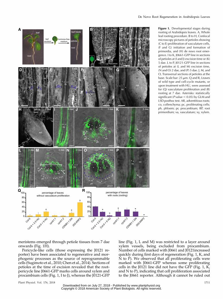
meristems emerged through petiole tissues from 7 daeonwards (Fig. 1H).Pericycle-like cells (those expressing the J0121 re-
porter) have been associated to regenerative and mor-phogenic processes as the source of reprogrammablecells (Sugimoto et al., 2010; Chen et al., 2014). Sections ofpetioles at the time of excision revealed that the root-pericycle line J0661-GFP marks cells around xylem andprocambium cells (Fig. 1, I to J), whereas the J0121-GFP
line (Fig. 1, L and M) was restricted to a layer aroundxylem vessels, being excluded from procambium.Number of cells marked with J0661 and J0121increasedquickly during first days of regeneration (Fig. 1, K, andN to P). We observed that all proliferating cells weremarked with J0661-GFP whereas some proliferatingcells in the J0121 line did not have the GFP (Fig. 1, K,and N to P), indicating that cell proliferation associatedto the J0661 reporter. Although it cannot be ruled out
Figure 1. Developmental stages duringrooting of Arabidopsis leaves. A, Wholeleaf rooting procedure. B to H, Confocalmicroscopy pictures of petioles showing(C to E) proliferation of vasculature cells,(F and G) initiation and formation ofprimordia, and (H) de novo root emer-gence. I to K, J0661-GFP line in sectionsof petioles at (I and J) excision time or (K)5 dae. L to P, J0121-GFP line in sectionsof petioles at (L and M) excision time,(N and O) 2 dae, and (P) 5 dae. J, M, andO, Transversal sections of petioles at thebase. Scale bar: 25 mm. Q and R, Leavesof wild type and cell-cycle mutants, orupon treatment with HU, were assessedfor (Q) vasculature proliferation and (R)rooting at 7 dae. Asterisks: statisticallysignificant (P value, 0.05) by GLM andLSD posthoc test. AR, adventitious roots;co, collenchyma; pc, proliferating cells;ph, phloem; pr, procambium; RP, rootprimordium; va, vasculature; xy, xylem.
Plant Physiol. Vol. 176, 2018 1711
De Novo Root Regeneration in Arabidopsis Leaves
www.plantphysiol.orgon July 27, 2018 - Published by Downloaded from Copyright © 2018 American Society of Plant Biologists. All rights reserved.

that J0661-GFP is activated in proliferating cells, it ispossible that xylem and procambium proliferate aspart of the reprogramming process. In addition, weobserved that primordia at early stages of developmentwere marked with J0121-GFP (Fig. 1P), establishing anassociation between de novo primordia formation andJ0121 identity, similarly to other developmental or re-generative processes such as callus or lateral root for-mation (Dubrovsky el al., 2006; Sugimoto et al., 2010).
We next studied mutants defective in cell cycle pro-gression, at the G1/S transition, such as the KIP-RELATED PROTEIN2 (KRP2) overexpressor, and at theG2/M transition, such as cyclinB1;1 (cycb1;1) and cycb1;2mutants and a dominant negative form of the CDKB1;1kinase (CDKB1;1 DN161). Percentage of petioles showingvasculature-associated proliferation was reduced inPro35S:KRP2 and CDKB1;1 DN161 lines (Fig. 1Q), whereasonly size of proliferating mass of cells was reduced in therest of the lines. In addition, Pro35S:KRP2 blocked de novoorgan regeneration, whereas cycb1;1 andCDKB1;1DN161showed a significant reduction in the number of petiolesregenerating roots (Fig. 1R). We also chemically inacti-vated theG1/S transition by incubating leaveswith either2.5 or 5mM hydroxyurea (HU).We observed a significantdecrease in rate of petioles showing vascular-associatedproliferation and subsequent new root formation by theHU treatment (Fig. 1, Q and R; Supplemental Fig. S2, Aand B). HU treatment did not associatewith increased celldeath around the vasculature near the leaf excision siteupon Trypan Blue staining (Supplemental Fig. S2C).Taken together, these results indicate that cell divisionactivation of vasculature cells is the first and requiredstage for de novo organogenesis during rooting of leaves.
Cytokinin Biosynthesis and Response during De NovoRoot Regeneration
As we had found an association between de novoroot regeneration and J0661 and J0121 identities, andcallus originates from J0121-marked cells after hormo-nal induction (Sugimoto et al., 2010), we hypothesizedthat vasculature proliferation required for leaf rootingcould be a type of callus. Cytokinin and auxin signalingare required for callus formation and regeneration, andthus we tested if these two hormones were involved inthe developmental pathway leading to de novo organregeneration from leaves.
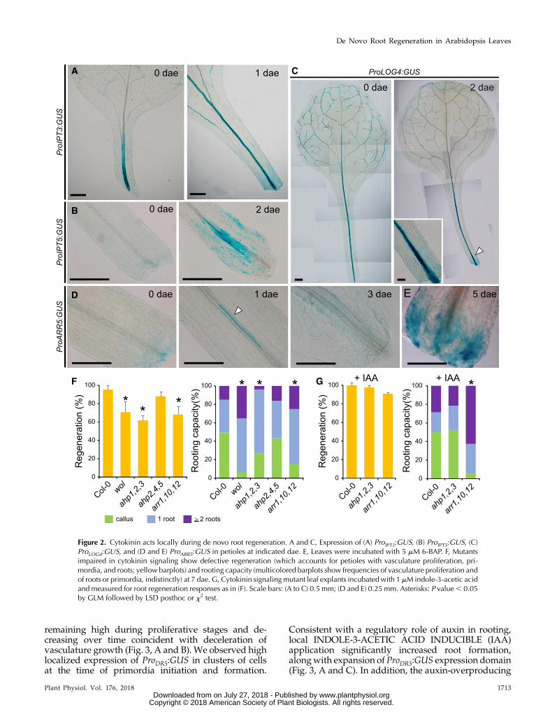
First, we investigated cytokinin biosynthesis andsignaling (Zürcher and Müller, 2016). We foundenriched expression of ProIPT3:GUS in petioles right af-ter excision (Fig. 2A). ProIPT5:GUSwas highly expressedin vascular-associated cells in the petiole base at 2 dae(Fig. 2B), whereas ProLOG4:GUS, which was originallyexpressed in leaf vasculature at 0 dae, increased ex-pression at the petiole base over time (Fig. 2C, arrow-head). Cytokinin signaling, as reported by ProARR5:GUS(D’Agostino et al., 2000), was restricted to a subset ofvascular-associated cells near the petiole base at 2 dae,which associated with proliferation of vasculature, to
decrease at later time points (Fig. 2D) and it did notshow expression during de novo primordia formation.Consistent with ARABIDOPSIS RESPONSE REGU-LATORS5 (ARR5) reporting primary cytokinin responseduring petiole vasculature proliferation (D’Agostinoet al., 2000), incubation of leaf explants with syntheticcytokinin 6-benzylaminopurine (6-BAP) increasedProARR5:GUS expression in the petiole vasculature andbasal region (Fig. 2E).
Next, we studied the ability of several cytokininsignaling mutant combinations in ARABIDOPSIS HISKINASE4 (AHK4) and ARABIDOPSIS HIS PHOSPHO-TRANSFER PROTEIN1 (AHP1) to AHP5 and ARR1,ARR10, and ARR12 genes in regulating both vascula-ture proliferation and de novo root regeneration. Wequantified petioles regenerating as petioles showingvasculature proliferation/thickening, root primordiaformation, or visible roots. Leaf petioles of wooden leg(wol, a dominant negative mutant in AHK4 receptor)and ahp1 ahp2 ahp3 and arr1 arr10 arr12 loss-of-functionmutants displayed lower regeneration percentageat 7 dae (Fig. 2F). Interestingly, wol, ahp1 ahp2 ahp3,and arr1 arr10 arr12 mutants were also defective inhormone-induced callus formation from different tis-sue explants, such as leaves, cotyledons, and roots(Supplemental Fig. S3), indicating that specific cytoki-nin signaling is required for both callus formation andvasculature proliferation in leaf petioles. Despite cyto-kinin signaling being required for vasculature prolif-eration in petioles during rooting, for those leaf petiolesof cytokinin signaling mutants that regenerated, wedetected a higher number of roots (which we catego-rized by frequencies in numbers of roots and desig-nated as “rooting capacity”; Fig. 2F). Higher auxin-to-cytokinin ratios have been shown to induce specifica-tion and growth of new root primordia (Müller andSheen, 2008). Thus, we wondered if we could alternew primordia initiation by altering hormone ratios.Cytokinin treatment increased vascular prolifera-tion in a concentration- and time-dependent manner(Supplemental Fig. S2, D and E). We observed that re-generation deficiencies of most cytokinin signalingmutants could be compensated by low levels of exog-enous auxin (Fig. 2G) that also increased vasculatureproliferation at the expense of reducing rooting capac-ity in the ahp1 ahp2 ahp3 mutant (Fig. 2G). Taken to-gether, these results indicate a dual role for cytokininfirst as a positive activator of vasculature cell division,and second as a negative regulator of root primordiainitiation.
Specific Auxin Signaling Factors Regulate De NovoRoot Regeneration
Next, we investigated auxin signaling during rootingof leaves using the DR5 reporter line (Ulmasov et al.,1997). ProDR5:GUSwas expressed in vascular-associatedcells at the proximal region of the petiole, as early as12 h after excision (hae), to increase quickly to 1 dae,
1712 Plant Physiol. Vol. 176, 2018
Bustillo-Avendaño et al.
www.plantphysiol.orgon July 27, 2018 - Published by Downloaded from Copyright © 2018 American Society of Plant Biologists. All rights reserved.
remaining high during proliferative stages and de-creasing over time coincident with deceleration ofvasculature growth (Fig. 3, A and B). We observed highlocalized expression of ProDR5:GUS in clusters of cellsat the time of primordia initiation and formation.
Consistent with a regulatory role of auxin in rooting,local INDOLE-3-ACETIC ACID INDUCIBLE (IAA)application significantly increased root formation,alongwith expansion of ProDR5:GUS expression domain(Fig. 3, A and C). In addition, the auxin-overproducing
Figure 2. Cytokinin acts locally during de novo root regeneration. A and C, Expression of (A) ProIPT3:GUS, (B) ProIPT5:GUS, (C)ProLOG4:GUS, and (D and E) ProARR5:GUS in petioles at indicated dae. E, Leaves were incubated with 5 mM 6-BAP. F, Mutantsimpaired in cytokinin signaling show defective regeneration (which accounts for petioles with vasculature proliferation, pri-mordia, and roots; yellow barplots) and rooting capacity (multicolored barplots show frequencies of vasculature proliferation andof roots or primordia, indistinctly) at 7 dae. G, Cytokinin signalingmutant leaf explants incubatedwith 1 mM indole-3-acetic acidand measured for root regeneration responses as in (F). Scale bars: (A to C) 0.5 mm; (D and E) 0.25 mm. Asterisks: P value, 0.05by GLM followed by LSD posthoc or x2 test.
Plant Physiol. Vol. 176, 2018 1713
De Novo Root Regeneration in Arabidopsis Leaves
www.plantphysiol.orgon July 27, 2018 - Published by Downloaded from Copyright © 2018 American Society of Plant Biologists. All rights reserved.
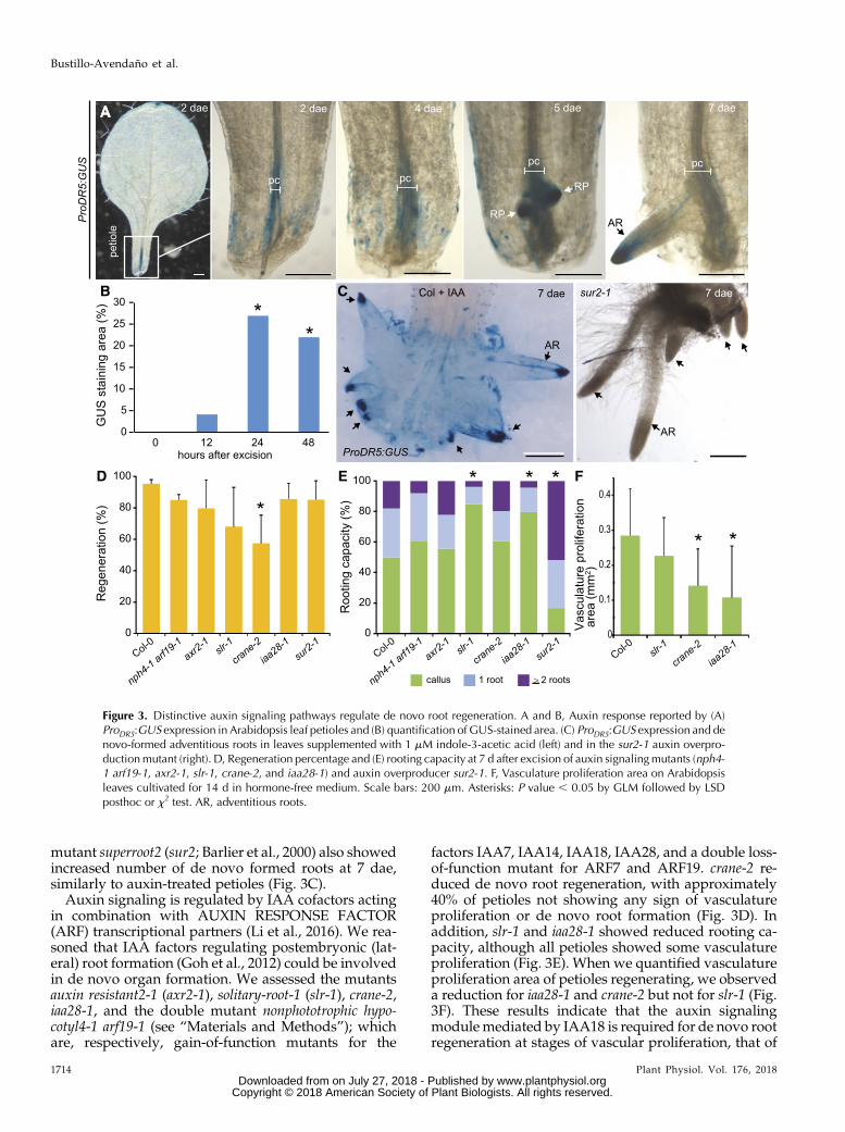
mutant superroot2 (sur2; Barlier et al., 2000) also showedincreased number of de novo formed roots at 7 dae,similarly to auxin-treated petioles (Fig. 3C).
Auxin signaling is regulated by IAA cofactors actingin combination with AUXIN RESPONSE FACTOR(ARF) transcriptional partners (Li et al., 2016). We rea-soned that IAA factors regulating postembryonic (lat-eral) root formation (Goh et al., 2012) could be involvedin de novo organ formation. We assessed the mutantsauxin resistant2-1 (axr2-1), solitary-root-1 (slr-1), crane-2,iaa28-1, and the double mutant nonphototrophic hypo-cotyl4-1 arf19-1 (see “Materials and Methods”); whichare, respectively, gain-of-function mutants for the
factors IAA7, IAA14, IAA18, IAA28, and a double loss-of-function mutant for ARF7 and ARF19. crane-2 re-duced de novo root regeneration, with approximately40% of petioles not showing any sign of vasculatureproliferation or de novo root formation (Fig. 3D). Inaddition, slr-1 and iaa28-1 showed reduced rooting ca-pacity, although all petioles showed some vasculatureproliferation (Fig. 3E). When we quantified vasculatureproliferation area of petioles regenerating, we observeda reduction for iaa28-1 and crane-2 but not for slr-1 (Fig.3F). These results indicate that the auxin signalingmodulemediated by IAA18 is required for de novo rootregeneration at stages of vascular proliferation, that of
Figure 3. Distinctive auxin signaling pathways regulate de novo root regeneration. A and B, Auxin response reported by (A)ProDR5:GUS expression in Arabidopsis leaf petioles and (B) quantification of GUS-stained area. (C) ProDR5:GUS expression and denovo-formed adventitious roots in leaves supplemented with 1 mM indole-3-acetic acid (left) and in the sur2-1 auxin overpro-ductionmutant (right). D, Regeneration percentage and (E) rooting capacity at 7 d after excision of auxin signalingmutants (nph4-1 arf19-1, axr2-1, slr-1, crane-2, and iaa28-1) and auxin overproducer sur2-1. F, Vasculature proliferation area on Arabidopsisleaves cultivated for 14 d in hormone-free medium. Scale bars: 200 mm. Asterisks: P value , 0.05 by GLM followed by LSDposthoc or x2 test. AR, adventitious roots.
1714 Plant Physiol. Vol. 176, 2018
Bustillo-Avendaño et al.
www.plantphysiol.orgon July 27, 2018 - Published by Downloaded from Copyright © 2018 American Society of Plant Biologists. All rights reserved.
IAA28 for vascular proliferation and root initiation,whereas IAA14 appears to be required only for de novoroot initiation.We also investigated if these mutants were affected in
hormone-induced callus formation. We found that onlycrane-2 showed reduction in all explants assayed afterhormonal incubation, whereas axr2-1 intriguingly showedan increase for callus formed from root explants(Supplemental Fig. S4, A to C). These results indicate thatvasculature proliferation during rooting and hormone-induced callus use the auxin signaling pathway medi-ated by IAA18. Our previous results also showed thatcytokinin signaling required for vasculature proliferationduring de novo organogenesis was also required forhormone-induced callus formation, suggesting that vas-culature proliferation is a type of callus. We investigatedABERRANT LATERAL ROOT FORMATION4 (ALF4)during leaf rooting, as aberrant lateral root formation4 (alf4),in this case, alf4-1 mutants, have been linked to callus for-mation (Sugimoto et al., 2010) and vascular connectionduring graft establishment (Melnyk et al., 2015). We ob-served that during de novo root formation, vasculatureproliferation is reducedby 2.5-fold in alf4-1mutants,whichis accompaniedby 15-fold decrease in de novo formed rootand primordia (Supplemental Fig. S5). Based on these re-sults, we designated the vasculature proliferation devel-opmental stage as the endogenous callus formation.
Auxin Signaling Factors Are Required for De Novo OrganRegeneration in Leaf Blades
In contrast to endogenous callus formation observedin petioles of whole leaves during rooting, limitedvasculature proliferation was observed during rootingof leaf blades (Liu et al., 2014). We wondered to whatextent auxin and cytokinin signaling factors regulatingproliferation at the petiole base would be involved inrooting of leaf blades. When we assessed rooting ca-pacity in the leaf blades of these mutants, we observedthat crane-2 and iaa28-1 displayed a reduction in rootingcapacity whereas slr-1 presented moderate althoughnonsignificant reductions (Supplemental Fig. S6A). woland arr1 arr10 arr12 mutants were similarly affected asslr-1, whereas no change was detected for ahp1 ahp2ahp3 and ahp2 ahp4 ahp5 mutants (Supplemental Fig.S6B). As IAA18 and IAA28 are required for endogenouscallus formation during whole leaf rooting and areshared with leaf blade rooting, it is possible that denovo root regeneration in leaf blades could also involvean endogenous callus developmental program.
Local Auxin Accumulation at the Petiole Base IsDependent on Polar Auxin Transport
As localized auxin signalingwas required forwhole leafrooting, we wondered about the source of auxin. YUCCA-mediated auxin biosynthesis was shown to be ubiqui-tously enhanced in the mesophyll of leaf blades shortly
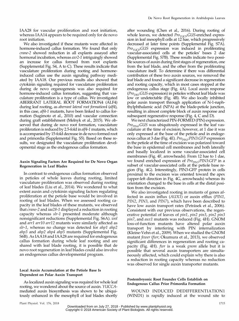
after wounding (Chen et al., 2016). During rooting ofwhole leaves, we detected ProYUC9:GUS-enriched expres-sion in leaf mesophyll cells at 12 hae, which progressivelydecreased at later time points (Supplemental Fig. S7A).ProYUC8:GUS expression was induced in proliferatingvascular-associated cells at the petioles’ bases 2 dae(Supplemental Fig. S7B). These results indicate two possi-ble sources of auxin duringfirst stages of regeneration, onefrom the leaf blade, and the other from the proliferatingvasculature itself. To determine if there was differentialcontribution of these two auxin sources, we removed theleaf blade and found a significant decrease in regenerationand rooting capacity, which in most cases stopped at theendogenous callus stage (Fig. 4A). Local auxin response(ProDR5:GUS expression) in petioles without leaf blade waslow or undetectable (Fig. 4B). We also locally inhibitedpolar auxin transport through application of N-1-naph-thylphthalamic acid (NPA) at the blade-petiole junction,resulting in almost complete block of auxin response andsubsequent regenerative response (Fig. 4, C and D).
Wenext characterized PIN-FORMED (PIN) expression.ProPIN4:GUS was ubiquitously expressed in the leaf vas-culature at the time of excision; however, at 1 dae it wasonly expressed at the base of the petiole and in endoge-nous callus at 3 dae (Fig. 4E). ProPIN3:PIN3:GFP expressionin the petiole at the time of excisionwas polarized towardthe base in epidermal cell membranes and both laterallyand basally localized in some vascular-associated cellmembranes (Fig. 4F, arrowheads). From 12 hae to 1 dae,we found enriched expression of ProPIN3:PIN3:GFP in asubset of vascular-associated cells at the petiole base re-gion (Fig. 4G). Interestingly, PIN3-GFP protein in cellsproximal to the excision was oriented toward the apex(upper-left direction in Fig. 4G, arrowheads) whereas itsorientation changed to the base in cells at the distal posi-tion from the excision.
We also investigated rooting in mutants of genes af-fected in auxin influx (AUX1) or auxin efflux (PIN1,PIN2, PIN3, and PIN7), which have been described tohave low auxin transport rates (Petrásek et al., 2006).Consistent with our previous observations, the regen-erative potential of leaves of pin1, pin2 pin3, pin2 pin3pin7, and aux1 mutants was reduced (Fig. 4H). GNOMloss-of-function mutants have altered polar auxintransport by interfering with PIN internalization(Kleine-Vehn et al., 2009). Whenwe studied the GNOMmutant fewer (fwr; Okumura et al., 2013), we observedsignificant differences in regeneration and rooting ca-pacity (Fig. 4H). fwr is a weak gnom allele but it ispossible that several auxin transporters are simulta-neously affected, which could explain why there is alsoa reduction in rooting capacity whereas no reductionwas observed for single auxin transporter mutants.
Postembryonic Root Founder Cells Establish onEndogenous Callus Prior Primordia Formation
WOUND INDUCED DEDIFFERENTIATION1(WIND1) is rapidly induced at the wound site to
Plant Physiol. Vol. 176, 2018 1715
De Novo Root Regeneration in Arabidopsis Leaves
www.plantphysiol.orgon July 27, 2018 - Published by Downloaded from Copyright © 2018 American Society of Plant Biologists. All rights reserved.
Figure 4. Polar auxin transport from the leaf blade to the petiole is required for rooting. A and B, Distal blade excision reduces (A)regeneration rate and rooting capacity at 7 dae and (B) ProDR5:GUS expression at 12 hae and 3 dae. C andD, Local treatment at theblade-petiole junction with 1%NPA reduces (C) regeneration rate and rooting capacity at 7 dae, and (D) ProDR5:GUS expression.E to G, Expression of (E) ProPIN4:GUS and (F and G) ProPIN3:PIN3:GFP. Arrowheads point to membrane-localized PIN-GFP. Scalebars: (B, D, F, and G) 0.2 and (E) 0.5 mm. H, Regeneration and rooting capacity in auxin transport mutants at 7 dae. Asterisks:P value , 0.05 by GLM followed by LSD posthoc or x2 test.
1716 Plant Physiol. Vol. 176, 2018
Bustillo-Avendaño et al.
www.plantphysiol.orgon July 27, 2018 - Published by Downloaded from Copyright © 2018 American Society of Plant Biologists. All rights reserved.
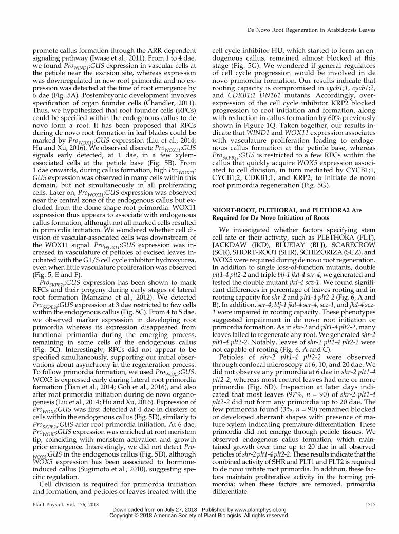
promote callus formation through the ARR-dependentsignaling pathway (Iwase et al., 2011). From 1 to 4 dae,we found ProWIND1:GUS expression in vascular cells atthe petiole near the excision site, whereas expressionwas downregulated in new root primordia and no ex-pression was detected at the time of root emergence by6 dae (Fig. 5A). Postembryonic development involvesspecification of organ founder cells (Chandler, 2011).Thus, we hypothesized that root founder cells (RFCs)could be specified within the endogenous callus to denovo form a root. It has been proposed that RFCsduring de novo root formation in leaf blades could bemarked by ProWOX11:GUS expression (Liu et al., 2014;Hu and Xu, 2016). We observed discrete ProWOX11:GUSsignals early detected, at 1 dae, in a few xylem-associated cells at the petiole base (Fig. 5B). From1 dae onwards, during callus formation, high ProWOX11:GUS expression was observed in many cells within thisdomain, but not simultaneously in all proliferatingcells. Later on, ProWOX11:GUS expression was observednear the central zone of the endogenous callus but ex-cluded from the dome-shape root primordia. WOX11expression thus appears to associate with endogenouscallus formation, although not all marked cells resultedin primordia initiation. We wondered whether cell di-vision of vascular-associated cells was downstream ofthe WOX11 signal. ProWOX11:GUS expression was in-creased in vasculature of petioles of excised leaves in-cubatedwith the G1/S cell cycle inhibitor hydroxyurea,evenwhen little vasculature proliferation was observed(Fig. 5, E and F).ProSKPB2s:GUS expression has been shown to mark
RFCs and their progeny during early stages of lateralroot formation (Manzano et al., 2012). We detectedProSKPB2s:GUS expression at 3 dae restricted to few cellswithin the endogenous callus (Fig. 5C). From 4 to 5 dae,we observed marker expression in developing rootprimordia whereas its expression disappeared fromfunctional primordia during the emerging process,remaining in some cells of the endogenous callus(Fig. 5C). Interestingly, RFCs did not appear to bespecified simultaneously, supporting our initial obser-vations about asynchrony in the regeneration process.To follow primordia formation, we used ProWOX5:GUS.WOX5 is expressed early during lateral root primordiaformation (Tian et al., 2014; Goh et al., 2016), and alsoafter root primordia initiation during de novo organo-genesis (Liu et al., 2014; Hu andXu, 2016). Expression ofProWOX5:GUS was first detected at 4 dae in clusters ofcells within the endogenous callus (Fig. 5D), similarly toProSKPB2s:GUS after root primordia initiation. At 6 dae,ProWOX5:GUS expression was enriched at root meristemtip, coinciding with meristem activation and growthprior emergence. Interestingly, we did not detect Pro-WOX5:GUS in the endogenous callus (Fig. 5D), althoughWOX5 expression has been associated to hormone-induced callus (Sugimoto et al., 2010), suggesting spe-cific regulation.Cell division is required for primordia initiation
and formation, and petioles of leaves treated with the
cell cycle inhibitor HU, which started to form an en-dogenous callus, remained almost blocked at thisstage (Fig. 5G). We wondered if general regulatorsof cell cycle progression would be involved in denovo primordia formation. Our results indicate thatrooting capacity is compromised in cycb1;1, cycb1;2,and CDKB1;1 DN161 mutants. Accordingly, over-expression of the cell cycle inhibitor KRP2 blockedprogression to root initiation and formation, alongwith reduction in callus formation by 60% previouslyshown in Figure 1Q. Taken together, our results in-dicate that WIND1 and WOX11 expression associateswith vasculature proliferation leading to endoge-nous callus formation at the petiole base, whereasProSKPB2s:GUS is restricted to a few RFCs within thecallus that quickly acquire WOX5 expression associ-ated to cell division, in turn mediated by CYCB1;1,CYCB1;2, CDKB1;1, and KRP2, to initiate de novoroot primordia regeneration (Fig. 5G).
SHORT-ROOT, PLETHORA1, and PLETHORA2 AreRequired for De Novo Initiation of Roots
We investigated whether factors specifying stemcell fate or their activity, such as PLETHORA (PLT),JACKDAW (JKD), BLUEJAY (BLJ), SCARECROW(SCR), SHORT-ROOT (SHR), SCHIZORIZA (SCZ), andWOX5were required during de novo root regeneration.In addition to single loss-of-function mutants, doubleplt1-4 plt2-2 and triple blj-1 jkd-4 scr-4, we generated andtested the double mutant jkd-4 scz-1. We found signifi-cant differences in percentage of leaves rooting and inrooting capacity for shr-2 and plt1-4 plt2-2 (Fig. 6, A andB). In addition, scr-4, blj-1 jkd-4 scr-4, scz-1, and jkd-4 scz-1 were impaired in rooting capacity. These phenotypessuggested impairment in de novo root initiation orprimordia formation. As in shr-2 and plt1-4 plt2-2, manyleaves failed to regenerate any root. We generated shr-2plt1-4 plt2-2. Notably, leaves of shr-2 plt1-4 plt2-2 werenot capable of rooting (Fig. 6, A and C).
Petioles of shr-2 plt1-4 plt2-2 were observedthrough confocal microscopy at 6, 10, and 20 dae. Wedid not observe any primordia at 6 dae in shr-2 plt1-4plt2-2, whereas most control leaves had one or moreprimordia (Fig. 6D). Inspection at later days indi-cated that most leaves (97%, n = 90) of shr-2 plt1-4plt2-2 did not form any primordia up to 20 dae. Thefew primordia found (3%, n = 90) remained blockedor developed aberrant shapes with presence of ma-ture xylem indicating premature differentiation. Theseprimordia did not emerge through petiole tissues. Weobserved endogenous callus formation, which main-tained growth over time up to 20 dae in all observedpetioles of shr-2 plt1-4 plt2-2. These results indicate that thecombined activity of SHR and PLT1 and PLT2 is requiredto de novo initiate root primordia. In addition, these fac-tors maintain proliferative activity in the forming pri-mordia; when these factors are removed, primordiadifferentiate.
Plant Physiol. Vol. 176, 2018 1717
De Novo Root Regeneration in Arabidopsis Leaves
www.plantphysiol.orgon July 27, 2018 - Published by Downloaded from Copyright © 2018 American Society of Plant Biologists. All rights reserved.
Figure 5. Root founder cells establish on endogenous callus prior de novo primordium formation. A to F, Expression of (A)ProWIND1:GUS, (B, E, and F) ProWOX11:GUS, (C) ProSKP2Bs:GUS, and (D) ProWOX5:GUS in leaf petioles at indicated dae. E, Controlconditions or (F) upon 5 mM HU treatment. GUS staining time in (E) was set for no expression. Scale bars: 200 mm. G, Regen-eration percentage and rooting capacity at 7 dae of mutants impaired in cell cycle progression and HU-treated Col-0 leaves.Asterisks: P value, 0.05 by GLM followed by LSD posthoc or x2 test. DN, de novo-formed root; pc, proliferating cells; RFC, rootfounder cell; RP, root primordium; va, vasculature; xy, xylem.
1718 Plant Physiol. Vol. 176, 2018 www.plantphysiol.orgon July 27, 2018 - Published by Downloaded from
Copyright © 2018 American Society of Plant Biologists. All rights reserved.
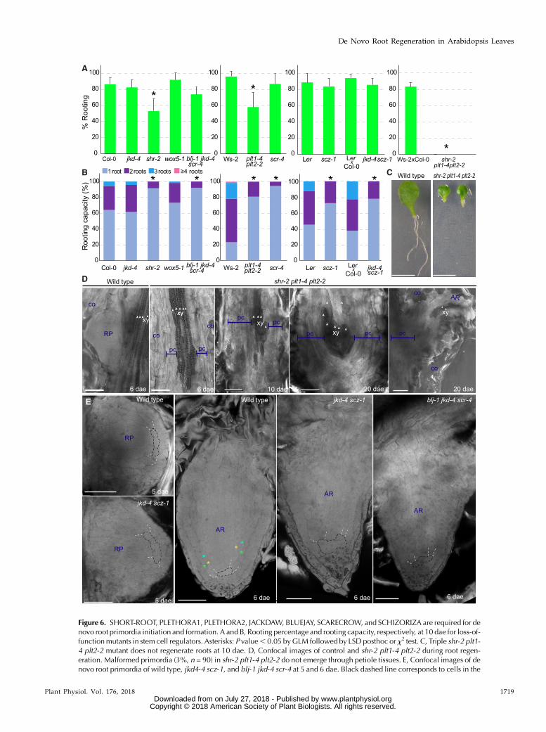
Figure 6. SHORT-ROOT, PLETHORA1, PLETHORA2, JACKDAW, BLUEJAY, SCARECROW, and SCHIZORIZA are required for denovo root primordia initiation and formation. A and B, Rooting percentage and rooting capacity, respectively, at 10 dae for loss-of-functionmutants in stem cell regulators. Asterisks: P value, 0.05 byGLM followed by LSD posthoc or x2 test. C, Triple shr-2 plt1-4 plt2-2 mutant does not regenerate roots at 10 dae. D, Confocal images of control and shr-2 plt1-4 plt2-2 during root regen-eration. Malformed primordia (3%, n = 90) in shr-2 plt1-4 plt2-2 do not emerge through petiole tissues. E, Confocal images of denovo root primordia of wild type, jkd4-4 scz-1, and blj-1 jkd-4 scr-4 at 5 and 6 dae. Black dashed line corresponds to cells in the
Plant Physiol. Vol. 176, 2018 1719
De Novo Root Regeneration in Arabidopsis Leaves
www.plantphysiol.orgon July 27, 2018 - Published by Downloaded from Copyright © 2018 American Society of Plant Biologists. All rights reserved.
JACKDAW, BLUEJAY, SCARECROW, and SCHIZORIZAAre Required for De Novo Formation of Root Primordia
We observed formation of root primordia in peti-oles of jkd-4 scz-1 and blj-1 jkd-4 scr-4 through confocalmicroscopy. We observed abnormal formative divi-sions in jkd-4 scz-1 primordia at 5 dae, which did notorganize properly in layers or rows as compared tocontrol roots (Fig. 6E). At 6 dae, we observed reducedand disorganized number of cell rows in jkd-4 scz-1 and blj-1 jkd-4 scr-4 primordia. Although endoder-mis, cortex, and middle cortex could be identified incontrol roots at this developmental stage based on posi-tion, corresponding rows in jkd-4 scz-1 and blj-1 jkd-4 scr-4could not be identified (Fig. 6E). These results suggestthat cell lineages or positional identity could not be cor-rectly established in these mutants during de novo rootformation.
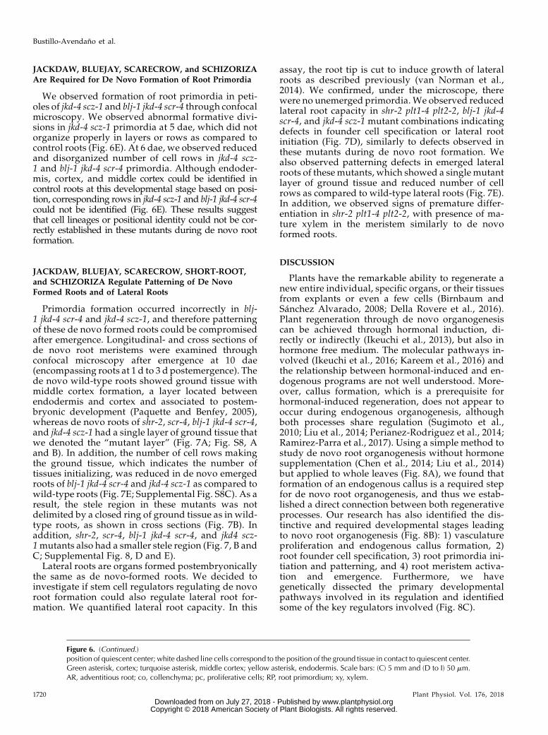
JACKDAW, BLUEJAY, SCARECROW, SHORT-ROOT,and SCHIZORIZA Regulate Patterning of De NovoFormed Roots and of Lateral Roots
Primordia formation occurred incorrectly in blj-1 jkd-4 scr-4 and jkd-4 scz-1, and therefore patterningof these de novo formed roots could be compromisedafter emergence. Longitudinal- and cross sections ofde novo root meristems were examined throughconfocal microscopy after emergence at 10 dae(encompassing roots at 1 d to 3 d postemergence). Thede novo wild-type roots showed ground tissue withmiddle cortex formation, a layer located betweenendodermis and cortex and associated to postem-bryonic development (Paquette and Benfey, 2005),whereas de novo roots of shr-2, scr-4, blj-1 jkd-4 scr-4,and jkd-4 scz-1 had a single layer of ground tissue thatwe denoted the “mutant layer” (Fig. 7A; Fig. S8, Aand B). In addition, the number of cell rows makingthe ground tissue, which indicates the number oftissues initializing, was reduced in de novo emergedroots of blj-1 jkd-4 scr-4 and jkd-4 scz-1 as compared towild-type roots (Fig. 7E; Supplemental Fig. S8C). As aresult, the stele region in these mutants was notdelimited by a closed ring of ground tissue as in wild-type roots, as shown in cross sections (Fig. 7B). Inaddition, shr-2, scr-4, blj-1 jkd-4 scr-4, and jkd4 scz-1mutants also had a smaller stele region (Fig. 7, B andC; Supplemental Fig. 8, D and E).
Lateral roots are organs formed postembryonicallythe same as de novo-formed roots. We decided toinvestigate if stem cell regulators regulating de novoroot formation could also regulate lateral root for-mation. We quantified lateral root capacity. In this
assay, the root tip is cut to induce growth of lateralroots as described previously (van Norman et al.,2014). We confirmed, under the microscope, therewere no unemerged primordia. We observed reducedlateral root capacity in shr-2 plt1-4 plt2-2, blj-1 jkd-4scr-4, and jkd-4 scz-1 mutant combinations indicatingdefects in founder cell specification or lateral rootinitiation (Fig. 7D), similarly to defects observed inthese mutants during de novo root formation. Wealso observed patterning defects in emerged lateralroots of these mutants, which showed a single mutantlayer of ground tissue and reduced number of cellrows as compared to wild-type lateral roots (Fig. 7E).In addition, we observed signs of premature differ-entiation in shr-2 plt1-4 plt2-2, with presence of ma-ture xylem in the meristem similarly to de novoformed roots.
DISCUSSION
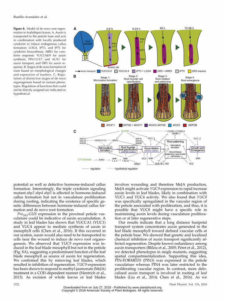
Plants have the remarkable ability to regenerate anew entire individual, specific organs, or their tissuesfrom explants or even a few cells (Birnbaum andSánchez Alvarado, 2008; Della Rovere et al., 2016).Plant regeneration through de novo organogenesiscan be achieved through hormonal induction, di-rectly or indirectly (Ikeuchi et al., 2013), but also inhormone free medium. The molecular pathways in-volved (Ikeuchi et al., 2016; Kareem et al., 2016) andthe relationship between hormonal-induced and en-dogenous programs are not well understood. More-over, callus formation, which is a prerequisite forhormonal-induced regeneration, does not appear tooccur during endogenous organogenesis, althoughboth processes share regulation (Sugimoto et al.,2010; Liu et al., 2014; Perianez-Rodriguez et al., 2014;Ramirez-Parra et al., 2017). Using a simple method tostudy de novo root organogenesis without hormonesupplementation (Chen et al., 2014; Liu et al., 2014)but applied to whole leaves (Fig. 8A), we found thatformation of an endogenous callus is a required stepfor de novo root organogenesis, and thus we estab-lished a direct connection between both regenerativeprocesses. Our research has also identified the dis-tinctive and required developmental stages leadingto novo root organogenesis (Fig. 8B): 1) vasculatureproliferation and endogenous callus formation, 2)root founder cell specification, 3) root primordia ini-tiation and patterning, and 4) root meristem activa-tion and emergence. Furthermore, we havegenetically dissected the primary developmentalpathways involved in its regulation and identifiedsome of the key regulators involved (Fig. 8C).
Figure 6. (Continued.)position of quiescent center; white dashed line cells correspond to the position of the ground tissue in contact to quiescent center.Green asterisk, cortex; turquoise asterisk, middle cortex; yellow asterisk, endodermis. Scale bars: (C) 5 mm and (D to I) 50 mm.AR, adventitious root; co, collenchyma; pc, proliferative cells; RP, root primordium; xy, xylem.
1720 Plant Physiol. Vol. 176, 2018
Bustillo-Avendaño et al.
www.plantphysiol.orgon July 27, 2018 - Published by Downloaded from Copyright © 2018 American Society of Plant Biologists. All rights reserved.
Vasculature Proliferation and EndogenousCallus Formation
Vasculature division is the first morphologicalchange we detect, and by chemically or geneticallyinhibiting vasculature proliferation, we affected denovo root organogenesis. We also observed that rootprimordia originated from vasculature. Pericycle-likecells, particularly those expressing the J0121 marker,have been associated to regenerative and morphogenicprocesses as the source of reprogrammable cells(Sugimoto et al., 2010; Chen et al., 2014). When weassessed the J0661 and J0121 pericycle markers, weobserved that vasculature proliferation associated toJ0661 identity, whereas some proliferating cells weredevoid of J0121 expression. Intriguingly, J0661 markscells around xylem and procambium in petioleswhereas J0121 only marks cells in contact to xylem.Proliferation competence, therefore, appears to involvedifferent cell types. Closer examination showed that allprimordia expressed a J0121 marker, which indicatesthat regeneration competence associates with J0121identity and shows parallelisms with callus and lateralroot formation. Cell lineage tracing using clonal
markers and live imaging could dissect the exact sourceof reprogrammable cells during de novo root regener-ation from whole leaves.
We also found that proliferating vascular cells ex-press WIND1, a positive regulator of wound-inducedcallus formation (Iwase et al., 2011), suggesting to-gether with J0121 analysis that these proliferating tis-sues could be a type of callus. Callus formation requiresconfluence of auxin and cytokinin responses in thesame set of cells (Gordon et al., 2007). In agreement withthis idea, our results show specific expression of auxinand cytokinin signaling reporters ProDR5:GUS andProARR5:GUS, respectively, in the vasculature. Theseresults also indicate that specific regulation was re-quired to induce auxin and cytokinin signaling at thepetiole base. In contrast to early notions that cytokininsare produced only in roots, it is now recognized thatthey are synthesized throughout the plant (Zürcher andMüller, 2016). Our results are consistent with ISO-PENTENYLTRANSFERASE3 (IPT3), IPT5, andLONELY GUY4 locally mediating cytokinin biosyn-thesis at the petiole base to contribute to vascular pro-liferation during root regeneration. Thus, cytokininsignaling mutants displayed reduced regeneration
Figure 7. JACKDAW, BLUEJAY, SCARE-CROW, SHORT-ROOT, and SCHIZO-RIZA regulate patterning ofpostembryonic roots. A and B, Confocal(A) longitudinal and (B) transversal sec-tions of de novo-formed roots at 10 dae(1 to 3 d post emergence). Transversalsections were taken at the end of thelateral root cap. Dashed lines and aster-isks: green, cortex; turquoise, middlecortex; yellow, endodermis; brown,stele; white, mutant undivided groundtissue. C, Number of cortical-like cell orundivided mutant cell rows in controlaccessions, jkd-4 scz-1 and blj-1 jkd-4scr-4. D, Lateral root capacity in Col-0,shr-2 plt1-4 plt2-2, jkd-4 scz-1, and blj-1 jkd-4 scr-4 and at 4 d after seed imbi-bition. E, Confocal longitudinal sectionsof emerged lateral roots of Col-0, shr-2plt1-4 plt2-2, jkd-4 scz-1, and blj-1 jkd-4scr-4 at 7 d after seed imbibition. Scalebars: 50mm.Asterisks: P value, 0.05 byGLM and LSD posthoc test. m, mutant;xy, xylem.
Plant Physiol. Vol. 176, 2018 1721
De Novo Root Regeneration in Arabidopsis Leaves
www.plantphysiol.orgon July 27, 2018 - Published by Downloaded from Copyright © 2018 American Society of Plant Biologists. All rights reserved.
potential as well as defective hormone-induced callusformation. Interestingly, the triple cytokinin signalingmutant ahp2 ahp4 ahp5 is affected in hormone-inducedcallus formation but not in vasculature proliferationduring rooting, indicating the existence of specific ge-netic differences between hormone-induced callus for-mation and de novo root formation.
ProDR5:GUS expression in the proximal petiole vas-culature could be indicative of auxin accumulation. Astudy in leaf blades has shown that YUCCA1 (YUC1)and YUC4 appear to mediate synthesis of auxin inmesophyll cells (Chen et al., 2016). If this occurred inour system, auxin would also need to be transported tocells near the wound to induce de novo root organo-genesis. We observed that YUC9 expression was in-duced in the leaf blade mesophyll but not in the petiole(Fig. 8A), suggesting a predominant function of the leafblade mesophyll as source of auxin for regeneration.We confirmed this by removing leaf blades, whichresulted in inhibition of regeneration. YUC9 expressionhas been shown to respond tomethyl-jasmonate (MeJA)treatment in a COI1-dependent manner (Hentrich et al.,2013). As excision of whole leaves or leaf blades
involves wounding and therefore MeJA production,MeJAmight activate YUC9 expression to rapid increaseauxin levels in leaf blades, likely in combination withYUC1 and YUC4 activity. We also found that YUC8was specifically upregulated in the vascular region ofthe petiole associated with proliferation, and thus, it ispossible that YUC8 might have a specific role inmaintaining auxin levels during vasculature prolifera-tion or at later regenerative steps.
Our results indicate that a long distance basipetaltransport system concentrates auxin generated in theleaf blade mesophyll toward defined vascular cells atthe petiole base. We showed that genetic and localizedchemical inhibition of auxin transport significantly af-fected regeneration. Despite known redundancy amongauxin transporters (Blilou et al., 2005; Péret et al., 2012),we detected phenotypes in single mutants, suggestingspatial compartmentalization. Supporting this idea,PIN-FORMED3 (PIN3) was expressed in the petiolevasculature whereas PIN4 was later restricted to theproliferating vascular region. In contrast, more delo-calized auxin transport is involved in rooting of leafblades (Liu et al., 2014; Chen et al., 2016). As we
Figure 8. Model of de novo root regen-eration in Arabidopsis leaves. A, Auxin istransported to the petiole base and actsin combination with locally producedcytokinin to induce endogenous callusformation: LOG4, IPT3, and IPT5 forcytokinin biosynthesis; ARR5 for cyto-kinin response; YUCCA8/9 for auxinsynthesis; PIN1/2/3/7 and AUX1 forauxin transport; and DR5 for auxin re-sponse. B, Stages of de novo formation ofroots based on morphological changesand expression of markers. C, Regu-lation of distinctive stages of de novoorganogenesis based on mutant pheno-types. Regulation of functions that couldnot be directly assigned are indicated ashypothetical.
1722 Plant Physiol. Vol. 176, 2018
Bustillo-Avendaño et al.
www.plantphysiol.orgon July 27, 2018 - Published by Downloaded from Copyright © 2018 American Society of Plant Biologists. All rights reserved.
observed predominant expression of ProDR5:GUS in theproximal petiole vasculature, it is possible that auxinwould need to be retained in this area. Our data suggesta model in which subcellular PIN3 localization shiftsfrom basal to apical membranes in vascular cells nearthe wound to redirect auxin flow backward and thusmaintaining high auxin levels in the proximal petiolevasculature. Interestingly, an auxin-dependent switchin PIN3 polarization contributing to auxin-flow rever-sal is involved in the shoot gravitropic response(Rakusová et al., 2016), where basal-to-apical shift inPIN localization has been described to depend onphosphorylation (Dai et al., 2012). It is thus tempting tospeculate that auxin-dependent phosphorylation ofPIN3 would be involved in maintaining high auxinlevels in the petiole base vasculature during root re-generation.The alf4 mutant (DiDonato et al., 2004) has been
linked to hormone-induced callus formation (Sugimotoet al., 2010), but not to wound-induced callus formationduring graft establishment (Melnyk et al., 2015). Weobserved reduced vasculature proliferation along withgreat reduction in de novo root formation from wholeleaves in alf4-1 mutants. As vasculature proliferationduring de novo root formation associates to J0121- andWIND1-marked cells, requires auxin and cytokininsignaling and involves ALF4, we propose it is a type ofcallus, and therefore we refer to it as “endogenouscallus” to differentiate it from callus obtained by ex-ogenous hormone supplementation. In our model,time-dependent auxin accumulation in a subset ofvascular cells activates proliferation, whereas cytoki-nins might regulate the expression of genes that aredirectly involved in callus formation (such asWIND1 orALF4) or that are downstream targets of the auxin sig-nal involved in callus formation (LATERAL ORGANBOUNDARIES DOMAIN factors; Schaller et al., 2015).Particularly, we found that IAA18 and IAA28 are bothinvolved in endogenous callus formation, althoughonly mutations in IAA18 affect whole regeneration re-sponse, whereas ARABIDOPSIS HIS PHOSPHO-TRANSFER PROTEIN1 (AHP1) to AHP3 andARABIDOPSIS RESPONSE REGULATOR1 (ARR1),ARR10, and ARR12 were involved in vasculature pro-liferation and regeneration response.
Specification of Root Founder Cells
Hormone-induced callus is organized in layersshowing root tissue identities that resemble a rootmeristem and therefore a new organ could be theo-retically initiated through a differentiation process(Sugimoto et al., 2010). WUSCHEL-RELATED HO-MEOBOX11 (WOX11) expression has been associatedto first cell-fate transition from regeneration-competent cells to root founder cells during leafblade rooting (Liu et al., 2014; Hu and Xu, 2016).WOX11 activates WOX5 during root formation in leafblades; however, we did not find WOX5 expression in
endogenous callus during de novo regeneration fromwhole leaves, suggesting that WOX11 could be in-volved in an earlier step in the reprograming process.On the other hand, specific expression associated toroot founder cell specification (SKP2B) revealed theestablishment of a cell lineage capable of forming anew root. These results indicate that additionalreprogramming processes are required.
PLT1 and PLT2 have been shown to be required toestablish pluripotency during de novo shoot regenera-tion (Kareem et al., 2015). Our results show that duringde novo root formation, PLT1 and PLT2, in combina-tionwith SHR, could also be involved in specification ofroot founder cells, which are pluripotent. In addition,persistent expression of PLT1, PLT2, and SHR appearsto be necessary during subsequent formative stages tomaintain primordia growth, as the very primordiafound in shr-2 plt1-4 plt2-2 quickly differentiate. Incontrast, in the de novo shoot regeneration system,transient induction of PLT2 has been shown to be suf-ficient to specify shoot progenitors, whereas subse-quent expression of other regulators is required toaccomplish de novo shoot formation from these pro-genitors (Kareem et al., 2015).
Root Primordia Initiation and Patterning
Multiple INDOLE-3-ACETIC ACID INDUCIBLE(IAA)-ARF modules cooperatively regulate lateral rootformation (Goh et al., 2012). We observed that factorsregulating auxin signaling, such as SOLITARY ROOT(IAA14), could be also involved in de novo root initia-tion; we detected decreased root capacity for slr-1,which could be indicative of fewer primordia initiation.In addition, the IAA28 module, which is upstream oflateral root founder cell specification (De Rybel et al.,2010), also regulates de novo root founder cell specifi-cation or initiation, although further experiments coulddissect more precisely at which stage IAA28 is in-volved. Our results show that factors primarily in-volved in formation of lateral roots are also affected inrooting of leaves, suggesting the existence of partiallyoverlapping auxin signaling modules during postem-bryonic root development. Conversely, cytokinin mu-tants (ahp1 ahp2 ahp3 and arr1 arr10 arr12) showedincreased rooting capacity and thus, a repressor role inde novo root initiation can be assigned for these factors,likely in an analog manner as their role during lateralroot initiation (Lavenus et al., 2013; Chang et al., 2015).In agreement with this model, we restored regenerationpotential of cytokinin signaling mutants by a moderateincrease in auxin levels.
PLT1 and PLT2 expression is considered to be a slowreadout of auxin response and prolonged auxin treat-ment results in PLT1 and PLT2 activation and the denovo specification of WOX5-marked stem cells(Mähönen et al., 2014). We found severe impairment inde novo primordia initiation in shr-2 plt1-4 plt2-2, andWOX5 expression during de novo primordia formation
Plant Physiol. Vol. 176, 2018 1723
De Novo Root Regeneration in Arabidopsis Leaves
www.plantphysiol.orgon July 27, 2018 - Published by Downloaded from Copyright © 2018 American Society of Plant Biologists. All rights reserved.
requires auxin input through an unknown pathway(Hu and Xu, 2016). Therefore, it is possible that PLT1and PLT2 are required for specification of WOX5-marked cells downstream of auxin during de novo or-gan initiation, which could occur in combination withactivity of WOX5 transcription factor itself and/orSHR, in turn acting in an auxin-independent pathway.Further experiments will be required to dissect themolecular pathway including PLTs, SHR, and WOX5.
We have also identified specific factors involved information of de novo root primordia. We have foundthat the stem cell regulators BLJ, JKD, SCR, SHR, andSCZ regulate ground tissue patterning and vasculatureformation prior emergence at the step of dome-shapeprimordia. Subsequently, more developed primordiaare not properly organized in cell layers or rows and bythe time of emergence, these defects persist and ag-gravate. Our results indicate that ground tissue pat-terning appears to be regulated in newly formed rootsat two levels: First, we observed impairment in asym-metric divisions specifying cortex, middle cortex, andendodermis in mutants of SHR and SCR, although afew asymmetric divisions were still observed. Inagreement, shr mutants have been shown to form en-dodermis in anchor roots (Lucas et al., 2011), which area type of adventitious root. Furthermore, we observedthat ground tissue asymmetric divisions were absent inmutant combinations of scr-4with bjl-1 and jkd-4 and indouble mutants jkd-4 scz-1. Interestingly, when westudied if these mutant combinations had defects in lat-eral roots, which are also organs formed postembryoni-cally, we also observed absence of ground tissueasymmetric divisions suggesting a conserved develop-mental program for endodermis and cortex specification.Second, we observed that SCR, JKD, BLJ, and SCZ couldfunction as ground tissue lineage determinants during denovo root organogenesis. The combined action of BLJ,JKD, and SCR is required to maintain the ground tissuelineage postembryonically, and lacking these three factorsresults in missing ground tissue initializations (hencefewer ground tissue cell rows are observed in cross sec-tions: Moreno-Risueno et al., 2015). We observed that denovo formed roots in blj-1 jkd-4 scr-4 and jkd-4 scz-1 mu-tants were missing ground tissue cell rows shortly afteremergence, which indicates incorrect specification ofground tissue initials during primordia formation or lateron in the course of development. Thus, it is possible thatSCZ, SHR, PLT1, and PLT2 function as lineage or cell fatedeterminants during postembryonic development, andparticularly so during de novo organogenesis.
MATERIALS AND METHODS
Growth Conditions
Seeds were surfaced-sterilized in 10% (m/v) NaClO and rinsed with sterilewater before being transferred to 120 3 120 3 10 mm petri dishes containing65 mL of one-half-Murashige & Skoog (MS) medium with 1% Suc and 10 g/LPlant Agar (Duchefa). After 2 d of stratification at 4°C in darkness, plates weretransferred to an MLR-352-PE growth chamber (Panasonic) at 22°C, 16/8
photoperiod or continuous light (50 mmol$m22$s21). Twelve d after germina-tion, the first pair of leaveswas excised across the junction of the petiole with thestem and transferred to Gamborg B5mediumwith 2.5% Suc, 10 g/LDifco Agar(Becton Dickinson), or 3 g/L Gelrite (Sigma-Aldrich) and Gamborg B5 vitaminmixture (Duchefa). Leaves after excision were grown in darkness at 22°C,routinely for 10 d, or for number of days indicated in corresponding experi-ment.
Hormonal and Inhibition Treatments
For exogenous hormone treatment, filter-sterilized indole-3-acetic acid,6-BAP, or thidiazuron (TDZ) stock solutions were added to warm growthmedium before pouring into plates to provide a final concentration of 1 mM
indole-3-acetic acid, 5 mM 6-BAP, or 4 mM TDZ, respectively. NPA was appliedlocally by preparing a lanoline solution containing 1%w/wNPA. For cell cyclearrest, leaf explants were incubated with growth medium supplemented with0.5, 2.5, or 5 mM hydroxyurea (Sigma-Aldrich).
Plant Material
Columbia-0 (Col-0), Landsberg erecta (Ler), and Wassilewskija-2 (Ws-2) ac-cessions were used as a genetic background as corresponding. The reporterlines ProIPT3:GUS, ProIPT5:GUS (Miyawaki et al., 2004), ProLOG4:GUS s (Kurohaet al., 2009) obtained from RIKEN, and ProARR5:GUS (D’Agostino et al., 2000)were used for tracing cytokinin biosynthesis and signaling during rooting.ProYUC8:GUS, ProYUC9:GUS (Hentrich et al., 2013) and ProDR5:GUS (Ulmasovet al., 1997) were used to investigate auxin biosynthesis and signaling; ProPIN3:PIN3:GFP (Xu et al., 2006) and ProPIN4:GUS (Friml et al., 2004) were used forauxin transport. To trace the molecular mechanisms during de novo root for-mation we used: ProWIND1:GUS (Iwase et al., 2011), ProWOX11:GUS (Liu et al.,2014), ProWOX5:GUS (Sarkar et al., 2007), and ProSKP2Bs:GUS (Manzano et al.,2012), which corresponds to a promoter deletion containing 0.5 Kb upstreamfrom the ATG. The J0121 and J0661 lines (Laplaze et al., 2005) were used tolocate pericycle-like cells during rooting. The following mutant lines wereused: wol-1 (Scheres et al., 1995), ahp1 ahp2 ahp3 (Hutchison et al., 2006), ahp2ahp4 ahp5, arr1 arr10 arr12 (Mason et al., 2005), aux1-22 (Bennett et al., 1996),axr2-1 (Timpte et al., 1994), slr-1 (Fukaki et al., 2005), crane-2 (Uehara et al.,2008), sur2-1 (Delarue et al., 1998), and iaa28-1 (Rogg et al., 2001), which wereobtained from NASC; and pin1, pin2 pin3, pin2 pin3 pin7 (Blilou et al., 2005), fwr(Okumura et al., 2013), cyclinb1;1 (cycb1;1) and cycb1;2 (Nowack et al., 2012), andCDKB1;1 DN161 and Pro35S:KRP2 (Boudolf et al., 2004; Verkest et al., 2005). Wealso used the following stem cell niche mutants: blj-1 jkd-4 scr-4 (Moreno-Risueno et al., 2015), scr-4 (Fukaki et al., 1996), shr-2 ProSHR:SHR:GR(Levesque et al., 2006), plt1-4 plt2-2 (Aida et al., 2004), jkd-4 (Welch et al., 2007),scz-1 (Mylona et al., 2002), and wox5-1 (Sarkar et al., 2007), and jkd-4 scz-1 andshr-2 plt1-4 plt2-2 ProSHR:SHR:GR (generated in this study).
Genotyping
The triple mutant shr-2 plt1-4 plt2-2 ProSHR:SHR:GR was generated bycrossing shr-2 ProSHR:SHR:GR and plt1-4 plt2-2. F2 seedlings were grown andpreselected on one-half-MS medium with 10 mM dexamethasone followed byDNA extraction and genotyping by PCR to finally obtain homozygous lines.The following oligonucleotides were used for genotyping shr-2 (while dis-criminating ProSHR:SHR:GR): F 59-CCAATACCATCCCGCCAC-39 and R 59-TGAACCGGTCATGCGGTTG-39; for plt1-4: F 59-AGACGGCCACGCCAA-GAC-39 and R 59CTAGTATCACGACATTATTTGC-39; and for plt-2-2: F 59-ACCTACAGTCGTCACTTGTGC-39 and R 59-ACTCTTGTCTCGTCATG-TTTTTC-39. T-DNA insertions of PLT1 and PLT2 were amplified using LB 59-CATTTTATAATAACGCTGCGGACATCTAC-39 and respective reverseprimer. Double mutant jkd-4 scz-1 was generated by crossing jkd-4 and scz-1.DNA extraction from F2 seedlings was used for genotyping by PCR. The fol-lowing oligonucleotides were used for genotyping jkd-4: F 59-GGATGAAAG-CAATGCAAAACA-39 and R 59-AATGTCGGGATGATGAACTCC-39. TheT-DNA insertion in the jkd-4 line was genotyped using respective forwardprimer and RB 59-TCAAACAGGATTTTCGCCTGCT-39. For scz-1 genotyping,a SCZ fragment was amplified using F 59-CGAAGGTCAAGGCAAAGCTG-39and R 59-GAGCAACAGGCTTGACATGG-39primers, followed by digestionwith NlaIII restriction enzyme. Upon digestion, an SCZ fragment from scz-1 renders a band of 900 bp, while the wild-type fragment is cut by the enzyme,resulting in two fragments of 530 bp and 360 bp.
1724 Plant Physiol. Vol. 176, 2018
Bustillo-Avendaño et al.
www.plantphysiol.orgon July 27, 2018 - Published by Downloaded from Copyright © 2018 American Society of Plant Biologists. All rights reserved.
Cell Death, GUS Staining, and Microscopy Analysis
In leaf tissues,dyingcellswerevisualizedbyLactophenol-TrypanBluestainingasdescribed in Pavet et al. (2005). For GUS staining, leaf explants were incubated at37°C for variable times (4 h to 24 h) in multiwell plates in the presence of the GUSstaining solution as described in Pérez-Pérez et al. (2010) orManzano et al. (2012). Tobleach chlorophyll, two different methods were used indistinctly, with similar re-sults. For the first method, samples were dehydrated after GUS staining using in-creasing ethanol concentrations (15, 50, 70, 96, and 100%) for 15min in each one, andkept overnight in 100% ethanol; then samples were rehydrated following the sameethanol concentration series to 15% ethanol and mounted in 15% glycerol. For thesecondmethod, sampleswerefixed in 96%ethanol for 48 h andwashedwith 0.1 MPbuffer (pH 6.8) before being transferred to a clearing solution (80 g chloral hydrateand 30mLdistilledwater). Leaf explantsweremounted on slides using amixture of80 g chloral hydrate, 20 mL distilled water, and 10mL glycerol. After GUS staining,pictures were taken in a DM 2000with a DCF300 camera (Leica), or in a bright-fieldAX70 microscope (Olympus) equipped with a PM-C35DX microphotography sys-tem (Olympus). The area of proliferating tissues (callus or vasculature) was man-ually drawn frommicroscopic images using a Bamboo tablet (Wacon) and areas ordiameters of their best-fitting ellipses were measured with the software ImageJ(v1.50; National Institutes of Health).
Microscopy Analysis
Leaf petioles (for studying developmental stages during rooting) and matureembryoswerestainedusinganAnilineBluestainingmethodasdescribed inBougourdet al. (2000). Processed sampleswere observedwith a TCS SP8 laser scanning confocalmicroscope (Leica) with the settings described in Bougourd et al. (2000). A J0121 flu-orescent reporter line was processed before imaging as indicated in Kurihara et al.(2015). Cross sections (10 mm of thickness) of J0121 petioles were obtained using aVibratome 1000 Plus (TedPella)when indicated.Otherfluorescent reporter lineswereincubated in a methanol/acetone solution for 20 min at 220°C, and immediatelytransferred to a 0.1-M P buffer (pH 6.8). Imaging was performed by confocal micros-copyusing aDigital EclipseC1 (Nikon) equippedwithEZ-C1 control software (NikonInstruments) or a TCSSP8 laser scanningmicroscope (Leica). GFPwas excitedwith anArgon laser at 488 nm and emissionwas collected at 505 nm to 530 nm,whereas YFPwas excited at 514 nm and emission collected at 535 nm to 560 nm. To excludeautofluorescence contamination, sample emission was collected at 605 nm to 675 nmbefore excitationwith anHe-Ne laser at 543 nm, and used as a background reference.Root samples for radial and longitudinal analyses in patterning studies were fluo-rescently stained with 10 mM propidium iodide (Sigma-Aldrich), and imaged usingstandard settings on a TCS SP8 confocal microscope (Leica).
Quantification and Statistical Analysis
Regeneration percentage was scored as the number of leaf explants that showedswelling of petioles, proliferation of vascular-associated cells, and/or outgrowth ofroots at the proximal region of the petiole, at 7 dae or as indicated in correspondingexperiments. Rooting percentagewas scored as the number of leaf explants showingrootsat10daeoras indicated.Proliferationofvascular-associatedcellsandnumberofde novo formed roots were scored on individual leaf samples and used to estimaterooting capacity categories at indicated days in corresponding experiments. Ten-daedenovo-formedrootswereusedforestablishingdifferences intissuepatterningusingnumber of cortical cells. The number of cortical cells was quantified using confocalcross sections taken at the site of lateral root cap ending. To determine lateral rootcapacity, root meristems of 4 d postimbibition seedlings were removed and thenumber of lateral rootsquantified3d later.Rootswereobservedat themicroscope toconfirm there were not unemerged primordia. Data values referred to % rooting, %regeneration, number of cortical cells, lateral root capacity, and vasculature or callusarea were statistically analyzed by a univariate general linear model (GLM) andANOVAwith a least significant difference (LSD) posthoc test, using SPSS Statistics21 software (IBM). For rooting capacity, x2 test was performed to assay if thereweredifferences in distribution frequency between lines, analyzed two-by-two, using thesoftware Centurion XVI.I (STATGRAPHICS). Significant differences were collectedwith 5% level of significance (P value, 0.05).
Supplemental Data
The following supplemental materials are available.
Supplemental Figure S1. Rooting of whole leaves and leaf blades of Ara-bidopsis wild-type accessions.
Supplemental Figure S2. Vascular proliferation and callus formation dur-ing rooting of whole leaves.
Supplemental Figure S3. AHK4, AHP1, AHP2, AHP3, AHP4, and AHP5,and ARR1, ARR10, and ARR12 cytokinin signaling factors regulatehormone-induced callus formation.
Supplemental Figure S4. IAA7/AXR2 and IAA18 auxin signaling factorsregulate hormone-induced callus formation.
Supplemental Figure S5. ALF4 is required for vasculature proliferationand de novo root formation.
Supplemental Figure S6. IAA7/AXR2, CRANE/IAA18, and IAA28 sig-naling factors regulate de novo root formation in leaf blades.
Supplemental Figure S7. The YUC8 and YUC9 auxin biosynthesis genesare induced in excised leaves.
Supplemental Figure S8. SCARECROW and SHORT-ROOT regulateground tissue patterning of de novo formed roots.
ACKNOWLEDGMENTS
We are especially indebted to M.A. Fernández-López for her expert techni-cal assistance. We thank Dr. M. Pernas for providing the scz-1 mutant andadvice about its genotyping, and two anonymous reviewers for their usefulsuggestions.
Received July 19, 2017; accepted December 10, 2017; published December 12,2017.
LITERATURE CITED
Aida M, Beis D, Heidstra R, Willemsen V, Blilou I, Galinha C, NussaumeL, Noh YS, Amasino R, Scheres B (2004) The PLETHORA genes mediatepatterning of the Arabidopsis root stem cell niche. Cell 119: 109–120
Atta R, Laurens L, Boucheron-Dubuisson E, Guivarc’h A, Carnero E,Giraudat-Pautot V, Rech P, Chriqui D (2009) Pluripotency of Arabi-dopsis xylem pericycle underlies shoot regeneration from root and hy-pocotyl explants grown in vitro. Plant J 57: 626–644
Barlier I, Kowalczyk M, Marchant A, Ljung K, Bhalerao R, Bennett M,Sandberg G, Bellini C (2000) The SUR2 gene of Arabidopsis thalianaencodes the cytochrome P450 CYP83B1, a modulator of auxin homeo-stasis. Proc Natl Acad Sci USA 97: 14819–14824
Bellini C, Pacurar DI, Perrone I (2014) Adventitious roots and lateral roots:similarities and differences. Annu Rev Plant Biol 65: 639–666
Bennett MJ, Marchant A, Green HG, May ST, Ward SP, Millner PA,Walker AR, Schulz B, Feldmann KA (1996) Arabidopsis AUX1 gene: apermease-like regulator of root gravitropism. Science 273: 948–950
Birnbaum KD, Sánchez Alvarado A (2008) Slicing across kingdoms: re-generation in plants and animals. Cell 132: 697–710
Blilou I, Xu J, Wildwater M, Willemsen V, Paponov I, Friml J, Heidstra R,Aida M, Palme K, Scheres B (2005) The PIN auxin efflux facilitatornetwork controls growth and patterning in Arabidopsis roots. Nature433: 39–44
Boudolf V, Vlieghe K, Beemster GT, Magyar Z, Torres Acosta JA, Maes S,van der Schueren E, Inzé D, De Veylder L (2004) The plant-specificcyclin-dependent kinase CDKB1;1 and transcription factor E2Fa-DPacontrol the balance of mitotically dividing and endoreduplicating cellsin Arabidopsis. Plant Cell 16: 2683–2692
Bougourd S, Marrison J, Haseloff J (2000) Technical advance: an anilineblue staining procedure for confocal microscopy and 3D imaging ofnormal and perturbed cellular phenotypes in mature Arabidopsis em-bryos. Plant J 24: 543–550
Chandler JW (2011) Founder cell specification. Trends Plant Sci 16: 607–613Chandler JW, Werr W (2015) Cytokinin-auxin crosstalk in cell type speci-
fication. Trends Plant Sci 20: 291–300Chang L, Ramireddy E, Schmülling T (2015) Cytokinin as a positional cue
regulating lateral root spacing in Arabidopsis. J Exp Bot 66: 4759–4768Chen L, Tong J, Xiao L, Ruan Y, Liu J, Zeng M, Huang H, Wang JW, Xu L
(2016) YUCCA-mediated auxin biogenesis is required for cell fate tran-sition occurring during de novo root organogenesis in Arabidopsis.J Exp Bot 67: 4273–4284
Plant Physiol. Vol. 176, 2018 1725
De Novo Root Regeneration in Arabidopsis Leaves
www.plantphysiol.orgon July 27, 2018 - Published by Downloaded from Copyright © 2018 American Society of Plant Biologists. All rights reserved.
Chen X, Qu Y, Sheng L, Liu J, Huang H, Xu L (2014) A simple methodsuitable to study de novo root organogenesis. Front Plant Sci 5: 208
D’Agostino IB, Deruère J, Kieber JJ (2000) Characterization of the re-sponse of the Arabidopsis response regulator gene family to cytokinin.Plant Physiol 124: 1706–1717
Dai M, Zhang C, Kania U, Chen F, Xue Q, McCray T, Li G, Qin G,Wakeley M, Terzaghi W, Wan J, Zhao Y, et al (2012) A PP6-typephosphatase holoenzyme directly regulates PIN phosphorylation andauxin efflux in Arabidopsis. Plant Cell 24: 2497–2514
Delarue M, Prinsen E, Onckelen HV, Caboche M, Bellini C (1998) Sur2mutations of Arabidopsis thaliana define a new locus involved in thecontrol of auxin homeostasis. Plant J 14: 603–611
Della Rovere FD, Fattorini L, D’Angeli S, Veloccia A, Falasca G,Altamura MM (2013) Auxin and cytokinin control formation of thequiescent centre in the adventitious root apex of Arabidopsis. Ann Bot112: 1395–1407
Della Rovere FD, Fattorini L, Ronzan M, Falasca G, Altamura MM (2016)The quiescent center and the stem cell niche in the adventitious roots ofArabidopsis thaliana. Plant Signal Behav 11: e1176660
De Rybel B, Vassileva V, Parizot B, Demeulenaere M, Grunewald W,Audenaert D, van Campenhout J, Overvoorde P, Jansen L, Vanneste S,Möller B, Wilson M, et al (2010) A novel aux/IAA28 signaling cascadeactivates GATA23-dependent specification of lateral root founder cellidentity. Curr Biol 20: 1697–1706
DiDonato RJ, Arbuckle E, Buker S, Sheets J, Tobar J, Totong R, Grisafi P,Fink GR, Celenza JL (2004) Arabidopsis ALF4 encodes a nuclear-localized protein required for lateral root formation. Plant J 37: 340–353
Drisch RC, Stahl Y (2015) Function and regulation of transcription factorsinvolved in root apical meristem and stem cell maintenance. Front PlantSci 6: 505
Dubrovsky JG, Gambetta GA, Hernández-Barrera A, Shishkova S,González I (2006) Lateral root initiation in Arabidopsis: developmentalwindow, spatial patterning, density and predictability. Ann Bot 97: 903–915
Efroni I, Mello A, Nawy T, Ip PL, Rahni R, DelRose N, Powers A, SatijaR, Birnbaum KD (2016) Root regeneration triggers an embryo-like se-quence guided by hormonal interactions. Cell 165: 1721–1733
El-Showk S, Ruonala R, Helariutta Y (2013) Crossing paths: cytokininsignalling and crosstalk. Development 140: 1373–1383
Fan M, Xu C, Xu K, Hu Y (2012) LATERAL ORGAN BOUNDARIES DO-MAIN transcription factors direct callus formation in Arabidopsis re-generation. Cell Res 22: 1169–1180
Forzani C, Aichinger E, Sornay E, Willemsen V, Laux T, Dewitte W,Murray JA (2014) WOX5 suppresses CYCLIN D activity to establishquiescence at the center of the root stem cell niche. Curr Biol 24: 1939–1944
Friml J, Yang X, Michniewicz M, Weijers D, Quint A, Tietz O, BenjaminsR, Ouwerkerk PB, Ljung K, Sandberg G, Hooykaas PJ, Palme K, et al(2004) A PINOID-dependent binary switch in apical-basal PIN polartargeting directs auxin efflux. Science 306: 862–865
Fukaki H, Fujisawa H, Tasaka M (1996) SGR1, SGR2, SGR3: novel geneticloci involved in shoot gravitropism in Arabidopsis thaliana. Plant Physiol110: 945–955
Fukaki H, Nakao Y, Okushima Y, Theologis A, Tasaka M (2005) Tissue-specific expression of stabilized SOLITARY-ROOT/IAA14 alters lateralroot development in Arabidopsis. Plant J 44: 382–395
Goh T, Kasahara H, Mimura T, Kamiya Y, Fukaki H (2012) MultipleAUX/IAA-ARF modules regulate lateral root formation: the role ofArabidopsis SHY2/IAA3-mediated auxin signalling. Philos Trans R SocLond B Biol Sci 367: 1461–1468
Goh T, Toyokura K, Wells DM, Swarup K, Yamamoto M, Mimura T,Weijers D, Fukaki H, Laplaze L, Bennett MJ, Guyomarc’h S (2016)Quiescent center initiation in the Arabidopsis lateral root primordia isdependent on the SCARECROW transcription factor. Development 143:3363–3371
Gordon SP, Heisler MG, Reddy GV, Ohno C, Das P, Meyerowitz EM(2007) Pattern formation during de novo assembly of the Arabidopsisshoot meristem. Development 134: 3539–3548
Hartmann HT, Kester DE, Davies FT, Geneve R (2010) Hartmann andKester’s Plant Propagation: Principles and Practices, 8th Ed. Prentice-Hall, Englewood Cliffs, NJ
Hayashi K, Hashimoto K, Kusaka N, Yamazoe A, Fukaki H, Tasaka M,Nozaki H (2006) Caged gene-inducer spatially and temporally controls
gene expression and plant development in transgenic Arabidopsis plant.Bioorg Med Chem Lett 16: 2470–2474
Hentrich M, Böttcher C, Düchting P, Cheng Y, Zhao Y, Berkowitz O,Masle J, Medina J, Pollmann S (2013) The jasmonic acid signalingpathway is linked to auxin homeostasis through the modulation ofYUCCA8 and YUCCA9 gene expression. Plant J 74: 626–637
Heyman J, Cools T, Vandenbussche F, Heyndrickx KS, van Leene J,Vercauteren I, Vanderauwera S, Vandepoele K, De Jaeger G, van derStraeten D, De Veylder L (2013) ERF115 controls root quiescent centercell division and stem cell replenishment. Science 342: 860–863
Hu X, Xu L (2016) Transcription factors WOX11/12 directly activate WOX5/7 topromote root primordia initiation and organogenesis. Plant Physiol 172: 2363–2373
Hutchison CE, Li J, Argueso C, Gonzalez M, Lee E, Lewis MW, MaxwellBB, Perdue TD, Schaller GE, Alonso JM, Ecker JR, Kieber JJ (2006) TheArabidopsis histidine phosphotransfer proteins are redundant positiveregulators of cytokinin signaling. Plant Cell 18: 3073–3087
Ikeuchi M, Ogawa Y, Iwase A, Sugimoto K (2016) Plant regeneration:cellular origins and molecular mechanisms. Development 143: 1442–1451
Ikeuchi M, Sugimoto K, Iwase A (2013) Plant callus: mechanisms of in-duction and repression. Plant Cell 25: 3159–3173
Iwase A, Mitsuda N, Koyama T, Hiratsu K, Kojima M, Arai T, Inoue Y,Seki M, Sakakibara H, Sugimoto K, Ohme-Takagi M (2011) The AP2/ERF transcription factor WIND1 controls cell dedifferentiation in Ara-bidopsis. Curr Biol 21: 508–514
Kareem A, Durgaprasad K, Sugimoto K, Du Y, Pulianmackal AJ, TrivediZB, Abhayadev PV, Pinon V, Meyerowitz EM, Scheres B, Prasad K(2015) PLETHORA genes control regeneration by a two-step mecha-nism. Curr Biol 25: 1017–1030
Kareem A, Radhakrishnan D, Sondhi Y, Aiyaz M, Roy MV, Sugimoto K,Prasad K (2016) De novo assembly of plant body plan: a step ahead ofDeadpool. Regeneration (Oxf) 3: 182–197
Kleine-Vehn J, Huang F, Naramoto S, Zhang J, Michniewicz M, OffringaR, Friml J (2009) PIN auxin efflux carrier polarity is regulated by PI-NOID kinase-mediated recruitment into GNOM-independent traffickingin Arabidopsis. Plant Cell 21: 3839–3849
Kurihara D, Mizuta Y, Sato Y, Higashiyama T (2015) ClearSee: a rapidoptical clearing reagent for whole-plant fluorescence imaging. Devel-opment 142: 4168–4179
Kuroha T, Tokunaga H, Kojima M, Ueda N, Ishida T, Nagawa S, FukudaH, Sugimoto K, Sakakibara H (2009) Functional analyses of LONELYGUY cytokinin-activating enzymes reveal the importance of the directactivation pathway in Arabidopsis. Plant Cell 21: 3152–3169
Kuroha T, Ueguchi C, Sakakibara H, Satoh S (2006) Cytokinin receptorsare required for normal development of auxin-transporting vasculartissues in the hypocotyl but not in adventitious roots. Plant Cell Physiol47: 234–243
Laplaze L, Parizot B, Baker A, Ricaud L, Martinière A, Auguy F, FrancheC, Nussaume L, Bogusz D, Haseloff J (2005) GAL4-GFP enhancer traplines for genetic manipulation of lateral root development in Arabidopsisthaliana. J Exp Bot 56: 2433–2442
Lavenus J, Goh T, Roberts I, Guyomarc’h S, Lucas M, De Smet I, FukakiH, Beeckman T, Bennett M, Laplaze L (2013) Lateral root developmentin Arabidopsis: fifty shades of auxin. Trends Plant Sci 18: 450–458
Levesque MP, Vernoux T, Busch W, Cui H, Wang JY, Blilou I, Hassan H,Nakajima K, Matsumoto N, Lohmann JU, Scheres B, Benfey PN (2006)Whole-genome analysis of the SHORT-ROOT developmental pathwayin Arabidopsis. PLoS Biol 4: e143
Li SB, Xie ZZ, Hu CG, Zhang JZ (2016) A review of auxin response factors(ARFs) in plants. Front Plant Sci 7: 47
Liu J, Sheng L, Xu Y, Li J, Yang Z, Huang H, Xu L (2014) WOX11 and 12 areinvolved in the first-step cell fate transition during de novo root orga-nogenesis in Arabidopsis. Plant Cell 26: 1081–1093
Lucas M, Swarup R, Paponov IA, Swarup K, Casimiro I, Lake D, Peret B,Zappala S, Mairhofer S, Whitworth M, Wang J, Ljung K, et al (2011)Short-Root regulates primary, lateral, and adventitious root developmentin Arabidopsis. Plant Physiol 155: 384–398
Mähönen AP, Ten Tusscher K, Siligato R, Smetana O, Díaz-Triviño S,Salojärvi J, Wachsman G, Prasad K, Heidstra R, Scheres B (2014)PLETHORA gradient formation mechanism separates auxin responses.Nature 515: 125–129
Manzano C, Ramirez-Parra E, Casimiro I, Otero S, Desvoyes B, De RybelB, Beeckman T, Casero P, Gutierrez C, C Del Pozo J (2012) Auxin and
1726 Plant Physiol. Vol. 176, 2018
Bustillo-Avendaño et al.
www.plantphysiol.orgon July 27, 2018 - Published by Downloaded from Copyright © 2018 American Society of Plant Biologists. All rights reserved.
epigenetic regulation of SKP2B, an F-box that represses lateral rootformation. Plant Physiol 160: 749–762
Mason MG, Mathews DE, Argyros DA, Maxwell BB, Kieber JJ, AlonsoJM, Ecker JR, Schaller GE (2005) Multiple type-B response regulatorsmediate cytokinin signal transduction in Arabidopsis. Plant Cell 17:3007–3018
Melnyk CW, Schuster C, Leyser O, Meyerowitz EM (2015) A develop-mental framework for graft formation and vascular reconnection inArabidopsis thaliana. Curr Biol 25: 1306–1318
Miyawaki K, Matsumoto-Kitano M, Kakimoto T (2004) Expression ofcytokinin biosynthetic isopentenyltransferase genes in Arabidopsis:tissue specificity and regulation by auxin, cytokinin, and nitrate. Plant J37: 128–138
Moreno-Risueno MA, Sozzani R, Yardımcı GG, Petricka JJ, Vernoux T,Blilou I, Alonso J, Winter CM, Ohler U, Scheres B, Benfey PN (2015)Transcriptional control of tissue formation throughout root develop-ment. Science 350: 426–430
Moubayidin L, Salvi E, Giustini L, Terpstra I, Heidstra R, Costantino P,Sabatini S (2016) A SCARECROW-based regulatory circuit controlsArabidopsis thaliana meristem size from the root endodermis. Planta 243:1159–1168
Mylona P, Linstead P, Martienssen R, Dolan L (2002) SCHIZORIZAcontrols an asymmetric cell division and restricts epidermal identity inthe Arabidopsis root. Development 129: 4327–4334
Müller B, Sheen J (2008) Cytokinin and auxin interaction in root stem-cellspecification during early embryogenesis. Nature 453: 1094–1097
Nowack MK, Harashima H, Dissmeyer N, Zhao X, Bouyer D, Weimer AK,De Winter F, Yang F, Schnittger A (2012) Genetic framework of cyclin-dependent kinase function in Arabidopsis. Dev Cell 22: 1030–1040
Okumura K, Goh T, Toyokura K, Kasahara H, Takebayashi Y, Mimura T,Kamiya Y, Fukaki H (2013) GNOM/FEWER ROOTS is required for theestablishment of an auxin response maximum for Arabidopsis lateralroot initiation. Plant Cell Physiol 54: 406–417
Paquette AJ, Benfey PN (2005) Maturation of the ground tissue of the rootis regulated by gibberellin and SCARECROW and requires SHORT-ROOT. Plant Physiol 138: 636–640
Pavet V, Olmos E, Kiddle G, Mowla S, Kumar S, Antoniw J, Alvarez ME,Foyer CH (2005) Ascorbic acid deficiency activates cell death and dis-ease resistance responses in Arabidopsis. Plant Physiol 139: 1291–1303
Péret B, Swarup K, Ferguson A, Seth M, Yang Y, Dhondt S, James N,Casimiro I, Perry P, Syed A, Yang H, Reemmer J, et al (2012) AUX/LAXgenes encode a family of auxin influx transporters that perform distinct func-tions during Arabidopsis development. Plant Cell 24: 2874–2885
Pérez-Pérez JM, Candela H, Robles P, López-Torrejón G, del Pozo JC,Micol JL (2010) A role for AUXIN RESISTANT3 in the coordination ofleaf growth. Plant Cell Physiol 51: 1661–1673
Perianez-Rodriguez J, Manzano C, Moreno-Risueno MA (2014) Post-embryonic organogenesis and plant regeneration from tissues: twosides of the same coin? Front Plant Sci 5: 219
Petrásek J, Mravec J, Bouchard R, Blakeslee JJ, Abas M, Seifertová D,Wisniewska J, Tadele Z, Kubes M, Covanová M, Dhonukshe P, SkupaP, et al (2006) PIN proteins perform a rate-limiting function in cellularauxin efflux. Science 312: 914–918
Pi L, Aichinger E, van der Graaff E, Llavata-Peris CI, Weijers D, HennigL, Groot E, Laux T (2015) Organizer-derived WOX5 signal maintainsroot columella stem cells through chromatin-mediated repression ofCDF4 expression. Dev Cell 33: 576–588
Rakusová H, Abbas M, Han H, Song S, Robert HS, Friml J (2016) Ter-mination of shoot gravitropic responses by auxin feedback on PIN3polarity. Curr Biol 26: 3026–3032
Ramirez-Parra E, Perianez-Rodriguez J, Navarro-Neila S, Gude I,Moreno-Risueno MA, Del Pozo JC (2017) The transcription factorOBP4 controls root growth and promotes callus formation. NewPhytol 213: 1787–1801
Rogg LE, Lasswell J, Bartel B (2001) A gain-of-function mutation in IAA28suppresses lateral root development. Plant Cell 13: 465–480
Sabatini S, Heidstra R, Wildwater M, Scheres B (2003) SCARECROW isinvolved in positioning the stem cell niche in the Arabidopsis rootmeristem. Genes Dev 17: 354–358
Sarkar AK, Luijten M, Miyashima S, Lenhard M, Hashimoto T, NakajimaK, Scheres B, Heidstra R, Laux T (2007) Conserved factors regulatesignalling in Arabidopsis thaliana shoot and root stem cell organizers.Nature 446: 811–814
Schaller GE, Bishopp A, Kieber JJ (2015) The yin-yang of hormones: cy-tokinin and auxin interactions in plant development. Plant Cell 27: 44–63
Scheres B, Di Laurenzio L, Willemsen V, Hauser MT, Janmaat K, Weis-beek P, Benfey PN (1995) Mutations affecting the radial organisation ofthe Arabidopsis root display specific defects throughout the embryonicaxis. Development 121: 53–62
Skoog F, Miller CO (1957) Chemical regulation of growth and organ for-mation in plant tissues cultured in vitro. Symp Soc Exp Biol 11: 118–130
Sugimoto K, Jiao Y, Meyerowitz EM (2010) Arabidopsis regeneration frommultiple tissues occurs via a root development pathway. Dev Cell 18:463–471
Tian H, Jia Y, Niu T, Yu Q, Ding Z (2014) The key players of the primaryroot growth and development also function in lateral roots in Arabi-dopsis. Plant Cell Rep 33: 745–753
Timpte C, Wilson AK, Estelle M (1994) The axr2-1 mutation of Arabidopsisthaliana is a gain-of-function mutation that disrupts an early step inauxin response. Genetics 138: 1239–1249
Uehara T, Okushima Y, Mimura T, Tasaka M, Fukaki H (2008) Domain IImutations in CRANE/IAA18 suppress lateral root formation and affectshoot development in Arabidopsis thaliana. Plant Cell Physiol 49: 1025–1038
Ulmasov T, Murfett J, Hagen G, Guilfoyle TJ (1997) Aux/IAA proteinsrepress expression of reporter genes containing natural and highly ac-tive synthetic auxin response elements. Plant Cell 9: 1963–1971
van den Berg C, Willemsen V, Hendriks G, Weisbeek P, Scheres B (1997)Short-range control of cell differentiation in the Arabidopsis root meri-stem. Nature 390: 287–289
van Norman JM, Zhang J, Cazzonelli CI, Pogson BJ, Harrison PJ, BuggTD, Chan KX, Thompson AJ, Benfey PN (2014) Periodic root branchingin Arabidopsis requires synthesis of an uncharacterized carotenoid de-rivative. Proc Natl Acad Sci USA 111: E1300–E1309
Verkest A, Manes CL, Vercruysse S, Maes S, van der Schueren E,Beeckman T, Genschik P, Kuiper M, Inzé D, De Veylder L (2005) Thecyclin-dependent kinase inhibitor KRP2 controls the onset of the endo-reduplication cycle during Arabidopsis leaf development through in-hibition of mitotic CDKA;1 kinase complexes. Plant Cell 17: 1723–1736
Welch D, Hassan H, Blilou I, Immink R, Heidstra R, Scheres B (2007)Arabidopsis JACKDAW and MAGPIE zinc finger proteins delimitasymmetric cell division and stabilize tissue boundaries by restrictingSHORT-ROOT action. Genes Dev 21: 2196–2204
Xu J, Hofhuis H, Heidstra R, Sauer M, Friml J, Scheres B (2006) A mo-lecular framework for plant regeneration. Science 311: 385–388
Zhang Y, Jiao Y, Liu Z, Zhu YX (2015) ROW1 maintains quiescent centreidentity by confining WOX5 expression to specific cells. Nat Commun 6:6003
Zürcher E, Müller B (2016) Cytokinin synthesis, signaling, and function—advances and new insights. Int Rev Cell Mol Biol 324: 1–38
Plant Physiol. Vol. 176, 2018 1727
De Novo Root Regeneration in Arabidopsis Leaves
www.plantphysiol.orgon July 27, 2018 - Published by Downloaded from Copyright © 2018 American Society of Plant Biologists. All rights reserved.