REGULATION OF EUKARYOTIC CELL PHYSIOLOGY USING ORGANIC BIOELECTRONICS
46
From DEPARTMENT OF NEUROSCIENCE Karolinska Institutet, Stockholm, Sweden REGULATION OF EUKARYOTIC CELL PHYSIOLOGY USING ORGANIC BIOELECTRONICS Karl Svennersten Stockholm 2011
Transcript of REGULATION OF EUKARYOTIC CELL PHYSIOLOGY USING ORGANIC BIOELECTRONICS
From DEPARTMENT OF NEUROSCIENCE
Karolinska Institutet, Stockholm, Sweden
REGULATION OF EUKARYOTIC CELL
PHYSIOLOGY USING ORGANIC
BIOELECTRONICS
Karl Svennersten
Stockholm 2011

All previously published papers were reproduced with permission from the publisher. Published by Karolinska Institutet. Printed by Reproprint AB © Karl Svennersten, 2011 ISBN 978-91-7457-285-8

ABSTRACT The field of organic bioelectronics deals with the implementation of conducting polymer science in biology and medicine. The purpose of this thesis was to develop organic bioelectronic devices for the regulation of eukaryotic cell physiology. The specific physiological concepts that the devices aimed to address were activation of voltage-gated ion channels, cellular mechanotransduction and cell adhesion. The conductive polymers that were used in these devices were polypyrrole and poly(3,4-ethylenedioxythiophene). These polymers are biocompatible which makes them suitable for studying cell physiology. Five devices are presented in the thesis. These are: a nano-fiber scaffold coated with a conducting polymer for electric cell stimulation with high charge transfer capacity, a microfabricated chip comprising microactuators for mechanical stimulation on a cellular level, a conducting polymer surface and a planar electrochemical transistor for control of cell adhesion, and finally a conducting polymer surface with self-disintegrating properties for non-invasive cell release.
To evaluate the ability of these devices to address the key physiological concepts they were designed for. A number of cell lines with individual characteristics were chosen. Electric stimulation via conducting polymer coated nano-fibers was tested with a neuroblastoma cell line expressing voltage-operated Ca2+ channels. A renal epithelial cell line was used to investigate the microactuator chip for mechanical cell stimulation. Renal epithelial cells were also used when investigating devices designed to regulate cell adhesion by interacting with extracellular serum proteins. A human bladder carcinoma cell line was used to examine the self-disintegrating polymer surface for non-invasive cell detachment of adherent cells. The results of the thesis show that it is possible to activate voltage-gated ion channels, induce stimuli that trigger cellular mechanotransduction, and control cell adhesion using organic bioelectronics.

LIST OF PUBLICATIONS The thesis is based on the following papers: (Additional work is listed on page 28)
I. Nano-fiber scaffold electrodes based on PEDOT for cell stimulation. Maria H. Bolin*, Karl Svennersten*, Xiangjun Wang, Ioannis S. Chronakis, Agneta Richter-Dahlfors, Edwin W.H. Jager and Magnus Berggren. Sensors and Actuators B: Chemical, 2009, 142, 451-456. *contributed equally.
II. Mechanical stimulation of epithelial cells using polypyrrole microactuators. Karl Svennersten, Magnus Berggren, Agneta Richter-Dahlfors, Edwin WH Jager. Submitted manuscript.
III. Electrochemical modulation of epithelia formation using conducting polymers. Karl Svennersten*, Maria H Bolin*, Edwin W Jager, Magnus Berggren, Agneta Richter-Dahlfors. Biomaterials, 2009, 30, 6257-6264. *contributed equally.
IV. Active Control of Epithelial Cell-Density Gradients Grown Along the Channel of an Organic Electrochemical Transistor. Maria H. Bolin*, Karl Svennersten*, David Nilsson, Anurak Sawatdee, Edwin W. H. Jager, Agneta Richter-Dahlfors, and Magnus Berggren. Advanced Materials, 2009, 21, 4379-4382. *contributed equally.
V. Electronic control of cell detachment using a self-doped conducting polymer. Kristin M Persson, Roger Karlsson, Karl Svennersten, Edwin W H Jager, Agneta Richter-Dahlfors, Peter Konradsson, Magnus Berggren. Submitted manuscript.

Till Jenny


CONTENTS 1 Introduction ................................................................................................... 1
1.1 Bioelectronics ..................................................................................... 1 1.2 History of conducting polymers ......................................................... 2 1.3 Industrial applications of conducting polymers ................................. 2 1.4 The electronic features of conducting polymers ................................ 3
1.4.1 Doping of conjugated polymers ............................................. 3 1.4.2 Charge carriers ........................................................................ 3 1.4.3 Optical properties of conjugated polymers ............................ 4
1.5 Electrochemistry in organic electronics ............................................. 5 1.5.1 Reduction and oxidation ........................................................ 5 1.5.2 The electrochemical transistor ............................................... 6
1.6 The eukaryotic cell ............................................................................. 6 1.6.1 Voltage-gated ion channels .................................................... 7 1.6.2 Cellular mechanotransduction ............................................... 8 1.6.3 Ca2+ signaling ......................................................................... 9 1.6.4 Cell adhesion .......................................................................... 9 1.6.5 The extracellular matrix ....................................................... 10
1.7 Organic bioelectronics ...................................................................... 11 2 Aims of the thesis ....................................................................................... 12 3 Materials and methods ................................................................................ 13
3.1 Conducting polymers ........................................................................ 13 3.1.1 PEDOT ................................................................................. 13 3.1.2 PPy ........................................................................................ 13
3.2 Processing of devices ........................................................................ 13 3.3 Cell culture systems .......................................................................... 15
3.3.1 SH-SY5Y .............................................................................. 15 3.3.2 MDCK .................................................................................. 15 3.3.3 T24 ........................................................................................ 16
3.4 Imaging cell structure and function .................................................. 16 3.4.1 Fluorescence microscopy ..................................................... 17 3.4.2 Ratiometric calcium imaging ............................................... 17 3.4.3 Flow cytometry ..................................................................... 17
4 Results and discussion ................................................................................ 19 4.1 Electric cell stimulation (Paper I) ..................................................... 19 4.2 A microactuator for mechanical stimulation (Paper II) ................... 20 4.3 Regulating Cell adhesion (Paper III and IV) ................................... 21
4.3.1 Surface chemistry of cell culture substrates ........................ 21 4.3.2 Devices for electronically controlled cell culture surfaces . 22 4.3.3 Cell adhesion on biased PEDOT:Tosylate surfaces ............ 23
4.4 Electronic release of adherent cells (Paper V) ................................. 25 5 Conclusions and Perspectives .................................................................... 27 6 My scientific contribution .......................................................................... 28 7 Populärvetenskaplig sammanfattning ........................................................ 29 8 Acknowledgements .................................................................................... 31 9 References ................................................................................................... 32
LIST OF ABBREVIATIONS ATCC American Type Culture Collection ATP Adenosine-5’- triphosphate CCE Capacitive Ca2+ entry ECM Extracellular matrix ER Endoplasmic reticulum FN Fibronectin GAG Glycosaminoglycan IP3R Inositol 1,4,5-triphosphate receptor MscL Mechanosensitive channel of large conductance PEDOT Poly(3,4-ethylenedioxythiophene) PPy Polypyrrole PSS Poly(styrenesulfonate) RGD Arginine-glycine-aspartic acid SOCC Store-operated Ca2+ channel TRP Transient receptor potential VOCC Voltage-operated Ca2+ channel
1
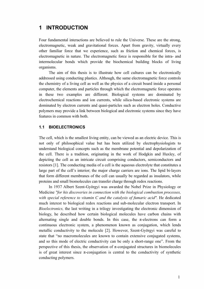
1 INTRODUCTION Four fundamental interactions are believed to rule the Universe. These are the strong, electromagnetic, weak and gravitational forces. Apart from gravity, virtually every other familiar force that we experience, such as friction and chemical forces, is electromagnetic in nature. The electromagnetic force is responsible for the intra- and intermolecular bonds which provide the biochemical building blocks of living organisms.
The aim of this thesis is to illustrate how cell cultures can be electronically addressed using conducting plastics. Although, the same electromagnetic force controls the chemistry of a living cell as well as the physics of a circuit board inside a personal computer, the elements and particles through which the electromagnetic force operates in these two examples are different. Biological systems are dominated by electrochemical reactions and ion currents, while silica-based electronic systems are dominated by electron currents and quasi-particles such as electron holes. Conductive polymers may provide a link between biological and electronic systems since they have features in common with both.
1.1 BIOELECTRONICS The cell, which is the smallest living entity, can be viewed as an electric device. This is not only of philosophical value but has been utilized by electrophysiologists to understand biological concepts such as the membrane potential and depolarization of the cell. There is a tradition, originating in the work of Hodgkin and Huxley, of depicting the cell as an intricate circuit comprising conductors, semiconductors and resistors [1]. The conducting media of a cell is the aqueous electrolyte that constitutes a large part of the cell’s interior; the major charge carriers are ions. The lipid bi-layers that form different membranes of the cell can usually be regarded as insulators, while proteins and small biomolecules can transfer charge through redox reactions.
In 1937 Albert Szent-Györgyi was awarded the Nobel Prize in Physiology or Medicine "for his discoveries in connection with the biological combustion processes, with special reference to vitamin C and the catalysis of fumaric acid". He dedicated much interest to biological redox reactions and sub-molecular electron transport. In Bioelectronics, the last writing in a trilogy investigating the electronic dimension of biology, he described how certain biological molecules have carbon chains with alternating single and double bonds. In this case, the π-electrons can form a continuous electronic system, a phenomenon known as conjugation, which lends metallic conductivity to the molecule [2]. However, Szent-Györgyi was careful to state that “no macromolecules are known to contain extensive conjugated systems, and so this mode of electric conductivity can be only a short-range one”. From the perspective of this thesis, the observation of π-conjugated structures in biomolecules is of great interest since π-conjugation is central to the conductivity of synthetic conducting polymers.
2
1.2 HISTORY OF CONDUCTING POLYMERS In 1967 Hideki Shirakawa of Kyoto University synthesized a thin polyacetylene film due to a fortunate mistake. When Shirakawa and his co-workers tried to reproduce the error they found that they had used nearly a thousand times more Ziegler-Natta catalyst than usual [3]. The film had a metallic color and was found to be highly conductive. Due to a chance meeting with guest lecturer Alan G. MacDiarmid, Shirakawa was introduced to poly(sulfur nitride) films which had similar conducting properties to polyacetylene films. MacDiarmid was working on poly(sulfur nitride) films together with Alan J Heeger, a physicist and colleague from Pennsylvania University. This meeting was the beginning of a fruitful collaboration, and Shirakawa joined MacDiarmid and Heeger as a guest scientist at Pennsylvania University.
Their collaboration led to the discovery that it was in fact chemical doping of the polymers that yielded the high conductivity [4]. In 2000 they were awarded the Nobel Prize in Chemistry "for the discovery and development of conductive polymers." Although the work of Heeger, MacDiarmid and Shirakawa had major bearing on the recognition of conducting polymers, they were not the first to discover the special properties of conjugated polymers. In 1963 Australian researchers published work on doped polypyrrole with conductivity comparable to more recent polymers [5]. The first functional organic electronic device was based on the conjugated biological polymer melanin, and it operated as an amorphous semiconductor threshold switch [6]. 1.3 INDUSTRIAL APPLICATIONS OF CONDUCTING POLYMERS The electronics industry is currently still dominated by silica-based products. Compared to silicon and other traditional electronic materials, conducting polymers have some promising advantages. Because of their organic origin they can be treated as combustible waste as long as they are not contaminated with conventional electronic materials. The raw materials of organic electronics are many times cheaper than those of conventional electronics which include a number of expensive trace metals. Organic materials can be harvested from both renewable and fossil resources while trace metals are a strictly limited resource. A further advantage is that organic electronics can be fabricated using traditional printing technologies which give low production costs.
Today, conducting polymers perform well in organic solar cells [7], luminescent devices [8] , organic electrochromic displays [9], and even transistors [10]. The QUE ProReader from Plastic Logic, a spinoff company of the Cavendish laboratory at Cambridge, is a good example of a successful product that is based on organic electronics. Thin Film Electronics is a Scandinavian company that develops products based on organic electronics; they focus on card games and intelligent labels.

3
1.4 THE ELECTRONIC FEATURES OF CONDUCTING POLYMERS A common feature of conducting polymers is that the main chain is π-conjugated. The mechanism behind conductivity due to π-conjugation can be explained as follows: In polymer chains with alternating single and double bonds, three of the four outer shell electrons of the carbon atom occupy hybridized states formed from one s and two p states. This is referred to as sp2 hybridization which leads to formation of the strong covalent σ-bond between the carbons in the backbone. Thus, one electron is left free in a pz orbital; these overlap among neighboring carbons and form a π-bond. Each delocalized orbital provides one free electron that theoretically yields metallic behavior to the polymer chain. However, due to the bond alternation, this system is unstable resulting in a band gap in the electronic spectrum. For polyacetylene the band gap is approximately 1.7 eV [4, 11], and for short-chain polyenes it is about 2.4 eV [4]. The band gap gives rise to semiconducting rather than metallic properties. 1.4.1 Doping of conjugated polymers The band gaps reducing the conductivity of conjugated polymers can be overcome by doping. As for inorganic semiconductors, doping can be used to increase the conductivity of an organic semiconductor by several orders of magnitude. However, one significant difference between the doping of inorganic semiconductors and organic semiconductors is the relative amount of dopant which is needed to induce a measurable effect. For inorganic semiconductors, a few parts per million of dopant is sufficient to increase the conductivity by several orders of magnitude. In contrast, several percent of dopant are needed to obtain the same effect for organic semiconductors. The explanation lies in the different physical mechanisms which are responsible for the doping of different classes of materials. In conducting polymers, the dopant and the polymer chain form an ionic complex.
There are several methods of doping conducting polymers, i.e. chemical, electrochemical, photo- and charge-injection doping. Chemical and electrochemical doping are the most commonly used. In chemical doping an oxidizing or reducing agent is mixed with the polymer. Oxidation of the polymer chain induces a positively charged unit in the conjugated system and is referred to as p-doping. Reduction of the polymer induces a negatively charged unit in the conjugated system and is known as n-doping [11]. Electrochemical doping depends on the same mechanism, but instead of an oxidizing or reducing agent, a potential is applied to drive the redox reaction. P-doping is most commonly used since those polymers that can be n-doped are usually unstable in the ambient atmosphere because they react with oxygen in the air. 1.4.2 Charge carriers The charge carriers in conducting polymers are quasi-particles, i.e. solitons, polarons and bipolarons. Solitons are found in polymers with a degenerate ground state such as trans-polyacetylene. In trans-polyacetylene the soliton is a domain boundary between the two possible ground state configurations, the “A” phase and the ”B” phase [12].
4
The soliton can be charged and spinless or neutral with an unpaired spin. Non-degenerate polymers, such as the polythiophenes, commonly exhibit polarons and bipolarons as charge carriers. A polaron can be thought of as a bound state of a charged soliton and a neutral soliton whose mid-gap energy states hybridize and form bonding and anti-bonding levels. A bipolaron can be thought of as a bound state of two charged solitons of similar charge.
Two main factors determine the conductivity of a conducting polymer, the number of charge carriers, and the arrier mobility. The conductivity as determined by the relationship between tors is defined as
charge c these fac
σ is the conductivity, n is the concentration of charge carriers, e is the charge of an electron, and μ is the charge carrier mobility. However, if the concentration of charge carriers increases above a certain level, the conductivity will start to decrease because of interactions between the charge carriers. The highest conductivity is obtained when the polymer has both charged and uncharged sites into which the charge carriers can move. Consequently, there is a finite window of high conductivity for conducting polymers; this window is found at the potential interval for which charge both can be injected into and withdrawn from the polymer [13]. 1.4.3 Optical properties of conjugated polymers Conducting polymers generally exhibit electro-chromic features which are associated with the quasi-particles induced by doping. The outcome of the electro-chromic effect is dependent on the band gap of the polymer. If the energy gap exceeds 3 eV the un-doped polymer is usually transparent, while the doped polymer is absorbing in the visible wave length. If the energy gap is small, i.e. 1-1.5 eV, the un-doped polymer will be colored and the doped polymer transparent [14]. This shift in the absorbance spectra may be used in conducting polymer displays operated by electrochemical doping [15, 16].
Conducting polymers can also be made to emit light as, for example, in organic light emitting diodes. In this case, light is emitted when a sufficient bias is applied to achieve injection of positive and negative charge carriers from opposite electrodes. Photo emission occurs when oppositely charged carriers within the polymer meet, and is due to formation of neutral bound excited states called excitons [17]. Excitons can also be formed when energy in the form of a photon matching the band gap of the polymer is transferred to the system. These excitons typically have a lifetime of nanoseconds; when the electronic ground state is restored, the polymer undergoes fluorescence, emitting light of a longer wavelength than the absorbed wavelength. Intermolecular interactions between polymers can induce spectral shifts and alteration in photoluminescent intensity [18]. This effect has been used in a special class of polymers which have been supplied with charged side chains rendering them soluble in aqueous solutions [19]. These polymers have found applications as markers for amyloidal structures in biological materials [20].

5
1.5 ELECTROCHEMISTRY IN ORGANIC ELECTRONICS Electrochemistry is the branch of chemistry that deals with the reactions that occur between an electronic conductor and an ionic conductor. The Italian physician Luigio Galvani is usually given credit as the founder of electrochemistry since he was the first to report on the connection between electricity and chemistry in his essay De Viribus Electricitatis in Motu Musculari Commentarius in the 18th century [21]. Later, in the mid 20th century, Hodgkin and Huxley would describe how the observations that Galvani had made were indeed due to electrochemical potentials produced by neuronal tissue to transmit signals throughout the body at high speed. Electrochemistry is essential to the understanding of organic electronics. The electrolytic ions migrate toward and into the anode or cathode, depending on whether they are negatively or positively charged. They subsequently donate or accept electrons, respectively, thereby taking part in the doping of the polymer (Figure 1.).
Figure 1. When a potential is applied over two conducting polymer electrodes in an electrolyte, anions will migrate into the anode to compensate for the oxidation of the polymer and cations will migrate into the cathode to compensate for the reduction.
1.5.1 Reduction and oxidation Electrochemical redox reactions are fundamental for the operation, as well as for electrochemical doping and undoping, of conducting polymer devices in electrolytes. The principles of these redox reactions can be illustrated by viewing the polymer and electrolyte as an electrochemical half cell.
A conducting polymer electrode of an arbitrary polymer which is submerged into an electrolyte and biased using a voltage source and a counter electrode will be reduced or oxidized depending on the polarity of the potential. At equilibrium the potential relates to the ratio of reduced and oxidized species according to the Nernst equation:
E is the applied potential, E0 is the redox potential, T is the temperature in Kelvin, n is the number of electrons transferred in the reaction, k is Boltzmann’s constant, and [Ox] and [Red] are the concentrations of oxidized and reduced species, respectively.
The redox reactions taking place at the respective electrodes are balanced by the
6
migration of mobile ions in and out of the polymer. These ions can be endogenous ions from the electrolyte or mobile dope ions from the polymer. 1.5.2 The electrochemical transistor The reduction and oxidation reactions described above introduce positive and negative charges which, depending on the ground state of the doped polymer, can undope the polymer or dope it even further. Since electrochemical doping affects the conductivity of the polymer, this phenomenon can be used to gate a current that runs through the polymer.
Some very early electrochemical transistors were junction transistors with an emitter, a base, and a collector but these were not made of conducting polymers [22]. The first electrochemical transistors based on conducting polymers was presented in 1984 [23]. Electrochemical transistors made of conducting polymers usually function according to the principles of the field effect transistor with a channel and a gate. But the gating of the channel in the electrochemical transistor is rather due to redox reactions than an electric field. True field effect transistors made of conducting polymers were presented in 1987 [24]. Today the use of these organic field effect transistors is advancing rapidly; they are, for example, successfully integrated as active components of displays.
The organic electrochemical transistor can have different designs. The most common are variations of the “sandwich” design, familiar from the field effect transistors in which the gate is situated on top of or below the channel. Since the gating in the electrochemical transistor is dependent on redox reactions in the channel, the gate can be an electrode but it might just as well be a reducing or oxidizing chemical compound. This has encouraged scientists to use the organic electrochemical transistor as a sensor for different compounds that are redox active or can be catalyzed by redox active enzymes [25]. 1.6 THE EUKARYOTIC CELL Cellular life can be divided into three major domains: archae, prokaryots and eukaryots. Among these it is only the eukaryots that can give rise to higher order organisms such as plants and animals. The eukaryotic cells of higher organisms are highly specialized and social. In some tissues such as muscle, the cells completely lose their single cell integrity and merge into a multinuclear syncytium with a common cytoplasm. In other tissues such as epithelia, the cells are still discrete entities but their cytoplasm is connected through gap junctions, and they are mechanically connected via desmosomes. Brief descriptions of relevant structures and features of the eukaryotic cell will be given in the following sections.
7
1.6.1 Voltage-gated ion channels In biological organisms, electrical signaling is dependent on voltage sensitive ion channels which traverse the plasma membrane. Electrical signaling and voltage-gated ion channels have an important role in the nervous system when information needs to be transported fast over long distances, for example, from the brain to the muscles in the legs. Voltage-gated ion channels react to changes in the membrane potential, and mediate physiological phenomena such as the action potential, contraction of muscle cells, and secretion of hormones and neurotransmitters. There are voltage-gated ion channels for all common physiological ions, i.e. K+, Na+, Ca2+, and Cl- [26-29]. The ion channels are highly selective for each of these ions and utilize them for different purposes.
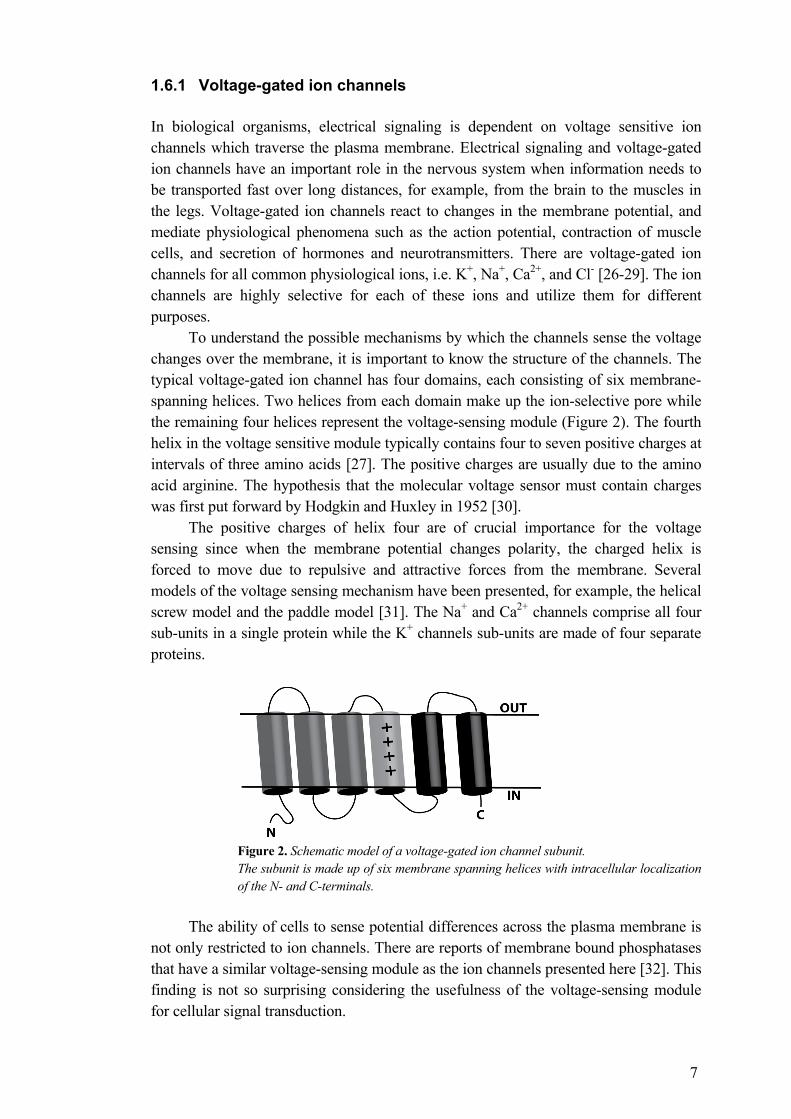
To understand the possible mechanisms by which the channels sense the voltage changes over the membrane, it is important to know the structure of the channels. The typical voltage-gated ion channel has four domains, each consisting of six membrane- spanning helices. Two helices from each domain make up the ion-selective pore while the remaining four helices represent the voltage-sensing module (Figure 2). The fourth helix in the voltage sensitive module typically contains four to seven positive charges at intervals of three amino acids [27]. The positive charges are usually due to the amino acid arginine. The hypothesis that the molecular voltage sensor must contain charges was first put forward by Hodgkin and Huxley in 1952 [30].
The positive charges of helix four are of crucial importance for the voltage sensing since when the membrane potential changes polarity, the charged helix is forced to move due to repulsive and attractive forces from the membrane. Several models of the voltage sensing mechanism have been presented, for example, the helical screw model and the paddle model [31]. The Na+ and Ca2+ channels comprise all four sub-units in a single protein while the K+ channels sub-units are made of four separate proteins.
Figure 2. Schematic model of a voltage-gated ion channel subunit. The subunit is made up of six membrane spanning helices with intracellular localization of the N- and C-terminals.
The ability of cells to sense potential differences across the plasma membrane is not only restricted to ion channels. There are reports of membrane bound phosphatases that have a similar voltage-sensing module as the ion channels presented here [32]. This finding is not so surprising considering the usefulness of the voltage-sensing module for cellular signal transduction.
8
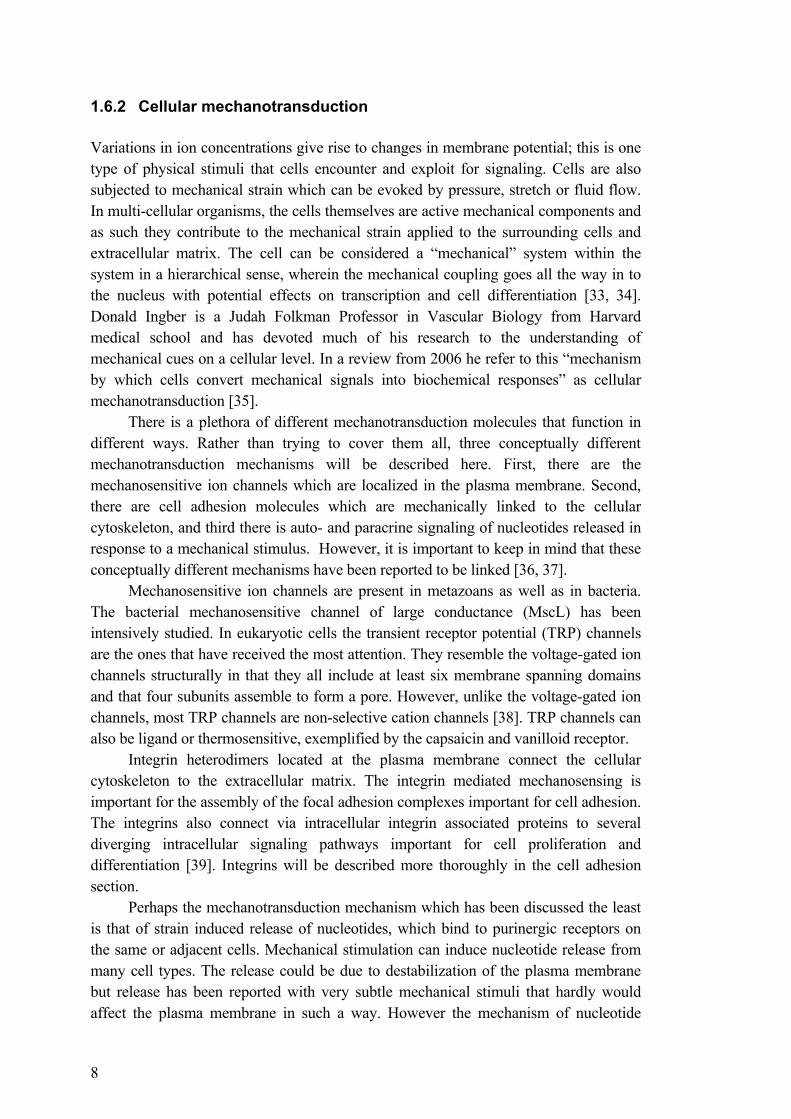
1.6.2 Cellular mechanotransduction Variations in ion concentrations give rise to changes in membrane potential; this is one type of physical stimuli that cells encounter and exploit for signaling. Cells are also subjected to mechanical strain which can be evoked by pressure, stretch or fluid flow. In multi-cellular organisms, the cells themselves are active mechanical components and as such they contribute to the mechanical strain applied to the surrounding cells and extracellular matrix. The cell can be considered a “mechanical” system within the system in a hierarchical sense, wherein the mechanical coupling goes all the way in to the nucleus with potential effects on transcription and cell differentiation [33, 34]. Donald Ingber is a Judah Folkman Professor in Vascular Biology from Harvard medical school and has devoted much of his research to the understanding of mechanical cues on a cellular level. In a review from 2006 he refer to this “mechanism by which cells convert mechanical signals into biochemical responses” as cellular mechanotransduction [35].
There is a plethora of different mechanotransduction molecules that function in different ways. Rather than trying to cover them all, three conceptually different mechanotransduction mechanisms will be described here. First, there are the mechanosensitive ion channels which are localized in the plasma membrane. Second, there are cell adhesion molecules which are mechanically linked to the cellular cytoskeleton, and third there is auto- and paracrine signaling of nucleotides released in response to a mechanical stimulus. However, it is important to keep in mind that these conceptually different mechanisms have been reported to be linked [36, 37].
Mechanosensitive ion channels are present in metazoans as well as in bacteria. The bacterial mechanosensitive channel of large conductance (MscL) has been intensively studied. In eukaryotic cells the transient receptor potential (TRP) channels are the ones that have received the most attention. They resemble the voltage-gated ion channels structurally in that they all include at least six membrane spanning domains and that four subunits assemble to form a pore. However, unlike the voltage-gated ion channels, most TRP channels are non-selective cation channels [38]. TRP channels can also be ligand or thermosensitive, exemplified by the capsaicin and vanilloid receptor.
Integrin heterodimers located at the plasma membrane connect the cellular cytoskeleton to the extracellular matrix. The integrin mediated mechanosensing is important for the assembly of the focal adhesion complexes important for cell adhesion. The integrins also connect via intracellular integrin associated proteins to several diverging intracellular signaling pathways important for cell proliferation and differentiation [39]. Integrins will be described more thoroughly in the cell adhesion section.
Perhaps the mechanotransduction mechanism which has been discussed the least is that of strain induced release of nucleotides, which bind to purinergic receptors on the same or adjacent cells. Mechanical stimulation can induce nucleotide release from many cell types. The release could be due to destabilization of the plasma membrane but release has been reported with very subtle mechanical stimuli that hardly would affect the plasma membrane in such a way. However the mechanism of nucleotide
9
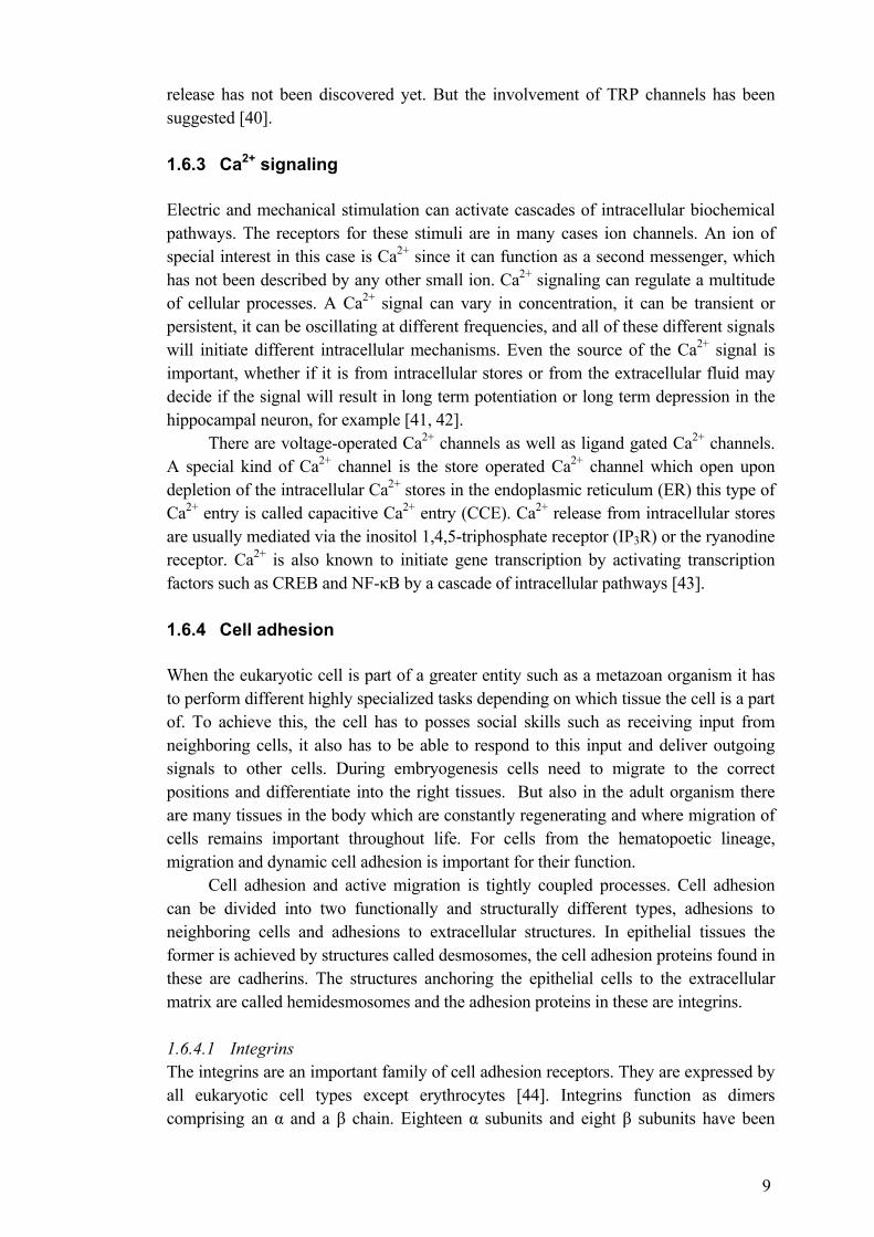
release has not been discovered yet. But the involvement of TRP channels has been suggested [40]. 1.6.3 Ca2+ signaling Electric and mechanical stimulation can activate cascades of intracellular biochemical pathways. The receptors for these stimuli are in many cases ion channels. An ion of special interest in this case is Ca2+ since it can function as a second messenger, which has not been described by any other small ion. Ca2+ signaling can regulate a multitude of cellular processes. A Ca2+ signal can vary in concentration, it can be transient or persistent, it can be oscillating at different frequencies, and all of these different signals will initiate different intracellular mechanisms. Even the source of the Ca2+ signal is important, whether if it is from intracellular stores or from the extracellular fluid may decide if the signal will result in long term potentiation or long term depression in the hippocampal neuron, for example [41, 42].
There are voltage-operated Ca2+ channels as well as ligand gated Ca2+ channels. A special kind of Ca2+ channel is the store operated Ca2+ channel which open upon depletion of the intracellular Ca2+ stores in the endoplasmic reticulum (ER) this type of Ca2+ entry is called capacitive Ca2+ entry (CCE). Ca2+ release from intracellular stores are usually mediated via the inositol 1,4,5-triphosphate receptor (IP3R) or the ryanodine receptor. Ca2+ is also known to initiate gene transcription by activating transcription factors such as CREB and NF-κB by a cascade of intracellular pathways [43]. 1.6.4 Cell adhesion When the eukaryotic cell is part of a greater entity such as a metazoan organism it has to perform different highly specialized tasks depending on which tissue the cell is a part of. To achieve this, the cell has to posses social skills such as receiving input from neighboring cells, it also has to be able to respond to this input and deliver outgoing signals to other cells. During embryogenesis cells need to migrate to the correct positions and differentiate into the right tissues. But also in the adult organism there are many tissues in the body which are constantly regenerating and where migration of cells remains important throughout life. For cells from the hematopoetic lineage, migration and dynamic cell adhesion is important for their function.
Cell adhesion and active migration is tightly coupled processes. Cell adhesion can be divided into two functionally and structurally different types, adhesions to neighboring cells and adhesions to extracellular structures. In epithelial tissues the former is achieved by structures called desmosomes, the cell adhesion proteins found in these are cadherins. The structures anchoring the epithelial cells to the extracellular matrix are called hemidesmosomes and the adhesion proteins in these are integrins.
1.6.4.1 Integrins The integrins are an important family of cell adhesion receptors. They are expressed by all eukaryotic cell types except erythrocytes [44]. Integrins function as dimers comprising an α and a β chain. Eighteen α subunits and eight β subunits have been
10
characterized in mammals and in vertebrates there are 24 known αβ heterodimers [45]. Some of the best known heterodimers are the vitronectin receptor αvβ3, the laminin receptor α2β1, the collagen receptor α3β1, and the fibronectin receptor α5β1.
Integrins not only mediate cell adhesion, but they are also signaling molecules. As such they can carry information in two directions, the terms outside-in and inside-out signaling is popular when describing the signaling activity of integrins. There is often an element of mechanotransduction associated with the signaling of integrins [46]. The α and β subunits of the heterodimer have different roles. The α-subunit is believed to determine the specificity of the extracellular ligand binding, while the β-subunit is primarily interacting with the intracellular components through its cytoplasmic tail.
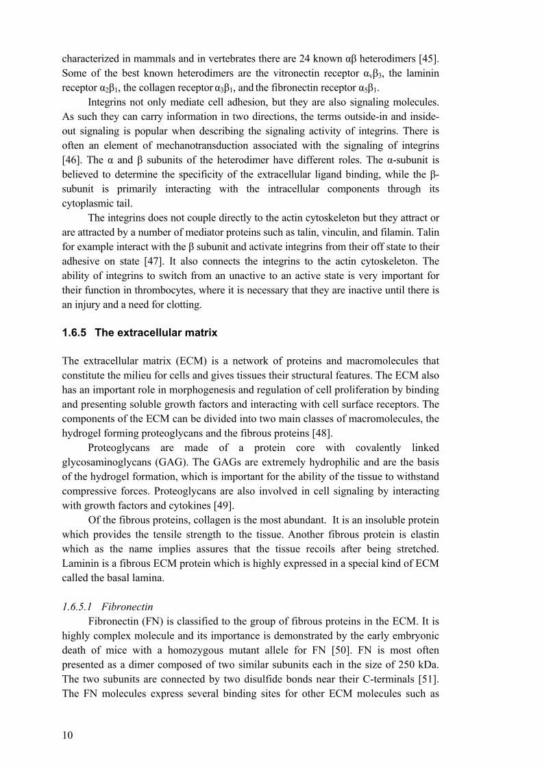
The integrins does not couple directly to the actin cytoskeleton but they attract or are attracted by a number of mediator proteins such as talin, vinculin, and filamin. Talin for example interact with the β subunit and activate integrins from their off state to their adhesive on state [47]. It also connects the integrins to the actin cytoskeleton. The ability of integrins to switch from an unactive to an active state is very important for their function in thrombocytes, where it is necessary that they are inactive until there is an injury and a need for clotting. 1.6.5 The extracellular matrix The extracellular matrix (ECM) is a network of proteins and macromolecules that constitute the milieu for cells and gives tissues their structural features. The ECM also has an important role in morphogenesis and regulation of cell proliferation by binding and presenting soluble growth factors and interacting with cell surface receptors. The components of the ECM can be divided into two main classes of macromolecules, the hydrogel forming proteoglycans and the fibrous proteins [48].
Proteoglycans are made of a protein core with covalently linked glycosaminoglycans (GAG). The GAGs are extremely hydrophilic and are the basis of the hydrogel formation, which is important for the ability of the tissue to withstand compressive forces. Proteoglycans are also involved in cell signaling by interacting with growth factors and cytokines [49].
Of the fibrous proteins, collagen is the most abundant. It is an insoluble protein which provides the tensile strength to the tissue. Another fibrous protein is elastin which as the name implies assures that the tissue recoils after being stretched. Laminin is a fibrous ECM protein which is highly expressed in a special kind of ECM called the basal lamina.
1.6.5.1 Fibronectin
Fibronectin (FN) is classified to the group of fibrous proteins in the ECM. It is highly complex molecule and its importance is demonstrated by the early embryonic death of mice with a homozygous mutant allele for FN [50]. FN is most often presented as a dimer composed of two similar subunits each in the size of 250 kDa. The two subunits are connected by two disulfide bonds near their C-terminals [51]. The FN molecules express several binding sites for other ECM molecules such as
11
collagen, heparin and other FN molecules. FN exists in to different forms as soluble FN in plasma and as fibrillar FN in the ECM. The fibrillisation of FN is an active process that is mediated by cells binding FN. The cell driven fibrillisation of FN is dependent on integrins.
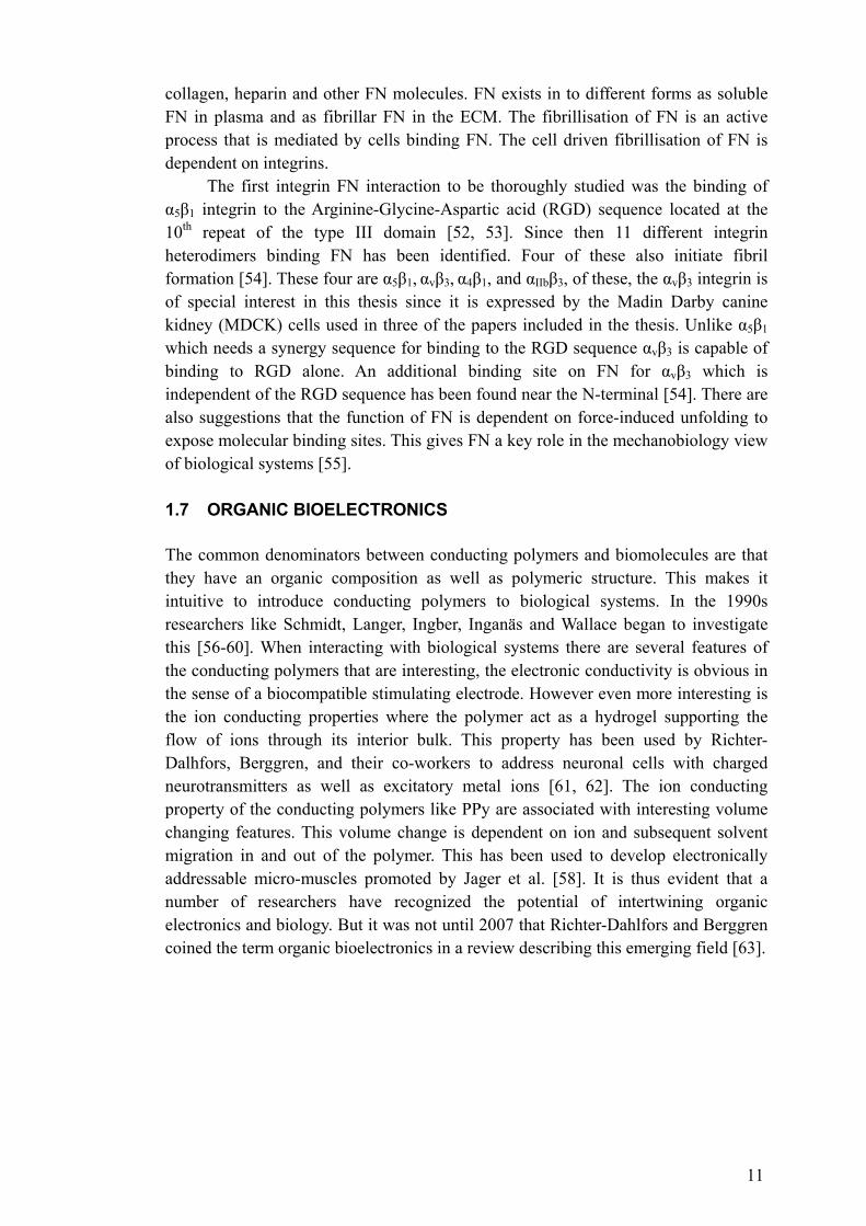
The first integrin FN interaction to be thoroughly studied was the binding of α5β1 integrin to the Arginine-Glycine-Aspartic acid (RGD) sequence located at the 10th repeat of the type III domain [52, 53]. Since then 11 different integrin heterodimers binding FN has been identified. Four of these also initiate fibril formation [54]. These four are α5β1, αvβ3, α4β1, and αIIbβ3, of these, the αvβ3 integrin is of special interest in this thesis since it is expressed by the Madin Darby canine kidney (MDCK) cells used in three of the papers included in the thesis. Unlike α5β1 which needs a synergy sequence for binding to the RGD sequence αvβ3 is capable of binding to RGD alone. An additional binding site on FN for αvβ3 which is independent of the RGD sequence has been found near the N-terminal [54]. There are also suggestions that the function of FN is dependent on force-induced unfolding to expose molecular binding sites. This gives FN a key role in the mechanobiology view of biological systems [55]. 1.7 ORGANIC BIOELECTRONICS The common denominators between conducting polymers and biomolecules are that they have an organic composition as well as polymeric structure. This makes it intuitive to introduce conducting polymers to biological systems. In the 1990s researchers like Schmidt, Langer, Ingber, Inganäs and Wallace began to investigate this [56-60]. When interacting with biological systems there are several features of the conducting polymers that are interesting, the electronic conductivity is obvious in the sense of a biocompatible stimulating electrode. However even more interesting is the ion conducting properties where the polymer act as a hydrogel supporting the flow of ions through its interior bulk. This property has been used by Richter-Dalhfors, Berggren, and their co-workers to address neuronal cells with charged neurotransmitters as well as excitatory metal ions [61, 62]. The ion conducting property of the conducting polymers like PPy are associated with interesting volume changing features. This volume change is dependent on ion and subsequent solvent migration in and out of the polymer. This has been used to develop electronically addressable micro-muscles promoted by Jager et al. [58]. It is thus evident that a number of researchers have recognized the potential of intertwining organic electronics and biology. But it was not until 2007 that Richter-Dahlfors and Berggren coined the term organic bioelectronics in a review describing this emerging field [63].

12
2 AIMS OF THE THESIS The aim of this thesis is to develop and evaluate organic electronic devices that address different levels of cellular physiology. The levels addressed are as follows:
1. Electrical activation of voltage-gated ion channels 2. Cellular mechanotransduction 3. Cell adhesion and proliferation by modulating cellular interaction with the
extracellular matrix 4. Detachment of cells without disrupting extracellular proteins
13
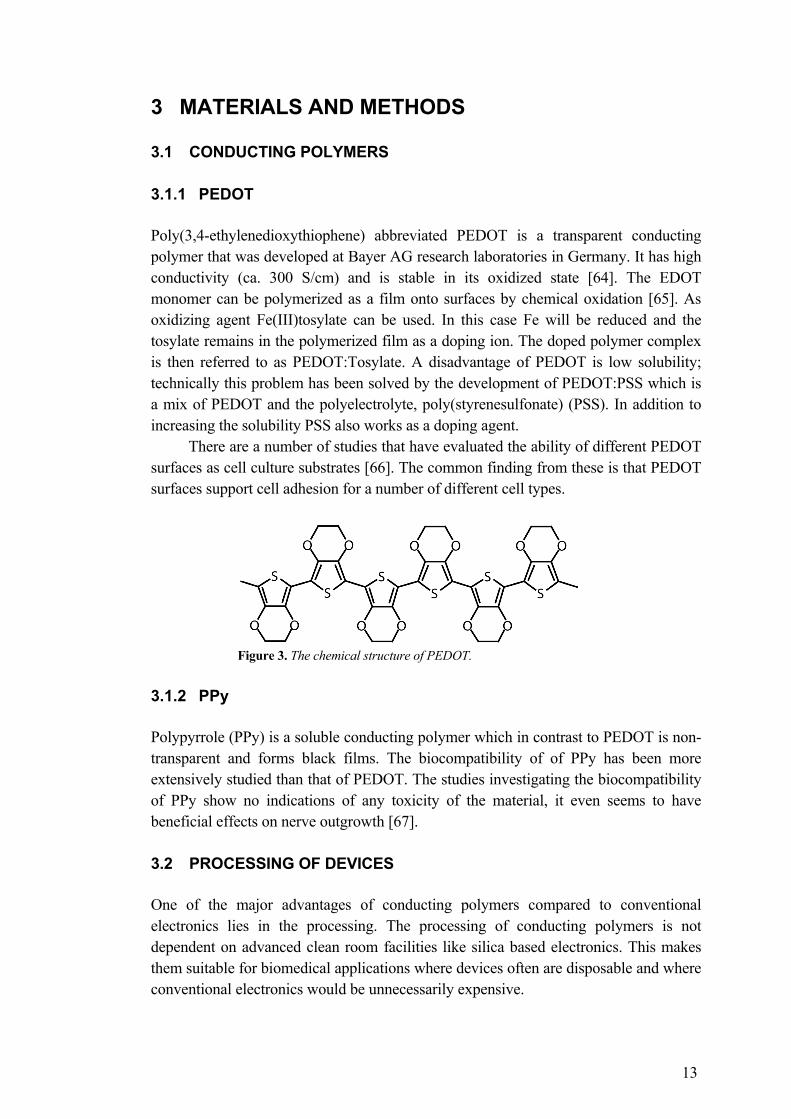
3 MATERIALS AND METHODS 3.1 CONDUCTING POLYMERS 3.1.1 PEDOT Poly(3,4-ethylenedioxythiophene) abbreviated PEDOT is a transparent conducting polymer that was developed at Bayer AG research laboratories in Germany. It has high conductivity (ca. 300 S/cm) and is stable in its oxidized state [64]. The EDOT monomer can be polymerized as a film onto surfaces by chemical oxidation [65]. As oxidizing agent Fe(III)tosylate can be used. In this case Fe will be reduced and the tosylate remains in the polymerized film as a doping ion. The doped polymer complex is then referred to as PEDOT:Tosylate. A disadvantage of PEDOT is low solubility; technically this problem has been solved by the development of PEDOT:PSS which is a mix of PEDOT and the polyelectrolyte, poly(styrenesulfonate) (PSS). In addition to increasing the solubility PSS also works as a doping agent.
There are a number of studies that have evaluated the ability of different PEDOT surfaces as cell culture substrates [66]. The common finding from these is that PEDOT surfaces support cell adhesion for a number of different cell types.
Figure 3. The chemical structure of PEDOT.
3.1.2 PPy Polypyrrole (PPy) is a soluble conducting polymer which in contrast to PEDOT is non-transparent and forms black films. The biocompatibility of of PPy has been more extensively studied than that of PEDOT. The studies investigating the biocompatibility of PPy show no indications of any toxicity of the material, it even seems to have beneficial effects on nerve outgrowth [67]. 3.2 PROCESSING OF DEVICES One of the major advantages of conducting polymers compared to conventional electronics lies in the processing. The processing of conducting polymers is not dependent on advanced clean room facilities like silica based electronics. This makes them suitable for biomedical applications where devices often are disposable and where conventional electronics would be unnecessarily expensive.
14
When the aim is to develop conducting polymer devices for biomedical applications it is well worth to spend a few lines of text to discuss the processing, since different physical and chemical processing steps can have large effects on the interaction between the devices and biological materials. Such effects can often be difficult to predict from the electronics perspective. Here, we will present some of the processing techniques used in this thesis. 3.2.1.1 Spin coating Spin coating is a procedure in which the centrifugal force is used to coat a flat surface with a material. In short, the surface to be coated is placed on a rotating disc, and the material is applied as a liquid at the center of surface and the liquid is then spread out on the surface due to the centrifugal force induced by the rotating disc.
The spin coating of conducting polymers in this thesis is made by adding a mixture of the monomer and an oxidizing agent which is spin coated and polymerized in situ. Once the oxidation agent and the monomer is mixed the polymerization begins a method to control the polymerization process and achieve smoth and homogenous films is to ad a strong base to the reaction mixture This method to polymerize conducting polymers in situ was presented by DeLeeuw et al. in 1994 [65]. 3.2.1.2 Barcoating Barcoating is a method to deploy a polymer film onto a substrate. The principle is that the polymer solution is evenly spread onto the substrate by a bar that is advanced over the substrate. This method allows for larger surfaces to be coated than the spin coating method. Similarly to the spin coating method the monomer and the other reagents are mixed before the coating. 3.2.1.3 Vapor phase polymerization Vapor phase polymerization is a good processing technique when the aim is to coat thin polymer films onto substrates with more complicated geometries than plain flat surfaces. Vapor deposition of conducting polymers was described by Mohammadi et al. [68]. The quality of the vapor deposited films can be enhanced if the oxidizing agent, preferably a ferric sulfonate is coated onto the surface in advance by spin or dip coating. When the monomer is vaporized onto the oxidizing agent it polymerizes and form a smooth homogenous film [69]. Physical properties of the film such as conductance, absorbance and thickness can be regulated by controlling the temperature during the polymerization process. 3.2.1.4 Electropolymerization Conducting polymers in solution can also be electropolymerized onto a conducting substrate the only limitation in this case is that the substrate itself has to be conducting. An advantage with electropolymerization is that the thickness of the film can be controlled with the amount of current that is applied. Also the selection of possible dope ions is huge with electropolymerization since the only criterion is basically that the doping agent carries a charge. In electropolymerizatiom the monomer must be present in an electrolyte, this has been used in a creative way by Martin and co-workers
15
[70]. They have used the extracellular fluid as electrolyte and electropolymerized conducting polymers in living nervous tissue. 3.2.1.5 Electrospinning Electrospinning is a process that uses an electric field to spin very fine fibers from a liquid solution. In principle a field is applied between a nozzle containing the liquid and a grounded collector. The potential charges the liquid and when it’s been sufficiently charged the electrostatic forces ejects the liquid from the nozzle which starts to polymerize in the flight and so the fiber is formed.
The phenomenon was observed already in the 16th century by William Gilbert who observed it when he brought a charged piece of amber close to a water droplet. The first patents that tried to commercialize electrospinning were filed in February 1902 by J.F Cooley. At present electrospinning is primarily used to fabricate nanofibres. 3.2.1.6 Photolithography Photolithography is a processing technique commonly used in the electronics industry. It uses light sensitive resists that can be patterned onto surfaces with photo masks. The patterned photoresists can then withstand the etching chemical that etches into the silicon wafer. In electronics processing, a silicon wafer can go through up to 50 cycles of etching and photoresist. 3.3 CELL CULTURE SYSTEMS The ability to grow cells was an important methodological advancement for contemporary biology and medicine. Early progress was made by Ringer in the 19th century when he was able to maintain a beating hearth outside of the donor animal by using different physiological salt solutions. The ability to culture cells has also been very important for research in virology and the development of vaccines, as viruses needs a cellular host for their propagation. 3.3.1 SH-SY5Y The SH-SY5Y cell line was first described in 1978 [71]. It is a subclone of the SK-N-SH neuroblastoma cell line which was isolated from a metastatic neuroblastoma site through a bone marrow biopsy from a four year old female. The SH-SY5Y cells have dopamine β-hydroxylase activity and express acetylcholine receptors [72]. The cells can be differentiated to a phenotype with more neurites using retinoic acid. The SH-SY5Y cells express voltage-operated Ca2+ channels [28]. Therefore they can be used to study voltage induced Ca2+ signaling. 3.3.2 MDCK The Madin Darby canine kidney (MDCK) cell line deserves a short presentation of its own as it has been used in three out of five papers in this thesis. The cell line was

16
isolated from the kidney of a healthy cocker spaniel in 1958 by S.H. Madin and N.B. Darby. The isolation of the MDCK cells has not been published but the isolation of bovine and ovine kidney cells by Madin and Darby was published in the same year [73]. The characterization of the MDCK cell line was published in 1966, at the time it was already established for the study of virus host-cell interactions [74]. In the early publications describing the MDCK cell line, it is reported as originating from a male animal. Although the characterization in 1966 failed to identify a Y chromosome and the cell line is described as originating from a female animal in the documentation from American type culture collection (ATCC). In 1969 Leighton and co-workers observed polarized structures such as tight junctions and microvillus processes in MDCK cultures propagated on glass surfaces [75]. These findings of the well retained differentiated phenotype of MDCK cells paved the way for their popularity as a model for the study of epithelia. The polarization of ion channels and the effect on trans epithelial potential differences can be studied by culturing the monolayer on porous membranes [76].
Because of its kidney origin and differentiated phenotype it has become popular with scientists studying renal physiology and tubulogenesis [77, 78]. The expression of integrins is fairly consistent to what you find in the distal tubuli [79, 80]. Another physiologically interesting feature of the MDCK cell line is that it can change between a migrating mesenchymal and a stationary epithelial phenotype [81]. This transduction can be induced by the hepatocyte growth factor (HGF) via the c-met receptor [82]. This is interesting both with regards to tubulogenesis but also in regards to how epithelial cancers become invasive. They also have interesting mechanosensing capabilities where purinergic receptors play an important role. 3.3.3 T24 The T24 bladder cancer cell line was isolated from an 81 year old female at the Karolinska hospital in 1970 by Dr J Bubeník [83]. As the MDCK cell line the T24 cells are derived from the urinary tract, an important difference is that unlike the MDCK cells the primary isolate is from cancerous tissue. The T24 cells grow as single cells and do not form epithelial monolayers. The hyaluronan binding, surface antigen CD44 is expressed by T24 cells [84]. 3.4 IMAGING CELL STRUCTURE AND FUNCTION During the last 20 to 30 years the use of fluorescence as readout in the life sciences has increased to become totally dominating. The raw data acquired, especially from microscopy is intuitive to understand and aesthetically pleasing to watch. Even though methods like radiolabeling and electron microscopy provide higher sensitivity and resolution, fluorescence based techniques has become popular due to low toxicity and user friendliness.
17
3.4.1 Fluorescence microscopy Fluorescence is light which is emitted when an electron in a substance is transitioned from an excited state to its ground state. The excited state of the electron can be induced by incoming light, this is called excitation. For a specific fluorophore i.e. a substance that is prone to fluorescence, there is a wavelength interval by which it is excited and another wavelength interval by which the fluorophore emits light. The excitation is usually caused by light of a shorter wavelength than the emitted light. The difference between these wavelengths is called Stokes shift.
Fluorophores can be conjugated to antibodies or other molecules with specificity for a target protein or structure. With the fluorophore bound to the target structure it can be visualized using fluorescence microscopy. In the fluorescence microscope a light source, for example a mercury lamp is used to excite the sample and then a set of filters is used to select emitted light of the right wavelength. Often a set of chromatic mirrors and filters can be used to direct the exciting light through the same lenses and optics as is used for imaging. The advantage of this compared to illuminating through the specimen is that the only light coming into the objective is emitted or reflected and because of stokes shift, reflected light can be filtered out. This technique increases the signal to noise ratio and is called epifluorescence.
Confocal microscopy is another common fluorescence microscopy technique. In confocal microscopy the light source is lasers with specific wavelengths. The key feature of the confocal microscope is the pin hole. The function of the pin hole is to remove out of focus light. In this way the image that is captured is only from the focal plane. By making images of different focal planes a 3D image can be reconstructed. 3.4.2 Ratiometric Ca2+ imaging Intracellular Ca2+ is an important second messenger that regulates many physiological functions in the cell. The use of fluorescent Ca2+ indicators has been very important for the study of intracellular Ca2+ dynamics. The indicators can be genetically encoded fluorescent proteins or engineered fluorescent chemicals [85].
The chemical indicators can be ratiometric or nonratiometric. When a dye is ratiometric it means that it absorbs light at different wavelengths depending on whether Ca2+ is bound or not while non-ratiometric dyes only absorbs light when Ca2+ is bound. A ratiometric dye that is commonly used is Fura-2, which has a peak absorbance at 340 nm in the unbound state which shifts to 380 nm when Ca2+ is bound.
Most chemical Ca2+ indicators are not permeable through the plasma membrane. This problem can be solved by adding an acetoxymetyl (AM) ester to the indicator. The AM ester makes the indicator permeable, but once inside the cell the AM ester is cleaved off by cellular esterase’s, and the indicator is trapped inside the cell [86]. 3.4.3 Flow cytometry Flow cytometry is a technology that is used to analyze cells or other particles in suspension. The basic principle is that the suspended sample flows in a narrow stream
18
past a detector that analyses every single particle. The detector can analyze parameters such as granularity and size by measuring the light scattering. If the sample is labeled with fluorescent probes cells can be analyzed with regard to fluorescence intensity. The fluorescence intensity in this case correlates to the amount target molecules for the probe which is present in the sample. Fluorescence activated cell sorting (FACS) is often used synonymously with flow cytometry. Nevertheless, it is a special kind of flow cytometry where the analyzed cells can be sorted into different containers depending on their fluorescent and light scattering parameters.
19
4 RESULTS AND DISCUSSION 4.1 ELECTRIC CELL STIMULATION (PAPER I) The use of conducting polymers as materials for stimulating electrodes in biological tissues is perhaps one of the most intuitive applications for these materials. In this application conducting polymers have congenital advantages such as low elastic modulus and high charge transfer properties [87, 88]. The low elastic modulus is beneficial since it gives less of a strain mismatch between the soft biological tissue and the electrode. The strain mismatch associated with metal electrodes is responsible for the failure of long term operation of the metal electrodes, since the mismatch induce inflammatory responses that cause scar formation with insulating fibrous tissue around the electrode which severely impairs its function. As for the high charge transfer of the conducting polymers it, at least theoretically, allows for a lower stimulation potential to be used to depolarize the target tissue. A lower stimulation potential is favorable since a higher potential is associated with hydrolysis and unwanted products thereof.
The higher charge transfer effect of conducting polymers is partly due to their higher surface to volume ratio than traditional conductors. This ratio can be increased further by depositing the conducting polymer film onto a nano- structured fibrous scaffold. These scaffolds can be produced in a variety of ways from different materials. Some of the methods to fabricate nanoscaled fibrous scaffolds comprise self-assembly, phase separation, and electrospinning [89]. There has been much interest in nanoscaled polymeric scaffolds in different cell culture applications because of their structural resemblance to the extracellular matrix. The material of choice can be either synthetic, biological, or different mixtures of these. For many cell biology applications though, the use of collagen gels is the most convenient matrix solution. However, synthetic materials have the advantage of being less inclined to include biological contamination.
In our study we coat PET nano-fibers with PEDOT:Tosylate to achieve a cell culture substrate which also can function as a stimulating electrode. In this case the nano-fibers have two purposes, they increase the surface area of the electrode thus facilitating charge transfer and they provide a structural morphology for the cells that resembles the extracellular matrix. The nano-fibers have a core shell structure with electrospun PET as the core and vapor phase polymerized PEDOT:Tosylate as the shell. We also tried to spin coat and chemically polymerize the PEDOT:Tosylate but this procedure buried the PET fibers in PEDOT:Tosylate abolishing the porous structure whilst the vapor phase polymerization yielded an even coating around the PET fibers conserving the nano-fiber morphology. The PET fiber that are inherently hydrophobic became super hydrophilic upon coating with PEDOT:Tosylate. PEDOT:Tosylate is a good choice when considering the long term stability of the electrode properties of the polymer when it is operated in physiological solution such as cell culture media [90].
The SH-SY5Y cells that we use to show proof of principle with the PEDOT
20
based nano-fiber scaffold electrode, was chosen since it is a robust cell line that express voltage-operated Ca2+ channels (VOCCs) even when they are undifferentiated [72, 91, 92]. In these experiments the pore size of the fibers which was less than 5-10 µm did not seem to promote infiltration of whole cells into the polymer mesh, though neurite extensions from the cells follow the topography of the nano-fibers. The small pore size itself should not pose a limitation for infiltration of neuronal cells. An increased infiltration of cells in the fiber mesh could probably be accomplished by incorporating neurotrophic ligands into the PEDOT polymer. We assayed the electrode quality of the nano-fibers by preloading the SH-SY5Y cells with the calcium sensitive dye Fura-2, and then stimulated the cells with -3.0 V through the conductive fibers, which activated VOCCs and subsequent Ca2+ influx. A second impulse resulted in a slower response with lower amplitude. This can be explained by desensitization of the cells via down regulation of the VOCCs which is known to be more efficient in undifferentiated SH-SY5Y cells than differentiated [93].
In conclusion this work shows how electrospinning and conducting polymers can be combined to fabricate an electrode material that can host and stimulate excitable cells. 4.2 A MICROACTUATOR FOR MECHANICAL STIMULATION (PAPER II) A cell is not an isolated entity but is naturally located in a milieu which in a multi-cellular organism is comprised of neighboring cells and an extracellular matrix. In this milieu the cell will be subjected to different physical stimuli of one which is mechanical stimuli. It is important for the cell to be able to sense and respond to these mechanical stimuli. This ability is referred to as cellular mechanotransduction and is essential for the regulation of many physiological functions [94-96]. For the study of mechanotransduction in cell and tissue cultures there is a number of different methods that can be used to supply mechanical stimulus, using stretchable silicon rubber substrates, glass micropipetts, flow chambers, and so on [97-99].
In the second paper of this thesis we contribute to the field of cellular mechanotransduction by developing a chip for mechanical stimulation on a cellular level. The chip has microactuator surfaces which are made of alternating lines of the photoresist SU-8 and PPy. PPy which is a water soluble conducting polymer expands when it is biased with a negative voltage in an electrolyte. The applied potential reduces the polymer, a reaction which is compensated for by intercalation of ions from the electrolyte. The ion migration will carry water into the polymer by osmosis and therefore force the polymer to expand [100]. As both topographical structures and electric potentials can affect cells the chip was designed to comprise control areas for these factors [27, 101]. The dimensions of the chip made it easy to handle and it could be used with conventional cell biology imaging techniques. We chose to investigate mechanical stimulation of MDCK cells, since mechanotransduction in renal epithelia is a physiologically relevant phenomena [102]. We investigated the role of the nucleotide adenosine-5’-triphosphate (ATP), in the mechanotransdution responses of the MDCK cells. This was done by using the enzyme ATPase to inhibit the effect of auto- and paracrine ATP signaling [103].
21
In comparison to how the conducting polymer is used in paper I, we have added another parameter between the electronic addressing and the cellular response. In this case the primary stimulus is the mechanical strain that is applied to the cell through the expansion of the polymer and the electroactivity of the polymer is secondary with regards to the cellular response. We also find that the response we trigger in the cells is to a major extent by the mechanical stimulation. The design of the chip made it possible to observe cellular responses that where due to the operation potentials of the chip itself.
From paper II we could draw some important conclusions. Such as that the chip is well designed to be used together with microscopy techniques used in cell biology. In addition, that the responses seen in the MDCK cells where mainly due to ATP mediated mechanotransduction but that there also seem to be mechanisms other than mechanotransduction involved as well. 4.3 REGULATING CELL ADHESION (PAPER III AND IV) 4.3.1 Surface chemistry of cell culture substrates Cell adhesion is fundamental for the development of multicellular organisms. Cell adhesion occurs between neighboring cells as well as to the extracellular matrix. Many of the protein complexes involved in cell adhesion supply direct cues to the individual cells on how to proliferate and differentiate. Structural biology studies have supplied comprehensive knowledge of the molecular structure and function of some of the macromolecular units involved in cell adhesion. Yet on a molecular scale not so much is known about the dynamics of these macromolecules. Especially about the physical properties of the immediate environment of the macromolecule and which impact this has for the fate of the living cell.
For the cultured cell an important part of the immediate environment is the solid support on which the cells adhere. This support may be glass, cell culture treated polystyrene or polycarbonate filters. Physical surface properties will have an effect on the behavior of cells and different cells will respond differently to the different properties. Physical factors of the surface that is central are surface energy, topography, surface charge and modulus. It is important to remember that cells do not adhere directly onto the surface but to extracellular proteins which can be supplied via the culture media or secreted by the cells themselves. It is via these extracellular proteins that the features of the substrate surface exert its effect on cell adhesion.
The surface energy and the closely related parameter wettability have a significant impact upon protein adsorption but also on the functional status of the adsorbed proteins. The relation between we ab and surface energy is described by the Young equation:
tt ilty
cos
Where θ is the measured contact angle between a liquid droplet and a solid surface, is the surface energy of the surrounding vapor, is the solid-liquid interfacial
energy, and is the liquid-vapor surface tension [104, 105].
22
The topography of a surface affects the behavior of cells cultured on the surface. Cells have been shown to adopt their morphology according to micro- and nano-structured surfaces [106, 107]. Topography can also have effects on wettability. On a structured hydrophobic surface a liquid may rest on top of the asperities and is then said to be in the Cassie-Baxter state, which results in a greater contact angle than if it would rest on the planar equivalent of the surface.
Surface charge also contribute to the wetting properties of a surface [108]. Surface charge could affect cell adhesion through different mechanisms. It could affect the conformation of adsorbed proteins as well as the adsorption itself. Unspecific binding of cells due to the charged phospholipid bilayer is also possible. The strongest argument against the effect of surface charge would be that in a solution with a high concentration of electrolytes the Debye length would not reach beyond the thickness of the adsorbed extracellular serum proteins. Therefore proteins interacting with the cells would be effectively shielded from the surface charge by the solvent ions. However, effects on cell adhesion due to surface charge has been reported, but that this is due to secondary effects cannot be excluded [109, 110]. Finally, the modulus of a surface has been reported to have effect not only on cell adhesion, but also on differentiation of stem cells [111, 112]. 4.3.2 Devices for electronically controlled cell culture surfaces When a conducting polymer undergoes oxidation or reduction it changes its chemical properties from that of its ground state. A conducting polymer cell culture surface would thus alter its cell adhesion supporting features upon reduction and oxidation. In paper III we present a spin coated cell culture dish that can be biased with a 1.5 V AA battery. The dish is similar to the cell culture dishes presented by Salto et al. which were coated with PEDOT:Tosylate by vapour phase polymerization [113]. Salto et al. show an effect on the surface energy upon electric addressing of the substrate. They also see effects on protein adsorption and cell adhesion. Therefore we thought this design would be promising to develop further. In paper III we also expand the device repertoire to comprise bar coated PEDOT:Tosylate surfaces as well. These surfaces were significantly easier to handle in the preparation for microscopy specimens.
The possibility to barcoat the PEDOT:Tosylate surfaces was a very important step for the development of the electrochemical transistors we present in paper IV. Electrochemical PEDOT:PSS transistors with a lateral design has been described previously [114]. The potential gradient along the channel of this kind of planar electrochemical transistor has been investigated [115]. To optimize the transistor for cell culture along the channel we scaled it up so that the channel measured 10 mm. We also chose to operate the transistor with a grounded drain contact, in contrast to previous set ups where the ground connects to the source. An effect of this is that the device will not truly act as a transistor with a binary on/off switch as the gate potential is applied. On the other hand, the advantage of this setup is that it provides a potential gradient that have an interval that support cell adhesion, and can be regulated by the gate.
23
4.3.3 Cell adhesion on biased PEDOT:Tosylate surfaces To study the effect on electrical addressing of PEDOT:Tosylate surfaces on cell adhesion we chose the MDCK cell line which has a well differentiated epithelial phenotype. Simple epithelium is functionally a 2D tissue resting on a basal membrane.
The primary finding from the study in paper III was that the number and quality of cells on the anode was affected as compared to cells on the cathode. We investigated the adhesion and proliferation of the MDCK cells cultured on the PEDOT:Tosylate electrodes. In the two electrode system described in paper III we saw that the oxidized positively biased electrode had a lower number of adhered cells then the negatively biased reduced electrode. We also saw a large fraction (20 %) of cells with fragmented chromatin nuclei on the oxidized electrode. Fragmented chromatin is an indication of apoptosis [116].
Since many adherent cell types including MDCK cells are dependent on survival signals from the ECM we hypothesized that the low adhesion and high number of unviable cells were connected [117, 118]. In a first attempt to test this hypothesis we allowed cells to adhere to the surface overnight. This was to let the cells form mature adhesions to the ECM as well as to neighboring cells before we applied the redox potential for another 24 h. Since the adhesions were already formed when the redox potential was applied the survival signaling should be ongoing and we should not see an increase in unviable cells. This was also the result from this experiment.
Then our focus turned to the ECM and the role of serum proteins in the observed difference between the electrodes. The first step in the investigation of this was to culture cells in serum free media and to observe the behavior on the biased electrodes. We saw that in this case overall cell adhesion was low regardless of which electrode was studied. However, when the electrodes were coated with FN and the cells were cultured under serum free condition and seeded onto the electrodes, the adhesion bias in favor of the reduced electrode was restored. Similarly, when FN receptors on the cell surface were blocked with soluble RGD peptides the adhesion was equally poor on the two electrodes. From this we concluded that the effect on cell adhesion was mediated via FN.
One immediate thought is that the electrodes exhibited different adsorptions of cell adhesion proteins like FN. We investigated the adsorption of precoated FN as well as adsorption of FN from the cell culture media. When the surface was precoated with FN an increased adsorption was seen on the reduced compared to the oxidized electrode. This is similar to what Salto et al. had observed for albumin [113]. But when we investigated adsorption of FN from serum we could not see a difference in the amount of adsorbed FN between the electrodes. Our conclusion from this was that the reduced surface has an increased adsorption of proteins in general but that is not the reason for the difference in adhesion of cells and it is more likely that the effect is mediated because of conformational changes of the adsorbed proteins.
In paper IV we present a continues range of redox potentials of the PEDOT:Tosylate surface to the cells. We found that from the oxidized source contact
24
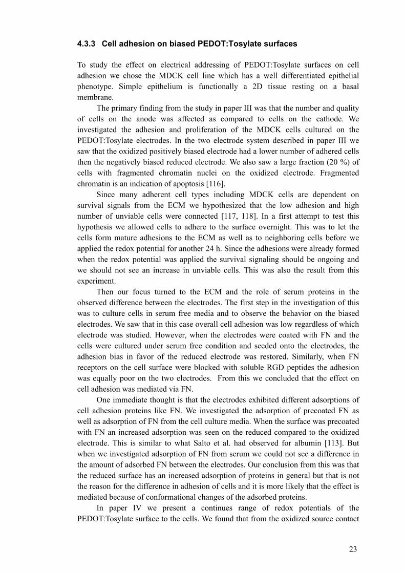
towards the reduced drain we saw a continuously increasing cell adhesion until a certain threshold where we saw a steep decrease in the cell adhesion. Whether the steep decrease is due to a threshold potential for cell adhesion, or is because of the non-linearity of the potential gradient remains to be investigated. When the gate potential was increased the steep decrease in cell adhesion was shifted towards the center of the channel (Figure 4).
Figure 4. Cells stained with FITC-Phalloidin. The rows shows the transistor channel addressed with three different gate voltages from top 0 V, 1.0 V, and 1.5 V.
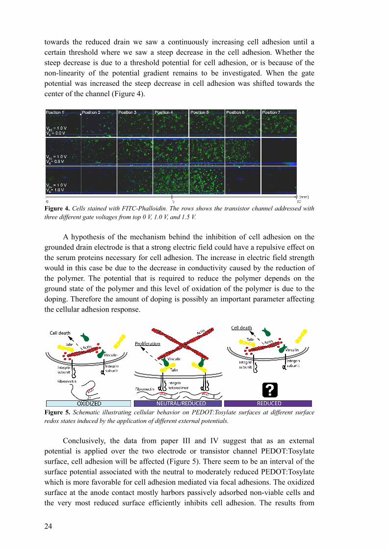
A hypothesis of the mechanism behind the inhibition of cell adhesion on the grounded drain electrode is that a strong electric field could have a repulsive effect on the serum proteins necessary for cell adhesion. The increase in electric field strength would in this case be due to the decrease in conductivity caused by the reduction of the polymer. The potential that is required to reduce the polymer depends on the ground state of the polymer and this level of oxidation of the polymer is due to the doping. Therefore the amount of doping is possibly an important parameter affecting the cellular adhesion response.
Figure 5. Schematic illustrating cellular behavior on PEDOT:Tosylate surfaces at different surface redox states induced by the application of different external potentials.
Conclusively, the data from paper III and IV suggest that as an external
potential is applied over the two electrode or transistor channel PEDOT:Tosylate surface, cell adhesion will be affected (Figure 5). There seem to be an interval of the surface potential associated with the neutral to moderately reduced PEDOT:Tosylate which is more favorable for cell adhesion mediated via focal adhesions. The oxidized surface at the anode contact mostly harbors passively adsorbed non-viable cells and the very most reduced surface efficiently inhibits cell adhesion. The results from
25
paper III indicates that the inhibition of cell adhesion at the anode is due to effects on serum proteins such as FN. Hypothesis’s regarding the mechanism of inhibition of adhesion seen on the most reduced surface in paper III is still to be tested (Figure 5). 4.4 ELECTRONIC RELEASE OF ADHERENT CELLS (PAPER V) The culture of adherent cells has a critical point when the cells need to be recovered for further subcultivation or analysis. The recovery includes detachment of the cells from their culture substrate. Typically, this is accomplished by enzymatic treatments, with for example trypsin, which cleaves the cell surface proteins. This treatment can have effect on the viability of cells and destroys surface antigens of the cells.
Enzyme free detachment of cells can also be accomplished by treatment with Ca2+ chelating buffers such as EDTA which binds the Ca2+ which is necessary for the function of integrins. This however is a rather inefficient method and both trypsin and EDTA lack the possibility to selectively detach desired cells. Okano and co-workers at Tokyo Women’s medical University have for a long time been developing a special polyacrylamide surface that is temperature sensitive. This surface will release cells at room temperature and allow adhesion at 37°C [119].
However, an intrinsic advantage of electronically addressed devices such as the one presented here is the high spatiotemporal resolution that can be accomplished compared to thermal or chemically applied surfaces. Electrically addressable polyelectrolyte cell culture substrate with micro-patterning potential has been shown by Vörös and co-workers [120, 121]. They use a conducting oxide surface to apply a potential that will detach a polyelectrolyte film.
In paper V we present a self doping conducting polymer film that upon application of a potential can disintegrate itself. This approach is conceptually different from that of Vörös since it is the conducting cell culture substrate that is the actively disintegrating component. The electronic detachment is based on thin films of the water-soluble PEDOT-S:H which is a derivative of PEDOT. This polymer is self doping due to the covalently linked sulfonyl side chain.
PEDOT-S:H can be barcoated onto any supporting substrate of choice. Even though the polymer is intrinsically conducting it is wise to use a conducting substrate to make sure that addressing of the film and subsequent disintegration is homogenous. For cell culture purposes it is preferable to use a transparent substrate as this makes it possible to analyses the cells using microscopy. Conducting transparent substrates could be indium tin oxide or PEDOT:PSS. We chose PEDOT:PSS because we found that PEDOT-S:H had good adhesive properties on this substrate.
When we cultured cells on the substrate we saw that the morphology of the cells differed somewhat from that of cells cultured on conventional cell culture plastic. We saw that actin stress fiber formation was less pronounced and that large focal adhesion complexes as visualized by vinculin staining were largely absent. We speculate that this might be due to the soft hydrogel like structure of the PEDOT-S:H surface as compared to the stiffer polystyrene cell culture plastic. This difference in morphology of the cells is not necessarily a flaw of the system, since the stiff cell culture plastic surface can be regarded as artificial and the soft PEDOT-S:H is more similar to the connective tissue
26
that cells experience in vivo. We did not see any signs of cytotoxicity due to the culture of cells on the PEDOT-S:H surface either, which favors the idea that it is a fairly good cell culture substrate.
The conclusions from paper V are that the PEDOT derivative PEDOT-S:H can be used as a cell culture substrate. Moreover, the self-disintegrating ability of the polymer allows for non-invasive recovering of adherent cells. Using flow cytometry and antibodies against the cell surface antigen CD44, we showed that the PEDOT-S:H method of cell detachment was superior to trypsin treatment with regards to conserving cell surface antigens.
27
5 CONCLUSIONS AND PERSPECTIVES Conducting polymers are complex and fascinating; the same can be said about eukaryotic cells. Cellular signaling and metabolism involves some of the functions exhibited by conducting polymers such as ion transport and electrochemical reactions. The work of this thesis illustrates how conducting polymers can address cell physiology from different aspects.
The important and evolutionary conserved voltage-gated ion channels that are expressed by many cells have been targeted using a PEDOT:Tosylate coated nano-fiber scaffold electrode. The L-type voltage-operated calcium channel could be activated by applying a depolarization potential via the nano-fiber scaffold electrode. The nano-fiber scaffold was demonstrated to be a functional cell culture substrate for the neuroblastoma cells that were used. Due to the nano-fiber structure of the electrode there is reason to assume that it possesses a high charge transfer capacity because of the high surface to volume ratio. This makes it very promising as a neural implant electrode.
There are a number of mechanisms that are suggested to be involved in cellular mechanotransduction which is a field of undisputed physiological relevance. However, there are still many white spots on the map of cellular mechanotransduction despite intense ongoing research. We have contributed with a microfabricated chip designed for the study of cellular mechanotransduction. The chip is evaluated using renal epithelial cells. We show that when stimulating these cells using the chip we activate a mechanotransduction mechanism based on autocrine nucleotide signaling.
Adherent cells are often dependent on survival signals mediated via their adhesion complexes. These adhesion complexes interact with extracellular proteins such as FN which is an important protein in the ECM. Using PEDOT:Tosylate surfaces we show how the surface potential and associated effects of the surface properties also influences the cells possibilities to interact with as well as adhere to the extracellular proteins. Using an electrochemical PEDOT:Tosylate transistor we can also show how complex this effect is, and that there is an interval in the electric surface potential for the PEDOT:Tosylate surface where adhesion is optimal.
The papers of this thesis have been investigating approaches to affect different cellular responses. In the last paper we show that conducting polymers also can be used when the aim is to accomplish a non-invasive treatment of eukaryotic cells. Adherent cells was harvested without causing any cytotoxicity and with their cell surface antigens intact, using a self-disintegrating PEDOT derivate. Conclusively, it has been a great pleasure to work on this thesis and be a part of a progressive field of interdisciplinary science. I would like to believe that we are only at the beginning of organic bioelectronics. One could speculate about future applications or medical devices but I would like to highlight that also the basic science of organic biolelectronics is truly captivating in its own right.

28
6 MY SCIENTIFIC CONTRIBUTION During my multidisciplinary thesis work, we have been developing organic bioelectronic devices for the study of cellular physiology. We combined electrospinning of nano-fibers with conducting polymers to achieve a new cell stimulation electrode. We have used microfabrication technology to develop a microactuator chip which was used to study the effect of cellular mechanotransduction on intracellular Ca2+ signaling in renal epithelial cells. We have highlighted the role of extracellular proteins in the cell adhesion response to electrically biased conducting polymer surfaces. We have shown how a planar electrochemical transistor can be used to achieve a cell density gradient that can be controlled by the gate potential. Finally, we have also developed and demonstrated how a self-disintegrating conducting polymer can be used for non-invasive cell detachment. RELATED WORK NOT INCLUDED IN THE THESIS Review Organic bioelectronics in nanomedicine. Karl Svennersten, Karin Larsson, Magnus Berggren, Agneta Richter-Dahlfors Biochimica et Biophysica Acta (BBA)-General Subjects, 2011, 1810(3), 276-285. Patents Method and system for extraction of cell types using cell surface receptor-specific attachement molecules. WO2010072256 Berggren, Rolf Magnus; Richter-Dahlfors, Agneta; Bolin, Maria; Svennersten, Karl; Jager, Edvin; Wang, Xiangjun Electrochemical active element for control of adhesion, growth and/or biofilm formation of prokaryotic cells. WO2010072257 Richter-Dahlfors, Agneta; Svennersten, Karl; Berggren, Rolf Magnus; Jager, Edwin; Persson, Kristin; Abdul, Kader
29
7 POPULÄRVETENSKAPLIG SAMMANFATTNING Plastelektronik kan användas för att påverka viktiga funktioner hos kroppens celler. Men varför vill man påverka funktioner hos kroppens celler och vad är plastelektronik? Många sjukdomar beror på att man förlorat celler med en viss funktion och då måste den här funktionen ersättas. I nervsystemet kan det handla om att man måste ersätta en förlorad elektrisk signal med hjälp av en inplanterad elektrod. I andra fall kan det handla om att man i laboratorium vill odla vävnader som hud eller ben för att kunna ersätta förlorad vävnad hos en patient. Då finns det ett behov av att styra funktionen hos cellerna i vävnaden så att den växer på ett tillfredställande sätt.
Plastelektronik är elektronik som tillverkats av plaster som kan leda elektrisk ström. Den här ledningsförmågan bygger på plastens kemiska struktur med omväxlande enkel- och dubbelbindningar som elektronerna kan röra sig fritt emellan. I praktiken så är strukturen med alternerande enkel- och dubbelbindningar instabil. Det medför att plasten bara fungerar som en halvledare. Men om man ”dopar” plasten så kan den få lika bra ledningsförmåga som metalliska ledare. Dopning är en process som även används inom konventionell halvledarelektronik. Dopning innebär att man skapar laddningsbärare i det halvledande materialet genom att tillsätta ett ämne som drar till sig eller lämnar ifrån sig elektroner.
Vi vill alltså styra processer i kroppens celler för att i förlägningen bota sjukdomar och plastelektronik är elektronik som tillverkats av elektriskt ledande plaster. Men hur kan plastelektronik användas för att kontrollera cellulära processer? I kroppens celler pågår en intensiv elektrokemisk aktivitet med jongradienter som byggs upp och elektroner som transporteras mellan olika molekyler. Signalprocessningen som hela tiden sker i en enda cell skulle kunna få vilken persondator som helst att framstå som en kulram i jämförelse. Ledningsförmågan i de ledande plasterna har en del gemensamt med signaleringen i cellerna. Elektrokemiska och jonledande förmågor är viktiga både för funktionen hos celler och ledande plaster.
I den här avhandlingen vill vi visa hur man kan använda ledande plaster för att styra olika viktiga funktioner hos kroppens celler. Till exempel visar vi hur man kan använda nanotrådar beklädda med en ledande plast för att stimulera nervceller. Nanotrådarna har en dubbel funktion, dels bidrar de till att skapa en miljö som nervcellerna trivs i och dels effektiviserar de överföringen av den elektriska signalen till nervcellen.
Alla celler i kroppen kan på ett eller annat sätt känna av mekaniska stimulin och beröring. I avhandlingen visar vi ett mikrochip som har beståndsdelar tillverkade av en ledande plast som sväller när den aktiveras elektriskt. Med hjälp av mikrochippet kan vi studera hur njurens celler påverkas av beröring. Hur njurceller påverkas av mekaniska stimuli är viktigt för njurens funktion. Cellerna i våra kroppar sitter fast i kroppens bindväv med hjälp av särskilda adhesionsmolekyler. När man odlar celler i laboratorium för forskning eller för medicinska ändamål så binder celler proteiner som fastnar på odlingsytan. Hur väl cellerna binder till proteinerna beror på ytans kemiska egenskaper. En yta av ledande plast kan ändra sina kemiska egenskaper om man tillför en elektrisk ström till ytan. Vi visar hur vi kan styra njurcellernas adhesion på en
30
ledande plastyta. Vi utvecklar detta med hjälp av en ledande plasttransistor som har en spänningsgradient där ser vi att det finns ett spännings intervall där njurcellerna trivs och att om spänningen understiger eller överstiger intervallet så vill inte njurcellerna växa på ytan.
Ett problem när man odlar celler eller vävnader i laboratorium är hur man ska få loss cellerna eller vävnaden från odlingsytan. Ett vanligt sätt att få loss celler och vävnader från odlingsytan är att behandla dem med enzymer som bryter ner proteiner som sitter utanpå cellerna. Den behandlingen kan ofta vara skadlig för cellerna. För att lösa det problemet så har vi använt en ledande plast som kan lösa upp sig själv när man lägger på en ström. Om man odlar celler på plasten så kan man få loss dem utan att behöva förstöra cellens proteiner med enzym. Vi provade den här ledande plastytan med celler från urinblåsan och såg att vi kunde få cellerna att lossna med hjälp av den självupplösande ledande plasten utan att förstöra cellens ytproteiner.
Sammanfattningsvis kan man säga att plastelektronikens unika egenskaper gör den särskilt interessant för att påverka flera olika funktioner hos kroppens celler i hopp om att kunna lindra och bota sjukdomar.
31
8 ACKNOWLEDGEMENTS No man is an island and this work would not have been possible without a number of people to whom I would like to express my sincerest gratitude: My main supervisor Professor Agneta Richter-Dahlfors for being the best coach one could wish for in the art of science. I can think of no other supervisor with whom I would have developed as much as a person and a scientist. Margret Wahlström thanks for co-ordinating everything and always being around for advice. Monica Rydén-Aulin for your endlessly good mood and Peter Kjäll for being my mentor and friend from the day I arrived in the lab.
Professor Magnus Berggren my co-supervisor for your never ending enthusiasm and the welcoming atmosphere in your group. Thanks to Maria Bolin for a great collaboration all the way from the start and Edwin Jager for your great project ideas and always having time. Kristin for your good spirit and patience even when things were progressing slowly, David for your knowledge I have read your thesis a thousand times, Roger Karlsson the master of organic synthesis, Peter Konradsson I will say hello to Jukka when I see him, and Sophie Lindesvik for co-ordinating everything down there.
Jorrit for all the fun in as well as outside the lab and sharing the agony of being a graduate student, Keira for your lovely attitude and understanding of sarcasm, Karin I take great pride in recruiting you to the group, Sindhu for the nice discussions about everything, Kader for being a great friend and for all the bold plans, Pelle for being the perfect student your enthusiasm and talent was really a vitamin injection for me, Susanne for being so smart and nice I am really happy that you will carry on the organic bioelectronics work, KJ Vasastan suits you, Ferdinand and Jakob the new nano-recruits, Arley me gustaría hacer una internado con usted en Colombia, Klas and Björn for raising the intellectual level of the scientific discussions when having lunch at hjulet, Marco for the company when I have been writing my thesis, Andreas for your healthy cynicism, Gabriella, Ann-Sofie, Sergei, Xianghui, Yunig, and Zhihua it’s a pleasure to share the corridor with you.
Ola it was a pleasure to share the corridor with you as well, double sided color print is really amazing, Shirin, Erik, Nina, Anna, Amilcar, Michalina, Goncalo, Karolina and everyone else from Olas group you made us feel very welcome when we arrived at neuroscience. And of course thanks to Mia and Marta.
Thanks to Rolf for your time on the SEM. Thanks to the people at our former department MTC especially Clemens, and the
rest in Galinas group, the coffe corner was great. Thanks to the people who has left the former department, Lina Guerra, Teresa Frisan and Anna Birgersdotter.
Thanks to Jan-Ingmar and Camilla and my fellow students at forskarskolan it was a great training for the quest that lay ahead.
Tack till Mamma och Lars-Ove och Pappa och Liisa och alla syskon, släktingar och vänner. Tack till Roger för en lång vänskap och många långa spännande diskussioner. Tack till Birgit för all hjälp i vårat kaosartade forskarhem. Framförallt vill jag tacka William, Olivia, Matilda och Mary världens snällaste och underbaraste barn och Jenny, jag älskar dig ditt fantastiska stöd och tålamod har betytt allt, ni är den bästa familj man kan önska sig.
32
9 REFERENCES 1. Hodgkin, A.L. and A.F. Huxley, Resting and Action Potentials in Single Nerve
Fibres. Journal of Physiology-London, 1945. 104(2): p. 176-195. 2. Szent-Györgyi, A., Bioelectronics : a study in cellular regulations, defense,
and cancer. 1968, New York: Academic Press. ix, 89 p. 3. Shirakawa, H., Nobel Lecture: The discovery of polyacetylene film—the
dawning of an era of conducting polymers. Reviews of Modern Physics, 2001. 73(3): p. 713.
4. Chiang, C.K., et al., Electrical Conductivity in Doped Polyacetylene. Physical Review Letters, 1977. 39(17): p. 1098.
5. Bolto, B., R. McNeill, and D. Weiss, Electronic Conduction in Polymers. III. Electronic Properties of Polypyrrole. Australian Journal of Chemistry, 1963. 16(6): p. 1090-1103.
6. Mcginnes.J, P. Corry, and P. Proctor, Amorphous-Semiconductor Switching in Melanins. Science, 1974. 183(4127): p. 853-855.
7. Brabec, C.J., Organic photovoltaics: technology and market. Solar Energy Materials and Solar Cells, 2004. 83(2-3): p. 273-292.
8. Zhou, T.X., et al., Stable and efficient electrophosphorescent organic light-emitting devices grown by organic vapor phase deposition. Applied Physics Letters, 2005. 86(2): p. -.
9. Argun, A.A., et al., Multicolored electrochromism polymers: Structures and devices. Chemistry of Materials, 2004. 16(23): p. 4401-4412.
10. Clemens, W., et al., From polymer transistors toward printed electronics. Journal of Materials Research, 2004. 19(7): p. 1963-1973.
11. Kaiser, A.B., Electronic transport properties of conducting polymers and carbon nanotubes. Reports on Progress in Physics, 2001. 64(1): p. 1-49.
12. Heeger, A.J., Semiconducting and metallic polymers: the fourth generation of polymeric materials. Synthetic Metals, 2001. 125(1): p. 23-42.
13. Ofer, D., R.M. Crooks, and M.S. Wrighton, Potential Dependence of the Conductivity of Highly Oxidized Polythiophenes, Polypyrroles, and Polyaniline - Finite Windows of High Conductivity. Journal of the American Chemical Society, 1990. 112(22): p. 7869-7879.
14. Patil, A.O., A.J. Heeger, and F. Wudl, Optical-Properties of Conducting Polymers. Chemical Reviews, 1988. 88(1): p. 183-200.
15. Andersson, P., et al., Active matrix displays based on all-organic electrochemical smart pixels printed on paper. Advanced Materials, 2002. 14(20): p. 1460-+.
16. Tehrani, P., et al., Evaluation of active materials designed for use in printable electrochromic polymer displays. Thin Solid Films, 2006. 515(4): p. 2485-2492.
17. Friend, R.H., et al., Electroluminescence in conjugated polymers. Nature, 1999. 397(6715): p. 121-128.
18. Langeveld-Voss, B.M.W., R.A.J. Janssen, and E.W. Meijer, On the origin of optical activity in polythiophenes. Journal of Molecular Structure, 2000. 521(1-3): p. 285-301.
19. Andersson, M., et al., Polythiophene with a Free Amino-Acid Side-Chain. Polymer Communications, 1991. 32(18): p. 546-548.
20. Nilsson, K.P.R., et al., Conjugated polyelectrolytes: Conformation-sensitive optical probes for detection of arnyloid fibril forrnation. Biochemistry, 2005.
33
44(10): p. 3718-3724. 21. Galvani, L., Aloysii Galvani de viribus electricitatis in motu musculari
commentarius. 1791, Bononiæ. 22. Letaw, H. and J. Bardeen, Electrolytic Analog Transistor. Journal of Applied
Physics, 1954. 25(5): p. 600-606. 23. Kittlesen, G.P., H.S. White, and M.S. Wrighton, Chemical derivatization of
microelectrode arrays by oxidation of pyrrole and N-methylpyrrole: fabrication of molecule-based electronic devices. Journal of the American Chemical Society, 1984. 106(24): p. 7389-7396.
24. Koezuka, H., A. Tsumura, and T. Ando, Field-effect transistor with polythiophene thin film. Synthetic Metals, 1987. 18(1-3): p. 699-704.
25. Macaya, D.J., et al., Simple glucose sensors with micromolar sensitivity based on organic electrochemical transistors. Sensors and Actuators B: Chemical, 2007. 123(1): p. 374-378.
26. MacKinnon, R., Potassium channels. FEBS Letters, 2003. 555(1): p. 62-65. 27. Catterall, W.A., Ion Channel Voltage Sensors: Structure, Function, and
Pathophysiology. Neuron. 67(6): p. 915-928. 28. Reuveny, E. and T. Narahashi, Two types of high voltage-activated calcium
channels in SH-SY5Y human neuroblastoma cells. Brain Research, 1993. 603(1): p. 64-73.
29. Jentsch, T.J., et al., The ClC Family of Voltage-Gated Chloride Channels: Structure and Functiona. Annals of the New York Academy of Sciences, 1993. 707(1): p. 285-293.
30. Hodgkin, A.L. and A.F. Huxley, A quantitative description of membrane current and its application to conduction and excitation in nerve. The Journal of Physiology, 1952. 117(4): p. 500-544.
31. Tombola, F., M.M. Pathak, and E.Y. Isacoff, How does voltage open an ion channel? Annual Review of Cell and Developmental Biology, 2006. 22: p. 23-52.
32. Murata, Y., et al., Phosphoinositide phosphatase activity coupled to an intrinsic voltage sensor. Nature, 2005. 435(7046): p. 1239-1243.
33. Wang, N., J.D. Tytell, and D.E. Ingber, Mechanotransduction at a distance: mechanically coupling the extracellular matrix with the nucleus. Nature Reviews Molecular Cell Biology, 2009. 10(1): p. 75-82.
34. Ingber, D.E., Tensegrity I. Cell structure and hierarchical systems biology. Journal of Cell Science, 2003. 116(7): p. 1157-1173.
35. Ingber, D.E., Cellular mechanotransduction: putting all the pieces together again. The FASEB Journal, 2006. 20(7): p. 811-827.
36. Hayakawa, K., H. Tatsumi, and M. Sokabe, Actin stress fibers transmit and focus force to activate mechanosensitive channels. Journal of Cell Science, 2008. 121(4): p. 496-503.
37. Du, S.Q., et al., Amiloride-sensitive ion channels in urinary bladder epithelium involved in mechanosensory transduction by modulating stretch-evoked adenosine triphosphate release. Urology, 2007. 69(3): p. 590-595.
38. Christensen, A.P. and D.P. Corey, TRP channels in mechanosensation: direct or indirect activation? Nature Reviews Neuroscience, 2007. 8(7): p. 510-521.
39. Schwartz, M.A. and D.W. DeSimone, Cell adhesion receptors in mechanotransduction. Current Opinion in Cell Biology, 2008. 20(5): p. 551-556.
40. Praetorius, H.A. and J. Leipziger, ATP release from non-excitable cells. Purinergic Signalling, 2009. 5(4): p. 433-446.
41. Nishiyama, M., et al., Calcium stores regulate the polarity and input
34
specificity of synaptic modfication. Nature, 2000. 408(6812): p. 584-588. 42. Taufiq, A.M., et al., Involvement of IP3 receptors in LTP and LTD induction
in guinea pig hippocampal CA1 neurons. Learning & Memory, 2005. 12(6): p. 594-600.
43. Clapham, D.E., Calcium signaling. Cell, 2007. 131(6): p. 1047-1058. 44. Hynes, R.O., Integrins - Versatility, Modulation, and Signaling in Cell-
Adhesion. Cell, 1992. 69(1): p. 11-25. 45. Barczyk, M., S. Carracedo, and D. Gullberg, Integrins. Cell and Tissue
Research, 2010. 339(1): p. 269-280. 46. Alenghat, F.J., et al., Mechanical Control of cAMP Signaling Through
Integrins Is Mediated by the Heterotrimeric G alpha s Protein. Journal of Cellular Biochemistry, 2009. 106(4): p. 529-538.
47. Tadokoro, S., et al., Talin binding to integrin beta tails: A final common step in integrin activation. Science, 2003. 302(5642): p. 103-106.
48. Frantz, C., K.M. Stewart, and V.M. Weaver, The extracellular matrix at a glance. J Cell Sci. 123(24): p. 4195-4200.
49. Kresse, H. and E. Schonherr, Proteoglycans of the extracellular matrix and growth control. Journal of Cellular Physiology, 2001. 189(3): p. 266-274.
50. George, E.L., et al., Defects in mesoderm, neural tube and vascular development in mouse embryos lacking fibronectin. Development, 1993. 119(4): p. 1079-1091.
51. Pankov, R. and K.M. Yamada, Fibronectin at a glance. J Cell Sci, 2002. 115(20): p. 3861-3863.
52. Pytela, R., M.D. Pierschbacher, and E. Ruoslahti, Identification and Isolation of a 140-Kd Cell-Surface Glycoprotein with Properties Expected of a Fibronectin Receptor. Cell, 1985. 40(1): p. 191-198.
53. Pierschbacher, M.D. and E. Ruoslahti, Variants of the Cell Recognition Site of Fibronectin That Retain Attachment-Promoting Activity. Proceedings of the National Academy of Sciences of the United States of America-Biological Sciences, 1984. 81(19): p. 5985-5988.
54. Leiss, M., et al., The role of integrin binding sites in fibronectin matrix assembly in vivo. Current Opinion in Cell Biology, 2008. 20(5): p. 502-507.
55. Smith, M.L., et al., Force-Induced Unfolding of Fibronectin in the Extracellular Matrix of Living Cells. PLoS Biol, 2007. 5(10): p. e268.
56. Adeloju, S.B. and G.G. Wallace, Conducting polymers and the bioanalytical sciences: new tools for biomolecular communications. A review. Analyst, 1996. 121(6): p. 699-703.
57. Garner, B., et al., Polypyrrole–heparin composites as stimulus-responsive substrates for endothelial cell growth. Journal of Biomedical Materials Research, 1999. 44(2): p. 121-129.
58. Jager, E.W.H., O. Inganäs, and I. Lundström, Microrobots for Micrometer-Size Objects in Aqueous Media: Potential Tools for Single-Cell Manipulation. Science, 2000. 288(5475): p. 2335-2338.
59. Wong, J.Y., R. Langer, and D.E. Ingber, Electrically conducting polymers can noninvasively control the shape and growth of mammalian cells. Proceedings of the National Academy of Sciences of the United States of America, 1994. 91(8): p. 3201-3204.
60. Schmidt, C.E., et al., Stimulation of neurite outgrowth using an electrically conducting polymer. Proceedings of the National Academy of Sciences of the United States of America, 1997. 94(17): p. 8948-8953.
61. Simon, D.T., et al., Organic electronics for precise delivery of neurotransmitters to modulate mammalian sensory function. Nature Materials,
35
2009. 8(9): p. 742-746. 62. Isaksson, J., et al., Electronic control of Ca2+ signalling in neuronal cells
using an organic electronic ion pump. Nature Materials, 2007. 6(9): p. 673-679.
63. Berggren, M. and A. Richter-Dahlfors, Organic bioelectronics. Advanced Materials, 2007. 19(20): p. 3201-3213.
64. Groenendaal, B.L., et al., Poly(3,4-ethylenedioxythiophene) and its derivatives: Past, present, and future. Advanced Materials, 2000. 12(7): p. 481-494.
65. Deleeuw, D.M., et al., Electroplating of Conductive Polymers for the Metallization of Insulators. Synthetic Metals, 1994. 66(3): p. 263-273.
66. Asplund, M., T. Nyberg, and O. Inganas, Electroactive polymers for neural interfaces. Polymer Chemistry, 2010. 1(9): p. 1374-1391.
67. George, P.M., et al., Three-dimensional conductive constructs for nerve regeneration. Journal of Biomedical Materials Research Part A, 2009. 91A(2): p. 519-527.
68. Mohammadi, A., et al., Chemical vapour deposition (CVD) of conducting polymers: Polypyrrole. Synthetic Metals, 1986. 14(3): p. 189-197.
69. Winther-Jensen, B.r., et al., Vapor Phase Polymerization of Pyrrole and Thiophene Using Iron(III) Sulfonates as Oxidizing Agents. Macromolecules, 2004. 37(16): p. 5930-5935.
70. Richardson-Burns, S.M., J.L. Hendricks, and D.C. Martin, Electrochemical polymerization of conducting polymers in living neural tissue. J Neural Eng, 2007. 4(2): p. L6-L13.
71. Biedler, J.L., et al., Multiple Neurotransmitter Synthesis by Human Neuroblastoma Cell Lines and Clones. Cancer Research, 1978. 38(11): p. 3751-3757.
72. Ridley, D.L., A. Rogers, and S. Wonnacott, Differential effects of chronic drug treatment on alpha3* and alpha7 nicotinic receptor binding sites, in hippocampal neurones and SH-SY5Y cells. Br J Pharmacol, 2001. 133(8): p. 1286-95.
73. Madin, S.H. and N.B. Darby, Established Kidney Cell Lines of Normal Adult Bovine and Ovine Origin. Proceedings of the Society for Experimental Biology and Medicine, 1958. 98(3): p. 574-576.
74. Gaush, C.R., W.L. Hard, and T.F. Smith, Characterization of an Established Line of Canine Kidney Cells (Mdck). Proceedings of the Society for Experimental Biology and Medicine, 1966. 122(3): p. 931-&.
75. Leighton, J., et al., Secretory Activity and Oncogenicity of a Cell Line (Mdck) Derived from Canine Kidney. Science, 1969. 163(3866): p. 472-&.
76. Simmons, N.L., Cultured Monolayers of Mdck Cells - a Novel Model System for the Study of Epithelial Development and Function. General Pharmacology, 1982. 13(4): p. 287-291.
77. Kim, M., et al., STAT1 Is Required for Redifferentiation during Madin-Darby Canine Kidney Tubulogenesis. Mol. Biol. Cell. 21(22): p. 3926-3933.
78. Rogers, K.K., et al., The Rho family of small GTPases is involved in epithelial cystogenesis and tubulogenesis. Kidney Int, 2003. 63(5): p. 1632-1644.
79. Schoenenberger, C.A., et al., Integrin Expression and Localization in Normal Mdck Cells and Transformed Mdck Cells Lacking Apical Polarity. Journal of Cell Science, 1994. 107: p. 527-541.
80. Kreidberg, J.A. and J.M. Symons, Integrins in kidney development, function, and disease. American Journal of Physiology - Renal Physiology, 2000. 279(2): p. F233-F242.
36
81. Hay, E.D. and A. Zuk, Transformations between Epithelium and Mesenchyme - Normal, Pathological, and Experimentally-Induced. American Journal of Kidney Diseases, 1995. 26(4): p. 678-690.
82. Balkovetz, D.F., Hepatocyte growth factor and Madin-Darby canine kidney cells: In vitro models of epithelial cell movement and morphogenesis. Microscopy Research and Technique, 1998. 43(5): p. 456-463.
83. Otoole, C., et al., Cellular Immunity to Human Urinary-Bladder Carcinoma .1. Correlation to Clinical Stage and Radiotherapy. International Journal of Cancer, 1972. 10(1): p. 77-&.
84. Fitzgerald, K.A., et al., Ras, protein kinase C zeta, and I kappa B kinases 1 and 2 are downstream effecters of CD44 during the activation of NF-kappa B by hyaluronic acid fragments in T-24 carcinoma cells. Journal of Immunology, 2000. 164(4): p. 2053-2063.
85. Paredes, R.M., et al., Chemical calcium indicators. Methods, 2008. 46(3): p. 143-151.
86. Takahashi, A., et al., Measurement of intracellular calcium. Physiological Reviews, 1999. 79(4): p. 1089-1125.
87. Green, R.A., et al., Conducting polymers for neural interfaces: challenges in developing an effective long-term implant. Biomaterials, 2008. 29(24-25): p. 3393-9.
88. Asplund, M., T. Nyberg, and O. Inganas, Electroactive polymers for neural interfaces. Polymer Chemistry. 1(9): p. 1374-1391.
89. Ma, Z., et al., Potential of nanofiber matrix as tissue-engineering scaffolds. Tissue Eng, 2005. 11(1-2): p. 101-9.
90. Nyberg, T., A. Shimada, and K. Torimitsu, Ion conducting polymer microelectrodes for interfacing with neural networks. Journal of Neuroscience Methods, 2007. 160(1): p. 16-25.
91. Smart, D., A. Wandless, and D.G. Lambert, Activation of phospholipase C in SH-SY5Y neuroblastoma cells by potassium-induced calcium entry. Br J Pharmacol, 1995. 116(2): p. 1797-800.
92. Dajas-Bailador, F.A., A.J. Mogg, and S. Wonnacott, Intracellular Ca2+ signals evoked by stimulation of nicotinic acetylcholine receptors in SH-SY5Y cells: contribution of voltage-operated Ca2+ channels and Ca2+ stores. J Neurochem, 2002. 81(3): p. 606-14.
93. Passafaro, M., F. Clementi, and E. Sher, Metabolism of omega-conotoxin-sensitive voltage-operated calcium channels in human neuroblastoma cells: modulation by cell differentiation and anti-channel antibodies. J Neurosci, 1992. 12(9): p. 3372-9.
94. Cho, H., et al., A novel mechanosensitive channel identified in sensory neurons. European Journal of Neuroscience, 2006. 23(10): p. 2543-2550.
95. Hahn, C. and M.A. Schwartz, Mechanotransduction in vascular physiology and atherogenesis. Nature Reviews Molecular Cell Biology, 2009. 10(1): p. 53-62.
96. Chalfie, M., Neurosensory mechanotransduction. Nature Reviews Molecular Cell Biology, 2009. 10(1): p. 44-52.
97. Nagayama, K., S. Yanagihara, and T. Matsumoto, A novel micro tensile tester with feed-back control for viscoelastic analysis of single isolated smooth muscle cells. Medical Engineering & Physics, 2007. 29(5): p. 620-628.
98. Young, E.W.K. and C.A. Simmons, Macro- and microscale fluid flow systems for endothelial cell biology. Lab on a Chip, 2010. 10(2): p. 143-160.
99. Benes, A.J., et al., A New Vacuum-Operated Stress-Providing Instrument That Applies Static or Variable Duration Cyclic Tension or Compression to Cells-
37
Invitro. Journal of Cell Science, 1985. 75(Apr): p. 35-42. 100. Smela, E. and N. Gadegaard, Surprising volume change in PPy(DBS): An
atomic force microscopy study. Advanced Materials, 1999. 11(11): p. 953-+. 101. Andersson, A.S., et al., Nanoscale features influence epithelial cell
morphology and cytokine production. Biomaterials, 2003. 24(20): p. 3427-3436.
102. Nauli, S.M., et al., Loss of Polycystin-1 in Human Cyst-Lining Epithelia Leads to Ciliary Dysfunction. Journal of the American Society of Nephrology, 2006. 17(4): p. 1015-1025.
103. Praetorius, H.A., J. Frokiaer, and J. Leipziger, Transepithelial pressure pulses induce nucleotide release in polarized MDCK cells. American Journal of Physiology-Renal Physiology, 2005. 288(1): p. F133-F141.
104. Adamson, A.W., A.P. Gast, and Editors, Physical Chemistry of Surfaces, Sixth Edition. 1997: Wiley. 784 pp.
105. Young, T., An Essay on the Cohesion of Fluids. Philosophical Transactions of the Royal Society of London, 1805. 95: p. 65-87.
106. Flemming, R.G., et al., Effects of synthetic micro- and nano-structured surfaces on cell behavior. Biomaterials, 1999. 20(6): p. 573-88.
107. Teixeira, A.I., et al., Epithelial contact guidance on well-defined micro- and nanostructured substrates. J Cell Sci, 2003. 116(Pt 10): p. 1881-92.
108. Bodhak, S., S. Bose, and A. Bandyopadhyay, Role of surface charge and wettability on early stage mineralization and bone cell-materials interactions of polarized hydroxyapatite. Acta Biomaterialia, 2009. 5(6): p. 2178-2188.
109. Bodhak, S., S. Bose, and A. Bandyopadhyay, Electrically polarized HAp-coated Ti: in vitro bone cell-material interactions. Acta Biomater, 2010. 6(2): p. 641-51.
110. Ohgaki, M., et al., Manipulation of selective cell adhesion and growth by surface charges of electrically polarized hydroxyapatite. J Biomed Mater Res, 2001. 57(3): p. 366-73.
111. Engler, A.J., et al., Matrix Elasticity Directs Stem Cell Lineage Specification. Cell, 2006. 126(4): p. 677-689.
112. Teixeira, A.I., et al., The promotion of neuronal maturation on soft substrates. Biomaterials, 2009. 30(27): p. 4567-4572.
113. Salto, C., et al., Control of Neural Stem Cell Adhesion and Density by an Electronic Polymer Surface Switch. Langmuir, 2008. 24(24): p. 14133-14138.
114. Nilsson, D., et al., Bi-stable and dynamic current modulation in electrochemical organic transistors. Advanced Materials, 2002. 14(1): p. 51-54.
115. Robinson, N.D., et al., On the current saturation observed in electrochemical polymer transistors. Journal of the Electrochemical Society, 2006. 153(3): p. H39-H44.
116. Gerschenson, L.E. and R.J. Rotello, Apoptosis - a Different Type of Cell-Death. Faseb Journal, 1992. 6(7): p. 2450-2455.
117. Ilic, D., et al., Extracellular matrix survival signals transduced by focal adhesion kinase suppress p53-mediated apoptosis. Journal of Cell Biology, 1998. 143(2): p. 547-560.
118. Frisch, S.M. and H. Francis, Disruption of Epithelial Cell-Matrix Interactions Induces Apoptosis. Journal of Cell Biology, 1994. 124(4): p. 619-626.
119. Yang, J., et al., Cell sheet engineering: Recreating tissues without biodegradable scaffolds. Biomaterials, 2005. 26(33): p. 6415-6422.
120. Guillaume-Gentil, O., et al., Polyelectrolyte coatings with a potential for electronic control and cell sheet engineering. Advanced Materials, 2008.
38
20(3): p. 560-+. 121. Guillaume-Gentil, O., et al., Electrochemically switchable platform for the
micro-patterning and release of heterotypic cell sheets. Biomedical Microdevices. 13(1): p. 221-230.


![Biosensors & Bioelectronics - OMICS International | Open … · Biosensors & Bioelectronics ... Mi-Kyung Park, Materials Research and Education Center, ... [18]. E2 phage was provided](https://static.fdocuments.in/doc/165x107/5acd99ae7f8b9a93268dcd3a/biosensors-bioelectronics-omics-international-open-bioelectronics-mi-kyung.jpg)