Understanding metabolic regulation and cellular resource ...
Upload
padhu-pattabiramanCategory
view
215download
0description
Regulation of Cellular ProteinPhosphatase-1 (PP1) byPhosphorylation of the CPI-17Family, C-kinase-activatedPP1 Inhibitors*□S
Published, JBC Papers in Press, October 21, 2009, DOI 10.1074/jbc.R109.059972
Masumi Eto1
From the Department of Molecular Physiology and Biophysics and KimmelCancer Center, Thomas Jefferson University, Philadelphia, Pennsylvania 19107
The regulatory circuit controlling cellular protein phospha-tase-1 (PP1), an abundant group of Ser/Thr phosphatases,involves phosphorylation of PP1-specific inhibitor proteins.Malfunctions of these inhibitor proteins have been linked to avariety of diseases, including cardiovascular disease and cancer.Upon phosphorylation at Thr38, the 17-kDa PP1 inhibitor pro-tein, CPI-17, selectively inhibits a specific form of PP1, myosinlight chain phosphatase, which transduces multiple kinase sig-nals into the phosphorylation of myosin II and other proteins.Here, the mechanisms underlying PP1 inhibition and thekinase/PP1 cross-talk mediated by CPI-17 and its related pro-teins, PHI, KEPI, and GBPI, are discussed.
The reciprocal activities of protein kinases and phosphatasesdetermine protein phosphorylation levels in cells. PP12 dephos-phorylates phospho-Ser/Thr residues of proteins to regulatemultiple signaling pathways at various cellular loci (1–3). Cel-lular PP1 is associated with PP1 regulatory proteins/subunits attheir PP1-binding site, known as the RVXF motif. The bindingof PP1 regulatory proteins thus confers substrate specificity andlocalization on cellular PP1. Nearly 100 polypeptides have beenidentified as PP1 regulatory proteins, and these account for thewide spectrum of PP1 function (1–3). In addition, eukaryoticcells express several PP1 inhibitor proteins that play importantroles in regulating cellular PP1. The first generation of PP1inhibitor proteins involves inhibitor-1, inhibitor-2, DARPP32,and NIPP-1, which potently inhibit the free catalytic subunit ofPP1, but these inhibitor proteins weremuch less potent toward
purified PP1 holoenzymes, MLCP and glycogen-bound PP1.Therefore, cellular PP1 holoenzymes were thought to undergosubunit dissociation prior to the inhibition of PP1 by the inhib-itor proteins (1, 2). However, the number of PP1 holoenzymesthat do undergo subunit dissociation in cells remains unclear.MLCP is a trimeric PP1 holoenzyme, consisting of a PP1∂
isoform and a regulatory complex of MYPT1 (akaMBS, M110)and a 21-kDa accessory subunit, and vital to control cellularphosphorylation in response to various signals (4). MYPT1 andPP1 bind through the MYPT1 KVKF segment, as well as itseight-repeat ankyrin motif at the N-terminal domain (5). Bind-ing of the N-terminal 300-residue domain of MYPT1 is suffi-cient to allosterically regulate PP1 activity. The MYPT1 C-ter-minal domain directly binds to substrates, including myosinand ezrin/radixin/moesin (4). MLCP activity is reversibly regu-lated in response to various signals. For example, in smoothmuscle, activation of the G-protein-coupled receptor inhibitsMLCP, resulting in increased Ca2� sensitivity of myosin phos-phorylation and contraction, whereas cyclic nucleotide signalscan activate MLCP to induce smooth muscle relaxation (6).MLCP inhibition occurs upon MYPT1 phosphorylation atThr696 and Thr853 (4). On the other hand, protein kinase G canactivateMLCP(7).TheseregulatorysignalsareMYPT1isoform-dependent (8), suggesting an important role for MYPT1 inMLCP regulation. In addition, we identified the MLCP inhibi-tor protein, named CPI-17, which transduces G-protein signalsinto MLCP inhibition (9, 10). Based on sequence similarity,three CPI-17 homologs in the human genome, PHI, KEPI, andGBPI, were characterized as PP1 inhibitors (11–13). EachCPI-17 family member carries a PHIN domain, in which thesequences are �41% identical to CPI-17 (Fig. 1A). Indeed, allCPI-17 family members potently inhibit MLCP activity, whichsuggests new avenues for PP1 holoenzyme inhibition. Thisminireview will focus on CPI-17 and its homologs (whoseamino acid sequences differ significantly from other PP1 inhib-itor proteins), highlight critical findings from CPI-17 studies,and discuss the role of other CPI-17 family members in regu-lating PP1 activity.
Structure and Function of CPI-17
Amino Acid Sequence of CPI-17—The CPI-17 gene(PPP1R14A, chromosome 19) encodes a 147-residue polypep-tide in which �85% of the amino acids are identical withinmammals (10) (Fig. 1A). A splice variant of CPI-17 (CPI-17�)lacking exon 2 exists in human smooth muscle cells, althoughwhether this form is physiologically relevant is not known (seebelow) (14). Zebrafish express a similar gene, although towhichCPI-17 family member this gene product is functionally relatedis unclear. No homologous genes have been detected in fruitfly, nematode, and yeast, suggesting that the CPI-17 familyemerged at a late stage in evolution. Phosphorylation of CPI-17at Thr38 is necessary and sufficient to convert the protein into apotent MLCP inhibitor (9, 10). No homology is detectedbetween the CPI-17 family and other classes of PP1 inhibitors,such as inhibitor-1 and inhibitor-2, even though phosphoryla-
* This work was supported, in whole or in part, by National Institutes of HealthGrant HL083261. This minireview will be reprinted in the 2009 MinireviewCompendium, which will be available in January, 2010.
□S The on-line version of this article (available at http://www.jbc.org) containssupplemental Figs. 1 and 2.
1 To whom correspondence should be addressed. E-mail: [email protected].
2 The abbreviations used are: PP1, Ser/Thr protein phosphatase-1; MLCP,myosin light chain phosphatase; MYPT1, myosin phosphatase-targetingsubunit-1; CPI-17, C-kinase-activated PP1 inhibitor of 17 kDa; PHI, PP1holoenzyme inhibitor; KEPI, kinase C-enhanced PP1 inhibitor; GBPI, gastro-intestinal- and brain-specific PP1 inhibitor; PHIN, PP1 holoenzyme inhibi-tory; PKC, protein kinase C; ROCK, RhoA-activated coiled-coil kinase; ILK,integrin-linked kinase; PKA, protein kinase A; ERK1/2, extracellular signal-regulated kinase-1/2; PNG, phospholipase C-neighboring gene; I-2,inhibitor-2.
THE JOURNAL OF BIOLOGICAL CHEMISTRY VOL. 284, NO. 51, pp. 35273–35277, December 18, 2009© 2009 by The American Society for Biochemistry and Molecular Biology, Inc. Printed in the U.S.A.
DECEMBER 18, 2009 • VOLUME 284 • NUMBER 51 JOURNAL OF BIOLOGICAL CHEMISTRY 35273
MINIREVIEW This paper is available online at www.jbc.org
at Duke U
niversity, on October 25, 2010
ww
w.jbc.org
Dow
nloaded from
http://www.jbc.org/content/suppl/2009/12/07/284.51.35273.DC1.htmlhttp://www.jbc.org/content/suppl/2009/10/21/R109.059972.DC1.htmlSupplemental Material can be found at:
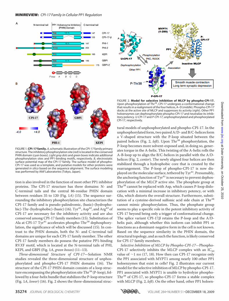
tion is also involved in the function of most other PP1 inhibitorproteins. The CPI-17 structure has three domains: N- andC-terminal tails and the central 86-residue PHIN domainbetween residues 35 to 120 (Fig. 1A) (15). The sequence sur-rounding the inhibitory phosphorylation site characterizes theCPI-17 family and is pseudo-palindromic, (basic)-(hydropho-bic)-Thr-(hydrophobic)-(basic) (16). Tyr41, Asp42, and Arg43 ofCPI-17 are necessary for the inhibitory activity and are alsoconserved among CPI-17 family members (15). Substitution ofAla at CPI-17 Tyr41 accelerates phospho-Thr38 dephosphory-lation, the significance of which will be discussed (15). In con-trast to the PHIN domain, both the N- and C-terminal taildomains are unique for each CPI-17 family member. The otherCPI-17 family members do possess the putative PP1-bindingRVXF motif, which is located at the N-terminal tails of PHI,KEPI, and GBPI (Fig. 1A, green boxes) (11–13).Three-dimensional Structure of CPI-17—Solution NMR
studies revealed the three-dimensional structure of unphos-phorylated and phospho-CPI-17 PHIN domains (16). Thestructure of the CPI-17 PHIN domain consists of a loop struc-ture encompassing the phosphorylation siteThr38 (P-loop), fol-lowed by a four-helix bundle that stabilizes the P-loop structure(Fig. 1A, lower) (16). Fig. 2 shows the three-dimensional struc-
tural models of unphosphorylated and phospho-CPI-17. In theunphosphorylated form, two pairedA/D- andB/C-helices forma V-shaped structure with the P-loop situated between thepaired helices (Fig. 2, left). Upon Thr38 phosphorylation, theP-loop becomes more solvent-exposed and, in doing so, gener-ates torque in the A-helix. This twisting of the A-helix rolls theA-B-loop up to align the B/C-helices in parallel with the A/D-helices (Fig. 2, center). The newly aligned four helices are thenstabilized through a hydrophobic core that is created by therearrangement. The P-loop of phospho-CPI-17 is now dis-played on themolecular surface, tethered byTyr41. Presumably,the anchoring function of Tyr41 is necessary to prevent dephos-phorylation of the MLCP active site. The phosphate group atThr38 cannot be replaced with Asp, which causes P-loop dislo-cation with a minimal increase in inhibitory potency, or withGlu, which distorts the overall structure. Furthermore, substi-tution of a cysteine-derived sulfonic acid side chain at Thr38cannot mimic phosphorylation. Thus, the phosphate groupseems to play a specific role in the potent inhibitory activity ofCPI-17 beyond being only a trigger of conformational change.The splice variant CPI-17� retains the P-loop and the A/D-helix pair, although whether this isoform can inhibit PP1 orfunctions as a dominant-negative form in the cell is not known.Based on the sequence similarity in the PHIN domain, thestructural topology, and as such the function, is likely conservedfor CPI-17 family members.Selective Inhibition of MLCP by Phospho-CPI-17—Phospho-
CPI-17 selectively inhibits the MLCP complex with an IC50value of �1 nM (17, 18). How then can CPI-17 recognize onlythe PP1 associated with MYPT1 among nearly 100 other PP1holoenzymes that exist in cells? Fig. 2 illustrates our currentmodel for the selective inhibition ofMLCPby phospho-CPI-17.PP1 associated with MYPT1 is unable to hydrolyze phospho-Thr38 of CPI-17, so phospho-CPI-17 forms a stable complexwith MLCP (Fig. 2, left). On the other hand, other PP1 holoen-
FIGURE 1. CPI-17 family. A, schematic illustration of the CPI-17 family primarystructure. The inhibitory phosphorylation site (red) is located in the conservedPHIN domain (cyan boxes). Light gray dots and green boxes indicate additionalphosphorylation sites and PP1-binding motifs, respectively. B, electrostaticsurface potential map of the CPI-17 family. The surface model of phospho-CPI-17 was used as a template, and putative models for other proteins weregenerated in silico based on the sequence alignment. The surface modelingwas performed by Altif Laboratories (Tokyo, Japan).
FIGURE 2. Model for selective inhibition of MLCP by phospho-CPI-17.Upon phosphorylation of Thr38, CPI-17 undergoes a conformational changethat results in a realignment of the four helices, A–D (middle). Phospho-CPI-17docks at the active site of MLCP and suppresses its activity (right). Other PP1holoenzymes can dephosphorylate phospho-CPI-17 and neutralize its inhib-itory potency. U-CPI-17 and P-CPI-17, unphosphorylated and phosphorylatedCPI-17, respectively.
MINIREVIEW: CPI-17 Family in Cellular PP1 Regulation
35274 JOURNAL OF BIOLOGICAL CHEMISTRY VOLUME 284 • NUMBER 51 • DECEMBER 18, 2009
at Duke U
niversity, on October 25, 2010
ww
w.jbc.org
Dow
nloaded from
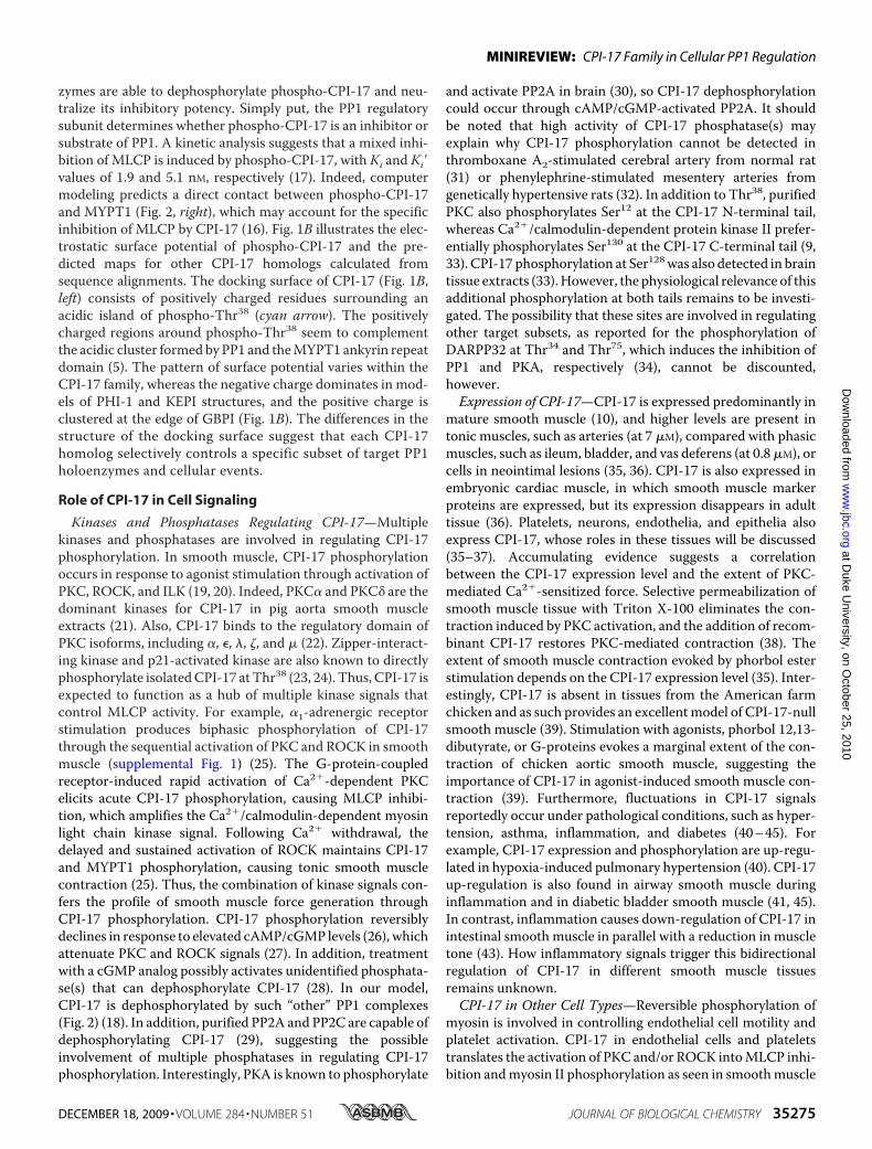
zymes are able to dephosphorylate phospho-CPI-17 and neu-tralize its inhibitory potency. Simply put, the PP1 regulatorysubunit determines whether phospho-CPI-17 is an inhibitor orsubstrate of PP1. A kinetic analysis suggests that a mixed inhi-bition of MLCP is induced by phospho-CPI-17, with Ki and Ki�values of 1.9 and 5.1 nM, respectively (17). Indeed, computermodeling predicts a direct contact between phospho-CPI-17and MYPT1 (Fig. 2, right), which may account for the specificinhibition of MLCP by CPI-17 (16). Fig. 1B illustrates the elec-trostatic surface potential of phospho-CPI-17 and the pre-dicted maps for other CPI-17 homologs calculated fromsequence alignments. The docking surface of CPI-17 (Fig. 1B,left) consists of positively charged residues surrounding anacidic island of phospho-Thr38 (cyan arrow). The positivelycharged regions around phospho-Thr38 seem to complementthe acidic cluster formed by PP1 and theMYPT1 ankyrin repeatdomain (5). The pattern of surface potential varies within theCPI-17 family, whereas the negative charge dominates in mod-els of PHI-1 and KEPI structures, and the positive charge isclustered at the edge of GBPI (Fig. 1B). The differences in thestructure of the docking surface suggest that each CPI-17homolog selectively controls a specific subset of target PP1holoenzymes and cellular events.
Role of CPI-17 in Cell Signaling
Kinases and Phosphatases Regulating CPI-17—Multiplekinases and phosphatases are involved in regulating CPI-17phosphorylation. In smooth muscle, CPI-17 phosphorylationoccurs in response to agonist stimulation through activation ofPKC, ROCK, and ILK (19, 20). Indeed, PKC� and PKC� are thedominant kinases for CPI-17 in pig aorta smooth muscleextracts (21). Also, CPI-17 binds to the regulatory domain ofPKC isoforms, including �, �, �, �, and � (22). Zipper-interact-ing kinase and p21-activated kinase are also known to directlyphosphorylate isolatedCPI-17 atThr38 (23, 24). Thus, CPI-17 isexpected to function as a hub of multiple kinase signals thatcontrol MLCP activity. For example, �1-adrenergic receptorstimulation produces biphasic phosphorylation of CPI-17through the sequential activation of PKC and ROCK in smoothmuscle (supplemental Fig. 1) (25). The G-protein-coupledreceptor-induced rapid activation of Ca2�-dependent PKCelicits acute CPI-17 phosphorylation, causing MLCP inhibi-tion, which amplifies the Ca2�/calmodulin-dependent myosinlight chain kinase signal. Following Ca2� withdrawal, thedelayed and sustained activation of ROCK maintains CPI-17and MYPT1 phosphorylation, causing tonic smooth musclecontraction (25). Thus, the combination of kinase signals con-fers the profile of smooth muscle force generation throughCPI-17 phosphorylation. CPI-17 phosphorylation reversiblydeclines in response to elevated cAMP/cGMP levels (26), whichattenuate PKC and ROCK signals (27). In addition, treatmentwith a cGMP analog possibly activates unidentified phosphata-se(s) that can dephosphorylate CPI-17 (28). In our model,CPI-17 is dephosphorylated by such “other” PP1 complexes(Fig. 2) (18). In addition, purified PP2A and PP2C are capable ofdephosphorylating CPI-17 (29), suggesting the possibleinvolvement of multiple phosphatases in regulating CPI-17phosphorylation. Interestingly, PKA is known to phosphorylate
and activate PP2A in brain (30), so CPI-17 dephosphorylationcould occur through cAMP/cGMP-activated PP2A. It shouldbe noted that high activity of CPI-17 phosphatase(s) mayexplain why CPI-17 phosphorylation cannot be detected inthromboxane A2-stimulated cerebral artery from normal rat(31) or phenylephrine-stimulated mesentery arteries fromgenetically hypertensive rats (32). In addition to Thr38, purifiedPKC also phosphorylates Ser12 at the CPI-17 N-terminal tail,whereas Ca2�/calmodulin-dependent protein kinase II prefer-entially phosphorylates Ser130 at the CPI-17 C-terminal tail (9,33). CPI-17 phosphorylation at Ser128was also detected in braintissue extracts (33).However, the physiological relevance of thisadditional phosphorylation at both tails remains to be investi-gated. The possibility that these sites are involved in regulatingother target subsets, as reported for the phosphorylation ofDARPP32 at Thr34 and Thr75, which induces the inhibition ofPP1 and PKA, respectively (34), cannot be discounted,however.Expression of CPI-17—CPI-17 is expressed predominantly in
mature smooth muscle (10), and higher levels are present intonic muscles, such as arteries (at 7 �M), compared with phasicmuscles, such as ileum, bladder, and vas deferens (at 0.8�M), orcells in neointimal lesions (35, 36). CPI-17 is also expressed inembryonic cardiac muscle, in which smooth muscle markerproteins are expressed, but its expression disappears in adulttissue (36). Platelets, neurons, endothelia, and epithelia alsoexpress CPI-17, whose roles in these tissues will be discussed(35–37). Accumulating evidence suggests a correlationbetween the CPI-17 expression level and the extent of PKC-mediated Ca2�-sensitized force. Selective permeabilization ofsmooth muscle tissue with Triton X-100 eliminates the con-traction induced by PKC activation, and the addition of recom-binant CPI-17 restores PKC-mediated contraction (38). Theextent of smooth muscle contraction evoked by phorbol esterstimulation depends on the CPI-17 expression level (35). Inter-estingly, CPI-17 is absent in tissues from the American farmchicken and as such provides an excellentmodel of CPI-17-nullsmooth muscle (39). Stimulation with agonists, phorbol 12,13-dibutyrate, or G-proteins evokes a marginal extent of the con-traction of chicken aortic smooth muscle, suggesting theimportance of CPI-17 in agonist-induced smooth muscle con-traction (39). Furthermore, fluctuations in CPI-17 signalsreportedly occur under pathological conditions, such as hyper-tension, asthma, inflammation, and diabetes (40–45). Forexample, CPI-17 expression and phosphorylation are up-regu-lated in hypoxia-induced pulmonary hypertension (40). CPI-17up-regulation is also found in airway smooth muscle duringinflammation and in diabetic bladder smooth muscle (41, 45).In contrast, inflammation causes down-regulation of CPI-17 inintestinal smooth muscle in parallel with a reduction in muscletone (43). How inflammatory signals trigger this bidirectionalregulation of CPI-17 in different smooth muscle tissuesremains unknown.CPI-17 in Other Cell Types—Reversible phosphorylation of
myosin is involved in controlling endothelial cell motility andplatelet activation. CPI-17 in endothelial cells and plateletstranslates the activation of PKC and/or ROCK intoMLCP inhi-bition andmyosin II phosphorylation as seen in smoothmuscle
MINIREVIEW: CPI-17 Family in Cellular PP1 Regulation
DECEMBER 18, 2009 • VOLUME 284 • NUMBER 51 JOURNAL OF BIOLOGICAL CHEMISTRY 35275
at Duke U
niversity, on October 25, 2010
ww
w.jbc.org
Dow
nloaded from
(46, 47). In Purkinje neurons, CPI-17 is involved in long-termsynaptic depression (37). The synaptic depression of cerebellarPurkinje cells occurs through PKC-mediated chronic internal-ization of the AMPA receptor in response to glutamate release.Neutralization of endogenous CPI-17 in Purkinje cells usingsmall interfering RNA or a blocking antibody results in rapidrecovery of membrane current upon glutamate stimulation(37), suggesting that metabotropic Glu receptor-induced acti-vation of PKC causes CPI-17 phosphorylation and subsequentMLCP inhibition, thus maintaining AMPA receptor internal-ization (37). Furthermore, CPI-17 drives cell proliferation byactivating the mitogen-activated protein kinase signaling path-way (48). Growth factor signals induce phosphorylation ofmer-lin, a product of the neurofibromatosis type 2 gene, whichrelieves inhibition of the ERK1/2 signal. Merlin is phosphorylatedby a subset of protein kinases, including ROCK, p21-activatedkinase, and PKC, which are also capable of phosphorylating CPI-17. Overexpression of CPI-17 down- and up-regulatesMLCP andmerlin phosphorylation, respectively, and attenuates the tumorsuppression activity ofmerlin (48, 49).Over 90%of cancer cells arederived fromepithelial cells, inwhich trace amounts ofCPI-17 areexpressed (35, 36). The role of CPI-17 in normal epitheliumremains to be investigated.
Functions of Other CPI-17 Family Members
PHI—Both PHI-1 and PHI-2 are products of PNG(PPP1R14B) (50). PNG was originally discovered on chromo-some 11 as a candidate gene involved in multiple endocrineneoplasia type 1, although later studies eliminated that possi-bility. Two potential initiationATG sequences exist in the PNGtranscript (11, 50). Initiation at the first ATG yields a 203-resi-due polypeptide, named PHI-2, whereas the other in-frameATG initiates translation for the 147-residue polypeptide,PHI-1 (Fig. 1A, red triangles) (11). PHI-1 is ubiquitously andabundantly expressed in various tissues and cultured cells. Incontrast, PHI-2 expression is restricted to muscle tissues (11).Immunohistochemical analysis showed a significant differencebetween CPI-17 and PHI-1/2 localization, with the antibodyrecognizing both PHI-1 and PHI-2 heavily staining skeletalmuscle capillary endotheliumand the juxtamembrane region ofthe ileac smoothmuscle layer (51). Recombinant PHI-1 inhibitsPP1 and the purified MLCP complex upon phosphorylationat Thr57 (11). Phosphorylated PHI-1 evokes contraction ofskinned smooth muscle strips (52). However, the inhibitorypotency of PHI-1 for the MLCP complex (IC50 � 50 nM) issignificantly lower comparedwith that of CPI-17 (IC50 � 1 nM),suggesting novel target PP1 holoenzymes for PHI-1. PurifiedPKC and ROCK are capable of phosphorylating PHI-1 at Thr57and other undetermined site(s) (52), whereas ILK phosphory-lates PHI-1 exclusively at Thr57 (52). Activation of G-proteins insmooth muscle tissues induces the phosphorylation of endo-genous PHI-1 (53, 54). On the other hand, reconstitution ofunphosphorylated PHI-1 does not restore phorbol ester-in-duced contraction of CPI-17-null chicken smooth muscle (39).Therefore, PHI-1 is not involved in PKC-mediatedMLCP inhi-bition. The endothelial expression of PHI-1 is involved in cellmigration (55). Endogenous PHI-1 accumulates at the leadingedge of endothelial cells, and gene silencing of PHI-1 can retard
cell migration. Interestingly, PHI-1 knockdown does not affectthe phosphorylation status ofMLCP substrate proteins, such asmyosin light chain and ezrin/radixin/moesin (55), suggestingthat PHI-1 controls a novel subset of PP1 holoenzymes in endo-thelial cells.KEPI and GBPI—KEPI (PPP1R14C, chromosome 6) was dis-
covered as a protein that is up-regulated in brain tissue isolatedfrom morphine-addicted mice (12). In terms of amino acidsequence, KEPI seems to be more closely related to PHI-1 thanCPI-17. The phosphorylation of KEPI at Thr75 by PKC is suffi-cient to convert this protein into a potent PP1 inhibitor (12). APP1-binding motif (-KVFF-) exists in the N-terminal tail ofKEPI (Fig. 1A, green boxes). Indeed, PP1 coprecipitates withbeads conjugated to unphosphorylatedKEPI (56). Purified PKCand ILK phosphorylate recombinant KEPI at Thr73, the inhib-itory phosphorylation site (12, 57). Phospho-KEPI inhibits thepurified MLCP complex and isolated PP1 with IC50 values of8 and 0.1 nM, respectively (57). Therefore, phospho-KEPIpotently inhibits the PP1 holoenzyme, but the N-terminalKVFF sequence of KEPI may affect its inhibitory potency.Recently, KEPI was rediscovered in a group of genes that aredown-regulated in breast tumor cells, along with a knowntumor suppressor, EGR1 (early growth response gene-1) (58).Ectopic expression of KEPI in MCF-7 cells restores the expres-sion of EGR1 and its downstream proteins, such as PTEN(phosphatase tensin homolog). Although both CPI-17 andKEPI are involved in cell growth regulation, there is a clearcontrast in their downstream signals, suggesting that differentpools of target PP1 holoenzymes exist for each inhibitor (48,58). GBPI (PPP1R14D, chromosome 15) was discovered as ahomolog of KEPI (13). The gene transcribes two splicing vari-ants, GBPI andGBPI-2 (Fig. 1A). GBPI includes an intact PHINdomain whose sequence is 35% identical to CPI-17. On theother hand, the testis-specific GBPI-2mRNA includes a frame-shift at theA-B-loop and as such is unlikely to inhibit PP1.GBPIphosphorylated by PKC inhibits isolated PP1with an IC50 valueof 3 nM. Phosphorylation of GBPI with PKA eliminates itsinhibitory potency. A PP1-binding motif, KVHW, is found inthe N-terminal tail (Fig. 1A, green boxes), which is necessary forthe inhibition of the isolated PP1 catalytic subunit (13).WhetherGBPI is capable of inhibiting PP1holoenzymes has yetto be tested. Interestingly, GBPI enhances PP2A activity follow-ing its phosphorylation by PKC (13).
Cellular Regulation of PP1 Holoenzymes via PP1Inhibitor Proteins
After the discovery of CPI-17 and CPI-17 family members, itbecomesclear thatPP1 inhibitorproteinscharacterizedpreviouslyalso engage in the control of cellular PP1 holoenzymes in theabsence of subunit dissociation. For example, PP1 I-2 inhibits acomplex of PP1 and amicrotubule-binding kinase, Nek2, throughthe conservedC-terminal domain of I-2 (59), which directly docksat the active site of PP1 in the co-crystal model of the PP1�I-2complex (60). In addition, PP1 inhibitor-1 and inhibitor-3 alsoinhibit PP1 holoenzymes (reviewed in Ref. 3). Thus, each PP1inhibitor proteinmay target a specific subset of PP1 holoenzymes,and there are more PP1 inhibitor proteins that transduce kinasesignals into phosphatases as �100 polypeptides function as PP1
MINIREVIEW: CPI-17 Family in Cellular PP1 Regulation
35276 JOURNAL OF BIOLOGICAL CHEMISTRY VOLUME 284 • NUMBER 51 • DECEMBER 18, 2009
at Duke U
niversity, on October 25, 2010
ww
w.jbc.org
Dow
nloaded from
regulatory subunits. A proteomic approach (“PP1 inhibitome”)will be useful to gain a full understanding of the specific combina-tions of kinases, PP1 holoenzymes, and inhibitor proteins (supple-mental Fig. 2). As discussed here, CPI-17, as well as other PP1inhibitor proteins, plays vital roles in signal transduction, control-ling both amplitude and duration of phosphorylation. AdditionalPP1 inhibitor proteins will surely be rediscovered as disease-caus-ing genes and recognized as novel therapeutic targets.
REFERENCES1. Cohen, P. T. (2002) J. Cell Sci. 115, 241–2562. Ceulemans, H., and Bollen, M. (2004) Physiol. Rev. 84, 1–393. Virshup, D. M., and Shenolikar, S. (2009)Mol. Cell 33, 537–5454. Matsumura, F., and Hartshorne, D. J. (2008) Biochem. Biophys. Res. Com-
mun. 369, 149–1565. Terrak, M., Kerff, F., Langsetmo, K., Tao, T., and Dominguez, R. (2004)
Nature 429, 780–7846. Somlyo, A. P., and Somlyo, A. V. (2003) Physiol. Rev. 83, 1325–13587. Surks, H. K., Mochizuki, N., Kasai, Y., Georgescu, S. P., Tang, K. M., Ito,
M., Lincoln, T.M., andMendelsohn,M. E. (1999) Science 286, 1583–15878. Richards, C. T., Ogut, O., and Brozovich, F. V. (2002) J. Biol. Chem. 277,
4422–44279. Eto, M., Ohmori, T., Suzuki, M., Furuya, K., and Morita, F. (1995) J. Bio-
chem. 118, 1104–110710. Eto, M., Senba, S., Morita, F., and Yazawa, M. (1997) FEBS Lett. 410,
356–36011. Eto, M., Karginov, A., and Brautigan, D. L. (1999) Biochemistry 38,
16952–1695712. Liu, Q. R., Zhang, P.W., Zhen, Q.,Walther, D.,Wang, X. B., andUhl, G. R.
(2002) J. Biol. Chem. 277, 13312–1332013. Liu, Q. R., Zhang, P. W., Lin, Z., Li, Q. F., Woods, A. S., Troncoso, J., and
Uhl, G. R. (2004) Biochem. J. 377, 171–18114. Yamawaki, K., Ito, M., Machida, H., Moriki, N., Okamoto, R., Isaka, N.,
Shimpo, H., Kohda, A., Okumura, K., Hartshorne, D. J., and Nakano, T.(2001) Biochem. Biophys. Res. Commun. 285, 1040–1105
15. Hayashi, Y., Senba, S., Yazawa, M., Brautigan, D. L., and Eto, M. (2001)J. Biol. Chem. 276, 39858–39863
16. Eto, M., Kitazawa, T., Matsuzawa, F., Aikawa, S., Kirkbride, J. A., Isozumi,N., Nishimura, Y., Brautigan, D. L., and Ohki, S. Y. (2007) Structure 15,1591–1602
17. Senba, S., Eto, M., and Yazawa, M. (1999) J. Biochem. 125, 354–36218. Eto, M., Kitazawa, T., and Brautigan, D. L. (2004) Proc. Natl. Acad. Sci.
U.S.A. 101, 8888–889319. Kitazawa, T., Eto,M.,Woodsome, T. P., and Brautigan, D. L. (2000) J. Biol.
Chem. 275, 9897–990020. Huang, J., Mahavadi, S., Sriwai, W., Hu, W., and Murthy, K. S. (2006)
Biochem. J. 396, 193–20021. Eto,M., Kitazawa, T., Yazawa,M.,Mukai, H., Ono, Y., and Brautigan, D. L.
(2001) J. Biol. Chem. 276, 29072–2907822. Zemlickova, E., Johannes, F. J., Aitken, A., and Dubois, T. (2004) Biochem.
Biophys. Res. Commun. 316, 39–4723. MacDonald, J. A., Eto, M., Borman, M. A., Brautigan, D. L., and Haystead,
T. A. J. (2001) FEBS Lett. 493, 91–9424. Takizawa, N., Koga, Y., and Ikebe, M. (2002) Biochem. Biophys. Res. Com-
mun. 297, 773–77825. Dimopoulos, G. J., Semba, S., Kitazawa, K., Eto, M., and Kitazawa, T.
(2007) Circ. Res. 100, 121–12926. Etter, E. F., Eto, M., Wardle, R. L., Brautigan, D. L., and Murphy, R. A.
(2001) J. Biol. Chem. 276, 34681–3468527. Kitazawa, T., Semba, S., Huh, Y. H., Kitazawa, K., and Eto, M. (2009)
J. Physiol. 587, 3587–360328. Bonnevier, J., and Arner, A. (2004) J. Biol. Chem. 279, 28998–2900329. Takizawa, N., Niiro, N., and Ikebe, M. (2002) FEBS Lett. 515, 127–13230. Ahn, J. H.,McAvoy, T., Rakhilin, S. V., Nishi, A., Greengard, P., andNairn,
A. C. (2007) Proc. Natl. Acad. Sci. U.S.A. 104, 2979–298431. Neppl, R. L., Lubomirov, L. T., Momotani, K., Pfitzer, G., Eto, M., and
Somlyo, A. V. (2009) J. Biol. Chem. 284, 6348–636032. Freitas, M. R., Eto, M., Kirkbride, J. A., Schott, C., Sassard, J., and Stoclet,
J. C. (2009) Fundam. Clin. Pharmacol. 23, 169–17833. Dubois, T., Howell, S., Zemlickova, E., Learmonth, M., Cronshaw, A., and
Aitken, A. (2003) Biochem. Biophys. Res. Commun. 302, 186–19234. Bibb, J. A., Snyder, G. L., Nishi, A., Yan, Z.,Meijer, L., Fienberg, A. A., Tsai,
L. H., Kwon, Y. T., Girault, J. A., Czernik, A. J., Huganir, R. L., Hemmings,H. C., Jr., Nairn, A. C., and Greengard, P. (1999) Nature 402, 669–671
35. Woodsome, T. P., Eto, M., Everett, A., Brautigan, D. L., and Kitazawa, T.(2001) J. Physiol. 535, 553–564
36. Kim, J. I., Young, G. D., Jin, L., Somlyo, A. V., and Eto, M. (2009) Histo-chem. Cell Biol. 132, 191–198
37. Eto, M., Bock, R., Brautigan, D. L., and Linden, D. J. (2002) Neuron 36,1145–1158
38. Kitazawa, T., Takizawa, N., Ikebe, M., and Eto, M. (1999) J. Physiol. 520,139–152
39. Kitazawa, T., Polzin, A. N., and Eto, M. (2004) J. Physiol. 557, 515–52840. Dakshinamurti, S., Mellow, L., and Stephens, N. L. (2005) Pediatr. Pulmo-
nol. 40, 398–40741. Sakai, H., Chiba, Y., Hirano, T., and Misawa, M. (2005) Mol. Pharmacol.
68, 145–15142. Morin, C., Sirois,M., Echave, V., andRousseau, E. (2008)Am. J. Respir. Cell
Mol. Biol. 39, 638–64343. Ohama, T., Hori, M., Sato, K., Ozaki, H., and Karaki, H. (2003) J. Biol.
Chem. 278, 48794–4880444. Xie, Z., Su, W., Guo, Z., Pang, H., Post, S. R., and Gong, M. C. (2006)
Cardiovasc. Res. 69, 491–50145. Chang, S., Hypolite, J. A., DiSanto, M. E., Changolkar, A., Wein, A. J., and
Chacko, S. (2006) Am. J. Physiol. Renal Physiol. 290, F650–F65646. Kolosova, I. A., Ma, S. F., Adyshev, D. M., Wang, P., Ohba, M., Natarajan,
V., Garcia, J. G., and Verin, A. D. (2004) Am. J. Physiol. Lung Cell. Mol.Physiol. 287, L970–L980
47. Watanabe, Y., Ito, M., Kataoka, Y., Wada, H., Koyama, M., Feng, J., Shiku,H., and Nishikawa, M. (2001) Blood 97, 3798–3805
48. Jin, H., Sperka, T., Herrlich, P., and Morrison, H. (2006) Nature 442,576–579
49. Thurneysen, C., Opitz, I., Kurtz, S., Weder, W., Stahel, R. A., and Felley-Bosco, E. (2009) Lung Cancer 64, 140–147
50. Lagercrantz, J., Carson, E., Larsson, C., Nordenskjold, M., and Weber, G.(1996) Genomics 31, 380–384
51. Tountas, N. A., Mandell, J. W., Everett, A. D., and Brautigan, D. L. (2004)Histochem. Cell Biol. 121, 343–350
52. Deng, J. T., Sutherland, C., Brautigan, D. L., Eto, M., and Walsh, M. P.(2002) Biochem. J. 367, 517–524
53. El-Touhky, A., Given, A. M., Cochard, A., and Brozovich, F. V. (2005)FEBS Lett. 579, 4271–4277
54. Pang, H., Guo, Z., Xie, Z., Su, W., and Gong, M. C. (2006) Am. J. Physiol.Cell Physiol. 290, C892–C899
55. Tountas, N. A., and Brautigan, D. L. (2004) J. Cell Sci. 117, 5905–591256. Gong, J. P., Liu, Q. R., Zhang, P. W., Wang, Y., and Uhl, G. R. (2005)
Neuroscience 132, 713–72757. Erdodi, F., Kiss, E., Walsh, M. P., Stefansson, B., Deng, J. T., Eto, M.,
Brautigan, D. L., andHartshorne, D. J. (2003) Biochem. Biophys. Res. Com-mun. 306, 382–387
58. Wenzel, K., Daskalow, K., Herse, F., Seitz, S., Zacharias, U., Schenk, J. A.,Schulz, H., Hubner, N., Micheel, B., Schlag, P. M., Osterziel, K. J., Ozcelik,C., Scherneck, S., and Jandrig, B. (2007) Biol. Chem. 388, 489–495
59. Eto,M., Elliott, E., Prickett, T. D., and Brautigan, D. L. (2002) J. Biol. Chem.277, 44013–44020
60. Hurley, T. D., Yang, J., Zhang, L., Goodwin, K. D., Zou, Q., Cortese, M.,Dunker, A. K., and DePaoli-Roach, A. A. (2007) J. Biol. Chem. 282,28874–28883
MINIREVIEW: CPI-17 Family in Cellular PP1 Regulation
DECEMBER 18, 2009 • VOLUME 284 • NUMBER 51 JOURNAL OF BIOLOGICAL CHEMISTRY 35277
at Duke U
niversity, on October 25, 2010
ww
w.jbc.org
Dow
nloaded from