Regulat ion of Mammary Stem/Progen itor Cells by PTEN/ … · Regulat ion of Mammary Stem/Progen...
14
Regulation of Mammary Stem/Progenitor Cells by PTEN/ Akt/b-Catenin Signaling Hasan Korkaya 1 *, Amanda Paulson 1 , Emmanuelle Charafe-Jauffret 2 , Christophe Ginestier 1 , Marty Brown 1 , Julie Dutcher 1 , Shawn G. Clouthier 1 , Max S. Wicha 1 1 Comprehensive Cancer Center, Department of Internal Medicine, University of Michigan, Ann Arbor, Michigan, United States of America, 2 Laboratory of Molecular Oncology, Marseille Cancer Research Institute, UMR 599 Inserm/Institut Paoli – Calmettes, Marseille, France Abstract Recent evidence suggests that many malignancies, including breast cancer, are driven by a cellular subcomponent that displays stem cell-like properties. The protein phosphatase and tensin homolog (PTEN) is inactivated in a wide range of human cancers, an alteration that is associated with a poor prognosis. Because PTEN has been reported to play a role in the maintenance of embryonic and tissue-specific stem cells, we investigated the role of the PTEN/Akt pathway in the regulation of normal and malignant mammary stem/progenitor cell populations. We demonstrate that activation of this pathway, via PTEN knockdown, enriches for normal and malignant human mammary stem/progenitor cells in vitro and in vivo. Knockdown of PTEN in normal human mammary epithelial cells enriches for the stem/progenitor cell compartment, generating atypical hyperplastic lesions in humanized NOD/SCID mice. Akt-driven stem/progenitor cell enrichment is mediated by activation of the Wnt/b-catenin pathway through the phosphorylation of GSK3-b. In contrast to chemotherapy, the Akt inhibitor perifosine is able to target the tumorigenic cell population in breast tumor xenografts. These studies demonstrate an important role for the PTEN/PI3-K/Akt/b-catenin pathway in the regulation of normal and malignant stem/ progenitor cell populations and suggest that agents that inhibit this pathway are able to effectively target tumorigenic breast cancer cells. Citation: Korkaya H, Paulson A, Charafe-Jauffret E, Ginestier C, Brown M, et al. (2009) Regulation of Mammary Stem/Progenitor Cells by PTEN/Akt/b-Catenin Signaling. PLoS Biol 7(6): e1000121. doi:10.1371/journal.pbio.1000121 Academic Editor: Craig Thomas Jordan, University of Rochester Medical Center, United States of America Received December 30, 2008; Accepted April 21, 2009; Published June 2, 2009 Copyright: ß 2009 Korkaya et al. This is an open-access article distributed under the terms of the Creative Commons Attribution License, which permits unrestricted use, distribution, and reproduction in any medium, provided the original author and source are credited. Funding: This work was funded by the National Institutes of Health (NIH) grants CA129765 and CA101860, by the Taubman Institute, and in part by the University of Michigan Cancer Center NIH support grant 5 P 30 CA46592. The funders had no role in study design, data collection and analysis, decision to publish, or preparation of the manuscript. Competing Interests: MSW holds equity in and is a scientific consultant for OncoMed Pharmaceuticals. Abbreviations: ALDH, aldehyde dehydrogenase; Bio, 6-bromoindirubin-39-oxime; ER, estrogen receptor; mTOR, mammalian target of rapamycin; NMEC, normal mammary epithelial cell; PTEN, phosphatase and tensin homolog; SP, side population. * E-mail: [email protected] Introduction There is increasing evidence that a variety of cancers, including those of the breast, may be driven by a component of tumor- initiating cells that retain stem cell-like properties. These properties include self-renewal, which drives carcinogenesis, as well as differentiation, which contributes to tumor cellular heterogeneity [1]. A number of signaling pathways have been found to play a role in mammary stem cell self-renewal, including Wnt, Notch, and Hedgehog [2–4]. In addition, the PTEN (phosphatase and tensin homolog deleted on chromosome 10) tumor suppressor gene, one of the most frequently mutated genes in human malignancies, has also been suggested to play a role in stem cell self-renewal [5]. PTEN acts as a lipid phosphatase to dephosphorylate phosphatidylinositol (3-5)-trisphosphate (PIP 3 ), antagonizing the PI3-K/Akt pathway. Deletion of PTEN results in increased activation of the PI3-K/Akt pathway, which correlates with poor prognosis in breast cancer patients [6]. Furthermore, deletion or reduced expression of PTEN in a wide variety of human tumors is associated with resistance to conventional therapeutic agents and relapse following initial treatment [7,8]. In prostate tumors, loss of PTEN expression predicts progression to invasive and metastatic disease [9]. Deletion of PTEN in murine models of prostate cancer results in expansion of the prostate stem/progenitor cell population and initiation of prostate tumors resembling those in humans [10]. In the hematopoietic system, PTEN deletion induces excessive proliferation of hematopoietic stem cells with subsequent depletion of this cell population in the bone marrow [11,12]. This PTEN deficiency also results in the induction of myeloproliferative disorders that progress to leukemia [11,12]. Recent studies have suggested that cancer stem cells, by virtue of their resistance to chemotherapy and radiation therapy, may contribute to tumor resistance and relapse [13,14]. The PTEN/ PI3-K/Akt pathway has been described as a major pathway conferring resistance to conventional therapies in multiple tumor types [15–17]. Using a large-scale RNA interference genetic screen, Berns et al. identified PTEN as the modulator of drug resistance in breast cancer [17]. Patients with HER2 amplified breast tumors that also contain PTEN deletions are resistant to Trastuzumab treatment [8]. Because cancer stem cells have been found to be resistant to radiation and chemotherapy, we postulated that the PTEN/Akt pathway may play a role in the regulation of mammary stem/progenitor cells. Thus, we examined PLoS Biology | www.plosbiology.org 1 June 2009 | Volume 7 | Issue 6 | e1000121
Transcript of Regulat ion of Mammary Stem/Progen itor Cells by PTEN/ … · Regulat ion of Mammary Stem/Progen...
Regulation of Mammary Stem/Progenitor Cells by PTEN/Akt/b-Catenin SignalingHasan Korkaya1*, Amanda Paulson1, Emmanuelle Charafe-Jauffret2, Christophe Ginestier1, Marty
Brown1, Julie Dutcher1, Shawn G. Clouthier1, Max S. Wicha1
1Comprehensive Cancer Center, Department of Internal Medicine, University of Michigan, Ann Arbor, Michigan, United States of America, 2 Laboratory of Molecular
Oncology, Marseille Cancer Research Institute, UMR 599 Inserm/Institut Paoli – Calmettes, Marseille, France
Abstract
Recent evidence suggests that many malignancies, including breast cancer, are driven by a cellular subcomponent thatdisplays stem cell-like properties. The protein phosphatase and tensin homolog (PTEN) is inactivated in a wide range ofhuman cancers, an alteration that is associated with a poor prognosis. Because PTEN has been reported to play a role in themaintenance of embryonic and tissue-specific stem cells, we investigated the role of the PTEN/Akt pathway in theregulation of normal and malignant mammary stem/progenitor cell populations. We demonstrate that activation of thispathway, via PTEN knockdown, enriches for normal and malignant human mammary stem/progenitor cells in vitro and invivo. Knockdown of PTEN in normal human mammary epithelial cells enriches for the stem/progenitor cell compartment,generating atypical hyperplastic lesions in humanized NOD/SCID mice. Akt-driven stem/progenitor cell enrichment ismediated by activation of the Wnt/b-catenin pathway through the phosphorylation of GSK3-b. In contrast to chemotherapy,the Akt inhibitor perifosine is able to target the tumorigenic cell population in breast tumor xenografts. These studiesdemonstrate an important role for the PTEN/PI3-K/Akt/b-catenin pathway in the regulation of normal and malignant stem/progenitor cell populations and suggest that agents that inhibit this pathway are able to effectively target tumorigenicbreast cancer cells.
Citation: Korkaya H, Paulson A, Charafe-Jauffret E, Ginestier C, Brown M, et al. (2009) Regulation of Mammary Stem/Progenitor Cells by PTEN/Akt/b-CateninSignaling. PLoS Biol 7(6): e1000121. doi:10.1371/journal.pbio.1000121
Academic Editor: Craig Thomas Jordan, University of Rochester Medical Center, United States of America
Received December 30, 2008; Accepted April 21, 2009; Published June 2, 2009
Copyright: ! 2009 Korkaya et al. This is an open-access article distributed under the terms of the Creative Commons Attribution License, which permitsunrestricted use, distribution, and reproduction in any medium, provided the original author and source are credited.
Funding: This work was funded by the National Institutes of Health (NIH) grants CA129765 and CA101860, by the Taubman Institute, and in part by theUniversity of Michigan Cancer Center NIH support grant 5 P 30 CA46592. The funders had no role in study design, data collection and analysis, decision to publish,or preparation of the manuscript.
Competing Interests: MSW holds equity in and is a scientific consultant for OncoMed Pharmaceuticals.
Abbreviations: ALDH, aldehyde dehydrogenase; Bio, 6-bromoindirubin-39-oxime; ER, estrogen receptor; mTOR, mammalian target of rapamycin; NMEC, normalmammary epithelial cell; PTEN, phosphatase and tensin homolog; SP, side population.
* E-mail: [email protected]
Introduction
There is increasing evidence that a variety of cancers, includingthose of the breast, may be driven by a component of tumor-initiating cells that retain stem cell-like properties. Theseproperties include self-renewal, which drives carcinogenesis, aswell as differentiation, which contributes to tumor cellularheterogeneity [1]. A number of signaling pathways have beenfound to play a role in mammary stem cell self-renewal, includingWnt, Notch, and Hedgehog [2–4]. In addition, the PTEN(phosphatase and tensin homolog deleted on chromosome 10)tumor suppressor gene, one of the most frequently mutated genesin human malignancies, has also been suggested to play a role instem cell self-renewal [5]. PTEN acts as a lipid phosphatase todephosphorylate phosphatidylinositol (3-5)-trisphosphate (PIP3),antagonizing the PI3-K/Akt pathway. Deletion of PTEN results inincreased activation of the PI3-K/Akt pathway, which correlateswith poor prognosis in breast cancer patients [6]. Furthermore,deletion or reduced expression of PTEN in a wide variety ofhuman tumors is associated with resistance to conventionaltherapeutic agents and relapse following initial treatment [7,8].In prostate tumors, loss of PTEN expression predicts progression to
invasive and metastatic disease [9]. Deletion of PTEN in murinemodels of prostate cancer results in expansion of the prostatestem/progenitor cell population and initiation of prostate tumorsresembling those in humans [10]. In the hematopoietic system,PTEN deletion induces excessive proliferation of hematopoieticstem cells with subsequent depletion of this cell population in thebone marrow [11,12]. This PTEN deficiency also results in theinduction of myeloproliferative disorders that progress to leukemia[11,12].Recent studies have suggested that cancer stem cells, by virtue
of their resistance to chemotherapy and radiation therapy, maycontribute to tumor resistance and relapse [13,14]. The PTEN/PI3-K/Akt pathway has been described as a major pathwayconferring resistance to conventional therapies in multiple tumortypes [15–17]. Using a large-scale RNA interference geneticscreen, Berns et al. identified PTEN as the modulator of drugresistance in breast cancer [17]. Patients with HER2 amplifiedbreast tumors that also contain PTEN deletions are resistant toTrastuzumab treatment [8]. Because cancer stem cells have beenfound to be resistant to radiation and chemotherapy, wepostulated that the PTEN/Akt pathway may play a role in theregulation of mammary stem/progenitor cells. Thus, we examined
PLoS Biology | www.plosbiology.org 1 June 2009 | Volume 7 | Issue 6 | e1000121

the PI3-K/Akt pathway and characterized its downstreamsignaling components for their role in regulating mammarystem/progenitor cells. In the present study, we demonstrate thatthe PI3-K/Akt pathway plays an important role in regulating theAldefluor-positive cell population, which is enriched in mammarystem/progenitor cells, by mediating Wnt/b-catenin signalingthrough phosphorylation of GSK3-b. Furthermore, we demon-strate that the Akt inhibitor perifosine is able to target normal andmalignant Aldefluor-positive mammary epithelial cells in vitro andin mouse xenograft models.
Results
The PTEN/PI3-K/Akt/b-Catenin Pathway Is Activated inMammospheres as Compared with Monolayer CulturesWe have previously demonstrated that primitive mammary stem/
progenitor cells are enriched in vitro in floating spherical coloniestermed mammospheres. Mammospheres are composed of a smallnumber of cells with stem cell-like properties including the ability toform secondary mammospheres as well as the ability to undergomultilineage differentiation [18]. In addition,we recently reported theenrichment of mammary stem/progenitor cells within the aldehydedehydrogenase (ALDH)-expressing cell population as assessed by theAldefluor assay [19]. When these primitive mammary cells arecultured in the presenceof serumonanadhesive substratum, they losethese primitive properties and undergo differentiation. Recent studiessuggest that signal transduction pathways including the PTEN/PI3-K/Akt play a role in embryonic and tissue-specific stem cell self-renewal [10,20,21].To determine whether this pathway was activated in primitive
mammary cells, we compared the levels of PTEN and Aktphosphorylation and its downstream targets in normal mammarystem and progenitor cells in mammospheres compared with thosein cells induced to undergo differentiation in monolayer cultures.Activation of the PTEN/PI3-K/Akt pathway was assessed byWestern blotting using phospho-specific antibodies. As comparedto adherent cultures, normal mammary epithelial cells (NMECs) in
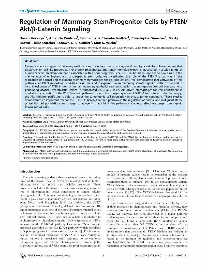
mammosphere cultures expressed increased Ser380 phosphoryla-tion of PTEN (Figure 1A), which results in its conformationalchanges masking the PDZ binding domain [22]. PTEN, throughits lipid phosphatase activity, antagonizes PI3-K/Akt signaling.We detected increased Akt Ser473 phosphorylation in mammo-spheres as compared with monolayer cultures, suggesting thatinactivation of PTEN results in increased Akt phosphorylation inmore primitive cells (Figure 1A). Akt has a number of knowndownstream targets including GSK3-b, which regulates the Wnt/b-catenin pathway. As compared to differentiated cells, cells inmammospheres displayed increased levels of GSK3-b phosphor-ylation and b-catenin activation (Figure 1A). b-catenin has beendemonstrated to play an important role in the development ofmammary stem cells in mouse models [23], suggesting that thispathway may also be active in human mammary stem/progenitorcells in mammospheres.We have previously reported an enrichment of mammary stem/
progenitor cellswithin the cell population expressing ofALDH,whichcan be detected by the enzymatic Aldefluor assay [19,24]. Consistentwith this, we found significantly higher ALDH expression inmammospheres as compared to attached mammary epithelial cellcultures (Figure 1A). We also analyzed activation of the Wnt/b-catenin pathway in ALDH-expressing cells. Aldefluor-positive cellsshowed significantly higher b-catenin activation as well as increasedGSK3-b phosphorylation as compared with Aldefluor-negative cells(Figure S1), suggesting that mammary stem/progenitor cells displayactivation of the Wnt/b-catenin pathway.
Activation of the PTEN/PI3-K/Akt Pathway Results inEnrichment of the Mammary Stem/Progenitor CellPopulation In VitroTo examine the functional role of the PI3-K/Akt/GSK3-b/b-
catenin pathway, we used both gain-of-function and loss-of-function strategies. To activate this pathway, PTEN levels weredecreased by using a PTEN shRNA DsRed-labeled lentivirus.Inclusion of the DsRed label allowed us to eliminate noninfectedcells by flow cytometry. As shown in Figure 1B, we achievedgreater than an 80% reduction in PTEN protein expression inmammospheres as assessed by Western blotting. Knockdown ofPTEN in these cells resulted in increased levels of Aktphosphorylation, GSK3-b phosphorylation, and b-catenin activa-tion (Figure 1B). In addition, activation of this pathway furtherincreased expression of ALDH1 (Figure 1B).To examine the functional role of the PTEN/PI3-K/Akt
pathway in human mammary stem/progenitor cell fate, wemeasured the effect of PTEN knockdown on mammosphereformation. Knockdown of PTEN resulted in an increase in thenumber of primary and secondary mammospheres as compared tocontrol, p,0.01 (Figure 1C and 1D). Furthermore, this increasewas maintained upon serial passage to tertiary mammospheres(Figure 1D). To provide additional evidence that this pathwayenriches for mammary stem/progenitor cells, we determined theeffect of PTEN knockdown on the percentage of cells expressingALDH as assessed by the Aldefluor assay. Primary NMECs fromreduction mammoplasties contain between 4% and 9% Aldefluor-positive cells, which increases to 14%–19% in primary mammo-spheres, consistent with the enrichment of stem/progenitor cellswhen grown in suspension cultures (Figure S2A). PTENknockdown increased the proportion of Aldefluor-positive cells(p,0.01) in mammospheres more than 2-fold to 37%–41%(Figure 1E). Thus, knockdown of PTEN resulted in enrichmentof mammary stem/progenitor cells in vitro as determined byboth mammosphere and Aldefluor assays. We previously observedthat while mammary stem/progenitor cells are enriched in
Author Summary
Healthy adult tissues are maintained through the regulat-ed proliferation of a subset of cells known as tissue stemand progenitor cells. Many cancers, including breastcancer, also are thought to arise from and be maintainedby a small population of cells that display stem cell-likeproperties. These so-called ‘‘cancer stem cells’’ may alsocontribute to tumor spread (metastasis), resistance totreatment, and disease relapse. Effective, long-lastingcancer treatments likely will need to target and eliminatethese cancer stem cells specifically. Regulatory pathwaysresponsible for maintaining cancer stem cells thereforemay be promising targets for treatment. Breast cancers inhumans frequently display abnormalities in the PTEN/PI3K/Akt pathway. We demonstrate using cell culture and amouse model of breast cancer that stem or progenitorcells in both normal breast tissue and breast tumors aredependent for their continued growth on this pathwayand on the Wnt/b-catenin pathway. We further show thatthe drug perifosine, which inhibits the kinase Akt, is ablespecifically to reduce the population of breast cancer stemor progenitor cells growing in mice. Our findings supportthe idea that drugs that selectively target breast cancerstem cells through the PTEN/PI3K/Akt pathway mayreduce tumor growth and metastasis and result inimproved patient outcomes.
PTEN/Akt Regulates Mammary Stem/Progenitor Cells
PLoS Biology | www.plosbiology.org 2 June 2009 | Volume 7 | Issue 6 | e1000121
mammosphere cultures, they are depleted in attachment cultures(Figure S2B). Cells with PTEN knockdown maintained a higherpercentage of Aldelfuor-positive cells in attachment culture (FigureS2B) as well as in suspension culture (Figure S2A). Furthermore,PTEN knockdown increased phospho-Akt expression in cells growneither in attachment or suspension cultures (Figure S2C). Theseresults suggest that PTEN knockdown is able to enrich for the stem/progenitor cell population independent of culture conditions.
PTEN Knockdown in NMECs Induces MorphologicalChanges with Features of Atypical Hyperplasia inHumanized NOD/SCID MiceWe previously used a mouse model described by Proia and
Kuperwasser [25] in which NMECs form outgrowths in NOD/
SCID mice whose mammary fat pads have been humanized bythe introduction of both irradiated and non-irradiated humanmammary fibroblasts. We used this system to examine the effectsof PTEN knockdown on mammary development. Serial dilutionsof flow cytometry-sorted cells were introduced into the humanizedfat pads of NOD/SCID mice. As indicated in Table S1, at alldilutions, NMECs with PTEN knockdown were more efficient ingenerating outgrowths than DsRed control cells. While at least10,000 control cells were required for efficient outgrowthformation, as few as 250 PTEN knockdown cells generatedoutgrowths in 50% of the mice, indicating that PTEN knockdownincreased the frequency of multipotent mammary stem/progen-itors. In addition, we observed significant morphological alter-ations in structures generated by PTEN knockdown in NMECscompared to DsRed controls (Figure 2A). PTEN knockdown cells
Figure 1. Activation of PTEN/PI3-K/Akt/GSK3-b/b-catenin signaling in mammospheres. (A) After 7–10 d of culture, mammospheres andadherent NMECs were analyzed by Western blotting for activation of the PI3-K/Akt pathway and its downstream targets. Mammospheres ascompared to adherent NMEC cultures demonstrated increased phosphorylation of PTEN, Akt, GSK3-b, and activated b-catenin (ABC). Mammospheresbut not the adherent cells also expressed the marker ALDH1. (B) Knockdown of PTEN expression via shRNA lentivirus infection led to further increasesin phospho-Akt, phospho-GSK3-b, and activated b-catenin levels compared with DsRed lentiviral-infected control mammospheres. (C) PTENknockdown led to an increase in the number of mammosphere forming cells. The efficiency of lentivirus infection was demonstrated by DsRedexpression (inserts). (D) DsRed control and PTEN knockdown mammospheres were cultured for three passages, and the number of mammospheresgenerated per 10,000 cells was determined. (E) Adherent NMECs were infected with control or PTEN lentiviral constructs and maintained inattachment cultures for 7 d. The cells from these attachment cultures were assessed for their mammosphere-forming ability. As indicated, cells withPTEN knockdown generated more mammospheres than control cells Scale bars in (C) = 100 mm. Each data point in (D) and (E) represents themean6SD of three independent experiments.doi:10.1371/journal.pbio.1000121.g001
PTEN/Akt Regulates Mammary Stem/Progenitor Cells
PLoS Biology | www.plosbiology.org 3 June 2009 | Volume 7 | Issue 6 | e1000121
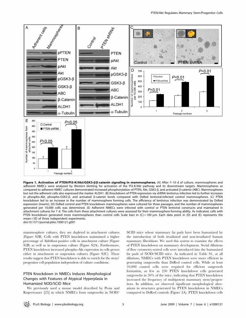
produced much larger structures that displayed significantmorphologic alterations. Knockdown of PTEN was confirmedby the lack of PTEN expression in PTEN knockdown outgrowthscompared to controls (Figure 2B, a and h). We used immunohis-tochemical staining for markers of myoepithelial, basal, andluminal epithelial cells to ascertain the effects of PTEN knockdownon cellular differentiation. Outgrowths generated by control-infected NMECs consisted of ductal structures that werecharacterized by a single layer of myoepithelial cells, whichexpressed smooth muscle actin recapitulating the architecture ofnormal mammary ducts in humans. In contrast, structuresgenerated by PTEN shRNA-infected cells were characterized bygross disorganization with increased numbers of smooth muscleactin expressing myoepithelial cells distributed throughout thegland (Figure 2B, b and i). Glands produced by control cellscontained only a small number of cells expressing the primitivecytokeratins 5/6, whereas the frequency of these cells was greatlyincreased in PTEN knockdown structures (Figure 2B, c and j).Examination of epithelial markers also revealed significant
differences between structures derived from PTEN knockdownand control cells. In control structures, the majority of the luminalepithelial cells expressed the luminal marker CK18, whereasexpression of this marker occurred only in a subfraction of PTENknockdown cells (Figure 2B, d and k). Estrogen receptor (ER) wasexpressed in luminal epithelial cells in structures generated fromDsRed control cells, but not in structures derived from PTENknockdown cells (Figure 2B, e and l). Furthermore, structures withPTEN knockdown displayed significant increases in proliferatingcells as determined by Ki67 expression (Figure 2B, f and m).Consistent with the in vitro experiments, PTEN knockdown alsoincreased the proportion of cells expressing ALDH1 (Figure 2B, gand n). These experiments confirm and extend the in vitro findingsand suggest that in addition to resulting in enrichment of the stem/progenitor cell pool, activation of the PTEN/PI3-K/Akt pathwayaffects cellular growth and differentiation. This results in thegeneration of cells displaying increased proliferation, with aberrantdifferentiation resulting in increased expression of primitive andbasal markers and decreased expression of luminal epithelial
Figure 2. Knockdown of PTEN in NMECs generates disorganized hyperplastic lesions in humanized NOD/SCID mice. (A) Humanmammary outgrowths generated from control or PTEN knockdown NMECs in humanized NOD/SCID mice exhibited an altered morphology byhematoxylin and eosin staining. (B) PTEN staining demonstrated reduced PTEN protein expression in outgrowths generated from PTEN knockdowncells as compared to the controls (a and h). Smooth muscle actin (SMA) staining revealed disorganized myoepithelial structures in PTEN knockdownoutgrowths compared to an organized layer of myoepithelial cells in controls (b and i). Outgrowths with PTEN knockdown showed increased CK5/6expression (c and j) and decreased CK18 expression (d and k), as well as a lack of ERa expression compared to control cells (e and l). PTEN knockdownoutgrowths displayed increased proliferation characterized by Ki67 staining compared to controls (f and m). Increased ALDH1 expression wasdemonstrated in PTEN knockdown structures (g and n). Scale bars = 100 mm. Data are representative of experiments with five mice in each group.doi:10.1371/journal.pbio.1000121.g002
PTEN/Akt Regulates Mammary Stem/Progenitor Cells
PLoS Biology | www.plosbiology.org 4 June 2009 | Volume 7 | Issue 6 | e1000121
markers. All of these histopathologic features are characteristic ofatypical ductal hyperplasia, a premalignant lesion that mayprogress to invasive breast cancer.
Inhibition of PI3-K/Akt Signaling SuppressesMammosphere Formation In Vitro and Generation ofOutgrowths in NOD/SCID MouseTo further characterize the pathways regulating mammary
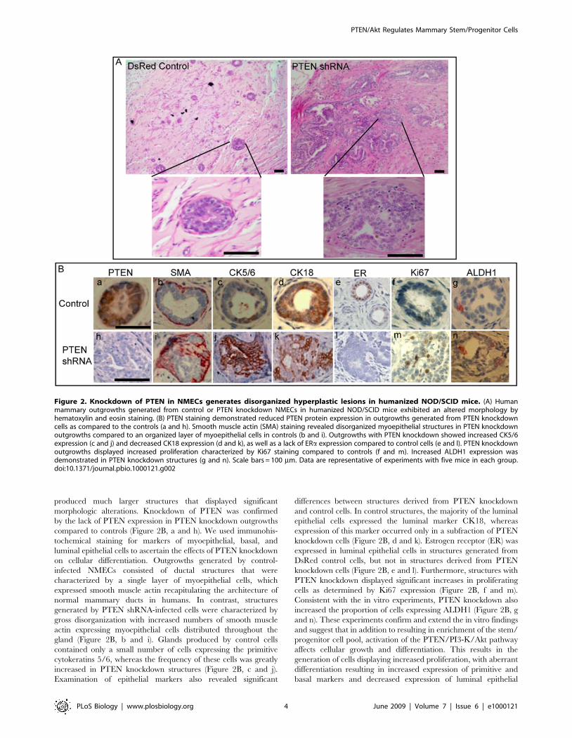
stem/progenitor cell self-renewal, we used inhibitors of PI3-K/Aktsignaling as well as its downstream targets, GSK3-b andmammalian target of rapamycin (mTOR). As demonstrated inFigure 3A, treatment of NMECs with the PI3-K inhibitorLY294002 or the Akt inhibitor IV or perifosine reduced thenumber of both primary (unpublished data) and secondarymammospheres (p,0.001). Furthermore, primary mammospherestreated with inhibitors of PI3-K or Akt completely failed to formtertiary mammospheres (unpublished data). To determine whetherthe mTOR pathway, which is downstream of Akt signaling, playsa role in this process, we examined the effect of rapamycin, aninhibitor of this pathway, on mammosphere formation. As shownin Figure 3A and Figure S2, rapamycin had little effect onsecondary mammosphere formation, suggesting that mTOR was
not responsible for mediating the effects of Akt signaling. Incontrast to rapamycin, PI3-K and Akt inhibitors suppressed theformation of secondary mammospheres in both control and PTENknockdown NMECs, supporting the importance of this pathway inmammary stem/progenitor cell self-renewal (Figure 3A). Todetermine whether PTEN knockdown affected cellular sensitivityto Akt inhibition, we performed dose response studies withperifosine. As shown in Figure 3B, 2 mM perifosine inhibited Aktphosphorylation by more than 50% in PTEN knockdown cellswhile having no demonstrable effect on control cells. Consistentwith the effects on mammosphere formation, the Akt inhibitorperifosine significantly reduced the proportion of Aldefluor-positive cells in mammospheres (Figure 3C). The effect ofperifosine on PI3-K/Akt signaling has previously been reported[26,27]. In that study, the authors found that 10 mM perifosine didnot have any inhibitory effect on the MAPK pathway [27]. Wealso failed to detect an effect of perifosine on MAPK activity in oursystem (unpublished data).To determine whether the PI3-K/Akt pathway was also critical
for mammary development in vivo, we examined the effect ofthe Akt inhibitor perifosine on the development of humanmammary structures generated by control DsRed infected or
Figure 3. Effect of PI3-K/Akt inhibitors on mammosphere formation, ALDH1 expression, and mammary development in NOD/SCIDmice. (A) Treatment of mammospheres with the PI3-K inhibitor LY294002 (1 mM), Akt inhibitor IV (2 mM), or perifosine (5 mM) inhibitedmammosphere formation in control cells, whereas PTEN knockdown NMECs were sensitive to lower doses of these inhibitors: LY294002 (0.5 mM), Aktinhibitor IV (1 mM), or perifosine (2 mM). In contrast, the mTOR inhibitor rapamycin (0.5 mM) had little effect on these cells. (B) Perifosine doseresponse was tested in both control and PTEN knockdown cells. As indicated, PTEN knockdown cells with higher Akt activity are more sensitive toperifosine than control cells with an IC50 of 5 mM. (C) The effect of perifosine on the Aldefluor-positive population of NMECs was measured by theAldefluor assay. Treatment of NMECs with perifosine over 5 d reduced the Aldefluor-positive population in primary mammospheres by more than50%. (D) Perifosine treatment of mice implanted with control or PTEN knockdown NMECs completely blocked the formation of outgrowths in NOD/SCID humanized fat pads compared to saline-treated control mice. High magnification (insets) shows persistence of the inoculated cells. Scalebars = 100 mm. Data represent the mean6SD of three independent experiments.doi:10.1371/journal.pbio.1000121.g003
PTEN/Akt Regulates Mammary Stem/Progenitor Cells
PLoS Biology | www.plosbiology.org 5 June 2009 | Volume 7 | Issue 6 | e1000121
PTEN knockdown NMECs. NOD/SCID mice implanted withcontrol or PTEN knockdown NMECs were treated withintraperitoneal injections of perifosine (30 mg/kg) 4 days a weekfor five weeks or a saline control. Consistent with previousexperiments, control DsRed NMECs generated mammary ductalstructures with normal morphology, PTEN knockdown NMECsgenerated ductal hyperplasias in saline-treated control mice. Incontrast, administration of perifosine completely inhibited out-growth formation by both control and PTEN knockdown NMECs(Figure 3D). These results further support the in vitro experimentsby demonstrating a critical role for Akt signaling in normalmammary development.
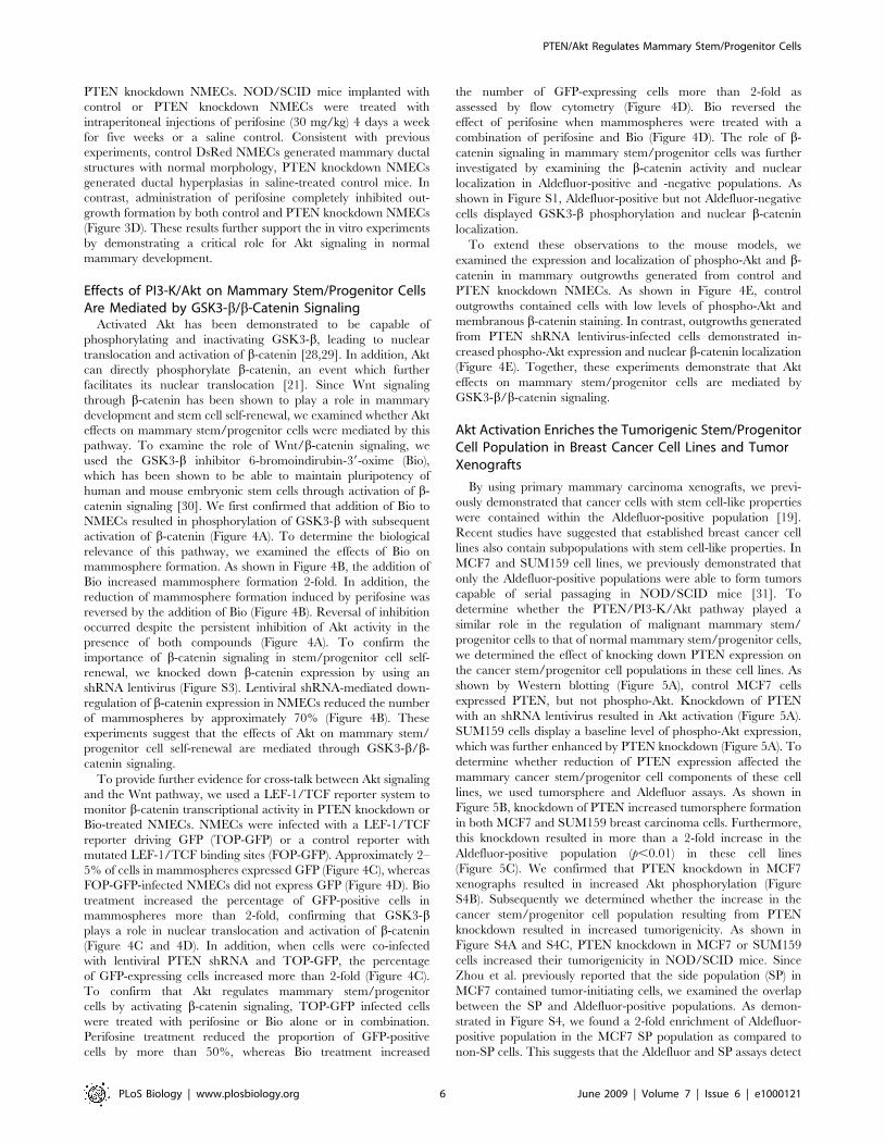
Effects of PI3-K/Akt on Mammary Stem/Progenitor CellsAre Mediated by GSK3-b/b-Catenin SignalingActivated Akt has been demonstrated to be capable of
phosphorylating and inactivating GSK3-b, leading to nucleartranslocation and activation of b-catenin [28,29]. In addition, Aktcan directly phosphorylate b-catenin, an event which furtherfacilitates its nuclear translocation [21]. Since Wnt signalingthrough b-catenin has been shown to play a role in mammarydevelopment and stem cell self-renewal, we examined whether Akteffects on mammary stem/progenitor cells were mediated by thispathway. To examine the role of Wnt/b-catenin signaling, weused the GSK3-b inhibitor 6-bromoindirubin-39-oxime (Bio),which has been shown to be able to maintain pluripotency ofhuman and mouse embryonic stem cells through activation of b-catenin signaling [30]. We first confirmed that addition of Bio toNMECs resulted in phosphorylation of GSK3-b with subsequentactivation of b-catenin (Figure 4A). To determine the biologicalrelevance of this pathway, we examined the effects of Bio onmammosphere formation. As shown in Figure 4B, the addition ofBio increased mammosphere formation 2-fold. In addition, thereduction of mammosphere formation induced by perifosine wasreversed by the addition of Bio (Figure 4B). Reversal of inhibitionoccurred despite the persistent inhibition of Akt activity in thepresence of both compounds (Figure 4A). To confirm theimportance of b-catenin signaling in stem/progenitor cell self-renewal, we knocked down b-catenin expression by using anshRNA lentivirus (Figure S3). Lentiviral shRNA-mediated down-regulation of b-catenin expression in NMECs reduced the numberof mammospheres by approximately 70% (Figure 4B). Theseexperiments suggest that the effects of Akt on mammary stem/progenitor cell self-renewal are mediated through GSK3-b/b-catenin signaling.To provide further evidence for cross-talk between Akt signaling
and the Wnt pathway, we used a LEF-1/TCF reporter system tomonitor b-catenin transcriptional activity in PTEN knockdown orBio-treated NMECs. NMECs were infected with a LEF-1/TCFreporter driving GFP (TOP-GFP) or a control reporter withmutated LEF-1/TCF binding sites (FOP-GFP). Approximately 2–5% of cells in mammospheres expressed GFP (Figure 4C), whereasFOP-GFP-infected NMECs did not express GFP (Figure 4D). Biotreatment increased the percentage of GFP-positive cells inmammospheres more than 2-fold, confirming that GSK3-bplays a role in nuclear translocation and activation of b-catenin(Figure 4C and 4D). In addition, when cells were co-infectedwith lentiviral PTEN shRNA and TOP-GFP, the percentageof GFP-expressing cells increased more than 2-fold (Figure 4C).To confirm that Akt regulates mammary stem/progenitorcells by activating b-catenin signaling, TOP-GFP infected cellswere treated with perifosine or Bio alone or in combination.Perifosine treatment reduced the proportion of GFP-positivecells by more than 50%, whereas Bio treatment increased
the number of GFP-expressing cells more than 2-fold asassessed by flow cytometry (Figure 4D). Bio reversed theeffect of perifosine when mammospheres were treated with acombination of perifosine and Bio (Figure 4D). The role of b-catenin signaling in mammary stem/progenitor cells was furtherinvestigated by examining the b-catenin activity and nuclearlocalization in Aldefluor-positive and -negative populations. Asshown in Figure S1, Aldefluor-positive but not Aldefluor-negativecells displayed GSK3-b phosphorylation and nuclear b-cateninlocalization.To extend these observations to the mouse models, we
examined the expression and localization of phospho-Akt and b-catenin in mammary outgrowths generated from control andPTEN knockdown NMECs. As shown in Figure 4E, controloutgrowths contained cells with low levels of phospho-Akt andmembranous b-catenin staining. In contrast, outgrowths generatedfrom PTEN shRNA lentivirus-infected cells demonstrated in-creased phospho-Akt expression and nuclear b-catenin localization(Figure 4E). Together, these experiments demonstrate that Akteffects on mammary stem/progenitor cells are mediated byGSK3-b/b-catenin signaling.
Akt Activation Enriches the Tumorigenic Stem/ProgenitorCell Population in Breast Cancer Cell Lines and TumorXenografts
By using primary mammary carcinoma xenografts, we previ-ously demonstrated that cancer cells with stem cell-like propertieswere contained within the Aldefluor-positive population [19].Recent studies have suggested that established breast cancer celllines also contain subpopulations with stem cell-like properties. InMCF7 and SUM159 cell lines, we previously demonstrated thatonly the Aldefluor-positive populations were able to form tumorscapable of serial passaging in NOD/SCID mice [31]. Todetermine whether the PTEN/PI3-K/Akt pathway played asimilar role in the regulation of malignant mammary stem/progenitor cells to that of normal mammary stem/progenitor cells,we determined the effect of knocking down PTEN expression onthe cancer stem/progenitor cell populations in these cell lines. Asshown by Western blotting (Figure 5A), control MCF7 cellsexpressed PTEN, but not phospho-Akt. Knockdown of PTENwith an shRNA lentivirus resulted in Akt activation (Figure 5A).SUM159 cells display a baseline level of phospho-Akt expression,which was further enhanced by PTEN knockdown (Figure 5A). Todetermine whether reduction of PTEN expression affected themammary cancer stem/progenitor cell components of these celllines, we used tumorsphere and Aldefluor assays. As shown inFigure 5B, knockdown of PTEN increased tumorsphere formationin both MCF7 and SUM159 breast carcinoma cells. Furthermore,this knockdown resulted in more than a 2-fold increase in theAldefluor-positive population (p,0.01) in these cell lines(Figure 5C). We confirmed that PTEN knockdown in MCF7xenographs resulted in increased Akt phosphorylation (FigureS4B). Subsequently we determined whether the increase in thecancer stem/progenitor cell population resulting from PTENknockdown resulted in increased tumorigenicity. As shown inFigure S4A and S4C, PTEN knockdown in MCF7 or SUM159cells increased their tumorigenicity in NOD/SCID mice. SinceZhou et al. previously reported that the side population (SP) inMCF7 contained tumor-initiating cells, we examined the overlapbetween the SP and Aldefluor-positive populations. As demon-strated in Figure S4, we found a 2-fold enrichment of Aldefluor-positive population in the MCF7 SP population as compared tonon-SP cells. This suggests that the Aldefluor and SP assays detect
PTEN/Akt Regulates Mammary Stem/Progenitor Cells
PLoS Biology | www.plosbiology.org 6 June 2009 | Volume 7 | Issue 6 | e1000121
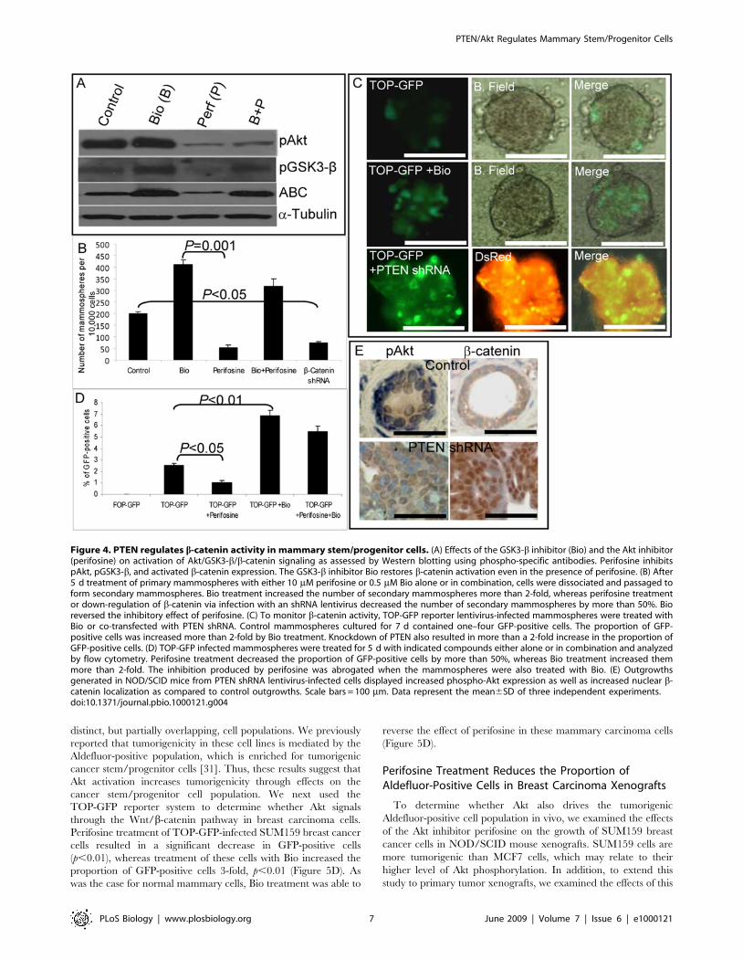
distinct, but partially overlapping, cell populations. We previouslyreported that tumorigenicity in these cell lines is mediated by theAldefluor-positive population, which is enriched for tumorigeniccancer stem/progenitor cells [31]. Thus, these results suggest thatAkt activation increases tumorigenicity through effects on thecancer stem/progenitor cell population. We next used theTOP-GFP reporter system to determine whether Akt signalsthrough the Wnt/b-catenin pathway in breast carcinoma cells.Perifosine treatment of TOP-GFP-infected SUM159 breast cancercells resulted in a significant decrease in GFP-positive cells(p,0.01), whereas treatment of these cells with Bio increased theproportion of GFP-positive cells 3-fold, p,0.01 (Figure 5D). Aswas the case for normal mammary cells, Bio treatment was able to
reverse the effect of perifosine in these mammary carcinoma cells(Figure 5D).
Perifosine Treatment Reduces the Proportion ofAldefluor-Positive Cells in Breast Carcinoma Xenografts
To determine whether Akt also drives the tumorigenicAldefluor-positive cell population in vivo, we examined the effectsof the Akt inhibitor perifosine on the growth of SUM159 breastcancer cells in NOD/SCID mouse xenografts. SUM159 cells aremore tumorigenic than MCF7 cells, which may relate to theirhigher level of Akt phosphorylation. In addition, to extend thisstudy to primary tumor xenografts, we examined the effects of this
Figure 4. PTEN regulates b-catenin activity in mammary stem/progenitor cells. (A) Effects of the GSK3-b inhibitor (Bio) and the Akt inhibitor(perifosine) on activation of Akt/GSK3-b/b-catenin signaling as assessed by Western blotting using phospho-specific antibodies. Perifosine inhibitspAkt, pGSK3-b, and activated b-catenin expression. The GSK3-b inhibitor Bio restores b-catenin activation even in the presence of perifosine. (B) After5 d treatment of primary mammospheres with either 10 mM perifosine or 0.5 mM Bio alone or in combination, cells were dissociated and passaged toform secondary mammospheres. Bio treatment increased the number of secondary mammospheres more than 2-fold, whereas perifosine treatmentor down-regulation of b-catenin via infection with an shRNA lentivirus decreased the number of secondary mammospheres by more than 50%. Bioreversed the inhibitory effect of perifosine. (C) To monitor b-catenin activity, TOP-GFP reporter lentivirus-infected mammospheres were treated withBio or co-transfected with PTEN shRNA. Control mammospheres cultured for 7 d contained one–four GFP-positive cells. The proportion of GFP-positive cells was increased more than 2-fold by Bio treatment. Knockdown of PTEN also resulted in more than a 2-fold increase in the proportion ofGFP-positive cells. (D) TOP-GFP infected mammospheres were treated for 5 d with indicated compounds either alone or in combination and analyzedby flow cytometry. Perifosine treatment decreased the proportion of GFP-positive cells by more than 50%, whereas Bio treatment increased themmore than 2-fold. The inhibition produced by perifosine was abrogated when the mammospheres were also treated with Bio. (E) Outgrowthsgenerated in NOD/SCID mice from PTEN shRNA lentivirus-infected cells displayed increased phospho-Akt expression as well as increased nuclear b-catenin localization as compared to control outgrowths. Scale bars = 100 mm. Data represent the mean6SD of three independent experiments.doi:10.1371/journal.pbio.1000121.g004
PTEN/Akt Regulates Mammary Stem/Progenitor Cells
PLoS Biology | www.plosbiology.org 7 June 2009 | Volume 7 | Issue 6 | e1000121
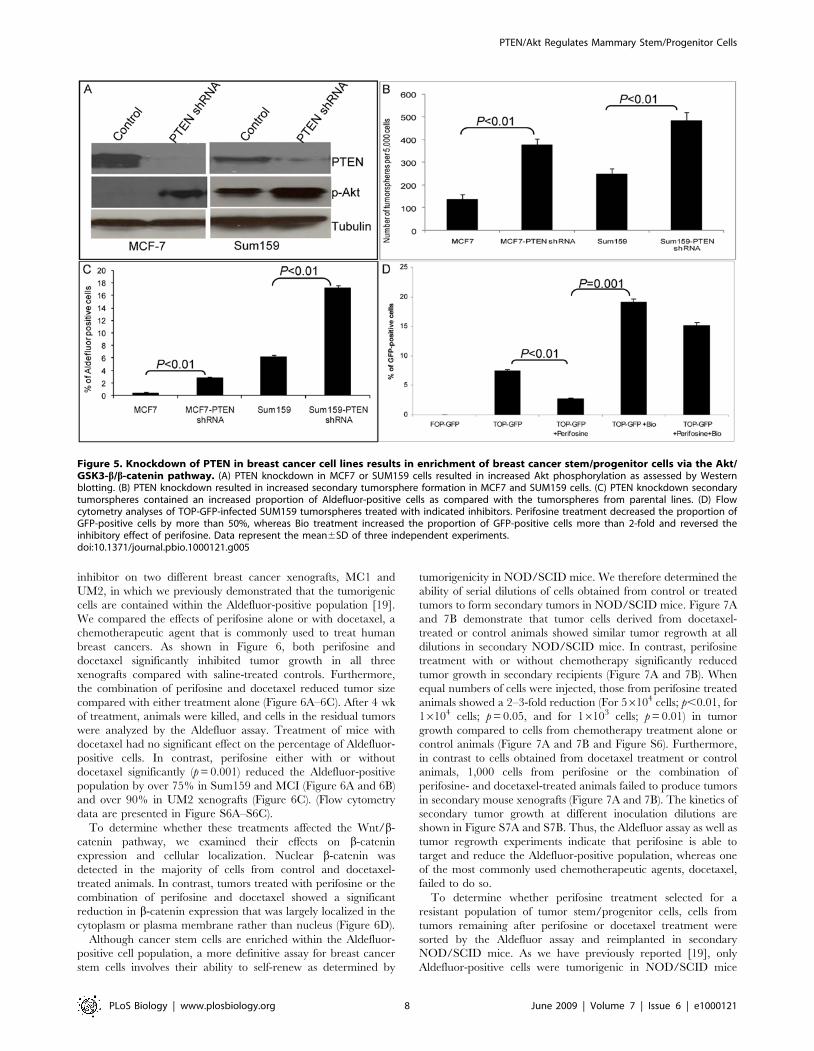
inhibitor on two different breast cancer xenografts, MC1 andUM2, in which we previously demonstrated that the tumorigeniccells are contained within the Aldefluor-positive population [19].We compared the effects of perifosine alone or with docetaxel, achemotherapeutic agent that is commonly used to treat humanbreast cancers. As shown in Figure 6, both perifosine anddocetaxel significantly inhibited tumor growth in all threexenografts compared with saline-treated controls. Furthermore,the combination of perifosine and docetaxel reduced tumor sizecompared with either treatment alone (Figure 6A–6C). After 4 wkof treatment, animals were killed, and cells in the residual tumorswere analyzed by the Aldefluor assay. Treatment of mice withdocetaxel had no significant effect on the percentage of Aldefluor-positive cells. In contrast, perifosine either with or withoutdocetaxel significantly (p=0.001) reduced the Aldefluor-positivepopulation by over 75% in Sum159 and MCI (Figure 6A and 6B)and over 90% in UM2 xenografts (Figure 6C). (Flow cytometrydata are presented in Figure S6A–S6C).To determine whether these treatments affected the Wnt/b-
catenin pathway, we examined their effects on b-cateninexpression and cellular localization. Nuclear b-catenin wasdetected in the majority of cells from control and docetaxel-treated animals. In contrast, tumors treated with perifosine or thecombination of perifosine and docetaxel showed a significantreduction in b-catenin expression that was largely localized in thecytoplasm or plasma membrane rather than nucleus (Figure 6D).Although cancer stem cells are enriched within the Aldefluor-
positive cell population, a more definitive assay for breast cancerstem cells involves their ability to self-renew as determined by
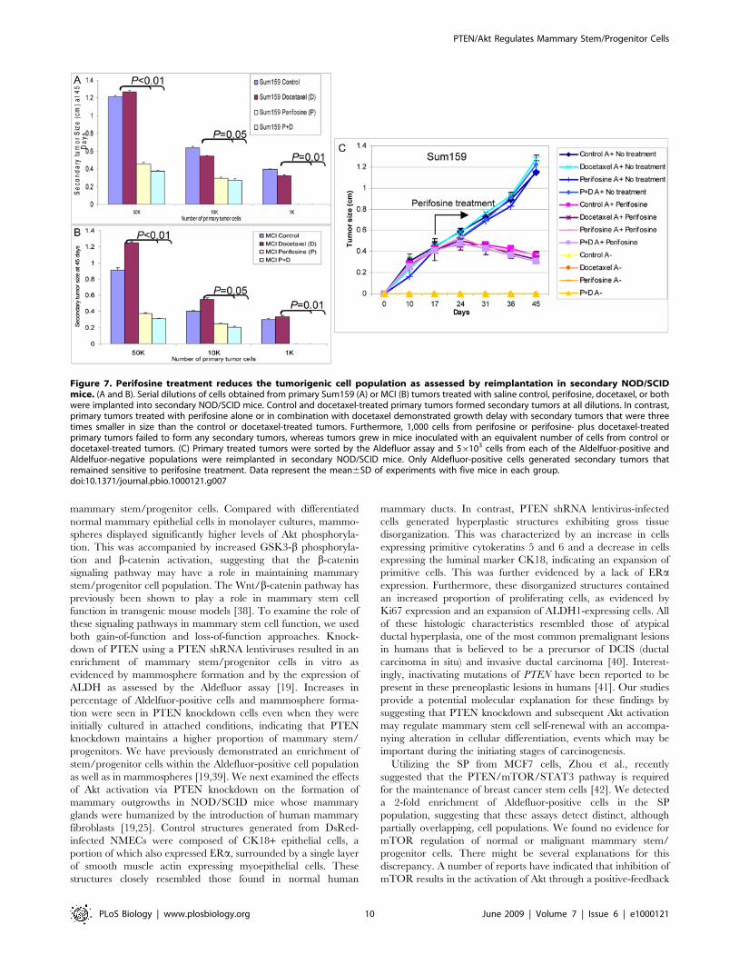
tumorigenicity in NOD/SCID mice. We therefore determined theability of serial dilutions of cells obtained from control or treatedtumors to form secondary tumors in NOD/SCID mice. Figure 7Aand 7B demonstrate that tumor cells derived from docetaxel-treated or control animals showed similar tumor regrowth at alldilutions in secondary NOD/SCID mice. In contrast, perifosinetreatment with or without chemotherapy significantly reducedtumor growth in secondary recipients (Figure 7A and 7B). Whenequal numbers of cells were injected, those from perifosine treatedanimals showed a 2–3-fold reduction (For 56104 cells; p,0.01, for16104 cells; p=0.05, and for 16103 cells; p=0.01) in tumorgrowth compared to cells from chemotherapy treatment alone orcontrol animals (Figure 7A and 7B and Figure S6). Furthermore,in contrast to cells obtained from docetaxel treatment or controlanimals, 1,000 cells from perifosine or the combination ofperifosine- and docetaxel-treated animals failed to produce tumorsin secondary mouse xenografts (Figure 7A and 7B). The kinetics ofsecondary tumor growth at different inoculation dilutions areshown in Figure S7A and S7B. Thus, the Aldefluor assay as well astumor regrowth experiments indicate that perifosine is able totarget and reduce the Aldefluor-positive population, whereas oneof the most commonly used chemotherapeutic agents, docetaxel,failed to do so.To determine whether perifosine treatment selected for a
resistant population of tumor stem/progenitor cells, cells fromtumors remaining after perifosine or docetaxel treatment weresorted by the Aldefluor assay and reimplanted in secondaryNOD/SCID mice. As we have previously reported [19], onlyAldefluor-positive cells were tumorigenic in NOD/SCID mice
Figure 5. Knockdown of PTEN in breast cancer cell lines results in enrichment of breast cancer stem/progenitor cells via the Akt/GSK3-b/b-catenin pathway. (A) PTEN knockdown in MCF7 or SUM159 cells resulted in increased Akt phosphorylation as assessed by Westernblotting. (B) PTEN knockdown resulted in increased secondary tumorsphere formation in MCF7 and SUM159 cells. (C) PTEN knockdown secondarytumorspheres contained an increased proportion of Aldefluor-positive cells as compared with the tumorspheres from parental lines. (D) Flowcytometry analyses of TOP-GFP-infected SUM159 tumorspheres treated with indicated inhibitors. Perifosine treatment decreased the proportion ofGFP-positive cells by more than 50%, whereas Bio treatment increased the proportion of GFP-positive cells more than 2-fold and reversed theinhibitory effect of perifosine. Data represent the mean6SD of three independent experiments.doi:10.1371/journal.pbio.1000121.g005
PTEN/Akt Regulates Mammary Stem/Progenitor Cells
PLoS Biology | www.plosbiology.org 8 June 2009 | Volume 7 | Issue 6 | e1000121
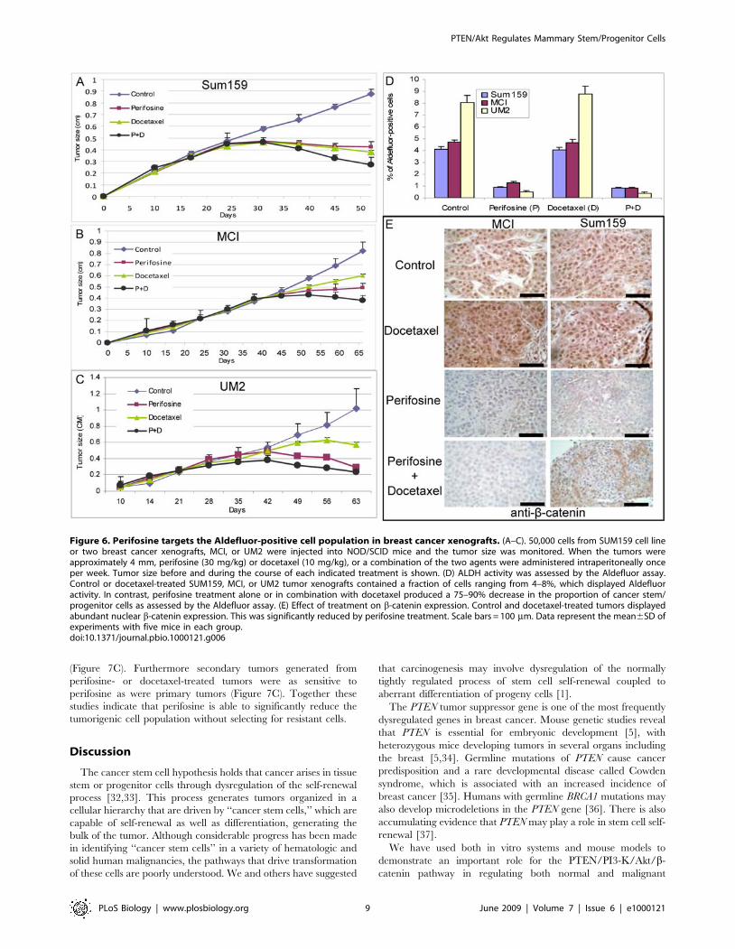
(Figure 7C). Furthermore secondary tumors generated fromperifosine- or docetaxel-treated tumors were as sensitive toperifosine as were primary tumors (Figure 7C). Together thesestudies indicate that perifosine is able to significantly reduce thetumorigenic cell population without selecting for resistant cells.
Discussion
The cancer stem cell hypothesis holds that cancer arises in tissuestem or progenitor cells through dysregulation of the self-renewalprocess [32,33]. This process generates tumors organized in acellular hierarchy that are driven by ‘‘cancer stem cells,’’ which arecapable of self-renewal as well as differentiation, generating thebulk of the tumor. Although considerable progress has been madein identifying ‘‘cancer stem cells’’ in a variety of hematologic andsolid human malignancies, the pathways that drive transformationof these cells are poorly understood. We and others have suggested
that carcinogenesis may involve dysregulation of the normallytightly regulated process of stem cell self-renewal coupled toaberrant differentiation of progeny cells [1].The PTEN tumor suppressor gene is one of the most frequently
dysregulated genes in breast cancer. Mouse genetic studies revealthat PTEN is essential for embryonic development [5], withheterozygous mice developing tumors in several organs includingthe breast [5,34]. Germline mutations of PTEN cause cancerpredisposition and a rare developmental disease called Cowdensyndrome, which is associated with an increased incidence ofbreast cancer [35]. Humans with germline BRCA1 mutations mayalso develop microdeletions in the PTEN gene [36]. There is alsoaccumulating evidence that PTEN may play a role in stem cell self-renewal [37].We have used both in vitro systems and mouse models to
demonstrate an important role for the PTEN/PI3-K/Akt/b-catenin pathway in regulating both normal and malignant
Figure 6. Perifosine targets the Aldefluor-positive cell population in breast cancer xenografts. (A–C). 50,000 cells from SUM159 cell lineor two breast cancer xenografts, MCI, or UM2 were injected into NOD/SCID mice and the tumor size was monitored. When the tumors wereapproximately 4 mm, perifosine (30 mg/kg) or docetaxel (10 mg/kg), or a combination of the two agents were administered intraperitoneally onceper week. Tumor size before and during the course of each indicated treatment is shown. (D) ALDH activity was assessed by the Aldefluor assay.Control or docetaxel-treated SUM159, MCI, or UM2 tumor xenografts contained a fraction of cells ranging from 4–8%, which displayed Aldefluoractivity. In contrast, perifosine treatment alone or in combination with docetaxel produced a 75–90% decrease in the proportion of cancer stem/progenitor cells as assessed by the Aldefluor assay. (E) Effect of treatment on b-catenin expression. Control and docetaxel-treated tumors displayedabundant nuclear b-catenin expression. This was significantly reduced by perifosine treatment. Scale bars = 100 mm. Data represent the mean6SD ofexperiments with five mice in each group.doi:10.1371/journal.pbio.1000121.g006
PTEN/Akt Regulates Mammary Stem/Progenitor Cells
PLoS Biology | www.plosbiology.org 9 June 2009 | Volume 7 | Issue 6 | e1000121
mammary stem/progenitor cells. Compared with differentiatednormal mammary epithelial cells in monolayer cultures, mammo-spheres displayed significantly higher levels of Akt phosphoryla-tion. This was accompanied by increased GSK3-b phosphoryla-tion and b-catenin activation, suggesting that the b-cateninsignaling pathway may have a role in maintaining mammarystem/progenitor cell population. The Wnt/b-catenin pathway haspreviously been shown to play a role in mammary stem cellfunction in transgenic mouse models [38]. To examine the role ofthese signaling pathways in mammary stem cell function, we usedboth gain-of-function and loss-of-function approaches. Knock-down of PTEN using a PTEN shRNA lentiviruses resulted in anenrichment of mammary stem/progenitor cells in vitro asevidenced by mammosphere formation and by the expression ofALDH as assessed by the Aldefluor assay [19]. Increases inpercentage of Aldelfuor-positive cells and mammosphere forma-tion were seen in PTEN knockdown cells even when they wereinitially cultured in attached conditions, indicating that PTENknockdown maintains a higher proportion of mammary stem/progenitors. We have previously demonstrated an enrichment ofstem/progenitor cells within the Aldefluor-positive cell populationas well as in mammospheres [19,39]. We next examined the effectsof Akt activation via PTEN knockdown on the formation ofmammary outgrowths in NOD/SCID mice whose mammaryglands were humanized by the introduction of human mammaryfibroblasts [19,25]. Control structures generated from DsRed-infected NMECs were composed of CK18+ epithelial cells, aportion of which also expressed ERa, surrounded by a single layerof smooth muscle actin expressing myoepithelial cells. Thesestructures closely resembled those found in normal human
mammary ducts. In contrast, PTEN shRNA lentivirus-infectedcells generated hyperplastic structures exhibiting gross tissuedisorganization. This was characterized by an increase in cellsexpressing primitive cytokeratins 5 and 6 and a decrease in cellsexpressing the luminal marker CK18, indicating an expansion ofprimitive cells. This was further evidenced by a lack of ERaexpression. Furthermore, these disorganized structures containedan increased proportion of proliferating cells, as evidenced byKi67 expression and an expansion of ALDH1-expressing cells. Allof these histologic characteristics resembled those of atypicalductal hyperplasia, one of the most common premalignant lesionsin humans that is believed to be a precursor of DCIS (ductalcarcinoma in situ) and invasive ductal carcinoma [40]. Interest-ingly, inactivating mutations of PTEN have been reported to bepresent in these preneoplastic lesions in humans [41]. Our studiesprovide a potential molecular explanation for these findings bysuggesting that PTEN knockdown and subsequent Akt activationmay regulate mammary stem cell self-renewal with an accompa-nying alteration in cellular differentiation, events which may beimportant during the initiating stages of carcinogenesis.Utilizing the SP from MCF7 cells, Zhou et al., recently
suggested that the PTEN/mTOR/STAT3 pathway is requiredfor the maintenance of breast cancer stem cells [42]. We detecteda 2-fold enrichment of Aldefluor-positive cells in the SPpopulation, suggesting that these assays detect distinct, althoughpartially overlapping, cell populations. We found no evidence formTOR regulation of normal or malignant mammary stem/progenitor cells. There might be several explanations for thisdiscrepancy. A number of reports have indicated that inhibition ofmTOR results in the activation of Akt through a positive-feedback
Figure 7. Perifosine treatment reduces the tumorigenic cell population as assessed by reimplantation in secondary NOD/SCIDmice. (A and B). Serial dilutions of cells obtained from primary Sum159 (A) or MCI (B) tumors treated with saline control, perifosine, docetaxel, or bothwere implanted into secondary NOD/SCID mice. Control and docetaxel-treated primary tumors formed secondary tumors at all dilutions. In contrast,primary tumors treated with perifosine alone or in combination with docetaxel demonstrated growth delay with secondary tumors that were threetimes smaller in size than the control or docetaxel-treated tumors. Furthermore, 1,000 cells from perifosine or perifosine- plus docetaxel-treatedprimary tumors failed to form any secondary tumors, whereas tumors grew in mice inoculated with an equivalent number of cells from control ordocetaxel-treated tumors. (C) Primary treated tumors were sorted by the Aldefluor assay and 56103 cells from each of the Aldelfuor-positive andAldelfuor-negative populations were reimplanted in secondary NOD/SCID mice. Only Aldefluor-positive cells generated secondary tumors thatremained sensitive to perifosine treatment. Data represent the mean6SD of experiments with five mice in each group.doi:10.1371/journal.pbio.1000121.g007
PTEN/Akt Regulates Mammary Stem/Progenitor Cells
PLoS Biology | www.plosbiology.org 10 June 2009 | Volume 7 | Issue 6 | e1000121
loop [43–46]. Furthermore, recent studies have shown thatmammary stem cells are not contained within the SP population[47]. In contrast to the report by Zhou et al., we used bothpharmacologic and genetic approaches to elucidate the down-stream signaling pathways of Akt in both normal and malignantmammary stem/progenitor cells. Treatment of NMECs with thePI3-K inhibitor Ly294002, or the Akt inhibitors IV and perifosine,reduced mammosphere formation and the Aldefluor-positive cellpopulation. The effect of perifosine on the PI3-K/Akt pathway haspreviously been described [26,48]. It has also been reported thatAkt may regulate Wnt signaling through Akt phosphorylation andinactivation of GSK3-b, which in turn mediates b-catenindegradation [28] or, directly by phosphorylating b-catenin onserine 552, promoting the nuclear translocation of b-catenin [21].Thus, by these two mechanisms, Akt activation promotes theactivation and accumulation of nuclear b-catenin [49]. Wedemonstrated in mammospheres that Akt phosphorylation wasassociated with increased phosphorylation of GSK3-b andactivation of b-catenin. GSK3-b targets b-catenin for ubiquitin-mediated degradation through phosphorylation of its N-terminalserine and threonine residues [50]. To demonstrate the impor-tance of b-catenin in mammosphere formation, we used a b-catenin shRNA lentivirus to knock down b-catenin expression.This resulted in significantly reduced mammosphere formation.Furthermore, we demonstrated that the GSK3-b inhibitor Biorescued the effects of Akt inhibition on mammosphere formation.To examine more directly the activation of the Wnt pathway, weused a TOP-GFP reporter system that is activated by b-cateninsignaling. The Akt inhibitor perifosine significantly reduced theproportion of TOP-GFP expressing cells, an effect that wasreversed by Bio. The relevance of these findings to mammarydevelopment in vivo was determined by examining the expressionand cellular localization of phospho-Akt and b-catenin inmammary outgrowths generated from control and PTENknockdown NMECs. While cells from control outgrowths showedminimal phospho-Akt expression and membranous b-cateninlocalization, outgrowths from PTEN shRNA lentivirus-infectedcells demonstrated increased phospho-Akt expression and nuclearb-catenin localization. The importance of this pathway wasdemonstrated by the ability of the Akt inhibitor perifosine tocompletely block mammary development in the mouse model.Together these in vitro and mouse experiments suggest that theeffects of Akt on mammary stem/progenitor cells are mediated byGSK3-b phosphorylation and b-catenin activation.To determine whether the PTEN/Akt/b-catenin signaling
pathway also plays a role in the regulation of malignant mammarystem/progenitor cells, we used both breast cancer cell lines and aprimary tumor xenograft. Although Hollestelle et al. reported anactivating PIK3CAmutation in MCF7 cells, they did not determinewhether this mutation resulted in activation of the PI3K/Aktpathway [51]. In contrast, Neve et al. screened 38 breast cancercell lines including MCF7 for various pathways and found thatMCF7 cells displayed significantly low levels of the PI3/Aktactivation when compared with other cell lines [52]. This wasconfirmed in a recent report demonstrating that despite harboringa PIK3CA helical mutation, MCF7 cells displayed a low level ofAKT phosphorylation [53]. In addition, these authors found nocorrelation between similar activating mutations of PIK3CA inprimary tumors and patient survival or the chemoresistance [53].Most importantly, they found no evidence that these mutations inPIK3CA resulted in activation of Akt pathway as compared withthe wild-type PIK3CA, but there was a strong correlation betweenPTEN inactivating mutation and activation of Akt signaling [53].In agreement with latter reports, we found that PTEN knockdown
in MCF7 or SUM159 resulted in activation of PI3-K/Aktpathway. We demonstrated that activation of this pathwaythrough knockdown of PTEN significantly increased tumorsphereformation and the ALDH-expressing cell population, indicating anenrichment of cancer stem/progenitor cells. Moreover, enrich-ment of the cancer stem/progenitor cell population directlycorrelated with increased tumorigenicity. Since this populationmediates tumorigenesis [31], it suggests that Akt activationenhances tumorigenesis through effects on the cancer stem/progenitor cell population. Furthermore, as was the case forNMECs, we demonstrated that Akt effects on mammarycarcinoma stem/progenitor cells are mediated by Wnt/b-cateninsignaling. By using the TOP-GFP reporter system, we found thatinhibition of Akt signaling significantly reduced the number ofGFP-positive cells, an effect that was rescued with the GSK3-binhibitor Bio. These findings are in agreement with the findings ofLi et al., who reported that Wnt-induced mouse mammary tumorsshow expansion of stem/progenitor population as characterized byincreased stem cell markers [54]. Furthermore the Wnt/b-cateninpathway has been shown to play a role in mediating the radiationresistance of mouse mammary progenitor cells [55].In addition to its effects on the stem/progenitor cell population,
Akt has been demonstrated to play a role in chemoresistance [15–17]. Recent evidence utilizing in vitro systems [56], animal models[13], and clinical trials have suggested that breast cancer stem cellsare relatively resistant to both radiation and chemotherapy [57].Improved clinical outcomes may require the development ofstrategies that are able to target this cancer stem cell population.The demonstration that Akt signaling plays a prominent role in stemcell self-renewal makes it an attractive target for such strategies. Weused the Akt inhibitor perifosine, an orally bioactive alkylpho-spholipid, to determine its effects on Aldefluor-positive cellpopulation. We demonstrate both in in vitro and in mousexenograft models that perifosine is able to target the Aldefluor-positive tumorigenic cell population as determined by the Aldefluorassay and by reduced tumorigenicity upon serial transplantation.Furthermore, tumorigenic Aldefluor-positive cells remaining afterperifosine treatment generated secondary tumors that were stillsensitive to perifosine. In contrast, the chemotherapeutic agentdocetaxel, although capable of causing tumor regression, failed totarget the Aldefluor-positive cell population. If cancer stem cellsindeed contribute to tumor resistance and relapse, then the additionof agents that are capable of targeting these cells may increase theclinical efficacy of current therapies. Our studies identify inhibitorsof the PI3-K/Akt/Wnt signaling pathway as potential agents fortherapeutic targeting of cancer stem cells.
Materials and Methods
Cell Lines and ReagentsThe MCF7 cell line was maintained in RPMI supplemented
with 5% fetal bovine serum (FBS), 5 mg/ml insulin, and antibiotic-antimycotic. The SUM159 cell line was maintained in Ham’s F12medium supplemented (with 5% FBS, 5 mg/ml insulin, 1 mg/mlhydrocortisone, and antibiotic/antimycotic 10,000 units/mlpenicillin G sodium, 10,000 mg/ml streptomycin sulfate, and25 mg/ml amphotericin B). LY294002, Akt inhibitor IV, and Biowere purchased from Millipore; perifosine was obtained fromKeryx Biopharmaceuticals, and docetaxel (Taxotere) was fromSanofi Aventis.Antibodies to phospho-Akt, Akt, b-catenin, GSK3-b, phospho-
GSK3-b Ser9, PTEN, and phospho-PTEN Ser380 were purchasedfrom Cell Signaling Technology. The a-tubulin antibody was fromSanta Cruz Biotechnology, the ALDH1 antibody was from BD
PTEN/Akt Regulates Mammary Stem/Progenitor Cells
PLoS Biology | www.plosbiology.org 11 June 2009 | Volume 7 | Issue 6 | e1000121
Transduction Laboratory, and the active-b-catenin (anti-ABC),clone 8E7 mouse monoclonal antibody was from MilliporeCorporation.The primary antibodies for human smooth muscle actin, Ki67,
CK5/6, and CK18 were purchased from Zymed laboratories(Invitrogen).
Dissociation of Normal Mammary TissuesMammary tissue from reduction mammoplasties was dissociat-
ed as previously described [19]. The single-cell suspension wasused for various experiments including sphere formation, flowcytometry analyses, and lentiviral infections. At least threedifferent patient samples (reduction mammoplasties) were usedfor each experiment involving NMECs.
Mammosphere/Tumorsphere AssaySingle NMECs were plated on 1% agarose coated plates at a
density of 16105 cells/ml and grown for 7–10 d. Subsequentcultures after dissociation of primary spheres were plated onultra-low attachment plates at a density of 56103–16104 cells/ml. Mammosphere cultures were grown in a serum-freemammary epithelium basal medium as previously described [18].Tumorspheres were cultured under identical conditions tomammospheres.
Lentiviral Constructs and Infection of NMECs and BreastCancer Cell LinesAll lentiviral constructs were prepared by the University of
Michigan Vector or shRNA core facilities. The primers targetingthe human PTEN short hairpin sequences were purchased fromIntegrated DNA Technologies. The forward primer TCCCA-GGTGAAGGTATGTTCCTCCAATCTAAAGGATTGGAGG-AATATATCTTCACCTGGGATTTTTTC and the reverseprimer TCGAGAAAAAATCCCAGGTGAAGATATATTCC-TCCAATCCTTTAGATTGGAGGAACATACCTTCACCTG-GAC were digested with the Xho1 enzyme, annealed, and clonedinto the pLentilox 3.7 vector. After confirmation of DNAsequences, the vectors were infected into 293 host cells to produceviruses at the University of Michigan Vector Core facility.Resulting lentiviral PTEN shRNA has been used to transfectNMECs and breast cancer cell lines. Two different CTNNB1shRNA lentiviral vectors were purchased from University ofMichigan shRNA core facility. TOP-GFP and FOP-GFP lentiviralreporter vectors were kindly provided by Irving L. Weissman(Stanford University School of Medicine, Stanford, California).
Aldefluor Assay and Flow CytometryTo measure and isolate cells with high ALDH activity, the
Aldefluor assay was carried out according to manufacturer’s(Stemcell Technologies) guidelines. Briefly, dissociated single cellsfrom cell lines or from primary mammospheres were suspended inAldefluor assay buffer containing an ALDH substrate, bodipy-aminoacetaldehyde (BAAA) at 1.5 mM, and incubated for 40 minat 37uC. To distinguish between ALDH-positive and -negativecells, a fraction of cells was incubated under identical condition inthe presence of a 10-fold molar excess of the ALDH inhibitor,diethylamino benzaldehyde (DEAB). This results in a significantdecrease in the fluorescent intensity of ALDH-positive cells andwas used to compensate the flow cytometer.
Lentivirus Infection of CellsNMECs, SUM159, and MCF7 cell lines were infected with the
lentivirus expressing human PTEN shRNA or control lentiviruses.
The pLentilox 3.7 vector contains DsRed sequences in itsbackbone for selection. Following 12–16 h of incubation insuspension cultures of NMECs or monolayer cultures of breastcancer cell lines, the viruses were replaced with fresh medium.
Implantation of Cells in NOD/SCID MiceAll mice were housed in the AAALAC-accredited specific
pathogen-free rodent facilities at the University of Michigan.Mice were housed on sterilized, ventilated racks and suppliedwith commercial chow and sterile water both previouslyautoclaved. All experimentation involving live mice wereconducted in accordance with standard operating proceduresapproved by the University Committee on the Use and Care ofAnimals at the University of Michigan. The fat pads of three weekold NOD/SCID mice (NOD.CB17-Prkdcscid/J, stock number001303) purchased from the Jackson Laboratories were clearedand replaced with 1:1 mixture of irradiated and nonirradiatedhuman fibroblasts obtained from reduction mammoplasties.Within 2–3 wk, human fibroblasts generated a humanizedmammary fat pad in mice. These resulting fat pads were injectedwith NMECs infected with DsRed or PTEN shRNA lentiviralvectors. A 60-d release 0.18-mg estrogen pellet (InnovativeResearch of America) was implanted subcutaneously in eachmouse. The mice were killed after 4–8 wk and the fat pads wereanalyzed for outgrowths.An identical protocol was followed for injection of breast cancer
cell lines and xenografts, except that they were injected directlyinto the fat pads of NOD/SCID mice. Indicated dilutions of celllines or xenografts were directly injected into fat pads of NOD/SCID mice. We used five mice for each experiment.
ImmunoblottingCells were lysed in Laemmli buffer, boiled, and subjected to
SDS-PAGE. The proteins were transferred onto NitrocelluloseMembranes (Pierce) using semi dry Trans-Blot (Bio RadLaboratories). Blots were first incubated in TBS blocking buffercontaining either 2% milk or 2% BSA (for phospho-specificantibodies) for 1 or 2 h at room temperature and then with therespective primary antibodies diluted in TBST (containing 0.1%Tween20 and 2% BSA) either overnight at 4uC, or 2 h at roomtemperature. Subsequently, blots were washed and incubated withappropriate secondary antibodies (GE Healthcare) in TBST anddetected using SuperSignal West Pico Chemiluminescent Sub-strate (Pierce).
ImmunostainingFor immunohistochemistry, paraffin-embedded sections were
deparaffinized in xylene and rehydrated in graded alcohol.Antigen enhancement was done by incubating the sections incitrate buffer pH6 (Dakocytomation) as recommended. Stainingwas done using peroxidase histostain-Plus Kit (Zymed) accordingto the manufacturer’s protocol. Sections were incubated withprimary antibodies for 1 h. Following the incubation with broad-spectrum secondary antibody and the HRP-conjugated streptavi-din, AEC (Zymed) was used as substrate for peroxidase. Slideswere counter-stained with hematoxylin and coverslipped usingglycerin.For fluorescent staining, cells were fixed with 95% methanol at
220uC for 10 min. After rehydrating in PBS, cells were incubatedwith respective antibodies at room temperature for one hour,washed and incubated 30 min with FITC-conjugated secondaryantibodies. The nuclei were stained with DAPI/antifade (Invitro-gen) and coverslipped. Sections were examined with a fluorescentmicroscope (Leica).
PTEN/Akt Regulates Mammary Stem/Progenitor Cells
PLoS Biology | www.plosbiology.org 12 June 2009 | Volume 7 | Issue 6 | e1000121
Statistical AnalysesStatistical differences for the number of mammospheres, GFP-
positive cells, Aldefluor assays and tumor growths were deter-mined using Students t-test.
Supporting Information
Figure S1 b-catenin and phospho-GSK3-b analyzed inAldefluor-positive and Aldefluor-negative NMECs. Alde-fluor-positive cells express b-catenin with nuclear localization ascompared to cytoplasmic b-catenin in Aldefluor-negative cells.Aldefluor-positive cells also showed higher expression of phospho-GSK3-b as compared to the Aldefluor-negative cells. Scalebars = 100 mm.Found at: doi:10.1371/journal.pbio.1000121.s001 (0.41 MB TIF)
Figure S2 PTEN knockdown increases normal mam-mary stem/progenitor cells. (A) Mammary epithelial cellsfrom reduction mammoplasties contain 5–7% Aldefluor-positivecells, and that increases to 15–18% in primary mammospheres.PTEN knockdown increases the Aldefluor-positive population inprimary mammospheres as compared to that of control mammo-spheres. (B) Down-regulation of PTEN using lentiviral shRNA alsomaintains a higher level of Aldefluor-positive cells grown underadherent conditions. (C) Phospho-Akt expression measured byimmunofluorescent staining in adherent versus mammospherecultures from control and PTEN knockdown cells is shown. Ahigher Akt activity (p-Akt) was observed in cells with PTENknockdown cultured in both adherent culture and mammo-spheres.Found at: doi:10.1371/journal.pbio.1000121.s002 (0.53 MB TIF)
Figure S3 Infection of NMECs with a lentiviral b-cateninshRNA produced a 50% reduction in the level of b-catenin protein expression as well as a significantreduction in active b-catenin as assessed by Westernblotting.Found at: doi:10.1371/journal.pbio.1000121.s003 (0.06 MB TIF)
Figure S4 MCF7 cells were analyzed for the sidepopulation as assessed by Hoechst dye exclusion, andapproximately one million SP or non-SP cells weresorted. Subsequently, SP or non-SP cells were analyzed by theAldefluor assay. As indicated there was approximately a 2-foldenrichment of Aldelfuor-positive population in SP as compared tonon-SP, which showed a similar percentage of Aldefluor-positivecells as seen in unfractionated MCF7 cells.Found at: doi:10.1371/journal.pbio.1000121.s004 (0.19 MB TIF)
Figure S5 PTEN knockdown activates Akt and accela-rates tumor growth. A) Representative tumors from MCF7-GFP control and MCF7-PTEN shRNA xenografts. (B) Tumorsections from MCF7-GFP and MCF7-PTEN shRNA stained withphospho-Akt antibodies demonstrated higher Akt phosphorylationin PTEN knockdown MCF7 xenografts. (C) Relative tumorgrowth of MCF7-GFP and MCF7-PTEN shRNA in NOD/SCIDmice.Found at: doi:10.1371/journal.pbio.1000121.s005 (0.94 MB TIF)
Figure S6 Representative flow cytometry results ofAldefluor assays performed on primary SUM159, MC1or UM2 tumor xenografts. Treatment of cells with DEABresulted in inhibition of ALDH1 activity. Following the compen-sation of the FITC channel based on DEAB inhibition, the cellswere gated and the Aldefluor-positive cells were analyzed in theabsence of the inhibitor. Perifosine or perifosine+docetaxel treatedtumors displayed a 75–90% reduction in the Aldefluor-positivepopulation as compared to the control or docetaxel treatedSUM159, MC1, or UM2 tumors.Found at: doi:10.1371/journal.pbio.1000121.s006 (0.59 MBTIF)
Figure S7 Reimplantation of primary SUM159 and MC1tumors treated with saline, docetaxel, perifosine, orboth demonstrated different kinetics of tumor growthin secondary mice. 50,000 or 10,000 cells from perifosine orthe combination of perifosine- and docetaxel-treated miceproduced a significant delay in growth in secondary mice.Moreover, 1,000 tumor cells from control or docetaxel-treatedmice formed tumors when transplanted into secondary mice, whilethe same number of cells from perifosine or the combination ofperifosine- and docetaxel-treated primary tumors failed to formsecondary tumors.Found at: doi:10.1371/journal.pbio.1000121.s007 (0.17 MBTIF)
Table S1 Mammary outgrowths in humanized NOD/SCID mouse. Serial dilutions of NMECs infected with DsRed orPTEN lentiviral shRNA were injected into the humanizedmammary fat pads of NOD/SCID mice. Implantation of as fewas 250 PTEN knockdown cells generated outgrowths in two out offour mice (p,0.01). In contrast, 5,000 DsRed-infected NMECsfailed to generate outgrowths (p,0.005). In addition, PTENknockdown cells formed larger outgrowths as compared to controlDsRed cells.Found at: doi:10.1371/journal.pbio.1000121.s008 (0.07 MBTIF)
Acknowledgments
We would like to thank Dr. Thomas Giordano for providing breastreductions, the University of Michigan Cancer Center Flow Cytometrycore for flow cytometry, the University of Michigan Vector core forlentiviral constructs, Denise Poirier for her administrative assistance, Dr.Irving L. Weissman (Stanford University School of Medicine, Stanford,California) for providing the LEF-1/TCF reporter constructs, Dr. GabrielaDontu and Dr. Suling Liu for their critical reading of the manuscript, andAshley Amick for her help in maintaining cell lines.
Author Contributions
The author(s) have made the following declarations about theircontributions: Conceived and designed the experiments: HK MW.Performed the experiments: HK AP MB JD. Analyzed the data: HKMW. Contributed reagents/materials/analysis tools: ECJ CG SGC. Wrotethe paper: HK MW.
References
1. Wicha MS, Liu S, Dontu G (2006) Cancer stem cells: an old idea–a paradigmshift. Cancer Res 66: 1883–1890; discussion 1895–1886.
2. Liu S, Dontu G, Mantle ID, Patel S, Ahn NS, et al. (2006) Hedgehog signalingand Bmi-1 regulate self-renewal of normal and malignant human mammarystem cells. Cancer Res 66: 6063–6071.
3. Dontu G, Jackson KW, McNicholas E, Kawamura MJ, Abdallah WM, et al.(2004) Role of Notch signaling in cell-fate determination of human mammarystem/progenitor cells. Breast Cancer Res 6: R605–615.
4. Smalley MJ, Dale TC (1999) Wnt signalling in mammalian development andcancer. Cancer Metastasis Rev 18: 215–230.
5. Di Cristofano A, Pesce B, Cordon-Cardo C, Pandolfi PP (1998) PTEN isessential for embryonic development and tumour suppression. Nat Genet 19:348–355.
6. Perez-Tenorio G, Stal O (2002) Activation of AKT/PKB in breast cancerpredicts a worse outcome among endocrine treated patients. Br J Cancer 86:540–545.
PTEN/Akt Regulates Mammary Stem/Progenitor Cells
PLoS Biology | www.plosbiology.org 13 June 2009 | Volume 7 | Issue 6 | e1000121
7. Shoman N, Klassen S, McFadden A, Bickis MG, Torlakovic E, et al. (2005)Reduced PTEN expression predicts relapse in patients with breast carcinomatreated by tamoxifen. Mod Pathol 18: 250–259.
8. Nagata Y, Lan KH, Zhou X, Tan M, Esteva FJ, et al. (2004) PTEN activationcontributes to tumor inhibition by trastuzumab, and loss of PTEN predictstrastuzumab resistance in patients. Cancer Cell 6: 117–127.
9. Schmitz M, Grignard G, Margue C, Dippel W, Capesius C, et al. (2007)Complete loss of PTEN expression as a possible early prognostic marker forprostate cancer metastasis. Int J Cancer 120: 1284–1292.
10. Wang S, Garcia AJ, Wu M, Lawson DA, Witte ON, et al. (2006) PTEN deletionleads to the expansion of a prostatic stem/progenitor cell subpopulation andtumor initiation. Proc Natl Acad Sci U S A 103: 1480–1485.
11. Zhang J, Grindley JC, Yin T, Jayasinghe S, He XC, et al. (2006) PTENmaintains haematopoietic stem cells and acts in lineage choice and leukaemiaprevention. Nature 441: 518–522.
12. Yilmaz OH, Valdez R, Theisen BK, Guo W, Ferguson DO, et al. (2006) PTENdependence distinguishes haematopoietic stem cells from leukaemia-initiatingcells. Nature 441: 475–482.
13. Shafee N, Smith CR, Wei S, Kim Y, Mills GB, et al. (2008) Cancer stem cellscontribute to cisplatin resistance in Brca1/p53-mediated mouse mammarytumors. Cancer Res 68: 3243–3250.
14. Hambardzumyan D, Squatrito M, Holland EC (2006) Radiation resistance andstem-like cells in brain tumors. Cancer Cell 10: 454–456.
15. Han Z, Hong L, Han Y, Wu K, Han S, et al. (2007) Phospho Akt mediatesmultidrug resistance of gastric cancer cells through regulation of P-gp, Bcl-2 andBax. J Exp Clin Cancer Res 26: 261–268.
16. Frattini M, Saletti P, Romagnani E, Martin V, Molinari F, et al. (2007) PTENloss of expression predicts cetuximab efficacy in metastatic colorectal cancerpatients. Br J Cancer 97: 1139–1145.
17. Berns K, Horlings HM, Hennessy BT, Madiredjo M, Hijmans EM, et al. (2007)A Functional Genetic Approach Identifies the PI3K Pathway as a MajorDeterminant of Trastuzumab Resistance in Breast Cancer. Cancer Cell 12:395–402.
18. Dontu G, Abdallah WM, Foley JM, Jackson KW, Clarke MF, et al. (2003) Invitro propagation and transcriptional profiling of human mammary stem/progenitor cells. Genes Dev 17: 1253–1270.
19. Ginestier C, Hur MH, Charafe-Jauffret E, Monville F, Dutcher J, et al. (2007)ALDH1 Is a Marker of Normal and Malignant Human Mammary Stem Cellsand a Predictor of Poor Clinical Outcome. Cell Stem Cell 1: 555–567.
20. Hambardzumyan D, Becher OJ, Rosenblum MK, Pandolfi PP, Manova-Todorova K, et al. (2008) PI3K pathway regulates survival of cancer stem cellsresiding in the perivascular niche following radiation in medulloblastoma in vivo.Genes Dev 22: 436–448.
21. He XC, Yin T, Grindley JC, Tian Q, Sato T, et al. (2007) PTEN-deficientintestinal stem cells initiate intestinal polyposis. Nat Genet 9: 189–198.
22. Vazquez F, Ramaswamy S, Nakamura N, Sellers WR (2000) Phosphorylation ofthe PTEN tail regulates protein stability and function. Mol Cell Biol 20:5010–5018.
23. Li Y, Rosen JM (2005) Stem/progenitor cells in mouse mammary glanddevelopment and breast cancer. J Mammary Gland Biol Neoplasia 10: 17–24.
24. Charafe-Jauffret E, Ginestier C, Iovino F, Wicinski J, Cervera N, et al. (2009)Breast Cancer Cell Lines Contain Functional Cancer Stem Cells with MetastaticCapacity and a Distinct Molecular Signature. Cancer Res 69: 1302–1313.
25. Proia DA, Kuperwasser C (2006) Reconstruction of human mammary tissues ina mouse model. Nat Protoc 1: 206–214.
26. Hideshima T, Catley L, Yasui H, Ishitsuka K, Raje N, et al. (2006) Perifosine, anoral bioactive novel alkylphospholipid, inhibits Akt and induces in vitro and invivo cytotoxicity in human multiple myeloma cells. Blood 107: 4053–4062.
27. Leleu X, Jia X, Runnels J, Ngo HT, Moreau AS, et al. (2007) The Akt pathwayregulates survival and homing in Waldenstrom macroglobulinemia. Blood 110:4417–4426.
28. Pap M, Cooper GM (1998) Role of glycogen synthase kinase-3 in thephosphatidylinositol 3-Kinase/Akt cell survival pathway. J Biol Chem 273:19929–19932.
29. Yost C, Torres M, Miller JR, Huang E, Kimelman D, et al. (1996) The axis-inducing activity, stability, and subcellular distribution of beta-catenin isregulated in Xenopus embryos by glycogen synthase kinase 3. Genes Dev 10:1443–1454.
30. Sato N, Meijer L, Skaltsounis L, Greengard P, Brivanlou AH (2004)Maintenance of pluripotency in human and mouse embryonic stem cellsthrough activation of Wnt signaling by a pharmacological GSK-3-specificinhibitor. Nat Med 10: 55–63.
31. Korkaya H, Paulson A, Iovino F, Wicha MS (2008) HER2 regulates themammary stem/progenitor cell population driving tumorigenesis and invasion.Oncogene 27: 6120–6130.
32. Trumpp A, Wiestler OD (2008) Mechanisms of disease: cancer stem cells-targeting the evil twin. Nat Clin Pract Oncol 5: 337–347.
33. Korkaya H, Wicha MS (2007) Selective targeting of cancer stem cells: a newconcept in cancer therapeutics. BioDrugs 21: 299–310.
34. Podsypanina K, Ellenson LH, Nemes A, Gu J, Tamura M, et al. (1999)Mutation of PTEN/Mmac1 in mice causes neoplasia in multiple organ systems.Proc Natl Acad Sci U S A 96: 1563–1568.
35. Lloyd KM 2nd, Dennis M (1963) Cowden’s disease. A possible new symptomcomplex with multiple system involvement. Ann Intern Med 58: 136–142.
36. Saal LH, Gruvberger-Saal SK, Persson C, Lovgren K, Jumppanen M, et al.(2008) Recurrent gross mutations of the PTEN tumor suppressor gene in breastcancers with deficient DSB repair. Nat Genet 40: 102–107.
37. Naka K, Ohmura M, Hirao A (2007) Regulation of the self-renewal ability oftissue stem cells by tumor-related genes. Cancer Biomark 3: 193–201.
38. Brennan KR, Brown AM (2004) Wnt proteins in mammary development andcancer. J Mammary Gland Biol Neoplasia 9: 119–131.
39. Dontu G, Al-Hajj M, Abdallah WM, Clarke MF, Wicha MS (2003) Stem cells innormal breast development and breast cancer. Cell Prolif 36 Suppl 1: 59–72.
40. Arpino G, Laucirica R, Elledge RM (2005) Premalignant and in situ breastdisease: biology and clinical implications. Ann Intern Med 143: 446–457.
41. Bose S, Crane A, Hibshoosh H, Mansukhani M, Sandweis L, et al. (2002)Reduced expression of PTEN correlates with breast cancer progression. HumPathol 33: 405–409.
42. Zhou J, Wulfkuhle J, Zhang H, Gu P, Yang Y, et al. (2007) Activation of thePTEN/mTOR/STAT3 pathway in breast cancer stem-like cells is required forviability and maintenance. Proc Natl Acad Sci U S A 104: 16158–16163.
43. Wang X, Yue P, Chan CB, Ye K, Ueda T, et al. (2007) Inhibition ofmammalian target of rapamycin induces phosphatidylinositol 3-kinase-depen-dent and Mnk-mediated eukaryotic translation initiation factor 4E phosphor-ylation. Mol Cell Biol 27: 7405–7413.
44. Wan X, Harkavy B, Shen N, Grohar P, Helman LJ (2007) Rapamycin inducesfeedback activation of Akt signaling through an IGF-1R-dependent mechanism.Oncogene 26: 1932–1940.
45. O’Reilly KE, Rojo F, She QB, Solit D, Mills GB, et al. (2006) mTOR inhibitioninduces upstream receptor tyrosine kinase signaling and activates Akt. CancerRes 66: 1500–1508.
46. Sun SY, Rosenberg LM, Wang X, Zhou Z, Yue P, et al. (2005) Activation of Aktand eIF4E survival pathways by rapamycin-mediated mammalian target ofrapamycin inhibition. Cancer Res 65: 7052–7058.
47. Stingl J, Eirew P, Ricketson I, Shackleton M, Vaillant F, et al. (2006) Purificationand unique properties of mammary epithelial stem cells. Nature 439: 993–997.
48. Jin X, Gossett DR, Wang S, Yang D, Cao Y, et al. (2004) Inhibition of AKTsurvival pathway by a small molecule inhibitor in human endometrial cancercells. Br J Cancer 91: 1808–1812.
49. Monick MM, Carter AB, Robeff PK, Flaherty DM, Peterson MW, et al. (2001)Lipopolysaccharide activates Akt in human alveolar macrophages resulting innuclear accumulation and transcriptional activity of beta-catenin. J Immunol166: 4713–4720.
50. Munemitsu S, Albert I, Rubinfeld B, Polakis P (1996) Deletion of an amino-terminal sequence beta-catenin in vivo and promotes hyperphosporylation of theadenomatous polyposis coli tumor suppressor protein. Mol Cell Biol 16:4088–4094.
51. Hollestelle A, Elstrodt F, Nagel JH, Kallemeijn WW, Schutte M (2007)Phosphatidylinositol-3-OH kinase or RAS pathway mutations in human breastcancer cell lines. Mol Cancer Res 5: 195–201.
52. Neve RM, Chin K, Fridlyand J, Yeh J, Baehner FL, et al. (2006) A collection ofbreast cancer cell lines for the study of functionally distinct cancer subtypes.Cancer Cell 10: 515–527.
53. Stemke-Hale K, Gonzalez-Angulo AM, Lluch A, Neve RM, Kuo WL, et al.(2008) An Integrative Genomic and Proteomic Analysis of PIK3CA, PTEN, andAKT Mutations in Breast Cancer. Cancer Res 68: 6084–6091.
54. Li Y, Welm B, Podsypanina K, Huang S, Chamorro M, et al. (2003) Evidencethat transgenes encoding components of the Wnt signaling pathway preferen-tially induce mammary cancers from progenitor cells. Proc Natl Acad Sci U S A100: 15853–15858.
55. Woodward WA, Chen MS, Behbod F, Alfaro MP, Buchholz TA, et al. (2007)WNT/beta-catenin mediates radiation resistance of mouse mammary progen-itor cells. Proc Natl Acad Sci U S A 104: 618–623.
56. Ma S, Lee TK, Zheng BJ, Chan KW, Guan XY (2008) CD133+ HCC cancerstem cells confer chemoresistance by preferential expression of the Akt/PKBsurvival pathway. Oncogene 27: 1749–1758.
57. Li X, Lewis MT, Huang J, Gutierrez C, Osborne CK, et al. (2008) Intrinsicresistance of tumorigenic breast cancer cells to chemotherapy. J Natl Cancer Inst100: 672–679.
PTEN/Akt Regulates Mammary Stem/Progenitor Cells
PLoS Biology | www.plosbiology.org 14 June 2009 | Volume 7 | Issue 6 | e1000121