Regional Veterinary Laboratories Surveillance Report 2006 ... · PDF fileRegional Veterinary...
16
Regional Veterinary Laboratories Surveillance Report 2006 Sligo RVL Athlone RVL Cork RVL Limerick RVL Kilkenny RVL Dublin Regional Veterinary Laboratories Surveillance Report 2006
Transcript of Regional Veterinary Laboratories Surveillance Report 2006 ... · PDF fileRegional Veterinary...
Regional Veterinary Laboratories Surveillance Report 2006
Sligo RVL
Athlone RVL
Cork RVL
Limerick RVLKilkenny RVL
Dublin
Regional Veterinary Laboratories Surveillance Report 2006


�
Regional Veterinary Laboratories Surveillance Report 2006
This is the second Annual Report on Disease Surveillance activities
of the Department of Agriculture, Fisheries and Food (DAFF)
Regional Veterinary Laboratory Service. The report comprises an
analysis of selected areas of diagnostic submissions to the six
Regional Veterinary Laboratories (RVLs) in Athlone, Cork, Dublin,
Kilkenny, Limerick and Sligo, during the year 2006. Its purpose is
to provide an overview of the most significant causes of disease
diagnosed through examination of farm animal submissions. It is
hoped that this information will be of value to herdowners, veterinary
practitioners, animal health researchers and farm advisors in their
task of maintaining and improving the health and welfare status of
Irish livestock.
The Irish livestock population comprises over 6.5 million cattle,
approximately 4.2 million sheep, 1.6 million pigs and over 1� million
poultry. The economic value of the output from Irish livestock in
2006 was €�,441 million at farm gate prices. Its annual value is
dependent on maintaining freedom from exotic diseases such as
Foot and mouth disease, classical and African swine fever and avian
influenza (H5N1). The high health status of Irish livestock greatly
facilitates its access to international export markets.
The Department’s Laboratory Service provides diagnostic pathology
facilities and expertise of national scope to those involved in
animal production. Clinical samples from live animals, or from
carcases of animals that died, can be submitted by herdowners,
in consultation with their veterinary practitioners, for laboratory
analysis and investigation. The results of these investigations are
individually reported in order to facilitate optimal treatment, control
and prevention within the individual herd. Each year this nationwide
laboratory diagnostic service thereby contributes to the dual benefits
of increased national economic output, as well as improved welfare
of Irish farm animals. The Department’s Veterinary Laboratory
Service also functions as a key component of Ireland’s national
animal disease surveillance program - which provides continuous
surveillance for exotic, zoonotic and emerging diseases. It does this
both through the laboratory investigations of disease incidents and
outbreaks, as well as by retrospective analysis of laboratory results.
This report provides information on the relative frequency with
which various diseases or agents are identified in submissions to
the Department’s Veterinary Laboratories. These data should not
be interpreted as representing the actual incidence of any particular
disease within the Irish livestock population – as it is inevitable that
there is a degree of bias in the selection of samples for laboratory
examination. The decision to submit, for example, is at the discretion
of individual herdowners and their veterinary practitioners. This
may be influenced by factors such as severity and duration of the
problem, number of animals affected, distance from the nearest
laboratory, etc. However, the laboratories provide a unique source
of information on the occurrence of a wide range of infectious,
metabolic, toxic and environmental causes of ill health and deaths in
farm animals throughout the country.
The information contained in this report is the result of diagnostic
investigations undertaken by veterinary pathologists and laboratory
analysts in the Department’s Regional Veterinary Laboratories in
Athlone, Cork, Dublin, Kilkenny, Limerick and Sligo. Their work is
supported by the specialist Virology, Bacteriology and Pathology
Divisions of the Central Veterinary Research Laboratory at
Backweston. The recording, reporting and retrieval of data have been
greatly facilitated through operation of the Laboratory Information
Management System (LIMS), which encompasses the entire
laboratory network.
The data was analysed and collated by Jim Barry, William Byrne,
Mícheál Casey, Paul Collery, John Fagan, Alan Johnson, Cepta Joyce,
John Moriarty, Peter O’Neill and Cosme Sanchez-Miguel.
Contents:
• Bovine mortality diagnoses
• Ovine mortality diagnoses
• Porcine mortality diagnoses
• Bovine abortion agents detected
• Ovine abortion agents detected
• Calf enteritis causes detected
• Bovine mastitis pathogens detected
• Antibiotic sensitivity profiles
• Equine infectious anaemia
• Surveillance of wild birds for avian influenza
IntRodctIon

4
Regional Veterinary Laboratories Surveillance Report 2006
Carcasses submitted to Regional Veterinary Laboratories are subjected to post-mortem examinations and ancillary tests as deemed necessary by the examining pathologist. Submissions may represent individual losses – or may be part of a larger problem within the herd of origin. The submitting veterinary surgeon is responsible for informing the herd owner of the findings, as well as for recommending control procedures. When the laboratory examinations and tests are completed, the pathologists record their diagnoses in the LIMS. The most significant associated microbiological agent detected in each case is also recorded.The present report has been compiled from diagnostic findings recorded by pathologists in all of the Regional Veterinary Laboratories. The following presentation of diagnosed causes of mortality is broken down by animal age groups. These represent the different phases of animal rearing and development. Carcasses unsuitable for examination have been omitted.
neonatal calves (birth to one month)The most commonly diagnosed causes of mortality in calves from birth to about one month of age are shown in Figure 1.Enteric infections (i.e. infectious calf diarrhoeas) continued to be a very important cause of death – accounting for about one third of all losses in this age group. Septicaemias or bacteraemias accounted for about 20 per cent of deaths. Respiratory infections were responsible for about a further 10 per cent.Analysis of the specific pathogens detected from calf enteric infections is also included later in this report (Table 7). For calves with septicaemia or bacteraemia, E. coli (58 per cent) and Salmonella Dublin (18 per cent) were the most frequently detected pathogens. Mannheimia haemolytica (27 per cent) was the most frequently isolated respiratory pathogen. Bovine viral diarrhoea virus infection (BVD) and clostridial disease were responsible for 1.4 per cent and 1.8 per cent of losses, respectively. Poor navel hygiene post-calving is reflected in the number of diagnoses of navel ill or joint ill.
Figure 1: Most commonly diagnosed causes of mortality in calves from birth to one month of age (n = 764).
0% 10% 20% 30% 40%
Enteric infection
Septicemia/bacteraemia
Respiratory infection
Naval ill/joint ill
Peritontis
Hereditary anddevelopmental anomalies
Clostridial disease
Dystoia/anoxia/hypoxia
BVD/mucosal disease
Diagnosis not reached
Various other diagnoses
Gastro-intestinalobstructional/torsion
0%
10%
20%
30%
40%
50%
60%
70%
80%
67.8%
32.2%
Colostrumdeprived(ZST < 16)
Adequatecolostrum(ZST > 16)
Figure 2: Colostrum deprivation in young calves – ZST values in carcasses of calves less than two weeks of age submitted to the Regional Veterinary Laboratories in 2006. (n = 444).
dIagnoSed cauSeS of boVIne moRtaLIty

5
Regional Veterinary Laboratories Surveillance Report 2006
colostrum status of young calves as indicated by results of the ZSt testHypogammaglobulinaemia (below-normal blood antibody concentration) is considered to be a major contributory factor in many neonatal deaths. Newborn calves are inherently susceptible to various infections because they are born with very low levels of antibodies. However, they are able to absorb intact antibodies across their intestinal wall if they receive sufficient maternal antibody-rich colostrum in the vital first hours of life. At this time, the digestive tract is uniquely adapted to enable the absorption of immunoglobulins. Failure of a calf to receive adequate colostrum within these first hours of life leaves it inherently susceptible to infectious diseases, including enteritis, septicaemia and joint ill.In the laboratory, the level of absorbed immunoglobulins can be measured in blood samples using the zinc sulphate turbidity (ZST) test. Nearly sixty-eight per cent of calves under two weeks of age, submitted to the Regional Veterinary Laboratories in 2006, were colostrum-deprived (Figure 2). This is based on a ZST result of 16 units or less. It highlights the finding that two thirds of the calves
that died in the first fortnight of life were inherently susceptible to infections due to sub-optimal levels of protective maternal antibodies. Most of the diagnoses in these cases were of infectious diseases including septicaemias, enteritis and joint ill. This reinforces the conventional wisdom that ensuring adequate and timely consumption of colostrum by the newborn calf is the single most valuable measure in the prevention of neonatal disease.
calves (one to three months)Respiratory infections were the most frequently identified causes of deaths in calves in the one to three-month age range – accounting for about 25 per cent of all deaths (Figure �). This probably reflects housing conditions, waning maternal immunity, and high levels of pathogen challenge. Mannheimia haemolytica (16 per cent) and Pasteurella multocida (14 per cent) were the most frequently detected respiratory pathogens.Enteric infections (1� per cent), gastro-intestinal obstruction/torsion (seven per cent) and septicaemia or bacteraemia (seven per cent), were the next most frequently diagnosed conditions. The gastro-
Figure 3: Most commonly diagnosed causes of mortality in calves from one to three months of age (n = 549).
10% 20% 30%0%
Peritonitis
Respiratory infection
Enteric infections
Gastro-intestinal obstruction/torsion
Clostridial disease
Abomasal ulcer/perf/peritonitis
Lead Poisoning
Naval ill/joint ill
BVD/mucosal disease
Diagnosis not reached
Various other diagnoses
Septicaemia/bacteraemia
10% 20% 30% 40%
Respiratory infection
Clostridial disease
BVD/mucosal disease
Parasitic gastro-enteritis
Septicemia/bacteraemia
Enteric infection
Peritonitis
Poisoning
Cerebro-cortical necrosis
Various other diagnoses
Diagnosis not reached
Parasitic pneumonia
0%
Figure 4: Most commonly diagnosed causes of mortality in calves between three and 12 months of age (n = 323).

6
Regional Veterinary Laboratories Surveillance Report 2006
intestinal obstruction or torsion group included gastric, intestinal and mesenteric torsions. Of the septicaemia or bacteraemia diagnoses, Salmonella Dublin (�9 per cent) was the most frequently isolated pathogen.
Weanlings (three months to one year)For calves older than three months, respiratory infections become an increasingly important cause of death, being responsible for �4 per cent of all deaths (Figure 4). As with the other age groups, Mannheimia haemolytica (2� per cent) and Pasteurella multocida (12 per cent) were the most frequently detected agents.
Clostridial diseases were also a significant cause of loss within this age group (9 per cent), with blackleg being the most frequently diagnosed (Table 2). Parasitic diseases (gastro-enteritis and bronchitis) and BVD/mucosal disease, were also diagnosed more frequently in this age-group.
adults (bovines over one year)The most commonly diagnosed causes of mortality in bovines over
12 months of age are shown in Figure 5. Diagnoses were more
varied for adult animals – reflecting the less intensive conditions
under which they are reared, and the more diverse challenges to
which they are exposed. Although respiratory infections continued to
be the most frequent diagnosis – they only accounted for about 15
per cent of deaths.
PoisoningsIn bovines, lead was the most frequently detected toxic cause
of death. It was detected in all age groups - demonstrating the
continuing risk of inappropriately stored batteries or lead-based
paint to livestock (Table 1). Plant poisonings such as ragwort, fern
(bracken) and yew tree were also diagnosed.
Table 1: Types of poisonings diagnosed according to age group.
Diagnosis Neonatal calves
Calves Weanlings Adults
Ragwort poisoning 4 6
Lead poisoning 2 12 � 4
Copper poisoning 2 �
Fern poisoning 1
Yew tree poisoning 1
Rodenticide 1
Total � 12 10 14
clostridial diseasesDespite the availability of an effective vaccine, blackleg was the most frequently diagnosed clostridial disease in 2006 (Table 2).
Table 2: Diagnoses associated with clostridial infections or toxaemias.
DiagnosisNeonatal calves
Calves Weanling Adults
Blackleg 2 6 14 4
Black disease �
Clostridial enterotoxaemia 6 4 6 2
Malignant oedema 1 � � 1
Abomasitis- emphysematous
2 � 2
Botulism 2 5
Bacillary haemoglobinuria 1
Enteritis � �
Peritonitis – Cl. Sordellii 1
Total 14 19 28 16
Figure 5: Most commonly diagnosed causes of mortality in bovines over 12 months of age (n = 211).
10% 20% 30%0%
Babesiosis
Respiratory infection
Poisoning
Clostridial disease
Peritonitis
Encephalitis/meningitis
GIT obstruction/torsion
Hepatitis
BVD/mucosal disease
Various other diagnoses
Diagnosis not reached
Septicaemia/bacteraemia

7
Regional Veterinary Laboratories Surveillance Report 2006
10% 20%
Pneumonia
Septicaemia
Metritis
Louping ill
Enteritis
Pregnancy toxaemia
Parasitic gastro-enteritis
Hypomagnesaemia
Fatty liver
Fascioliasis chronic
Rumenal acidosis
Urolithiasis
Clostridialenterotoxaemia
Peritonitis
Mesenteric torsion
Haemorrhage
Other diagnoses
Diagnosis not reached
Listeriosis
0%
Poisoning
Figure 7: Diagnosed causes of mortality in sheep over six months of age (n = 192).
Figure 6: Causes of mortality in lambs less than six months of age (n = 317).
5% 10% 15% 20% 25%
Pneumonia
Septicaemia
Parasitic gastro-enteritis
Inadequate nutrition
Colisepticaemia
Nephrosis
Coccidiosis
Naval/joint Ill
Trauma
Swayback
Poisoning
Pasteurellosis
Enterotoxaemia
Clostridial enterotoxaemia
No diagnosis
Other diagnoses
Enteritis
0%
dIagnoSed cauSeS of oVIne moRtaLItyLambs under six months of ageFigure 6 illustrates the relative frequency of diagnoses for deaths of lambs of less than six months of age. Diagnoses which were recorded at a frequency of less than 1.5 per cent of the total have been grouped together as ‘Other diagnoses’.
Pneumonia, septicaemia, parasitic-gastroenteritis and enteritis of non-parasitic origin were the most commonly recorded causes of mortality in lambs less than six months of age. The most frequently isolated pathogen associated with pneumonia was Mannheimia haemolytica, while coliform bacterial species and Mannheimia haemolytica were most frequently isolated from lambs with septicaemia.
Sheep older than six monthsFigure 7 illustrates the relative frequency with which each condition was diagnosed in sheep greater than six months of age. Diagnoses recorded at a frequency of less than 1.5 per cent are grouped as ‘Other diagnoses’. The category ‘Diagnosis not reached’ includes cases for which the carcase was deemed unsuitable for diagnostic purposes – usually due to the occurrence of excessive autolysis prior to submission.Again, in this age group, the most commonly diagnosed condition was pneumonia. Septicaemias, parasitic gastroenteritis and non-parasitic enteritis were relatively less important than in the under six-month age group.The most frequently identified aetiological agent, associated with pneumonia was Mannheimia haemolytica.

8
Regional Veterinary Laboratories Surveillance Report 2006
dIagnoSed cauSeS of PoRcIne moRtaLItyFigure 8 shows a summary of the most common diagnoses recorded following the post-mortem examination of pig carcases, or part-carcases, in 2006. Part-carcases accounted for a higher percentage of porcine carcase submissions than for other species. This may be due in part to the fact that more post-mortems are carried out by private veterinary practitioners – either on-farm or in abattoirs. It is therefore important to note that the recorded frequency of porcine diagnoses in this report, including non-diagnoses, may be biased by the selection of specific tissues prior to submission to the laboratory.The data for pigs is not presented for individual age groups because this information was often not available for part-carcase submissions.
Pneumonia, post-weaning multisystemic wasting syndrome (PMWS)1, and enteritis were most frequently diagnosed as causes of mortality in pigs by the RVLs in 2006.
1 A diagnosis of PMWS is based on the demonstration of porcine corona virus 2 (PCV 2) in tissues by immuno-histochemistry, together with the presence of consistent histopathological lesions.
aboRtIon In cattLe and SheePIdentified causes of bovine abortionThe Regional Veterinary Laboratories undertake field and laboratory investigations of abortions on behalf of veterinary practitioners and herdowners. Once an infectious agent has been identified from individual cases or outbreaks, effective measures can be taken on-farm to limit or prevent further losses. Some of the agents involved in bovine and ovine abortions are also potentially zoonotic. The fact that they can cause disease in humans is an additional reason to investigate such incidents and to identify and record any such infectious agents. The Regional Laboratories also play an important diagnostic role in support of the Department’s Brucellosis eradication program.Samples of foetal abomasal contents, foetal organs, or placentas are examined in the RVLs by selective culture procedures for Brucella, Salmonella and other bacterial species.
Results of foetal examinations in the RVLs for the last three years show that the numbers of abortions due to Brucella abortus infection have been declining. This reflects the major progress of the Brucellosis eradication program in recent years (See Table � and Figure 9){see page 8}.
Table 3: Results for samples subjected to selective bacterial culture for Brucella abortus in the six RVLs.
Year Number tested (*) Number positive
Per cent positive
2004 2�59 6 0.25 %2005 2472 � 0.12 %2006 2182 0 0.00 %
* In addition to bovine foetuses and placentas, this data also includes sheath washings and semen samples from bulls.
The year 2006 was the first year since the creation of the Veterinary Laboratory Service in which Brucella abortus was not identified in any of the almost two thousand foetuses and/or placentas tested by the RVLs.
The main bacterial causes of abortion identified in 2006 are shown in (Table 4). Arcanobacterium pyogenes (formerly Actinomyces pyogenes, Corynebacterium pyogenes) was the most commonly identified organism.
Salmonella Dublin was the second most commonly detected (6.1%) cause of bovine abortion in 2006. The monthly isolation rate of Salmonella Dublin showed a seasonal variation - which reached a peak in the month of November (Figure 10). The organism was isolated from 18 per cent (51) of aborted specimens in November.
Figure 8: Diagnosed causes of porcine mortality (n = 117).
5% 10% 15% 20% 25% 30%
Pneumonia
PMWS
Enteritis
Enterotoxaemia
PRRS
Septicaemia
Intestinal torsion
Meningitis
Colisepticaemia
No diagnosis
Miscellaneous
Peritonitis
0%
Table 4: The main bacterial organisms isolated from aborted bovine foetal materials (n = 1977).
Bacterial species isolated Number samples positive
Per cent samples positive
Arcanobacterium pyogenes
121 6.9 %
Salmonella Dublin 1�6 6.1 %Bacillus licheniformis 82 4.1 %Listeria monocytogenes 26 1.� %Aspergillus fumigatus 10 0.5 %Brucella abortus 0 0 %

9
Regional Veterinary Laboratories Surveillance Report 2006
The protozoan Neospora caninum is a well-recognised cause of bovine abortions, stillbir ths and congenitally-infected calves. Dogs are the definitive hosts and cattle are the intermediate hosts for this protozoal pathogen. There are at least two routes of transmission: ingestion of oocysts by the dam (horizontal spread) and transplacental infection from infected dams to their offspring in-utero (vertical spread).A diagnosis of neosporosis in the foetus may be made by histopathological examination of tissues (foetal brain and heart) and by serology on foetal blood or fluids (in more mature foetuses) (Table 5). Detection of Neospora antibodies in blood samples from the dam will also assist in reaching a diagnosis.
Table 5: Results of various other specific diagnostic procedures and tests for further causes of bovine abortion.
Abortifacient Foetuses tested
Number positive
Per cent positive
Neospora caninum 677 �91 5.8 %Leptospira hardjo 428 202 4.7 %BVD virus 408 26� 6.4 %
1 Foetal antibodies detected (19 further suspected cases based on histopathological findings).2 Foetal antibodies detected or agent detected by fluorescent antibody test. 3 Agent detected by ELISA Antigen or PCR.
The bacterial agent Leptospira interrogans (serovar hardjo) was identified as the cause of abortion in just under 5 per cent of bovine foetal submissions (Table 5). This diagnosis was based on identification of specific antibodies in foetal serum or fluids, or by detection of antigen in tissues by fluorescent antibody tests (FAT). The fact that a foetus may be expelled up to 12 weeks after the dam has been infected is a complicating factor for diagnosis, as antibody levels may decline in the period between initial infection and expulsion of the foetus.Delivery of a fresh foetus to the laboratory is very important in order to optimise the chances of a causative agent being identified. Autolysis of tissues (decomposition) reduces the sensitivity of the FAT and foetal serology tests. In some cases, poor preservation may render a foetus completely unsuitable for laboratory examination.Bovine viral diarrhoea (BVD) has a wide spectrum of clinical manifestations – including abortion, the birth of weak calves, and congenital deformities. Subclinical persistently infected (PI) animals represent an important threat to the breeding herd. It is often the case that the diagnosis of BVD virus in an aborted foetus is the first indication of the presence of a PI animal within the herd. This should be followed up by on-farm screening to detect and remove any other PI animals.A range of other laboratory examinations may be undertaken on abortion submissions. The scope of these depends on factors such as the clinical history and initial gross post-mortem findings, e.g. tests for bovine herpes virus 1 (Infectious bovine rhinotracheitis: IBR), nitrate poisoning, mineral deficiencies, Coxiella burnetti, etc. The incidence of these other causes of abortion was less significant in the present analysis of 2006 laboratory results.
Identified causes of ovine abortionThe most commonly identified cause of ovine abortion in 2006 was Chlamydophila abortus - the causal agent of enzootic abortion of ewes (EAE) (Table 6). A diagnosis of EAE is made on the basis of one or more of the following: a positive result for antibodies to chlamydial-related antigens2; positive Ziehl-Neelsen acid-fast staining of antigen within histologically-prepared fixed section(s) of placenta; passage in cell culture and subsequent fluorescent antibody microscopic examination.Toxoplasmosis (infection with Toxoplasma gondii) was the second most commonly identified cause of abortion in 2006. A diagnosis of toxoplasmosis is made based on the detection of antibodies
5.0%
10.0%
15.0%
20.0%
25.0%
30.0%
35.0%
Bovine foetal submissions to Limerick RVL: 1976-2006Brucella abortus positive
1976197719781979198019811982198319841985198619871988
19901991
19941995199619971998199920002001
20052006
1989
19921993
200220032004
0.0%
Figure 9: Brucella abortus positive bovine foetal submissions to Limerick RVL 1976 to 2006.
20%
Jan Feb Mar Apr May Jun Jul Aug Sep Oct Nov Dec
18%
16%
14%
12%
10%
8%
6%
4%
2%
0%
Figure 10: Salmonella Dublin positive abortion cases as a percentage of all abortion cases submitted for each month.
2Clearview Chlamydia (Unipath) for detection of Chlamydia trachomatis

10
Regional Veterinary Laboratories Surveillance Report 2006
in foetal fluids to Toxoplasma gondii, and/or the finding of histopathological lesions of protozoal encephalitis in brain tissues.
Other ovine abortifacients identified on culture of foetal tissues included Campylobacter sp., Aspergillus fumigatus and Arcanobacterium pyogenes.Table 6: Results of various specific diagnostic procedures and tests for causes of abortion in sheep (n = 250).
Abortion aetiology Number samples Positive
Per cent samples positive
Enzootic abortion 56 22.4%Toxoplasmosis 46 18.4%Bacterial abortion 8 �.2%
Neither of the principal causes of abortion in sheep – Chlamydophila abortus and Toxoplasma gondii – are detected by routine culture of foetal stomach contents. Therefore, it is most important in cases of ovine abortion to submit a sample of fresh placenta - including at least one cotyledon – along with the aborted foetus.Because of the greater difficulty in detecting these agents, it may also be necessary to submit foetal and placental material from a number of aborted ewes. A negative result for foetal material from a single ewe is insufficient to rule out infectious abortion in a flock. Blood samples collected from aborted ewes may also be of value, in addition to the foetus and placental material submitted.
Procedures for submission of samples for laboratory investigationIt is important to ensure that samples are submitted to the Laboratories in suitable clean containers – as they could pose a health and safety hazard for postal and laboratory staff if improperly packed.The packaging and transport of diagnostic specimens in the EC is governed by the European Agreement for Transportation of Dangerous Goods regulations 2007 (ADR 2007). Full details can be obtained from the UNECE website at http://www.unece.org/trans/danger/publi/adr/adr2007/07ContentsE.html.Briefly, packaging comprises three container layers. Samples should be packaged within a primary container wrapped in absorbent material. These should be placed in a leak-proof plastic container. The leak-proof plastic container containing the samples should be placed in an outer box or padded envelope labelled with the words ‘DIAGNOSTIC SPECIMEN’ on the outside. A completed RVL submission form should also be included in the outer box or padded envelope. This should include the herdowner’s name, address, herd/flock number, as well as animal details including clinical history and vaccination status. Copies of the RVL submission form can be obtained from any Regional Veterinary Laboratory.
cauSeS of caLf enteRItIS Enteritis was the most commonly identified cause of death in calves less than one month of age in 2006 (Figure 1). As for 2005, rotavirus and Cryptosporidium were the two most frequently detected pathogens in calf faecal samples – accounting for about 25 per cent of submissions examined in each case (Table 7). As in 2005, K99-E. coli and coronavirus were less frequently identified.Serum samples were collected from a proportion of the calves that died of enteritis and were examined for levels of maternal immunoglobulins (Figure 11). The results showed that 67.8 per cent of these calves had an inadequate level of maternally derived immunoglobulins (ZST result of less than 16 units). This supports the advice that the most effective measure to reduce deaths due to calf diarrhoea is to ensure adequacy of colostrum intake within hours of birth.
As all of the infectious agents involved in the pathogenesis of calf enteritis are spread by ingestion, the importance of maintaining good hygiene at all times in the calving and calf-rearing areas cannot be overstressed. Surfaces should be kept clean and bedding should be replaced regularly.More specific prevention and control measures for calf diarrhoea can be taken if the causative agent has been identified. Where samples are to be submitted for laboratory examination, they should be from a number of recently infected animals (preferably untreated).
Figure 13: Cryptosporidium oocysts in a bovine faecal sample demonstrated by MZN stain (magnification x1000) Photo: Paul Brennan.
Figure 12: Cryptosporidium oocysts in a bovine faecal sample demonstrated by fluorescent antibody test (C. parvum) (magnification x200 and x1000, repectively). Photos: Martin Hill.
Table 7: Pathogenic agents detected in samples submitted to the six RVLs for calf enteritis tests in 2006.
Number tested
Number positive
Per cent positive
Rotavirus 2969 768 25.9 %Cryptosporidium 297� 771 25.9 %Coronavirus 2941 74 2.5 %Escherichia coli K99 2241 27 1.2 %Salmonella species 298� 109 �.7 %
0-5 (No Immunoglobins) 6-15 (Inadequate)16-20 (Marginal) 21 or >21 (Adequate)
19.7%
12.5%
40.5%
27.3%
Figure 11: Results of zinc sulphate turbidity (ZST) tests for maternally derived immunoglobulins in serum samples from calves up to two weeks of age for which a sample had also been submitted for calf enteritis pathogen detection (n = 447).

11
Regional Veterinary Laboratories Surveillance Report 2006
More than �,�00 bovine milk samples were submitted to the Regional Laboratories for bacteriological examination in 2006. As in 2005, the submissions followed a seasonal pattern (Figure 14), with numbers rising through the year and peaking in October and November. The latter are the typical dry-off months for spring calving dairy herds.
Contamination of samples continued to be a problem associated with milk sample submissions. Issues included:• Samples collected into non-sterile containers.• Inadequate preparation of the teat ends prior to sampling.• Collection of samples with dirty hands.• Samples not being refrigerated after collection.• Samples not being submitted to the laboratory within 24
hours of collection.Typically, contaminated samples yield mixed growths of coliform bacteria on culture - from which it is difficult to identify a likely causative agent.
As in 2005, Staphylococcus aureus was the most commonly isolated pathogen – being cultured from 4�.7 per cent of the milk samples submitted during the year (Figure 15). This contagious pathogen continues to cause problems for farmers, as it is associated with both clinical and subclinical mastitis. The isolation rate of the pathogen varied by month – lowest in the spring months and rising to almost 60 per cent of samples received in October (Figure 16).
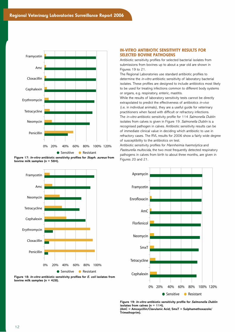
Staphylococcus aureus is well known for being a resilient organism when associated with mastitis – particularly in older cows. Apart from culling, antibiotic treatment during the dry period is considered to be the best treatment option. In-vitro antibiotic sensitivity profiles for Staph. aureus and E. coli isolates from bovine milk samples are shown in Figure 17 and Figure 18.As expected, many of the Staph. aureus isolates were penicillin resistant (56 per cent). However, the proportion of isolates resistant to most of the other antibiotics on test was low.In contrast, E. coli showed greater resistance. Of the antibiotics on test, only framycetin and amoxycillin/clavulanic acid showed activity against more than three-quarters of the isolates.
0%
10%
20%
30%
40%
50%
60%
Jan May Jun Jul Aug Sep Oct Nov DecFeb AprMar
Figure 16: Staphyloccocus aureus isolation rates by month.
0
100
200
300
400
500
600
Jan May Jun Jul Aug Sep Oct Nov DecFeb AprMar
Figure 14: Milk sample submission by month in 2006.
Figure 15: Mastitis pathogen isolation rates (n = 3,339 milk samples cultured).
10% 20% 30% 40% 50%
Staphyloccusaureus
Escherichia colicoliforms
Streptococcusuberis
Streptococcusagalactiae
Other Staphyloccusspecies
Other Streptococcusspecies
Bacilluscereus
Corynebacteriumbovis
Arcanobacteriumpyogenes
Yeast species
Others
No dominantgrowth
Streptococcusdysgalactiae
0%
boVIne maStItIS PathogenS
12
Regional Veterinary Laboratories Surveillance Report 2006
In-VItRo antIbIotIc SenSItIVIty ReSuLtS foR SeLected boVIne PathogenSAntibiotic sensitivity profiles for selected bacterial isolates from submissions from bovines up to about a year old are shown in Figures 19 to 21.The Regional Laboratories use standard antibiotic profiles to determine the in-vitro antibiotic sensitivity of laboratory bacterial isolates. These profiles are designed to include antibiotics most likely to be used for treating infections common to different body systems or organs, e.g. respiratory, enteric, mastitis.While the results of laboratory sensitivity tests cannot be directly extrapolated to predict the effectiveness of antibiotics in-vivo (i.e. in individual animals), they are a useful guide for veterinary practitioners when faced with difficult or refractory infections.The in-vitro antibiotic sensitivity profile for 114 Salmonella Dublin isolates from calves is given in Figure 19. Salmonella Dublin is a recognised pathogen in calves. Antibiotic sensitivity results can be of immediate clinical value in deciding which antibiotic to use in refractory cases. The RVL results for 2006 show a fairly wide degree of susceptibility to the antibiotics on test.Antibiotic sensitivity profiles for Mannheimia haemolytica and Pasteurella multocida, the two most frequently detected respiratory pathogens in calves from birth to about three months, are given in Figures 20 and 21.
20% 40% 60% 80% 100% 120%
Apramycin
Framycetin
Enrofloxacin
Florfenicol
Neomycin
SmxT
Tetracycline
Cephalexin
ResistantSensitive
AmC
0%
Figure 19: In-vitro antibiotic sensitivity profile for Salmonella Dublin isolates from calves (n = 114). (AmC = Amoxycillin/Clavulanic Acid; SmxT = Sulphamethoxazole/Trimethoprim).
20% 40% 60% 80% 100%
ResistantSensitive
0%
Framycetin
Amc
Neomycin
Erythromycin
Tetracycline
Cloxacillin
Penicillin
Cephalexin
Figure 18: In-vitro antibiotic sensitivity profiles for E. coli isolates from bovine milk samples (n = 428).
20% 40% 60% 80% 100% 120%
Framycetin
Amc
Cloxacillin
Erythromycin
Tetracycline
Neomycin
Penicillin
ResistantSensitive
Cephalexin
0%
Figure 17: In-vitro antibiotic sensitivity profiles for Staph. aureus from bovine milk samples (n = 584).

1�
Regional Veterinary Laboratories Surveillance Report 2006
20% 40% 60% 80% 100%
ResistantSensitive
0%
Florfenicol
Enrofloxacin
Tetracycline
Cephalexin
SmxT
Penicillin
Streptomycin
AmC
Figure 21: Antibiotic sensitivity profiles for Pasteurella multocida isolates from bovines up to one year of age (n = 62).
20% 40% 60% 80% 100%
ResistantSensitive
0%
Florfenicol
AmC
Enrofloxacin
Cephalexin
Penicllin
Tetracyline
Streptomycin
SmxT
Figure 20: In-vitro antibiotic sensitivity profiles for Mannheimia haemolytica isolates from bovines up to one year (n = 71). (AmC = Amoxycillin/Clavulanic Acid; SmxT = Sulphamethoxazole/Trimethoprim).
Although not shown in the graph – as they were only added to test profiles at the end of the year – Mannheimia haemolytica isolates (N = 7) were found to be sensitive to the antibiotics Tulathromycin, Tilmicosin, Marbofloxacin and Ceftiofur. Only one of ten Pasteurella multocida isolates showed resistance to each of the antibiotics Tulathromycin and Tilmicosin – and none of the ten was resistant to Marbofloxacin and Ceftiofur.
During 2006, 1,477 tissue samples from 808 wild birds submitted
to the RVLs were tested by real-time PCR for avian influenza (Figure
25). These mainly comprised wild bird deaths reported by members
of the public to the Department of Agriculture Avian Influenza
Hotline phone number - with the carcases being submitted to the
Regional Veterinary Laboratories by the District Veterinary Offices.
Necropsy examination was carried out to identify lesions suggestive
of avian influenza infection. Appropriate tissues samples were
collected for real time PCR assay for avian influenza virus by Virology
Division of CVRL at Backweston.
All samples examined were negative for highly pathogenic avian
influenza virus H5N1. While three specimens were positive on
screening for the matrix gene of avian influenza – one from a mute
swan (Cygnus olor) in Donegal, one from a common shelduck
(Tadorna tadorna) in Cork, and one from a common guillemot (Uria
aalge) in Kerry – the only isolate was a non-pathogenic subtype
(H11) from the shelduck in Cork.
SuRVeILLance of WILd bIRdS foR aVIan InfLuenZa, 2006
Figure 25: Map dot distribution illustration of number of wild bird carcases per county submitted to the RVLs in 2006 for examination for avian influenza. Dots are on a per-county basis and do not represent geographical locations within counties.
14
Regional Veterinary Laboratories Surveillance Report 2006
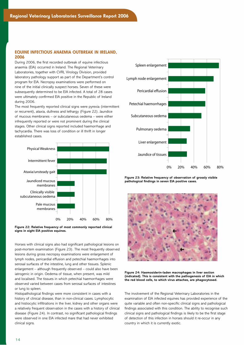
equIne InfectIouS anaemIa outbReak In IReLand, 2006During 2006, the first recorded outbreak of equine infectious anaemia (EIA) occurred in Ireland. The Regional Veterinary Laboratories, together with CVRL Virology Division, provided laboratory pathology support as part of the Department’s control program for EIA. Necropsy examinations were performed on nine of the initial clinically suspect horses. Seven of these were subsequently determined to be EIA infected. A total of 28 cases were ultimately confirmed EIA positive in the Republic of Ireland during 2006.The most frequently reported clinical signs were pyrexia (intermittent or recurrent), ataxia, dullness and lethargy (Figure 22). Jaundice of mucous membranes – or subcutaneous oedema – were either infrequently reported or were not prominent during the clinical stages. Other clinical signs reported included haemorrhage and tachycardia. There was loss of condition or ill thrift in longer established cases.
Horses with clinical signs also had significant pathological lesions on post-mortem examination (Figure 2�). The most frequently observed lesions during gross necropsy examinations were enlargement of lymph nodes, pericardial effusion and petechial haemorrhages into serosal surfaces of the intestine, lung and other tissues. Splenic enlargement – although frequently observed – could also have been iatrogenic in origin. Oedema of tissue, when present, was mild and localised. The tissues in which petechial haemorrhages were observed varied between cases from serosal surfaces of intestines or lung to spleen.Histopathological findings were more consistent in cases with a history of clinical disease, than in non-clinical cases. Lymphocytic and histiocytic infiltrations in the liver, kidney and other organs were a relatively frequent observation in the cases with a history of clinical disease (Figure 24). In contrast, no significant pathological findings were observed in one EIA infected mare that had never exhibited clinical signs.
The involvement of the Regional Veterinary Laboratories in the examination of EIA infected equines has provided experience of the quite variable and often non-specific clinical signs and pathological findings associated with this condition. The ability to recognise such clinical signs and pathological findings is likely to be the first stage of detection of this infection in horses should it re-occur in any country in which it is currently exotic.
0% 20% 40% 60% 80%
Spleen enlargement
Lymph node enlargement
Pericardial effusion
Liver enlargement
Jaundice of tissues
Subcutaneous oedema
Pulmonary oedema
Petechial haemorrhages
Figure 23: Relative frequency of observation of grossly visible pathological findings in seven EIA positive cases.
Figure 24: Haemosiderin-laden macrophages in liver section (indicated). This is consistent with the pathogenesis of EIA in which the red blood cells, to which virus attaches, are phagocytosed.
0% 20% 40% 60% 80%
Physical Weakness
Intermittent fever
Ataxia/unsteady gait
Clinically visible subcutaneous oedema
Pale mucousmembranes
Jaundiced mucousmembranes
Figure 22: Relative frequency of most commonly reported clinical signs in eight EIA positive equines.

Name Grade Address Phone Fax* Email
Athlone RVL
Fagan, John SRO Coosan, Athlone, Co. Westmeath 09064 75514 09064 75215 [email protected]
Murray, Gerard RO Coosan, Athlone, Co. Westmeath 09064 75514 09064 75215 [email protected]
O’Donovan, Jim RO Coosan, Athlone, Co. Westmeath 09064 75514 09064 75215 [email protected]
Cork RVL
Power, Eugene SRO Model Farm Road, Bishopstown, Cork 4 021 454�9�1 021 454615� [email protected]
Gomez, Parada M. RO Model Farm Road, Bishopstown, Cork 4 021 454�9�1 021 454615� [email protected]
Sanchez, Cosme RO Model Farm Road, Bishopstown, Cork 4 021 454�9�1 021 454615� [email protected]
Kilkenny RVL
Moriarty, John SRO Leggatsrath, Hebron Road, Kilkenny 056 77 21688 056 77
64741
Jahns, Hanne RO Leggatsrath, Hebron Road, Kilkenny 056 77 21688 056 77
64741
Toolan, Donal RO Leggatsrath, Hebron Road, Kilkenny 056 77 21688 056 77
64741
Limerick RVL
Johnson, Alan RO Knockalisheen, Limerick 061 452911 061 451849 [email protected]
Kelly, Dave RO Knockalisheen, Limerick 061 452911 061 451849 [email protected]
Sligo RVL
Casey, Micheal SRO Fawcett’s Bridge, Doonally, Co. Sligo 071 9142191 071 9145900 [email protected]
O’Muireagain, Colm RO Fawcett’s Bridge, Doonally, Co. Sligo 071 9142191 071 9145900 [email protected]
Barrett, Damien RO Fawcett’s Bridge, Doonally, Co. Sligo 071 9142191 071 9145900 [email protected]
Dublin RVL
Byrne, William SRO Backweston Laboratory Complex,
Youngs Cross, Celbridge, Co. Kildare
01 6157115/61572�5 01 6157199 [email protected]
Brady, Colm RO Backweston Laboratory Complex,
Youngs Cross, Celbridge, Co. Kildare
01 6157115/61572�8 01 6157199 [email protected]
Sharpe, Ann RO Backweston Laboratory Complex,
Youngs Cross, Celbridge, Co. Kildare
01 6157115/6157220 01 6157199 [email protected]
*All faxes should be marked ‘for the attention of...’ and the name of the intended recipient.
Pro
duce
d by
IFP M
edia
w
ww.if
pmed
ia.c
om
RegIonaL VeteRInaRy LaboRatoRy contact detaILS
CVRL BACkwESToN MAiN oFFiCE PhoNE: 01 6157106 FAx: 01 6157199

16
Regional Veterinary Laboratories Surveillance Report 2006