Regional variations of urokinase-type plasminogen activator in human colorectal cancer: A...
7
Int. J Cancer: 60,308-314 (1995) Publication of the International Unlon Against Cancer Publication de I'Union Internationale Contre le Cancer ici 1995 Wilcy-Liss, Inc. REGIONAL VARIATIONS OF UROKINASE-TYPE PLASMINOGEN ACTIVATOR IN HUMAN COLORECTAL CANCER: A QUANTITATIVE STUDY BY IMAGE ANALYSIS Kevin TAN',De5mond G. POWE, Trevor GRAY. David R. TURNER and Robert E. HEWITT Department of Histopathology, Uniwrsi& of Nottingham Medical School, Queen's Medical Centre, Nottingham NG7 2UH, UK. Qualitative histological studies in the distribution of urokinase- type plasminogen activator (uPA) in human colorectal carcino- mas have been well documented. However, to our knowledge the histological distribution of this enzyme has not been quanti- fied in any tumour. For the present image analysis study, uPA was demonstrated in sections of human colorectal cancer using immunoperoxidasetechnique. A total of 9 colorectal carcinoma cases were used, in which 132 regions were analysed. Within each region, staining intensity measurements were made at evenly spaced intervals. Samples of normal mucosa from 6 cases were also studied. Enzyme levels were assessed with staining intensity measurements. For each section, a negative control section was included, in which the primary antibody was omitted. Staining for uPA was quantified for each region in the test section, and the measurement for the correspondingregion of the negative control was then subtracted. The enzyme uPA was localised more at the tumour edge than in the tumour centre or distant host tissue. These differences were highly significant (p < 0.OOOI). There was also a highly significant difference in staining intensity when tumour regions adjacent to pushing edge were compared with those adjacent to infiltrating edge (p < 0.000 I). Infiltratingtumours showed stronger stain- ing for uPA than tumours with pushing edges. Since invasive activity is thought to be maximal at the edge of the tumour. localisation of uPA at this site is consistent with the role of this enzyme in the process of tumour invasion. c 1995 Wilry-Liss. Inc. Urokinasc-type plasminogen activator (uPA) is involved in various physiological and pathological processes, including angiogenesis, salivary morphogenesis, trophoblastic invasion and tumour invasion. Thc enzyme promotes the conversion of plasminogen to the active serine protease plasmin. Plasmin can also activate procollagenases. As a result, uPA activity can lead to the breakdown of a wide range of extracellular matrix proteins, including laminin, fibronectin and collagens. The enzyme is regulated by (i) pro-uPA activation; (ii) plasmino- gen activator inhibitors PAL1 and PAI-2; (iii) a cell surface receptor specific for uPA, which binds both pro-uPA and active uPA; and (iv) altered expression of the uPA gene (Alexander and Werb, 1991; Dan0 et a/., 1985). Numerous studies suggest that uPA plays an important role in tumour invasion (Ossowski, 1992; Baker etal., 1990; Reich et a/., 1988), and many observational studies have shown in- creased uPA expression in cancer. Raised levels of the enzyme have been shown in cultured colorectal tumour cell lines (Boyd et al., 1988; Ossowski, 1992) and in extracts from human colon carcinoma tissue (Sier et al., 1991b; Sim et al., 1988). Immuno- histochemical studies have produced differing results regard- ing the tumoural distribution of uPA. Some have demon- strated antigen localisation in the neoplastic cell component of colorectal carcinomas (Sier et al., 1991a; Kohga et al., 1985, 1989), while others report staining localisation to the connec- tive tissue stroma (Grondahl-Hansen eta/., 1991; Koretz ef a!., 1993). In addition, while some studies report an increase in staining for urokinase at the invasive edge of colorectal carcinomas (Kohga et al., 1985; Buo et al., 1993), others do not report any difference between tumour centre and periphery (Sieretal., 19916; Kohgaetal., 1989). In the present study, image analysis techniques have been used to provide an objective assessment of uPA distribution in colorectal carcinomas, as demonstrated by immunoperoxidase staining. Since uPA is thought to play a role in tumour invasion, the highest levels of the enzyme would be expected at sites of maximal invasive activity. Levels of uPA would there- fore be expected to be higher at the invasive edge, where there is abundant morphological evidence of invasive activity, than in the tumour centre, where such evidence is lacking (Gabbert, 1985). Higher levels of the enzyme might also be expected in areas where the invading edge has an infiltrating rather than a pushing character, since the infiltrating pattern has been associated with a more aggressive tumour phenotype (Jass et al., 1986). Evidence from the present study supports both of these hypotheses. MATERIAL AND METHODS Antibodies The following antibodies were used at the given dilutions: clone 6 mouse anti-human uPA monoclonal antibody (MAb) (gift from J. Grondahl-Hansen) diluted 1:lOOO (0.2 pgiml) (Grondahl-Hansen et al., 1991); rabbit anti-human colla- gen-IV anti-serum (Euro-Path, Cornwall, UK) diluted 1:250; biotinylated rabbit anti-mouse antibody (Dako, High Wy- combe. UK) diluted 1500; horseradish peroxidase-conjugated swine anti-rabbit antibody (Dako) diluted 1:50; mouse IgG,, kappa (MOPC 31C), myeloma antibody (Sigma, Poole, UK) diluted 1:25,000 (0.2 pgiml). Tissue samples Samples of human normal mucosa and carcinomas were obtained from bowel specimens received by the Department of Histopathology, Queen's Medical Centre, Nottingham, UK. A total of 9 colorectal carcinoma cases were obtained. Adjacent normal mucosae from 6 of these cases were also taken. Within 1 hr of surgical excision, fresh tissue samples were quenched in isopentane pre-cooled in liquid nitrogen and stored at - 70°C until required. Cryostat sections, 6 km in thickness, were fixed in absolute acetone for 20 min at 4°C then air-dried for 15 min at room temperature (RT). Histological diagnoses were: Dukes' stage A (n = 1); Dukes' stage B (n = 5); Dukes' stage C (n = 3). All cases were moderately differentiated adenocarci- nomas except for one Dukes' stage B case, which was focally poorly differentiated. Inimunocytochernistry A 3-step immunoperoxidase technique using the avidin- biotin-peroxidase complex (ABC) (Dako) (Hsu et al., 1981) was employed for uPA detection. Incubation with the primary antibody was carried out overnight at 4°C. Second and third step incubations were carried out for 30 min at RT. Anti-serum was diluted using 1:20 non-immune swine serum: Tris-buffered saline (TBS)/O.l% Triton X100, pH 7.6. Detection was achieved using a DAB chromogen, which was subsequently 'To whom correspondence and reprint requests should be sent. c/o Dr. R.E. Hewitt, Department of Histopathology, Queen's Medical Centre, Nottingham NG7 ?UH, UK. Received: June 2, 1994 and in revised form September 20, 1994
Transcript of Regional variations of urokinase-type plasminogen activator in human colorectal cancer: A...

Int. J Cancer: 60,308-314 (1995) Publication of the International Unlon Against Cancer Publication de I'Union Internationale Contre le Cancer
i c i 1995 Wilcy-Liss, Inc.
REGIONAL VARIATIONS OF UROKINASE-TYPE PLASMINOGEN ACTIVATOR IN HUMAN COLORECTAL CANCER: A QUANTITATIVE STUDY BY IMAGE ANALYSIS Kevin TAN', De5mond G. POWE, Trevor GRAY. David R. TURNER and Robert E. HEWITT Department of Histopathology, Uniwrsi& of Nottingham Medical School, Queen's Medical Centre, Nottingham NG7 2UH, UK.
Qualitative histological studies in the distribution of urokinase- type plasminogen activator (uPA) in human colorectal carcino- mas have been well documented. However, to our knowledge the histological distribution of this enzyme has not been quanti- fied in any tumour. For the present image analysis study, uPA was demonstrated in sections of human colorectal cancer using immunoperoxidase technique. A total of 9 colorectal carcinoma cases were used, in which 132 regions were analysed. Within each region, staining intensity measurements were made at evenly spaced intervals. Samples of normal mucosa from 6 cases were also studied. Enzyme levels were assessed with staining intensity measurements. For each section, a negative control section was included, in which the primary antibody was omitted. Staining for uPA was quantified for each region in the test section, and the measurement for the corresponding region of the negative control was then subtracted. The enzyme uPA was localised more at the tumour edge than in the tumour centre or distant host tissue. These differences were highly significant (p < 0.OOOI). There was also a highly significant difference in staining intensity when tumour regions adjacent to pushing edge were compared with those adjacent to infiltrating edge (p < 0.000 I). Infiltrating tumours showed stronger stain- ing for uPA than tumours with pushing edges. Since invasive activity is thought to be maximal at the edge of the tumour. localisation of uPA at this site is consistent with the role of this enzyme in the process of tumour invasion. c 1995 Wilry-Liss. Inc.
Urokinasc-type plasminogen activator (uPA) is involved in various physiological and pathological processes, including angiogenesis, salivary morphogenesis, trophoblastic invasion and tumour invasion. Thc enzyme promotes the conversion of plasminogen to the active serine protease plasmin. Plasmin can also activate procollagenases. As a result, uPA activity can lead to the breakdown of a wide range of extracellular matrix proteins, including laminin, fibronectin and collagens. The enzyme is regulated by ( i ) pro-uPA activation; (ii) plasmino- gen activator inhibitors PAL1 and PAI-2; (iii) a cell surface receptor specific for uPA, which binds both pro-uPA and active uPA; and (iv) altered expression of the uPA gene (Alexander and Werb, 1991; Dan0 et a/., 1985).
Numerous studies suggest that uPA plays an important role in tumour invasion (Ossowski, 1992; Baker etal., 1990; Reich et a/., 1988), and many observational studies have shown in- creased uPA expression in cancer. Raised levels of the enzyme have been shown in cultured colorectal tumour cell lines (Boyd et al., 1988; Ossowski, 1992) and in extracts from human colon carcinoma tissue (Sier et al., 1991b; Sim et al., 1988). Immuno- histochemical studies have produced differing results regard- ing the tumoural distribution of uPA. Some have demon- strated antigen localisation in the neoplastic cell component of colorectal carcinomas (Sier et al., 1991a; Kohga et al., 1985, 1989), while others report staining localisation to the connec- tive tissue stroma (Grondahl-Hansen eta/., 1991; Koretz ef a!., 1993). In addition, while some studies report an increase in staining for urokinase at the invasive edge of colorectal carcinomas (Kohga et al., 1985; Buo et al., 1993), others do not report any difference between tumour centre and periphery (Sieretal., 19916; Kohgaetal., 1989).
In the present study, image analysis techniques have been used to provide an objective assessment of uPA distribution in
colorectal carcinomas, as demonstrated by immunoperoxidase staining. Since uPA is thought to play a role in tumour invasion, the highest levels of the enzyme would be expected at sites of maximal invasive activity. Levels of uPA would there- fore be expected to be higher at the invasive edge, where there is abundant morphological evidence of invasive activity, than in the tumour centre, where such evidence is lacking (Gabbert, 1985). Higher levels of the enzyme might also be expected in areas where the invading edge has an infiltrating rather than a pushing character, since the infiltrating pattern has been associated with a more aggressive tumour phenotype (Jass et al., 1986). Evidence from the present study supports both of these hypotheses.
MATERIAL AND METHODS Antibodies
The following antibodies were used at the given dilutions: clone 6 mouse anti-human uPA monoclonal antibody (MAb) (gift from J. Grondahl-Hansen) diluted 1:lOOO (0.2 pgiml) (Grondahl-Hansen et al., 1991); rabbit anti-human colla- gen-IV anti-serum (Euro-Path, Cornwall, UK) diluted 1:250; biotinylated rabbit anti-mouse antibody (Dako, High Wy- combe. UK) diluted 1500; horseradish peroxidase-conjugated swine anti-rabbit antibody (Dako) diluted 1:50; mouse IgG,, kappa (MOPC 31C), myeloma antibody (Sigma, Poole, UK) diluted 1:25,000 (0.2 pgiml).
Tissue samples Samples of human normal mucosa and carcinomas were
obtained from bowel specimens received by the Department of Histopathology, Queen's Medical Centre, Nottingham, UK. A total of 9 colorectal carcinoma cases were obtained. Adjacent normal mucosae from 6 of these cases were also taken. Within 1 hr of surgical excision, fresh tissue samples were quenched in isopentane pre-cooled in liquid nitrogen and stored at - 70°C until required. Cryostat sections, 6 km in thickness, were fixed in absolute acetone for 20 min at 4°C then air-dried for 15 min at room temperature (RT). Histological diagnoses were: Dukes' stage A (n = 1); Dukes' stage B (n = 5); Dukes' stage C (n = 3). All cases were moderately differentiated adenocarci- nomas except for one Dukes' stage B case, which was focally poorly differentiated.
Inimunocytochernistry A 3-step immunoperoxidase technique using the avidin-
biotin-peroxidase complex (ABC) (Dako) (Hsu et al., 1981) was employed for uPA detection. Incubation with the primary antibody was carried out overnight at 4°C. Second and third step incubations were carried out for 30 min at RT. Anti-serum was diluted using 1:20 non-immune swine serum: Tris-buffered saline (TBS)/O.l% Triton X100, p H 7.6. Detection was achieved using a DAB chromogen, which was subsequently
'To whom correspondence and reprint requests should be sent. c / o Dr. R.E. Hewitt, Department of Histopathology, Queen's Medical Centre, Nottingham NG7 ?UH, UK.
Received: June 2, 1994 and in revised form September 20, 1994
UROKINASE QUANTITATION LN TUMOURS 309
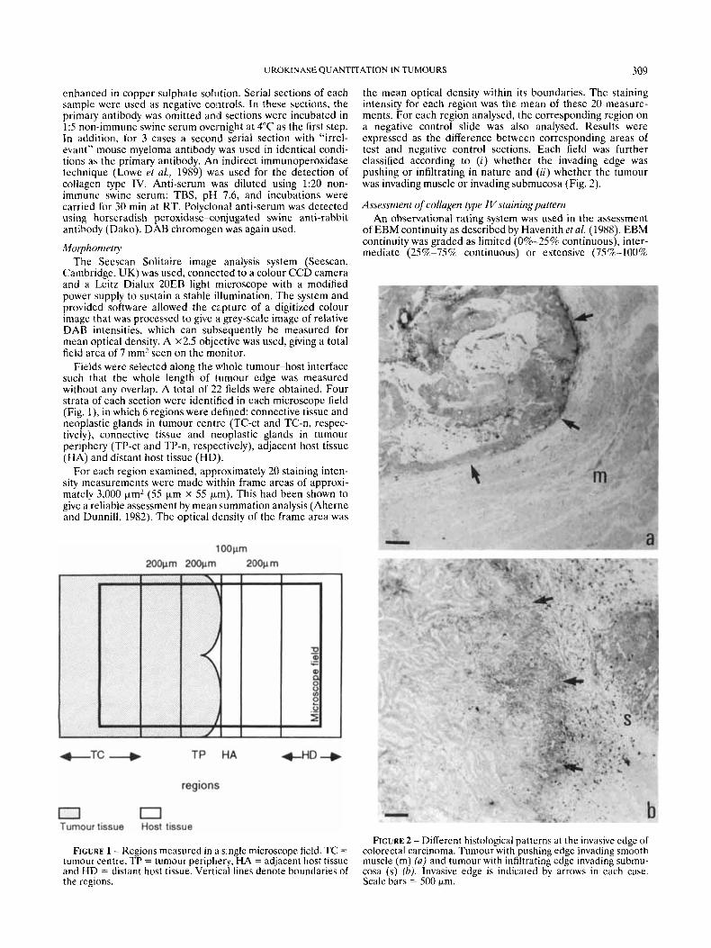
enhanced in copper sulphate solution. Serial sections of each sample were used as negative controls. In these sections, the primary antibody was omitted and sections were incubated in 1 5 non-immune swine serum overnight at 4°C as the first step. In addition, for 3 cases a second serial section with “irrel- evant” mouse myeloma antibody was used in identical condi- tions as the primary antibody. An indirect immunoperoxidase technique (Lowe et al., 1989) wa.s used for the detection of collagen type IV. Anti-serum was diluted using 1:20 non- immune swine serum: TBS, pH 7.6, and incubations were carried for 30 min at RT. Polyclonal anti-serum was detected using horseradish peroxidase-conjugated swine anti-rabbit antibody (Dako). DAB chromogen was again used.
Motphometv The Secscan Solitaire image analysis system (Seescan,
Cambridgc, UK) was used, connected to a colour CCD camera and a Leitz Dialux 20EB light microscope with a modified power supply to sustain a stable illumination. The system and provided software allowed the calpture of a digitized colour image that was processed to give a grey-scale image of relative DAB intensities, which can subsequently be measured for mean optical density. A ~ 2 . 5 objective was used, giving a total field area of 7 mm2 seen on the monitor.
Fields were selected along the whole tumour-host interface such that the whole length of tumour cdge was measured without any overlap. A total of 22 fields were obtained. Four strata of each section were identified in each microscope field (Fig. l), in which 6 regions were defined: connective tissue and neoplastic glands in tumour centrl- (TC-ct and TC-n, respec- tively), connective tissue and neoplastic glands in tumour periphery (TP-ct and TP-n, respectively), adjacent host tissue (HA) and distant host tissue (HD)
For each region examined, approximately 20 staining inten- sity measurements were made within frame areas of approxi- mately 3,000 Fm2 (55 Fm x 55 p i ) . This had been shown to give a reliable assessment by mean summation analysis (Aherne and Dunnill, 1982). The optical density of the frame area was
the mean optical density within its boundaries. The staining intensity for each region was the mean of these 20 measure- ments. For each region analysed, the corresponding region on a negative control slide was also analysed. Results were expressed as the difference between corresponding areas of test and negative control sections. Each field was further classified according to (i) whether the invading edge was pushing or infiltrating in nature and (ii) whether the tumour was invading muscle or invading submucosa (Fig. 2).
Assessment of collagen type Nstainingpattem An observational rating system was used in the assessment
of EBM continuity as described by Havenith et al. (1988). EBM continuity was graded as limited (0%-25% continuous), inter- mediate (2.5%-75% continuous) or extensivc (7S%-100%
FIGURE 2 - Different histological patterns a t the invasive edge of colorectal carcinoma. Tumour with pushing edge invading smooth muscle (m) ( a ) and tumour with infiltrating edge invading submu- cosa (s) (b). lnvasive edge is indicated by arrows in each case. Scale bars = 500 km.
FIGURE 1 - Regions measured in a single microscope field. TC = tumour centre, TP = turnour periphery, HA = adjacent host tissue and HD = distant host tissue. Vertical lines denote boundaries of the regions.

310 TAN E T A L
continuous). Fields corresponding to those observed in the quantitation of uPA were used in this evaluation. Slides stained for uPA were also examined by light microscopy and the staining pattern described.
Statistical analysis Comparisons of staining intensities between regions, types
of invading edge and types of tissue invaded were analysed statistically using analysis of variance (ANOVA) and the paired t-test. ANOVA was also used to assess the relationship between uPA distribution and EBM continuity. Calculations were performed using SPSS statistical software.
RESULTS Qualitative assessment of uPA staining
On simple observation, uPA staining appeared to be stron- ger in regions HA and T P than in regions H D and TC. Intense staining was generally seen along the whole of the tumour’s invading edge. Muscularis propria and muscularis mucosa generally showed stronger staining than submucosa. On higher magnification, most staining was detected in the spindle- shaped (fibroblast-like) cells in region TP. Staining was de- tected in most of the endothelial cells within tumour tissue but only in some of the endothelial cells in host tissue. There was diffuse staining of neoplastic cells and in the interstitial spaces at the invasive edge (Fig. 3). There was minimal peroxidase staining in all 6 cases of adjacent normal colonic tissue in the corresponding regions and cell types.
Similarly, minimal staking was observed in the slides where the uPA antibody was omitted and replaced by “irrelevant” mouse myeloma antibody. The non-specific staining patterns for both slides were identical in each of the 3 cases studied (Fig. 3), providing further evidence for the specificity of the uPA staining reaction.
Quantitative assessment of uPA staining by image analysis In general the highest measured values (i.e., most intense
staining) were found in the connective tissues at the tumour- host interface (regions TP-ct and HA). The least values were in the connective tissues away from the tumour edge (regions TC-ct and HD) and the neoplastic cells in tumour centre (Fig. 40). It can be seen from Figure 4b that the non-specific staining varies considerably in the different regions. A higher back- ground was obtained for neoplastic cells than over connective tissues. Measurement of uPA-specific staining intensities would be very unreliable if this was not taken into account.
The significance of differences between uPA staining inten- sities in different tumour regions are shown by the p values given in Table I. There are statistically significant differences in the distribution of uPA among all the regions ( F = 14.1, p < 0.0001). Regions with the strongest uPA staining were TP-ct, H A and TC-ct, in descending order. Region H A had stronger uPA staining than region H D ( T = 4.3, p < 0.0001). Region TP showed more uPA staining than region TC. A comparison between staining intensities of the connective tissues in these 2 regions (TP-ct with TC-ct) gave a statistically significant difference ( T = 3 . 5 , ~ < 0.001). Within the tumour, connective tissues tended to have more intense staining than neoplastic cells, such that a significant difference was seen between TP-ct and TP-n ( p < 0.05).
When regions adjacent to pushing and infiltrating invading edges were considered separately, the same staining patterns were observed for each group (Fig. 5) . Regions adjacent to infiltrating edges generally showed stronger staining than regions adjacent to pushing edges, and a highly significant difference was seen between them (F = 1 5 . 2 , ~ < 0.0001). The 2 regions that showed the greatest difference in staining intensity between pushing and infiltrating edge were H A and TP-ct. More obvious increases in uPA staining intensity at the
FIGURE 3 - Distribution of uPA staining in colorectal carci- noma. The most intense staining was ,in the connective tissue at the turnour edge (arrows, a). Negative controls showed an absence of staining (b). “Irrelevant” mouse myeloma antibody staining was identical to negative control staining (c). Scale bars = 100 Fm.
invasive edge where submucosa rather than muscle was being invaded were observed (Fig. 6).
Comparison of uPA staining intensity with EBM continuity EBM was demonstrated by immunostaining for type IV
collagen and assessed by an observational rating system (Table 11). Many more EBM breaks were observed in region T P than in region TC. Most glands in region TP had limited or intermediate EBM continuity, especially those at the tumour- host interface. Glands in region TC generally showed continu- ous EBM. Thinning of EBM was seen in a few T C regions, but

UROKINASE QUANTITATION IN TUMOURS 31 1
0 20 K-1 L n=22 I
0.3
0.2
0.1
0.0
T C - C t T P - c t HA HD . T C - n T P - n
ragion
T T I I
W test W negative control
T T
D
T C - c t T P - c t HA HD . T C - n T P - n
region
FIGURE 4 - Graphs showing regional variations in uPA staining intensity for all cases. (a) Morphometric values for staining intensity when negative controls were taken into consideration. (b) Morphometric values of staining, intensity for test and negative controls. Error bars show standard deviation.
seldom did complete breaks occur. Examination of the nega- tive controls confirmed the specificity of the collagen type IV anti-serum.
EBM continuity in regions TC and TP was compared collectively with uPA staining intensity in the corresponding connective tissue regions (TC-ct and HA, respectively) (Fig. 7 ) . No evidence of a significant correlation was seen.
DISCL SSION Regional variations in uPA staining intensity
Results of our image analysis showed a highly significant increase in uPA staining intensity at the invasive edge of colorectal carcinoma. Many studies have indicated that uPA plays an important role in tumour invasion. The distribution of uPA demonstrated in the present study is therefore consistent
TABLE I - p VALUES OF PLANNED COMPARISONS OF uPA STAINING INTENSITY BETWEEN DIFFERENT REGIONS
Repion TC-ct TP-ct HA HD TC-n TP-n
TC-ct (0.078) TP-ct (0.135) 0.001 HA(O.109) nls nls
TC-n (0.041) n/s * * nls TP-n(0.058) nls * * nls nls
Mean values of staining intensity in parentheses. *Pairs of regions significantly different at the 0.05 level. nls = not signifi- cant, TC = tumour centre, TP = tumour periphery, HA = adjacent host tissue, HD = distant host tissue, ct = connective tissue, n = neoplastic glands.
HD (0.038) nls 0.0001
1 H pushing edge (n=8 or more) T infiltrating edge ( k l l or more) T
0.10
0.00 T C - c t T P - c t HA HD . T C - n T P - n
region
FIGURE 5 - Graph comparing uPA staining intensity in regions adjacent to pushing edge with staining in regions adjacent to infiltrating edge. Negative control values were taken into consider- ation. Error bars show standard deviation.
with the hypothesis that in colorectal carcinoma invasive activity is most marked in the invasive edge (Gabbert, 1985; Hewitt et al., 1993). Previous qualitative studies have given conflicting reports about comparative levels of uPA staining in central and peripheral regions of colorectal carcinomas. Inves- tigators using polyclonal anti-uPA did not find any difference in staining levels between tumour centre and periphery (Ko- hga et al., 1989). Others using anti-uPA MAbs (as used in the present study) have shown increased staining for the enzyme at the invasive edge (Buo et al., 1993). Discrepancies between these qualitative studies may be due to differences in the specificity of antibodies used, to sampling error or perhaps to subjective factors. Using image analysis to provide a more objective and quantitative assessment of staining intensities, we show a highly significant increase in uPA staining in connective tissue at the invasive edge of colorectal carcinoma ( p < 0.001). This is consistent with the hypothesis that uroki- nase plays a role in invasive activity and that this activity is most marked at the invasive edge of colorectal carcinomas.
Rectal carcinoma tumours with predominantly infiltrating edges are thought to have a more aggressive phenotype and are associated with a worse prognosis than tumours with predomi- nantly pushing edges (Jass et al., 1986). We find that in areas where the invasive edge is infiltrating in character, the connec- tive tissues in and around the tumour show significantly

312 TAN ET.4L
0 3
0 2
0 1
muscle invasion (n=13 or more)
subrnucosa invasion (n=6 or more)
0 0 T C - c t T P - c t HA HD . T C - n T P - n
region
FIGURE 6 - Graph comparing uPA staining intensity in areas where tumour is invading muscle and where tumour invades submucosal connective tissue. Negative control values were taken into consideration. Error bars show standard deviation.
TABLE I1 - RESULTS OF EBM CONTINUITY ASSESSMENT
Tumoui Field Tumour centre
1 1
4 4 5 5 5 5 5
1 2 1 1
3 1 2 1 2 3 4 5 1 2 1 2 1 2 3 1 2
7
+++ +++ ++ +++ ++ ++ +++
+++ +++ ++ ++
+++ +++ ++ +++ +++ ++ ++ ++ +++
+++
-
Turnour periphery
++ ++ ++ + + + ++ +++ + + + +
++ ++ + ++ ++ + ++ ++ ++ ++
+: 0%-25c/o continuous or limited, + +: 25%-75% continuous or intermediate, + + +: 75%-100% continuous or extensive, -: no measurement taken.
stronger staining for uPA than those in arcas where the invasive edge is pushing in charactcr ( p < 0.0001). It is possible that high lcvels of uPA expression lead to the development of an infiltrating rather than pushing edge, and this suggests the possibility that assays for uPA level or activity might assist the prognostic evaluation of patients with colorec- tal cancer.
When the typc of tissue invaded was taken into account, higher expression of uPA was seen at the edge of the tumour-invading submucosa (regions HA and TP-ct). This finding may be due to the differences between cell types present in submucosa and muscularis propria of the bowel wall. Induction of uPA may occur more readily in fibroblasts of
m c C .- - m c al
4
3 n.
0.3
0.2
0.1
0.0
I +
9
+
t + i 0 + * *
+ I I I
0 1 2 3 4
EBM continuity
FIGURE 7 - Scattergraph comparing uPA staining intensity with EBM continuity. Each point represents values for corresponding areas on serial sections stained for uPA and type IV collagen. EBM continuity scale: 1 = 0%-25% continuity or limited, 2 = 25%-75% continuity or intermediate and 3 = 75%-100% continu- ity or extensive.
the submucosa than in smooth muscle cells of the muscularis propria. This result suggests that the level of expression of uPA depends on the type of host cells with which the tumour cells interact. This supports the findings of previous studies that point to the possible importance of local tissue environmental factors in influencing a tumour’s invasive activity (Hewitt et nl., 1994).
Like Grondahl-Hansen et al. (1991), we found that fibroblast in the tumour stroma stained intensely for uPA. However, unlike this group, we also detected staining in neoplastic cells at the edge of the tumour, as well as a general increase in staining all along the invading edge. This difference might be due to our use of a more sensitive immunostaining technique (Hsu et al., 1981). Staining of the neoplastic cells at the invasive edge might be due to production of the enzyme by these cells or to the presence of high levels of uPA receptors on these cells leading to increased binding of uPA to the cell surface. Consistent with the latter explanation. Pyke et al. (1991) have demonstrated by in situ hybridization that colorectal cancer cells at the tumour-stromal interface of invasive foci show increased expression of uPA receptor mRNA.
Consistent with the observed endothelial cell staining, sev- eral studies have documented increased levels of enzyme expression in endothelial cells. Certain eytokines such as tumour necrosis factor (TNF) (Niedbala and Stein-Picarella, 1992), interleukin-I (IL-I) (Niedbala and Stein-Picarella, 1991), interleukin-4 (IL-4) (Wojta et al., 1993), lipopolysaccha- ride (LPS) (Niedbala and Stein-Picarella, 1991) and lympho- toxin (Van Hinsbergh et al., 1990) promote the expression, synthesis and secretion of uPA in endothelial cells. The cnzyme uPA may be involved in angiogenesis, where it is needed for extracellular matrix remodelling as it sprouts through tissues. Also, the enzyme may have becn taken up from the environment, which would be abundant in uPA that had been produced by other cells. Several studies have documented the identification of uPA receptors on endothelial

UROKINASE QUANTITATION IN TUMOURS 313
cells in cell cultures (Hajjar and IHamel, 1990; Langer et a/., 1993).
Comparison of UPA distribution with EBM continuity Thc relationship between enzyme concentrations of uPA
and EBM continuity was studied to test the hypothesis that EBM breaks would be associated with increased levels of uPA since it is known that basement membrane degradation in- volves the plasminogen activator system (Dan0 et aL, 1985). When tumours were considered on a case by case basis and the tumour centre was compared with the tumour periphery, we made two observations: first, that uPA staining was generally more intense at the tumour periphery, and second, that the EBM was generally lcss continuous at the tumour periphery (Fig. 8). These two observations suggested the possibility of an inverse relationship between uPA level and EBM continuity. However, we found no evidence of such a relationship when EBM scores and uPA staining measurements were compared col lcct ive I y .
It is possible that a relationship exists between uPA levels and EBM continuity but that thi9 relationship is obscured by section to section variation in staining efficiency. It is also possible that a relationship exists< between the levels of active enzyme and EBM continuity but that this relationship is obscured by staining for the latent form of the enzyme, which is also recognized by the antibody used here (Grondahl-Hansen et al., 1991).
There are natural inhibitors of uPA and plasmin. They include plasminogen activator inhibitor (PA1)-1, PAI-2, prote- ase nexin 1, a? -anti-plasmin arid aprotinin (Alexander and Wcrb, 1991). PAI-1 and PAI-2 are known to be increased in human colorectal carcinoma (Sier et al., 19916). As the balance between plasminogen activators and their inhibitors is believed to be important in determining the invasiveness and metastatic potential of tumour cells, it may be valuable to evaluate the relative levels of these different c'omponents in future quantita- tive immunohistochemical studies.
Alternatively, it may be that E,BM deficiency at the invasive edge is not primarily due to increased degradation by enzymes such as uPA. Rather, the failure to synthesise basement membrane components (Gabbert, 1985; Warburton et aL, 1986) or the failure to assemble them (Daneker et al., 1987) may be more important. Gabberlr (1985) suggested that the loss of EBM could be the result of the inability of poorly differenti- ated or dedifferentiated tumour cells to synthesise the macro- molecules necessary to assemble a regular and continuous basement membrane. This explamtion does not contradict the idea of uPA having a role in tumour invasion because in- creased levels of uPA at the invading edge of a tumour may still be involved in the degradation (of extracellular matrix compo- nents other than EBM.
Conclusion Results of the present image analysis study provide objective
evidence that stromal uPA levels are higher at the invasive edge of colorectal carcinomas than in the tumour centre. In addition. the demonstration of stronger stromal uPA staining adjacent to those invasive edges with an infiltrating character represents a novel finding. These results are consistent with the model that uPA expression is associated with the invasive activity of colorectal cancer arid that this invasive activity is most marked at the invasive edge, particularly where there is an infiltrating pattern of invasion.
ACKNOWLEDGEMENTS
We are grateful to Dr. I. Leach for histological grading of the tumours and to Dr. G. Kuk for statistical advice. We also
FIGURE 8 -Comparison of uPA staining pattern (u) and colla- gen type IV (EBM) staining pattern (b) in serial sections. uPA staining is most intense in connective tissues at the invading edge, which shows an infiltrating character. In the section stained for collagen type IV there is a lack of EBM staining around tumour glands at the invasive edge. Scale bars = 200 p,m.
thank Mr. W. Brackenbury and Mr. D. Hughes for their skilled photographic assistance. This work was supported by depart- mental funds.
314 TAN ETAL.
REFERENCES
AHERNE, W.A. and DUNNILL, M.S., Mophometry. E. Arnold, London (1982). &EX4NDER, C.M. and WERB, Z., Extracellular matrix degradation. In: E.D. Hay (ed.), Cell biology of extracellular matrix, Plenum, New York (1991). BAKER, M.S., BLEAKLEY, P., WOODROW, G.C. and DOE, W.F.. Inhibi- tion of cancer cell urokinase plasminogen activator by its specific inhibitor PAL2 and subsequent effects on extracellular matrix degrada- tion. Cancer Res., 50,4676-4684 (1990). BOYD, D., FLORENT, G., KIM, P. and BRATTAIN, M., Determination of the levels of urokinase and its receptor in human colon carcinoma cell lines. CancerRes., 48,3112-3116 (1988). Buo, L., LYBERG, T., JORGENSEN, L., JOHANSEN, H.T. and AASEN, A.O., Location of plasminogen activator (PA) and PA inhibitor in human colorectal adenocarcinomas. Actu pathol. microbiol. immunol. Scand., 101,235-241 (1993). DANEKER, G.W., MERCURIO, A.M., GUERRA, L., WOLF, B., SALEM, R.R., BAGLI, D.J. and STEELE, G.D., Laminin expression in colorectal carcinomas varying in degree of differentiation. Arch. Surg., 122, 1470-1474 (1987). DAN0, K., ANDREASEN, P.A., GRONDAHL-HANSEN, J., KRISTENSEN, P., NIELSEN. D.S. and SKRIVER, L., Plasminogen activators, tissue degrada- tion and cancer. Adv. Cancer Res., 44,139-266 (1985). GABBERT, H., Mechanisms of tumour invasion-evidence from in vivo observations. Cuncer Metastasis Rev., 4,293-309 (1985). GRONDAHL-HANSEN, J., RALFKJAER, E., KIRKEBY, L.T., KRISTENSEN, P., LUND, L.R. and DAN^, K., Localisation of urokinase-type plasmino- gen activator in stromal cells in adenocarcinomas of the colon in humans. Amer. J. Pathol., 138,111-1 17 (1991). HAJJAR. K.A. and HAMEL, N.M., Identification and characterisation of human endothelial cell membrane binding site for tissue plasminogen activator and urokinase. J. biol. Chem., 265,2908-2916 (1990). HAVENITH. M.G.. ARENDS. J.W.. SIMON. R.. Vo~ovrcs. A.. WIGGERS. T. and BOSMAN, F.T., Type IV collagen immunoreactivity in colorectal cancer Cancer, 62,2207-2211 (1988) HEWITT, R.E., KEEBLE, W.. POWE, D.G., WILLIAMSON, R.J. and TURNER, D.R., The influence of local tissue environment on epithelial basement membrane continuity in colorectal carcinomas. Znt. J. Can- cer, 56,675-680 (1994). HEWITT, R.E., POWE, D.G., CARTER, I. TURNER, D.R., Desmopla- sia and its relevance to colorectal invasion. Ztit. J. Cancer, 53, 62-69 (1993). Hsu, S.M., RAINE, L. and FANGER. H., Use of avidin-biotin- peroxidase complex (ABC) in immunoperoxidase techniques-a com- parison between ABC and unlabelled antibody (PAP) procedures. J. Histochem. Cytockem., 24,577-580 (1981). JASS, J.R., ATKIN, W.S., CUZICK, J., BUSSEY, H.J.R., MORSON, B.C., NORTHOVER, J.M.A. and TODD, I.P., The grading of rectal cancer: historical perspectives and a multivariate analysis of 447 cases. Histopatho OD, 10,437-459 (1986). KOHGA, S., HARVEY, S.R., SUZUMIYA, J., SUMIYOSHI, A. and MARKUS, G., Comparison of the immunohistochemical localisation of urokinase in normal and cancerous human colon tissue. Fihrinolysis, 3, 17-22 (1 989). KOHGA, S., HARVEY, S.R., WEAVER, R.M. and MARKUS, G., Localisa- tion of plasminogen activators in human colon cancer by immunoper- oxidase staining. Cancer Res., 45,1787-1796 (1985).
KORETZ, K., MOLLER. P. and SCHWARTZ-ALBIEZ, R., Plasminogen activators and plasminogen activator inhibitors in human colorectal carcinoma tissues are not expressed by the tumour cells. Europ. J. Cancer, 29A, 1184-1189 (1993). LANCER, D.J., Kuo, A,, KARIKO, K., AHUJA, M., KLUGHERZ, B.D., IVANICS. K.M., HOXIE, J.A., WILLIAMS, W.V., LIANG, B.T. and CINES, D.B., Regulation of the endothelial cell urokinase-type plasminogen activator receptor. Evidence for cyclic AMP-dependent and protein kinase C-dependent pathways. Circul. Res., 72,33&340 (1993). LOWE, J., MACLENNAN, K.A., POWE, D.G. and PALMER, J.B., Micro- glial cells in human brain have phenotypic characteristics related to possible function as dendritic antigen presenting cells. J. Pathol., 159,
NIEDBALA, M.J. and STEIN-PICARELLA, M., Tumour necrosis factor induction of urokinase-type plasminogen activator in human endothe- lial cells. Biomed. Biochim. Actu, 50,427436 (1991). NIEDBALA, M.J. and STEIN-PICARELLA, M., Tumour necrosis factor regulation of endothelial cell extracellular proteolysis: the role of urokinase plasminogen activator. Biol. Chem. Hoppe-Sqler, 373, 555- 566 (1992). OSSOWSKI, L., Invasion of connective tissue by human carcinoma cell lines-requirement for urokinase, urokinase receptor and interstitial collagenase. Cancer Res., 52,6754-6760 (1992). PYKE, C., KRISTENSEN, P., RALFKIAER, E., GRONDAHL-HANSEN, J., ERIKSEN, J., BLASI, F. and DAN^. K.. Urokinase-type plasminogen activator is expressed in stromal cells and its receptors in cancer cells at invasive foci in human colon adenocarcinomas. Amer. J. Pathol., 138,
REICH, R., THOMPSON, E.W., IWAMOTO, Y., MARTIN, G.R., DEASON, J.R., FULLER, G.C. and MISKIN, R., Effects of inhibitors of plasmino- gen activator, serine proteinases and collagenase IV on the invasion of basement membrane by metastatic cells. Cancer Res., 48, 3307-3312 (1988). SIER, C.F.M., FELLBAUM, C., VERSPAGET, H.W., SCHMITT, M., GRIF- FIOEN, G., GRAEFF, H., HOFLER, H. and LAMERS, C.B.H.W., Immuno- localisation of urokinase-type plasminogen activator in adenomas and carcinomas of the colorectum. Histopathology, 19,231-237 (1991~).
QUAX. P.H.A., DOOIJEWAARD, G., DE BRUIN, P.A.F. and LAMERS. C.B.H.W., Imbalance of plasminogen activators and their inhibitors in human colorectal neoplasia. Gustroenterology, 101, 1522-1528 (1991b). SIM, P.S., STEPHENS, R.W., FAYLE, D.R.H. and DOE, W.F.. Urokinase- type plasminogen activator in colorectal carcinomas and adenomatous polyps-quantitative expression of active and proenzyme. Int. J. Cancer, 42,483488 (1988). VAN HINSBERGH, V.W., VAN DEN BERG, E.A., FIERS, W. and DOOIJEW- AARD, G., Tumour necrosis factor induces the production of urokinase- type plasminogen activator by human endothelial cells. Blood, 75, 1991-1998 (1990). WARBURTON, M.J., FERNS, S.A. and HYNES, N.E., Collagen processing in ras-transfected mouse mammary epithelial cells. Biocbem. biopbys. Res. Comm., 137,161-166 (1986). WOJTA, J., GALLICCHIO, M., ZOELLNER, H.. FILONZI, E.L., HAMILTON, J.A. and MCGRATH, K., Interleukin-4 stimulates expression of uroki- nase-type plasminogen activator in human foreskin microvascular endothelial cells. Blood, 81,3285-3292 (1993).
143-149 (1989).
1059-1067 (1991).
SIER, C.F.M.. VERSPAGET, H.W., GRIFFIOEN, G., VERHEIJEN, J.H.,


![Arecombinantchimeric plasminogenactivatorwithhighaffinity for … › content › pnas › 88 › 22 › 10337.full.pdf · urokinase-type plasminogen activator [scuPA(32kDa)], afi-brin-selective](https://static.fdocuments.in/doc/165x107/5f1cd2e4e4e08d6801761b19/arecombinantchimeric-plasminogenactivatorwithhighaffinity-for-a-content-a-pnas.jpg)