Anesthesia for the emergency patient - American Association of
Regional Anesthesia in the Patient ReceivingAntithrombotic or Thrombolytic Therapy
American Society of Regional Anesthesia and Pain MedicineEvidence-Based Guidelines (Fourth Edition)
Terese T. Horlocker, MD,* Erik Vandermeuelen, MD,† Sandra L. Kopp, MD,* Wiebke Gogarten, MD,‡Lisa R. Leffert, MD,§ and Honorio T. Benzon, MD||
(Reg Anesth Pain Med 2018;43: 263–309)
Evolving standards for the prevention of perioperative venousthromboembolism (VTE) and the introduction of increasingly
potent antithrombotic medications have resulted in concerns re-garding the heightened risk of neuraxial bleeding. Furthermore,societies and organizations seeking to address these concernsthrough guidelines in perioperative management have issuedconflicting recommendations. In response to these patient safetyissues and the need for a more international approach to manage-ment, the American Society of Regional Anesthesia and PainMedicine (ASRA), in conjunction with the European Society ofAnaesthesiology (ESA), convened its Fourth Consensus Confer-ence on Regional Anesthesia and Anticoagulation. Portions ofthe material presented here were published in the 1998, 2003,and 2010 ASRA Consensus Documents as well as the 2010 ESAGuidelines.1–8 The information has been updated to incorporatedata available since the time of its publication.
Based on limited clinical and animal data, these guidelinesdo not define standard of care. They are not intended to replaceclinical judgment as applied to a specific patient scenario. Variancesfrom recommendations contained in this document may be accept-able based on the judgment of the responsible anesthesiologist.Importantly, the authors consistently tended toward conservativerecommendations. The consensus statements are designed to en-courage safe and quality patient care, but cannot guarantee aspecific outcome. As with any clinical guideline recommenda-tion, these are subject to revision as knowledge of specific compli-cations advances.
The first 2 consensus conferences focused on neuraxialblocks and anticoagulants in surgical patients, with limited infor-mation on the management of thromboprophylaxis in the parturi-ent. However, the hypercoagulability associated with pregnancyand the puerperium has resulted in an increased number of par-turients receiving antithrombotic therapy for the treatment andprevention of thromboembolism.9–14 The third consensus confer-ence, based on limited information, recommended that parturientsbe managed similarly to nonpregnant patients undergoing neuraxialblock.7 More recently, the National Partnership for Maternal
Safety (NPMS)13 developed a consensus bundle on VTE, whichwill likely result in more parturients receiving thromboprophylaxis.Integration of these recommendations in a patient population wherethere is a lack of a comparable “alternative” analgesic techniquehas again raised concern regarding the timing of epidural catheterplacement/removal and initiation of postpartum thromboprophylaxisand is addressed in this update. Of note, ASRA collaborated withthe NPMS and Society for Obstetric Anesthesia and Perinatology(SOAP) to develop a unified set of recommendations.15
The 2010 consensus conference also addressed, for the firsttime, the risk of significant bleeding in patients undergoing plexusand peripheral neural blockade. This section also is extensivelyupdated as more information regarding frequency and severityof bleeding complications associated with nonneuraxial techniqueshas become available.
Finally, recent publications of cases of epidural hematomaduring interventional pain procedures in patients receiving anti-platelet agents suggested a need for separate recommendationsfor these patients,16–18 as the current ASRA guidelines for theplacement of epidural and spinal catheters do not recommend ces-sation of these antiplatelet agents for epidural procedures, nor dothe guidelines differentiate between interventional pain proce-dures and perioperative regional anesthesia blocks.7 A summaryof the interventional spine and pain guidelines, highlighting thedifferences, is included. In addition, the guidelines consider themanagement of patients undergoing pain (both neuraxial and pe-ripheral) procedures in combination with antithrombotic therapy.
These recommendations are intended for use by anesthesiol-ogists and other physicians and health care providers performingneuraxial and peripheral regional anesthetic/analgesic blockade.However, these recommendations may also serve as a resourcefor other health care providers involved in the management of pa-tients who have undergone similar procedures (eg, myelography,lumbar puncture).
METHODSA systematic and general review of the relevant literature be-
tween 2010 and 2017 was performed. Published reports onpharmacokinetics and pharmacodynamics of antithrombotic med-ications, series of patient receiving these medications while under-going neuraxial and peripheral blockade, and case reports ofneuraxial and perineural bleeding (both spontaneous and regionalanesthesia associated) were identified. Members of the task forcerepresented the specialties of regional anesthesia/acute pain andinterventional pain. The members represented ASRA, SOAP,and ESA.
Since the last consensus document, many new oral anticoag-ulant medications have been approved by the US Food and DrugAdministration (FDA). Nearly all were released with a black boxwarning regarding the risk of spinal hematoma. In addition, many
From the *Mayo Clinic, Rochester, MN; †Katholieke Universiteit, Leuven,Belgium; ‡Bielefeld Hospital, Bielefeld, Germany; §Massachusetts GeneralHospital, Boston, MA; and ||Northwestern University, Chicago, IL.Accepted for publication January 21, 2018.Address correspondence to: Terese T. Horlocker, MD, Department of
Anesthesiology and Perioperative Medicine, Mayo Clinic, 200 First St. SW,Rochester, MN 55905 (e‐mail: [email protected]).
The authors declare no conflict of interest.Copyright © 2018 by American Society of Regional Anesthesia and Pain
MedicineISSN: 1098-7339DOI: 10.1097/AAP.0000000000000763
REGIONAL ANESTHESIA AND ACUTE PAIN
SPECIAL ARTICLE
Regional Anesthesia and Pain Medicine • Volume 43, Number 3, April 2018 263

have labeling that describes time intervals between discontinua-tion of the anticoagulant and a surgical procedure or neuraxialblock, the timing of epidural catheter after administration of theanticoagulant, and the timing of subsequent dosing followingneuraxial catheter removal. In the absence of larger series and casereports, these recommendations are often pharmacologicallybased. For example, recommended time intervals between discon-tinuation of drug during therapeutic anticoagulation and subse-quent neuraxial block are 5 half-lives (and is dependent on renalfunction). This allows for resolution of 97% of the anticoagulanteffect. With lower levels of anticoagulation associated with pro-phylaxis, only a 2-half-life interval is required. Similarly, a recentFDA Drug Safety Communication recommended a 4-hour timeinterval between catheter removal and subsequent low-molecular-weight heparin (LMWH) administration.19 The new recommen-dation is based on the work of Rosencher et al,20 who proposedsubsequent dosing of antithrombotic therapy based on 8 hours mi-nus the time it takes for the anticoagulant to reach peak effect.Rosencher et al20 cited the work of Bouma and Mosnier,21 whonoted that it takes approximately 8 hours for a platelet plug to be-come stable. For example, the efficacy of thrombolytics therapyfollowing a cerebral embolic clot markedly decreases after 6 to8 hours.22 This implies that anticoagulants will have a hard timelysing a clot after 8 hours. Given the 4-hour time to peak effectwith LMWH, the time to subsequent dosing following cathetermanipulation would be 8 hours minus 4 hours, or 4 hours.23
The American Society of Regional Anesthesia and Pain Medicinehas consistently incorporated FDA-approved labeling into practicerecommendations and as such adopted the changes. This time in-terval is also consistent with the ESA,8 Scandinavian,24 and mostrecent German guidelines (all of which incorporate a pharmaco-logic approach to newer anticoagulantswhere data are sparse).8,23,25
Recommendations are frequently made with a time interval,rather than an exact time (eg, 4–6 hours), representing normal var-iation in pharmacologic activity. In patients without additional co-morbidities that affect metabolism, excretion, and/or distribution,the shorter time may be observed. Significant renal insufficiency(in a renally excreted medication) is not included in this time inter-val and will require longer periods. Also to be considered are apatient history of bruising/bleeding, the presence of multiple med-ications affecting hemostasis, and the proposed regional technique(deep vs superficial, continuous vs single injection).
Strength and Grade of RecommendationsThe recommendations presented are based on a thorough
evaluation of the available information using a grading systembased on level of evidence and strength of recommendation. Thelevel of evidence classification combines an objective description ofthe types of studies/expert consensus supporting the recommen-dation. Unfortunately, with a complication as rare as spinal hema-toma, in randomized clinical trials and meta-analyses, the highest(A) level of evidence is not available. Numerous observationaland epidemiologic series (typically, level of evidence B) have doc-umented the conditions for safe performance of neuraxial anesthesiaand analgesia in the anticoagulated patient. However, high-qualityevidencemay come from large observational/epidemiologic seriesyielding very large risk reduction.26 Hence, depending on the riskreduction, recommendations from these sources may be categorizedas level of evidence A or B. Recommendations derived from casereports or expert opinion merit a C level of evidence. Often, recom-mendations involving the anesthetic management of new antithrom-botic agents, for which data involving safety and/or risk are sparse,are based on the pharmacology of hemostasis-altering drugs, riskof surgical bleeding, and expert opinion (C level of evidence).
The grade of recommendation indicates the strength of theguideline and the degree of consensus agreement. For example,grade 1 represents general agreement in the efficacy, grade 2 notesconflicting evidence or opinion on the usefulness, and grade 3suggests that the procedure may not be useful (and possibly harm-ful). These guidelines do not contain any grade 3 recommendations.The phrase “we recommend” is used for strong recommendations(grades 1A, 1B, and 1C), and “we suggest” for weaker recommen-dations (grades 2A, 2B, and 2C). In cases where the evidence isscant (such as with the new oral anticoagulants), the authors highlyvalued patient safety and proposed conservative (ie, longer) timesfor interruption of therapy prior to neural blockade. These willlikely be revised as additional information regarding blood levelsand anticoagulant effect, as well as the introduction of reversalagents, is presented.
Finally, although there are several new sections representingthe recently introduced antiplatelet and anticoagulant medications,as well as revised recommendations of previously included agents,in some cases the management has remained unchanged. In orderto facilitate review, the status of the current recommendations isnoted in each section.
Current Recommendations for the Prevention andTreatment of Venous Thromboembolism
Venous thromboembolism is an important health care prob-lem and a significant source of morbidity and mortality. Nearlyall hospitalized patients have at least 1 risk factor for thrombo-embolism; approximately 40% have 3 or more risk factors27
(Table 1). Consequently, the majority of hospitalized patients arecandidates for thromboprophylaxis.
The agent, dosing regimen, and duration of thromboprophylaxisare based on identification of risk factors, both individual (eg, age,sex, previous history of thromboembolism) and group-specific(eg, primary reason for hospitalization, surgery, medical illness)27,28
TABLE 1. Risk Factors for Venous Thromboembolism
SurgeryTrauma (major trauma or lower-extremity injury)Immobility, lower-extremity paresisCancer (active or occult)Cancer therapy (hormonal, chemotherapy, angiogenesis inhibitors,radiotherapy)
Venous compression (tumor, hematoma, arterial abnormality)Previous VTEIncreasing agePregnancy and the postpartum periodEstrogen-containing oral contraceptives or hormone replacementtherapy
Selective estrogen receptor modulatorsErythropoiesis-stimulating agentsAcute medical illnessInflammatory bowel diseaseNephrotic syndromeMyeloproliferative disordersParoxysmal nocturnal hemoglobinuriaObesityCentral venous catheterizationInherited or acquired thrombophilia
Adapted from Geerts et al,27 with permission.
Horlocker et al Regional Anesthesia and Pain Medicine • Volume 43, Number 3, April 2018
264 © 2018 American Society of Regional Anesthesia and Pain Medicine

(Tables 1 and 2). Depending on the risks of thromboembolismand bleeding, thromboprophylaxis may be achieved with compres-sion stockings, intermittent compression devices, with medications(chemoprophylaxis), or a combination of both.28,29 Because an in-dividualized approach to thromboprophylaxis is complex, mostrecommendations are group specific, with modifications basedon the presence/absence of additional risk factors. Guidelinesfor antithrombotic therapy including appropriate pharmacologicagent, degree of anticoagulation desired, and duration of therapycontinue to evolve.30,31
A review of the evolution of practice guidelines and thestrength/grade of recommendations noted that (1) the vast major-ity of guidelines are based on lower levels of evidence or expertopinion—level A recommendations (derived from randomizedclinical trials) are rare; and (2) bias may exist due to funding of in-dustry trials (in restricted patient populations), as well as conflictof interest by the guideline-writing groups.32,33 The AmericanCollege of Chest Physicians (ACCP) thoroughly addressed theseissues in their most recent edition of The Antithrombotic Therapyand Prevention of Thrombosis, ninth edition (AT9), in 2012. Thelead investigator, Gordon H. Guyatt,31 noted AT9 of the evidence-based clinical practice guidelines differed substantially from theprior version both in process and in content. The most importantchanges in the content of AT9 resulted directly from a change inthe process of reconciling intellectual and financial conflicts of in-terest. The new process gave primary leadership and responsibilityfor each article in the document not to a thrombosis expert but to amethodologist who, in almost all cases, also is a practicing physi-cian without important conflicts of interest. In addition, there wasamore rigorous reevaluation of the evidence that distinguished be-tween a surrogate outcome (asymptomatic thrombosis detected onimaging) and patient important outcomes (symptomatic thrombo-sis vs bleeding). The more rigorous and critical evaluation ofthe literature resulted in many weak recommendations (2A–C) re-placing the strong recommendations (1A–C) of previous editions.One remarkable result was a major change on the use of aspirinin thromboprophylaxis in orthopedic surgery. The authors of theeighth edition (AT8) had concluded that there was “high-qualityevidence justifying a strong recommendation against aspirin asthe sole agent for thromboprophylaxis in surgical patients.”27,31
After reevaluation of the evidence and inclusion of a very large
trial, the AT9 authors concluded “that the trial provides moderate-quality evidence supporting the use of aspirin in patients under-going major orthopedic procedures, which is now offered as anoption for thromboprophylaxis in patients undergoing major or-thopedic procedures.”31,34 Thus, with AT8, for patients undergoingmajor orthopedic surgery, LMWH, fondaparinux, and vitamin Kagonists were the only recommended agents of thrombo-prophylaxis.27 However, with the AT9 guidelines, clinicians maynow choose from LMWH, fondaparinux, vitamin K agonists,dabigatran, rivaroxaban, apixaban, low-dose unfractionated hepa-rin (UFH), aspirin, or an intermittent pneumatic compressivedevice.29 It is interesting to note that the American Academy ofOrthopaedic Surgeons published guidelines in 2011 for the pre-vention of venous thromboembolic disease for patients undergo-ing elective hip and knee arthroplasty with essentially the sameconclusion (http://www.aaos.org/uploadedFiles/PreProduction/Quality/Guidelines_and_Reviews/VTE_full_guideline_10.31.16.pdf. Accessed March 4, 2017). The American Academy ofOrthopaedic Surgeons evidence-based guidelines recommended“the use of pharmacologic agents and/or mechanical compressivedevices for the prevention of VTE in patients undergoing electivehip or knee arthroplasty and who are not at elevated risk beyondthat of the surgery itself for VTE or bleeding.” However, notingthat “current evidence is unclear about which prophylactic strategy(or strategies) is/are optimal or suboptimal, we were unable to rec-ommend for or against specific prophylactics in these patients.”
In general, AT9 represents a “less aggressive” set of guide-lines with more options for thromboprophylaxis. Selection of an-tithrombotic agents that are efficacious but have less impact onhemostasis (such as aspirin compared with LMWH in orthopedicpatients) will theoretically decrease the severity and frequencyof serious bleeding implications—both surgical and anesthesiarelated—and improve patient safety.
As with previous advisories, ASRA recommendations incor-porate the dosing regimen approved by the FDA, as well as theACCP antithrombotic guidelines.
1.0 Administration of Thromboprophylaxis1.1 For each of the antithrombotic agents, we recommend
that clinicians follow the FDA-approved dosing and ACCPmanagement guidelines (grade 1A).
Remarks: There is no change in this recommendation.
TABLE 2. Recommendations for Thromboprophylaxis in Nonorthopedic Surgical Patients
Risk of Major Bleeding Complications
Risk of Symptomatic VTE Average Risk (∼1%) High Risk (∼2%) or Severe Consequences
Very low (<0.5%) No specific prophylaxisLow (∼1.5%) Mechanical prophylaxis, preferably with IPCModerate (∼3.0%) LDUH, LMWH, or mechanical prophylaxis,
preferably with IPCMechanical prophylaxis, preferably with IPC
High (∼6.0%) LDUH or LMWH plus mechanical prophylaxiswith ES or IPC
Mechanical prophylaxis, preferably with IPC, until riskof bleeding diminishes and pharmacologicprophylaxis can be added
High-risk cancer surgery LDUH or LMWH plus mechanical prophylaxiswith ES or IPC and extended-durationprophylaxis with LMWH postdischarge
Mechanical prophylaxis, preferably with IPC, until riskof bleeding diminishes and pharmacologicprophylaxis can be added
High risk, LDUH, and LMWHcontraindicated or not available
Fondaparinux or low-dose aspirin (160 mg);mechanical prophylaxis, preferably with IPC;or both
Mechanical prophylaxis, preferably with IPC, until riskof bleeding diminishes and pharmacologicprophylaxis can be added
ES indicates elastic stockings; IPC, intermittent pneumatic compression; LDUH, low-dose UFH.Adapted from Gould et al,28 with permission.
Regional Anesthesia and Pain Medicine • Volume 43, Number 3, April 2018 Regional Anesthesia and Anticoagulation
© 2018 American Society of Regional Anesthesia and Pain Medicine 265

PerioperativeManagement of Antithrombotic andAntiplatelet Therapy
Long-term anticoagulation with warfarin is often indicatedfor patients with a history of VTE, mechanical heart valves, andatrial fibrillation. In addition, patients with bare metal or drug-eluting coronary stents require antiplatelet therapy with aspirinand thienopyridine derivatives (eg, clopidogrel) for varying dura-tions. These patients may present for elective or urgent surgicalprocedures. Perioperative management involves balancing therisks of surgical bleeding and thromboembolism. Minor proce-dures may not require interruption of antithrombotic or antiplate-let therapy. However, continuation of these medications in thesetting of a major surgery increases the risk of bleeding. Thus,it is critical to determine whether the planned procedure necessi-tates interruption of antithrombotic/antiplatelet therapy and, ifso, whether the patient will need bridging therapy to minimizethe risk of thromboembolism during the time the antithromboticeffect is subtherapeutic. In many patients, antithrombotic therapymay be safely interrupted until adequate surgical hemostasis isachieved. In other patients, bridging anticoagulation with UFHor LMWH is required until the time of surgery (and reinitiatedin the immediate postoperative period). It may also be necessaryto postpone elective surgeries in patients where a suitable “bridge”has not been identified and antithrombotic therapy is critical; pre-mature discontinuation of dual antiplatelet therapy in patients withcoronary stents has been associated with stent thrombosis, myo-cardial infarction, and death35,36 (Tables 3 and 4).
Evidence-based guidelines for the perioperative managementof antithrombotic therapy have been established by the ACCP.36
In general, in patients at moderate to high risk of thromboembo-lism, bridging therapy is recommended (and the prevention ofthromboembolism is valued over the potential for increased surgicalbleeding). Notably, 3 different bridging regimens are presented;
selection is based on the potential consequences of perioperativebleeding. Conversely, no bridging therapy is recommended forpatients at low risk of thromboembolism. While the recommenda-tions for management are relatively simple, complexity arises inthe determination of who is “high risk.” This evaluation is perhapsbest performed within an integrated multidisciplinary clinic bythrombophilia experts.37 Timing of resumption of antithrombotictherapy postoperatively also is based on the relative risks of bleed-ing and thrombosis. In general, it is recommended that patientsat low risk of bleeding initiate antithrombotic therapy 24 hourspostoperatively, whereas those at high risk of bleeding wait for48 to 72 hours.36,38 These are longer time intervals than were pre-viously recommended.39
Incidence, Risk Factors, and Neurologic Outcomeof Spinal Hematoma
Spinal hematoma, defined as symptomatic bleeding withinthe spinal neuraxis, is a rare and potentially catastrophic complica-tion of spinal or epidural anesthesia. The actual incidence of neu-rologic dysfunction resulting from hemorrhagic complicationsassociated with central neural blockade is unknown. In an exten-sive review of the literature, Tryba40 identified 13 cases of spinalhematoma following 850,000 epidural anesthetics and 7 casesamong 650,000 spinal techniques. Based on these observations,the calculated incidence is approximated to be less than 1 in150,000 epidural and less than 1 in 220,000 spinal anesthetics.41
However, the series involved in these calculations involved pa-tients who were not receiving thromboprophylaxis. Recent caseseries and epidemiologic surveys suggest that the risk has in-creased.2,42,43 In addition, without a mandatory reporting systemand/or centralized registry, it is likely that many spinal hematomasremain unreported, and the frequency is higher than calculated.
Hemorrhage into the spinal canal most commonly occurs inthe epidural space, likely because of the prominent epidural ve-nous plexus, although anesthetic variables, such as needle sizeand catheter placement, may also affect the site of clinically signif-icant bleeding.44,45 In a review of the literature from 1906 through1994, Vandermeulen et al43 reported 61 cases of spinal hematomaassociated with epidural or spinal anesthesia; 60% of cases oc-curred in the last decade of the study period. In 42 (68%) of the61 patients, the spinal hematomas associated with central neural
TABLE 3. Perioperative Management of Patients on Warfarin
Preoperative• Discontinue warfarin at least 5 days before elective procedure• Assess INR 1–2 d prior to surgery, if >1.5, consider 1–2 mg oralvitamin K
• Reversal for urgent surgery/procedure, consider 2.5–5 mg oralor IV vitamin K; for immediate reversal, consider PCCs,fresh frozen plasma
• Patients at high risk of thromboembolism○ Bridge with therapeutic SC LMWH (preferred) or IV UFH○ Last dose of preoperative LMWH administered 24 h beforesurgery, administer half of the daily dose
○ Intravenous heparin discontinued 4–6 h before surgery• No bridging necessary for patients at low risk of thromboembolism
Postoperative• Patients at low risk of thromboembolism
○ Resume warfarin on POD• Patients at high risk of thromboembolism (who receivedpreoperative bridging therapy)○ Minor surgical procedure—resume therapeutic LMWH 24 hpostoperatively
○ Major surgical procedure—resume therapeutic LMWH 48–72 hpostoperatively or administer low-dose LMWH
• Assess bleeding risk and adequacy of hemostasis whenconsidering timing of the resumption of LMWH or UFH therapy
Recommendations from Douketis et al.36
TABLE 4. Perioperative Management of Patients on AntiplateletTherapy
Patients with coronary stents• Elective surgery postponed for the following durations if aspirinand thienopyridine (eg, clopidogrel or prasugrel) therapy mustbe discontinued○ Bare metal stents: 6 wk○ Drug-eluting stents: 6 mo
• If surgery cannot be postponed, continue dual antiplatelet therapythroughout perioperative period
Patients at high risk of cardiac events (exclusive of coronary stents)• Continue aspirin throughout the perioperative period• Discontinue clopidogrel/prasugrel 5 d prior to surgery• Resume thienopyridine 24 h postoperatively
Patients at low risk of cardiac events• Discontinue dual antiplatelet therapy 7–10 d prior to surgery• Resume antiplatelet therapy 24 h postoperatively
Recommendations from Douketis et al.36
Horlocker et al Regional Anesthesia and Pain Medicine • Volume 43, Number 3, April 2018
266 © 2018 American Society of Regional Anesthesia and Pain Medicine

blockade occurred in patients with evidence of hemostatic abnor-mality. Twenty-five of the patients had received intravenous (IV)or subcutaneous (SC) (unfractionated or low molecular weight)heparin, whereas an additional 5 patients were presumably admin-istered heparin as theywere undergoing avascular surgical procedure.In addition, 12 patients had evidence of coagulopathy or thrombo-cytopenia or were treated with antiplatelet medications (aspirin,indomethacin, ticlopidine), oral anticoagulants (phenprocoumon),thrombolytics (urokinase), or dextran 70 immediately before orafter the spinal or epidural anesthetic. Needle and catheter place-ment was reported to be difficult in 15 (25%) or bloody in 15(25%) patients. Overall, in 53 (87%) of the 61 cases, either aclotting abnormality or needle placement difficulty was present.A spinal anesthetic was performed in 15 patients. The remaining46 patients received an epidural anesthetic, including 32 patientswith an indwelling catheter. In 15 of these 32 patients, the spinalhematoma occurred immediately after the removal of the epiduralcatheter. Nine of these catheters were removed during therapeuticlevels of heparinization. Neurologic compromise presented asprogression of sensory or motor block (68% of patients) or bowel/bladder dysfunction (8% of patients), not severe radicular backpain. Importantly, although only 38% of patients had partial orgood neurologic recovery, spinal cord ischemia tended to be re-versible in patients who underwent laminectomy within 8 hoursof onset of neurologic dysfunction43(Table 5).
The need for prompt diagnosis and intervention in the eventof a spinal hematoma was also demonstrated in 2 reviews of theAmerican Society of Anesthesiologists (ASA) Closed Claims da-tabase involving claims related to nerve injury.46,47 Cheney et al46
examined the claims of nerve injury associated with general or re-gional block between 1990 and 1999 and noted that spinal cord in-juries were the leading cause of claims in the 1990s. Furthermore,spinal hematomas accounted for nearly half of the spinal cordinjuries. Patient care was rarely judged to have met standards be-cause of delay in the diagnosis and resultant poor outcome. Con-sequently, the median payment was very high.46 An in-depthanalysis of the claims related to nerve injury following regionalanesthesia between 1980 and 1999 reported 36 spinal hematomas,associated mainly with vascular or orthopedic surgical proce-dures. Three-fourths of patients had evidence of a preexisting oriatrogenic coagulation abnormality.47 More than half of the pa-tients received IV heparin during a vascular surgical or diagnosticprocedure, often in combination with other medications that im-pair coagulation. Consistent with Vandermeulen et al,43 the pre-senting symptom was increased motor block (83% of cases),rather than back pain (25% of cases). Importantly, the presenceof postoperative numbness or weakness was typically attributed
to local anesthetic effect rather than spinal cord ischemia, whichdelayed the diagnosis. Although the symptoms were noted typi-cally on postoperative day 1 (POD1), often 24 hours or moreelapsed prior to diagnosis.47 There were permanent deficits in90% of patients.
It is impossible to conclusively determine the risk factors forthe development of spinal hematoma in patients undergoingneuraxial blockade solely through review of the case series, whichrepresent only patients with the complication and do not definethose who underwent uneventful neuraxial analgesia. However,large inclusive surveys that evaluate the frequencies of complica-tions (including spinal hematoma), as well as identify subgroupsof patients with higher or lower risk, enhance risk stratifica-tion. Moen et al42 investigated serious neurologic complicationsamong 1,260,000 spinal and 450,000 epidural blocks performedin Sweden over a 10-year period. Twenty-four of the 33 spinal he-matomas occurred in the last 5 years of the decade surveyed.Among the 33 spinal hematomas, 24 occurred in females; 25 wereassociated with an epidural technique. A coagulopathy (existingor acquired) was present in 11 patients; 2 of these patients wereparturients with HELLP syndrome (hemolysis, elevated liver en-zymes, and low platelets). Pathology of the spine was present in6 patients. The presenting complaint was typically lower-extremityweakness. Only 5 of 33 patients recovered neurologically (becauseof delay in the diagnosis/intervention). These demographics, riskfactors, and outcomes confirm those of previous series. However,the methodology allowed for calculation of frequency of spinalhematoma among patient populations. For example, the risk asso-ciated with epidural analgesia in women undergoing childbirthwas significantly less (1 in 200,000) than that in elderly womenundergoing knee arthroplasty (1 in 3600, P < 0.0001). Likewise,women undergoing hip fracture surgery under spinal anesthesiahad an increased risk of spinal hematoma (1 in 22,000) comparedwith all patients undergoing spinal anesthesia (1 in 480,000). Amore recent review by Ehrenfeld et al48involving 43,000 ortho-pedic patients undergoing epidural blockade reported a similarfrequency, 1.38/10,000 (95% confidence interval, 0–0.002).
Overall, these series suggest that the risk of clinically signif-icant bleeding varies with age, associated abnormalities of the spi-nal cord or vertebral column, the presence of an underlyingcoagulopathy, difficulty during needle placement, and an indwell-ing neuraxial catheter during sustained anticoagulation (particu-larly with standard heparin or LMWH), perhaps in an additiveversus synergistic multifactorial manner. They also consistentlydemonstrate the need for prompt diagnosis and intervention.
Fibrinolytic and Thrombolytic TherapyPharmacology of Fibrinolytics/Thrombolytics
The fibrinolytic system dissolves intravascular clots as a re-sult of the action of plasmin. Plasmin is produced by the cleavageof a single peptide bond of the inactive precursor, plasminogen.The resulting compound is a nonspecific protease capable of dis-solving fibrin clots and other plasma proteins, including severalcoagulation factors. Exogenous plasminogen activators such asstreptokinase and urokinase not only dissolve thrombus but alsoaffect circulating plasminogen. Endogenous t-PA formulations(alteplase, tenecteplase) are more fibrin-selective and have lesseffect on circulating plasminogen. Clot lysis leads to elevation offibrin degradation products, which themselves have an anticoagu-lant effect by inhibiting platelet aggregation. Patients who receivefibrinolytic therapy frequently receive IV heparin to maintain anactivated partial thromboplastin time (aPTT) of 1.5 to 2 times nor-mal and often an antiplatelet agent such as aspirin or clopidogrel.While the plasma half-life of thrombolytic drugs is only hours, it
TABLE 5. NeurologicOutcome inPatientsWith SpinalHematomaFollowing Neuraxial Blockade*
Interval Between Onsetof Paraplegia and Surgery
Good(n = 15)
Partial(n = 11)
Poor(n = 29)
<8 h (n = 13) 6 4 3Between 8 and 24 h (n = 7) 1 2 4>24 h (n = 12) 2 0 10No surgical intervention(n = 13)
4 1 8
Unknown (n = 10) 2 4 4
*Neurologic outcome was reported for 55 of 61 cases of spinal hema-toma following neuraxial blockade.
Adapted from Vandermeulen et al,43 with permission.
Regional Anesthesia and Pain Medicine • Volume 43, Number 3, April 2018 Regional Anesthesia and Anticoagulation
© 2018 American Society of Regional Anesthesia and Pain Medicine 267

may take days for the thrombolytic effect to resolve; fibrinogenand plasminogen are maximally depressed at 5 hours after throm-bolytic therapy and remain significantly depressed at 27 hours(Fig. 1). The decrease in coagulation factor levels is greater withstreptokinase compared with t-PA therapy. However, the fre-quency of hemorrhagic events is similar.50 Importantly, originalcontraindications to thrombolytic therapy included surgery orpuncture of noncompressible vessels within 10 days.51
Case Reports of Spontaneous and RegionalAnesthesia–Related Spinal Hematomas Related toThrombolytic Therapy
There are no large series addressing regional anesthesia inthe patient receiving fibrinolytic/thrombolytic therapy. The major-ity of published reports involve spontaneous spinal or epiduralhematomas after thrombolytic therapy.52–68 Recent cases involvethrombolysis for myocardial infarction. Bleeding has been re-ported at all spinal levels—cervical, thoracic, and lumbar.
To date, there are 6 cases of spinal hematoma involving theconcomitant use of neuraxial anesthesia and fibrinolytic/thrombolytictherapy. Five cases appeared in the literature69–73; 1 additional casewas reported through the MedWatch system. (The MedWatch pro-gram was initiated in 1993. Reporting of serious adverse events byhealth care professionals and hospitals is voluntary. Confidentialityis maintained. However, manufacturers and distributors of FDA-approved pharmaceuticals have mandatory reporting requirements.The FDA estimates that less than 1% of serious adverse drug reac-tions are reported.) Two of the spinal hematomas (including theMedWatch case) occurred in patients who underwent a neuraxialtechnique (epidural anesthesia for lithotripsy, epidural steroid injec-tion [ESI]) and subsequently complained of myocardial ischemiaandwere treatedwith a thrombolytic.7,69 The potential for significantspinal bleeding was not appreciated by the interventional cardiolo-gists, despite recent neuraxial needle placement in these 2 patients.
2.0 Anesthetic Management of the Patient ReceivingThrombolytic Therapy
Patients receiving fibrinolytic/thrombolytic medicationsare at risk of serious hemorrhagic events, particularly thosewho have undergone an invasive procedure. Recommenda-tions are based on the profound effect on hemostasis, the useof concomitant heparin and/or antiplatelet agents (which fur-ther increase the risk of bleeding), and the potential for spon-taneous neuraxial bleeding with these medications.
2.1 In patients scheduled to receive thrombolytic therapy,we recommend that the patient be queried andmedical recordreviewed for a recent history of lumbar puncture, spinal orepidural anesthesia, or ESI to allow appropriate monitoring.Guidelines detailing original contraindications to thrombo-lytic drugs suggest avoidance of these drugs for 10 days follow-ing puncture of noncompressible vessels (grade 1A).
Remarks: There is no change in this recommendation.2.2 In patients who have received fibrinolytic and throm-
bolytic drugs, we recommend against performance of spinalor epidural anesthetics except in highly unusual circum-stances (grade 1A).
Remarks: There is no change in this recommendation.2.3 Data are not available to clearly outline the length of
time neuraxial puncture should be avoided after discontinua-tion of these drugs. However, a 48-hour time interval and doc-umentation of normalization of clotting studies (includingfibrinogen) are suggested (grade 2C).
Remarks: There is no change in this recommendation.2.4 In those patients who have received neuraxial blocks
at or near the time of fibrinolytic and thrombolytic therapy,we recommend that neurological monitoring should be con-tinued for an appropriate interval. It may be that the intervalof monitoring should not be more than 2 hours between neu-rologic checks. If neuraxial blocks have been combined withfibrinolytic and thrombolytic therapy and ongoing epiduralcatheter infusion, we recommend the infusion should be lim-ited to drugs minimizing sensory and motor block to facilitateassessment of neurologic function (grade 1C).
Remarks: There is no change in this recommendation.
FIGURE 1. Activated partial thromboplastin time before treatment, 1 to 6 hours after SC injection of 5000U of heparin, and after prophylacticheparin treatment of surgical patients. The horizontal bars show themean values. The aPTTwas significantly prolonged for 1 to 5 hours afterinjection of heparin. Adapted from Gallus et al,49 with permission.
Horlocker et al Regional Anesthesia and Pain Medicine • Volume 43, Number 3, April 2018
268 © 2018 American Society of Regional Anesthesia and Pain Medicine

2.5 There is no definitive recommendation for removal ofneuraxial catheters in patients who unexpectedly receive fibri-nolytic and thrombolytic therapy during a neuraxial catheterinfusion. We suggest the measurement of fibrinogen level (oneof the last clotting factors to recover) to evaluate the presenceof residual thrombolytic effect and appropriate timing of cath-eter removal (grade 2C).
Remarks: There is no change in this recommendation.
Intravenous and SubcutaneousUnfractionated HeparinPharmacology of Unfractionated Heparin
The major anticoagulant effect of heparin is due to a uniquepentasaccharide that binds to antithrombin with high affinity andis present in approximately one-third of heparin molecules. Bind-ing of this heparin pentasaccharide to antithrombin accelerates itsability to inactivate thrombin (factor IIa), factor Xa, and factorIXa. Anticoagulant activities of UFH depend on both the num-ber of heparin molecules with the pentasaccharide chain and thesize of the molecules containing the pentasaccharide sequence.Larger-molecular-weight heparins will catalyze inhibition of bothfactors IIa and Xa. Smaller-molecular-weight heparins will cata-lyze inhibition of only factor Xa.74,75 Intravenous injection resultsin immediate anticoagulant activity, whereas SC injection resultsin a 1- to 2-hour delay. The anticoagulant effect of heparin isboth dose and molecular size dependent and is not linear, but in-creases disproportionately with increasing doses. For example,the biologic half-life of heparin increases from 30 minutes after25 U/kg IV, to 60 minutes with 100 U/kg IV, and to 150 minuteswith a bolus of 400 U/kg IV.75,76 When the SC route is selectedfor delivery of therapeutic anticoagulation, the dose of heparin ishigher than the IV route to compensate for the reduced bioavail-ability associated with SC administration.
When given in therapeutic doses, the anticoagulant effect ofheparin is typically monitored with the aPTT. However, this testdoes not directly measure heparin and is affected by physiologicand analytic variables. Anti–factor Xa testing offers improvementsover aPTT testing for accurate measurement of heparin levels.Clinical data from the last 10 to 20 years suggest that anti–factorXa monitoring may offer a smoother dose-response curve and re-quire fewer blood samples and dosage adjustments.77 The acti-vated clotting time (ACT) is typically used to monitor higherdoses given during cardiopulmonary bypass. Adequate therapeu-tic effect (in patients with VTE or unstable angina) is achieved
with a prolongation of the aPTT to between 1.5 and 2.5 timesthe baseline value,74 heparin level of 0.2 to 0.4 U/mL, oranti–factor Xa level of 0.3 to 0.7 U/mL.78 Administration oflow-dose (5000 U) SC heparin for prophylaxis of deep venousthrombosis does not significantly prolong the aPTT in the ma-jority of patients and is typically not monitored. However, itcan result in unpredictable (10-fold variability) and therapeuticblood concentrations of heparin in some patients within 2 hoursafter administration.49
One of the advantages of heparin anticoagulation is that its ef-fect may be rapidly reversed with protamine. Each milligram ofprotamine can neutralize 100U of heparin. Neutralization of subcu-taneously administered heparinmay require a prolonged infusion ofprotamine because of the continued absorption.74 Finally, followingadministration of heparin for more than 5 days, there is a subset ofpatients who will develop a decrease in the platelet count.76
Risk Factors for Spinal Hematoma in the HeparinizedPatient Undergoing Neuraxial Blockade
The combination of spinal or epidural needle insertion in thepresence of sustained therapeutic anticoagulation with heparin isassociated with increased risk. Much of our information about thisassociation comes from a report of 342 patients who deliberatelyreceived systemic therapeutic heparin after lumbar puncture.79
Three factors associated with increased risk were identified: lessthan 60-minute time interval between the administration of hepa-rin and lumbar puncture, traumatic needle placement, and con-comitant use of other anticoagulants (aspirin). These risk factorshave been verified in subsequent large reviews of case reports ofhematomas associated with neuraxial procedures in the presenceof UFH45,80,81(Table 6).
Intravenous Unfractionated HeparinIntraoperative heparinization typically involves injection of
5000 to 10,000 U of heparin intravenously during the operativeperiod, particularly in the setting of vascular surgery to prevent co-agulation during cross-clamping of arterial vessels.75 Neuraxialanesthetic techniques are often attractive for these patients, butmay be associated with an increased risk of epidural hematoma,as demonstrated by case series, epidemiologic surveys, and theASA Closed Claims database.42,43,82,83 Maintaining a 1-hour in-terval between needle placement and heparinization, as well asavoidance of other hemostasis-altering medications, decreasesthe risk of significant bleeding.
TABLE 6. Risk Factors and Estimated Incidence for Spinal Hematoma and Neuraxial Anesthesia
Relative Risk ofSpinal Hematoma
Estimated Incidencefor Epidural Anesthesia
Estimated Incidencefor Spinal Anesthesia
No heparinAtraumatic 1.00 1:220,000 1:320,000Traumatic 11.2 1:20,000 1:29,000With aspirin 2.54 1:150,000 1:220,000
Heparin anticoagulation following neuraxial procedureAtraumatic 3.16 1:70,000 1:100,000Traumatic 112 1:2000 1:2900Heparin >1 h after puncture 2.18 1:100,000 1:150,000Heparin <1 h after puncture 25.2 1:8700 1:13,000With aspirin 26 1:8500 1:12,000
Data from Stafford-Smith,45 with permission.
Regional Anesthesia and Pain Medicine • Volume 43, Number 3, April 2018 Regional Anesthesia and Anticoagulation
© 2018 American Society of Regional Anesthesia and Pain Medicine 269

Management of a traumatic neuraxial procedure must alsobe considered. Previous case reports suggest that presence of abloody tap or a traumatic regional block is an associated factorin approximately 50% of spinal hematomas.43 Although someinvestigators have recommended cancellation of the surgical pro-cedures should these events occur,80 there are no clinical datato support this recommendation.67,84 Direct communication withthe surgeon and a specific risk-benefit decision about proceedingin each case are warranted.
Heparinization may be continued into or initiated in the post-operative period. However, the removal of a neuraxial catheter inthe presence of heparin therapy increases the risk of hematomaformation. In the series by Vandermeulen et al,43 half of the spinalhematomas associated with systemic heparinization were detectedat the time of catheter removal. The risk of hematoma resultingfrom catheter removal has led to the recommendation that, inpatients who have undergone systemic heparinization, heparinshould be discontinued for 2 to 4 hours, and the coagulation statusassessed prior to neuraxial catheter manipulation or removal.
Heparinization During Cardiopulmonary BypassSince the publication of the initial ASRA guidelines in 1998,4
there have been continued discussions regarding the relative risk(and benefit) of neuraxial anesthesia and analgesia in the patientundergoing heparinization for cardiopulmonary bypass.85–89 Un-fortunately, although there is improved analgesia, pulmonaryfunction, and cardiac arrhythmia, there is no reduction in hospitalstay, myocardial infarction, or mortality. To date, there is a singlecase of spinal hematoma following the full heparinization associ-ated with cardiopulmonary bypass.90 However, these series in-volve small numbers of patients. Using a mathematical analysisof the probability of predicting a rare event (based on the totalsof 4583 epidural and 10,840 spinal anesthetics reported withoutcomplications), Ho et al91 estimated the risk of hematoma to beapproximately 1:1528 for epidural and 1:3610 for spinal technique.Thus, this analgesic technique remains controversial in that the riskappears too great for the perceived benefits. A review has recom-mended certain precautions to be taken to minimize the risk82:
(1) Neuraxial blocks should be avoided in a patient with knowncoagulopathy from any cause.
(2) Surgery should be delayed 24 hours in the event of atraumatic tap.
(3) Time from instrumentation to systemic heparinization shouldexceed 60 minutes.
(4) Heparin effect and reversal should be tightly controlled(smallest amount of heparin for the shortest duration com-patible with therapeutic objectives).
(5) Epidural catheters should be removed when normal coagu-lation is restored, and patients should be closely monitoredpostoperatively for signs and symptoms of hematomaformation.
Subcutaneous Unfractionated HeparinLow-dose SC UFH is commonly used for prophylaxis
against development of VTE in general and urologic surgery.92
Administration of 5000 U of heparin subcutaneously twice daily(BID) or thrice daily (TID) has been used extensively and effec-tively for prophylaxis against deep venous thrombosis. There isoften no detectable change in the clotting parameters, as measuredby the aPTT, anti–factor Xa level, or heparin level. However, thereis a minority of patients, perhaps up to 15%, who may developmeasurable changes in coagulation, although the aPTT rarely ex-ceeds 1.5 times the normal level and normalizeswithin 4 to 6 hours
after administration.49 There is a smaller subset (2%–4%) of pa-tients who may become therapeutically anticoagulated duringSC heparin therapy.
The widespread use of SC heparin and paucity of complica-tions suggests that there is little risk of spinal hematoma associ-ated with this therapy. There are 9 published series totaling morethan 9000 patients who have received this therapy without compli-cations.4 Two recent series, with a combined total of more than4000 patients who received epidural analgesia in the presence ofTID 5000 U heparin, reported no spinal hematomas.93,94 Thereare only 4 case reports of neuraxial hematomas, 3 epidural43 and1 spinal,95 during neuraxial block with the use of SC heparin.
The safety of higher-dose SC UFH (doses >5000 U or totaldaily dose >15,000 U) remains controversial because of themarked variability in patient response to these dosing regimens.Specifically, because the anticoagulant effect of heparin is nonlin-ear and increases disproportionately with increasing doses, ad-ministration of more than 5000 U will increase the intensity andduration of the anticoagulant effect.75 For example, in 1 study in-volving obstetric patients, 6 of 11 women receiving therapeuticSCUFH still had an elevated aPTT 12 hours after their last dose.10
Timing of assessment of coagulation status for residual heparineffect is based on dose and frequency of dosing. For example,for individual heparin dose of 7500 to 10,000 U BID or a dailydose of 20,000 U or less, it is suggested neuraxial block occur12 hours after SC heparin administration and assessment of coag-ulation status. Likewise, for therapeutic UFH (eg, individual dose>10,000 U SC per dose or >20,000-U total daily dose), it is sug-gested neuraxial block occur 24 hours after SC heparin adminis-tration and assessment of coagulation status.
The updated ASRA recommendations on unfractionated SCheparin differ significantly from the previous guidelines. Specifi-cally, the 2010 statements suggested the following:
In patients receiving prophylaxis with SC UFH withdosing regimens of 5000 U BID, there is no contraindi-cation to the use of neuraxial techniques. The risk ofneuraxial bleeding may be reduced by delay of the heparininjection until after the block and may be increased indebilitated patients after prolonged therapy. In patients re-ceiving doses greater than 10,000 U of UFH daily or morethan BID dosing of UFH, we suggest that neuraxial blockbe avoided (grade 2C).
The new guidelines allow for 5000 U TID but also suggestthat the timing of needle placement and catheter removal—regardless of BID or TID dosing—coincide with lower levels ofanticoagulant activity. These updated recommendations are notthe result of additional case reports of spinal hematoma in this pa-tient population but are consistent with recent trends/conceptsof perioperative thromboprophylaxis, which recommend dosingregimens that minimize residual anticoagulant at the time of sur-gery as well as allow for a delay in initiation of postoperativethromboprophylaxis until hemostasis is ensured.10,28,29,36 Theserecommendations are based on the pharmacology of SC 5000-Udose of UFH, which results in an anticoagulant effect 1 hourafter administration that persists 4 to 6 hours,76,96 hence the 4-to 6-hour delay after administration for needle placement. Finally,these recommendations are consistent with those of the ESA,8 (aswell as British97 and German guidelines,25 which permit an inter-national set of guidelines) (Table 7).
3.0 Anesthetic Management of the Patient ReceivingUnfractionated Heparin
Anesthetic management of the heparinized patient wasestablishedmore than 2 decades ago. Initial recommendations
Horlocker et al Regional Anesthesia and Pain Medicine • Volume 43, Number 3, April 2018
270 © 2018 American Society of Regional Anesthesia and Pain Medicine
have been supported by in-depth reviews of case series, casereports of spinal hematoma, and theASAClosedClaims Project.
3.1 We recommend daily review of the patient's medicalrecord to determine the concurrent use of medications that af-fect other components of the clotting mechanisms. Thesemed-ications include antiplatelet medications, LMWH, and oralanticoagulants (grade 1B).
Remarks: There is no change in this recommendation.3.2 Since heparin-induced thrombocytopenia may occur
during heparin administration, we recommend that patientsreceiving IVor SC UFH for more than 4 days have a plateletcount assessed prior to neuraxial block or catheter removal(grade 1C).
Remarks: There is no change in this recommendation.3.3 Intravenous heparin3.3.1 Discontinue heparin infusion 4 to 6 hours and ver-
ify normal coagulation status prior to neuraxial blockade(grade 1A).
Remarks: There is no change in this recommendation.3.3.2 Avoid neuraxial techniques in patients with other
coagulopathies (grade 1A).Remarks: There is no change in this recommendation.3.3.3 Delay heparin administration for 1 hour after nee-
dle placement (grade 1A).Remarks: There is no change in this recommendation.
3.3.4 Remove indwelling neuraxial catheters 4 to 6 hoursafter the last heparin dose (and after assessment of the pa-tient's coagulation status); reheparinize 1 hour after catheterremoval (grade 1A).
Remarks: There is no change in this recommendation.3.3.5Monitor the patient postoperatively to provide early
detection of motor blockade and consider use of minimal con-centration of local anesthetics to enhance the early detection ofa spinal hematoma (grade 1A).
Remarks: There is no change in this recommendation.3.3.6 Although the occurrence of a bloody or difficult
neuraxial needle placement may increase risk, there are nodata to support mandatory cancellation of a case. Directcommunication with the surgeon and a specific risk-benefitdecision about proceeding in each case are warranted(grade 1A).
Remarks: There is no change in this recommendation.3.3.6 Currently, insufficient data and experience are
available to determine if the risk of neuraxial hematoma is in-creased when combining neuraxial techniques with the fullanticoagulation of cardiac surgery. We suggest postoperativemonitoring of neurologic function and selection of neuraxialsolutions that minimize sensory and motor block to facilitatedetection of new/progressive neurodeficits (grade 2C).
Remarks: There is no change in this recommendation.
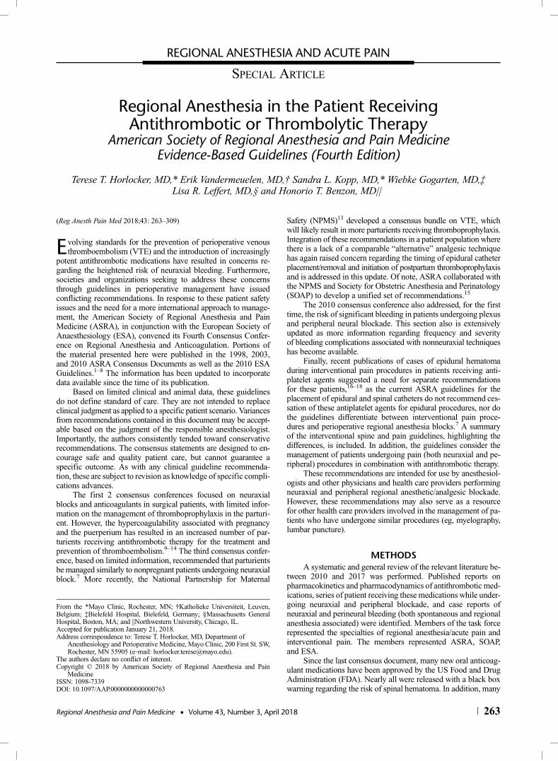
TABLE 7. European Society of Anaesthesiology's Recommended Time Intervals Before and After Neuraxial Puncture or CatheterRemoval*
Time BeforePuncture/Catheter
Manipulation or Removal
Time AfterPuncture/Catheter
Manipulation or Removal Laboratory Tests
UFHs (for prophylaxis, ≤15,000 IU/d) 4–6 h 1 h Platelets during treatmentfor >5 d
UFHs (for treatment) IV 4–6 h 1 h aPTT, ACT, plateletsSC 8–12 h 1 h
LMWHs (for prophylaxis) 12 h 4 h Platelets during treatmentfor >5 d
LMWHs (for treatment) 24 h 4 h Platelets during treatmentfor >5 d
Fondaparinux (for prophylaxis, 2.5 mg/d) 36–42 h 6–12 h (Anti–factor Xa, standardizedfor specific agent)
Rivaroxaban (for prophylaxis, 10 mg daily) 22–26 h 4–6 h (Anti–factor Xa, standardizedfor specific agent)
Apixaban (for prophylaxis, 2.5 mg BID) 26–30 h 4–6 h (Anti–factor Xa, standardizedfor specific agent)
Dabigatran (for prophylaxis, 150–220 mg) Contraindicated accordingto the manufacturer
6 h TT
Coumarins INR ≤1.4 After catheter removal INRHirudins (desirudin) 8–10 h 2–4 h aPTT, ECTArgatroban 4 h 2 h aPTT, ECT, ACTAcetylsalicylic acid None NoneClopidogrel 7 d After catheter removalTiclopidine 10 d After catheter removalPrasugrel 7–10 d 6 h after catheter removalTicagrelor 5 d 6 h after catheter removalCilostazol 42 h 5 h after catheter removalNSAIDs None None
*All time intervals refer to patients with normal renal function. Prolonged time interval in patients with hepatic insufficiency.Adapted from Gogarten et al,8 with permission.
Regional Anesthesia and Pain Medicine • Volume 43, Number 3, April 2018 Regional Anesthesia and Anticoagulation
© 2018 American Society of Regional Anesthesia and Pain Medicine 271

3.4 Subcutaneous heparin3.4.1 Preoperative low-dose UFH for thromboprophylaxis.
We suggest, in patients receiving SC low-dose UFHwith dosingregimens of 5000 U BID or TID, neuraxial block occur 4 to6 hours after heparin administration, or coagulation statusbe assessed (grade 2C).
Remarks: There is no change in this recommendation.3.4.2 Preoperative “higher-dose” UFH for thrombo-
prophylaxis (eg, individual heparin dose of 7500–10,000 U BIDor a daily dose of ≤20,000 U). We suggest neuraxial block oc-cur 12 hours after SC heparin administration and assessmentof coagulation status (grade 2C).
Remarks: This is a new recommendation that addresses thehigher-dose UFH thromboprophylaxis in the pregnant patient.
3.4.3 Preoperative therapeutic UFH (eg, individual dose>10,000 U SC per dose or >20,000-U total daily dose).We sug-gest neuraxial block occur 24 hours after SC heparin admin-istration and assessment of coagulation status (grade 2C).
Remarks: This is a new recommendation that addresses thehigher-doseUFH therapeutic anticoagulation in the pregnant patient.
3.4.4 Postoperative low-dose UFH. There is no contrain-dication to maintaining neuraxial catheters in the presenceof low-dose UFH. We suggest catheter removal occur 4 to6 hours after heparin administration. Subsequent heparinadministration may occur 1 hour after catheter removal(grade 2C).
Remarks: There is no change in this recommendation.3.4.5 Postoperative “higher-dose” UFH. The safety of in-
dwelling neuraxial catheters in patients receiving dosesgreater than 5000 U or greater than 15,000 U of UFH dailyhas not been established.We suggest that the risk and benefitsbe assessed on an individual basis and that techniques to facil-itate detection of new/progressive neurodeficits (eg, enhancedneurologic monitoring occur and neuraxial solutions to mini-mize sensory and motor block) be applied (grade 2C).
Remarks: There is no change in this recommendation.
Low-Molecular-Weight HeparinPharmacology, Monitoring, and Reversal of theAnticoagulant Effect of LMWH
Low-molecular-weight heparins are used for both prophylaxisand treatment of arterial and VTE. The biochemical and pharmaco-logic properties of LMWH differ from those of UFH.74,98–101 Mostrelevant are the prolonged half-life and irreversibility with prot-amine. Anti–factor Xa levels peak 3 to 5 hours after administra-tion. The elimination half-life of LMWH is 3 to 6 hours afterSC injection in patients with normal renal function and is doseindependent. In patients with severe renal insufficiency, the anti-coagulant effect is exaggerated, and the elimination half-life maybe prolonged up to 16 hours.98 Prolonged LMWH therapy maybe associated with an accumulation of anti–factor Xa activityand fibrinolysis.102
The anticoagulant effect of LMWH is most readily assessedby the anti–factor Xa activity. However, the anti–factor Xa activityis typically not monitored except with high-dose (therapeutic)applications. Anti–factor Xa activity may be assessed prior toneuraxial blockade. Importantly, the clinical significance of theresidual anti–factor Xa effect is unknown. For example, whiletargeted levels have been established for therapeutic anticoagula-tion, the level of residual anti–factor Xa acceptable for “safe” per-formance of neuraxial block remains undetermined.98
While the anticoagulant effects of standard heparin are neu-tralized by an equimolar dose of protamine, because of reducedprotamine binding to LMWH fractions, only the anti–factor IIa
activity of LMWH is completely reversed, whereas anti–factorXa activity is not fully neutralized. Both anti–factor IIa and anti–factor Xa activity may return up to 3 hours after protamine reversal.
Low-molecular-weight heparins vary both biochemicallyand pharmacologically, including molecular weight, anti–factorIIa and anti–factor Xa activities, and plasma half-life. However,there are no adequate trials comparing the efficacy and safety of1 LMWH to another, and it is not possible to recommend 1 specificLMWH over another.74 Experience in Europe suggests that the rateof spinal hematoma is similar among LMWH preparations.103
Spinal and Epidural Anesthesia in the PatientReceiving LMWH
In 1993, enoxaparin was the first LMWH to be introducedfor general use in the United States. Labeled indications includedthromboprophylaxis after major joint replacement. The initialdose scheduling was 30 mg every 12 hours, with the first dose ad-ministered as soon as possible after surgery. In the first 5 years,more than 40 spinal hematomas were reported through theMedWatch system.2 The risk of spinal hematoma was estimatedto be approximately 1 in 3000 continuous epidural anestheticscompared with 1 in 40,000 spinal anesthetics.104 However, thiswas likely an underestimation; in addition to the spinal hemato-mas reported at the time of the first ASRA consensus conference,therewere approximately 20 that had occurred but were not yet re-ported to the MedWatch system. The frequency was attributed toBID dosing (compared with once-daily dosing as administered inEurope) in the presence of an indwelling neuraxial catheter. How-ever, 20 years later in Sweden, Moen et al42 reported a 1:3600 fre-quency of spinal hematomas amongwomen undergoing total kneereplacement (with once-daily LMWH), which is strikingly similarto the frequency associated with BID administered LMWH calcu-lated by Horlocker and Wedel.2
Risk Factors for Spinal Hematomas WithLMWH Thromboprophylaxis
Based on an examination of the published cases, MedWatchreports, and clinical experience in Europe and North America,specific risk factors have been proposed.2,3,42 It is not possibleto stratify the individual risk factors or determine interactions be-tween risk factors. In summary, age and sex appear to be signifi-cant patient factors, perhaps through vertebral canal compromise(smaller volume need to produce critical ischemic pressure) and/ordrug effect (exaggerated response to LMWH, renal insuffi-ciency). Finally, the additive, if not synergistic, effect of multiplehemostasis-altering medications cannot be overstated andmay el-evate the risk of once-daily LMWH to that of BID dosing.42
On November 6, 2013, the FDA released a Drug SafetyCommunication regarding updated recommendations to decreasethe risk of neuraxial bleeding and paralysis in patients on LMWHs19
(Table 8). The recommendations were based on a series of 100confirmed spinal hematomas occurring between July 20, 1992,and January 31, 2013, which were associated with enoxaparinthromboprophylaxis and neuraxial anesthesia. The case serieswere submitted to the FDA by the manufacturer of enoxaparin(Lovenox; Sanofi-Aventis, Paris, France). Importantly, the major-ity of these were included in the series of spinal hematomasreported in the 1998 and 2002 ASRA recommendations. Al-though all 100 spinal hematomas involved enoxaparin, thenew timing recommendations will be added to the labels of allLMWHs. Specifically:
• For enoxaparin, placement or removal of a neuraxial cathetershould be delayed for at least 12 hours after administration of
Horlocker et al Regional Anesthesia and Pain Medicine • Volume 43, Number 3, April 2018
272 © 2018 American Society of Regional Anesthesia and Pain Medicine
prophylactic doses such as those used for prevention of deepvein thrombosis (30 mg BID or 40 mg once daily). Longer de-lays (24 hours) are appropriate to consider for patients receiv-ing higher therapeutic doses of enoxaparin (1 mg/kg BID or1.5 mg/kg once daily).
• A postprocedure dose of enoxaparin should usually be given nosooner than 4 hours after catheter removal.
• In all cases, a benefit-risk assessment should consider both therisk of thrombosis and the risk of bleeding in the context ofthe procedure and patient risk factors.
The first and third statements are compatible with previousASRA guidelines. Conversely, the second recommends a 4-hourtime interval prior to administration of a postprocedure dose ofLMWH (rather than the 2-hour time interval recommended byASRA). Importantly, among the 100 confirmed spinal hematomas,nonewere associated with a 0- to 4-hour time interval between cath-eter removal and subsequent LMWH dosing (Table 8).19 As pre-viously mentioned, it appears that the new recommendation isbased on the recommendation of Rosencher et al,20 who proposedsubsequent dosing of antithrombotic therapy based on 8 hours mi-nus the time it takes for the anticoagulant to reach peak effect(which is 4 hours for LMWH).
Therapeutic (Off-label) ApplicationsBridging anticoagulation aims tominimize the risk of arterial
thromboembolism, such as stroke and systemic embolism, in pa-tients with a mechanical heart valve or atrial fibrillation and tominimize the risk of recurrent thrombosis in patients with priorVTE. During bridging, either standard or LMWH is administeredduring the 10- to 12-day period that warfarin is discontinued andthe prothrombin time (PT) allowed to normalize (Table 3). Low-molecular-weight heparin has been demonstrated to be effica-cious as a “bridge therapy” for these patients.74,105 High-dose(therapeutic-dose) LMWHbridging involves administering a dose
that is similar to that used for the treatment of acute VTE or anacute coronary syndrome, such as enoxaparin 1 mg/kg every12 hours, enoxaparin 1.5 mg/kg daily, dalteparin 120 U/kg every12 hours, dalteparin 200 U/kg daily, or tinzaparin 175 U/kg daily.It is advisable to maintain peak anti–factor Xa levels between0.5 and 1 U/mL (measured 3–4 hours after the LMWH dose, withthe assay calibrated to the specific LMWH administered). Withthese doses, it is critical that the last preoperative dose should oc-cur at least 24 hours preoperatively.36 A recent series of 19 patientstherapeutically anticoagulated with enoxaparin preoperativelydemonstrated the likelihood of residual anti–factor Xa activity,even if the 24-hour interval is maintained.106 Eleven of 19 patientsstill had anti–factor Xa activity that would place them at or abovethe lower limit for the peak target therapeutic range for VTE pro-phylaxis. One of these patients had an anti–factor Xa level withinthe target therapeutic range for thrombosis treatment. All but1 patient, whose sample was drawn at 23.25 hours, would havemet the ASRA-recommended time-based guideline (a minimumof 24 hours from the last dose). Although the small number of pa-tients makes it difficult to definitively identify risk factors thatmay increase the possibility of residual anti–factor Xa level activ-ity, this series validates previously reported results that suggestthat patients with lower creatinine clearance (CrCl) values andincreased age may be at particular risk of an exaggerated and/orprolonged response and are at risk of bleeding complications(including spinal hematoma)2,107(Table 8).
Both the FDA andACCP recommend a reduction in the ther-apeutic dosing in patients who have severe renal disease (CrCl<30 mL/min).108 However, recent reviews have suggested that areduction in LMWH dose should be made even with CrCl be-tween 30 and 50 mL/min because of the bleeding risk.107 It is alsointeresting that many of the patients in the series by Henshawet al,106 for example, an 85-year-old, 53-kg woman with a calcu-lated CrCl of 45 mL/min, received 60 mg BID. Her anti–factorXa activity was 0.54 international unit (IU)/mL—still within treat-ment therapeutic peak target range of 0.5 to 0.8 IU/mL. Of note,there have been no spinal hematomas reported in patients whohave undergone bridging therapy with subsequent neuraxial block.However, these recent data, combined with the increased avail-ability of the anti–factor Xa assay, suggest that in patients whomay be at risk of significant residual LMWH effect an assessmentmay bemade prior to neuraxial block. It is also important to deter-mine when the first postoperative dose is anticipated becausethese patients are often aggressively anticoagulated postopera-tively. It is recommended that therapeutic-dose LMWH be re-sumed 24 hours after non–high-bleeding-risk surgery and 48 to72 hours after high-bleeding-risk surgery.36
4.0AnestheticManagement of the Patient ReceivingLMWHNorth American recommendations have drawn on the
extensive European experience in the development of practiceguidelines for the management of patients undergoing spinaland epidural blocks while receiving perioperative LMWH.Previous consensus recommendations have appeared to de-crease the risk. Concern remains for higher-dose applications,where sustained therapeutic levels of anticoagulation are pres-ent, as well as early postprocedural dosing.
4.1 The anti–factor Xa level is not predictive of the risk ofbleeding, although it may be useful in monitoring efficacy oftherapy with therapeutic (high dose) regimens. We recommendagainst the routine use ofmonitoring of the anti–factorXa level.An acceptable level of residual anti–factor Xa level for perfor-mance of neuraxial block remains undetermined (grade 1A).
Remarks: The increased availability of anti–factor Xa activ-ity level allows for preoperative assessment of residual anticoag-ulant effect in patients on higher-dose LMWH.
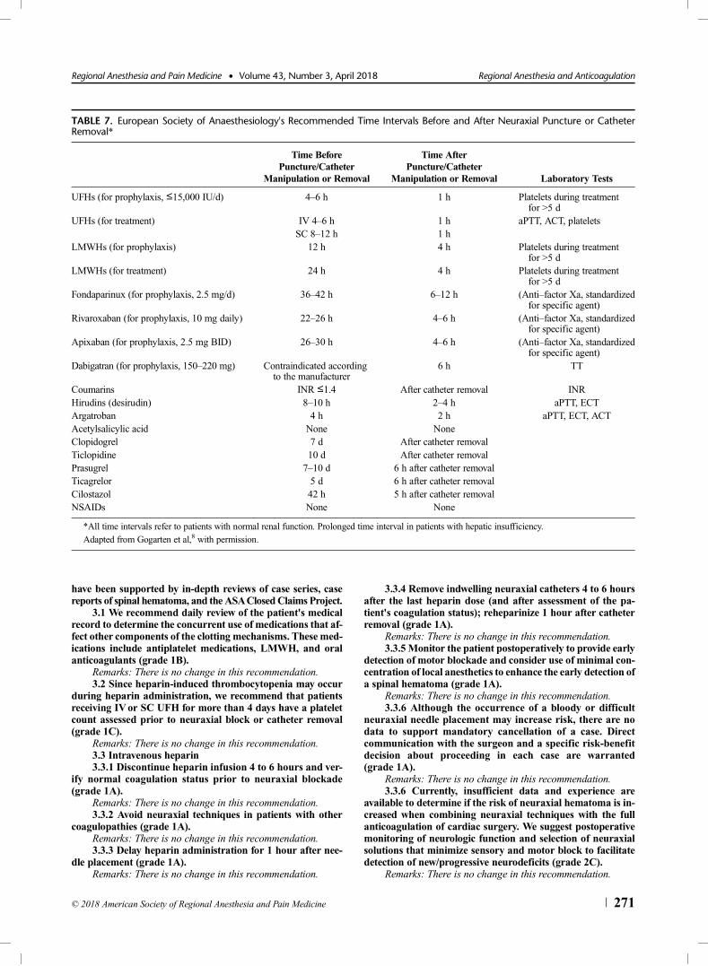
TABLE 8. Patient, Anesthetic, and LMWHRisk Factors*AssociatedWith Spinal Hematoma
n
Patient factorsFemale sex 72Elderly (≥65 y) 70Abnormalities of spinal cord or vertebral column 20Patients at increased risk of hemorrhage† 47Renal insufficiency 7
Anesthetic factorsTraumatic needle/catheter placement 26Epidural technique 54Indwelling epidural catheter during LMWH administration 36
LMWH dosing factorsImmediate preoperative administration (<12 h) 5Intraoperative administration 7Early postoperative administration (<12 h) 17Administration close to indwelling catheter removal (<12 h) 1Twice-daily administration (vs once daily administration) 48Higher LMWH dose than that in the label 1Concomitant medications affecting hemostasis 43
*More than 1 risk factor may have been present in a single case.Adapted from the FDA Drug Safety Communication.19
Regional Anesthesia and Pain Medicine • Volume 43, Number 3, April 2018 Regional Anesthesia and Anticoagulation
© 2018 American Society of Regional Anesthesia and Pain Medicine 273

4.2Antiplatelet or oral anticoagulantmedications admin-istered in combination with LMWH increase the risk of spinalhematoma. Education of the entire patient care team is neces-sary to avoid potentiation of the anticoagulant effects. We rec-ommend against concomitant administration of medicationsaffecting hemostasis, such as antiplatelet drugs, standard hep-arin, or dextran, regardless of LMWH dosing regimen whenthere is an indwelling neuraxial catheter (grade 1A).
Remarks: There is no change in this recommendation.4.3 Since heparin-induced thrombocytopenia (HIT) may
occur during LMWHadministration, we recommend that pa-tients receiving LMWH for greater than 4 days have a plateletcount assessed prior to neuraxial block or catheter removal(grade 1C).
Remarks: There is no change in this recommendation.4.4 The presence of blood during needle and catheter
placement does not necessitate postponement of surgery. Wesuggest that initiation of LMWH therapy in this setting shouldbe delayed for 24 hours postoperatively and that this consider-ation be discussed with the surgeon (grade 2C).
Remarks: There is no change in this recommendation.4.5 Preoperative LMWH4.5.1We recommend that needle placement should occur
at least 12 hours after a prophylactic LMWHdose (grade 1C).Remarks: Previously recommended was a 10- to 12-hour
range. This recommendation incorporates labeling changes madeby the FDA.
4.5.2 In patients administered a dose of LMWH 2 hourspreoperatively (general surgery patients), we recommendagainst neuraxial techniques because needle placement wouldoccur close to peak anticoagulant activity (grade 1A).
Remarks: There is no change in this recommendation.4.5.3 In patients receiving higher (therapeutic) doses of
LMWH, such as enoxaparin 1mg/kg every 12 hours, enoxaparin1.5 mg/kg daily, dalteparin 120 U/kg every 12 hours, dalteparin200 U/kg daily, or tinzaparin 175 U/kg daily, we recommenddelay of at least 24 hours prior to needle/catheter placement(grade 1C). Consider checking anti–factor Xa activity level,particularly in elderly patients and patients with renal insuffi-ciency. An acceptable level of residual anti–factor Xa activity toproceedwith neuraxial block remains undetermined (grade 2C).
Remarks: Residual anti–factor Xa activity may be presenteven after 24 hours. Assessment, especially in patients with mod-erate to severe renal insufficiency, may be considered.
4.6 Postoperative LMWH4.6.1 Twice-daily prophylactic dosing. This dosage regi-
men is associated with an increased risk of spinal hematoma.We recommend the first dose of LMWH should be adminis-tered the following day and no earlier than 12 hours afterneedle/catheter placement, regardless of anesthetic technique,and only in the presence of adequate (surgical) hemostasis. In-dwelling catheters should be removed prior to initiation ofLMWH thromboprophylaxis. Administration of LMWH shouldbe delayed for 4 hours after catheter removal (grade 1C).
Remarks: Previously recommended was a first dose 24 hoursafter needle/catheter placement and a delay of LMWH dosing foronly 2 hours after catheter removal. These recommendations in-corporate labeling changes made by the FDA.
4.6.2 Single daily prophylactic dosing. We recommendthe first postoperative LMWH dose should be administeredat least 12 hours after needle/catheter placement. The secondpostoperative dose should occur no sooner than 24 hours afterthe first dose. Indwelling neuraxial catheters do not representincreased risk and may be maintained. However, no addi-tional hemostasis alteringmedications should be administered
because of the additive effects. The catheter should be re-moved 12 hours after the last dose of LMWH. SubsequentLMWHdosing should occur at least 4 hours after catheter re-moval (grade 1C).
Remarks: Previously recommended was a 10- to 12-hourrange for both needle/catheter placement and catheter removal.Subsequent LMWH was previously 2 hours after catheter re-moval. These recommendations incorporate labeling changesmade by the FDA.
4.6.3 Single or BID therapeutic dosing. Therapeutic-doseLMWH may be resumed 24 hours after non–high-bleeding-risk surgery and 48 to 72 hours after high-bleeding-risk sur-gery. We recommend that indwelling neuraxial catheters beremoved 4 hours prior to the first postoperative dose and atleast 24 hours after needle/catheter placement, whichever isgreater (grade 1C).
Remarks: There is no change in this recommendation.
Anti–Factor Xa AgentsFondaparinux
Fondaparinux (Arixtra), an injectable synthetic pentasac-charide, was approved in December 2001. The FDA releasedfondaparinux with a black box warning similar to that of LMWHsand heparinoids. Fondaparinux produces its antithrombotic effectthrough factor Xa inhibition. The plasma half-life of fondaparinuxis 21 hours, allowing for single-daily dosing, with the first doseadministered 6 hours postoperatively.109 Investigators reporteda spinal hematoma among the initial dose-ranging study (at adose that was subsequently determined to be twice that requiredfor thromboprophylaxis).109,110 No additional spinal hematomaswere reported in the combined series of 3600 patients who under-went spinal or epidural anesthesia in combination with fondaparinuxthromboprophylaxis. However, the conditions for performance ofneuraxial block were strictly controlled. Patients were included insubsequent clinical trials only if needle placement was atraumaticand accomplished on the first attempt. In addition, indwelling epi-dural catheters were removed 2 hours prior to fondaparinux ad-ministration. These strict parameters suggested that neuraxialblockade in patients with planned fondaparinux thromboprophylaxismay not be feasible in clinical practice. For example, in a prospec-tive series, less than 40% of neuraxial blocks were successful with1 pass.44 A recent series of 1631 patients undergoing continuousneuraxial or deep peripheral block reported no serious hemor-rhagic complications. However, the catheters were removed 36 hoursafter the last dose of fondaparinux, and subsequent dosing wasdelayed for 12 hours after catheter removal.111 Although theseresults are reassuring, the deviation from the manufacturer's sug-gested dosing guidelines is of concern.
5.0 Anesthetic Management of the Patient ReceivingFondaparinux
5.1 Based on the sustained and irreversible antithrom-botic effect, early postoperative dosing, and the spinal hema-toma reported during initial clinical trials, we recommendthat until further clinical experience is available performanceof neuraxial techniques should occur under conditions used inclinical trials (single needle pass, atraumatic needle place-ment, avoidance of indwelling neuraxial catheters). If this isnot feasible, an alternate method of prophylaxis should beconsidered (grade 1C).
Remarks: There is no change in this recommendation.5.2 We suggest that neuraxial catheters 6 hours be re-
moved prior to the first (postoperative) dose (grade 2C).Remarks: There is no change in this recommendation.
Horlocker et al Regional Anesthesia and Pain Medicine • Volume 43, Number 3, April 2018
274 © 2018 American Society of Regional Anesthesia and Pain Medicine

New (or Direct) Oral Anti–Factor Xa AgentsThe new oral anti–factor Xa antiagents (rivaroxaban,
apixaban, edoxaban, and betrixaban) are used in the primary pre-vention of VTE after elective total hip replacement surgery, theprevention of stroke and systemic embolism in adult patients withnonvalvular atrial fibrillation (NVAF), and the prevention andtreatment of (recurrent) VTE and pulmonary embolism (PE)112–117
(Table 9). These drugs are at least as effective anticoagulants asthe vitamin K antagonists but seem to be safer in terms of bleed-ing, have a rapid onset of action and a short half-life, and are de-void of the need for routine laboratory monitoring. Until recently,any specific antidotes were lacking.
RivaroxabanRivaroxaban (Xarelto) is an oral inhibitor of the active site of
factor Xa. It is used in the same indications as dabigatran and hasan oral bioavailability and a plasma protein binding of 80% to100% and 90%, respectively.121 Two to 4 hours after oral inges-tion, maximum plasma levels will be reached. Approximatelyone-third of the drug is eliminated in an unchanged formthrough the kidney, whereas the remaining two-thirds will bemetabolized by the liver and excreted in equal amounts by thekidney and the gut.121–123 The terminal elimination half-life is5 to 9 hours in healthy patients but is prolonged to 11 to 13 hoursin the elderly (rivaroxaban [full prescribing information], availableat https://www.xarelto-us.com/shared/product/xarelto/prescribing-information.pdf, accessed March 4, 2017). In individuals withmild, moderate, and severe renal impairment, rivaroxaban expo-sure was increased 1.4-, 1.5-, and 1.6 fold, respectively.
Twenty milligrams BID is used in the prevention of systemicembolism in patients with NVAF and for the prevention of recur-rent of VTE or PE, whereas 15 mg BID for 3 weeks followed by20 mg once a day is advised for the treatment of VTE and PE.In the prevention of VTE, a dose of 10 mg once daily is started6 to 10 hours after orthopedic surgery (rivaroxaban [full prescrib-ing information], available at https://www.xarelto-us.com/shared/product/xarelto/prescribing-information.pdf, accessed March 4,2017). The manufacturer does not recommend a dose adjustmentin patients with a CrCl of greater than 30 mL/min. Rivaroxabanshould be avoided when the CrCl is less than 30 mL/min, withthe exception of patients with NVAF having a CrCl of 15 to30 mL/min, where a dose reduction to 15 mg once daily applies.Rivaroxaban should also not be used in patients treated with cyto-chrome P450 3A4 (CYP3A4) and P-glycoprotein (P-gp) inhibi-tors and in the presence of hepatic disease associated withcoagulopathy and clinically relevant bleeding risk, including Child-Pugh B and C cirrhotic patients.
Rivaroxaban prolongs the international normalized ratio(INR) in a dose-dependent way, but the results are not always re-liable because of an important interassay variability dependenton the reagent used.124 At best, the PT can give some qualitativeinformation. The aPTT is even less sensitive, has a nonlineardose-dependent prolongation and an important interassay variabil-ity, and is not suited to qualitatively and quantitatively assess theeffects of rivaroxaban.124 The best method to assess rivaroxabanis the use of chromogenic anti–factor Xa assays developed forthe measurement of direct factor Xa inhibitors using specificrivaroxaban calibrators.124
There are minimal clinical data on the use of neuraxial anes-thesia in rivaroxaban-treated patients.23 Product labeling states,“Epidural or spinal hematomas have occurred in patients treatedwith rivaroxaban who received neuraxial anesthesia or under-went spinal puncture” (rivaroxaban [full prescribing information],available at https://www.xarelto-us.com/shared/product/xarelto/
prescribing-information.pdf, accessed March 4, 2017). However,no details regarding risk factors or frequency are reported. A re-viewof the literature noted 7 neuraxial hematomas associated withrivaroxaban125–134(Table 10). Three of the patients had undergonea neuraxial block. However, in 2 of the cases, an alternate agentof thromboprophylaxis was used immediately postoperatively,and the hematomas occurred after hospital discharge.126,127 Inthe remaining case, the epidural catheter was removed 18 hours af-ter the first dose of rivaroxaban, a shorter time interval than the26 hours (2 elimination half-lives as is recommended by the man-ufacturer in elderly patients (rivaroxaban [full prescribing infor-mation], available at https://www.xarelto-us.com/shared/product/xarelto/prescribing-information.pdf, accessed March 4, 2017).125
If a neuraxial block is considered in patients treated with pro-phylactic rivaroxaban (≤10mg/d), a time interval of 22 to 26 hoursshould be observed between the last dose of rivaroxaban and thesubsequent neuraxial puncture and/or catheter manipulation/withdrawal. In the presence of a CrCl of less than 50 mL/minand/or when greater than 10mg/d is used, this time interval shouldbe extended to 44 to 65 hours. The absence of any remaining an-ticoagulant activity may be documented using a chromogenicanti–factor Xa assay calibrated for rivaroxaban. Importantly, al-though it appears “safe” to proceed with neuraxial block if zeroanti–factor Xa activity is noted, the residual level acceptable forperformance of neuraxial remains unknown. The first or next doseof rivaroxaban should be given only 6 hours after the neuraxialpuncture or neuraxial catheter withdrawal.
ApixabanApixaban (Eliquis) is an oral highly selective reversible fac-
tor Xa inhibitor. The drug has an oral bioavailability of 51% to85% and a plasma protein binding of approximately 87%.121
The maximum plasma concentration is reached 2 to 4 hours afteroral intake. Approximately 27% of the clearance is renal; the re-mainder will undergo hepatic or biliary transformation prior to be-ing eliminated via the gut.121,135 The terminal elimination half-lifein healthy patients is 10 to 15 hours and is onlymarginally influencedby the renal function, with half-life increasing up to only 17.5 hoursin the presence of moderate to severe renal insufficiency (apixaban[full prescribing information], available at http://packageinserts.bms.com/pi/pi_eliquis.pdf, accessed March 4, 2017).119 Apixabanshould not be used in patients with a CrCl of less than 15 mL/minand in patients with Child-Pugh class B and C hepatic impair-ment. The concomitant use of strong inhibitors of CYP3A4 andP-gp should also be avoided and may be an indication for a 50%dose reduction of apixaban.
Apixaban is used in a dose of 5 mg BID in the prevention ofstroke and systemic embolism in NVAF. This dose is reduced to2.5 mg BID in patients with at least 2 of the following conditions:age 80 years or older, a serum creatinine 1.5 mg/dL or greater, or abody weight 60 kg or less. A dose reduction is not necessary in allof the other indications (apixaban [full prescribing information],available at http://packageinserts.bms.com/pi/pi_eliquis.pdf, accessedMarch 4, 2017). Following elective hip or knee replacement sur-gery, a dose of 2.5 mg BID is used and started 12 to 24 hours aftersurgery. In the treatment of VTE or PE, a starting dose of 10 mgBID is used for 7 days, followed by 5 mg BID. For the subsequentprevention of recurrent VTE and PE, a dose of 2.5 mg BIDis recommended.
Both the PT and the aPTT are not suited to qualitatively andquantitatively assess the effects of apixaban.136 These tests pro-duce unreliable results because of a low sensitivity for apixabanand a large interassay variability dependent on the reagents used.The aPTTalso displays a nonlinear dose-dependent prolongation.136
Regional Anesthesia and Pain Medicine • Volume 43, Number 3, April 2018 Regional Anesthesia and Anticoagulation
© 2018 American Society of Regional Anesthesia and Pain Medicine 275

TABL
E9.
New
(Dire
ct)O
ralA
nti–Factor
XaAn
ticoa
gulants
Betrixaba
n*Rivaroxab
an†
Apixaba
n‡Edoxaba
n§
Indications
anddosing
Prophylaxisof
VTEin
adultp
atientshospitalized
foran
acutemedicalillnesswho
areatrisk
ofthromboem
bolic
complications
dueto
moderateor
severerestrictedmobility
andotherriskfactorsforVTE
Reductionof
riskof
stroke
andsystem
icem
bolism
inNVA
F:Reductionof
riskof
stroke
andsystem
icem
bolism
inNVAF:
5mgBID
(EU,U
nitedStates)
Reductionof
riskof
stroke
andsystem
icem
bolism
inNVAF:
Inpatientswith
CrCl5
0–95
mL/m
in:6
0mg
QD(EU,U
nitedStates)
Inpatientswith
CrCl>
50mL/m
in:2
0mgQD
(EuropeanUnion
[EU],UnitedStates)
Inpatientswith
atleast2
ofthefollowing
characteristics:age≥8
0y,body
weight≤
60kg,
orserum
creatinine≥1
.5mg/dL
:2.5mgBID
(EU,U
nitedStates)
Donotu
sein
patientswith
CrCl>
95mL/m
in(U
nitedStates)
Inpatientswith
CrCl1
5–50
mL/m
in:1
5mgQD
(EU,U
nitedStates)
Inpatientswith
CrCl1
5–50
mL/m
in:3
0mg
QD(U
nitedStates)
Treatm
ento
fVTE,P
E,and
reductionin
therisk
ofrecurrentV
TEandof
PE:
TreatmentofV
TEandPE
:10mgBID
for7
d,followed
by5mgBID
(EU,U
nitedStates)
Inpatientswith
CrCl>
30mL/m
in:1
60mg
everyday(Q
D)firstd
ay,then80
mgQD
(UnitedStates)
Inpatientswith
≥1of
thefollowingclinical
factors:CrCl15–50
mL/minor
body
weight≤
60kg,orthe
concom
itantuseof
P-gp
inhibitors:30mgQD(EU)
Inpatientswith
aCrCl>
15mL/m
in:1
5mgBID
forthefirst2
1dof
theinitialtreatmento
facute
VTEor
PE.A
ftertheinitialtreatmentp
eriod,
20mgQDfortheremaining
treatmentand
the
long-term
reductionin
theriskof
recurrence
ofVTEandof
PE(EU,U
nitedStates)
Treatm
ento
fVTEandPE
:60mgQD
(EU,U
nitedStates)
Inpatientswith
≥1of
thefollowingclinical
factors:CrCl15–50
mL/minor
body
weight≤
60kg,orthe
concom
itantuseof
P-gp
inhibitors:30mgQD(EU,U
nitedStates)
Reductionin
theriskof
recurrentV
TEandPE
followinginitialtherapy:
60mgQD(EU)
Inpatientswith
CrCl15–50
mL/minor
body
weight≤
60kg,orthe
concom
itantuseof
P-gp
inhibitors:30mgQD(EU)
Inpatientswith
aCrCl1
5–50
mL/m
inandahigh
bleeding
risk,consider15
mgQDinsteadof
20mg
QDfortheremaining
treatmentand
thelong-term
reductionin
theriskof
recurrence
ofVTEandof
PE(EU)
Prophylaxisof
VTE
followingTHAor
TKA:10mgQD
(EU,U
nitedStates)
Preventionof
atherothrombotic
eventsinadultpatients
afteranacutecoronary
syndrome(coadm
inistered
with
acetylsalicylicacid[ASA
])aloneor
with
ASA
plus
clopidogrelorticlopidine):2.5mgBID
(EU)
Reductionin
theriskof
recurrentV
TE
andPE
followinginitialtherapy:
2.5mgBID
(EU,U
nitedStates)
Prophylaxisof
VTE
followingTH
Aor
TKA:
2.5mgBID
(EU,U
nitedStates)
Inpatientswith
CrCl1
5–30
mL/m
in:8
0mgQD,
then
40mgQD(U
nitedStates)
Renalclearance
5%–11%
112,113,118
35%
27%
50%
CrCl,mL/m
in≥9
060–89
30–59
15–29
<15(off-dialysis)
≥80
50–79
30–49
15–29
<15(off-dialysis)
≥80
50–79
30–49
15–29
<15(off-dialysis)
≥80
50–79
30–49
15–29
<15(off-dialysis)
Elim
inationhalf-lifet ½
β,h
37h1
14,115
Nodata
Nodata
Nodata
Nodata
5–9(young),11–13(elderly)†
8.71
16,117
9.01
16,117
9.51
16,117
13.2117
15.1119
14.6119
17.6119
17.3119
Nodata
10–14†
8.41
209.41
2016.9
Nodata
*Betrix
aban
fullprescribinginform
ation(http
s://w
ww.accessdata.fda.gov/drugsatfd
a_docs/label/2017/208383S0
00lbl.pdf,accessedNovem
ber15,2017).
†Xarelto:Eparproductinform
ation
(http://www.ema.europa.eu/docs/en_GB/docum
ent_library/EPA
R_-_P
roduct_Information/human/000944/WC500057108.pdf,
accessed
Decem
ber22,2015,2011;
rivaroxaban
fullprescribinginform
ation(https://w
ww.xarelto-us.com/shared/product/x
arelto/prescrib
ing-inform
ation.pdf,accessed
March
4,2017).
‡Apixabanfullprescribinginform
ation(http
://packageinserts.bms.com/pi/p
i_eliquis.pdf,accessed
March
4,2017;apixaban:Epar—
productinformation(http
://www.ema.europa.eu/docs/en_GB/docum
ent_
library/EPA
R_-_P
roduct_Information/human/002148/WC500107728.pdf,accessed
Decem
ber22,2
015).
§Edoxaban:
Epar—
productinform
ation(http
://www.ema.europa.eu/docs/en_GB/docum
ent_lib
rary/EPA
R_-_P
roduct_Information/human/002629/WC500189045.pdf,accessed
22/12/20105,
2015);Savaysa
fullprescribinginform
ation(http://dsi.com
/prescrib
ing-inform
ation-portlet/getPIContent?productNam
e=Savaysa&
inlin
e=true,accessedMarch
4,2017).
Horlocker et al Regional Anesthesia and Pain Medicine • Volume 43, Number 3, April 2018
276 © 2018 American Society of Regional Anesthesia and Pain Medicine

Chromogenic anti–factor Xa assays developed for the measure-ment of direct factor Xa inhibitors and using specific calibratorsfor apixaban are the monitoring tests of choice.136
There are very few prospective data concerning the use ofneuraxial blocks in apixaban-treated patients. A literature searchup to November 2017 found no reports of epidural/spinal bleedingassociated with a neuraxial anesthesia, although a single spontane-ous hematoma has been reported.134 Thus, after a cautious risk-benefit analysis, there is justification to proceed with a neuraxialblock; there should be at least a 26- to 30-hour time interval
between the last dose of prophylactic dose of apixaban (2.5 mg/d)and the subsequent neuraxial puncture and/or catheter manipulation/removal. If a dose of 5 mg/d is used and/or in patients with a se-rum creatinine of 1.5 mg/dL or greater and aged 80 years or olderor a body weight 60 kg or less, a time interval of 40 to 75 hoursshould be observed. The absence of any remaining apixaban ac-tivity can be documented using a chromogenic anti–factor Xaassay calibrated for apixaban. The next dose of apixaban shouldbe administered at least 6 hours after the neuraxial puncture orwithdrawal of the neuraxial catheter.
TABLE 10. Case Reports of Spinal Hematoma in Patients on New Oral Anticoagulants
Drug History Outcome Comments
Dabigatran128 72 y, M, 150 mg BID dabigatran for AF;T2–T7 burst fracture after a fall, 3 hafter last dose of dabigatran; bonyretropulsion and epidural hematomaon MRI
Fresh frozen plasma, factor VII;decompression initially performed;PSF done 7 d later when coagulationstatus was normal; bowel functionreturned but bladder function did notand unable to walk
Spontaneous hematoma
Dabigatran129 70 y, 150 mg dabigatran for AF; tetraplegia12 h after dabigatran; C2–C4 hematomaon CT
PCC; urgent C2–C4 laminectomy;complete neurological recovery
Spontaneous hematoma
Rivaroxaban130 61 y, F, 10 mg rivaroxaban started 8 hafter tibial osteotomy (general anesthesia);T8 paraplegia 22 h after last dose;C2–C8 SEH on MRI
Rivaroxaban discontinued; spontaneousrecovery, without surgery,4 h after onset
Spontaneous hematoma; patient wasalso on ibuprofen postoperatively
Rivaroxaban126 69 y, F, THR; 40 mg/d enoxaparin started12 h after epidural, continued for 4 d;10 mg rivaroxaban started on POD5,22 h after enoxaparin; neck pain andLE weakness 5 d after rivaroxabanstarted, 14 h after last dose; C2–C4SEH on CT
Symptoms improved 4 h later, completerecovery 48 h after symptoms
Spontaneous hematoma
Rivaroxaban131 69 y, M, 20 mg rivaroxaban for AF;T8 paraplegia; T3-conus subduralhematoma on MRI
Steroids, lumbar and cervical drainsplaced 8 d later (surgery notrecommended); no recovery
Spontaneous hematoma
Rivaroxaban132 58 y, M, 20 mg rivaroxaban daily for AF;interscapular pain after 30 d on rivaroxaban;bilateral LE weakness; C7–T2 intraduralhematoma on MRI
IV dexamethasone, sensory/motorimprovement after 1 d; C7 corpectomydone of fourth day; almost completerecovery
Spontaneous hematoma. Patient hadTHR (spinal) 3 wk before episode,rivaroxaban stopped 3 d beforesurgery and restarted a few dayslater; he was asymptomatic beforeepisode
Rivaroxaban133 72 y, M, 20 mg daily rivaroxaban ! 3 y for AF;interscapular pain flaccid paraplegia; T6–T8subdural hematoma on MRI
PCC; laminectomy next day, IV steroids;no improvement at 6-mo follow-up
Spontaneous hematoma
Rivaroxaban127 53 y, F, TKR (spinal); 5–7 mg warfarin onhospital day 1 and POD2–POD3, and 40 mgenoxaparin on POD1–POD3. Switched torivaroxaban on POD4 (dose not stated);leg pain on POD6 and numbness on POD7;L4–5 SEH on MRI
Emergency laminectomy IVC filter;residual peroneal anesthesia andneurogenic bladder at 2-y follow-up
Patient had spinal stenosis; SEH mayhave been initiated or aggravated bythe multiple anticoagulants and theintake of rivaroxaban while warfarinwas still fully active
Rivaroxaban125 59 y, F, TKR (CSE), postoperative epiduralinfusion; 10 mg rivaroxaban started thenight of surgery, epidural catheter removed18 h after rivaroxaban; rivaroxaban repeated6 h after removal; bladder incontinence andback pain 12 h after last dose of rivaroxaban;L2–L4 SEH on CT
Emergency laminectomy; completerecovery after 5 d
Short interval between rivaroxabandose and removal of epiduralcatheter (18 h); 6-h interval betweencatheter removal and resumptionof rivaroxaban (6 h) may be just alittle short
Apixaban134 78 M, apixaban 2.5 mg BID and clopidogrelafter coronary stent placement; back pain;hyperintense fluid level (subarachnoidhemorrhage) on MRI
Anticoagulants dcd; complete recoveryat 3 wk
Spontaneous spinal subarachnoidhemorrhage
AF indicates atrial fibrillation; CT, computed tomography; IVC, inferior vena cava; LE, lower extremity; PCC, prothrombin complex concentrate;PSF, posterior spinal fusion; SEH, spinal-epidural hematoma; THR, total hip replacement; TKR, total knee replacement.
Regional Anesthesia and Pain Medicine • Volume 43, Number 3, April 2018 Regional Anesthesia and Anticoagulation
© 2018 American Society of Regional Anesthesia and Pain Medicine 277

EdoxabanEdoxaban (Savaysa; Daiichi Sankyo Inc, Parsippany, New
Jersey) is also is an oral highly selective reversible factor Xa inhib-itor. Following oral ingestion, maximumplasma levels are reachedwithin 1 to 2 hours. Edoxaban has an absolute bioavailability anda plasma protein binding of approximately 62% and 55%, respec-tively. The kidney clears approximately 50% of the administereddose, whereas metabolism and biliary/intestinal excretion accountfor the remainder. The elimination half-life after oral administra-tion is 10 to 14 hours in healthy patients.137–141 In the presenceof mild, moderate, and severe renal insufficiency, the total exposureto edoxaban is increased by 32%, 74%, and 72%, resulting in an elim-ination half-life of 8.4, 9.45, and 16.9 hours, respectively.120,142–145
Edoxaban is used in the prevention of stroke and systemicembolism in patients with NVAF and the treatment of VTE and ofPE (Savaysa [full prescribing information], available at http://dsi.com/prescribing-information-portlet/getPIContent?productName=Savaysa&inline=true, accessed March 4, 2017). A dose of 60 mgonce daily is recommended in both indications. The dose shouldbe reduced to 30 mg/d in NVAF patients with CrCl of 15 to49 mL/min and in patients treated for VTE/PE with body weightof 60 kg or less and/or concomitant use of P-gp inhibitors.146–150
Interestingly, edoxaban should not be used in NVAF patients witha CrCl of greater than 95 mL/min because of an increased risk ofischemic stroke (Savaysa [full prescribing information], availableat http://dsi.com/prescribing-information-portlet/getPIContent?productName=Savaysa&inline=true, accessed March 4, 2017).The 30-mg daily dose was also effective in the prevention ofVTE following prosthetic surgery of the knee and hip,151,152 butthis indication has not been registered in the United States orin Europe.
The PT and aPTT should not be used to assess the antico-agulant activity of edoxaban as the compound behaves in muchthe same way as apixaban.153 Edoxaban is best assessed usingtailor-made chromogenic anti–factor Xa assays.153
There have been no reports of any neuraxial bleeding in asso-ciation with neuraxial anesthesia up to November 2017, but theuse of neuraxial anesthetic techniques in edoxaban-treated patientsshould be cautiously considered as there are no data. There shouldbe at least a 20- to 28-hour time interval (2 half-lives) between thelast dose of preventive dose of edoxaban (≤30 mg/d) and the sub-sequent neuraxial puncture and/or catheter manipulation/removal.In case of a higher dose (>30 mg/d) and/or in patients with CrCl15 to 49 mL/min, a body weight 60 kg or less, and/or the concom-itant use of P-gp inhibitors, a time interval of 40 to 70 hours (4–5half-lives) should be observed. The presence of any residualedoxaban activity can be excluded using a chromogenic anti–factorXa assay calibrated for edoxaban. As with the other new oral an-ticoagulants, an acceptable level of residual edoxaban activity toproceed with neuraxial block remains undetermined. The nextdose of edoxaban should be administered at least 6 hours afterthe neuraxial puncture or withdrawal of the neuraxial catheter.
BetrixabanThe latest arrival in the family of the oral anti–factor Xa
agents is betrixaban (Bevyxxa; Portola Pharmaceuticals, Inc,San Francisco, California). Betrixaban is also an oral reversible se-lective factor Xa inhibitor that recently received FDA approval.After oral intake, peak plasma levels will be reached 3 to 4 hours.The drug has an oral bioavailability of approximately 34% and anin vitro plasma protein binding of 60%. The excretion ofbetrixaban occurs mainly through the liver.112–115,154 Hepatic me-tabolism is minimal (<1%), and the unchanged drug is primarilyexcreted via a P-gp–dependent mechanism in the hepatobiliary
route. As a result, 82% to 89% of the drug can be found in the fe-ces, whereas the remainder (5%–11% of an orally administereddose) is eliminated through the kidney, which makes betrixabanthe least kidney-dependent factor Xa inhibitor.112,113,118 Theresulting terminal half-life is 37 hours, whereas the pharmacody-namic half-life is 19 to 27 hours.114,115,118 Compared with healthysubjects, renal impairment will still cause an increase of the patientexposure to the drug by 1.89-, 2.27-, and 2.63-fold in the presenceof mild, moderate, and severe renal insufficiency, respectively.118
Betrixaban is used in prophylaxis of VTE in adult patientshospitalized for an acute medical illness (who are not undergoingsurgery) and are at risk of thromboembolic complications due tomoderate or severe restricted mobility and other risk factors forVTE.118 Treatment is initiated with a single dose of 160 mg,followed by 80mg once daily. In patients with severe renal impair-ment or treated with P-gp inhibitors, the dose should be reduced toa single dose of 80 mg initially, followed by 40 mg once daily.118
As with the other factor Xa inhibitors, routine monitoring ofthe anticoagulant effect is not necessary. However, if necessary,any betrixaban effect should assessed using a calibrated chromo-genic anti–factor Xa assay.114,115,155
As of August 2017, there have been no reports of neuraxialbleeding in patients treated with betrixaban who underwent aneuraxial anesthetic technique. Still, the use of epidural or spinalanesthesia in these patients should once again be carefully consid-ered as clinical experience with betrixaban is lacking. The drugwas released with a black box warning regarding spinal/epiduralhematoma in patients who are receiving neuraxial anesthesia orundergoing spinal puncture.118 The FDA-approved labeling rec-ommends that prior to neuraxial intervention the potential benefitversus the risk in anticoagulated patients or in patients to beanticoagulated for thromboprophylaxis should be consideredand that an epidural catheter should be placed/removed no earlierthan 72 hours after the last administration of betrixaban, whilethe next dose of betrixaban should be administered no earlierthan 5 hours after the removal of the catheter. If traumatic punctureoccurs, administration of betrixaban should be delayed for72 hours.118
Using a pharmacologic approach, if a neuraxial block is con-sidered in patients receiving Betrixaban, a minimum time intervalof 72 hours should be respected between the last prophylactic doseof betrixaban and the following neuraxial puncture and/or cathetermanipulation/withdrawal. In patients with CrCl of 15 to 29 mL/minand/or the concomitant use of other P-gp inhibitors (or if in thefuture higher doses should be used), a time interval of 76 to135 hours (5 half-lives) should be observed. The presence ofany residual betrixaban activity can be excluded using a chromo-genic anti–factor Xa assay calibrated for betrixaban. The nextdose of betrixaban should be administered at least 5 hours afterthe neuraxial puncture or withdrawal of the neuraxial catheter.
Reversal of Oral Anti–Factor Xa AgentsSpecific antidotes that reverse the anticoagulant effects of the
oral anti–factor Xa agents are still lacking. Most of the limiteddata available on the reversal of novel oral anticoagulants arebased on preclinical data, animal experiments, studies in healthyvolunteers, and a limited number of case reports. Fortunately, allof these agents have, at least in healthy patients, a relatively shorthalf-life. If there is time to await spontaneous clearance, discontin-uation of the drug is a possible strategy.156,157
The reversal of factor Xa inhibitors still depends on the use ofprohemostatic agents such as prothrombin complex concentrates(PCCs), activated PCC or factor VIII inhibitor bypassing activity,and recombinant factor VIIa. The recommended doses are 25 to
Horlocker et al Regional Anesthesia and Pain Medicine • Volume 43, Number 3, April 2018
278 © 2018 American Society of Regional Anesthesia and Pain Medicine

50 IU/kg, 90 to 100 IU/kg, and 90 μg/kg IV when PCC, activatedPCC, or recombinant factor VIIa is used, respectively.157–159 If re-combinant factor VIIa is used, the potential risk of thromboticevents must always be taken into account.160 Factor Xa inhibitorsall have a high degree of protein binding and are therefore notsuited to be eliminated via hemodialysis.
Two promising antidotes for factor Xa inhibitors are cur-rently under investigation. The first is a recombinant protein,named PRT064445 (also known as andexanet alfa or andexanet),which that serves as a universal antidote for factor Xa inhib-itors, LMWH, and pentasaccharide.161,162 A recent multicenter,prospective, open-label, single-group study evaluated 67 patientswho had an acute major bleeding within 18 hours after the admin-istration of a factor Xa inhibitor. Andexanet substantially reducedanti–factor Xa activity, with effective hemostasis occurring in79% of the patients.163 PER977 (also known as ciraparantag) isanother compound that binds and neutralizes heparins, dabigatran,and all factor Xa inhibitors in preclinical models.164 In healthyvolunteers, it was found to completely reverse the anticoagulanteffects of both enoxaparin and edoxaban.165,166 However, furtherclinical studies are needed before both of these compounds willbecome available.
6.0 Anesthetic Management of the Patient ReceivingRivaroxaban
6.1We suggest that rivaroxaban be discontinued 72 hoursprior to neuraxial block. Consider checking rivaroxaban oranti–factor Xa activity level if less than 72 hours. An accept-able level of residual rivaroxaban activity to proceed withneuraxial block remains undetermined (grade 2C).
Remarks: This is a new recommendation.6.2 We suggest that neuraxial catheters be removed
6 hours prior to the first (postoperative) dose (grade 2C).Remarks: This is a new recommendation.6.3 With unanticipated administration with indwelling
catheter, we suggest that rivaroxaban dosing be held for 22to 26 hours before or an anti–factor Xa assay calibrated torivaroxabanbe assessedbefore the catheter is removed (grade 2C).
Remarks: This is a new recommendation.7.0AnestheticManagement of thePatientReceivingApixaban7.1 We suggest that apixaban be discontinued 72 hours
prior to neuraxial block. Consider checking apixaban oranti–factor Xa activity level if less than 72 hours. An accept-able level of residual apixaban activity to proceedwith neuraxialblock remains undetermined (grade 2C).
Remarks: This is a new recommendation.7.2 We suggest that neuraxial catheters be removed
6 hours prior to the first (postoperative) dose (grade 2C).Remarks: This is a new recommendation.7.3 With unanticipated administration with indwelling
catheter, we suggest that apixaban dosing be held for 26 to30 hours or an anti–factorXa assay calibrated to apixaban be-fore the catheter is removed (grade 2C).
Remarks: This is a new recommendation.8.0AnestheticManagement of thePatientReceivingEdoxaban8.1 We suggest that edoxaban be discontinued 72 hours
prior to neuraxial block. Consider checking edoxaban oranti–factor Xa activity level if less than 72 hours. An accept-able level of residual edoxaban activity to proceed withneuraxial block remains undetermined (grade 2C).
Remarks: This is a new recommendation.8.2 We suggest that neuraxial catheters be removed
6 hours prior to the first (postoperative) dose (grade 2C).Remarks: This is a new recommendation.8.3 With unanticipated administration with indwelling
catheter, we suggest that edoxaban dosing be held for 20 to
28 hours or an anti–factor Xa assay calibrated to edoxabanbefore the catheter is removed (grade 2C).
Remarks: This is a new recommendation.9.0 Anesthetic Management of the Patient Receiving
Betrixaban9.1 We suggest that betrixaban be discontinued a mini-
mum of 3 days prior to neuraxial block. Consider checkingbetrixaban or anti–factor Xa level if less than 3 days (grade 2C).
Remarks: This is a new recommendation.9.2 We suggest against the performance of neuraxial
blocks in patients with a CrCl of less than 30 mL/min(grade 2C).
Remarks: This is a new recommendation.9.3 We suggest that neuraxial catheters 5 hours be re-
moved prior to next dose (grade 2C).Remarks: This is a new recommendation.9.4 With unanticipated administration with indwelling
catheter,we suggest that betrixabandosing be held for 72hours,then the catheter removed (grade 2C).
Remarks: This is a new recommendation.
Direct Thrombin InhibitorsParenteral Thrombin Desirudin, Bivalirudin,and Argatroban)
Recombinant hirudin derivatives, including desirudin (Revasc)and bivalirudin (Angiomax), inhibit both free and clot-boundthrombin. Argatroban (Acova), an L-arginine derivative, has asimilar mechanism of action. These medications are indicatedfor the treatment and prevention of thrombosis in patients withHITand as an adjunct to angioplasty procedures.167,168 Desirudinis approved for prevention of VTE/PE following hip replace-ment.169 The anticoagulant effect of thrombin inhibitors is moni-tored by the aPTT and is present for 1 to 3 hours after IVadministration. Hemorrhagic complications, particularly whencombined with thrombolytic or antiplatelet agents, may be lifethreatening. There is no “antidote”; the antithrombin effect cannotbe reversed pharmacologically. Although there are no case reportsof spinal hematoma related to neuraxial anesthesia among patientswho have received a thrombin inhibitor, spontaneous intracranialbleeding has been reported. The lack of information available andthe approved applications of these agents (typically patients withHITwhowill need therapeutic levels of anticoagulation) make pa-tients who are receiving these medications poor candidates forneuraxial blockade.
10.0AnestheticManagement of PatientsReceivingParenteralThrombin Inhibitors (Desirudin, Bivalirudin, and Argatroban)
10.1 In patients receiving parenteral thrombin inhibitors,we recommend against the performance of neuraxial tech-niques (grade 2C).
Remarks: There is no change in this recommendation.
Oral Thrombin Inhibitors (Dabigatran)Dabigatran (Pradaxa) is an oral competitive direct inhibitor
of both free and clot-bound thrombin with a bioavailability andplasma protein binding of approximately 7% and 35%, re-spectively.170–172 Peak plasma levels will be reached within 0.5to 2 hours after administration (Table 11). The drug is primarily(≥80%) eliminated through the kidney in an unchanged formand has a terminal elimination half-life of 12 to 17 hours aftermultiple doses in healthy patients. A mild (CrCl 50–80 mL/min)to moderate (CrCl 30–49 mL/min) renal insufficiency increasesthe half-life up to 18 hours and 28 hours in severe renal insuffi-ciency (CrCl 15–29 mL/min).170,172,173
Regional Anesthesia and Pain Medicine • Volume 43, Number 3, April 2018 Regional Anesthesia and Anticoagulation
© 2018 American Society of Regional Anesthesia and Pain Medicine 279

A dose of 150 mg BID is used in the treatment of VTE andPE, the prevention of recurrent VTE and PE, and in the preventionof systemic embolism in NVAF. A dose reduction to 75mg BID isrecommended for the latter indication in the presence of a CrCl of15 to 29 mL/min and in the presence of CrCl of 30 to 49 mL/minwith the concomitant use of the P-gp inhibitors.174 In the preven-tion of VTE following orthopedic surgery, the first oral dose ofdabigatran 110 mg is given 1 to 4 hours postoperatively, followedby 220 mg BID on subsequent days. Except for the NVAF patients,dabigatran should not be used in patients with a CrCl of less than30 mL/min or on dialysis (Pradaxa [full prescribing information],available at http://docs.boehringer-ingelheim.com/Prescribing%20Information/PIs/Pradaxa/Pradaxa.pdf, accessed March 4, 2017).
The PT and the INR lack any reliable correlation todabigatran plasma levels and should not be used for the monitor-ing of dabigatran.175 In contrast, the aPTT is prolonged in adose-dependent but nonlinear correlation and with considerableinterindividual and interassay variability. The aPTT can onlybe used to provide rapid qualitative information in emergencysituations.175–178 The thrombin time (TT) and the dilute TT(dTT) are best suited to monitor dabigatran.176,178,179 The latteris now routinely available as the Hemoclot thrombin inhibitorassay.180 These tests, as well as the chromogenic anti–factor Xaassays used to monitor the other novel oral anticoagulants, maynot be readily available in many institutions.
There is limited experiencewith dabigatran and neuraxial an-esthesia, and none with the use of indwelling epidural catheters.Neuraxial blockade was performed in approximately 70% of allpatients in the first studies with dabigatran, but the drug wasstarted postoperatively, and neuraxial catheters were removed atleast 2 hours, but mostly 4 to 6 hours before the first dose ofdabigatran.181–184 Dabigatran was not used if an indwelling epidu-ral catheter remained in place for postoperative pain relief.182,184,185
There have been 2 neuraxial hematomas associated with patientsreceiving dabigatran for chronic atrial fibrillation (one spontane-ous and one related to a trauma/fall).128,129 Although there areno reports up to November 2017 of a spinal/epidural hematomaafter neuraxial anesthesia, it is still too early to make any firm en-dorsements on the use of neuraxial anesthetic (catheter) techniquesin dabigatran-treated patients. Therefore, any recommendationson the use of neuraxial techniques in dabigatran-treated patientsare based on expert opinion and on the pharmacokinetics of thedrugs involved.
The use of a neuraxial block in dabigatran-treated patientsshould be carefully and individually considered. The 12- to 17-hourhalf-life of dabigatran in healthy patients suggests a time intervalof 34 hours (ie, 2 half-lives) between the last prophylactic dose(≤220 mg/d) of dabigatran and the subsequent neuraxial punctureand/or catheter manipulation or withdrawal. This time intervalshould be extended to 48 to 85 hours (4–5 half-lives) when higherdoses (>220 mg/d) are used and up to 72 to 90 hours in patientswith a CrCl of 30 to 49 mL/min. In case of any doubt, the absenceof any residual anticoagulant dabigatran activity can be docu-mented using the dTT. The first or next dose of dabigatran shouldbe given at least 6 hours after the neuraxial puncture or neuraxialcatheter withdrawal.
Reversal of DabigatranIn 2013, Schiele et al186 described idarucizumab as a mono-
clonal antibody fragment that binds to dabigatran and reversesits anticoagulant effects both in vitro and in vivo in rats. A recentclinical trial in patients with bleeding or requiring urgent surgerydemonstrated that idarucizumab completely reversed the antico-agulant effect of dabigatran within minutes.174 In October 2015,idarucizumab was approved by the FDA to be used in adult
TABLE 11. Oral Antithrombin Agents (Dabigatran)*
Indications and dosing Reduction of risk of stroke and systemic embolism in NVAF:In patients with CrCl >30 mL/min: 150 mg BID (EU, United States)In patients with CrCl 30–50 mL/min and high bleeding risk: consider 110 mg BID (EUIn patients with CrCl 15–30 mL/min: 75 mg BID (United States)Treatment of VTE and PE:In patients with CrCl >30 mL/min: 150 mg BID after 5–10 d of parenteral anticoagulation (EU, United States)In patients with CrCl 30–50 mL/min and high bleeding risk: consider 110 mg BID (EU)Reduction in the risk of recurrent VTE and PE:In patients with CrCl >30 mL/min: 150 mg BID after previous treatment (EU, United States)In patients with CrCl 30–50 mL/min and high bleeding risk: consider 110 mg BID (EU)Prophylaxis of VTE following THA:In patients with CrCl >30 mL/min: 110 mg QD first day, then 220 mg QD (United States)In patients with CrCl >50 mL/min: 110 mg QD first day, then 220 mg QD (EU)In patients with CrCl 30–50 mL/min, or aged ≥75 or concomitant use of P-gp inhibitors: 75 mg QD first day,then 150 mg QD (EU)
Prophylaxis of VTE following TKA:In patients with CrCl >50 mL/min: 110 mg QD first day, then 220 mg QD (EU)In patients with CrCl 30–50 mL/min, concomitant use of P-gp inhibitors: 75 mg QD first day, then 150 mg QD (EU)
Renal clearance ≥80%CrCl, mL/min ≥80 50–79 30–49 15–29 <15 (Off-dialysis)Elimination half-life t½β, h 12–17170 16.6173 18.7173 27.5173 No data
*Pradaxa full prescribing information (available at http://docs.boehringer-ingelheim.com/Prescribing%20Information/PIs/Pradaxa/Pradaxa.pdf,accessed March 4, 2017); dabigatran: Epar—product information (available at http://www.ema.europa.eu/docs/en_GB/document_library/EPAR_-_Product_Information/human/000829/WC500041059.pdf, accessed December 22, 2015).
EU indicates European Union; THA, total hip arthroplasty; TKA, total knee arthroplasty.
Horlocker et al Regional Anesthesia and Pain Medicine • Volume 43, Number 3, April 2018
280 © 2018 American Society of Regional Anesthesia and Pain Medicine

patients treated with dabigatran when rapid reversal of its antico-agulant effects is required in situations of emergency surgery/urgent procedures or life-threatening or uncontrolled bleeding(idarucizumab [full prescribing information, http://docs.boehringer-ingelheim.com/Prescribing%20Information/PIs/Praxbind/Praxbind.pdf, accessedMarch 4, 2017). The recommendeddose is 5 g via an IV infusion/injection.
11.0 Anesthetic Management of the Patient ReceivingDabigatran
Dabigatran is highly dependent (>80%) on renal excre-tion. Estimated CrCl tends to overestimate actual renal func-tion. Furthermore, renal function may be further impairedperioperatively.
11.1We suggest that dabigatran be discontinued 120 hoursprior to neuraxial block. However, if renal function has beenreliably determined, and there are no additional risk factorsfor bleeding (eg, age >65 years, hypertension, concomitantantiplatelet medications),187 a more graded approach maybe considered.
Remarks: This is a new recommendation.11.1.1We suggest that dabigatran be discontinued 72 hours
in patients with a CrCl 80mL/min or greater. Consider checkingdTTor ecarin clotting time (ECT) if less than 72 hours. An ac-ceptable level of residual dabigatran activity to proceed withneuraxial block remains undetermined (grade 2C).
Remarks: This is a new recommendation.11.1.2 We suggest that dabigatran be discontinued
96 hours in patients with a CrCl of 50 to 79mL/min. Considerchecking dTT or ECT if less than 96 hours. An acceptablelevel of residual dabigatran activity to proceed with neuraxialblock remains undetermined (grade 2C).
Remarks: This is a new recommendation.11.1.3 We suggest that dabigatran be discontinued
120 hours in patients with a CrCl of 30 to 49 mL/min. Con-sider checking dTTor ECT if less than 120 hours. An accept-able level of residual dabigatran activity to proceed withneuraxial block remains undetermined (grade 2C).
Remarks: This is a new recommendation.11.1.4 We suggest against the performance of neuraxial
blocks in patientswith aCrCl of less than 30mL/min (grade 2C).Remarks: This is a new recommendation.11.2 We suggest that neuraxial catheters be removed
6 hours prior to the first (postoperative) dose (grade 2C).Remarks: This is a new recommendation.11.3 With unanticipated administration with indwelling
catheter, we suggest that dabigatran dosing be held for 34 to36 hours or the dTTor ECTassessed before the catheter is re-moved (grade 2C).
Remarks: This is a new recommendation.
Vitamin K Antagonists (Warfarin)Warfarin Pharmacology
Oral anticoagulants, includingwarfarin, exert their anticoagulanteffect by interfering with the synthesis of the vitamin K–dependent
clotting factors VII, IX, X, and II (thrombin). The effects of war-farin are not apparent until there is a significant amount of bio-logically inactive clotting factors and are dependent on factorhalf-life.188 Clinical experiencewith patients who are congenitallydeficient in factor II, IX, or X suggests that a factor activity levelof 40% for each factor is adequate for normal or near-normal he-mostasis.189 Bleeding may occur if the level of any clotting factoris decreased to 20% to 40% of baseline. During the first few daysof therapy, the PT reflects primarily a reduction of factor VII,the half-life of which is approximately 6 hours (Table 12). Aftera single dose, marked prolongation of the INR may occur, al-though adequate factor levels are still present.190 Discontinuationof warfarin requires normalization of the INR to ensure adequateactivities of all the clotting factors.
Clinical Use of WarfarinThe measured response to anticoagulant therapy at the initi-
ation of treatment varies significantly. Some of the variabilitymay be attributed to age, female sex, preexisting medical condi-tions (low patient weight or liver, cardiac, and renal disease), race,and drug interactions that are associated with an enhancedresponse to warfarin and/or a lower dose requirement for mainte-nance anticoagulation1,188,191,192 (Table 13). Women and the el-derly are more susceptible to warfarin. In a study of 12,202patients on the effect of age and sex on warfarin maintenance dos-ing, it was noted that warfarin dose was inversely related to ageand strongly associated with sex.193 The association of renal in-sufficiency and increased response towarfarin has also been notedby Benzon et al.194 One patient in their study evaluating the con-centration of clotting factors after discontinuation of warfarinhad renal insufficiency (blood urea nitrogen of 64 mg/mL, creati-nine of 5.3 mg/dL, and a glomerular filtration rate of 10 mL/min).Her INR 5 days after discontinuation of warfarin was 0.9, but herclotting factors VII, IX, X, and II were 159%, 55%, 37%, and 41%respectively.
There are many drug interactions with warfarin therapy thatpotentiate the anticoagulant effect, including concomitant admin-istration of antiplatelet medications, heparin, and LMWH.188,195,196
Recent clinical studies have demonstrated that patients with vari-ations in the CYP2C9 and/or VKORC1 genes are at risk of in-creased bleeding and require lower doses of warfarin.188 TheACCP recommended against the use of pharmacogenetic-basedinitial dosing until randomized data showed it to be beneficial.188
Recent studies did not resolve the issue. One study showed agreater mean percentage time in the therapeutic INR in the pa-tients who had genotype-guided dosing of warfarin comparedwith a clinically guided group.197 However, 2 other studies didnot show a difference.198,199 As the advantage of genetic-baseddosing of warfarin has not been uniformly shown, to date,adjusted-dose warfarin is the most common agent used for throm-boembolism prophylaxis after hip and knee replacement surgeryin the United States.
TABLE 12. Half-lives of Vitamin K–Dependent Clotting Factors
Factor Half-Life, h
Factor VII 6–8Factor IX 24Factor X 25–60Factor II 50–80
TABLE 13. Patient Characteristics Associated With IncreasedSensitivity to Warfarin
Age >65 yFemale sexWeight <100 lbLiver, cardiac, or renal diseaseAsian ancestryExcessive surgical blood loss
Regional Anesthesia and Pain Medicine • Volume 43, Number 3, April 2018 Regional Anesthesia and Anticoagulation
© 2018 American Society of Regional Anesthesia and Pain Medicine 281
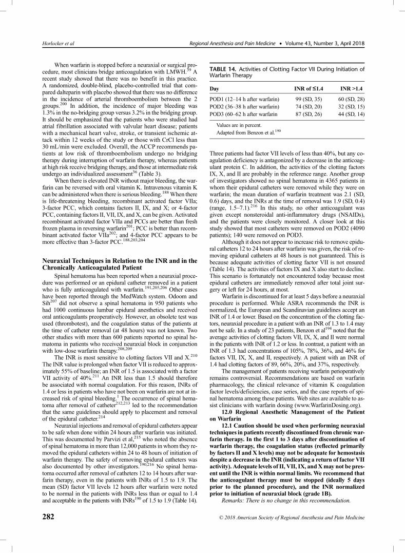
When warfarin is stopped before a neuraxial or surgical pro-cedure, most clinicians bridge anticoagulation with LMWH.39 Arecent study showed that there was no benefit in this practice.A randomized, double-blind, placebo-controlled trial that com-pared dalteparin with placebo showed that there was no differencein the incidence of arterial thromboembolism between the 2groups.200 In addition, the incidence of major bleeding was1.3% in the no-bridging group versus 3.2% in the bridging group.It should be emphasized that the patients who were studied hadatrial fibrillation associated with valvular heart disease; patientswith a mechanical heart valve, stroke, or transient ischemic at-tack within 12 weeks of the study or those with CrCl less than30 mL/min were excluded. Overall, the ACCP recommends pa-tients at low risk of thromboembolism undergo no bridgingtherapy during interruption of warfarin therapy, whereas patientsat high risk receive bridging therapy, and those at intermediate riskundergo an individualized assessment36 (Table 3).
When there is elevated INRwithout major bleeding, the war-farin can be reversed with oral vitamin K. Intravenous vitamin Kcan be administeredwhen there is serious bleeding.188When thereis life-threatening bleeding, recombinant activated factor VIIa;3-factor PCC, which contains factors II, IX, and X; or 4-factorPCC, containing factors II, VII, IX, and X, can be given. Activatedrecombinant activated factor VIIa and PCCs are better than freshfrozen plasma in reversing warfarin201; PCC is better than recom-binant activated factor VIIa202; and 4-factor PCC appears to bemore effective than 3-factor PCC.188,203,204
Neuraxial Techniques in Relation to the INR and in theChronically Anticoagulated Patient
Spinal hematoma has been reported when a neuraxial proce-dure was performed or an epidural catheter removed in a patientwho is fully anticoagulated with warfarin.191,205,206 Other caseshave been reported through the MedWatch system. Odoom andSih207 did not observe a spinal hematoma in 950 patients whohad 1000 continuous lumbar epidural anesthetics and receivedoral anticoagulants preoperatively. However, an obsolete test wasused (thrombotest), and the coagulation status of the patients atthe time of catheter removal (at 48 hours) was not known. Twoother studies with more than 600 patients reported no spinal he-matoma in patients who received neuraxial block in conjunctionwith low-dose warfarin therapy.208,209
The INR is most sensitive to clotting factors VII and X.210
The INR value is prolonged when factor VII is reduced to approx-imately 55% of baseline; an INR of 1.5 is associated with a factorVII activity of 40%.211 An INR less than 1.5 should thereforebe associated with normal coagulation. For this reason, INRs of1.4 or less in patients who have not been on warfarin are not at in-creased risk of spinal bleeding.1 The occurrence of spinal hema-toma after removal of catheter212,213 led to the recommendationthat the same guidelines should apply to placement and removalof the epidural catheter.214
Neuraxial injections and removal of epidural catheters appearto be safe when done within 24 hours after warfarin was initiated.This was documented by Parvizi et al,215 who noted the absenceof spinal hematoma inmore than 12,000 patients inwhom they re-moved the epidural catheters within 24 to 48 hours of initiation ofwarfarin therapy. The safety of removing epidural catheters wasalso documented by other investigators.190,216 No spinal hema-toma occurred after removal of catheters 12 to 14 hours after war-farin therapy, even in the patients with INRs of 1.5 to 1.9. Themean (SD) factor VII levels 12 hours after warfarin were notedto be normal in the patients with INRs less than or equal to 1.4and acceptable in the patients with INRs190 of 1.5 to 1.9 (Table 14).
Three patients had factor VII levels of less than 40%, but any co-agulation deficiency is antagonized by a decrease in the anticoag-ulant protein C. In addition, the activities of the clotting factorsIX, X, and II are probably in the reference range. Another groupof investigators showed no spinal hematoma in 4365 patients inwhom their epidural catheters were removed while they were onwarfarin; the mean duration of warfarin treatment was 2.1 (SD,0.6) days, and the INRs at the time of removal was 1.9 (SD, 0.4)(range, 1.5–7.1).216 In this study, no other anticoagulant wasgiven except nonsteroidal anti-inflammatory drugs (NSAIDs),and the patients were closely monitored. A closer look at thisstudy showed that most catheters were removed on POD2 (4090patients); 140 were removed on POD3.
Although it does not appear to increase risk to remove epidu-ral catheters 12 to 24 hours after warfarin was given, the risk of re-moving epidural catheters at 48 hours is not guaranteed. This isbecause adequate activities of clotting factor VII is not ensured(Table 14). The activities of factors IX and X also start to decline.This scenario is fortunately not encountered today because mostepidural catheters are immediately removed after total joint sur-gery or left for 24 hours, at most.
Warfarin is discontinued for at least 5 days before a neuraxialprocedure is performed. While ASRA recommends the INR isnormalized, the European and Scandinavian guidelines accept anINR of 1.4 or lower. Based on the concentration of the clotting fac-tors, neuraxial procedure in a patient with an INR of 1.3 to 1.4 maynot be safe. In a study of 23 patients, Benzon et al194 noted that theaverage activities of clotting factors VII, IX, X, and II were normalin the patients with INR of 1.2 or less. In contrast, a patient with anINR of 1.3 had concentrations of 105%, 78%, 36%, and 46% forfactors VII, IX, X, and II, respectively. A patient with an INR of1.4 had clotting factors of 89, 66%, 20%, and 37%, respectively.
The management of patients receiving warfarin perioperativelyremains controversial. Recommendations are based on warfarinpharmacology, the clinical relevance of vitamin K coagulationfactor levels/deficiencies, case series, and the case reports of spi-nal hematoma among these patients. Web sites are available to as-sist clinicians with warfarin dosing (www.WarfarinDosing.org).
12.0 Regional Anesthetic Management of the Patienton Warfarin
12.1 Caution should be used when performing neuraxialtechniques in patients recently discontinued from chronic war-farin therapy. In the first 1 to 3 days after discontinuation ofwarfarin therapy, the coagulation status (reflected primarilyby factors II and X levels) may not be adequate for hemostasisdespite a decrease in the INR (indicating a return of factor VIIactivity). Adequate levels of II, VII, IX, andXmay not be pres-ent until the INR is within normal limits. We recommend thatthe anticoagulant therapy must be stopped (ideally 5 daysprior to the planned procedure), and the INR normalizedprior to initiation of neuraxial block (grade 1B).
Remarks: There is no change in this recommendation.
TABLE 14. Activities of Clotting Factor VII During Initiation ofWarfarin Therapy
Day INR of ≤1.4 INR >1.4
POD1 (12–14 h after warfarin) 99 (SD, 35) 60 (SD, 28)POD2 (36–38 h after warfarin) 74 (SD, 20) 32 (SD, 15)POD3 (60–62 h after warfarin 87 (SD, 26) 44 (SD, 14)
Values are in percent.Adapted from Benzon et al.190
Horlocker et al Regional Anesthesia and Pain Medicine • Volume 43, Number 3, April 2018
282 © 2018 American Society of Regional Anesthesia and Pain Medicine

12.2 We recommend against the concurrent use of medi-cations that affect other components of the clotting mecha-nisms and may increase the risk of bleeding complicationsfor patients receiving oral anticoagulants and do so withoutinfluencing the INR. These medications include aspirin andother NSAIDs, thienopyridines, UFH, and LMWH (grade 1A).
Remarks: There is no change in this recommendation.12.3 In patients who are likely to have an enhanced re-
sponse to the drug, we recommend that a reduced dose be ad-ministered (grade 1B).
Remarks: There is no change in this recommendation.12.4 In patients receiving an initial dose of warfarin prior
to surgery, we suggest the INR should be checked prior toneuraxial block if the first dose was given more than 24 hoursearlier or a second dose of oral anticoagulant has been admin-istered (grade 2C).
Remarks: There is no change in this recommendation.12.5 In patients receiving low-dose warfarin therapy dur-
ing epidural analgesia, we suggest that their INR be moni-tored on a daily basis (grade 2C).
Remarks: There is no change in this recommendation.12.6 Neurologic testing of sensory and motor function
should be performed routinely during epidural analgesia forpatients on warfarin therapy. To facilitate neurologic evalua-tion, we recommend that the type of analgesic solution be tai-lored to minimize the degree of sensory and motor blockade(grade 1C).
Remarks: There is no change in this recommendation.12.7 As thromboprophylaxis with warfarin is initiated,
we suggest that neuraxial catheters be removed when theINR is less than 1.5. While removal of epidural catheters 12to 24 hours after warfarin was given does not appear to repre-sent increase risk, the risk of removing epidural catheters at48 hours is not guaranteed.
Remarks: This is a new recommendation based on recentlaboratory studies assessing factor levels and INR as well as clin-ical studies in patients receiving warfarin during epiduralcatheterization.
12.8 In patients with INR of greater than 1.5 but lessthan 3, the increase in risk with progressive INR prolongationremains unknown. We suggest indwelling catheters may bemaintained with caution, based on INR and duration of war-farin therapy (grade 2C).
Remarks: This is a new recommendation based on recentlaboratory studies assessing factor levels and INR as well as clin-ical studies in patients receiving warfarin during epiduralcatheterization.
12.9 In patients with an INR of greater than 3, we recom-mend that the warfarin dose be held or reduced in patientswith indwelling neuraxial catheters (grade 1A). We can makeno definitive recommendation regarding the management tofacilitate removal of neuraxial catheters in patients with ther-apeutic levels of anticoagulation during neuraxial catheter in-fusion (grade 2C).
Remarks: There is no change in this recommendation.12.10 We suggest that neurologic assessment be continued
for at least 24 hours following catheter removal (grade 2C).Remarks: There is no change in this recommendation.
Antiplatelet MedicationsAntiplatelet agents include NSAIDs, thienopyridine derivatives/
platelet ADP antagonists (ticlopidine, clopidogrel, prasugrel),platelet glycoprotein (GP) IIb/IIIa receptor antagonists (abciximab,eptifibatide, and tirofiban), platelet P2Y12 receptor antagonists
(ticagrelor), and platelet phosphodiesterase (PDE) IIIA inhibitors(cilostazol). It is important to note the pharmacologic differencesamong the drugs with antiplatelet effects.
Nonsteroidal Anti-inflammatory DrugsCyclooxygenase (COX) exists in 2 forms. Cyclooxygenase 1
regulates constitutive mechanisms, whereas COX-2mediates painand inflammation. Nonsteroidal anti-inflammatory drugs inhibitplatelet COX and prevent the synthesis of thromboxane A2. Plate-lets from patients who have been taking these medications havenormal platelet adherence to subendothelium and normal primaryhemostatic plug formation. Depending on the dose administered,aspirin (and other NSAIDs) may produce opposing effectson the hemostatic mechanism. For example, platelet COX isinhibited by low-dose aspirin (60–325 mg/d), whereas largerdoses (1.5–2 g/d) will also inhibit the production of prostacyclin(a potent vasodilator and platelet aggregation inhibitor) by vascu-lar endothelial cells and thus result in a paradoxical thrombogeniceffect.217,218 As a result, low-dose aspirin (81–325 mg/d) is theo-retically a greater risk factor for bleeding than higher doses. Thereis consensus that the optimal dose of aspirin for prevention ofmyocardial infarction, stroke, or vascular death lies within the nar-row range of 75 to 160 mg/d.34 Spontaneous219 and postoperative(unrelated to neuraxial technique)220 spinal hematomas have beenreported with low-dose aspirin therapy.
Platelet function is affected for the life of the platelet follow-ing aspirin ingestion; other nonsteroidal analgesics produce ashort-term defect, which normalizes within 3 days; for short-acting NSAIDS such as ibuprofen, diclofenac, and indomethacin,the effect on platelet aggregation is normalized after 24 hours, andmore than 50% is restored after 6 hours.221
Celecoxib (Celebrex) is an anti-inflammatory agent that pri-marily inhibits COX-2, an inducible enzyme that is not expressedin platelets and thus does not cause platelet dysfunction.222 Aftersingle dosing and multidosing, there have not been findings ofsignificant disruption of platelet aggregation; nor is there a historyof undesirable bleeding events.
ThienopyridinesThe antiplatelet effect of the thienopyridine derivatives,
ticlopidine (Ticlid), clopidogrel (Plavix), and prasugrel (Effient)results from inhibition of ADP-induced platelet aggregation.These antiplatelet agents, used in the prevention of cerebrovascu-lar thromboembolic events, affect both primary and secondaryplatelet aggregation. Thienopyridines also interfere with platelet-fibrinogen binding and subsequent platelet-platelet interactions.223
All 3 are prodrugs that must undergo metabolic activation throughthe hepatic CYP450 system to generate the active metabolites thatinhibit the platelet P2Y12 receptor.34 Thienopyridine derivativesdemonstrate both time- and dose-dependent effects. For example,steady state is achievedwithin 7 days for clopidogrelwith doses of75 mg/d. However, steady-state levels of clopidogrel are reachedwithin 2 to 15 hours with 300- to 600-mg loading doses.39,224
Prasugrel is typically administered with a loading dose that resultsin 50% of platelet aggregation within 1 hour of administration(prasugrel [full prescribing information], available at http://uspl.lilly.com/effient/effient.html#pi, accessed March 4, 2017). Com-pared with clopidogrel, prasugrel exhibits a more rapid onset ofaction, a higher potency, and more consistent antiplatelet effects.It is also associated with a higher frequency of major bleeding.34
Although often administered in combination with aspirin,the concomitant use of thienopyridines and aspirin may be associ-ated with increased risk of hemorrhagic events. Serious hemato-logic adverse reactions, including agranulocytosis, thrombotic
Regional Anesthesia and Pain Medicine • Volume 43, Number 3, April 2018 Regional Anesthesia and Anticoagulation
© 2018 American Society of Regional Anesthesia and Pain Medicine 283

thrombocytopenic purpura, and aplastic anemia, have resultedin placement of a black box warning on ticlopidine. Because ofthese toxicities, ticlopidine has been essentially replaced withclopidogrel and prasugrel.34
Labeling of the thienopyridine derivatives recommends thatif a patient is to undergo an elective procedure and an antiplateleteffect is not desired, ticlopidine should be discontinued for 10 to14 days (ticlopidine [full prescribing information], available athttp://www.accessdata.fda.gov/drugsatfda_docs/nda/2000/19979-s019_ticlid.pdf, accessed March 4, 2017). Clopidogrel should bediscontinued 5 days (clopidogrel [full prescribing information],available at http://packageinserts.bms.com/pi/pi_plavix.pdf, accessedMarch 4, 2017), and prasugrel discontinued 7 days (prasugrel [fullprescribing information], available at http://uspl.lilly.com/effient/effient.html#pi, accessed March 4, 2017), prior to surgery. TheACCP recommends discontinuation of clopidogrel and prasugrelfor 5 days (Table 4).36 Although it is possible to assess residualclopidogrel effect using assays of platelet function (eg, PFA II,P2Y12 assay), only a normal result would be reassuring.225 Thus,the clinical significance of assay findings is uncertain, and the as-say results have not been shown to predict clinical outcomes, andthe clinical applicability of these tests remains undetermined atthis time.36 The potency of these medications is demonstratedby recent reports of spontaneous spinal hematomas duringclopidogrel therapy.226–228
TicagrelorTicagrelor (Brilinta) represents a new class of non-
thienopyridine platelet inhibitors designed to address the limita-tions of current oral platelet drugs. Ticagrelor completely butreversibly inhibits ADP-induced platelet activation, unlike thethienopyridines (eg, clopidogrel, prasugrel). Ticagrelor also actsdirectly on the P2Y12 receptor and does not require CYP biotrans-formation. After a loading dose, an antiplatelet effect is observedwithin 30 minutes, whereas maximum effect is achieved within2 hours. Ticagrelor has been studied in acute coronary syndromein combination with aspirin. Maintenance doses of aspirin greaterthan 100 mg decrease the effectiveness. In theory, the reversi-ble antiplatelet effect may improve the perioperative safety ofticagrelor relative to the other agents. However, medication label-ing recommends that when possible ticagrelor should “bediscontinued at least 5 days prior to any surgery” (ticagrelor [fullprescribing information], available at http://www.azpicentral.com/brilinta/brilinta.pdf#page=1, accessed March 4, 2017).
In Europe, labeling of clopidogrel, prasugrel, and ticagrelorrecommends that they all be discontinued 7 days prior to electivesurgery. Hence, a somewhat longer interval is typically advised in“European” recommendations.8,25,97,229
CangrelorCangrelor (Kengreal) is a direct and reversible IV P2Y12 in-
hibitor. It is indicated as an adjunct to percutaneous coronary in-tervention (PCI) to reduce the risk of periprocedural myocardialinfarction, repeat coronary revascularization, and stent thrombosisin patients who have not been treated with another P2Y12 inhibi-tor and are not being given a GP IIb/IIa inhibitor.
The dosage of the drug is 30-μg/kg bolus followed by an in-fusion 4 μg/kg per minute. Its onset is rapid, and its offset short. Itsantiplatelet effect is seen within 2 minutes of administration andinhibits platelet aggregation by 95% to 100%.230 Its plasmahalf-life is 3 to 6 minutes, and platelet recovery is quick; 80%and 90% of the samples recover in 60 and 90 minutes, respec-tively.231 The adverse effects of cangrelor include bleeding, dys-pnea, and decreased renal function. The dyspnea is seen with
ticagrelor and cangrelor and may be related to the drug-inducedaccumulation of adenosine with inhibition of the P2Y12 effecton sensory neurons and stimulation of the adenosine receptorson bronchopulmonary vagal fibers. Although there is no clear ev-idence of acute renal toxicity, animal studies suggested thatcangrelor may injure renal tubules.
Two earlier studies on the efficacy of cangrelor, theCHAMPION PLATFORM and CHAMPION PCI trials, were notsupportive. However, the CHAMPION PHOENIX trial, whereinthe definition of myocardial infarction was based on the universaldefinition, showed superiority of the drug over clopidogrel in termsof reduction of periprocedural myocardial infarction with no differ-ence in the incidence of severe bleeding between the 2 drugs.232 Pa-tients given cangrelor for PCI are usually continued on one of theoral P2Y12 inhibitors. Both clopidogrel and prasugrel will not workwhile the cangrelor is being infused because their metabolite cannotbind to the receptor while it is being occupied by cangrelor. Ticagrelor,on the other hand, has a binding site separate from cangrelor. Forthese reasons, clopidogrel and prasugrel should be given (imme-diately) after discontinuation of cangrelor, whereas ticagrelor canbe given during or immediately after the infusion.230 The recom-mended loading doses are 600 mg for clopidogrel, 60 mg forprasugrel, and 180 mg for ticagrelor.
The oral P2Y12 inhibitors need to be stopped for 5 to 10 daysbefore surgery. Cangrelor can therefore be used as a bridge ther-apy in these situations. A study showed efficacy of cangrelor overplacebo; the incidence of greater than 60% platelet inhibition wasmore than 80% of the patients on cangrelor versus 19% for pla-cebo, and therewas no significant difference in major bleeding.233
It is possible that perioperative anesthesiologists will encounterthis scenario more often in the future. In these cases, a minimumof 3-hour interval, preferably longer, should observed.
Platelet GP IIb/IIIa Receptor AntagonistsPlatelet GP IIb/IIIa receptor antagonists, including abciximab
(Reopro), eptifibatide (Integrilin), and tirofiban (Aggrastat), inhibitplatelet aggregation by interfering with platelet-fibrinogen andplatelet–von Willebrand factor binding. Because fibrinogen andvon Willebrand factor have multiple binding sites, they can bindto multiple platelets, causing cross-linking and platelet aggrega-tion. Conversely, inhibition of GP IIb/IIIa receptors blocks the fi-nal common pathway to platelet aggregation.223 The majority ofclinical trials involving the GP IIb/IIIa antagonists have evaluatedtheir use in the treatment of acute coronary syndrome (with orwithout PCI). Importantly, the GP IIb/IIIa antagonists are typi-cally administered in combination with aspirin and heparin. Con-traindications include a history of surgery within 4 to 6 weeks.Time to normal platelet aggregation following discontinuationof therapy ranges from 8 hours (eptifibatide, tirofiban) to 24 to48 hours (abciximab).34 Thrombocytopenia is a frequent adverseeffect.34 Platelet counts usually recover upon discontinuation oftherapy. During therapy with GP IIb/IIIa antagonists, labeling pre-cautions recommend that needle puncture sites should be mini-mized and monitored (abciximab [full prescribing information],available at https://www.drugs.com/pro/reopro.html, accessedMarch 4, 2017).
CilostazolCilostazol (Pletal) produces a selective inhibition of PDE
IIIA resulting in a reversible inhibition of platelet aggregation.Cilostazol is used in peripheral arterial vascular disease becauseof its vasodilatory properties (vascular muscle also containsPDE IIIA). It has a half-life of 11 hours, which is prolonged in pa-tients with severe renal impairment.34 The terminal half-life of
Horlocker et al Regional Anesthesia and Pain Medicine • Volume 43, Number 3, April 2018
284 © 2018 American Society of Regional Anesthesia and Pain Medicine

active metabolites is 21 hours. There are limited data on perioper-ative administration of cilostazol. However, a single case report ofspinal hematoma following epidural catheter removal in the pres-ence of cilostazol therapy has been reported.234 The ESA recom-mendations include maintaining a time interval of 2 eliminationhalf-lives (42 hours) after the last dose of cilostazol prior toneuraxial blockade, and the next dose administered at least 5 hoursafter neuraxial catheter manipulation/removal.8
DipyridamoleDipyridamole is a pyrimidopyrimidine derivative with vaso-
dilator and antiplatelet properties. The mechanism of action ofdipyridamole as an antiplatelet agent is controversial. Historically,the efficacy of immediate-release dipyridamole (alone or in com-bination with aspirin) was not established. However, a newextended-release formulation appears to be more efficacious thanaspirin in the prevention of stroke. However, the combination ofaspirin and extended-release dipyridamole is associated with morebleeding complications than clopidogrel.34,235 The terminal half-life is 10 hours. There are no data to support discontinuationof dipyridamole perioperatively. However, the combination ofdipyridamole and aspirin may increase the risk of bleeding. Al-though there are no spinal hematomas associated with neuraxialblock and dipyridamole, as will be noted in the upcoming sectionon plexus and peripheral block, 2 serious hematomas were re-ported in patients who underwent continuous interscalene236 orilioinguinal/iliohypogastric block237 while receiving dipyridamole.
Spinal Hematoma in Patients ReceivingAntiplatelet Medications
At the previous ASRAConsensus Conferences on NeuraxialAnesthesia and Anticoagulation, it was concluded NSAIDs didnot appear to present significant risk to patients for developingspinal-epidural hematomas for patients undergoing neuraxial tech-niques for surgery (nonchronic pain) procedures.3,6,7 Vandermeulenet al43 implicated antiplatelet therapy in 3 of the 61 cases of spinalhematoma occurring after spinal or epidural anesthesia. These pa-tients had received aspirin, indomethacin, or ticlopidine. Four ad-ditional case reports related to neuraxial techniques have beenpublished, 2 involving ketorolac and 2 involving a thienopyridinederivative.238–241 The paucity of case reports is important, giventhe prevalence of NSAID use among the general population.
Several large studies have demonstrated the relative safety ofcentral neural blockade in combination with NSAID therapy, al-though the total number of patients in this combined series is only4714.6 If low-dose aspirin creates the greatest impact on plateletfunction, patients receiving 60 to 325 mg aspirin would theoreti-cally represent the greatest risk of significant bleeding. However,the CLASP (Collaborative Low-Dose Aspirin Study in Preg-nancy) Group included 1422 high-risk obstetric patients, approx-imately 700 of whom were administered 60 mg aspirin dailywho underwent epidural anesthesia without any neurologic se-quelae.242 A recent prospective study evaluated the risk of neuro-logic complications following ESI. There were no spinal hematomasamong the 1214 patients, including the 32% of patients who re-ported NSAID use prior to injection.243 These results confirmthose of previous studies performed in obstetric and surgical pop-ulations. An exception to this are patients undergoing invasivepain procedures, among whom 3 spinal hematomas have been re-ported following spinal cord stimulator lead placement whilereceiving aspirin or other NSAIDs.16–18 The differences in thepain and surgical populations and their associated risks of signif-icant spinal bleeding will be discussed later.
There are minimal published data purporting the safety ofneuraxial blockade in the presence of thienopyridine derivativesor platelet GP IIb/IIIa receptor antagonists.244 Although the dataare inconsistent, increased perioperative bleeding in patientsundergoing cardiac and vascular surgery after receiving thieno-pyridines and GP IIb/IIIa antagonists has been noted.36,245,246 Ingeneral, the cardiac surgical246 and interventional radiology liter-ature recommends that elective surgery be delayed 24 to 48 hoursfollowing abciximab and 4 to 8 hours following eptifibatide ortirofiban.247 Surgery performed within 12 hours of abciximabadministration will most likely necessitate a platelet transfusion;After transfusion, the antibody redistributes to the transfusedplatelets, reducing the mean level of receptor blockade and im-proving platelet function. However, subsequent platelet transfu-sion may be necessary. There have been 3 spinal hematomasattributed to neuraxial techniques and ticlopidine or clopidogrel,including 1 patient undergoing a series of ESIs.238,241,248
Combination of Antiplatelet Medications WithAnticoagulants and Thrombolytics
Nonsteroidal anti-inflammatory drugs alone do not signifi-cantly increase the risk of spinal hematoma. However, combina-tion therapy with UFH, LMWH, oral anticoagulants, andthrombolytics has been demonstrated to increase the frequencyof spontaneous hemorrhagic complications, bleeding at puncturesites, and spinal hematoma.2,50,79 For example, in the series of40 spinal hematomas associated with LMWH reported in 1998,10 patients received concomitant antiplatelet medications.2 Theaddition of antiplatelet therapy to postoperative thromboprophylaxiswas implicated in a similar number of cases in the survey by Moenet al.42 Likewise, in a case report of spinal hematoma following ESI,Benzon et al238 noted the patient had received multiple antiplateletmedications, including clopidogrel and aspirin.
13.0 Anesthetic Management of the Patient ReceivingAntiplatelet Medications
Antiplatelet medications exert diverse effects on plateletfunction. The pharmacologic differences make it impossibleto extrapolate between the groups of drugs regarding thepractice of neuraxial techniques. Careful preoperative assess-ment of the patient to identify alterations of health that mightcontribute to bleeding is crucial. These conditions include ahistory of easy bruising/excessive bleeding, female sex, andincreased age.
13.1 Nonsteroidal anti-inflammatory drugs13.1.1 Nonsteroidal anti-inflammatory drugs appear to
represent no added significant risk of the development of spinalhematoma in patients having epidural or spinal anesthesia.Nonsteroidal anti-inflammatory drugs (including aspirin) donot create a level of risk that will interfere with the perfor-mance of neuraxial blocks. In patients receiving these medica-tions, we do not identify specific concerns as to the timing ofsingle-injection or catheter techniques in relationship to thedosing of NSAIDs, postoperative monitoring, or the timingof neuraxial catheter removal (grade 1A).
Remarks: There is no change in this recommendation.13.1.2 In patients receiving NSAIDS, we suggest caution
in the performance of neuraxial techniques if the concurrentuse of other medications affecting clotting mechanisms, suchas other (non-NSAID) antiplatelet agents, oral anticoagulants,UFH, andLMWH, is anticipated in the early postoperative periodbecause of the increased risk of bleeding complications. Cyclooxy-genase 2 inhibitors have minimal effect on platelet function andshould be considered in patients who require anti-inflammatorytherapy in the presence of anticoagulation (grade 2C).
Remarks: There is no change in this recommendation.
Regional Anesthesia and Pain Medicine • Volume 43, Number 3, April 2018 Regional Anesthesia and Anticoagulation
© 2018 American Society of Regional Anesthesia and Pain Medicine 285

13.2 Thienopyridines (ticlopidine, clopidogrel, prasugrel)13.2.1 Preoperative. Based on labeling and surgical/
procedural experience, the recommended time interval be-tween discontinuation of thienopyridine therapy and neuraxialblockade is 10 days for ticlopidine, 5 to 7 days for clopidogrel,and 7 to 10 days for prasugrel (grade 1C).
Remarks: Previous time intervals were 14 days for ticlopidineand 7 days for clopidogrel. The new time intervals reflect ACCPrecommendations. The recommendation for prasugrel is new.
13.2.2 Postoperative. In accordance with ACCP recom-mendations, thienopyridine therapymay be reinstituted 24 hourspostoperatively (grade 1A).
Remarks: This is a new recommendation.13.2.3 Neuraxial catheters should not be maintainedwith
prasugrel or ticagrelor because of the rapid onset. However,because the antiplatelet effect is not immediate with ticlopidineand clopidogrel, neuraxial cathetersmay bemaintained for 1 to2 days, provided a loading dose of the antiplatelet agent is notadministered (grade 2C).
Remarks: This is a new recommendation.13.2.4 Thienopyridine therapy may be resumed immedi-
ately after needle placement/catheter removal, provided aloading dose of the drugs is not administered. If a loading doseis administered, we suggest a time interval of 6 hours betweencatheter removal and administration (grade 2C).
Remarks: This is a new recommendation.13.4 Ticagrelor13.4.1 Preoperative. Based on labeling and surgical/
procedural experience, the recommended time interval betweendiscontinuation of ticagrelor therapy is 5 to 7 days (grade 1C).
Remarks: This is a new recommendation.13.4.2 Postoperative. In accordance with ACCP recom-
mendations, ticagrelor therapy may be reinstituted 24 hourspostoperatively (grade 1A).
Remarks: This is a new recommendation.13.4.3 Neuraxial catheters should not be maintainedwith
ticagrelor because of the rapid onset (grade 2C).Remarks: This is a new recommendation.13.4.4 Ticagrelor therapy may be resumed immediately
after needle placement/catheter removal, provided a loadingdose of the drugs is not administered. If a loading dose is ad-ministered, we suggest a time interval of 6 hours between cath-eter removal and administration (grade 2C).
Remarks: This is a new recommendation.13.5 Platelet GP IIb/IIa. The platelet GP IIb/IIIa inhibi-
tors exert a profound effect on platelet aggregation. Followingadministration, the time to normal platelet aggregation is 24to 48 hours for abciximab and 4 to 8 hours for eptifibatideand tirofiban.
13.5.1 Preoperative. We recommend that neuraxial tech-niques should be avoided until platelet function—as impactedby the GP IIb/IIIa inhibitor—has recovered. (Patients are typi-cally on dual therapy andmay still have residual NSAID effect.)
Remarks: There is no change to this recommendation.13.5.2 Postoperative. Although GP IIb/IIIa antagonists
are contraindicated within 4 weeks of surgery, should one beemergently administered in the postoperative period (followinga neuraxial technique), we recommend the infusion should belimited to drugs minimizing sensory and motor block to facili-tate assessment of neurologic function and that the patient becarefully monitored neurologically (grade 1C). Timing of cath-eter removal is based on ongoing risk of thromboembolism andneed for continued antithrombotic therapy and the potentialfor spinal bleeding during catheter maintenance and removal.
Remarks: This is a new recommendation.
13.6 Cilostazol. The risk of serious bleeding associatedwith neuraxial block performed ormaintained in the presenceof residual cilostazol effect is unknown.
13.6.1 Based on the elimination half-life, we suggest thatneuraxial techniques be avoided for 2 days after discontinua-tion of cilostazol (grade 2C).
Remarks: This is a new recommendation.13.6.2 We suggest that neuraxial catheters be removed
prior to reinstitution of cilostazol therapy postoperatively(grade 2c).
Remarks: This is a new recommendation.13.6.3 We suggest that the first postoperative dose of
cilostazol be administered 6 hours after neuraxial catheter re-moval (grade 2C).
Remarks: This is a new recommendation.13.7 Dipyridamole. The risk of dipyridamole in combina-
tion with aspirin therapy may represent an increased risk.13.7.1 Based on the elimination half-life, we suggest
discontinuing extended-release dipyridamole for 24 hours priorto neuraxial block. Aspirin may be continued perioperatively(grade 2C).
Remarks: This is a new recommendation.13.7.2 We suggest that neuraxial catheters be removed
prior to reinstitution of dipyridamole therapy postoperatively(grade 2C).
Remarks: This is a new recommendation.13.7.3 We suggest that the first postoperative dose of
dipyridamole be administered 6 hours after neuraxial cathe-ter removal (grade 2C).
Remarks: This is a new recommendation.13.8 Cangrelor. The risk of serious bleeding associated
with neuraxial block performed ormaintained in the presenceof residual cangrelor effect is unknown.
13.8.1 Based on the elimination half-life, we suggest thatneuraxial techniques be avoided for 3 hours after discontinu-ation of cangrelor (grade 2C).
Remarks: This is a new recommendation.13.8.2We suggest that neuraxial catheters be removed prior
to reinstitution of cangrelor therapy postoperatively (grade 2C).Remarks: This is a new recommendation.13.8.3 We suggest that the first postoperative dose of
cangrelor be administered 8 hours after neuraxial catheter re-moval (grade 2C).
Remarks: This is a new recommendation.
Herbal MedicationsThere is a widespread use of herbal medications in surgical
patients. Most patients do not volunteer information regarding herbalmedication use; obtaining such a history may be difficult.249–251
Morbidity and mortality associated with herbal use may be morelikely in the perioperative period because of the polypharmacyand physiological alterations that occur. Such complications in-clude bleeding from garlic, ginkgo and ginseng, and potentialinteraction between ginseng-warfarin3 (Table 15).
There have been case reports of spontaneous neuraxial bleed-ing following ingestion of garlic252 and ginkgo biloba.253–256
Despite the widespread use of herbal medications, there arefew controlled clinical trials of the efficacy (or adverse effects)and few outcome studies of the effects of herbal medicationson surgical patients; a prospective study including more than600 patients found no differences in surgical outcomes, includingbleeding, in patients reporting recent herbal therapy.257 However,although overall there does not appear to be a clinically significantincrease in surgical bleeding or spinal hematoma in patients
Horlocker et al Regional Anesthesia and Pain Medicine • Volume 43, Number 3, April 2018
286 © 2018 American Society of Regional Anesthesia and Pain Medicine

receiving herbal medications, data on the combination of herbaltherapy with other forms of anticoagulation are lacking. The con-current use of other medications affecting clotting mechanisms,such as oral anticoagulants or heparin, may increase the risk ofbleeding complications in these patients. Thus, it is often recom-mended that these medications be discontinued in anticipation ofsurgery, but there is no reason for cancellation of the procedureif patients have not done so.258
14.0 Anesthetic Management of the Patient ReceivingHerbal Therapy
14.1 The use of herbal medications does not create a levelof risk that will interfere with the performance of neuraxialblock. We recommend against the mandatory discontinuationof these medications or avoidance of regional anesthetic tech-niques in patients in whom these medications have been ad-ministered (grade 1C).
Remarks: There is no change in this recommendation.
Antithrombotic Therapy and PregnancyVenous thromboembolism is one of the most common
causes of maternal morbidity and mortality, especially in high-resource countries.13 Pharmacologic thromboprophylaxis hasbeen shown to significantly decrease morbidity, but possibly atthe cost of an increased incidence of bleeding.12,259 Common riskfactors that increase the incidence of thrombosis in pregnantwomen include increasing age, prolonged immobilization, obe-sity, thrombophilia, previous thromboembolism, and cesareandelivery.10,14 The puerperium, defined as the 6-week period fol-lowing delivery, is associated with a higher rate of thrombosisand PE than that associated with pregnancy itself.260,261
Although there is an increased risk of thrombosis duringnormal pregnancy, in the majority of healthy women with un-complicated pregnancies and vaginal deliveries, the benefits ofthromboprophylaxis do not outweigh the maternal and fetal risks.However, for maternal conditions such as acquired or inheritedthrombophilia and for thosewomen on extended bed rest, the ben-efits of thromboprophylaxis may outweigh the risks. The use ofanticoagulation for prevention of thromboembolism in patientsis becoming more frequent. However, the thromboprophylaxisrecommendations from the ACCP,14 the American College ofObstetricians and Gynecologists (ACOG),262 and the Royal
College of Obstetricians and Gynaecologists259 differ noticeably10
(Table 16). A recent review of post–cesarean delivery patientsfound that 1%, 35%, and 85% of patients would receive pharma-cologic prophylaxis under the ACOG, ACCP, and Royal Collegeof Obstetricians and Gynaecologists criteria, respectfully.11,263
In an effort to reduce the frequency of obstetric thromboem-bolism and improve maternal outcomes, the NPMS, a multidisci-plinary working group representing all major women's health careprofessional organizations, has recently published a ConsensusBundle on Venous Thromboembolism.13 The bundle advocatesfor more aggressive risk-based assessment of obstetric patients,which will ultimately result in not only an increased use, but alsolikely increased doses of antithrombotic agents. This will increasethe likelihood that pregnant women will be anticoagulated whenpresenting in labor (requesting analgesia) or for an urgent, non-elective cesarean delivery and ultimately impact on the choice ofanalgesia or anesthesia that is offered. It will be critical that proac-tive, multidisciplinary communication occur to help determine theappropriate anticoagulant doses and stop times based on the risksand benefits of VTE and neuraxial analgesia/anesthesia.
Physiologic Changes in Obstetric PatientsAffecting Thromboprophylaxis
Physiological changes during pregnancy result in changes inthe volume of distribution, clearance, bioavailability, and metabo-lism of many drugs.264 Available data from studies examining thepharmacokinetics and pharmacodynamics of UFH suggest that,compared with nonpregnant patients, aPTT response and durationof action of UFH may be decreased in obstetric patients.265,266
Similarly, for LMWH, peak anti–factor Xa levels, duration of ac-tion, and the total exposure to the drug over time (area under theplasma activity vs time curve) are lower in obstetric patients versusnonpregnant or postpartum patients.265,267,268 These changes areprimarily due to increased volume of distribution and clearance as-sociated with pregnancy andmay lead to both decreased peak effectand lower plasma concentrations over time after LMWH dosing.
Spinal Hematoma in the Obstetric PatientThe incidence of spinal hematoma after neuraxial blockade
(with or without altered hemostasis) in the obstetric patient is very
TABLE 15. Three Herbal Medications With the Greatest Impact on Hemostasis*
Herb Important Effects Perioperative ConcernsTime to Normal Hemostasis
After Discontinuation
Garlic Inhibition of platelet aggregation(may be irreversible)
Potential to increase bleeding, especially whencombined with other medications that inhibitplatelet aggregation
7 d
Increased fibrinolysisEquivocal antihypertensive activity
Ginkgo Inhibition of platelet-activating factor Potential to increase bleeding, especially whencombined with other medications that inhibitplatelet aggregation
36 h
Ginseng Lowers blood glucose Hypoglycemia 24 hPotential to increase risk of bleedingIncreased prothrombin and activated partial
PTs in animals Potential to decrease anticoagulant effectof warfarinOther diverse effects
*At this time, it is not deemed necessary to discontinue herbal medications and allow resolution of their effects on hemostasis prior to surgery oranesthesia.
Adapted from Horlocker et al,3 with permission.
Regional Anesthesia and Pain Medicine • Volume 43, Number 3, April 2018 Regional Anesthesia and Anticoagulation
© 2018 American Society of Regional Anesthesia and Pain Medicine 287

difficult to determine, although it is widely reported these patientshave a significantly lower incidence of complications than their el-derly counterparts.42,269 Moen et al42 reported 2 spinal hemato-mas among 200,000 epidural blocks for pain relief in labor: 1after a subarachnoid block and 1 following the removal of an epi-dural catheter. The authors reported the incidence of spinal hema-toma after obstetric epidural blockade was 1:200,000, which wassignificantly lower than the incidence of 1:3600 elderly femalesundergoing total knee arthroplasty. Bateman et al270 confirm thefindings of Moen et al42 that obstetric patients undergoing epidu-ral catheterization are at significantly lower risk of spinal hema-toma compared with perioperative (nonobstetric surgical) patientsundergoing epidural catheterization. There were 7 epidural hema-tomas among 142,287 patients undergoing epidural anesthesia/analgesia, for an overall risk of 1 in 20,326 epidural catheteriza-tions. However, none of the 79,837 obstetric patients who under-went epidural placement developed a neuraxial hematoma requiringdecompressive laminectomy (upper limit of 95% confidence in-terval, 1:21,643), compared with 7 of 62,450 patients who re-ceived perioperative epidural catheter placement (1:9000; 95%confidence interval, 1:22,189–1:4330). This difference in incidenceis even more striking when considering that bloody taps are morecommon in the obstetric population and have been reported to occurin approximately 3% of all obstetric epidural placements.271
The relatively hypercoagulable state of pregnancy may beprotective and offers 1 possible reason for the lower rate ofneuraxial hematomas in this population. The normal anatomicchanges that occur in the aging spine may provide another expla-nation for the differing incidence. Both Moen et al42 and Pöppinget al272 cited osteoporotic deformities as likely contributing to therisk of symptomatic vertebral canal bleeding after epidural block-ade in elderly women. The prevalence of vascular disease, osteo-porosis, and degenerative changes of the spine increases withage, ultimately resulting in a decrease in the volume of the epidu-ral space. In contrast, the younger obstetric patient has a morecompliant epidural space, with the ability to accommodate a largervolume of blood before the onset of symptoms. Magnetic reso-nance imaging (MRI) after epidural blood patch revealed bloodleaking out through the intravertebral foramina after injection inyoung individuals.273
Until 2011, all case reports of neuraxial hematomas in obstet-ric patients with altered hemostasis have occurred in parturientswith a preexisting coagulopathy (eg, thrombocytopenia, hemor-rhage, preeclampsia, thrombocytopenia, HELLP) at the time ofeither epidural placement or removal42,47,269,274–284 (Table 17).There are 2 recent cases of spinal hematoma involving LMWHand neuraxial anesthesia in obstetric patients: in one, the patientreceived therapeutic anticoagulation (enoxaparin 1 mg/kg BID)for a postpartum pulmonary embolus 3 days after a cesarean deliv-ery performed with a spinal catheter.286 In the second case, a post-partum patient developed a spinal hematoma after a combinedspinal-epidural (CSE) for cesarean delivery and enoxaparin thrombo-prophylaxis, but she had signs and symptoms of an expandingspinal hematoma before receiving the LMWH dose.285
Neuraxial and General Anesthesia in theObstetric Patient
Neuraxial block is particularly vital to the care of the obstet-ric patient compared with alternative modes of pain management.For labor, neuraxial analgesia provides superior pain relief to otheranalgesic modalities and decreases circulating catecholaminelevels, which may be particularly beneficial for patients with hy-pertensive disorders of pregnancy or preexisting comorbidities.Likewise, for cesarean delivery, neuraxial anesthesia has manymaternal287–299 and fetal298–302 benefits compared with generalanesthesia, including decreased risk of pulmonary complicationsawareness under anesthesia and surgical infections, as well as en-abling immediate postdelivery mother-infant bonding (Table 18).
Obstetric and Anesthetic ManagementThe peripartummanagement of the anticoagulated parturient
represents a significant clinical challenge to both the obstetricianand the anesthesiologist. The ACCP14 and the NPMS13 recom-mendations support proactive measures in the obstetric patient suchas (1) discontinuing anticoagulant therapy upon the onset of labor,and per the NPMS, (2) using 5000 SC UFH BID for thrombo-prophylaxis in antepartum inpatients near delivery to facilitateneuraxial analgesia and anesthesia when feasible (Table 19). Inthe event of unforeseen labor or urgent cesarean delivery, the
TABLE 16. Suggested Dosing Regimens for Prophylaxis Against Pregnancy-Related VTE
ACOG262 ACCP14 NPMS13
Prophylactic UFH 5000–10,000 U every 12 h 5000 U every 12 h • 5000–7500 U every 12 h in 1st trimester• 5000–7500 U every 12 h in 1st trimester • 7500–10,000 U every 12 h in 2nd trimester
• 10,000 U every 12 h in the 3rd trimester• 7500–10,000 U every 12 h in 2nd trimester
Hospitalized antepartum patients may receive5000 U every 12 h
• 10,000 U every 12 h in the 3rd trimesterunless aPTT is elevated
Minidose: 5000 U every 12 hProphylactic LMWH Dalteparin 5000 U once daily Dalteparin 5000 U once daily Dalteparin 5000 U once daily
Tinzaparin 4500 U once daily Tinzaparin 4500 U once daily Tinzaparin 4500 U once dailyEnoxaparin 40 mg once daily Enoxaparin 40 mg once daily Enoxaparin 40 mg once dailyAt extremes of body weight, modification
of dose may be requiredAt extremes of body weight,
modification of dose may be requiredAt extremes of body weight, modification
of dose may be requiredTherapeutic UFH ≥10,000 U every 12 h in doses adjusted
to target aPTT in the therapeutic rangeEvery 12-h dose adjusted to target a
midinterval aPTT in thetherapeutic range
≥10,000 U BID adjusted to midinterval aPTT
Therapeutic-doseLMWH*
Dalteparin 200 U/kg QD or dalteparin100 U/kg every 12 h
Dalteparin 200 U/kg QD or 100 U/kgevery 12 h
Dalteparin 200 U/kg QD
Tinzaparin 175 U/kg QD Tinzaparin 175 U/kg QD Tinzaparin 175 U/kg QDEnoxaparin 1 mg/kg every 12 h Enoxaparin 1 mg/kg every 12 h Enoxaparin 1 mg/kg every 12 h
*LMWH adjusted to a peak anti–factor Xa level of 0.5 to 1.0 U/mL (measured 3–4 hours after dose) or trough levels of 0.2 to 0.4 U/mL (measured12 hours after dose). Anti–factor Xa assay must be calibrated to specific LMWH.
Horlocker et al Regional Anesthesia and Pain Medicine • Volume 43, Number 3, April 2018
288 © 2018 American Society of Regional Anesthesia and Pain Medicine

TABLE 17. Spinal Hematomas in Obstetric Patients
Author Year Technique Coagulopathy Outcome Miscellaneous
Ballin275 1981 Epidural None Recovered • Spinal stenosisNewman278 1983 Epidural None Minimal weakness
and paresthesia• Presented 2 h after delivery
Roscoe and Barrington280 1984 Epidural None Residual leg weakness • Epidural ependymoma• Presented 3 d postpartum• Surgical treatment
Crawford269 1985 Epidural Unknown Recovered • Presented several weeks postpartumSibai et al282 1986 Epidural Thrombocytopenia Unknown • No informationScott and Hibbard281 1990 Epidural Not reported Improving • Surgical treatmentLao et al277 1993 Epidural Preeclampsia and lupus
anticoagulantResidual urinary andbowel dysfunction
• Presented 1 d postpartum• Surgical treatment
Abnormal aPTTYarnell and D'Alton283 1996 Epidural Elevated aPTT Mild weakness of
right leg• Presented 12 h after epidural
Elevated PT • Surgical treatment
Yuen et al284 1999 Epidural Severe pre-eclampsia Recovered • Presented within hours• Surgical laminectomyThrombocytopenia
Esler et al276 2001 Epidural None Recovered • Neurofibromatosis• Presented on 2nd postpartum day
Moen et al42 2004 Subarachnoid HELLP Unknown • Evidence of coagulopathyMoen et al42 2004 Epidural HELLP Unknown • Epidural catheter removed in setting
of coagulopathyNguyen et al279 2006 Epidural Post partum hemorrhage Recovered • Epidural catheter inadvertently
removed while coagulopathicThrombocytopeniaElevated thrombin time
Walters et al285 2012 CSE Preoperative: none Wheelchair bound withneuropathic pain
• Uneventful placement forcesarean deliveryPostoperative: enoxaparin
40 mg given the eveningof surgery after onsetof symptoms
• A few hours after the cesareandelivery, she reported new back painradiating to both legs
• Enoxaparin 40 mg that evening• 24 h later complete motor block,absent reflexes
• CT scan: no evidence of hematoma• 48 h later MRI scan without obvioushematoma
• Transferred to tertiary referral center.MRI reread as hematoma with caudaequina compression
• Surgical treatmentChiaghana et al286 2016 Continuous spinal Preoperative: UFH held
for 24 hFully recovered at18 mo
• Enoxaparin started 12 h after catheterremoval
ASA • POD3 dyspnea, PEPostoperative: enoxaparin1 mg/kg
• Enoxaparin increased to BID• POD 4 back pain and LE weakness• MRI T6 to sacrum hematoma• Decompressive laminectomy
Adapted from Horlocker et al,7 with permission.
Regional Anesthesia and Pain Medicine • Volume 43, Number 3, April 2018 Regional Anesthesia and Anticoagulation
© 2018 American Society of Regional Anesthesia and Pain Medicine 289

choice of analgesia and/or anesthesia should weigh the risks ofgeneral anesthesia and benefits of neuraxial anesthesia in the set-ting of the anticoagulant type, dose, time of administration, andpertinent laboratory values. The plan for reinitiating anticoagula-tion postpartum must also be considered when planning the anes-thetic management. In general, early postpartum, administrationof thromboprophylaxis is delayed until adequate hemostasis isachieved, and bleeding risk is decreased. For example, prophylac-tic LMWH may be started/restarted 6 to 12 hours after deliveryand no sooner than 4 hours after epidural catheter removal(whichever is later). Therapeutic LMWH may be administered24 hours after delivery, as long as hemostasis is ensured, and therehas not been a bloody or traumatic epidural.10 These time inter-vals are consistent with those in the nonobstetric patient.7
15.0AnestheticManagement of theAnticoagulatedParturient15.1 Given the limited pharmacologic data on antithrom-
botic agents in pregnancy and in the absence of a large seriesof neuraxial techniques in the pregnant population receivingprophylaxis or treatment for VTE, we suggest that the ASRAguidelines be applied to parturients (grade 2C).
Remarks: There is no change to this recommendation.15.2 However, in circumstances involving select high-risk
parturients receiving VTE prophylaxis and requiring urgentinterventions for maternal or fetal indications, the risk of gen-eral anesthesia may be greater than neuraxial anesthesia, andexceptions/modifications of the ASRA guidelines may be ap-propriate (grade 2C).
Remarks: This is a new recommendation.
Plexus and Peripheral Blockade in theAnticoagulated Patient
Although spinal hematoma is the most significant hemor-rhagic complication of regional anesthesia due to the catastrophicnature of bleeding into a fixed and noncompressible space, the as-sociated risk following plexus and peripheral techniques remainsundefined. The fear of bleeding, specifically in a deep, noncom-pressible site, is frequently a deterrent from performing peripheral
nerve blockade, even in patients who would likely benefit. Unfor-tunately, there continues to be a lack of investigations examiningthe frequency and severity of hemorrhagic complications follow-ing plexus or peripheral blockade in anticoagulated patients. Inaddition, there continue to be case reports of significant morbidityrelated to hematomas following peripheral nerve blockade incoagulopathic patients.236,237,303,304 Idestrup et al305 performed aprospective, observational study investigating the incidence of he-matoma formation and subsequent neurovascular compromise inpatients with femoral catheters who were started on rivaroxabanpostoperatively. Rivaroxaban was administered daily, and the fem-oral catheter was removed 20 hours after the first dose. No patientpresented with a hematoma causing neurovascular compromise,although ecchymosis in the groin or upper thigh was relativelycommon, with the highest incidence on POD3.
All published cases of clinically significant bleeding/bruisingafter plexus or peripheral techniques in patients with normal he-mostasis are reported in Table 20,306–318 and those receiving anti-thrombotic therapy are reported in Table 21.236,237,303,304,310,321–325
In all patients with neurodeficits, neurologic recoverywas completewithin 6 to 12 months. Thus, while bleeding into a neurovascularsheath may result in significant decreases in hematocrit, the ex-pandable nature of peripheral site may decrease the chance ofirreversible neural ischemia.
Of the 14 patients with bleeding complications following pe-ripheral or plexus block in patients without anticoagulation, 6 wereserious and required hospitalization, transfusion, and/or surgical in-tervention (including 1 emergency tracheostomy after traumaticstellate ganglion block). Therewere 18 cases (4 new cases since lastpublication) of hemorrhagic complications associated with periph-eral or plexus block in patients receiving antithrombotic therapypreblock and/or postblock (Table 21). Fifteen of these complicationswere serious, including 1 death due to massive hemorrhage follow-ing lumbar sympathetic block in a patient receiving clopidogrel. Inall but 1 patient, hospitalization was complicated and prolonged.
This series of 32 patients remains insufficient to make defin-itive recommendations. However, trends are evolving, which mayassist with patient management. For example, these cases continueto suggest that significant blood loss, rather than neural deficits,may be the most serious complication of nonneuraxial regionaltechniques in the anticoagulated patient. In addition, hemorrhagiccomplications following the deep plexus/deep peripheral techniques(eg, lumbar sympathetic, lumbar plexus, and paravertebral), particu-larly in the presence of antithrombotic therapy, are often serious anda source of major patient morbidity. While needle/or catheter place-ment may be described as difficult, there is often no evidence ofvessel trauma (including the patient death from massive bleeding).
16.0 Anesthetic Management of the Patient UndergoingPlexus or Peripheral Block
16.1 For patients undergoing perineuraxial, deep plexus,or deep peripheral block, we recommend that guidelines re-garding neuraxial techniques be similarly applied (grade 1C).
Remarks: There is no change in this recommendation.16.2 For patients undergoing other plexus or peripheral
techniques, we suggest management (performance, cathetermaintenance, and catheter removal) based on site compress-ibility, vascularity, and consequences of bleeding, should itoccur (grade 2C).
Remarks: This is a new recommendation.
Recommendations of the ESA for RegionalAnesthesia and Antithrombotic Agents
Consensus statements tend to reflect the clinical experi-ence and concerns of the conference participants. A number of
TABLE 18. Advantages of Neuraxial Versus General Anesthesiafor Cesarean Delivery in the Obstetric Patients
Maternal BenefitsDecreases anesthesia-related adverse events (eg, pulmonary orcardiac complications, cardiac arrest)287
Reduces risk of gastric aspiration288–290
Avoids hypertensive response to intubation in vulnerablepopulation (eg, preeclampsia)290
Avoids awareness under general anesthesia291
Reduces risk of intraoperative uterine atony and hemorrhage292–294
Reduces surgical site infection295
Provides superior quality with systemic opioid-sparing cesareananalgesia; reduces risk of chronic postdelivery pain296,297
Enables benefits of immediate postdelivery skin-to-skin bondingand breastfeeding298,299
Improves maternal and paternal participation in birth299
Fetal BenefitsReduces risks of respiratory depression at delivery, Apgar <7 at5 min, and admission to neonatal intensive care unit300,301
Avoids in utero exposure to induction/inhalational agents withpotential developmental neurotoxicity302
Enables benefits of immediate postdelivery skin-to-skin bondingand breastfeeding298,299
Horlocker et al Regional Anesthesia and Pain Medicine • Volume 43, Number 3, April 2018
290 © 2018 American Society of Regional Anesthesia and Pain Medicine
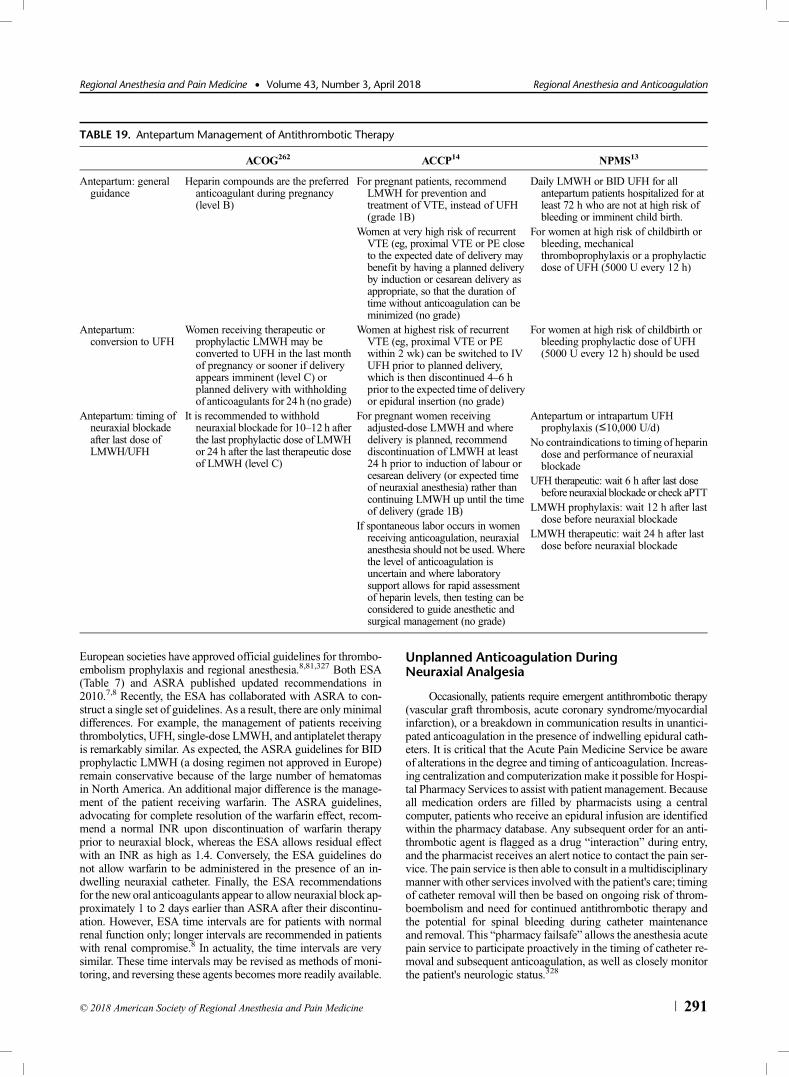
European societies have approved official guidelines for thrombo-embolism prophylaxis and regional anesthesia.8,81,327 Both ESA(Table 7) and ASRA published updated recommendations in2010.7,8 Recently, the ESA has collaborated with ASRA to con-struct a single set of guidelines. As a result, there are only minimaldifferences. For example, the management of patients receivingthrombolytics, UFH, single-dose LMWH, and antiplatelet therapyis remarkably similar. As expected, the ASRA guidelines for BIDprophylactic LMWH (a dosing regimen not approved in Europe)remain conservative because of the large number of hematomasin North America. An additional major difference is the manage-ment of the patient receiving warfarin. The ASRA guidelines,advocating for complete resolution of the warfarin effect, recom-mend a normal INR upon discontinuation of warfarin therapyprior to neuraxial block, whereas the ESA allows residual effectwith an INR as high as 1.4. Conversely, the ESA guidelines donot allow warfarin to be administered in the presence of an in-dwelling neuraxial catheter. Finally, the ESA recommendationsfor the neworal anticoagulants appear to allow neuraxial block ap-proximately 1 to 2 days earlier than ASRA after their discontinu-ation. However, ESA time intervals are for patients with normalrenal function only; longer intervals are recommended in patientswith renal compromise.8 In actuality, the time intervals are verysimilar. These time intervals may be revised as methods of moni-toring, and reversing these agents becomes more readily available.
Unplanned Anticoagulation DuringNeuraxial Analgesia
Occasionally, patients require emergent antithrombotic therapy(vascular graft thrombosis, acute coronary syndrome/myocardialinfarction), or a breakdown in communication results in unantici-pated anticoagulation in the presence of indwelling epidural cath-eters. It is critical that the Acute Pain Medicine Service be awareof alterations in the degree and timing of anticoagulation. Increas-ing centralization and computerization make it possible for Hospi-tal Pharmacy Services to assist with patient management. Becauseall medication orders are filled by pharmacists using a centralcomputer, patients who receive an epidural infusion are identifiedwithin the pharmacy database. Any subsequent order for an anti-thrombotic agent is flagged as a drug “interaction” during entry,and the pharmacist receives an alert notice to contact the pain ser-vice. The pain service is then able to consult in a multidisciplinarymanner with other services involvedwith the patient's care; timingof catheter removal will then be based on ongoing risk of throm-boembolism and need for continued antithrombotic therapy andthe potential for spinal bleeding during catheter maintenanceand removal. This “pharmacy failsafe” allows the anesthesia acutepain service to participate proactively in the timing of catheter re-moval and subsequent anticoagulation, as well as closely monitorthe patient's neurologic status.328
TABLE 19. Antepartum Management of Antithrombotic Therapy
ACOG262 ACCP14 NPMS13
Antepartum: generalguidance
Heparin compounds are the preferredanticoagulant during pregnancy(level B)
For pregnant patients, recommendLMWH for prevention andtreatment of VTE, instead of UFH(grade 1B)
Daily LMWH or BID UFH for allantepartum patients hospitalized for atleast 72 h who are not at high risk ofbleeding or imminent child birth.
Women at very high risk of recurrentVTE (eg, proximal VTE or PE closeto the expected date of delivery maybenefit by having a planned deliveryby induction or cesarean delivery asappropriate, so that the duration oftime without anticoagulation can beminimized (no grade)
For women at high risk of childbirth orbleeding, mechanicalthromboprophylaxis or a prophylacticdose of UFH (5000 U every 12 h)
Antepartum:conversion to UFH
Women receiving therapeutic orprophylactic LMWH may beconverted to UFH in the last monthof pregnancy or sooner if deliveryappears imminent (level C) orplanned delivery with withholdingof anticoagulants for 24 h (nograde)
Women at highest risk of recurrentVTE (eg, proximal VTE or PEwithin 2 wk) can be switched to IVUFH prior to planned delivery,which is then discontinued 4–6 hprior to the expected time of deliveryor epidural insertion (no grade)
For women at high risk of childbirth orbleeding prophylactic dose of UFH(5000 U every 12 h) should be used
Antepartum: timing ofneuraxial blockadeafter last dose ofLMWH/UFH
It is recommended to withholdneuraxial blockade for 10–12 h afterthe last prophylactic dose of LMWHor 24 h after the last therapeutic doseof LMWH (level C)
For pregnant women receivingadjusted-dose LMWH and wheredelivery is planned, recommenddiscontinuation of LMWH at least24 h prior to induction of labour orcesarean delivery (or expected timeof neuraxial anesthesia) rather thancontinuing LMWH up until the timeof delivery (grade 1B)
Antepartum or intrapartum UFHprophylaxis (≤10,000 U/d)
No contraindications to timing of heparindose and performance of neuraxialblockade
UFH therapeutic: wait 6 h after last dosebefore neuraxial blockade or check aPTT
LMWH prophylaxis: wait 12 h after lastdose before neuraxial blockade
If spontaneous labor occurs in womenreceiving anticoagulation, neuraxialanesthesia should not be used.Wherethe level of anticoagulation isuncertain and where laboratorysupport allows for rapid assessmentof heparin levels, then testing can beconsidered to guide anesthetic andsurgical management (no grade)
LMWH therapeutic: wait 24 h after lastdose before neuraxial blockade
Regional Anesthesia and Pain Medicine • Volume 43, Number 3, April 2018 Regional Anesthesia and Anticoagulation
© 2018 American Society of Regional Anesthesia and Pain Medicine 291

TABLE 20. Hemorrhagic Complications Following Plexus and Peripheral Block in Patients Without Antithrombotic Therapy
Reference Patient Information Block Type Clinical Course/Outcomes
Wheatley et al306 (1984) 59-y-old woman withchronic pelvic pain
4 Lumbar sympatheticblocks over a 5-d period(paravertebral approach5 cm lateral to the 2ndlumbar vertebrae)
• 30 min after the last block patient noted right lowerquadrant pain extending to her hip and flank
• Treated with unknown analgesics and dismissed home• The next morning patient was readmitted with feverand nausea and vomiting
• A palpable mass in the flank and right upper abdomenwas noted
• Abdominal CT scan revealed a large subcapsular hematomacausing considerable renal compression
• Patient was transfused with packed red blood cells• Pain persisted and hypertension developed betweenthe 9th and 12th day
• Surgical intervention revealed a massive subcapsularhematoma (1000 mL of blood was evacuated)
Stan et al307 (1995) No information Axillary (transarterial) • Patient developed small (<2 cm) hematoma• Resolved without sequelae
Kurzel et al308 (1996) 17-y-old admitted forinduction of labor
Bilateral pudendal block • Postpartum day 3 patient complained of abdominaldistention, back pain, pain in right inguinal area, and fever
• Patient was discharged and readmitted 2 d later withincreasing abdominal pain radiating to right lowerquadrant and flank
• CT scan revealed an infected, right retroperitonealhematoma
• Treated with broad-spectrum antibiotics and dismissedafter 3 d when fever and pain resolved
Mishio et al309 (1998) 62-y-old woman withsudden deafness
4 Sequential (on alternatedays) stellate ganglionblocks at C7, anteriorparatracheal approach
• 30 min after the 4th block, patient complained ofdiscomfort in her throat
• Patient was noted to have a Horner syndrome• 2 h later, patient complained of dyspnea and difficultyswallowing
• MRI scan revealed massive hemorrhage from clivus tothe diaphragm
• Patient required emergency tracheotomy due to glotticswelling and narrowing
• Multiple complications during the hospitalization• Stoma closed on POD33 and discharged on 41st day afterthe block
Ben-David and Stahl310
(1999)38-y-old healthy man,procedure on palmarsurface of wrist
Axillary (transarterial) • Large axillary hematoma causing paresthesias and radialnerve weakness
• Electromyogram at 4 wk demonstrated signs of neuropraxia• Complete recovery after 6 mo
Thomas et al311 (1999) 65-y-old womanundergoingthoracotomy forrecurrent esophagealhernia
Paravertebral (loss ofresistance to salinetechnique)
• Technical difficulty during block placement, with bloodaspiration on 2nd pass of the needle
• 150 mL of blood suctioned from the endotracheal tube• CT scan revealed small hematoma around thoracicspine and the aorta at the level of T6 and an area ofpulmonary hemorrhage in the left lower lobe
• Initial surgery canceled and patient discharged after 3 d• Readmitted 2 wk later for initial thoracotomy with noparavertebral block
Vaisman312 (2001) 40-y-old man with lefttesticular pain
Ilioinguinal • 3 h after block placement, patient returned toemergency room with nausea, dizziness, and worseningabdominal pain
• CT scan revealed left pelvic hematoma (approximately20 mL)
• Symptoms resolved with conservative management,and patient was discharged home after 2 d
Continued next page
Horlocker et al Regional Anesthesia and Pain Medicine • Volume 43, Number 3, April 2018
292 © 2018 American Society of Regional Anesthesia and Pain Medicine

Comparison Between the ASRA RegionalAnesthesia and Pain Guidelines
In response to recent publications of cases of epidural hema-toma during interventional pain procedures in patients receivingantiplatelet agents,16,17 attendees of the 11th Annual ASRA PainMedicine Meeting of ASRA, held November 15 to 18, 2012, inMiami, Florida, were surveyed. The purpose was to determinepractice patterns of pain physicians regarding continuance of con-currently administered anticoagulants, timing schedules for cessa-tion and resumption of use, and any use of “bridging” therapieswhen planning for various interventional pain procedures. Thesurvey items included specific practice characteristics andwhether active protocols were used. In addition, the survey que-ried the frequency of adherence to specific elements of the currentASRA practice guidelines for regional anesthesia in patients onantiplatelet and anticoagulant medications and/or if respondentsincorporated different protocols for different pain procedures.
Most of the respondents followed ASRA guidelines for anti-coagulants but not for antiplatelet agents. Two-thirds of the partic-ipants had separate protocols regarding aspirin or NSAIDs.Moreover, 55% stopped acetylsalicylic acid before spinal cordstimulation (SCS) trials and implants, and 32% stopped aspirinbefore ESIs. In addition, 17% admitted that they used differentprotocols for cervical spine injections as compared with lumbarspine injections. For example, although ESIs have been shownto be safe in patients who take NSAIDs in 1 study, 80% of the in-jections were done in the lumbar levels.243
These cases and survey results suggest that the risk of signif-icant bleeding in patients undergoing interventional pain proce-dures may have been underestimated. Indeed, all previousASRA guidelines (including the current fourth edition) do not rec-ommend discontinuation of aspirin or NSAIDs prior to the place-ment of epidural and spinal blocks; nor do the guidelinesdifferentiate between interventional pain procedures and perioper-ative regional anesthesia blocks, other than considering severity of
TABLE 20. (Continued)
Reference Patient Information Block Type Clinical Course/Outcomes
Borgeat et al313 (2001) No information Infraclavicular (modifiedapproach of the Rajtechnique)
• Patient developed hematoma at the puncture site• No information about outcome
Ekatodramis et al314
(2001)48-y-old woman withchronic regional painsyndrome of theright hand
Continuous interscalenecatheter
• 3 d postoperatively, the patient complained of blurredvision and painful swelling on right side of neck
• Ultrasound of neck revealed 4 ! 5-cm hematoma• Neurologic investigation confirmed Horner syndrome• Catheter removed immediately• 6 mo later, Horner syndrome began to improve• Complete resolution at 1 y
Ekatodramis et al314
(2001)20-y-old healthy womanundergoing rightshoulder Bankart repair
Continuous interscalenecatheter
• POD1 patient noted visual disturbances and neck swelling• Ultrasound of the neck revealed 3 ! 4-cm hematoma• Neurologic investigation confirmed Horner syndrome• Catheter removed immediately• 6 mo later, only slight ptosis present• Complete resolution at 1 y
Bergman et al315 (2003) No information Continuous axillarycatheter
• Patient developed axillary hematoma• Resolved spontaneously without sequelae
Amory et al316 (2003) 6-y-old healthy boy,left herniorrhaphy
Ilioinguinal/iliohypogastric • Large, obstructing subserosal hematoma, whichpresented as intestinal obstruction 5 d postoperatively
• Diagnostic laparoscopy and bowel resection with anuneventful recovery
Frigon et al317 (2006) 6-y-old healthy girl,appendectomy
Ilioinguinal/iliohypogastric • Hematoma (2 cm in diameter) in the wall of theterminal ileum noted upon opening peritoneum
• Hematoma was nonobstructing• Uneventful recovery
Singh et al318 (2014) 35-y-old woman Supraclavicular block • Paresthesia elicited on 3rd attempt(paresthesia technique) • At the end of procedure, complained of slight chest pain
in ipsilateral infraclavicular regionGiant cell tumor resectionof radius
• Chest x-ray negative• Adequate sensory/motor block• 12 h later, worsening chest pain and palpitations• Chest x-ray revealed massive fluid collection on theright side with partial collapse of right lung
• Chest tube placed with 1300 mL of blood drained andtransfused 4 U whole blood
• Intensive care unit for 5 d, dismissed in 10 d in satisfactorycondition
Adapted from Horlocker et al,7 with permission.
Regional Anesthesia and Pain Medicine • Volume 43, Number 3, April 2018 Regional Anesthesia and Anticoagulation
© 2018 American Society of Regional Anesthesia and Pain Medicine 293

TABLE 21. Hemorrhagic Complications Following Plexus and Peripheral Block in Patients Receiving Antithrombotic Therapy
Reference Patient InformationAnticoagulant/
Antiplatelet Agent(s) Block Type Clinical Course/Outcomes
Nielsen319
(1989)• 80-y-old man Preoperative • Intercostal nerve blocks
bilaterally T7–T11• Small chest wall hematoma noted onPOD3 at T7 before the last blockwas placed
• Cholecystectomy withsevere postoperative pain
• Warfarin preoperatively• Placed 20 h postoperatively• PT on operative day 13 s
• Over the next 2 d hematoma increasedto 30 × 65 cm and covered a large areaof the posterior and lateral chest walldown to flank and upper thigh
• Required transfusion of 8 U of packedred blood cells
• Heparin infusion was stopped• Patient recovered uneventfully but hadpain for 4 wk in the area ofthe hematoma
Postoperative • Blocks repeated 4 times(approximately every 7 h)
• Heparin 5000 Usubcutaneously every 8 h(started 8 h postoperativelyand discontinued on POD2)
• POD2 heparin bolus 4500 Uintravenously followed bycontinuous infusion(PTT ranged from 99–29 s)
Aida et al320
(1996)• 71-y-old woman Preoperative • Lumbar plexus block • Low back pain became more intense
1 d after the last block• Performed 4 times for 4 wk• None• Difficulty walking for several days• Level of L3 using loss of
resistance techniquePostoperative
• Pain still present 10 d after the block• Pain treated with mephenamicacid (NSAID)
• Pain due to herniatedlumbar disc
• CT scan and ultrasonography revealedrenal subcapsular hematoma
• Microscopic hematuria resolvedafter 2 wk
• Back pain resolved after 3 wk• Hematoma resorbed spontaneouslywithin 4 mo
Aida et al320
(1996)• 68-y-old woman Preoperative • Lumbar plexus block at the
level of L3 using loss ofresistance technique
• Low back pain became more intense1 d after the block• None
Postoperative • Pain remained severe and patient couldnot ambulate for several days
• Severe low back pain dueto spinal spondylosis
• Pain treated with 200 mg IVdiclofenac (NSAID) • 3 d after block CT scan and
ultrasonography revealed a renalsubcapsular hematoma
•Microscopic hematuria resolved after 1 wk• Pain markedly decreased after 2 wk• Discharged after a 10-d hospitalization• Pain resolved after 6 wk, hematomaresorbed within 4 mo
Klein et al321
(1997)• 67-y-old woman Preoperative Night of admission • Hospital day 7 (1 d after below the knee
amputation) patient complained ofright hip pain
• Incision and drainageof ankle
• Aspirin 325 mg/d
• Sciatic block (Labat approach),saphenous nerve blockposterior to thesartorius muscle
• Hospital day 14 severity ofpain worsened
• Repair of open rightcalcaneal fracture Postoperative
Hospital day 3(2nd procedure)
• Hospital day 15 patient was unableto move right leg
• POD1 enoxaparin 30 mgBID and aspirin 325 mg/d
• Open reduction internalfixation of ankle
• Abdominal CT revealed a largeretroperitoneal hematoma involvingthe right psoas muscle
• Enoxaparin continued30 mg every 12 h
• 3rd operative procedure was19½h after last doseof enoxaparin
• First postoperative enoxaparindose (after 3rd procedure)4.5 h after block placement
• Patient regained motor function overthe next 5 d
• Enoxaparin discontinued onhospital day 15 afterCT scan
• No subjective or objective evidenceof sensory deficit at the 4-mofollow-up visit
• Sciatic block (Raj approach)and lumbar plexus block(femoral approach)
Hospital day 6 (3rdprocedure)
• Right below knee amputation• Lumbar plexus block(posterior approach)—unableto place despitemultiple attempts
• Lumbar plexus block(femoral approach) andsciatic block (Raj approach)
• Patient underwent3 operative proceduresover a 6-d period
Continued next page
Horlocker et al Regional Anesthesia and Pain Medicine • Volume 43, Number 3, April 2018
294 © 2018 American Society of Regional Anesthesia and Pain Medicine

TABLE 21. (Continued)
Reference Patient InformationAnticoagulant/
Antiplatelet Agent(s) Block Type Clinical Course/Outcomes
Maier et al322
(2002)• 79-y-old woman Preoperative • Diagnostic lumbar
sympathetic blockat the L3 level
• No intravascular injection recognized• 9 h after the blockpatient complained ofburning groin and medial thigh pain
• Clopidogrel 75 mg/d• Generalized peripheralarterial disease • Discontinued 3 d prior to block
• Vital signs stable at the timePostoperative• Low-dose opioids decreasedpain, and patient walkedon the ward withoutcomplaints
• 1 h later patient wasfound pulseless
• Resuscitation attemptswere unsuccessful
• Autopsy revealed a massivecoagulated retroperitonealhematoma (about 2.3–3 L)
• Severe pain in lowerextremities • None
Maier et al322
(2002)• 71-y-old man Preoperative • Left-sided lumbar
sympathetic block usingradiographic control
• No vascular puncture during first block• Ticlopidine 500 mg/dPostoperative
• Within 12 h complained of numbnesson medial side of thigh and groin pain
• 6 d after the first block, asecond block was performed• Ticlopidine stopped 2 d after the
first block
• Left leg claudication
• Radiographic control usingcontrast medium confirmedintravascular needle position
• 2 d later, widespread skin hematomawas noted
• The night after the second block, thepatient complained of severe groinpain and decrease in blood pressure
• Needle was repositioned • CT scan revealed large retroperitonealhematoma
• Patient required a blood transfusionof an unknown amount
• Discharged 5 d later withoutmajor complaints
Weller et al323
(2003)• 85-y-old woman Preoperative • Single-injection sciatic (Labat
approach) and continuouslumbar plexus block(posterior approach)
• Blood was aspirated, catheterwithdrawn 2 cm and flushedwith saline, aspirationthen negative
• Lumbar plexus infusion discontinuedthe evening of POD1 subcutaneously• No anticoagulant medication
Postoperative • Lumbar plexus catheter removed at 10:40AM
• POD2 at 0900 enoxaparin30 mg
• Supplemental sciatic block atthe midpoint between theischial tuberosity and greatertrochanter, and a femoralblock at the groin
• 4 h later, patient complained of new,significant back pain
• Treated with morphine• POD3 right paravertebral pain, noneurological deficit
• POD4 CT scan revealed extensiveretroperitoneal hematoma thatextended from the retrohepatic spaceto the pelvis
• Transfused 4 U of packed red blood cells• Protracted postoperative course withacute renal failure, ileus, pulmonaryedema, and atrial fibrillation
• Enoxaparin continued 30 mgevery 12 h
• Never developed a neurologic deficit• POD5 extensive ecchymosis and painin flank and hip
• Dismissed from hospital on POD20
• Enoxaparin discontinuedon POD4
• Unicompartmental rightknee arthroplasty
Continued next page
Regional Anesthesia and Pain Medicine • Volume 43, Number 3, April 2018 Regional Anesthesia and Anticoagulation
© 2018 American Society of Regional Anesthesia and Pain Medicine 295

TABLE 21. (Continued)
Reference Patient InformationAnticoagulant/
Antiplatelet Agent(s) Block Type Clinical Course/Outcomes
Weller et al323
(2003)• 65-y-old man Preoperative • Single-injection lumbar
plexus block (posterior approach)and single-injection sciatic block
• POD3 patient complained of back painat the site of the lumbar plexus block• Left knee arthroscopy • Warfarin 5 mg/d
• Aspirin 81 mg BID• No technical difficulty noted
• CT scan revealed moderate-sizeretroperitoneal hematoma that appearedto originate in the psoas muscle
• Mechanical aortic valve• Patient admitted with INR5.19, coumadin stopped,and heparin started
• Next day INR measured 9.27,given 10 mg vitamin K and1 U fresh frozen plasma
• Transfused 2 U packed red blood cells
• Morning of surgery INR 0.92
• Discharged on POD10 with plan torestart anticoagulation 2 wkafter discharge
Postoperative• Heparin infusion initiated at1200 U/h 8 h after the lumbarplexus block
• aPTT on POD1 was 77.7 s• Coumadin restarted eveningof POD1
• POD3 aPTT >100 s andINR 1.4
• Heparin adjusted and POD4aPTT 60.2 s
• POD5 anticoagulationdiscontinued and vitamin K1 mg subcutaneously and 2 Ufresh frozen plasma given
Avelineet al324
(2004)
• 72-y-old woman Preoperative • Lumbar plexus block via theposterior approach
• On POD17, the patient complainedof progressive left leg motor deficitand left lumbar back pain
• Phenylindanedione stopped5 d before surgery • Unable to be placed after
3 attempts
• Total hip replacement
• Extensive ecchymosis on the left sideof her back with sensory and motordeficit in the distribution of the femoralnerve and lateral cutaneous nerve ofthe thigh (INR 3.5)
• Fascia iliaca compartment blockon first attempt
• Enoxaparin 60 mg BID started5 d before surgery and held24 h before surgery
• INR and aPTT normalPostoperative • CT scan revealed large left
retroperitoneal hematoma with anteriordisplacement of the left kidney anddiffusion of the hematoma into theleft psoas and iliac muscles
• Anticoagulation initiated at14 h with enoxaparin 40 mgand then increased to 60 mgonce daily on POD2
• Phenylindanedione restartedon POD3 and enoxaparinstopped on POD7
• Patient received 3 U of packedred blood cells and vitamin K
• Motor function started to progressivelyrecover on POD19
• Recovery was complete on POD45
• Heterozygotous Leidenmutation
Bickleret al325
(2006)
• 49-y-old man Preoperative • Continuous sciatic nerve catheter(lateral, midfemoral region)
• No difficulty noted with either catheterplacement• No anticoagulant medication
• Continuous femoral catheter • Nerve catheters removed on POD3approximately 3 h after theenoxaparin dose
Postoperative• POD2 enoxaparin 40 mg/dsubcutaneously
• At the time of catheter removal, swellingand discoloration were noted at boththe femoral and sciatic insertion sites
• POD4 massive swelling of right thighand extensive ecchymoses at both sites
• Sensation and strength were unaffected• Patient discharged on POD7, which was2 d later than expected
• No specific treatment for the hematoma
• POD5 enoxaparindiscontinued
• Right total knee replacement
Continued next page
Horlocker et al Regional Anesthesia and Pain Medicine • Volume 43, Number 3, April 2018
296 © 2018 American Society of Regional Anesthesia and Pain Medicine

TABLE 21. (Continued)
Reference Patient InformationAnticoagulant/
Antiplatelet Agent(s) Block Type Clinical Course/Outcomes
Bickleret al325
(2006)
• 78-y-old woman Preoperative • Continuous sciatic nerve catheter(lateral, midfemoral region)
• No difficulty noted with either catheterplacement• No anticoagulant medication• Total knee replacement
• Continuous femoral catheter • On POD2, the lateral thigh at the siteof the sciatic catheter was swollenand ecchymotic
Postoperative• POD1 enoxaparin 40 mg/dsubcutaneously
• The catheters were removed at this time,but the bruising increased over thenext 24 h
• No significant neurologic impairmentat the time of discharge on POD5
Bickleret al325
(2006)
• 48-y-old woman Preoperative • Continuous sciatic nerve catheter(lateral, midfemoral region)
• No difficulty noted with eithercatheter placement• No anticoagulant medication
• POD2 the dressings over the femoralcatheter insertion site were soaked with15–20 mL of blood, no hematoma noted
• Continuous femoral catheterPostoperative• POD1 enoxaparin 40 mg/dsubcutaneously
• The catheters were removed on POD2,and the femoral site continued to oozeblood and soaked another dressing
• No further bleeding on POD3• No delay in discharge
• Total knee replacement
Wiegelet al326
(2007)
• No information Preoperative • Continuous femoral catheter • Patient complained of inguinal pain,numbness, weakness of the thighon the 6th POD
• CT scan revealed a retroperitonealhematoma
• Retroperitoneal hematoma requiredsurgical intervention
• No further information
• Aspirin 1 g/d
Clendenenet al236
(2010)
• 66-y-old woman Preoperative • Continuous interscalene block • Nerve stimulator guided block withpuncture of external jugular veinduring placement
• Dipyridamole• Aspirin (stopped 1 wkpreoperative)
Postoperative
• Interscalene catheter noted to bedislodged in the am of POD1
• A 1-cm diameter hematoma notedat previous puncture site
• A 2nd continuous interscalene catheterplaced distal to previous puncture site
• Catheter was removed 72 h later onPOD5 at home
• POD8, lethargic, pale, with laboredbreathing and signs of infectionat surgical site
• Admitted with sepsis and noted tohave a hard mass at site or previousinterscalene catheter placement
• Cultures revealed Staphylococcus aureus• Discharged after 30-d hospital stay toextended care facility in good condition
• Dipyridamole
• Right shoulderhemiarthroplasty androtator cuff repair
Continued next page
Regional Anesthesia and Pain Medicine • Volume 43, Number 3, April 2018 Regional Anesthesia and Anticoagulation
© 2018 American Society of Regional Anesthesia and Pain Medicine 297

TABLE 21. (Continued)
Reference Patient InformationAnticoagulant/
Antiplatelet Agent(s) Block Type Clinical Course/Outcomes
Rodriguezet al303
(2011)
59-y-old woman undergoingbullet removal fromleft calf
Preoperative Single injection femoraland sciatic nerve blocks
• Preoperative laboratory values includeplatelet count 175,000, INR: 0.99,fibrinogen: 427 mg/dL, aPTT: 38.9 s
• Dismissed the same day withoutobvious complications
• 10 d postoperatively, the patient wasreadmitted with a large, soft hematomaat the proximal two-thirds of the thighthat extended to the knee
• CT scan revealed intermuscular andintramuscular hematoma of thequadriceps femoris muscle
• aPTTwas prolonged at 44.4 s, andbemiparin was stopped
• Patient diagnosed with a mild factor XIdeficiency (45%)
• 4 d after readmission, patient notedsevere paresis and quadriceps femorisand anesthesia to pinprick in saphenousnerve distribution
• Emergent surgical exploration revealeda 10-cm intraneural hematoma of thefemoral nerve extending towardpelvic cavity
• None
• Discharged after 22 d with residualparesis but able to crutch walk; 4 molater, able to walk without limping,although cutaneous anesthesia ofmedial side of the calf to medialmalleolus persisted
Postoperative
• Bemiparin 3500 IUsubcutaneously every 24 h
Parvaizet al237
(2012)
• 71-y-old man Preoperative • Ilioinguinal/iliohypogastricnerve block
• Placed uneventfully withlandmark-based technique• Inguinal hernia • Dipyridamole
• Aspirin (stopped 1 wkpreoperatively)
• Admitted 9 d later with right flankpain and hemoglobin 7.0 g/dL
Postoperative• Dipyridamole
• CT scan revealed large retroperitonealhematoma 22 × 10 × 7 cm
• Angiogram showed active bleeding fromdeep circumflex iliac artery, which wassuccessfully coiled
• Discharged 2 d later after a bloodtransfusion
Warneret al304
(2016)
• 87-y-old man Preoperative • Posterior lumbarplexus catheter
• Baseline platelet count 700,000 onhigh-dose hydroxyurea• Left total hip arthroplasty • Aspirin 325 mg BID
• Hemoglobin preoperatively 11.5 g/dL• Several passes required to place psoascatheter and blood aspirated on initialplacement. Catheter removed andreplaced with negative aspiration
• Moderate hypotension intraoperativelywith severe hypotension in thepostanesthesia care unit
• Hemoglobin 5.2 g/dL in thepostanesthesia care unit with a250-mL surgical blood loss
• Urgent CT scan of abdomen/pelvisrevealed a large retroperitonealhematoma
• No surgical intervention required• No neurologic sequelae
Postoperative• Myeloproliferative disorderand essential thrombocytosis • Aspirin 325 mg BID
Adapted from Horlocker et al,7 with permission.
Horlocker et al Regional Anesthesia and Pain Medicine • Volume 43, Number 3, April 2018
298 © 2018 American Society of Regional Anesthesia and Pain Medicine

needle/catheter trauma, degree of anticoagulation, and ability tocompress the site.
On the basis of these concerns, the ASRABoard of Directorsappointed a committee to develop separate guidelines for pain in-terventions. The committee has an international representationand was endorsed by the European Society of Regional Anaesthe-sia & Pain Therapy, the American Academy of Pain Medicine,the International Neuromodulation Society, the North AmericanNeuromodulation Society, and the World Institute of Pain.
Differences in Patient PopulationsPatients undergoing spine and pain procedures may be
treated differently from patients undergoing regional blocks forsurgery, acute pain, or obstetrics for several reasons. Pain proce-dures vary from minimally invasive procedures with high-risktargets (eg, percutaneous SCS lead placement, vertebral augmen-tation, deep visceral blocks, and spine interventions) to low-riskperipheral nerve blocks (Table 22). The ASRA guidelines maybe appropriate for the low- or intermediate-risk category, but thehigh-risk spine procedures require a more conservative approach.
For example, SCS involves the insertion of large-boreneedles and cephalad advancement of the electrode up to theT10 (for lumbar radiculitis) or the upper thoracic levels (forupper-extremity radiculopathy). Several advancements and retrac-tions of electrodes are needed to optimally place the electrodes,resulting in possible trauma to the vascular structures. Occasion-ally, 2 electrodes are placed to adequately cover the painful area(s). Most patients who are candidates for SCS placement had aprior laminectomy with postoperative scarring, further decreasingthe epidural space. In addition, patients with neck or back pain un-dergoing ESIs or other spinal interventions may have significantspinal abnormalities including spinal stenosis, ligamentum flavumhypertrophy, spondylolisthesis, or spondylosis, which may com-press the epidural venous plexus within a fixed and concealedspace. The risk of bleeding is further increased in pain patientstaking several concomitant analgesics and other medications withantiplatelet effects including NSAIDs and aspirin.
Chronic pain patients may also be on antiplatelet medications(aspirin or clopidogrel) for cardiovascular or central nervous sys-tem thrombotic/embolic prophylaxis. The risk is further increasedin patients on multiple anticoagulants.238
Stratification of Bleeding RiskThe interventional spine and pain recommendations are
based on both the risk of bleeding and the seriousness of hemor-rhagic sequelae and are stratified as to mild, intermediate, andhigh bleeding risks and made recommendations18 (Table 22).
Patients who undergo procedures in the minor risk category, in-cluding superficial peripheral nerve blocks and joint injections,may continue their anticoagulants. For those undergoing proce-dures that entail intermediate or major bleeding risks, the antico-agulants are discontinued. Intermediate-risk procedures includeneuraxial injections, visceral sympathetic blocks, and revisions ofthe pocket where the pump or generator is placed, whereas high-risk procedures include SCS and intrathecal pump placements,epiduroscopy, and vertebroplasty/kyphoplasty. Recommendationsfor anticoagulants, including heparin, LMWH, warfarin, and thenew oral anticoagulants, are very similar to previous and currentASRA consensus statements (and validate the survey results forwhat is considered an acceptable practice). However, there arema-jor differences in management of patients receiving antiplatelettherapy, particularly those receiving aspirin or NSAIDs scheduledto undergo intermediate- and high-risk pain procedures.
Aspirin, NSAIDs, and the P2Y12 InhibitorsSimilar to the regional anesthesia guidelines, aspirin and
NSAIDs may be continued when the procedure is low-risk painprocedures. However, antiplatelet therapy is discontinued whenmedium- and high-risk procedures are planned. The length of dis-continuation depends on the indication for aspirin administration,6 days for primary prevention (patients with no overt cardiovascu-lar disease) and 4 days for secondary prophylaxis, and is based ona study that showed platelet aggregation normalized with 4 to6 days in the patients treated with aspirin.329 For NSAIDs, an in-terval of 5 half-lives of the NSAID is recommended before theprocedure: 1 to 2 days for ibuprofen, diclofenac, ketorolac, indo-methacin; 4 days for naproxen and meloxicam; 6 days fornabumetone; and 10 days for oxaprozin and piroxicam. Aspirinand NSAIDs can be resumed a day after the procedure.330 Thereis no significant difference between the guidelines in managingthemore potent antiplatelet agents, including clopidogrel, ticlopidine,ticagrelor, and prasugrel.
Older Anticoagulants: Warfarin, Heparin, LMWH,Fibrinolytic Agents, and Fondaparinux
The ASRA interventional pain recommendations for patientsreceiving IV or SC UFH are somewhat more conservative thanthe regional guidelines. The pain guidelines recommend againstneuraxial procedures in patients on TID heparin.While the regionalguidelines allow neuraxial injections in patients receiving SC BIDheparin, the pain guidelines prefer that moderate- and high-risk pro-cedures not be performed. However, if an interventional procedureis indicated, then an interval of 6 hours (rather than 4–6 hours)should be followed. For IV heparin, the regional guidelines
TABLE 22. Stratification of Risk According to Procedures
Low Risk Intermediate Risk High Risk
Regional Guidelines Pain Guidelines Regional Guidelines Pain Guidelines Regional Guidelines Pain Guidelines
Superficial andcompressibleplexus/peripheralnerve blocks
Peripheral nerveblocks
Other procedures based onbased on compressibility,patient body habitus,comorbidities, and thedegree and duration ofanticoagulation
Neuraxial injections Neuraxial blocks Spinal cord stimulatortrial and implantDeep and noncompressible
plexus/peripheralnerve blocks
Facet proceduresIntrathecal pump trialand implant
Joint injections
Vertebroplasty andkyphoplasty
Visceral sympatheticblocks
Epiduroscopy
Sacroiliac joint andsacral lateralbranch blocks
Pocket revision
Narouze et al,18 with permission.
Regional Anesthesia and Pain Medicine • Volume 43, Number 3, April 2018 Regional Anesthesia and Anticoagulation
© 2018 American Society of Regional Anesthesia and Pain Medicine 299

recommend a 4- to 6-hour interval between administration of thedrug and neuraxial injection. In contrast, the pain guidelines rec-ommended an interval of 6 hours.
The guidelines on LMWH, warfarin, thrombolytic therapy,and fondaparinux are the same for both regional anesthesia/acute pain and interventional pain patients.
New Direct Oral Anticoagulants: Dabigatran,Rivaroxaban Apixaban, and Edoxaban
For the new direct oral anticoagulants, the ASRA guidelinesfor both regional anesthesia and pain recommended an interval of5 half-lives of the drug before a regional or a pain intervention.There are minor differences between the 2 guidelines. For resump-tion of the drug after a neuraxial procedure, the ASRA regionalguidelines recommended 6 hours, whereas the pain guidelinesrecommended 24 hours. The longer interval in the pain guide-lines is to provide a longer observation period after proceduresthat involve surgical dissection (ie, intrathecal pump placements,permanent spinal cord stimulator implantations). However, itis important to note that, for patients undergoing a surgical
procedure, initiation of thromboprophylaxis with the new oralanticoagulants is recommended a minimum of 24 hours, and per-haps as long as 72 hours, depending on bleeding risk.36,187,331
Hence, ASRA regional and pain guidelines are functionallythe same.
SUMMARYPractice guidelines or recommendations summarize evidence-
based reviews. However, the rarity of spinal hematoma defies aprospective randomized study, and there is no current laboratorymodel. As a result, these consensus statements represent the col-lective experience of recognized experts in the field of neuraxialanesthesia and anticoagulation. They are based on case reports,clinical series, pharmacology, hematology, and risk factors for sur-gical bleeding with appropriate grading of the level of evidenceand strength of the recommendations. An understanding of thecomplexity of this issue is essential to patient management; a“cookbook” approach is not appropriate. Rather, the decision toperform spinal or epidural anesthesia/analgesia and the timingof catheter removal in a patient receiving antithrombotic therapy
FIGURE 2. Procedural management checklist. Adapted from Narouze et al,18 with permission.
Horlocker et al Regional Anesthesia and Pain Medicine • Volume 43, Number 3, April 2018
300 © 2018 American Society of Regional Anesthesia and Pain Medicine

should be made on an individual basis, weighing the small,though definite risk of spinal hematoma with the benefits of re-gional anesthesia for a specific patient (and the risks of withhold-ing these benefits) (Fig. 2). Alternative anesthetic and analgesictechniques exist for patients to a patient whose risk of regional an-esthesia exceeds the expected benefit. The patient's coagulationstatus should be optimized at the time of spinal or epiduralneedle/catheter placement, and the level of anticoagulation mustbe carefully monitored during the period of epidural catheteriza-tion. Indwelling catheters should not be removed in the presenceof therapeutic anticoagulation because this appears to signifi-cantly increase the risk of spinal hematoma. It must also be re-membered that identification of risk factors and establishmentof guidelines will not completely eliminate the complication ofspinal hematoma. In Vandermeulen and colleagues'43 series, al-though 87% of patients had a hemostatic abnormality or difficultywith needle puncture, 13% had no identifiable risk factor. Vigi-lance in monitoring is critical to allow early evaluation of neuro-logic dysfunction and prompt intervention. Protocols must be inplace for urgent MRI and hematoma evacuation if there is achange in neurologic status. We must focus not only on the pre-vention of spinal hematoma, but also on rapid diagnosis and treat-ment in order to optimize neurologic outcome.
ACKNOWLEDGMENTSThe authors thank the leadership of SOAP, specifically Lisa
Leffert, Ruth Landau, Alex Butwick, and Brendan Carvalho, fortheir expert and gracious collaboration with the section on man-agement of the obstetric patient receiving antithrombotic therapy.
REFERENCES1. Enneking FK, Benzon H. Oral anticoagulants and regional anesthesia: a
perspective. Reg Anesth Pain Med. 1998;23:140–145.
2. Horlocker TT, Wedel DJ. Neuraxial block and low-molecular-weightheparin: balancing perioperative analgesia and thromboprophylaxis.Reg Anesth Pain Med. 1998;23:164–177.
3. Horlocker TT, Wedel DJ, Benzon H, et al. Regional anesthesia in theanticoagulated patient: defining the risks (the second ASRA consensusconference on neuraxial anesthesia and anticoagulation). Reg Anesth PainMed. 2003;28:172–197.
4. Liu SS, Mulroy MF. Neuraxial anesthesia and analgesia in the presence ofstandard heparin. Reg Anesth Pain Med. 1998;23:157–163.
5. Rosenquist RW, Brown DL. Neuraxial bleeding:fibrinolytics/thrombolytics. Reg Anesth Pain Med. 1998;23:152–156.
6. Urmey WF, Rowlingson J. Do antiplatelet agents contribute to thedevelopment of perioperative spinal hematoma? Reg Anesth Pain Med.1998;23:146–151.
7. Horlocker TT, Wedel DJ, Rowlingson JC, et al. Regional anesthesia inthe patient receiving antithrombotic or thrombolytic therapy:American Society of Regional Anesthesia and Pain Medicineevidence-based guidelines (third edition). Reg Anesth Pain Med.2010;35:64–101.
8. Gogarten W, Vandermeulen E, van Aken H, Kozek S, Llau JV, SamamaCM. Regional anaesthesia and antithrombotic agents: recommendationsof the European Society of Anaesthesiology. Eur J Anaesthesiol. 2010;27:999–1015.
9. Bates SM, Greer IA, Hirsh J, Ginsberg JS. Use of antithrombotic agentsduring pregnancy: the seventh ACCP conference on antithrombotic andthrombolytic therapy. Chest. 2004;126:627S–644S.
10. Bates SM, Middeldorp S, Rodger M, James AH, Greer I.Guidance for the treatment and prevention of obstetric-associatedvenous thromboembolism. J Thromb Thrombolysis.2016;41:92–128.
11. Friedman AM, Smiley RM. Expanding venous thromboembolismprophylaxis for at-risk obstetric patients: recommendations from thenational partnership bundle. Anesth Analg. 2016;123:806–808.
12. Leffert L, Landau R. Integrating the new thromboprophylaxis guidelinesinto obstetric anesthesia practice. Anesth Analg. 2016;123:809–811.
13. D'Alton ME, Friedman AM, Smiley RM, et al. National partnership formaternal safety: consensus bundle on venous thromboembolism.Anesth Analg. 2016;123:942–949.
14. Bates SM, Greer IA, Middeldorp S, et al. VTE, thrombophilia,antithrombotic therapy, and pregnancy: antithrombotic therapy andprevention of thrombosis, 9th ed: American College of Chest Physiciansevidence-based clinical practice guidelines. Chest. 2012;141:e691S–e736S.
15. Leffert L, Butwick A, Carvalho B, et al. The Society for ObstetricAnesthesia and Perinatology consensus statement on the anestheticmanagement of pregnant and postpartum women receivingthromboprophylaxis or higher dose anticoagulants [published onlineahead of print November 21, 2017]. Anesth Analg. 2017.
16. Giberson CE, Barbosa J, Brooks ES, et al. Epidural hematomas afterremoval of percutaneous spinal cord stimulator trial leads: two casereports. Reg Anesth Pain Med. 2014;39:73–77.
17. Buvanendran A, Young AC. Spinal epidural hematoma after spinal cordstimulator trial lead placement in a patient taking aspirin. Reg AnesthPain Med. 2014;39:70–72.
18. Narouze S, Benzon HT, Provenzano DA, et al. Interventional spine andpain procedures in patients on antiplatelet and anticoagulant medications:guidelines from the American Society of Regional Anesthesia and PainMedicine, the European Society of Regional Anaesthesia and PainTherapy, the American Academy of Pain Medicine, the InternationalNeuromodulation Society, the North American Neuromodulation Society,and the World Institute of Pain. Reg Anesth Pain Med. 2015;40:182–212.
19. FDA Drug Safety Communication. Updated recommendations todecrease risk of spinal column bleeding and paralyssis in patients on lowmolecular weigh heparins. Available at: http://wwwfdagov/downloads/Drugs/DrugSafety//UCM373735pdf.2013. Accessed March 4, 2017.
20. Rosencher N, Bonnet MP, Sessler DI. Selected new antithrombotic agentsand neuraxial anaesthesia for major orthopaedic surgery: managementstrategies. Anaesthesia. 2007;62:1154–1160.
21. Bouma BN, Mosnier LO. Thrombin activatable fibrinolysis inhibitor(TAFI)—how does thrombin regulate fibrinolysis? Ann Med. 2006;38:378–388.
22. IST-3 Collaborative Group, Sandercock P, Wardlaw JM, et al. Thebenefits and harms of intravenous thrombolysis with recombinant tissueplasminogen activator within 6 h of acute ischaemic stroke (the thirdinternational stroke trial [IST-3]): a randomised controlled trial.Lancet. 2012;379:2352–2363.
23. Benzon HT, Avram MJ, Green D, Bonow RO. New oral anticoagulantsand regional anaesthesia. Br J Anaesth. 2013;111(suppl 1):i96–i113.
24. Breivik H, Bang U, Jalonen J, Vigfússon G, Alahuhta S, Lagerkranser M.Nordic guidelines for neuraxial blocks in disturbed haemostasis from theScandinavian Society of Anaesthesiology and Intensive Care Medicine.Acta Anaesthesiol Scand. 2010;54:16–41.
25. Waurick K, Riess H, van Aken H, Kessler P, Gogarten W, Volk T.Rückenmarksnahe regionalanästhesien undthrombembolieprophylaxe/antithrombotische medikation. AnasthIntensivmed. 2014;55:464–492.
26. Guyatt GH, Cook DJ, Jaeschke R, Pauker SG, SchünemannHJ. Grades ofrecommendation for antithrombotic agents: American College of ChestPhysicians evidence-based clinical practice guidelines (8th edition).Chest. 2008;133:123S–131S.
27. Geerts WH, Bergqvist D, Pineo GF, et al. Prevention of venousthromboembolism: American College of Chest Physicians
Regional Anesthesia and Pain Medicine • Volume 43, Number 3, April 2018 Regional Anesthesia and Anticoagulation
© 2018 American Society of Regional Anesthesia and Pain Medicine 301

evidence-based clinical practice guidelines (8th edition).Chest. 2008;133:381S–453S.
28. Gould MK, Garcia DA, Wren SM, et al. Prevention of VTE innonorthopedic surgical patients: antithrombotic therapy and prevention ofthrombosis, 9th ed: American College of Chest Physiciansevidence-based clinical practice guidelines. Chest. 2012;141:e227S–e277S.
29. Falck-Ytter Y, Francis CW, Johanson NA, et al. Prevention of VTE inorthopedic surgery patients: antithrombotic therapy and prevention ofthrombosis, 9th ed: American College of Chest Physiciansevidence-based clinical practice guidelines. Chest. 2012;141:e278S–e325S.
30. ACCP-NHLBI national conference on antithrombotic therapy. AmericanCollege of Chest Physicians and the National Heart, Lung and BloodInstitute. Chest. 1986;89:1S–106S.
31. Guyatt GH, Akl EA, Crowther M, et al. Introduction to the ninth edition:antithrombotic therapy and prevention of thrombosis, 9th ed: AmericanCollege of Chest Physicians evidence-based clinical practice guidelines.Chest. 2012;141:48S–52S.
32. Shaneyfelt TM, Centor RM. Reassessment of clinical practice guidelines:go gently into that good night. JAMA. 2009;301:868–869.
33. Tricoci P, Allen JM, Kramer JM, Califf RM, Smith SC Jr. Scientificevidence underlying the ACC/AHA clinical practice guidelines.JAMA. 2009;301:831–841.
34. Eikelboom JW, Hirsh J, Spencer FA, Baglin TP, Weitz JI. Antiplateletdrugs: antithrombotic therapy and prevention of thrombosis, 9th ed:American College of Chest Physicians evidence-based clinical practiceguidelines. Chest. 2012;141:e89S–e119S.
35. American Society of Anesthesiologists Committee on Standards andPractice Parameters Practice alert for the perioperative management ofpatients with coronary artery stents: a report by the American Society ofAnesthesiologists committee on standards and practice parameters.Anesthesiology. 2009;110:22–23.
36. Douketis JD, Spyropoulos AC, Spencer FA, et al. Perioperativemanagement of antithrombotic therapy: antithrombotic therapy andprevention of thrombosis, 9th ed: American College of Chest Physiciansevidence-based clinical practice guidelines. Chest. 2012;141:e326S–e350S.
37. Dowling NF, Beckman MG, Manco-Johnson M, et al. The U.S.Thrombosis and Hemostasis Centers pilot sites program. J ThrombThrombolysis. 2007;23:1–7.
38. Liew A, Douketis J. Perioperative management of patients who arereceiving a novel oral anticoagulant. Intern Emerg Med. 2013;8:477–484.
39. Douketis JD, Berger PB, Dunn AS, et al. The perioperative managementof antithrombotic therapy: American College of Chest Physiciansevidence-based clinical practice guidelines (8th edition).Chest. 2008;133:299S–339S.
40. Tryba M. Epidural regional anesthesia and low molecular heparin: pro.Anasthesiol Intensivmed Notfallmed Schmerzther. 1993;28:179–181.
41. Horlocker TT, Wedel DJ. Anticoagulation and neuraxial block: historicalperspective, anesthetic implications, and risk management. Reg AnesthPain Med. 1998;23:129–134.
42. Moen V, Dahlgren N, Irestedt L. Severe neurological complications aftercentral neuraxial blockades in sweden 1990–1999. Anesthesiology. 2004;101:950–959.
43. Vandermeulen EP, van Aken H, Vermylen J. Anticoagulants andspinal-epidural anesthesia. Anesth Analg. 1994;79:1165–1177.
44. Horlocker TT, Wedel DJ, Schroeder DR, et al. Preoperative antiplatelettherapy does not increase the risk of spinal hematoma associated withregional anesthesia. Anesth Analg. 1995;80:303–309.
45. Stafford-Smith M. Impaired haemostasis and regional anaesthesia.Can J Anaesth. 1996;43:R129–R141.
46. Cheney FW, Domino KB, Caplan RA, Posner KL. Nerve injuryassociated with anesthesia: a closed claims analysis. Anesthesiology.1999;90:1062–1069.
47. Lee LA, Posner KL, Domino KB, Caplan RA, Cheney FW. Injuriesassociated with regional anesthesia in the 1980s and 1990s: a closedclaims analysis. Anesthesiology. 2004;101:143–152.
48. Ehrenfeld JM, Agarwal AK, Henneman JP, Sandberg WS. Estimating theincidence of suspected epidural hematoma and the hidden imaging cost ofepidural catheterization: a retrospective review of 43,200 cases.Reg Anesth Pain Med. 2013;38:409–414.
49. Gallus AS, Hirsh J, Tutle RJ, et al. Small subcutaneous doses of heparin inprevention of venous thrombosis. N Engl J Med. 1973;288:545–551.
50. Schulman S, Beyth RJ, Kearon C, Levine MN. Hemorrhagiccomplications of anticoagulant and thrombolytic treatment: AmericanCollege of Chest Physicians evidence-based clinical practice guidelines(8th edition). Chest. 2008;133:257S–298S.
51. Brunton LL, Lazo JS, Parker KL. Goodman & Gilman's ThePharmacological Basis of Therapeutics. New York: McGraw-Hill; 2006.
52. Chan KC, Wu DJ, Ueng KC, et al. Spinal epidural hematoma followingtissue plasminogen activator and heparinization for acute myocardialinfarction. Jpn Heart J. 2002;43:417–421.
53. Clark MA, Paradis NA. Spinal epidural hematoma complicatingthrombolytic therapy with tissue plasminogen activator—a case report.J Emerg Med. 2002;23:247–251.
54. Connolly ES Jr, Winfree CJ, McCormick PC. Management of spinalepidural hematoma after tissue plasminogen activator. A case report.Spine (Phila Pa 1976). 1996;21:1694–1698.
55. Cultrera F, Passanisi M, Giliberto O, Giuffrida M, Mancuso P, Ventura F.Spinal epidural hematoma following coronary thrombolysis. A casereport. J Neurosurg Sci. 2004;48:43–47.
56. DePorto R, Ahn JH, Gianutsos JG. Paraplegia subsequent toadministration of tissue plasminogen activator and intravenous heparinfollowingmyocardial infarction—a case report. J Spinal Cord Med. 2000;23:150–152.
57. Ferrante A, Pedi C, Centamore G, et al. A rare complication ofthrombolytic therapy: spinal epidural hematoma. A case report [in Italian].Ital Heart J Suppl. 2003;4:688–690.
58. Garcia Lopez A, Perez Lara JM, Herrainz Hidalgo R, Puente Gonzalo E.Spinal epidural hematoma following thrombolytic therapy for acutemyocardial infarction. Orthopedics. 1999;22:987–988.
59. Groen RJ, van Alphen HA. Operative treatment of spontaneous spinalepidural hematomas: a study of the factors determining postoperativeoutcome. Neurosurgery. 1996;39:494–508; discussion 508–509.
60. Haldar S, Hudsmith L, Munir S. Cervical extradural haematomafollowing thrombolysis. Heart. 2005;91:422.
61. Han YM, Kwak HS, Jin GY, Chung GH, Song KJ. Spinal epiduralhematoma after thrombolysis for deep vein thrombosis with subsequentpulmonary thromboembolism: a case report.Cardiovasc Intervent Radiol.2006;29:450–453.
62. Kim WJ, Hong YK, Yoo WH. Epidural hematoma mimicking transversemyelitis in a patient with primary antiphospholipid syndrome. RheumatolInt. 2008;28:709–712.
63. Ozgocmen S, Yoldas T, Kocakoc E, Ozkurt-Zengin F, Ardicoglu O. Spinalepidural hematoma associated with streptokinase treatment formyocardial infarction. Spinal Cord. 2004;42:374–377.
64. Sawin PD, Traynelis VC, Follett KA. Spinal epidural hematoma followingcoronary thrombolysis with tissue plasminogen activator. Report of twocases. J Neurosurg. 1995;83:350–353.
65. Song KJ, Lee KB. The poor outcome of the delayed diagnosis of acutespontaneous spinal epidural hematoma: two cases report. J Korean MedSci. 2005;20:331–334.
Horlocker et al Regional Anesthesia and Pain Medicine • Volume 43, Number 3, April 2018
302 © 2018 American Society of Regional Anesthesia and Pain Medicine
66. Toyonaga M, Hagiwara N, Irie F, et al. Acute cervical spinal epiduralhematoma during antithrombotic therapy: dual warnings againstantithrombotic therapy [in Japanese]. Rinsho Shinkeigaku. 2003;43:287–290.
67. Wulf H. Epidural anaesthesia and spinal haematoma. Can J Anaesth.1996;43:1260–1271.
68. Gupta K, Sharma R, Agrawal N, Puttegowda B, Basappa R, ManjunathCN. Spinal epidural hematoma—a rare and debilitating complication ofthrombolytic therapy. J Cardiovasc Dis Res. 2013;4:236–238.
69. Baron EM, Burke JA, Akhtar N, Young WF. Spinal epidural hematomaassociated with tissue plasminogen activator treatment of acutemyocardial infarction. Catheter Cardiovasc Interv. 1999;48:390–396.
70. Dickman CA, Shedd SA, Spetzler RF, Shetter AG, Sonntag VK. Spinalepidural hematoma associated with epidural anesthesia: complications ofsystemic heparinization in patients receiving peripheral vascularthrombolytic therapy. Anesthesiology. 1990;72:947–950.
71. Nava S, Rossignoli L, Tagariello V, Bertini L. Technical difficulties inepidural blocks and spinal bleeding complications [in Italian]. MinervaAnestesiol. 2001;67:881–886.
72. Onishchuk JL, Carlsson C. Epidural hematoma associated with epiduralanesthesia: complications of anticoagulant therapy. Anesthesiology. 1992;77:1221–1223.
73. Rabito SF, Ahmed S, Feinstein L, Winnie AP. Intrathecal bleeding afterthe intraoperative use of heparin and urokinase during continuous spinalanesthesia. Anesth Analg. 1996;82:409–411.
74. Hirsh J, Bauer KA, Donati MB, Gould M, Samama MM, Weitz JI.Parenteral anticoagulants: American College of Chest Physiciansevidence-based clinical practice guidelines (8th edition).Chest. 2008;133:141S–159S.
75. Hirsh J, Raschke R, Warkentin TE, Dalen JE, Deykin D, Poller L.Heparin: mechanism of action, pharmacokinetics, dosingconsiderations, monitoring, efficacy, and safety. Chest. 1995;108:258S–275S.
76. Garcia DA, Baglin TP, Weitz JI, SamamaMM. Parenteral anticoagulants:antithrombotic therapy and prevention of thrombosis, 9th ed: AmericanCollege of Chest Physicians evidence-based clinical practice guidelines.Chest. 2012;141:e24S–e43S.
77. Vandiver JW, Vondracek TG. Antifactor Xa levels versus activated partialthromboplastin time for monitoring unfractionated heparin.Pharmacotherapy. 2012;32:546–558.
78. Murray DJ, Brosnahan WJ, Pennell B, Kapalanski D, Weiler JM, Olson J.Heparin detection by the activated coagulation time: a comparison of thesensitivity of coagulation tests and heparin assays. J Cardiothorac VascAnesth. 1997;11:24–28.
79. Ruff RL, Dougherty JH Jr. Complications of lumbar puncture followed byanticoagulation. Stroke. 1981;12:879–881.
80. Rao TL, El-Etr AA. Anticoagulation following placement of epidural andsubarachnoid catheters: an evaluation of neurologic sequelae.Anesthesiology. 1981;55:618–620.
81. Vandermeulen E, Singelyn F, Vercauteren M, Brichant JF, Ickx BE,Gautier P. Belgian guidelines concerning central neural blockade inpatients with drug-induced alteration of coagulation: an update. ActaAnaesthesiol Belg. 2005;56:139–146.
82. Chaney MA. Intrathecal and epidural anesthesia and analgesia for cardiacsurgery. Anesth Analg. 1997;84:1211–1221.
83. Fox KA, Mehta SR, Peters R, et al. Benefits and risks of thecombination of clopidogrel and aspirin in patients undergoing surgicalrevascularization for non–ST-elevation acute coronary syndrome: theClopidogrel in Unstable angina to prevent Recurrent ischemic Events(CURE) trial. Circulation. 2004;110:1202–1208.
84. Wildsmith JA, McClure JH. Anticoagulant drugs and central nerveblockade. Anaesthesia. 1991;46:613–614.
85. Hammer GB, Ngo K,Macario A. A retrospective examination of regionalplus general anesthesia in children undergoing open heart surgery. AnesthAnalg. 2000;90:1020–1024.
86. Peterson KL, DeCampli WM, Pike NA, Robbins RC, Reitz BA. A reportof two hundred twenty cases of regional anesthesia in pediatric cardiacsurgery. Anesth Analg. 2000;90:1014–1019.
87. Sanchez R, Nygard E. Epidural anesthesia in cardiac surgery: is there anincreased risk? J Cardiothorac Vasc Anesth. 1998;12:170–173.
88. Ralley FE. Neuraxial anesthesia is contraindicated in patientsundergoing heparinization for surgery. Pro: neuraxial anesthesia shouldnot be used in patients undergoing heparinization for surgery.J Cardiothorac Vasc Anesth. 1996;10:957–960.
89. Turnbull KW. Neuraxial anesthesia is contraindicated in patientsundergoing heparinization for surgery. Con: neuraxial block is useful inpatients undergoing heparinization for surgery. J Cardiothorac VascAnesth. 1996;10:961–962.
90. Rosen DA, Hawkinberry DW2nd, Rosen KR, Gustafson RA, Hogg JP,Broadman LM. An epidural hematoma in an adolescent patient aftercardiac surgery. Anesth Analg. 2004;98:966–969.
91. Ho AM, Chung DC, Joynt GM. Neuraxial blockade and hematoma incardiac surgery: estimating the risk of a rare adverse event that has not(yet) occurred. Chest. 2000;117:551–555.
92. Collins R, Scrimgeour A, Yusuf S, Peto R. Reduction in fatal pulmonaryembolism and venous thrombosis by perioperative administration ofsubcutaneous heparin. Overviewof results of randomized trials in general,orthopedic, and urologic surgery. N Engl J Med. 1988;318:1162–1173.
93. Davis JJ, Bankhead BR, Eckman EJ, Wallace A, Strunk J.Three-times-daily subcutaneous unfractionated heparin and neuraxialanesthesia: a retrospective review of 928 cases. Reg Anesth Pain Med.2012;37:623–626.
94. Pace M, Koury K, Gulur P. Epidurals in patients receivingthromboprophylaxis with unfractionated heparin three times a day: thevalue of activated partial thromboplastin time testing.Anesth Analg. 2014;119:1215–1218.
95. Greaves JD. Serious spinal cord injury due to haematomyelia caused byspinal anaesthesia in a patient treated with low-dose heparin. Anaesthesia.1997;52:150–154.
96. Cooke ED, Lloyd MJ, Bowcock SA, Pilcher MF. Letter: monitoringduring low-dose heparin prophylaxis. N Engl J Med. 1976;294:1066–1067.
97. Working Party, Association of Anaesthetists of Great Britain & Ireland,Obstetric Anaesthetists' Association, Regional Anaesthesia UK. Regionalanaesthesia and patients with abnormalities of coagulation: theAssociation of Anaesthetists of Great Britain & Ireland The ObstetricAnaesthetists' Association Regional Anaesthesia UK. Anaesthesia. 2013;68:966–972.
98. Cosmi B, Hirsh J. Low molecular weight heparins. Curr Opin Cardiol.1994;9:612–618.
99. Holst J, Lindblad B, Bergqvist D, et al. Protamine neutralization ofintravenous and subcutaneous low-molecular-weight heparin(tinzaparin, logiparin). An experimental investigation in healthyvolunteers. Blood Coagul Fibrinolysis. 1994;5:795–803.
100. Kessler CM, Esparraguera IM, Jacobs HM, et al. Monitoring theanticoagulant effects of a low molecular weight heparin preparation.Correlation of assays in orthopedic surgery patients receiving ardeparinsodium for prophylaxis of deep venous thrombosis. Am J Clin Pathol.1995;103:642–648.
101. Levine MN, Planes A, Hirsh J, Goodyear M, Vochelle N, Gent M. Therelationship between anti–factor Xa level and clinical outcome in patientsreceiving enoxaparine low molecular weight heparin to prevent deep veinthrombosis after hip replacement. Thromb Haemost. 1989;62:940–944.
102. Lojewski B, Bacher P, Iqbal O, et al. Evaluation of hemostatic andfibrinolytic alterations associated with daily administration of
Regional Anesthesia and Pain Medicine • Volume 43, Number 3, April 2018 Regional Anesthesia and Anticoagulation
© 2018 American Society of Regional Anesthesia and Pain Medicine 303

low-molecular-weight heparin for a 12-week period. Semin ThrombHemost. 1995;21:228–239.
103. Tryba M. European practice guidelines: thromboembolism prophylaxisand regional anesthesia. Reg Anesth Pain Med. 1998;23:178–182.
104. Schroeder DR. Statistics: detecting a rare adverse drug reaction usingspontaneous reports. Reg Anesth Pain Med. 1998;23:183–189.
105. Heit JA. Perioperative management of the chronically anticoagulatedpatient. J Thromb Thrombolysis. 2001;12:81–87.
106. Henshaw DS, Turner JD, Forest DJ, Thompson GR, Weller RS. Residualenoxaparin activity, anti-Xa levels, and concerns about the AmericanSociety of Regional Anesthesia and Pain Medicine anticoagulationguidelines. Reg Anesth Pain Med. 2017;42:432–436.
107. Shaikh SA, Regal RE. Dosing of enoxaparin in renal impairment. P T.2017;42:245–249.
108. Whitlock RP, Sun JC, Fremes SE, Rubens FD, Teoh KH. Antithromboticand thrombolytic therapy for valvular disease: antithrombotic therapy andprevention of thrombosis, 9th ed: American College of Chest Physiciansevidence-based clinical practice guidelines. Chest. 2012;141:e576S–e600S.
109. Turpie AG, Gallus AS, Hoek JA. Pentasaccharide Investigators.A synthetic pentasaccharide for the prevention of deep-vein thrombosisafter total hip replacement. N Engl J Med. 2001;344:619–625.
110. Landow L. A synthetic pentasaccharide for the prevention of deep-veinthrombosis. N Engl J Med. 2001;345:291–292.
111. Singelyn FJ, Verheyen CC, Piovella F, van Aken HK, Rosencher N.EXPERT Study Investigators. The safety and efficacy of extendedthromboprophylaxis with fondaparinux after major orthopedic surgery ofthe lower limb with or without a neuraxial or deep peripheral nervecatheter: the expert study. Anesth Analg. 2007;105:1540–1547.
112. Cohen AT, Harrington R, Goldhaber SZ, et al. The design and rationale forthe acute medically ill venous thromboembolism prevention withextended duration betrixaban (APEX) study. Am Heart J. 2014;167:335–341.
113. Turpie AG, Bauer KA, Davidson BL, et al. A randomized evaluationof betrixaban, an oral factor Xa inhibitor, for prevention ofthromboembolic events after total knee replacement (expert). ThrombHaemost. 2009;101:68–76.
114. Chan NC, Hirsh J, Ginsberg JS, Eikelboom JW. Betrixaban (prt054021):pharmacology, dose selection and clinical studies. Future Cardiol. 2014;10:43–52.
115. Chan NC, Bhagirath V, Eikelboom JW. Profile of betrixaban and itspotential in the prevention and treatment of venous thromboembolism.Vasc Health Risk Manag. 2015;11:343–351.
116. Dias C, Moore KT, Murphy J, et al. Pharmacokinetics,pharmacodynamics, and safety of single-dose rivaroxaban in chronichemodialysis. Am J Nephrol. 2016;43:229–236.
117. Kubitza D, Becka M, Mueck W, et al. Effects of renal impairment onthe pharmacokinetics, pharmacodynamics and safety of rivaroxaban,an oral, direct factor Xa inhibitor. Br J Clin Pharmacol. 2010;70:703–712.
118. Highlights of Prescribing Information BEVYXXA. Available at: https://www.Accessdata.Fda.Gov/drugsatfda_docs/label/2017/208383s000lbl.Pdf. Accessed March 4, 2017.
119. Chang M, Yu Z, Shenker A, et al. Effect of renal impairment on thepharmacokinetics, pharmacodynamics, and safety of apixaban. J ClinPharmacol. 2016;56:637–645.
120. Ridout G, de la Motte S, Niemezyk S, et al. Effect of renal function onedoxaban pharmacokinetics (PK) and on population PK/PK-PD model.J Clin Pharmacol. 2009;49:1124–1124.
121. Eriksson BI, Quinlan DJ, Weitz JI. Comparative pharmacodynamics andpharmacokinetics of oral direct thrombin and factor Xa inhibitors indevelopment. Clin Pharmacokinet. 2009;48:1–22.
122. Laux V, Perzborn E, Kubitza D, Misselwitz F. Preclinical and clinicalcharacteristics of rivaroxaban: a novel, oral, direct factor Xa inhibitor.Semin Thromb Hemost. 2007;33:515–523.
123. Weitz JI, Eikelboom JW, Samama MM. New antithrombotic drugs:antithrombotic therapy and prevention of thrombosis, 9th ed: AmericanCollege of Chest Physicians evidence-based clinical practice guidelines.Chest. 2012;141:e120S–e151S.
124. Douxfils J, Mullier F, Loosen C, Chatelain C, Chatelain B, Dogne JM.Assessment of the impact of rivaroxaban on coagulation assays:laboratory recommendations for themonitoring of rivaroxaban and reviewof the literature. Thromb Res. 2012;130:956–966.
125. Madhisetti KR, Mathew M, George M, Pillai SS. Spinal epiduralhaematoma following rivaroxaban administration after total kneereplacement. Indian J Anaesth. 2015;59:519–521.
126. Ozel O, Demircay E, Kircelli A, Cansever T. Atypical presentation of anepidural hematoma in a patient receiving rivaroxaban after total hiparthroplasty. Orthopedics. 2016;39:e558–e560.
127. Radcliff KE, Ong A, Parvizi J, Post Z, Orozco F. Rivaroxaban-inducedepidural hematoma and cauda equina syndrome after total kneearthroplasty: a case report. Orthop Surg. 2014;6:69–71.
128. Truumees E, Gaudu T, Dieterichs C, Geck M, Stokes J. Epiduralhematoma and intraoperative hemorrhage in a spine trauma patient onpradaxa (dabigatran). Spine (Phila Pa 1976). 2012;37:E863–E865.
129. Bamps S, Decramer T, Vandenbussche N, et al. Dabigatran-associatedspontaneous acute cervical epidural hematoma. World Neurosurg. 2015;83:257–258.
130. Jaeger M, Jeanneret B, Schaeren S. Spontaneous spinal epiduralhaematoma during factor Xa inhibitor treatment (rivaroxaban).Eur Spine J. 2012;21(suppl 4):S433–S435.
131. Castillo JM, Afanador HF, Manjarrez E, Morales XA. Non-traumaticspontaneous spinal subdural hematoma in a patient with non-valvularatrial fibrillation during treatment with rivaroxaban.Am J Case Rep. 2015;16:377–381.
132. Zaarour M, Hassan S, Thumallapally N, Dai Q. Rivaasxroxaban-inducednontraumatic spinal subdural hematoma: an uncommon yetlife-threatening complication. Case Rep Hematol. 2015;2015:275380.
133. Dargazanli C, Lonjon N, Gras-Combe G. Nontraumatic spinal subduralhematoma complicating direct factor Xa inhibitor treatment (rivaroxaban):a challenging management. Eur Spine J. 2016;25(suppl 1):100–103.
134. Heckmann JG. Spinal subarachnoid hemorrhage in cortical superficialsiderosis after apixaban and clopidogrel therapy. J Thromb Thrombolysis.2016;41:654–655.
135. Raghavan N, Frost CE, Yu Z, et al. Apixaban metabolism andpharmacokinetics after oral administration to humans. Drug MetabDispos. 2009;37:74–81.
136. Douxfils J, Chatelain C, Chatelain B, Dogne JM, Mullier F. Impact ofapixaban on routine and specific coagulation assays: a practical laboratoryguide. Thromb Haemost. 2013;110:283–294.
137. Lip GY, Agnelli G. Edoxaban: a focused review of its clinicalpharmacology. Eur Heart J. 2014;35:1844–1855.
138. Matsushima N, Lee F, Sato T, Weiss D, Mendell J. Bioavailability andsafety of the factor Xa inhibitor edoxaban and the effects of quinidine inhealthy subjects. Clin Pharmacol Drug Dev. 2013;2:358–366.
139. Ogata K, Mendell-Harary J, Tachibana M, et al. Clinical safety,tolerability, pharmacokinetics, and pharmacodynamics of the novel factorXa inhibitor edoxaban in healthy volunteers. J Clin Pharmacol. 2010;50:743–753.
140. Plitt A, Giugliano RP. Edoxaban: review of pharmacology and key phase Ito III clinical trials. J Cardiovasc Pharmacol Ther. 2014;19:409–416.
141. Ruff CT, Giugliano RP, Antman EM, et al. Evaluation of the novel factorXa inhibitor edoxaban compared with warfarin in patients with atrialfibrillation: design and rationale for the Effective Anticoagulation With
Horlocker et al Regional Anesthesia and Pain Medicine • Volume 43, Number 3, April 2018
304 © 2018 American Society of Regional Anesthesia and Pain Medicine

Factor Xa Next Generation in Atrial Fibrillation—Thrombolysis inMyocardial Infarction Study 48 (ENGAGE AF-TIMI 48). Am Heart J.2010;160:635–641.
142. Koretsune Y, Yamashita T, Kimura T, Fukuzawa M, Abe K, Yasaka M.Short-term safety and plasma concentrations of edoxaban in Japanesepatients with non-valvular atrial fibrillation and severe renal impairment.Circ J. 2015;79:1486–1495.
143. Parasrampuria DA, Marbury T, Matsushima N, et al. Pharmacokinetics,safety, and tolerability of edoxaban in end-stage renal disease subjectsundergoing haemodialysis. Thromb Haemost. 2015;113:719–727.
144. Yamashita T, Koretsune Y, Yasaka M, et al. Randomized, multicenter,warfarin-controlled phase II study of edoxaban in Japanese patients withnon-valvular atrial fibrillation. Circ J. 2012;76:1840–1847.
145. Fuji T, Fujita S, Kawai Y, et al. A randomized, open-label trial of edoxabanin Japanese patients with severe renal impairment undergoing lower-limborthopedic surgery. Thromb J. 2015;13:6.
146. Mendell J, Chen S, He L, Desai M, Parasramupria DA. The effect ofrifampin on the pharmacokinetics of edoxaban in healthy adults.Clin Drug Investig. 2015;35:447–453.
147. Mendell J, Johnson L, Chen S. An open-label, phase 1 study to evaluatethe effects of hepatic impairment on edoxaban pharmacokinetics andpharmacodynamics. J Clin Pharmacol. 2015;55:1395–1405.
148. Mendell J, Noveck RJ, Shi M. Pharmacokinetics of the direct factor Xainhibitor edoxaban and digoxin administered alone and in combination.J Cardiovasc Pharmacol. 2012;60:335–341.
149. Mendell J, Noveck RJ, Shi M. A randomized trial of the safety,pharmacokinetics and pharmacodynamics of edoxaban, an oral factor Xainhibitor, following a switch from warfarin. Br J Clin Pharmacol. 2013;75:966–978.
150. Mendell J, Zahir H, Matsushima N, et al. Drug-drug interaction studies ofcardiovascular drugs involving P-glycoprotein, an efflux transporter, onthe pharmacokinetics of edoxaban, an oral factor Xa inhibitor. Am JCardiovasc Drugs. 2013;13:331–342.
151. Fuji T, Fujita S, Kawai Y, et al. Safety and efficacy of edoxaban in patientsundergoing hip fracture surgery. Thromb Res. 2014;133:1016–1022.
152. Fuji T, Fujita S, Tachibana S, Kawai Y. A dose-ranging study evaluatingthe oral factor Xa inhibitor edoxaban for the prevention of venousthromboembolism in patients undergoing total knee arthroplasty.J Thromb Haemost. 2010;8:2458–2468.
153. Douxfils J, Chatelain B, Chatelain C, Dogne JM, Mullier F. Edoxaban:impact on routine and specific coagulation assays. A practical laboratoryguide. Thromb Haemost. 2015;115:368–381.
154. Zhang D, He K, Raghavan N, et al. Comparative metabolism of14c-labeled apixaban in mice, rats, rabbits, dogs, and humans. DrugMetab Dispos. 2009;37:1738–1748.
155. Samama MM, Meddahi S, Samama CM. Pharmacology andlaboratory testing of the oral Xa inhibitors. Clin Lab Med. 2014;34:503–517.
156. Levi M, Eerenberg E, Kamphuisen PW. Bleeding risk and reversalstrategies for old and new anticoagulants and antiplatelet agents. J ThrombHaemost. 2011;9:1705–1712.
157. Pernod G, Albaladejo P, Godier A, et al. Management of major bleedingcomplications and emergency surgery in patients on long-termtreatment with direct oral anticoagulants, thrombin or factor-xa inhibitors:proposals of the working group on perioperative haemostasis(GIHP)—March 2013. Arch Cardiovasc Dis. 2013;106:382–393.
158. Heidbuchel H, Verhamme P, AlingsM, et al. EHRA practical guide on theuse of new oral anticoagulants in patients with non-valvular atrialfibrillation: executive summary. Eur Heart J. 2013;34:2094–2106.
159. Vanden Daelen S, Peetermans M, Vanassche T, Verhamme P,Vandermeulen E. Monitoring and reversal strategies for new oralanticoagulants. Expert Rev Cardiovasc Ther. 2015;13:95–103.
160. Jackson LR2nd, Becker RC. Novel oral anticoagulants: pharmacology,coagulation measures, and considerations for reversal. J ThrombThrombolysis. 2014;37:380–391.
161. Lu G, DeGuzman FR, Hollenbach SJ, et al. A specific antidote forreversal of anticoagulation by direct and indirect inhibitors of coagulationfactor Xa. Nat Med. 2013;19:446–451.
162. Gonsalves WI, Gupta V, Patnaik MM. Management of bleedingcomplications in patients on new oral anticoagulants. J Hematol Transf.2014;2:1015–1020.
163. Connolly SJ, Milling TJ Jr, Eikelboom JW, et al. Andexanet alfa for acutemajor bleeding associated with factor Xa inhibitors. N Engl J Med. 2016;375:1131–1141.
164. Ansell JE, Bakhru SH, Laulicht BE, et al. Use of PER977 to reverse theanticoagulant effect of edoxaban. N Engl J Med. 2014;371:2141–2142.
165. Ansell JE, Bakhru SH, Laulicht BE, et al. Single-dose ciraparantag safelyand completely reverses anticoagulant effects of edoxaban. ThrombHaemost. 2017;117:238–245.
166. Ansell JE, Laulicht BE, Bakhru SH, Hoffman M, Steiner SS, Costin JC.Ciraparantag safely and completely reverses the anticoagulant effects oflow molecular weight heparin. Thromb Res. 2016;146:113–118.
167. Carswell CI, Plosker GL. Bivalirudin: a review of its potential place in themanagement of acute coronary syndromes. Drugs. 2002;62:841–870.
168. Greinacher A, Lubenow N. Recombinant hirudin in clinical practice:focus on lepirudin. Circulation. 2001;103:1479–1484.
169. Eriksson BI, Wille-Jorgensen P, Kalebo P, et al. A comparison ofrecombinant hirudin with a low-molecular-weight heparin to preventthromboembolic complications after total hip replacement.N Engl J Med.1997;337:1329–1335.
170. Stangier J. Clinical pharmacokinetics and pharmacodynamics of the oraldirect thrombin inhibitor dabigatran etexilate. Clin Pharmacokinet. 2008;47:285–295.
171. Stangier J, Clemens A. Pharmacology, pharmacokinetics, andpharmacodynamics of dabigatran etexilate, an oral direct thrombininhibitor. Clin Appl Thromb Hemost. 2009;15(suppl 1):9S–16S.
172. Stangier J, Rathgen K, Stahle H, Gansser D, Roth W. Thepharmacokinetics, pharmacodynamics and tolerability of dabigatranetexilate, a new oral direct thrombin inhibitor, in healthy male subjects.Br J Clin Pharmacol. 2007;64:292–303.
173. Stangier J, Rathgen K, Stahle H, Mazur D. Influence of renal impairmenton the pharmacokinetics and pharmacodynamics of oral dabigatranetexilate: an open-label, parallel-group, single-centre study. ClinPharmacokinet. 2010;49:259–268.
174. Pollack CV Jr., Reilly PA, Eikelboom J, et al. Idarucizumab for dabigatranreversal. N Engl J Med. 2015;373:511–520.
175. Douxfils J, Mullier F, Robert S, Chatelain C, Chatelain B, Dogne JM.Impact of dabigatran on a large panel of routine or specific coagulationassays. Laboratory recommendations for monitoring of dabigatranetexilate. Thromb Haemost. 2012;107:985–997.
176. Lind SE, Boyle ME, Fisher S, Ishimoto J, Trujillo TC, Kiser TH.Comparison of the aptt with alternative tests for monitoring directthrombin inhibitors in patient samples. Am J Clin Pathol. 2014;141:665–674.
177. Lippi G, Ardissino D, Quintavalla R, Cervellin G. Urgent monitoring ofdirect oral anticoagulants in patients with atrial fibrillation: a tentativeapproach based on routine laboratory tests. J Thromb Thrombolysis. 2014;38:269–274.
178. Stangier J, Feuring M. Using the HEMOCLOT direct thrombin inhibitorassay to determine plasma concentrations of dabigatran. Blood CoagulFibrinolysis. 2012;23:138–143.
179. van Ryn J, Stangier J, Haertter S, et al. Dabigatran etexilate—a novel,reversible, oral direct thrombin inhibitor: interpretation of coagulation
Regional Anesthesia and Pain Medicine • Volume 43, Number 3, April 2018 Regional Anesthesia and Anticoagulation
© 2018 American Society of Regional Anesthesia and Pain Medicine 305

assays and reversal of anticoagulant activity. Thromb Haemost. 2010;103:1116–1127.
180. van Ryn J, Grottke O, SpronkH.Measurement of dabigatran in standardlyused clinical assays, whole blood viscoelastic coagulation, and thrombingeneration assays. Clin Lab Med. 2014;34:479–501.
181. Eriksson BI, Dahl OE, Büller HR, et al. A new oral direct thrombininhibitor, dabigatran etexilate, compared with enoxaparin for preventionof thromboembolic events following total hip or knee replacement: theBISTRO II randomized trial. J Thromb Haemost. 2005;3:103–111.
182. Eriksson BI, Dahl OE, Rosencher N, et al. Oral dabigatran etexilate vs.subcutaneous enoxaparin for the prevention of venous thromboembolismafter total knee replacement: the re-model randomized trial. J ThrombHaemost. 2007;5:2178–2185.
183. Fuji T, Fuijita S, Ujihira T, Sato T. Dabigatran etexilate prevents venousthromboembolism after total knee arthroplasty in Japanese patients witha safety profile comparable to placebo. J Arthroplasty. 2010;25:1267–1274.
184. RE-MOBILIZE Writing Committee, Ginsberg JS, Davidson BL, et al.Oral thrombin inhibitor dabigatran etexilate vs North Americanenoxaparin regimen for prevention of venous thromboembolism afterknee arthroplasty surgery. J Arthroplasty. 2009;24:1–9.
185. Rosencher N, Noack H, Feuring M, Clemens A, Friedman RJ,Eriksson BI. Type of anaesthesia and the safety and efficacy ofthromboprophylaxis with enoxaparin or dabigatran etexilate in majororthopaedic surgery: pooled analysis of three randomized controlled trials.Thromb J. 2012;10:9.
186. Schiele F, van Ryn J, Canada K, et al. A specific antidote fordabigatran: functional and structural characterization. Blood. 2013;121:3554–3562.
187. Doherty JU, Gluckman TJ, HuckerWJ, et al. 2017 ACC expert consensusdecision pathway for periprocedural management of anticoagulation inpatients with nonvalvular atrial fibrillation: a report of the AmericanCollege of Cardiology clinical expert consensus document task force.J Am Coll Cardiol. 2017;69:871–898.
188. Ansell J, Hirsh J, Hylek E, Jacobson A, Crowther M, Palareti G.Pharmacology and management of the vitamin K antagonists: AmericanCollege of Chest Physicians evidence-based clinical practice guidelines(8th edition). Chest. 2008;133:160S–198S.
189. Xi M, Beguin S, Hemker HC. The relative importance of the factors II,VII, IX and X for the prothrombinase activity in plasma of orallyanticoagulated patients. Thromb Haemost. 1989;62:788–791.
190. BenzonHT, AvramMJ, BenzonHA, Kirby-NolanM,Nader A. Factor VIIlevels and international normalized ratios in the early phase of warfarintherapy. Anesthesiology. 2010;112:298–304.
191. Wille-Jorgensen P, Jorgensen LN, Rasmussen LS. Lumbar regionalanaesthesia and prophylactic anticoagulant therapy. Is the combinationsafe? Anaesthesia. 1991;46:623–627.
192. Chan TY. Adverse interactions between warfarin and nonsteroidalantiinflammatory drugs: mechanisms, clinical significance, andavoidance. Ann Pharmacother. 1995;29:1274–1283.
193. Garcia D, Regan S, Crowther M, Hughes RA, Hylek EM. Warfarinmaintenance dosing patterns in clinical practice: implications for saferanticoagulation in the elderly population. Chest. 2005;127:2049–2056.
194. Benzon HT, Asher Y, Kendall MC, Vida L, McCarthy RJ, Green D.Clotting factor concentrations five days after discontinuation of warfarin.Reg Anesth Pain Med. 2018;43 [in press].
195. Hirsh J. Oral anticoagulant drugs. N Engl J Med. 1991;324:1865–1875.
196. Harder S, Thürmann P. Clinically important drug interactions withanticoagulants. An update. Clin Pharmacokinet. 1996;30:416–444.
197. Pirmohamed M, Burnside G, Eriksson N, et al. A randomized trial ofgenotype-guided dosing of warfarin. N Engl J Med. 2013;369:2294–2303.
198. Verhoef TI, Ragia G, de Boer A, et al. A randomized trial ofgenotype-guided dosing of acenocoumarol and phenprocoumon.N Engl JMed. 2013;369:2304–2312.
199. Kimmel SE, French B, Kasner SE, et al. A pharmacogenetic versus aclinical algorithm for warfarin dosing. N Engl J Med. 2013;369:2283–2293.
200. Douketis JD, Spyropoulos AC, Kaatz S, et al. Perioperative bridginganticoagulation in patients with atrial fibrillation. N Engl J Med. 2015;373:823–833.
201. Woo CH, Patel N, Conell C, et al. Rapid warfarin reversal in the setting ofintracranial hemorrhage: a comparison of plasma, recombinant activatedfactor VII, and prothrombin complex concentrate.World Neurosurg.2014;81:110–115.
202. Taketomi T, Szlam F, Levy JH, Tanaka KA. Warfarin reversal withprothrombin complex concentrate confers better antifibrinolytic activitycompared with recombinant activated factor VII. Blood CoagulFibrinolysis. 2008;19:106–108.
203. Voils SA, Holder MC, Premraj S, Catlin JR, Allen BR. Comparativeeffectiveness of 3- versus 4-factor prothrombin complex concentrate foremergent warfarin reversal. Thromb Res. 2015;136:595–598.
204. Al-Majzoub O, Rybak E, Reardon DP, Krause P, Connors JM. Evaluationof warfarin reversal with 4-factor prothrombin complex concentratecompared to 3-factor prothrombin complex concentrate at a tertiaryacademic medical center. J Emerg Med. 2016;50:7–13.
205. Woolson ST, Robinson RK, Khan NQ, Roqers BS, Maloney WJ. Deepvenous thrombosis prophylaxis for knee replacement: warfarin andpneumatic compression. Am J Orthop (Belle Mead NJ). 1998;27:299–304.
206. Badenhorst CH. Epidural hematoma after epidural pain control andconcomitant postoperative anticoagulation. Reg Anesth. 1996;21:272–273.
207. Odoom JA, Sih IL. Epidural analgesia and anticoagulant therapy.Experiencewith one thousand cases of continuous epidurals.Anaesthesia.1983;38:254–259.
208. Wu CL, Perkins FM. Oral anticoagulant prophylaxis and epidural catheterremoval. Reg Anesth. 1996;21:517–524.
209. Horlocker TT, Wedel DJ, Schlichting JL. Postoperative epidural analgesiaand oral anticoagulant therapy. Anesth Analg. 1994;79:89–93.
210. Shields RC,McBaneRD, Kuiper JD, Li H, Heit JA. Efficacy and safety ofintravenous phytonadione (vitamin K1) in patients on long-term oralanticoagulant therapy. Mayo Clin Proc. 2001;76:260–266.
211. Majerus PW, Broze GJ Jr, Miletich JP, Tollefsen DM. Anticoagulant,Antiplatelet, and Thrombolytic Drugs. In: Hardman JG, Limbird LE,Molinoff PB, Ruddon RW, Gilman AJ, eds. The Pharmacologic Basisof Therapeutics. 9th ed. New York, NY: MacMillan; 1990.
212. Janis KM. Epidural hematoma following postoperative epidural analgesia:a case report. Anesth Analg. 1972;51:689–692.
213. Helperin SW, Cohen DD. Hematoma following epidural anesthesia: reportof a case. Anesthesiology. 1971;35:641–644.
214. Horlocker TT. When to remove a spinal or epidural catheter in ananticoagulated patient. Reg Anesth. 1993;18:264–265.
215. Parvizi J, Viscusi ER, Frank HG, Sharkey PF, Hozack WJ, Rothman RR.Can epidural anesthesia and warfarin be coadministered? Clin OrthopRelat Res. 2007;456:133–137.
216. Liu SS, Buvanendran A, Viscusi ER, et al. Uncomplicated removal ofepidural catheters in 4365 patients with international normalized ratiogreater than 1.4 during initiation of warfarin therapy. Reg Anesth PainMed. 2011;36:231–235.
217. Amezcua JL, O'Grady J, Salmon JA, Moncada S. Prolonged paradoxicaleffect of aspirin on platelet behaviour and bleeding time in man.Thromb Res. 1979;16:69–79.
Horlocker et al Regional Anesthesia and Pain Medicine • Volume 43, Number 3, April 2018
306 © 2018 American Society of Regional Anesthesia and Pain Medicine

218. Godal HC, Eika C, Dybdahl JH, Daae L, Larsen S. Aspirin andbleeding-time. Lancet. 1979;1:1236.
219. Finsterer J, Seywald S, Stollberger C, et al. Recovery from acuteparaplegia due to spontaneous spinal, epidural hematoma underminimal-dose acetyl-salicylic acid. Neurol Sci. 2008;29:271–273.
220. Hyderally HA. Epidural hematoma unrelated to combined spinal-epiduralanesthesia in a patient with ankylosing spondylitis receiving aspirin aftertotal hip replacement. Anesth Analg. 2005;100:882–883.
221. Cronberg S, Wallmark E, Soderberg I. Effect on platelet aggregation oforal administration of 10 non-steroidal analgesics to humans. Scand JHaematol. 1984;33:155–159.
222. Leese PT, Hubbard RC, Karim A, Isakson PC, Yu SS, Geis GS. Effects ofcelecoxib, a novel cyclooxygenase-2 inhibitor, on platelet function inhealthy adults: a randomized, controlled trial. J Clin Pharmacol. 2000;40:124–132.
223. Patrono C, Baigent C, Hirsh J, Roth G. Antiplatelet drugs: AmericanCollege of Chest Physicians evidence-based clinical practice guidelines(8th edition). Chest. 2008;133:199S–233S.
224. Lange RA, Hillis LD. Antiplatelet therapy for ischemic heart disease.N Engl J Med. 2004;350:277–280.
225. Benzon HT, McCarthy RJ, Benzon HA, et al. Determination of residualantiplatelet activity of clopidogrel before neuraxial injections. Br JAnaesth. 2011;107:966–971.
226. Karabatsou K, Sinha A, Das K, Rainov NG. Nontraumatic spinal epiduralhematoma associated with clopidogrel. Zentralbl Neurochir. 2006;67:210–212.
227. Morales Ciancio RA, Drain O, Rillardon L, Guigui P. Acute spontaneousspinal epidural hematoma: an important differential diagnosis in patientsunder clopidogrel therapy. Spine J. 2008;8:544–547.
228. Sung JH, Hong JT, Son BC, Lee SW. Clopidogrel-induced spontaneousspinal epidural hematoma. J Korean Med Sci. 2007;22:577–579.
229. Vandermeulen E, Decoster J, Dewandre PY, et al. Central neural blockadein patients with a drug-induced alteration of coagulation. Third edition ofthe Belgian Association for Regional Anaesthesia (BARA) guidelines.Acta Anaesthesiol Belg. 2011;62:175–191.
230. Sible AM, Nawarskas JJ. Cangrelor: a new route for p2y12 inhibition.Cardiol Rev. 2017;25:133–139.
231. Akers WS, Oh JJ, Oestreich JH, Ferraris S, Wethington M, Steinhubl SR.Pharmacokinetics and pharmacodynamics of a bolus and infusion ofcangrelor: a direct, parenteral p2y12 receptor antagonist. J ClinPharmacol. 2010;50:27–35.
232. Bhatt DL, Stone GW,Mahaffey KW, et al. Effect of platelet inhibitionwithcangrelor during pci on ischemic events. N Engl J Med. 2013;368:1303–1313.
233. Angiolillo DJ, Firstenberg MS, Price MJ, et al. Bridging antiplatelettherapy with cangrelor in patients undergoing cardiac surgery: arandomized controlled trial. JAMA. 2012;307:265–274.
234. Kaneda T, Urimoto G, Suzuki T. Spinal epidural hematoma followingepidural catheter removal during antiplatelet therapy with cilostazol.J Anesth. 2008;22:290–293.
235. Sacco RL, Diener HC, Yusuf S, et al. Aspirin and extended-releasedipyridamole versus clopidogrel for recurrent stroke.N Engl J Med. 2008;359:1238–1251.
236. Clendenen SR, Robards CB, Wang RD, Greengrass RA. Case report:continuous interscalene block associated with neck hematoma andpostoperative sepsis. Anesth Analg. 2010;110:1236–1238.
237. Parvaiz MA, Korwar V, McArthur D, Claxton A, Dyer J, Isgar B. Largeretroperitoneal haematoma: an unexpected complication of ilioinguinalnerve block for inguinal hernia repair. Anaesthesia. 2012;67:80–81.
238. Benzon HT, Wong HY, Siddiqui T, Ondra S. Caution in performingepidural injections in patients on several antiplatelet drugs.Anesthesiology. 1999;91:1558–1559.
239. Gerancher JC, Waterer R, Middleton J. Transient paraparesis afterpostdural puncture spinal hematoma in a patient receiving ketorolac.Anesthesiology. 1997;86:490–494.
240. Gilbert A, Owens BD, Mulroy MF. Epidural hematoma after outpatientepidural anesthesia. Anesth Analg. 2002;94:77–78.
241. Kawaguchi S, Tokutomi S. A case of epidural hematoma associated withepidural catheterization which occurred on 12th days after the lastmedication of ticlopidine hydrochloride [in Japanese]. Masui. 2002;51:526–528.
242. Clasp: a randomised trial of low-dose aspirin for the prevention andtreatment of pre-eclampsia among 9364 pregnant women. CLASP(Collaborative Low-Dose Aspirin Study in Pregnancy) collaborativegroup. Lancet. 1994;343:619–629.
243. Horlocker TT, Bajwa ZH, Ashraf Z, et al. Risk assessment of hemorrhagiccomplications associated with nonsteroidal antiinflammatory medicationsin ambulatory pain clinic patients undergoing epidural steroid injection.Anesth Analg. 2002;95:1691–1697, table of contents.
244. Osta WA, Akbary H, Fuleihan SF. Epidural analgesia in vascular surgerypatients actively taking clopidogrel. Br J Anaesth. 2010;104:429–432.
245. Coukell AJ, Markham A. Clopidogrel. Drugs. 1997;54:745–750.
246. Dyke CM. Safety of glycoprotein iib-iiia inhibitors: a heart surgeon'sperspective. Am Heart J. 1999;138:307–316.
247. Shlansky-Goldberg R. Platelet aggregation inhibitors for use in peripheralvascular interventions: what can we learn from the experience in thecoronary arteries? J Vasc Interv Radiol. 2002;13:229–246.
248. Mayumi T, Dohi S. Spinal subarachnoid hematoma after lumbar puncturein a patient receiving antiplatelet therapy. Anesth Analg. 1983;62:777–779.
249. Eisenberg DM. Advising patients who seek alternative medical therapies.Ann Intern Med. 1997;127:61–69.
250. Elder NC, Gillcrist A, Minz R. Use of alternative health care by familypractice patients. Arch Fam Med. 1997;6:181–184.
251. Hensrud DD, Engle DD, Scheitel SM. Underreporting the use of dietarysupplements and nonprescription medications among patients undergoinga periodic health examination. Mayo Clin Proc. 1999;74:443–447.
252. Rose KD, Croissant PD, Parliament CF, Levin MB. Spontaneous spinalepidural hematoma with associated platelet dysfunction from excessivegarlic ingestion: a case report. Neurosurgery. 1990;26:880–882.
253. Gilbert GJ. Ginkgo biloba. Neurology. 1997;48:1137.
254. Matthews MK Jr. Association of ginkgo biloba with intracerebralhemorrhage. Neurology. 1998;50:1933–1934.
255. Rowin J, Lewis SL. Spontaneous bilateral subdural hematomas associatedwith chronic ginkgo biloba ingestion. Neurology. 1996;46:1775–1776.
256. Vale S. Subarachnoid haemorrhage associated with ginkgo biloba.Lancet. 1998;352:36.
257. Lee A, Chui PT, Aun CS, Lau AS, Gin T. Incidence and risk of adverseperioperative events among surgical patients taking traditional Chineseherbal medicines. Anesthesiology. 2006;105:454–461.
258. Moss J, Yuan CS. Herbal medicines and perioperative care.Anesthesiology. 2006;105:441–442.
259. Cantwell R, Clutton-Brock T, Cooper G, et al. Saving mothers' lives:reviewing maternal deaths to make motherhood safer: 2006–2008. Theeighth report of the confidential enquiries into maternal deaths in theUnited Kingdom. BJOG. 2011;118 suppl 1:1–203.
260. Greer IA. Prevention of venous thromboembolism in pregnancy. BestPract Res Clin Haematol. 2003;16:261–278.
261. Kujovich JL. Hormones and pregnancy: thromboembolic risks forwomen. Br J Haematol. 2004;126:443–454.
262. James A. Committee on Practice Bulletins—Obstetrics. Practice bulletinno. 123: thromboembolism in pregnancy. Obstet Gynecol. 2011;118:718–729.
Regional Anesthesia and Pain Medicine • Volume 43, Number 3, April 2018 Regional Anesthesia and Anticoagulation
© 2018 American Society of Regional Anesthesia and Pain Medicine 307

263. Palmerola KL, D'Alton ME, Brock CO, Friedman AM. A comparison ofrecommendations for pharmacologic thromboembolism prophylaxis aftercaesarean delivery from three major guidelines. BJOG. 2016;123:2157–2162.
264. Ansari J, Carvalho B, Shafer SL, Flood P. Pharmacokinetics andpharmacodynamics of drugs commonly used in pregnancy andparturition. Anesth Analg. 2016;122:786–804.
265. Brancazio LR, Roperti KA, Stierer R, Laifer SA. Pharmacokinetics andpharmacodynamics of subcutaneous heparin during the early thirdtrimester of pregnancy. Am J Obstet Gynecol. 1995;173:1240–1245.
266. Chunilal SD, Young E, Johnston MA, et al. The aPTT response ofpregnant plasma to unfractionated heparin. Thromb Haemost. 2002;87:92–97.
267. Casele HL, Laifer SA, Woelkers DA, Venkataramanan R. Changes in thepharmacokinetics of the low-molecular-weight heparin enoxaparinsodium during pregnancy. Am J Obstet Gynecol. 1999;181:1113–1117.
268. Lebaudy C, Hulot JS, Amoura Z, et al. Changes in enoxaparinpharmacokinetics during pregnancy and implications for antithrombotictherapeutic strategy. Clin Pharmacol Ther. 2008;84:370–377.
269. Crawford JS. Some maternal complications of epidural analgesia forlabour. Anaesthesia. 1985;40:1219–1225.
270. Bateman BT, Mhyre JM, Ehrenfeld J, et al. The risk and outcomes ofepidural hematomas after perioperative and obstetric epiduralcatheterization: a report from the multicenter perioperative outcomesgroup research consortium. Anesth Analg. 2012;116:1380–1385.
271. Paech MJ, Godkin R, Webster S. Complications of obstetric epiduralanalgesia and anaesthesia: a prospective analysis of 10,995 cases. Int JObstet Anesth. 1998;7:5–11.
272. Pöpping DM, Zahn PK, vanAken HK, Dasch B, Boche R, Pogatzki-ZahnEM. Effectiveness and safety of postoperative pain management: a surveyof 18 925 consecutive patients between 1998 and 2006 (2nd revision): adatabase analysis of prospectively raised data. Br J Anaesth. 2008;101:832–840.
273. Beards SC, Jackson A, Griffiths AG, Horsman EL. Magnetic resonanceimaging of extradural blood patches: appearances from 30 min to 18 h.Br J Anaesth. 1993;71:182–188.
274. Horlocker TT, Birnbach DJ, Connis RT, et al. Practice advisory for theprevention, diagnosis, and management of infectious complicationsassociated with neuraxial techniques: a report by the American Society ofAnesthesiologists task force on infectious complications associated withneuraxial techniques. Anesthesiology. 2010;112:530–545.
275. Ballin NC. Paraplegia following epidural analgesia. Anaesthesia. 1981;36:952–953.
276. Esler MD, Durbridge J, Kirby S. Epidural haematoma after dural puncturein a parturient with neurofibromatosis. Br J Anaesth. 2001;87:932–934.
277. Lao TT, Halpern SH, MacDonald D, Huh C. Spinal subdural haematomain a parturient after attempted epidural anaesthesia. Can J Anaesth. 1993;40:340–345.
278. Newman B. Postnatal paraparesis following epidural analgesia andforceps delivery. Anaesthesia. 1983;38:350–351.
279. Nguyen L, Riu B, Minville V, Chassery C, Catalaa I, Samii K. Epiduralhematoma after hemorrhagic shock in a parturient [in French. Can JAnaesth. 2006;53:252–257.
280. Roscoe MW, Barrington TW. Acute spinal subdural hematoma. A casereport and review of literature. Spine (Phila Pa 1976). 1984;9:672–675.
281. Scott DB, Hibbard BM. Serious non-fatal complications associatedwith extradural block in obstetric practice. Br J Anaesth. 1990;64:537–541.
282. Sibai BM, Taslimi MM, el-Nazer A, Amon E, Mabie BC, Ryan GM.Maternal-perinatal outcome associated with the syndrome of hemolysis,elevated liver enzymes, and low platelets in severepreeclampsia-eclampsia. Am J Obstet Gynecol. 1986;155:501–509.
283. Yarnell RW, D'Alton ME. Epidural hematoma complicating cholestasis ofpregnancy. Curr Opin Obstet Gynecol. 1996;8:239–242.
284. Yuen TS, Kua JS, Tan IK. Spinal haematoma following epiduralanaesthesia in a patient with eclampsia. Anaesthesia. 1999;54:350–354.
285. Walters MA, van de Velde M, Wilms G. Acute intrathecal haematomafollowing neuraxial anaesthesia: diagnostic delay after apparently normalradiological imaging. Int J Obstet Anesth. 2012;21:181–185.
286. Chiaghana CO, Bremer JM, Sappenfield JW, Wendling AL. Delayedneuraxial hematoma in parturient with fontan circulation followingneuraxial anesthesia for cesarean section. Reg Anesth Pain Med. 2016;41:773–775.
287. Guglielminotti J, Wong CA, Landau R, Li G. Temporal trends inanesthesia-related adverse events in cesarean deliveries, New York state,2003–2012. Anesthesiology. 2015;123:1013–1023.
288. D'Angelo R, Smiley RM, Riley ET, Segal S. Serious complications relatedto obstetric anesthesia: the serious complication repository project of thesociety for obstetric anesthesia and perinatology. Anesthesiology. 2014;120:1505–1512.
289. Kinsella SM,Winton AL, MushambiMC, et al. Failed tracheal intubationduring obstetric general anaesthesia: a literature review. Int J ObstetAnesth. 2015;24:356–374.
290. Knight M, Kenyon S, Brocklehurts P, Neilson J, Shakespeare J,Kurinczuk J. MBRRACE-UK Saving Lives, Improving Mothers' Care:Lessons Learned to Inform Future Maternity Care From the UK andIreland Confidential Enquiries Into Maternal Deaths and Morbidity2009–2012. Oxford: National Perinatal Epidemiology Unit, Universityof Oxford; 2014.
291. Pandit JJ, Andrade J, Bogod DG, et al. The 5th National Audit Project(NAP5) on accidental awareness during general anaesthesia: summary ofmain findings and risk factors. Anaesthesia. 2014;69:1089–1101.
292. Chang CC,Wang IT, Chen YH, Lin HC. Anesthetic management as a riskfactor for postpartum hemorrhage after cesarean deliveries. Am J ObstetGynecol. 2011;205:462.e1–462.e7.
293. Dyer RA, Els I, Farbas J, Torr GJ, Schoeman LK, James MF. Prospective,randomized trial comparing general with spinal anesthesia for cesareandelivery in preeclamptic patients with a nonreassuring fetal heart trace.Anesthesiology. 2003;99:561–569; discussion 5A–6A.
294. Lertakyamanee J, Chinachoti T, Tritrakarn T, Muangkasem J,Somboonnanonda A, Kolatat T. Comparison of general and regionalanesthesia for cesarean section: success rate, blood loss and satisfactionfrom a randomized trial. J Med Assoc Thai. 1999;82:672–680.
295. Tsai PS, Hsu CS, Fan YC, Huang CJ. General anaesthesia is associatedwith increased risk of surgical site infection after caesarean deliverycompared with neuraxial anaesthesia: a population-based study. Br JAnaesth. 2011;107:757–761.
296. Landau R, Bollag L, Ortner C. Chronic pain after childbirth. Int J ObstetAnesth. 2013;22:133–145.
297. Nikolajsen L, Sorensen HC, Jensen TS, Kehlet H. Chronic pain followingcaesarean section. Acta Anaesthesiol Scand. 2004;48:111–116.
298. Moore ER, Anderson GC, Bergman N, Dowswell T. Early skin-to-skincontact for mothers and their healthy newborn infants. CochraneDatabase Syst Rev. 2012:CD003519.
299. Smith J, Plaat F, Fisk NM. The natural caesarean: a woman-centredtechnique. BJOG. 2008;115:1037–1042; discussion 1042.
300. Algert CS, Bowen JR, Giles WB, Knoblanche GE, Lain SJ, Roberts CL.Regional block versus general anaesthesia for caesarean section andneonatal outcomes: a population-based study. BMC Med. 2009;7:20.
301. Beckmann M, Calderbank S. Mode of anaesthetic for category 1caesarean sections and neonatal outcomes. Aust N Z J Obstet Gynaecol.2012;52:316–320.
302. Palanisamy A. Maternal anesthesia and fetal neurodevelopment. Int JObstet Anesth. 2012;21:152–162.
Horlocker et al Regional Anesthesia and Pain Medicine • Volume 43, Number 3, April 2018
308 © 2018 American Society of Regional Anesthesia and Pain Medicine

303. Rodriguez J, Taboada M, Garcia F, Bermudez M, Amor M, Alvarez J.Intraneural hematoma after nerve stimulation–guided femoral block in apatient with factor XI deficiency: case report. J Clin Anesth. 2011;23:234–237.
304. Warner NS, Duncan CM, Kopp SL. Acute retroperitoneal hematoma afterpsoas catheter placement in a patient with myeloproliferativethrombocytosis and aspirin therapy. A A Case Rep. 2016;6:28–30.
305. Idestrup C, Sawhney M, Nix C, Kiss A. The incidence of hematomaformation in patients with continuous femoral catheters following totalknee arthroplasty while receiving rivaroxaban as thromboprophylaxis: anobservational study. Reg Anesth Pain Med. 2014;39:414–417.
306. Wheatley JK, Motamedi F, Hammonds WD. Page kidney resulting frommassive subcapsular hematoma. Complication of lumbar sympatheticnerve block. Urology. 1984;24:361–363.
307. Stan TC, KrantzMA, Solomon DL, Poulos JG, Chaouki K. The incidenceof neurovascular complications following axillary brachial plexus blockusing a transarterial approach. A prospective study of 1,000 consecutivepatients. Reg Anesth. 1995;20:486–492.
308. Kurzel RB, Au AH, Rooholamini SA. Retroperitoneal hematoma as acomplication of pudendal block. Diagnosis made by computedtomography. West J Med. 1996;164:523–525.
309. Mishio M, Matsumoto T, Okuda Y, Kitajima T. Delayed severe airwayobstruction due to hematoma following stellate ganglion block.Reg Anesth Pain Med. 1998;23:516–519.
310. Ben-David B, Stahl S. Axillary block complicated by hematoma andradial nerve injury. Reg Anesth Pain Med. 1999;24:264–266.
311. Thomas PW, Sanders DJ, Berrisford RG. Pulmonary haemorrhage afterpercutaneous paravertebral block. Br J Anaesth. 1999;83:668–669.
312. Vaisman J. Pelvic hematoma after an ilioinguinal nerve block fororchialgia. Anesth Analg. 2001;92:1048–1049.
313. Borgeat A, Ekatodramis G, Dumont C. An evaluation of theinfraclavicular block via a modified approach of the Raj technique.AnesthAnalg. 2001;93:436–441.
314. Ekatodramis G, Macaire P, Borgeat A. Prolonged horner syndrome due toneck hematoma after continuous interscalene block. Anesthesiology.2001;95:801–803.
315. Bergman BD, Hebl JR, Kent J, Horlocker TT. Neurologic complicationsof 405 consecutive continuous axillary catheters. Anesth Analg. 2003;96:247–252; table of contents.
316. Amory C, Mariscal A, Guyot E, Chauvet P, Leon A, Poli-Merol ML. Isilioinguinal/iliohypogastric nerve block always totally safe in children?Paediatr Anaesth. 2003;13:164–166.
317. Frigon C, Mai R, Valois-Gomez T, Desparmet J. Bowel hematomafollowing an iliohypogastric-ilioinguinal nerve block. Paediatr Anaesth.2006;16:993–996.
318. Singh SK, Katyal S, Kumar A, Kumar P. Massive hemothorax: a rarecomplication after supraclavicular brachial plexus block. Anesth EssaysRes. 2014;8:410–412.
319. Nielsen CH. Bleeding after intercostal nerve block in a patientanticoagulated with heparin. Anesthesiology. 1989;71:162–164.
320. Aida S, Takahashi H, Shimoji K. Renal subcapsular hematoma afterlumbar plexus block. Anesthesiology. 1996;84:452–455.
321. Klein SM, D'Ercole F, Greengrass RA,Warner DS. Enoxaparin associatedwith psoas hematoma and lumbar plexopathy after lumbar plexus block.Anesthesiology. 1997;87:1576–1579.
322. Maier C, Gleim M, Weiss T, Stachetzki U, Nicolas V, Zenz M. Severebleeding following lumbar sympathetic blockade in two patients undermedication with irreversible platelet aggregation inhibitors.Anesthesiology. 2002;97:740–743.
323. Weller RS, Gerancher JC, Crews JC, Wade KL. Extensive retroperitonealhematoma without neurologic deficit in two patients who underwentlumbar plexus block and were later anticoagulated. Anesthesiology. 2003;98:581–585.
324. Aveline C, Bonnet F. Delayed retroperitoneal haematoma after failedlumbar plexus block. Br J Anaesth. 2004;93:589–591.
325. Bickler P, Brandes J, Lee M, Bozic K, Chesbro B, Claassen J. Bleedingcomplications from femoral and sciatic nerve catheters in patientsreceiving low molecular weight heparin. Anesth Analg. 2006;103:1036–1037.
326. Wiegel M, Gottschaldt U, Hennebach R, Hirschberg T, Reske A.Complicati/Presented within hoursons and adverse effects associated withcontinuous peripheral nerve blocks in orthopedic patients. Anesth Analg.2007;104:1578–1582.
327. Gogarten W, van Aken H, Buttner J, Riess H, Wulf H, Burkle H.Regional anaesthesia and thromboembolism prophylasix/anticoagulation—revised recommendations of the german society ofanaesthesiology and intensive care medicine. Anasth Intensivemed. 2007;48:S109–S124.
328. Keegan MT, Horlocker TT. Epidural catheter removal beforeunanticipated anticoagulation: the pharmacy fail-safe. Anesthesiology.1999;91:328.
329. Zisman E, Erport A, Kohanovsky E, et al. Platelet function recovery aftercessation of aspirin: preliminary study of volunteers and surgical patients.Eur J Anaesthesiol. 2010;27:617–623.
330. Greenblatt DJ. Elimination half-life of drugs: value and limitations. AnnuRev Med. 1985;36:421–427.
331. Douketis JD, Syed S, Schulman S. Periprocedural management of directoral anticoagulants: comment on the 2015 American Society of RegionalAnesthesia and PainMedicine guidelines. Reg Anesth Pain Med. 2016;41:127–129.
Regional Anesthesia and Pain Medicine • Volume 43, Number 3, April 2018 Regional Anesthesia and Anticoagulation
© 2018 American Society of Regional Anesthesia and Pain Medicine 309