
Refractive Surgery Overview
147
1 Refractive Surgery Overview The goal of refractive surgery is deceptively straightforward and simple: To render the pt less reliant upon refractive accoutrements (ie, contacts and glasses). Ideally, a pt s/p refractive surgery would have 20/20 vision at all distances, under any lighting conditions, with no dysphotopsias (visual experiences that degrade vision quality), and with no risk of future negative repercussions vis a vis the long-term health and/or optical performance of the eye. Also ideally, the above could be achieved irrespective of pre-op refractive status and/or pre-existing ocular conditions. Current technology is unable to meet this lofty ideal, and thus refractive surgery necessitates compromises and trade-offs; eg, If you had to pick one, would you rather be spectacle-free at distance, or near? Would it be acceptable if you only needed glasses in dimly-lit restaurants? How bothersome would haloes around lights at night be? Because some aspect of the pt’s post-op visual life will be less than ideal, key to successful refractive surgery is 1) developing a solid understanding of the pt’s visual preferences and requirements, and 2) communicating effectively with the pt regarding what her post-op visual life will be; ie, establishing expectations that are realistic and achievable. Further, the surgeon must have a refined eye (ahem) for recognizing pre- existing ocular conditions that render the pt a poor surgical candidate (or increase the risk of complications down the road). Just as the goal of refractive surgery is simple, so too is its organization. Broadly speaking, there are but two types of surgery: Corneal and intraocular. Corneal (aka keratorefractive) procedures work by reshaping the cornea to offset the native refractive error of the eye; there are a plethora of techniques and technologies for doing this. In contrast, intraocular procedures are more limited in their variety: They involve either replacing the native lens with an IOL, or placing in the anterior chamber an accessory IOL that offsets the eye’s native refractive error.
Transcript of Refractive Surgery Overview
1
Refractive Surgery OverviewThe goal of refractive surgery is deceptively straightforward and simple: To render the pt less reliant upon refractive accoutrements (ie, contacts and glasses). Ideally, a pt s/p refractive surgery would have 20/20 vision at all distances, under any lighting conditions, with no dysphotopsias (visual experiences that degrade vision quality), and with no risk of future negative repercussions vis a vis the long-term health and/or optical performance of the eye. Also ideally, the above could be achieved irrespective of pre-op refractive status and/or pre-existing ocular conditions.
Current technology is unable to meet this lofty ideal, and thus refractive surgery necessitates compromises and trade-offs; eg, If you had to pick one, would you rather be spectacle-free at distance, or near? Would it be acceptable if you only needed glasses in dimly-lit restaurants? How bothersome would haloes around lights at night be? Because some aspect of the pt’s post-op visual life will be less than ideal, key to successful refractive surgery is 1) developing a solid understanding of the pt’s visual preferences and requirements, and 2) communicating effectively with the pt regarding what her post-op visual life will be; ie, establishing expectations that are realistic and achievable. Further, the surgeon must have a refined eye (ahem) for recognizing pre-existing ocular conditions that render the pt a poor surgical candidate (or increase the risk of complications down the road).
Just as the goal of refractive surgery is simple, so too is its organization. Broadly speaking, there are but two types of surgery: Corneal and intraocular. Corneal (aka keratorefractive) procedures work by reshaping the cornea to offset the native refractive error of the eye; there are a plethora of techniques and technologies for doing this. In contrast, intraocular procedures are more limited in their variety: They involve either replacing the native lens with an IOL, or placing in the anterior chamber an accessory IOL that offsets the eye’s native refractive error.
2
Refractive Surgery OverviewThe goal of refractive surgery is deceptively straightforward and simple: To render the pt less reliant upon refractive accoutrements (ie, contacts and glasses). Ideally, a pt s/p refractive surgery would have 20/20 vision at all distances, under any lighting conditions, with no dysphotopsias (visual experiences that degrade vision quality), and with no risk of future negative repercussions vis a vis the long-term health and/or optical performance of the eye. Also ideally, the above could be achieved irrespective of pre-op refractive status and/or pre-existing ocular conditions.
Current technology is unable to meet this lofty ideal, and thus refractive surgery necessitates compromises and trade-offs; eg, If you had to pick one, would you rather be spectacle-free at distance, or near? Would it be acceptable if you only needed glasses in dimly-lit restaurants? How bothersome would haloes around lights at night be? Because some aspect of the pt’s post-op visual life will be less than ideal, key to successful refractive surgery is 1) developing a solid understanding of the pt’s visual preferences and requirements, and 2) communicating effectively with the pt regarding what her post-op visual life will be; ie, establishing expectations that are realistic and achievable. Further, the surgeon must have a refined eye (ahem) for recognizing pre-existing ocular conditions that render the pt a poor surgical candidate (or increase the risk of complications down the road).
Just as the goal of refractive surgery is simple, so too is its organization. Broadly speaking, there are but two types of surgery: Corneal and intraocular. Corneal (aka keratorefractive) procedures work by reshaping the cornea to offset the native refractive error of the eye; there are a plethora of techniques and technologies for doing this. In contrast, intraocular procedures are more limited in their variety: They involve either replacing the native lens with an IOL, or placing in the anterior chamber an accessory IOL that offsets the eye’s native refractive error.
3
Refractive Surgery OverviewThe goal of refractive surgery is deceptively straightforward and simple: To render the pt less reliant upon refractive accoutrements (ie, contacts and glasses). Ideally, a pt s/p refractive surgery would have 20/20 vision at all distances, under any lighting conditions, with no dysphotopsias (visual experiences that degrade vision quality), and with no risk of future negative repercussions vis a vis the long-term health and/or optical performance of the eye. Also ideally, the above could be achieved irrespective of pre-op refractive status and/or pre-existing ocular conditions.
Current technology is unable to meet this lofty ideal, and thus refractive surgery necessitates compromises and trade-offs; eg, If you had to pick one, would you rather be spectacle-free at distance, or near? Would it be acceptable if you only needed glasses in dimly-lit restaurants? How bothersome would haloes around lights at night be? Because some aspect of the pt’s post-op visual life will be less than ideal, key to successful refractive surgery is 1) developing a solid understanding of the pt’s visual preferences and requirements, and 2) communicating effectively with the pt regarding what her post-op visual life will be; ie, establishing expectations that are realistic and achievable. Further, the surgeon must have a refined eye (ahem) for recognizing pre-existing ocular conditions that render the pt a poor surgical candidate (or increase the risk of complications down the road).
Just as the goal of refractive surgery is simple, so too is its organization. Broadly speaking, there are but two types of surgery: Corneal and intraocular. Corneal (aka keratorefractive) procedures work by reshaping the cornea to offset the native refractive error of the eye; there are a plethora of techniques and technologies for doing this. In contrast, intraocular procedures are more limited in their variety: They involve either replacing the native lens with an IOL, or placing in the anterior chamber an accessory IOL that offsets the eye’s native refractive error.

4
Refractive Surgery OverviewThe goal of refractive surgery is deceptively straightforward and simple: To render the pt less reliant upon refractive accoutrements (ie, contacts and glasses). Ideally, a pt s/p refractive surgery would have 20/20 vision at all distances, under any lighting conditions, with no dysphotopsias (visual experiences that degrade vision quality), and with no risk of future negative repercussions vis a vis the long-term health and/or optical performance of the eye. Also ideally, the above could be achieved irrespective of pre-op refractive status and/or pre-existing ocular conditions.
Current technology is unable to meet this lofty ideal, and thus refractive surgery necessitates compromises and trade-offs; eg, If you had to pick one, would you rather be spectacle-free at distance, or near? Would it be acceptable if you only needed glasses in dimly-lit restaurants? How bothersome would haloes around lights at night be? Because some aspect of the pt’s post-op visual life will be less than ideal, key to successful refractive surgery is 1) developing a solid understanding of the pt’s visual preferences and requirements, and 2) communicating effectively with the pt regarding what her post-op visual life will be; ie, establishing expectations that are realistic and achievable. Further, the surgeon must have a refined eye (ahem) for recognizing pre-existing ocular conditions that render the pt a poor surgical candidate (or increase the risk of complications down the road).
Just as the goal of refractive surgery is simple, so too is its organization. Broadly speaking, there are but two types of surgery: Corneal and intraocular. Corneal (aka keratorefractive) procedures work by reshaping the cornea to offset the native refractive error of the eye; there are a plethora of techniques and technologies for doing this. In contrast, intraocular procedures are more limited in their variety: They involve either replacing the native lens with an IOL, or placing in the anterior chamber an accessory IOL that offsets the eye’s native refractive error.

5
Refractive Surgery OverviewThe goal of refractive surgery is deceptively straightforward and simple: To render the pt less reliant upon refractive accoutrements (ie, contacts and glasses). Ideally, a pt s/p refractive surgery would have 20/20 vision at all distances, under any lighting conditions, with no dysphotopsias (visual experiences that degrade vision quality), and with no risk of future negative repercussions vis a vis the long-term health and/or optical performance of the eye. Also ideally, the above could be achieved irrespective of pre-op refractive status and/or pre-existing ocular conditions.
Current technology is unable to meet this lofty ideal, and thus refractive surgery necessitates compromises and trade-offs; eg, If you had to pick one, would you rather be spectacle-free at distance, or near? Would it be acceptable if you only needed glasses in dimly-lit restaurants? How bothersome would haloes around lights at night be? Because some aspect of the pt’s post-op visual life will be less than ideal, key to successful refractive surgery is 1) developing a solid understanding of the pt’s visual preferences and requirements, and 2) communicating effectively with the pt regarding what her post-op visual life will be; ie, establishing expectations that are realistic and achievable. Further, the surgeon must have a refined eye (ahem) for recognizing pre-existing ocular conditions that render the pt a poor surgical candidate (or increase the risk of complications down the road).
Just as the goal of refractive surgery is simple, so too is its organization. Broadly speaking, there are but two types of surgery: Corneal and intraocular. Corneal (aka keratorefractive) procedures work by reshaping the cornea to offset the native refractive error of the eye; there are a plethora of techniques and technologies for doing this. In contrast, intraocular procedures are more limited in their variety: They involve either replacing the native lens with an IOL, or placing in the anterior chamber an accessory IOL that offsets the eye’s native refractive error.

6
Refractive Surgery OverviewThe goal of refractive surgery is deceptively straightforward and simple: To render the pt less reliant upon refractive accoutrements (ie, contacts and glasses). Ideally, a pt s/p refractive surgery would have 20/20 vision at all distances, under any lighting conditions, with no dysphotopsias (visual experiences that degrade vision quality), and with no risk of future negative repercussions vis a vis the long-term health and/or optical performance of the eye. Also ideally, the above could be achieved irrespective of pre-op refractive status and/or pre-existing ocular conditions.
Current technology is unable to meet this lofty ideal, and thus refractive surgery necessitates compromises and trade-offs; eg, If you had to pick one, would you rather be spectacle-free at distance, or near? Would it be acceptable if you only needed glasses in dimly-lit restaurants? How bothersome would haloes around lights at night be? Because some aspect of the pt’s post-op visual life will be less than ideal, key to successful refractive surgery is 1) developing a solid understanding of the pt’s visual preferences and requirements, and 2) communicating effectively with the pt regarding what her post-op visual life will be; ie, establishing expectations that are realistic and achievable. Further, the surgeon must have a refined eye (ahem) for recognizing pre-existing ocular conditions that render the pt a poor surgical candidate (or increase the risk of complications down the road).
Just as the goal of refractive surgery is simple, so too is its organization. Broadly speaking, there are but two types of surgery: Corneal and intraocular. Corneal (aka keratorefractive) procedures work by reshaping the cornea to offset the native refractive error of the eye; there are a plethora of techniques and technologies for doing this. In contrast, intraocular procedures are more limited in their variety: They involve either replacing the native lens with an IOL, or placing in the anterior chamber an accessory IOL that offsets the eye’s native refractive error.

7
Refractive Surgery OverviewThe goal of refractive surgery is deceptively straightforward and simple: To render the pt less reliant upon refractive accoutrements (ie, contacts and glasses). Ideally, a pt s/p refractive surgery would have 20/20 vision at all distances, under any lighting conditions, with no dysphotopsias (visual experiences that degrade vision quality), and with no risk of future negative repercussions vis a vis the long-term health and/or optical performance of the eye. Also ideally, the above could be achieved irrespective of pre-op refractive status and/or pre-existing ocular conditions.
Current technology is unable to meet this lofty ideal, and thus refractive surgery necessitates compromises and trade-offs; eg, If you had to pick one, would you rather be spectacle-free at distance, or near? Would it be acceptable if you only needed glasses in dimly-lit restaurants? How bothersome would haloes around lights at night be? Because some aspect of the pt’s post-op visual life will be less than ideal, key to successful refractive surgery is 1) developing a solid understanding of the pt’s visual preferences and requirements, and 2) communicating effectively with the pt regarding what her post-op visual life will be; ie, establishing expectations that are realistic and achievable. Further, the surgeon must have a refined eye (ahem) for recognizing pre-existing ocular conditions that render the pt a poor surgical candidate (or increase the risk of complications down the road).
Just as the goal of refractive surgery is simple, so too is its organization. Broadly speaking, there are but two types of surgery: Corneal and intraocular. Corneal (aka keratorefractive) procedures work by reshaping the cornea to offset the native refractive error of the eye; there are a plethora of techniques and technologies for doing this. In contrast, intraocular procedures are more limited in their variety: They involve either replacing the native lens with an IOL, or placing in the anterior chamber an accessory IOL that offsets the eye’s native refractive error.
Before delving into specific surgical techniques, let’s touch briefly on the optics of refractive error,
starting with an overview of vergence

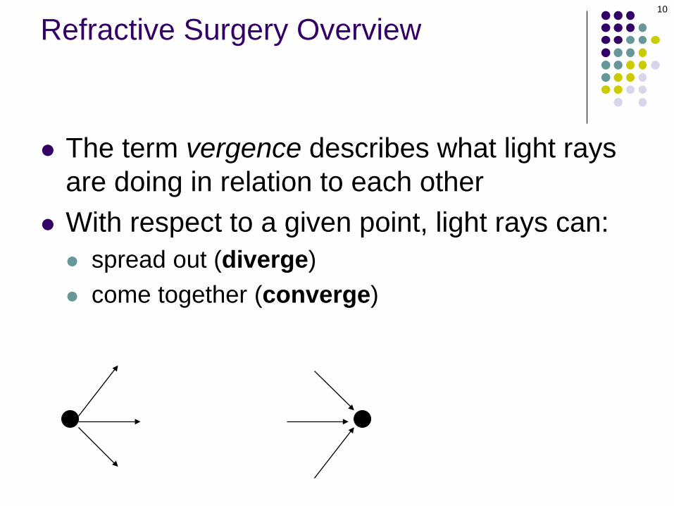
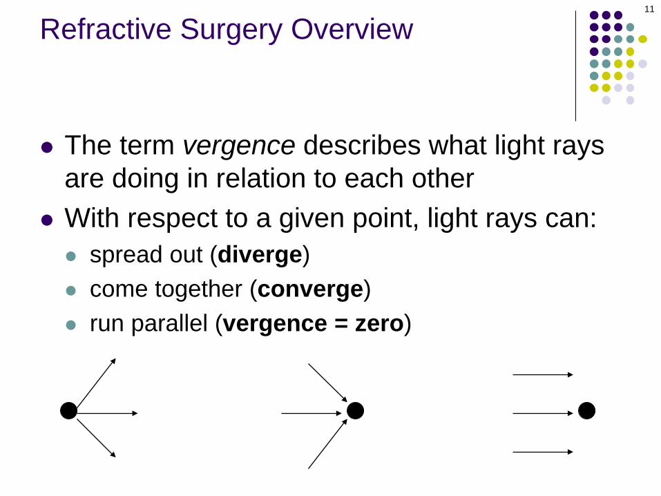
The term vergence describes what light rays are doing in relation to each other
8
Refractive Surgery Overview

The term vergence describes what light rays are doing in relation to each other
With respect to a given point, light rays can: spread out (diverge)
9
Refractive Surgery Overview
The term vergence describes what light rays are doing in relation to each other
With respect to a given point, light rays can: spread out (diverge) come together (converge)
10
Refractive Surgery Overview
The term vergence describes what light rays are doing in relation to each other
With respect to a given point, light rays can: spread out (diverge) come together (converge) run parallel (vergence = zero)
11
Refractive Surgery Overview
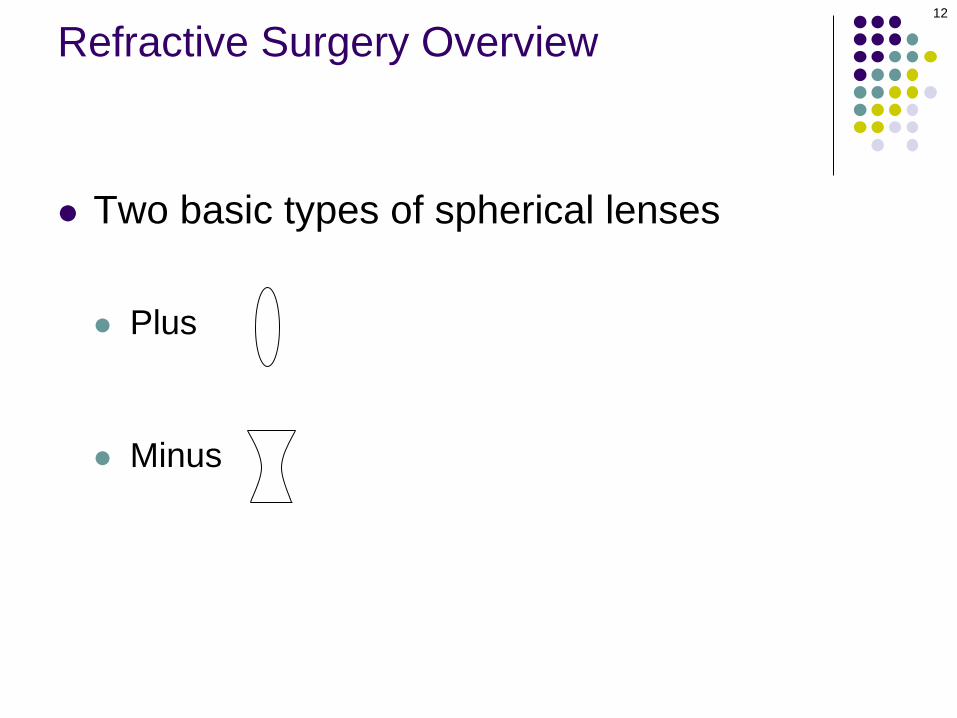
Two basic types of spherical lenses
Plus
Minus
12
Refractive Surgery Overview

Plus lens: induces convergence
In this example, a plus lens causes previously parallel rays to converge to a point
13
Refractive Surgery Overview
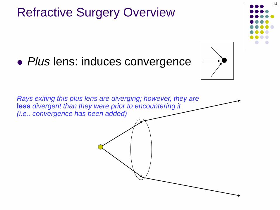
Plus lens: induces convergence
Rays exiting this plus lens are diverging; however, they are less divergent than they were prior to encountering it(i.e., convergence has been added)
14
Refractive Surgery Overview
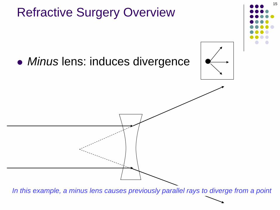
Minus lens: induces divergence
In this example, a minus lens causes previously parallel rays to diverge from a point
15
Refractive Surgery Overview
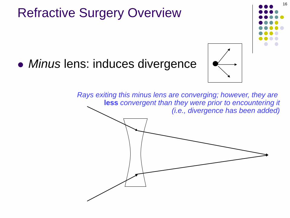
Minus lens: induces divergence
Rays exiting this minus lens are converging; however, they are less convergent than they were prior to encountering it
(i.e., divergence has been added)
16
Refractive Surgery Overview
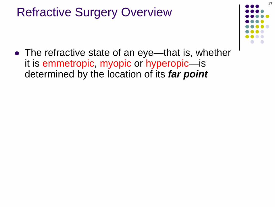
The refractive state of an eye—that is, whether it is emmetropic, myopic or hyperopic—is determined by the location of its far point
17
Refractive Surgery Overview
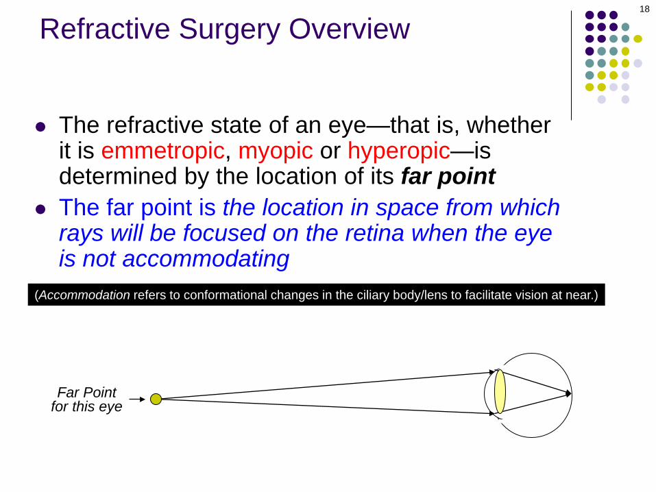
The refractive state of an eye—that is, whether it is emmetropic, myopic or hyperopic—is determined by the location of its far point
The far point is the location in space from which rays will be focused on the retina when the eye is not accommodating
(Accommodation refers to conformational changes in the ciliary body/lens to facilitate vision at near.)
Far Point for this eye
18
Refractive Surgery Overview
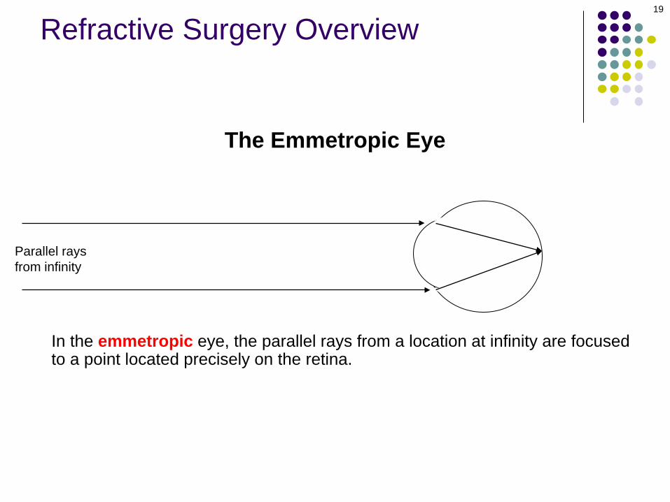
The Emmetropic Eye
In the emmetropic eye, the parallel rays from a location at infinity are focusedto a point located precisely on the retina.
19
Parallel rays from infinity
Refractive Surgery Overview
The Emmetropic Eye
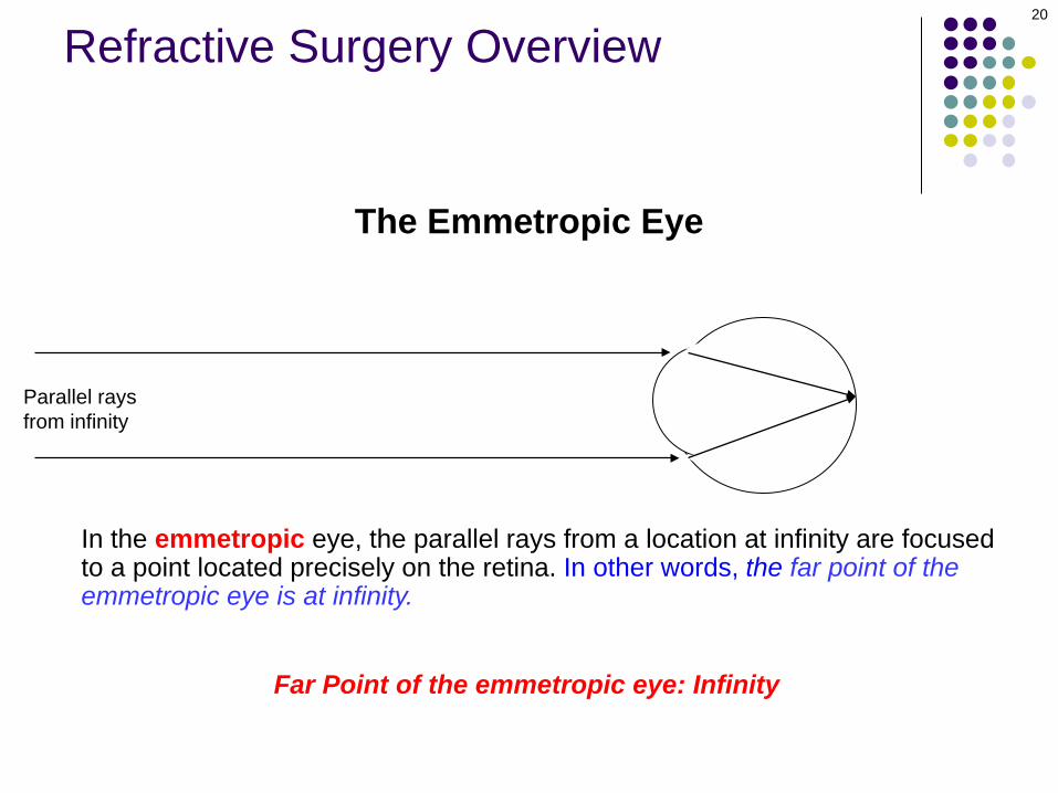
In the emmetropic eye, the parallel rays from a location at infinity are focusedto a point located precisely on the retina. In other words, the far point of theemmetropic eye is at infinity.
Far Point of the emmetropic eye: Infinity
20
Parallel rays from infinity
Refractive Surgery Overview

The Emmetropic Eye
In the emmetropic eye, the parallel rays from a location at infinity are focusedto a point located precisely on the retina. In other words, the far point of theemmetropic eye is at infinity. Thus, emmetropes see 20/20 (or better) at distance without correction.
Far Point of the emmetropic eye: Infinity
21
Parallel rays from infinity
Refractive Surgery Overview

The Myopic Eye
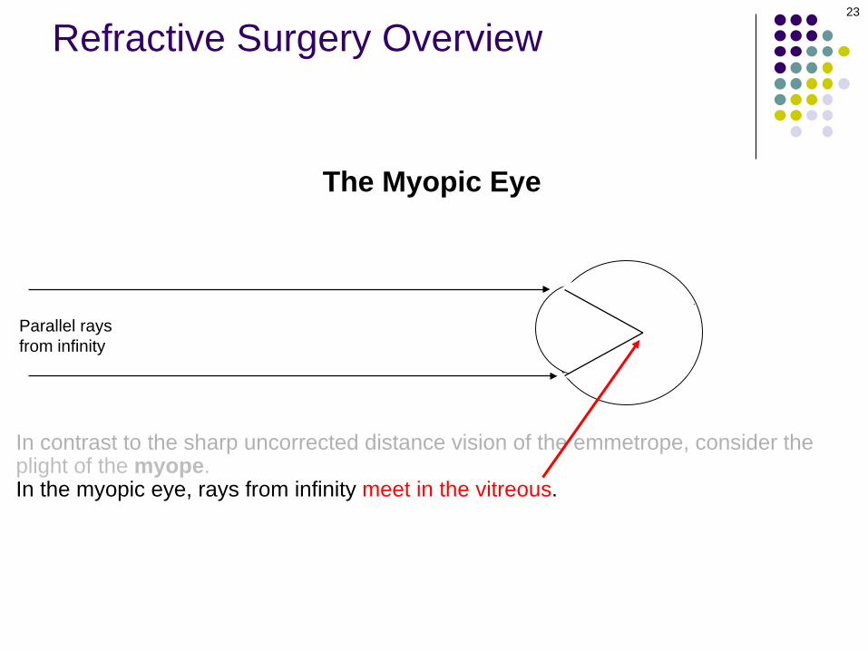
In contrast to the sharp uncorrected distance vision of the emmetrope, consider theplight of the myope.
22
Parallel rays from infinity
Refractive Surgery Overview
The Myopic Eye
In contrast to the sharp uncorrected distance vision of the emmetrope, consider theplight of the myope.In the myopic eye, rays from infinity meet in the vitreous.
23
Parallel rays from infinity
Refractive Surgery Overview

The Myopic Eye
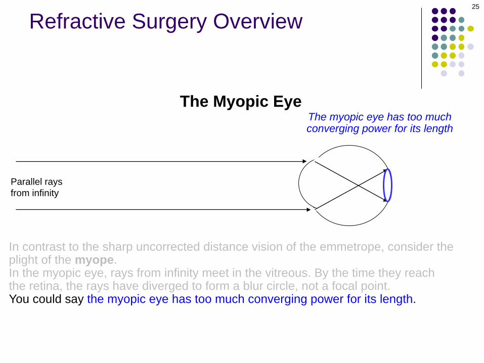
In contrast to the sharp uncorrected distance vision of the emmetrope, consider theplight of the myope.In the myopic eye, rays from infinity meet in the vitreous. By the time they reach the retina, the rays have diverged to form a blur circle, not a focal point.
24
Parallel rays from infinity
Refractive Surgery Overview
The Myopic EyeThe myopic eye has too muchconverging power for its length
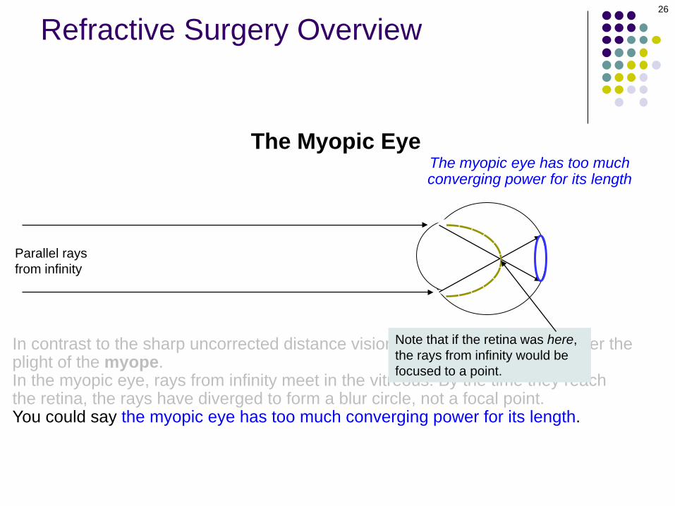
In contrast to the sharp uncorrected distance vision of the emmetrope, consider theplight of the myope.In the myopic eye, rays from infinity meet in the vitreous. By the time they reach the retina, the rays have diverged to form a blur circle, not a focal point.You could say the myopic eye has too much converging power for its length.
25
Parallel rays from infinity
Refractive Surgery Overview
In contrast to the sharp uncorrected distance vision of the emmetrope, consider theplight of the myope.In the myopic eye, rays from infinity meet in the vitreous. By the time they reach the retina, the rays have diverged to form a blur circle, not a focal point.You could say the myopic eye has too much converging power for its length.
Note that if the retina was here, the rays from infinity would be focused to a point.
The Myopic EyeThe myopic eye has too muchconverging power for its length
26
Parallel rays from infinity
Refractive Surgery Overview

The Myopic Eye
In contrast to the sharp uncorrected distance vision of the emmetrope, consider theplight of the myope.In the myopic eye, rays from infinity meet in the vitreous. By the time they reach the retina, the rays have diverged to form a blur circle, not a focal point.You could say the myopic eye has too much converging power for its length.To be focused on the retina, the Far Point of a myopic eye will have to offset its excess convergence with an equivalent amount of divergence. To accomplish this…
The myopic eye has too muchconverging power for its length
FarPoint?
27
Parallel rays from infinity
Refractive Surgery Overview
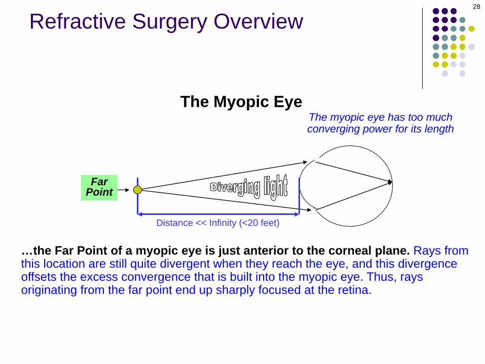
The Myopic Eye
Distance << Infinity (<20 feet)
…the Far Point of a myopic eye is just anterior to the corneal plane. Rays fromthis location are still quite divergent when they reach the eye, and this divergence offsets the excess convergence that is built into the myopic eye. Thus, rays originating from the far point end up sharply focused at the retina.
FarPoint
The myopic eye has too muchconverging power for its length
28
Refractive Surgery Overview

The Myopic Eye
Distance << Infinity (<20 feet)
…the Far Point of a myopic eye is just anterior to the corneal plane. Rays fromthis location are still quite divergent when they reach the eye, and this divergence offsets the excess convergence that is built into the myopic eye. Thus, rays originating from the far point end up sharply focused at the retina. This is why nearsighted individuals can read without glasses—they’re able to put the material at or near their far point.
FarPoint
The myopic eye has too muchconverging power for its length
29
Refractive Surgery Overview
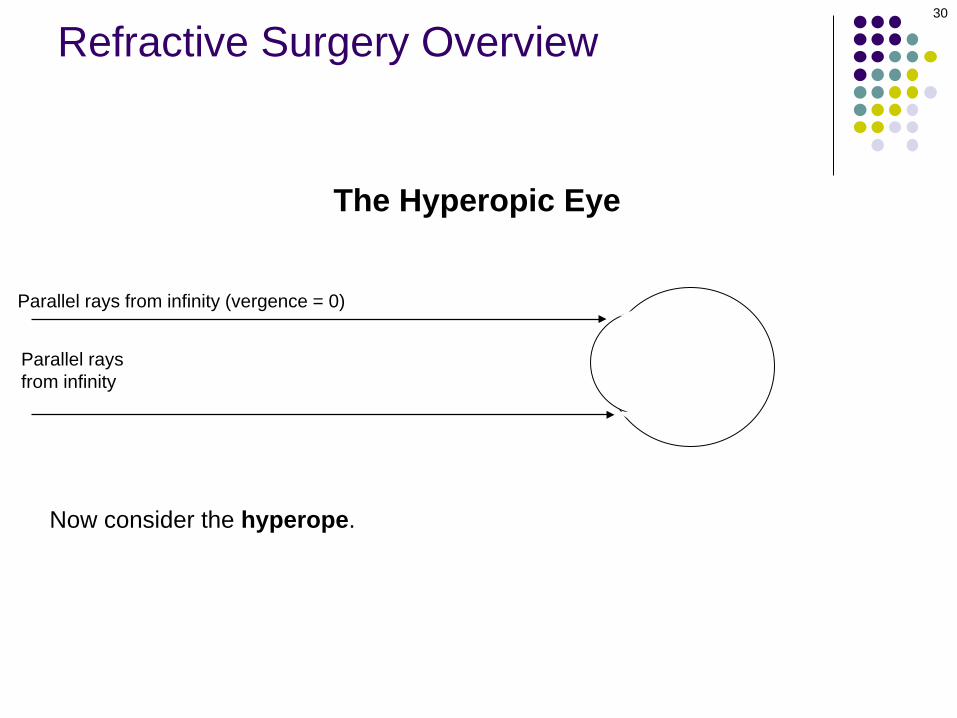
The Hyperopic Eye
Parallel rays from infinity (vergence = 0)
Now consider the hyperope.
30
Parallel rays from infinity
Refractive Surgery Overview

The Hyperopic Eye
Parallel rays from infinity (vergence = 0)
Now consider the hyperope.In the hyperopic eye, rays from infinity never meet—they run out of eyeball first. Thus, like the myopic eye, the rays form a blur circle, not a focal point, at the retina.
This is where the rays would meet if they hadn’t run into the retina.
31
Parallel rays from infinity
Refractive Surgery Overview
The Hyperopic Eye
Parallel rays from infinity (vergence = 0)
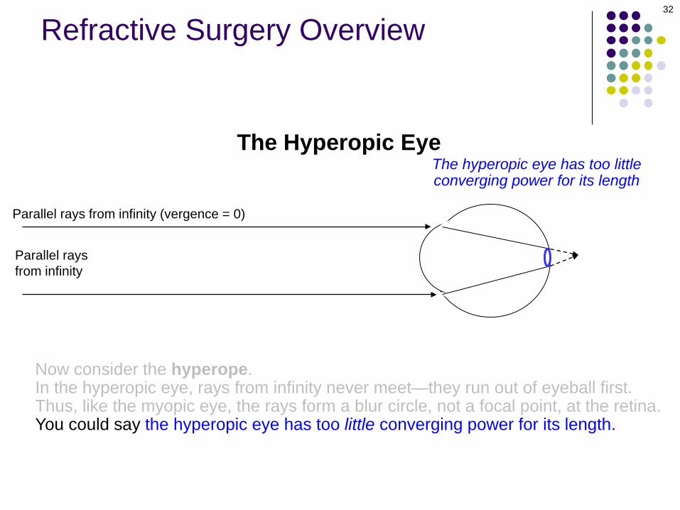
Now consider the hyperope.In the hyperopic eye, rays from infinity never meet—they run out of eyeball first. Thus, like the myopic eye, the rays form a blur circle, not a focal point, at the retina.You could say the hyperopic eye has too little converging power for its length.
The hyperopic eye has too littleconverging power for its length
32
Parallel rays from infinity
Refractive Surgery Overview

The Hyperopic Eye
Parallel rays from infinity (vergence = 0)
Now consider the hyperope.In the hyperopic eye, rays from infinity never meet—they run out of eyeball first. Thus, like the myopic eye, the rays form a blur circle, not a focal point, at the retina.You could say the hyperopic eye has too little converging power for its length.
33
Parallel rays from infinity
Refractive Surgery Overview
Note that if the retina was here, the rays from infinity would be focused to a point.
The hyperopic eye has too littleconverging power for its length

The Hyperopic Eye
Parallel rays from infinity (vergence = 0)
Now consider the hyperope.In the hyperopic eye, rays from infinity never meet—they run out of eyeball first. Thus, like the myopic eye, the rays form a blur circle, not a focal point, at the retina.You could say the hyperopic eye has too little converging power for its length.In order to be conjugate to the retina, the Far Point of a hyperopic eye mustcontribute convergence to compensate for this lack of converging power.To accomplish this…
FarPoint?
The hyperopic eye has too littleconverging power for its length
34
Parallel rays from infinity
Refractive Surgery Overview
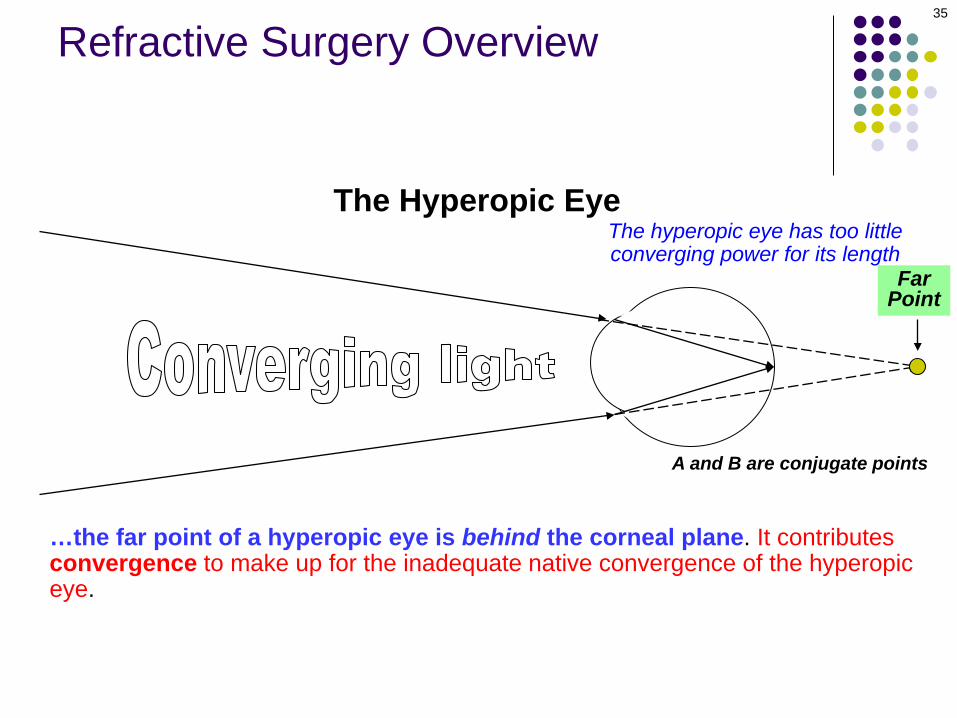
…the far point of a hyperopic eye is behind the corneal plane. It contributes convergence to make up for the inadequate native convergence of the hyperopic eye.
The Hyperopic Eye
FarPoint
The hyperopic eye has too littleconverging power for its length
A and B are conjugate points
35
Refractive Surgery Overview

…the far point of a hyperopic eye is behind the corneal plane. It contributes convergence to make up for the inadequate native convergence of the hyperopic eye. Thus, rays associated with the far point end up sharply focused at the retina.
The Hyperopic Eye
FarPoint
The hyperopic eye has too littleconverging power for its length
A and B are conjugate points
36
Refractive Surgery Overview
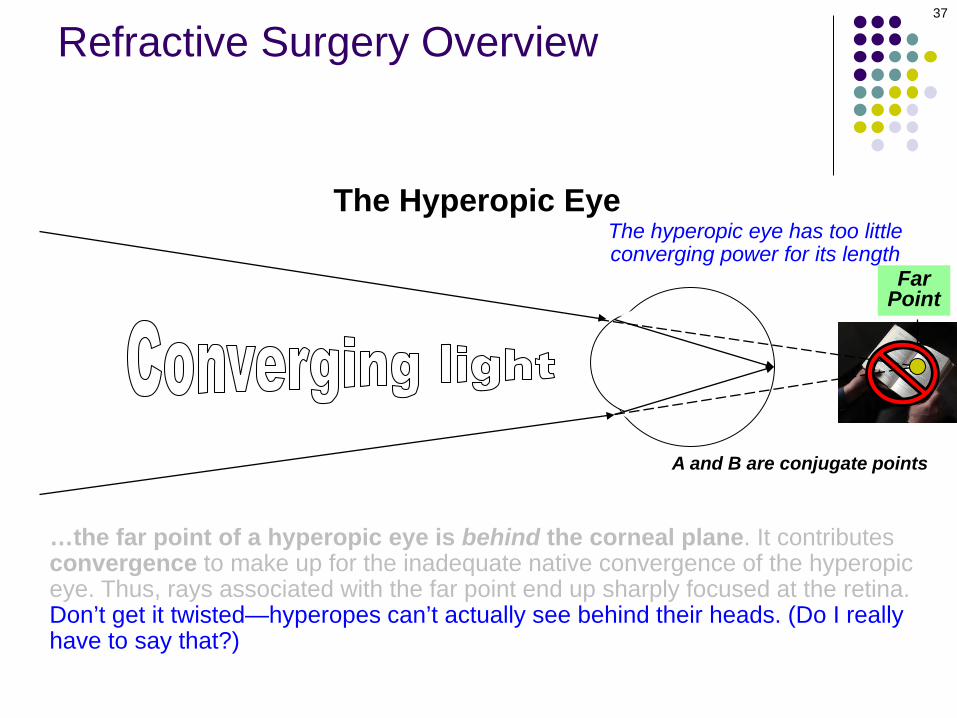
…the far point of a hyperopic eye is behind the corneal plane. It contributes convergence to make up for the inadequate native convergence of the hyperopic eye. Thus, rays associated with the far point end up sharply focused at the retina.Don’t get it twisted—hyperopes can’t actually see behind their heads. (Do I really have to say that?)
The Hyperopic Eye
FarPoint
The hyperopic eye has too littleconverging power for its length
A and B are conjugate points
37
Refractive Surgery Overview
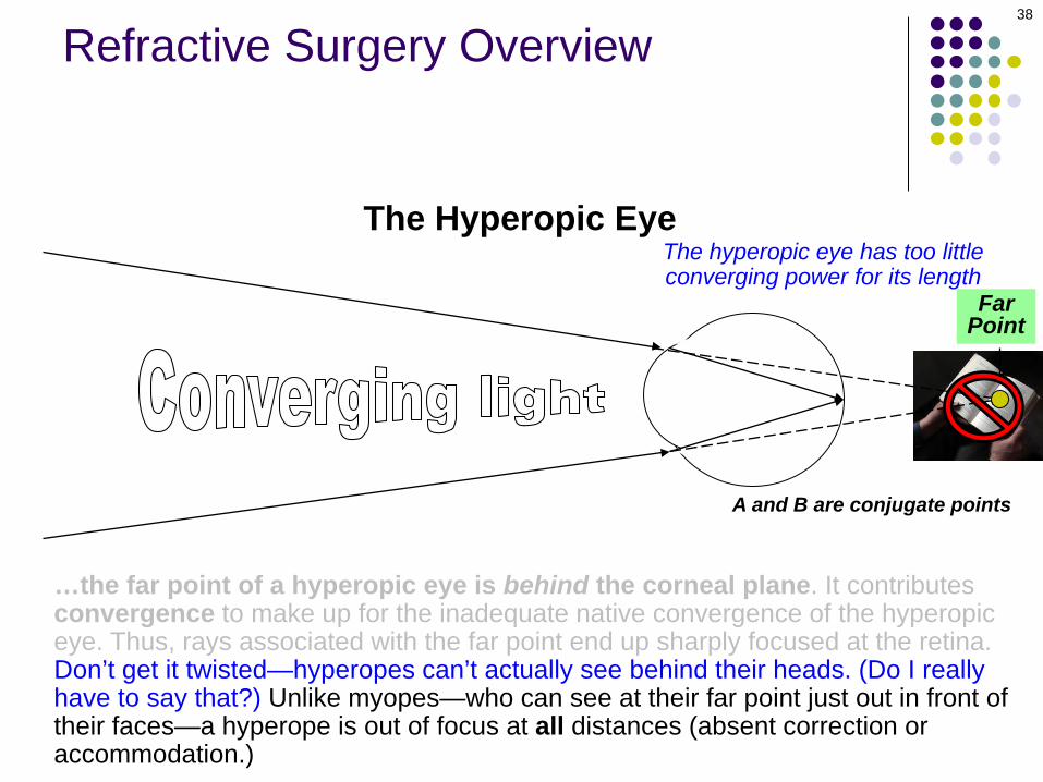
…the far point of a hyperopic eye is behind the corneal plane. It contributes convergence to make up for the inadequate native convergence of the hyperopic eye. Thus, rays associated with the far point end up sharply focused at the retina.Don’t get it twisted—hyperopes can’t actually see behind their heads. (Do I really have to say that?) Unlike myopes—who can see at their far point just out in front of their faces—a hyperope is out of focus at all distances (absent correction or accommodation.)
The Hyperopic Eye
FarPoint
The hyperopic eye has too littleconverging power for its length
A and B are conjugate points
38
Refractive Surgery Overview

The myopic eye has too much convergingpower for its length, as we said
Myopic Eye
39
Refractive Surgery Overview

The myopic eye has too much convergingpower for its length, as we said
Myopic Eye
Think of it this way: The myopic eye refracts light as if an extra ‘plus’ lens was built into it.This so-called error lens contributes the excess convergence that produces a myopicrefractive error.
40
Refractive Surgery Overview
Plus error lens

The myopic eye has too much convergingpower for its length, as we said
Myopic Eye
Minus corrective lens This explains why myopes wearminus lenses to correct theirrefractive error—minus lensesare needed to offset the excessconvergence induced by the pluserror lenses in their eyes.
Think of it this way: The myopic eye refracts light as if an extra ‘plus’ lens was built into it.This so-called error lens contributes the excess convergence that produces a myopicrefractive error.
41
Refractive Surgery Overview
Plus error lens
The myopic eye has too much convergingpower for its length, as we said
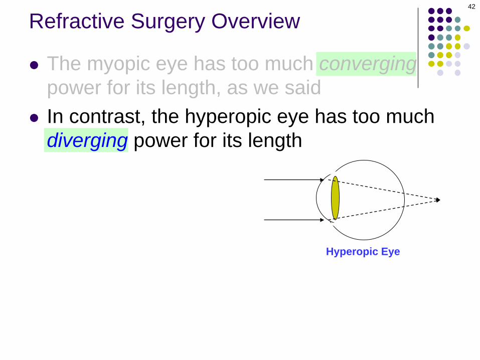
In contrast, the hyperopic eye has too much diverging power for its length
Hyperopic Eye
42
Refractive Surgery Overview
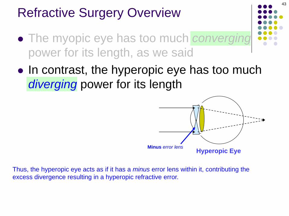
The myopic eye has too much convergingpower for its length, as we said
In contrast, the hyperopic eye has too much diverging power for its length
Hyperopic Eye
Thus, the hyperopic eye acts as if it has a minus error lens within it, contributing theexcess divergence resulting in a hyperopic refractive error.
43
Refractive Surgery Overview
Minus error lens

The myopic eye has too much convergingpower for its length, as we said
In contrast, the hyperopic eye has too much diverging power for its length
Hyperopic Eye
Plus corrective lensThis explains why hyperopeswear plus lenses to correct theirrefractive error—plus lenses areneeded to offset the excessdivergence induced by the minuserror lenses in their eyes.
Thus, the hyperopic eye acts as if it has a minus error lens within it, contributing theexcess divergence resulting in a hyperopic refractive error.
44
Refractive Surgery Overview
Minus error lens

45
Refractive Surgery Overview
Hyperopic EyeMinus error lensMyopic EyePlus error lens
The goal of refractive surgery is to produce an error-lens offset that is incorporated into the eye itself, rather than worn
on (CLs) or near (glasses) its anterior surface.

RefractiveSurgery
Corneal
46
Refractive Surgery Overview
Intraocular
As mentioned previously, refractive surgical procedures come in two basic forms—intraocular and corneal.
RefractiveSurgery
Corneal
47
Refractive Surgery Overview
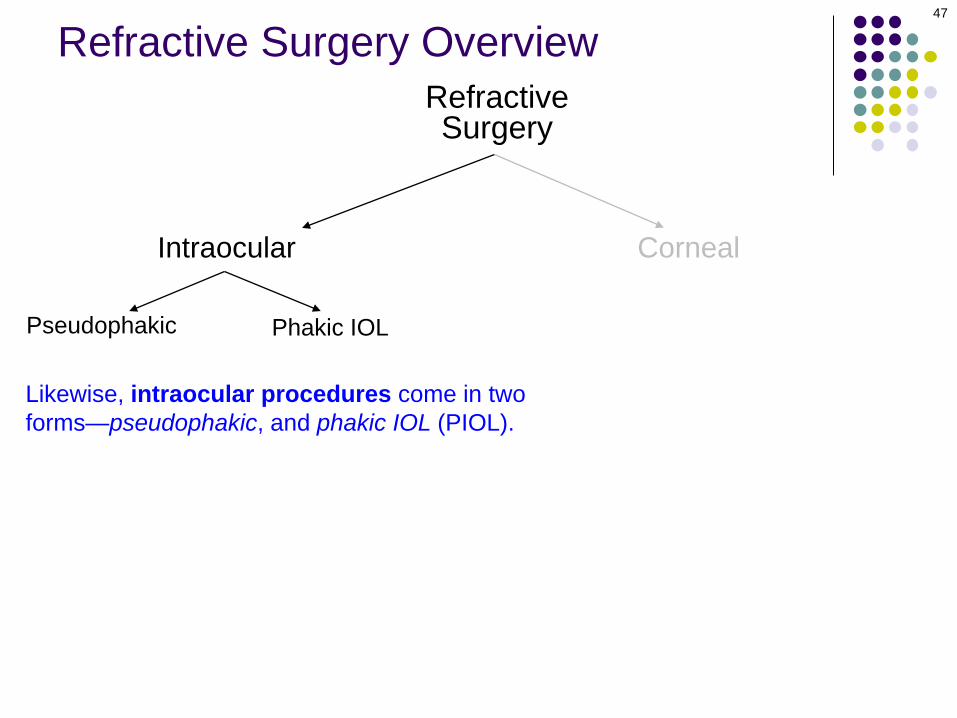
Intraocular
Likewise, intraocular procedures come in two forms—pseudophakic, and phakic IOL (PIOL).
Pseudophakic Phakic IOL
RefractiveSurgery
Corneal
48
Refractive Surgery Overview
Intraocular
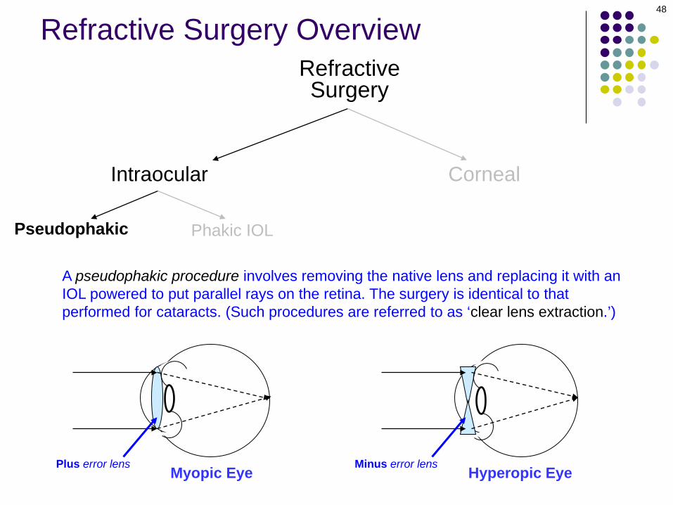
Pseudophakic
A pseudophakic procedure involves removing the native lens and replacing it with an IOL powered to put parallel rays on the retina. The surgery is identical to that performed for cataracts. (Such procedures are referred to as ‘clear lens extraction.’)
Myopic Eye Hyperopic EyePlus error lens Minus error lens
Phakic IOL
RefractiveSurgery
Corneal
49
Refractive Surgery Overview
Intraocular
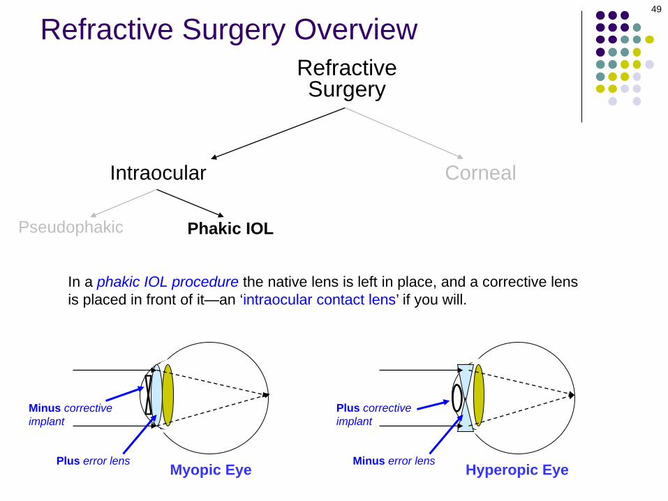
Pseudophakic Phakic IOL
In a phakic IOL procedure the native lens is left in place, and a corrective lens is placed in front of it—an ‘intraocular contact lens’ if you will.
Myopic EyePlus error lens Hyperopic EyeMinus error lens
Minus corrective implant
Plus corrective implant
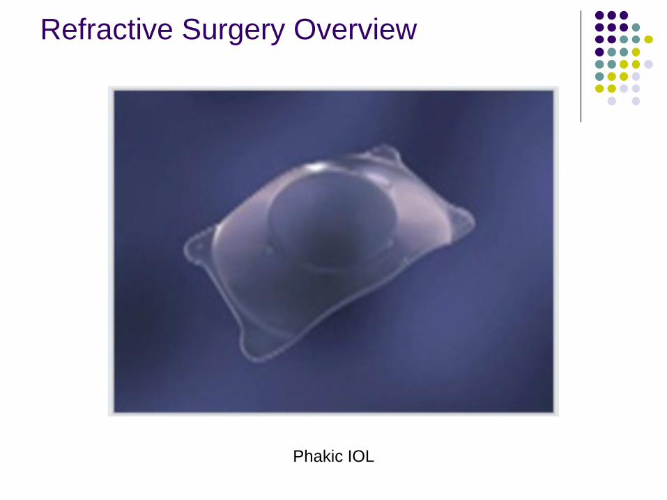
Phakic IOL
Refractive Surgery Overview

Phakic IOL
Refractive Surgery Overview
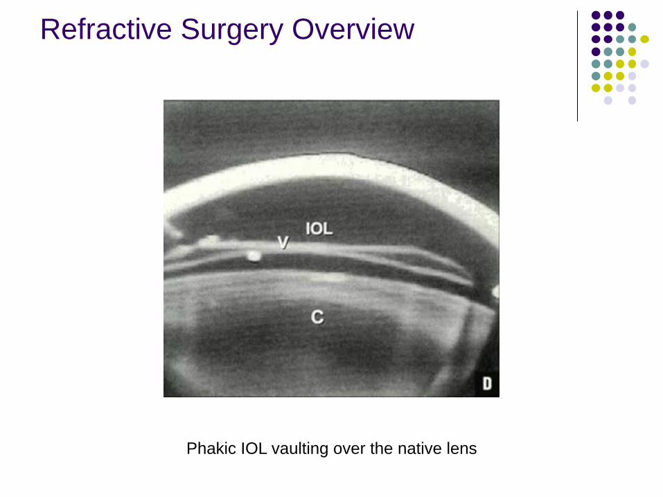
Phakic IOL vaulting over the native lens
Refractive Surgery Overview

RefractiveSurgery
Corneal
53
Refractive Surgery Overview
Intraocular
Pseudophakic Phakic IOL
Before we get into cornea-based refractive surgeries, let’s take a look at corneal optics
54
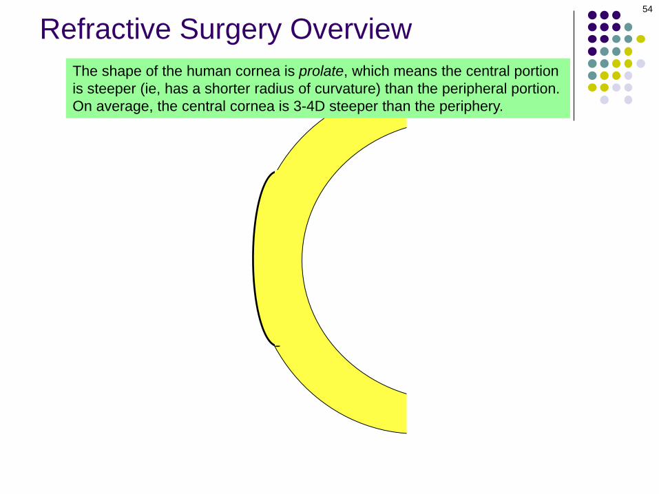
The shape of the human cornea is prolate, which means the central portion is steeper (ie, has a shorter radius of curvature) than the peripheral portion. On average, the central cornea is 3-4D steeper than the periphery.
Refractive Surgery Overview
55
Refractive Surgery Overview
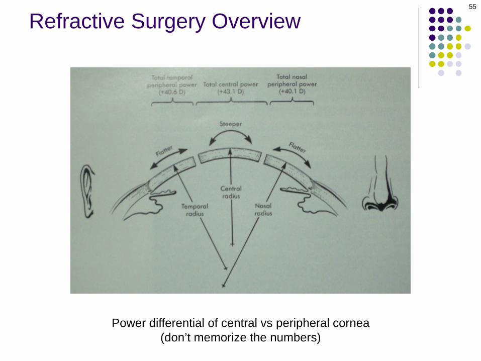
Power differential of central vs peripheral cornea (don’t memorize the numbers)
56
+49D
Light rays
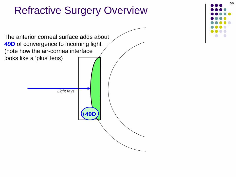
The anterior corneal surface adds about 49D of convergence to incoming light (note how the air-cornea interface looks like a ‘plus’ lens)
Refractive Surgery Overview

57
Light rays
-6D
However, the posterior corneal surface adds about 6D of divergence as light passes through it into the AC (note how the cornea-aqueous interface looks like a minus lens)
Refractive Surgery Overview

58
+49D
Light rays
-6D
+43D
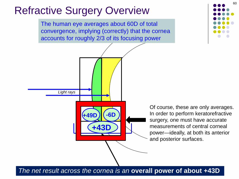
The net result across the cornea is an overall power of about +43D
Refractive Surgery Overview

59
+49D
Light rays
-6D
+43D
The net result across the cornea is an overall power of about +43D
The human eye averages about 60D of total convergence, implying (correctly) that the cornea accounts for roughly 2/3 of its focusing power
Refractive Surgery Overview
60
+49D
Light rays
-6D
+43D
The net result across the cornea is an overall power of about +43D
The human eye averages about 60D of total convergence, implying (correctly) that the cornea accounts for roughly 2/3 of its focusing power
Of course, these are only averages. In order to perform keratorefractive surgery, one must have accurate measurements of central corneal power—ideally, at both its anterior and posterior surfaces.
Refractive Surgery Overview
61
+D? -D?
+D?
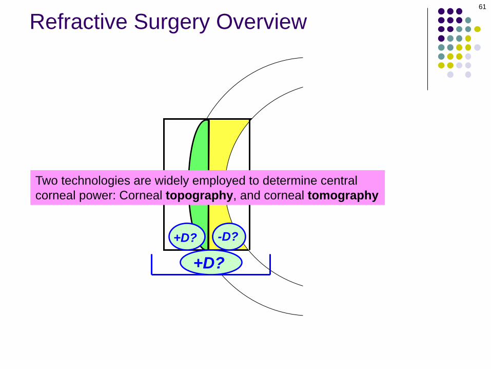
Two technologies are widely employed to determine central corneal power: Corneal topography, and corneal tomography
Refractive Surgery Overview

62
Refractive Surgery OverviewCorneal topography works by reflecting a set of concentric rings (collectively called a Placido disk) from the anterior corneal surface, and a computer analyzes the distances between, and shapes of, the reflected rings.

63
Refractive Surgery OverviewCorneal topography works by reflecting a set of concentric rings (collectively called a Placido disk) from the anterior corneal surface, and a computer analyzes the distances between, and shapes of, the reflected rings. Based on this analysis, the topographer creates a color-coded ‘map’ depicting the curvature across the corneal surface.

64
Refractive Surgery Overview
Corneal Placido-disk topography: Color map demonstrating with-the-rule astigmatism (ie, the cornea is steeper in its vertical meridian)
Corneal topography works by reflecting a set of concentric rings (collectively called a Placido disk) from the anterior corneal surface, and a computer analyzes the distances between, and shapes of, the reflected rings. Based on this analysis, the topographer creates a color-coded ‘map’ depicting the curvature across the corneal surface.
65
Refractive Surgery OverviewCorneal tomography works by mapping the anterior and posterior corneal surfaces in relation to one another. It allows for 3-D modeling of the cornea, including both anterior and posterior surface curvature and corneal thickness.
66
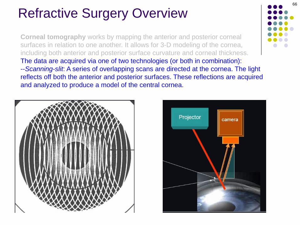
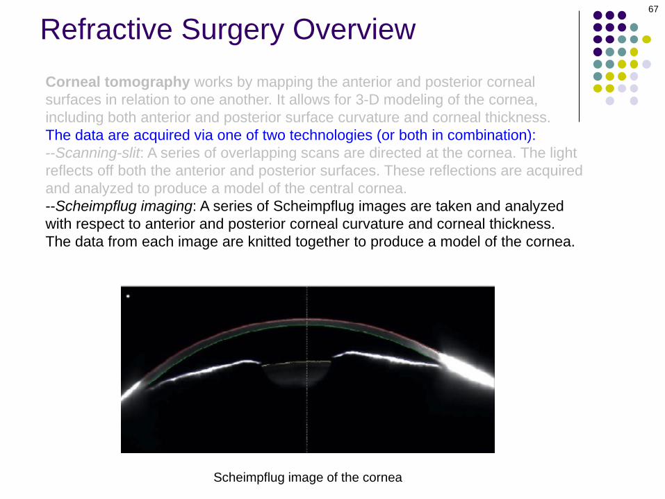
Refractive Surgery OverviewCorneal tomography works by mapping the anterior and posterior corneal surfaces in relation to one another. It allows for 3-D modeling of the cornea, including both anterior and posterior surface curvature and corneal thickness. The data are acquired via one of two technologies (or both in combination):--Scanning-slit: A series of overlapping scans are directed at the cornea. The light reflects off both the anterior and posterior surfaces. These reflections are acquired and analyzed to produce a model of the central cornea.
67
Refractive Surgery OverviewCorneal tomography works by mapping the anterior and posterior corneal surfaces in relation to one another. It allows for 3-D modeling of the cornea, including both anterior and posterior surface curvature and corneal thickness. The data are acquired via one of two technologies (or both in combination):--Scanning-slit: A series of overlapping scans are directed at the cornea. The light reflects off both the anterior and posterior surfaces. These reflections are acquired and analyzed to produce a model of the central cornea.--Scheimpflug imaging: A series of Scheimpflug images are taken and analyzed with respect to anterior and posterior corneal curvature and corneal thickness. The data from each image are knitted together to produce a model of the cornea.
Scheimpflug image of the cornea

68
Refractive Surgery Overview
Pentacam corneal tomographer readout

69
Refractive Surgery Overview
A) Anterior corneal values•K1, K2, Km: The two major meridians (K1, K2). Km is the average of K1 and K2•Rf, Rs, Rm: Radii corresponding with K1, K2, and Km, respectively•QS: Quality score (I.e. “OK,” “Data gaps,” “Fix,” “Model)•Axis: The meridian that requires no cylinder power to correct astigmatism•Astig: The central corneal astigmatismB) Posterior corneal valuesThe same variables described for the back of the cornea.C), D) Fuggedaboudit (too much for this overview)
Pentacam corneal tomographer readout

70
+D? -D?
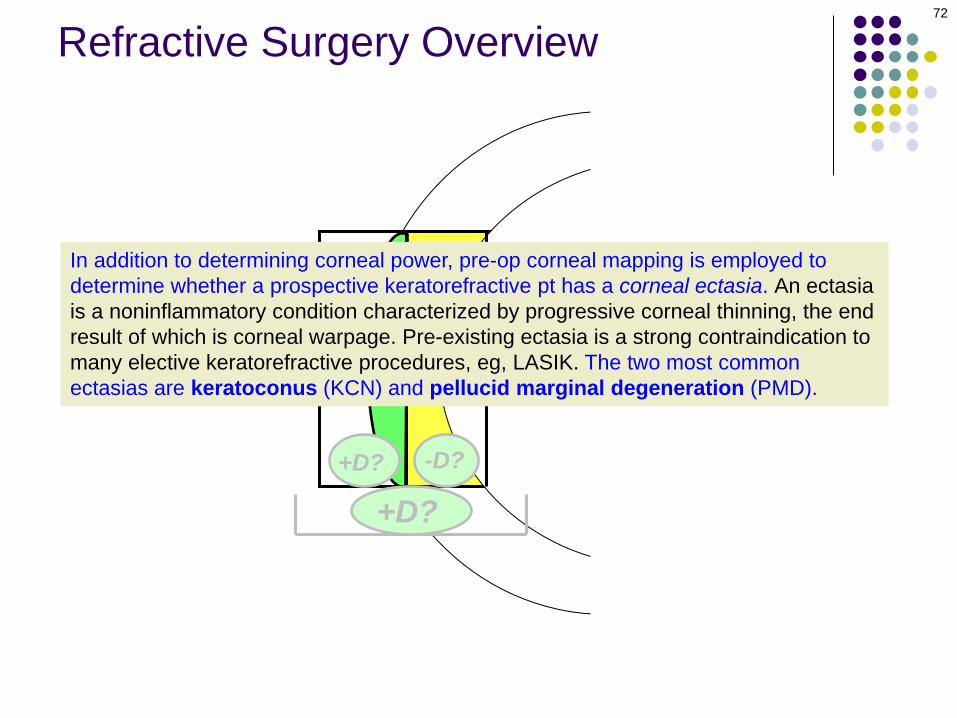
In addition to determining corneal power, pre-op corneal mapping is employed to determine whether a prospective keratorefractive pt has a corneal ectasia. An ectasia is a noninflammatory condition characterized by progressive corneal thinning, the end result of which is corneal warpage. Pre-existing ectasia is a strong contraindication to many elective keratorefractive procedures, eg, LASIK. The two most common ectasias are keratoconus (KCN) and pellucid marginal degeneration (PMD).
+D?
Refractive Surgery Overview

71
+D? -D?
In addition to determining corneal power, pre-op corneal mapping is employed to determine whether a prospective keratorefractive pt has a corneal ectasia. An ectasia is a noninflammatory condition characterized by progressive corneal thinning, the end result of which is corneal warpage. Pre-existing ectasia is a strong contraindication to many elective keratorefractive procedures, eg, LASIK. The two most common ectasias are keratoconus (KCN) and pellucid marginal degeneration (PMD).
+D?
Refractive Surgery Overview
72
+D? -D?
In addition to determining corneal power, pre-op corneal mapping is employed to determine whether a prospective keratorefractive pt has a corneal ectasia. An ectasia is a noninflammatory condition characterized by progressive corneal thinning, the end result of which is corneal warpage. Pre-existing ectasia is a strong contraindication to many elective keratorefractive procedures, eg, LASIK. The two most common ectasias are keratoconus (KCN) and pellucid marginal degeneration (PMD).
+D?
Refractive Surgery Overview

Refractive Surgery Overview
KCN

Topography in KCN: Classic inferior corneal steepening
Refractive Surgery Overview

Refractive Surgery Overview
Topography in PMD: Classic kissing doves
Refractive Surgery Overview
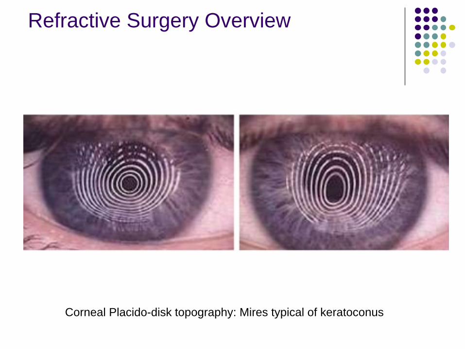
Corneal Placido-disk topography: Mires typical of keratoconus

RefractiveSurgery
Corneal
77
Refractive Surgery Overview
Intraocular
Pseudophakic Phakic IOL
Most corneal refractive surgeries involve altering the shape of the cornea in a way that impacts the vergence it imparts to incoming light.

RefractiveSurgery
Corneal
Incisional Laser
78
Refractive Surgery Overview
Intraocular
Pseudophakic Phakic IOL Other
Most corneal refractive surgeries involve altering the shape of the cornea in a way that impacts the vergence it imparts to incoming light. These alterations can involve incising the cornea, lasering it, or some other means.

RefractiveSurgery
Corneal
Incisional Laser
79
Refractive Surgery Overview
Intraocular
Pseudophakic Phakic IOL Other
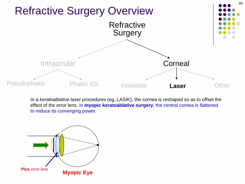
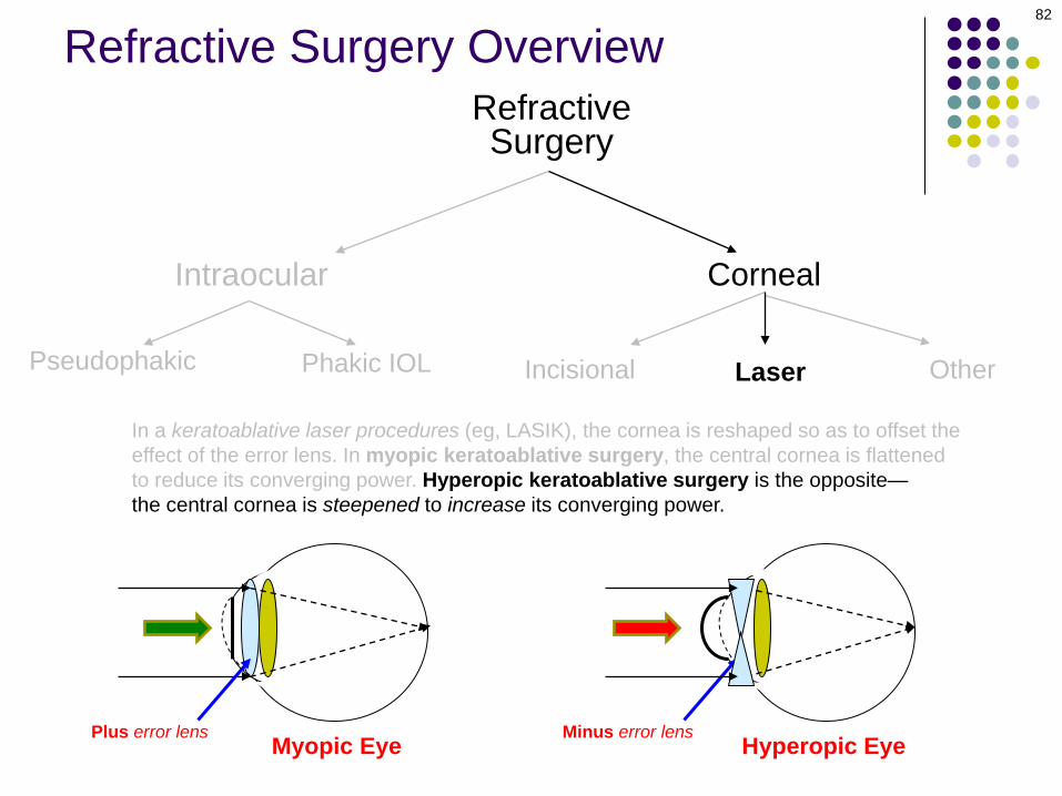
In a keratoablative laser procedures (eg, LASIK), the cornea is reshaped so as to offset the effect of the error lens..
RefractiveSurgery
Corneal
Incisional Laser
80
Refractive Surgery Overview
Intraocular
Pseudophakic Phakic IOL Other
Myopic EyePlus error lens
In a keratoablative laser procedures (eg, LASIK), the cornea is reshaped so as to offset the effect of the error lens. In myopic keratoablative surgery, the central cornea is flattened to reduce its converging power.

RefractiveSurgery
Corneal
Incisional Laser
81
Refractive Surgery Overview
Intraocular
Pseudophakic Phakic IOL Other
Myopic EyePlus error lens
In a keratoablative laser procedures (eg, LASIK), the cornea is reshaped so as to offset the effect of the error lens. In myopic keratoablative surgery, the central cornea is flattened to reduce its converging power. Hyperopic
Think of it as shaving down the peak of a mountain in order to make the structure more mesa-like
RefractiveSurgery
Corneal
Incisional Laser
82
Refractive Surgery Overview
Intraocular
Pseudophakic Phakic IOL Other
Hyperopic EyeMinus error lensMyopic EyePlus error lens
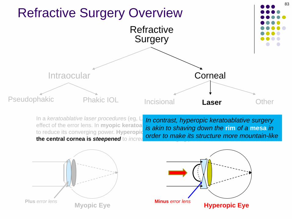
In a keratoablative laser procedures (eg, LASIK), the cornea is reshaped so as to offset the effect of the error lens. In myopic keratoablative surgery, the central cornea is flattened to reduce its converging power. Hyperopic keratoablative surgery is the opposite—the central cornea is steepened to increase its converging power.
RefractiveSurgery
Corneal
Incisional Laser
83
Refractive Surgery Overview
Intraocular
Pseudophakic Phakic IOL Other
Hyperopic EyeMinus error lensMyopic EyePlus error lens
In a keratoablative laser procedures (eg, LASIK), the cornea is reshaped so as to offset the effect of the error lens. In myopic keratoablative surgery, the central cornea is flattened to reduce its converging power. Hyperopic keratoablative surgery is the opposite—the central cornea is steepened to increase its converging power.
In contrast, hyperopic keratoablative surgery is akin to shaving down the rim of a mesa in order to make its structure more mountain-like

In a keratoablative laser procedures (eg, LASIK), the cornea is reshaped so as to offset the effect of the error lens. In myopic keratoablative surgery, the central cornea is flattened to reduce its converging power. Hyperopic keratoablative surgery is the opposite—the central cornea is steepened to increase its converging power.
RefractiveSurgery
Corneal
Incisional Laser
84
Refractive Surgery Overview
Intraocular
Pseudophakic Phakic IOL Other
Hyperopic EyeMinus error lensMyopic EyePlus error lens
Note that, by definition, keratoablative refractive surgery involves reshaping the central cornea (and thereby altering its refractive power) via the removal (by annihilation) of corneal tissue

In a keratoablative laser procedures (eg, LASIK), the cornea is reshaped so as to offset the effect of the error lens. In myopic keratoablative surgery, the central cornea is flattened to reduce its converging power. Hyperopic keratoablative surgery is the opposite—the central cornea is steepened to increase its converging power.
RefractiveSurgery
Corneal
Incisional Laser
85
Refractive Surgery Overview
Intraocular
Pseudophakic Phakic IOL Other
Hyperopic EyeMinus error lensMyopic EyePlus error lens
*One laser-based keratorefractive procedure does not involve tissue annihilation, rather, in it a section of corneal stroma is carved, then removed en bloc
Note that, by definition, keratoablative refractive surgery involves reshaping the central cornea (and thereby altering its refractive power) via the removal (by annihilation) of corneal tissue*
RefractiveSurgery
Corneal
Incisional Laser
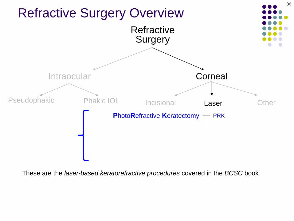
86
Intraocular
Pseudophakic Phakic IOL
PRKPhotoRefractive Keratectomy
Other
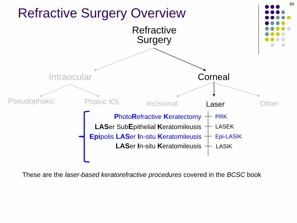
These are the laser-based keratorefractive procedures covered in the BCSC book
Refractive Surgery Overview
RefractiveSurgery
Corneal
Incisional Laser
87
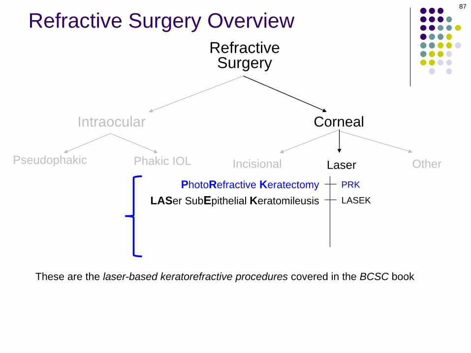
Intraocular
Pseudophakic Phakic IOL
PRK
LASEKPhotoRefractive Keratectomy
LASer SubEpithelial Keratomileusis
Other
These are the laser-based keratorefractive procedures covered in the BCSC book
Refractive Surgery Overview

RefractiveSurgery
Corneal
Incisional Laser
88
Intraocular
Pseudophakic Phakic IOL
PRK
LASEK
Epi-LASIK
PhotoRefractive KeratectomyLASer SubEpithelial Keratomileusis
Epipolis LASer In-situ Keratomileusis
Other
These are the laser-based keratorefractive procedures covered in the BCSC book
Refractive Surgery Overview
RefractiveSurgery
Corneal
Incisional Laser
89
Intraocular
Pseudophakic Phakic IOL
PRK
LASEK
LASIK
Epi-LASIK
LASer In-situ Keratomileusis
PhotoRefractive KeratectomyLASer SubEpithelial Keratomileusis
Epipolis LASer In-situ Keratomileusis
Other
These are the laser-based keratorefractive procedures covered in the BCSC book
Refractive Surgery Overview
RefractiveSurgery
Corneal
Incisional Laser
90
Intraocular
Pseudophakic Phakic IOL
PRK
LASEK
LASIK
SMILE
Epi-LASIK
LASer In-situ Keratomileusis
PhotoRefractive KeratectomyLASer SubEpithelial Keratomileusis
Epipolis LASer In-situ Keratomileusis
SMall-Incision Lenticule Extraction
Other
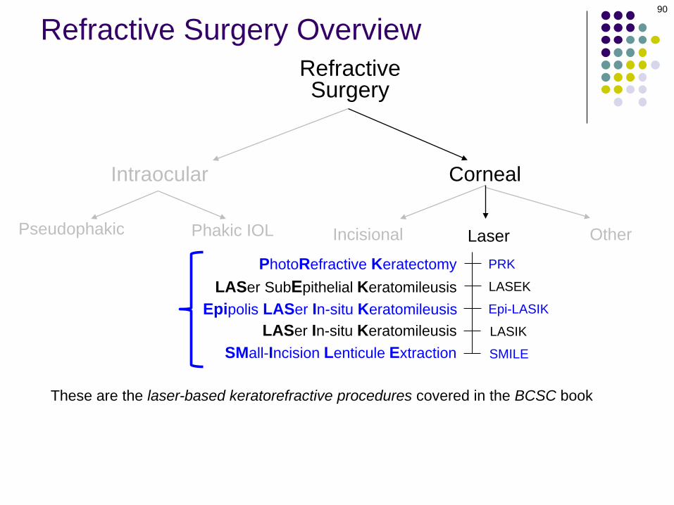
These are the laser-based keratorefractive procedures covered in the BCSC book
Refractive Surgery Overview

RefractiveSurgery
Corneal
Incisional Laser
91
Intraocular
Pseudophakic Phakic IOL
PRK
LASEK
LASIK
SMILE
Epi-LASIK
LASer In-situ Keratomileusis
PhotoRefractive KeratectomyLASer SubEpithelial Keratomileusis
Epipolis LASer In-situ Keratomileusis
SMall-Incision Lenticule Extraction
Other
These are the laser-based keratorefractive procedures covered in the BCSC book. All are ablative except for SMILE, which is the nonablative one referred to on a recent slide.
Refractive Surgery Overview

RefractiveSurgery
Corneal
Incisional Laser
92
Intraocular
Pseudophakic Phakic IOL
PRK
LASEK
LASIK
SMILE
Epi-LASIK
LASer In-situ Keratomileusis
PhotoRefractive KeratectomyLASer SubEpithelial Keratomileusis
Epipolis LASer In-situ Keratomileusis
SMall-Incision Lenticule Extraction
Other
These are the laser-based keratorefractive procedures covered in the BCSC book. All are ablative except for SMILE, which is the nonablative one referred to on a recent slide.
Refractive Surgery Overview
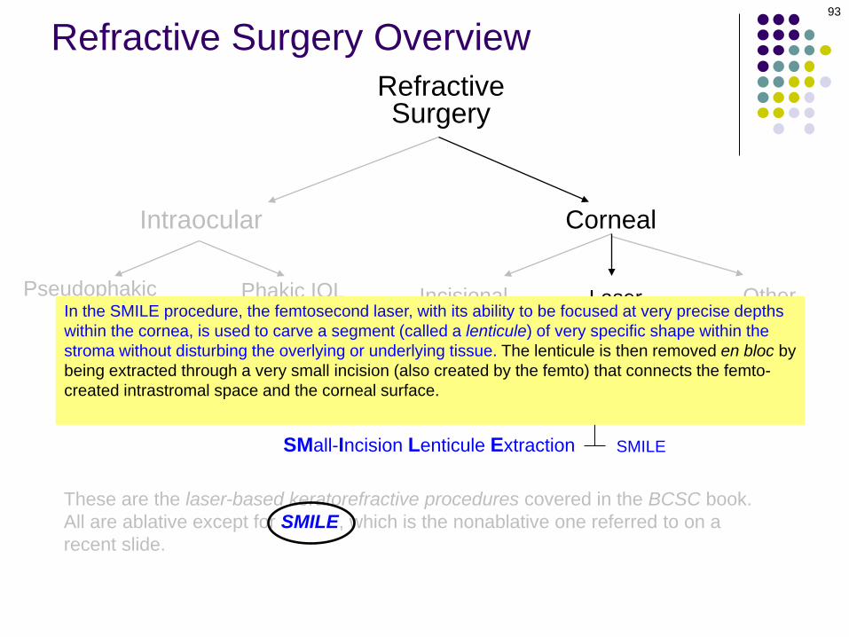
In the SMILE procedure, the femtosecond laser, with its ability to be focused at very precise depths within the cornea, is used to carve a segment (called a lenticule) of very specific shape within the stroma without disturbing the overlying or underlying tissue. The lenticule is then removed en bloc by being extracted through a very small incision (also created by the femto) that connects the femto-created intrastromal space and the corneal surface. The resulting loss of tissue reshapes the central corneal surface in a way that produces a desired change in its refractive power.
RefractiveSurgery
Corneal
Incisional Laser
93
Intraocular
Pseudophakic Phakic IOL
PRK
LASEK
LASIK
SMILE
Epi-LASIK
LASer In-situ Keratomileusis
PhotoRefractive KeratectomyLASer SubEpithelial Keratomileusis
Epipolis LASer In-situ Keratomileusis
SMall-Incision Lenticule Extraction
Other
These are the laser-based keratorefractive procedures covered in the BCSC book. All are ablative except for SMILE, which is the nonablative one referred to on a recent slide.
Refractive Surgery Overview
In the SMILE procedure, the femtosecond laser, with its ability to be focused at very precise depths within the cornea, is used to carve a segment (called a lenticule) of very specific shape within the stroma without disturbing the overlying or underlying tissue. The lenticule is then removed en bloc by being extracted through a very small incision (also created by the femto) that connects the femto-created intrastromal space and the corneal surface. The resulting loss of tissue reshapes the central corneal surface in a way that produces a desired change in its refractive power.

94
Refractive Surgery Overview
SMILE

RefractiveSurgery
Corneal
Incisional Laser
95
Intraocular
Pseudophakic Phakic IOL
PRK
LASEK
LASIK
SMILE
Epi-LASIK
LASer In-situ Keratomileusis
PhotoRefractive KeratectomyLASer SubEpithelial Keratomileusis
Epipolis LASer In-situ Keratomileusis
SMall-Incision Lenticule Extraction
Other
These are the laser-based keratorefractive procedures covered in the BCSC book. All are ablative except for SMILE, which is the nonablative one referred to on a recent slide.
In the SMILE procedure, the femtosecond laser, with its ability to be focused at very precise depths within the cornea, is used to carve a segment (called a lenticule) of very specific shape within the stroma without disturbing the overlying or underlying tissue. The lenticule is then removed en bloc by being extracted through a very small incision (also created by the femto) that connects the femto-created intrastromal space and the corneal surface. The resulting loss of tissue reshapes the central corneal surface in a way that produces a desired change in its refractive power.
Refractive Surgery Overview
RefractiveSurgery
Corneal
Incisional Laser
96
Intraocular
Pseudophakic Phakic IOL
PRK
LASEK
LASIK
SMILE
Epi-LASIK
LASer In-situ Keratomileusis
PhotoRefractive KeratectomyLASer SubEpithelial Keratomileusis
Epipolis LASer In-situ Keratomileusis
SMall-Incision Lenticule Extraction
Other
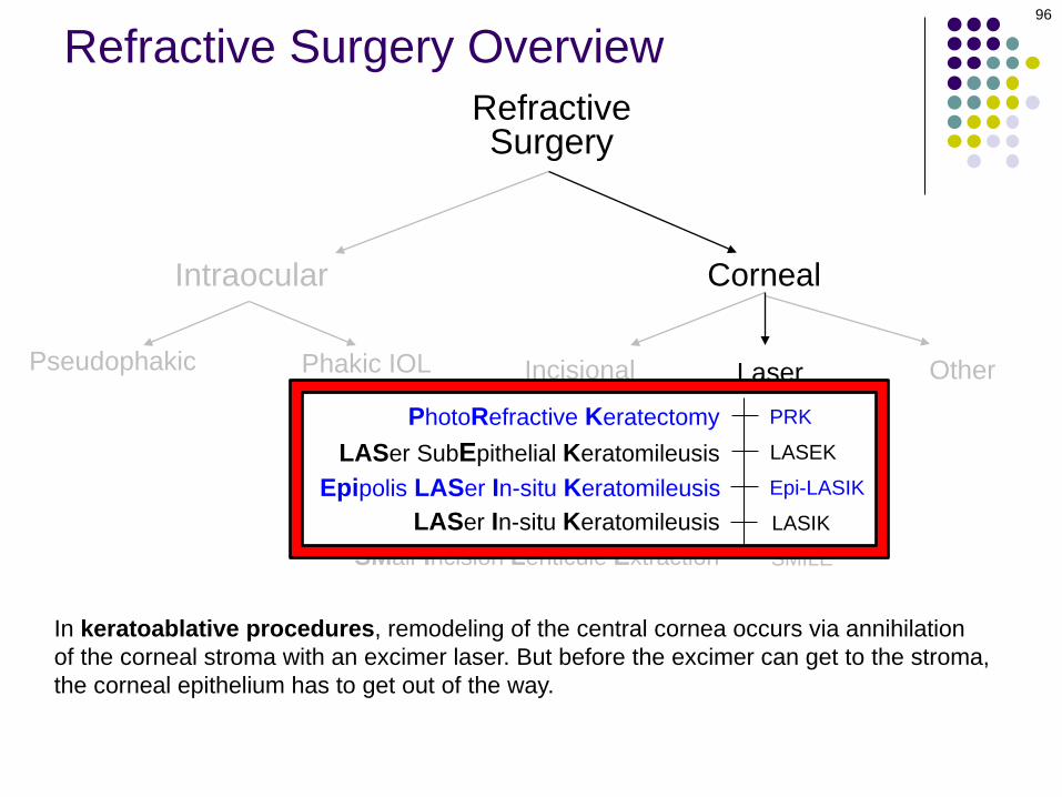
In keratoablative procedures, remodeling of the central cornea occurs via annihilation of the corneal stroma with an excimer laser. But before the excimer can get to the stroma, the corneal epithelium has to get out of the way.
Refractive Surgery Overview

RefractiveSurgery
Corneal
Incisional Laser
97
Intraocular
Pseudophakic Phakic IOL
PRK
LASEK
LASIK
SMILE
Epi-LASIK
LASer In-situ Keratomileusis
PhotoRefractive KeratectomyLASer SubEpithelial Keratomileusis
Epipolis LASer In-situ Keratomileusis
SMall-Incision Lenticule Extraction
Other
In keratoablative procedures, remodeling of the central cornea occurs via annihilation of the corneal stroma with an excimer laser. But before the excimer can get to the stroma, the corneal epithelium has to get out of the way. The four keratoablative procedures differ solely in how the epithelium is handled.
Refractive Surgery Overview
In keratoablative procedures, remodeling of the central cornea occurs via annihilation of the corneal stroma with an excimer laser. But before the excimer can get to the stroma, the corneal epithelium has to get out of the way. The four keratoablative procedures differ solely in how the epithelium is handled.
RefractiveSurgery
Corneal
Incisional Laser
98
Intraocular
Pseudophakic Phakic IOL
PRK
LASEK
LASIK
SMILE
Epi-LASIK
LASer In-situ Keratomileusis
PhotoRefractive KeratectomyLASer SubEpithelial Keratomileusis
Epipolis LASer In-situ Keratomileusis
SMall-Incision Lenticule Extraction
Other
Refractive Surgery Overview
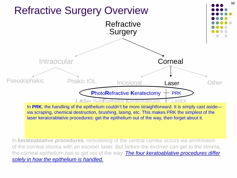
In PRK, the handling of the epithelium couldn’t be more straightforward. It is simply cast aside—via scraping, chemical destruction, brushing, lasing, etc. This makes PRK the simplest of the laser keratorablative procedures: get the epithelium out of the way, then forget about it. However, PRK is associated with several post-operative complications that render it problematic, chief among them being that it is associated with significant post-op pain as well as an increased risk of post-op haze formation—a potentially sight-threatening complication.

In keratoablative procedures, remodeling of the central cornea occurs via annihilation of the corneal stroma with an excimer laser. But before the excimer can get to the stroma, the corneal epithelium has to get out of the way. The four keratoablative procedures differ solely in how the epithelium is handled.
RefractiveSurgery
Corneal
Incisional Laser
99
Intraocular
Pseudophakic Phakic IOL
PRK
LASEK
LASIK
SMILE
Epi-LASIK
LASer In-situ Keratomileusis
PhotoRefractive KeratectomyLASer SubEpithelial Keratomileusis
Epipolis LASer In-situ Keratomileusis
SMall-Incision Lenticule Extraction
Other
Refractive Surgery Overview
In PRK, the handling of the epithelium couldn’t be more straightforward. It is simply cast aside—via scraping, chemical destruction, brushing, lasing, etc. This makes PRK the simplest of the laser keratorablative procedures: get the epithelium out of the way, then forget about it. However, PRK is associated with several post-operative complications that render it problematic, two of which are 1) it produces significant post-op pain, and 2) it is associated with an increased risk of post-op haze formation—a potentially sight-threatening development.
100
Refractive Surgery Overview
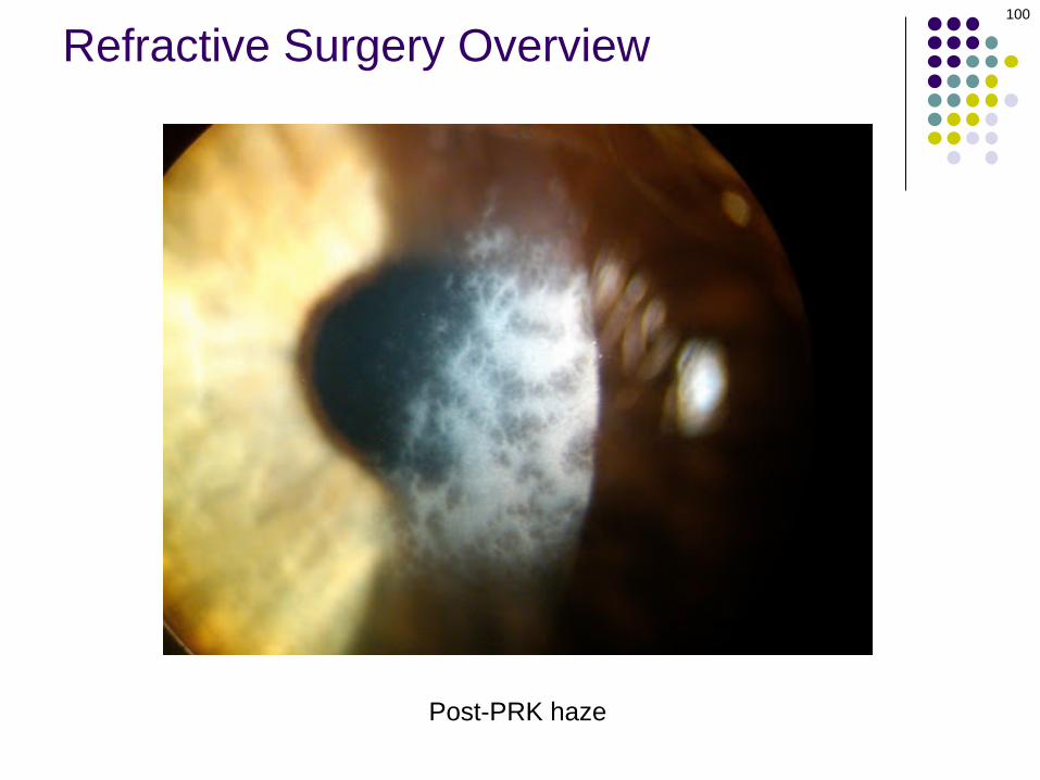
Post-PRK haze

RefractiveSurgery
Corneal
Incisional Laser
101
Intraocular
Pseudophakic Phakic IOL
PRK
LASEK
LASIK
SMILE
Epi-LASIK
LASer In-situ Keratomileusis
PhotoRefractive KeratectomyLASer SubEpithelial Keratomileusis
Epipolis LASer In-situ Keratomileusis
SMall-Incision Lenticule Extraction
Other
Refractive Surgery Overview
In keratoablative procedures, remodeling of the central cornea occurs via annihilation of the corneal stroma with an excimer laser. But before the excimer can get to the stroma, the corneal epithelium has to get out of the way. The four keratoablative procedures differ solely in how the epithelium is handled.
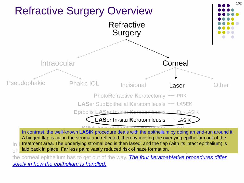
In contrast, the well-known LASIK procedure deals with the epithelium by doing an end-run around it. A hinged flap is cut in the stroma and reflected, thereby moving the overlying epithelium out of the treatment area. The underlying stromal bed is then lased, and the flap (with its intact epithelium) is laid back in place.
RefractiveSurgery
Corneal
Incisional Laser
102
Intraocular
Pseudophakic Phakic IOL
PRK
LASEK
LASIK
SMILE
Epi-LASIK
LASer In-situ Keratomileusis
PhotoRefractive KeratectomyLASer SubEpithelial Keratomileusis
Epipolis LASer In-situ Keratomileusis
SMall-Incision Lenticule Extraction
Other
Refractive Surgery Overview
In keratoablative procedures, remodeling of the central cornea occurs via annihilation of the corneal stroma with an excimer laser. But before the excimer can get to the stroma, the corneal epithelium has to get out of the way. The four keratoablative procedures differ solely in how the epithelium is handled.
In contrast, the well-known LASIK procedure deals with the epithelium by doing an end-run around it. A hinged flap is cut in the stroma and reflected, thereby moving the overlying epithelium out of the treatment area. The underlying stromal bed is then lased, and the flap (with its intact epithelium) is laid back in place. Far less pain; vastly reduced risk of haze formation.

Refractive Surgery Overview
LASIK
LASEK
RefractiveSurgery
Corneal
Incisional Laser
104
Intraocular
Pseudophakic Phakic IOL
PRK
LASIK
SMILE
Epi-LASIK
LASer In-situ Keratomileusis
PhotoRefractive KeratectomyLASer SubEpithelial Keratomileusis
Epipolis LASer In-situ Keratomileusis
SMall-Incision Lenticule Extraction
Other
Refractive Surgery Overview
In keratoablative procedures, remodeling of the central cornea occurs via annihilation of the corneal stroma with an excimer laser. But before the excimer can get to the stroma, the corneal epithelium has to get out of the way. The four keratoablative procedures differ solely in how the epithelium is handled.
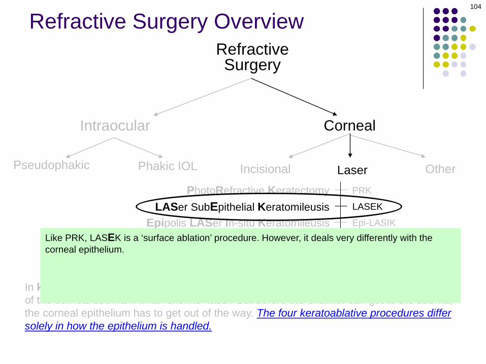
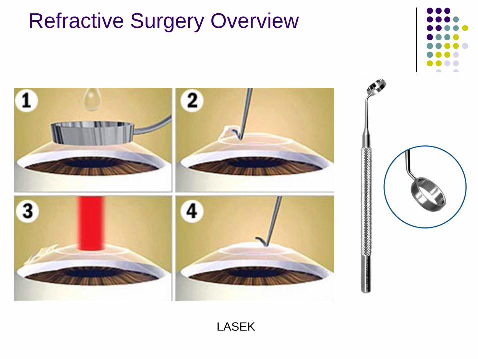
Like PRK, LASEK is a ‘surface ablation’ procedure. However, it deals very differently with the corneal epithelium. In LASEK, the epithelium is chemically devitalized and loosened by bathing it in an alcohol solution. The loosened epithelium is then folded back, and the ablation is performed. Following the ablation, this ‘epithelial flap’ is smoothed back into place and covered with a bandage CL. By re-positing the epithelium, LASEK avoids the large epi defect (and resulting severe pain) of PRK.

LASEK
RefractiveSurgery
Corneal
Incisional Laser
105
Intraocular
Pseudophakic Phakic IOL
PRK
LASIK
SMILE
Epi-LASIK
LASer In-situ Keratomileusis
PhotoRefractive KeratectomyLASer SubEpithelial Keratomileusis
Epipolis LASer In-situ Keratomileusis
SMall-Incision Lenticule Extraction
Other
Refractive Surgery Overview
In keratoablative procedures, remodeling of the central cornea occurs via annihilation of the corneal stroma with an excimer laser. But before the excimer can get to the stroma, the corneal epithelium has to get out of the way. The four keratoablative procedures differ solely in how the epithelium is handled.
Like PRK, LASEK is a ‘surface ablation’ procedure. However, it deals very differently with the corneal epithelium. In LASEK, the epithelium is chemically devitalized and loosened by bathing it in an alcohol solution. The loosened epithelium is then folded back, and the ablation is performed. Following the ablation, this ‘epithelial flap’ is smoothed back into place and covered with a bandage CL. By re-positing the epithelium, LASEK avoids the large epi defect (and resulting severe pain) of PRK.
LASEK
Refractive Surgery Overview

LASEK
RefractiveSurgery
Corneal
Incisional Laser
107
Intraocular
Pseudophakic Phakic IOL
PRK
LASIK
SMILE
Epi-LASIK
LASer In-situ Keratomileusis
PhotoRefractive KeratectomyLASer SubEpithelial Keratomileusis
Epipolis LASer In-situ Keratomileusis
SMall-Incision Lenticule Extraction
Other
Refractive Surgery Overview
In keratoablative procedures, remodeling of the central cornea occurs via annihilation of the corneal stroma with an excimer laser. But before the excimer can get to the stroma, the corneal epithelium has to get out of the way. The four keratoablative procedures differ solely in how the epithelium is handled.
Like PRK, LASEK is a ‘surface ablation’ procedure. However, it deals very differently with the corneal epithelium. In LASEK, the epithelium is chemically devitalized and loosened by bathing it in an alcohol solution. The loosened epithelium is then folded back, and the ablation is performed. Following the ablation, this ‘epithelial flap’ is smoothed back into place and covered with a bandage CL. By re-positing the epithelium, LASEK avoids the large epi defect (and resulting severe pain) of PRK.
Epipolis LASer In-situ Keratomileusis Epi-LASIK
RefractiveSurgery
Corneal
Incisional Laser
108
Intraocular
Pseudophakic Phakic IOL
PRK
LASEK
LASIK
SMILE
LASer In-situ Keratomileusis
PhotoRefractive KeratectomyLASer SubEpithelial Keratomileusis
SMall-Incision Lenticule Extraction
Other
Refractive Surgery Overview
In keratoablative procedures, remodeling of the central cornea occurs via annihilation of the corneal stroma with an excimer laser. But before the excimer can get to the stroma, the corneal epithelium has to get out of the way. The four keratoablative procedures differ solely in how the epithelium is handled.
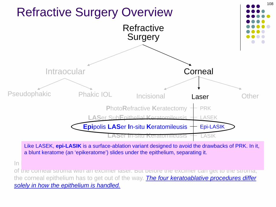
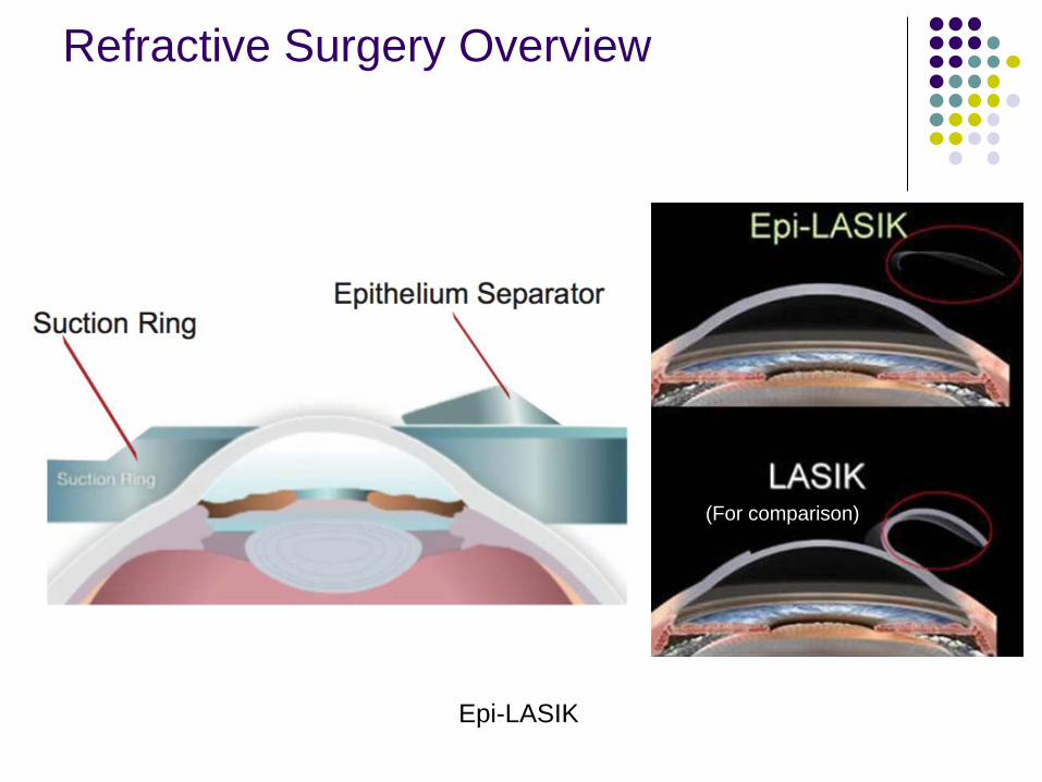
Like LASEK, epi-LASIK is a surface-ablation variant designed to avoid the drawbacks of PRK. In it, a blunt keratome (an ‘epikeratome’) slides under the epithelium, separating it. The epithelial flap thus created is folded back, then re-placed after the stroma has been ablated. (BTW, epipolis is a Greek word meaning ‘superficial.’)
Epi-LASIK
Refractive Surgery Overview
(For comparison)

Epipolis LASer In-situ Keratomileusis Epi-LASIK
RefractiveSurgery
Corneal
Incisional Laser
110
Intraocular
Pseudophakic Phakic IOL
PRK
LASEK
LASIK
SMILE
LASer In-situ Keratomileusis
PhotoRefractive KeratectomyLASer SubEpithelial Keratomileusis
SMall-Incision Lenticule Extraction
Other
Refractive Surgery Overview
In keratoablative procedures, remodeling of the central cornea occurs via annihilation of the corneal stroma with an excimer laser. But before the excimer can get to the stroma, the corneal epithelium has to get out of the way. The four keratoablative procedures differ solely in how the epithelium is handled.
Like LASEK, epi-LASIK is a surface-ablation variant designed to avoid the drawbacks of PRK. In it, a blunt keratome (an ‘epikeratome’) slides under the epithelium, separating it. The epithelial flap thus created is folded back, then re-placed after the stroma has been ablated. (BTW, epipolis is a Greek word meaning ‘superficial.’)
RefractiveSurgery
Corneal
Incisional Laser
111
Intraocular
Pseudophakic Phakic IOL
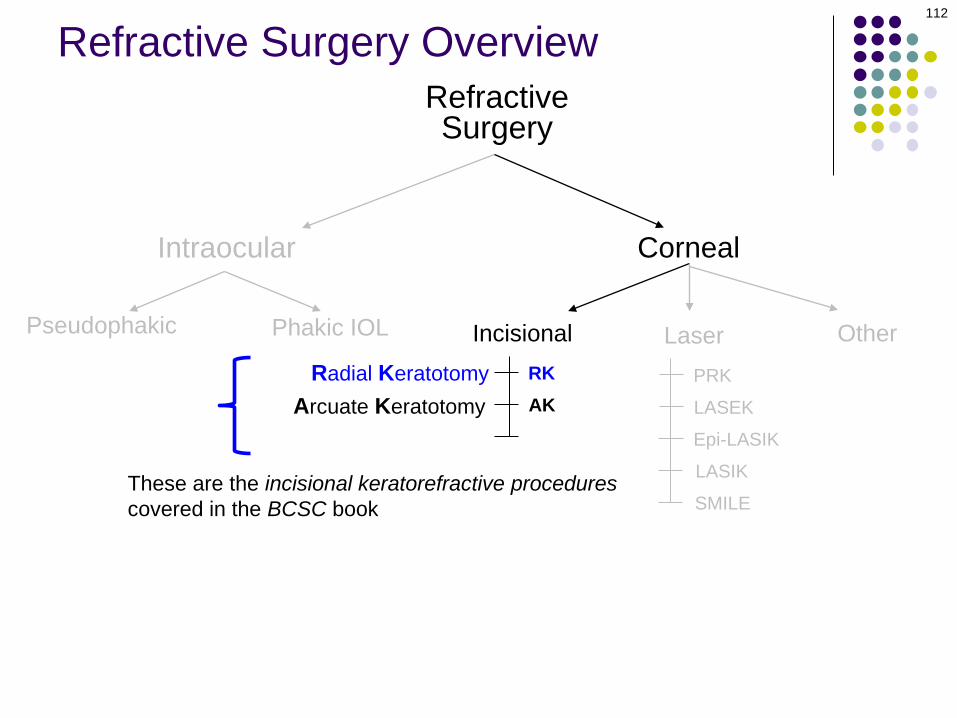
Radial Keratotomy
OtherRK
These are the incisional keratorefractive procedures covered in the BCSC book
PRK
LASEK
LASIK
SMILE
Epi-LASIK
Refractive Surgery Overview
RefractiveSurgery
Corneal
Incisional Laser
112
Intraocular
Pseudophakic Phakic IOL
Radial KeratotomyArcuate Keratotomy
OtherRKAK
These are the incisional keratorefractive procedures covered in the BCSC book
PRK
LASEK
LASIK
SMILE
Epi-LASIK
Refractive Surgery Overview

RefractiveSurgery
Corneal
Incisional Laser
113
Intraocular
Pseudophakic Phakic IOL
Radial KeratotomyArcuate Keratotomy
Limbal Relaxing Incisions
OtherRKAKLRI
These are the incisional keratorefractive procedures covered in the BCSC book
PRK
LASEK
LASIK
SMILE
Epi-LASIK
Refractive Surgery Overview

Arcuate KeratotomyLimbal Relaxing Incisions
RefractiveSurgery
Corneal
Incisional Laser
114
Intraocular
Pseudophakic Phakic IOLRadial Keratotomy
OtherRKAKLRI
PRK
LASEK
LASIK
Epi-LASIK
SMILE
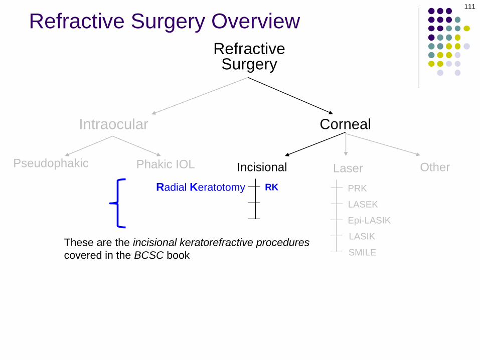
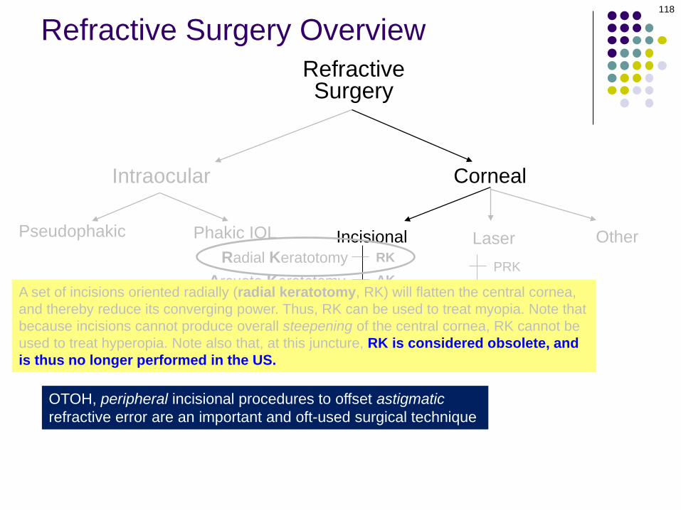
A set of incisions oriented radially (radial keratotomy, RK) will flatten the central cornea, and thereby reduce its converging power. Thus, RK can be used to treat myopia. Note that because incisions cannot produce overall steepening of the central cornea, RK cannot be used to treat hyperopia. Note also that RK is considered obsolete at this juncture, and is thus no longer performed in the US.
Myopic EyePlus error lens
Refractive Surgery Overview

115
Refractive Surgery Overview
Radial keratotomy

Arcuate KeratotomyLimbal Relaxing Incisions
RefractiveSurgery
Corneal
Incisional Laser
116
Intraocular
Pseudophakic Phakic IOLRadial Keratotomy
OtherRKAKLRI
PRK
LASEK
LASIK
Epi-LASIKA set of incisions oriented radially (radial keratotomy, RK) will flatten the central cornea, and thereby reduce its converging power. Thus, RK can be used to treat myopia. Note that because incisions cannot produce overall steepening of the central cornea, RK cannot be used to treat hyperopia. Note also that RK is considered obsolete at this juncture, and is thus no longer performed in the US.
Refractive Surgery Overview

Arcuate KeratotomyLimbal Relaxing Incisions
RefractiveSurgery
Corneal
Incisional Laser
117
Intraocular
Pseudophakic Phakic IOLRadial Keratotomy
OtherRKAKLRI
PRK
LASEK
LASIK
Epi-LASIKA set of incisions oriented radially (radial keratotomy, RK) will flatten the central cornea, and thereby reduce its converging power. Thus, RK can be used to treat myopia. Note that because incisions cannot produce overall steepening of the central cornea, RK cannot be used to treat hyperopia. Note also that, at this juncture, RK is considered obsolete, and is thus no longer performed in the US.
Refractive Surgery Overview
Radial KeratotomyArcuate Keratotomy
Limbal Relaxing Incisions
RefractiveSurgery
Corneal
Incisional Laser
118
Intraocular
Pseudophakic Phakic IOL OtherRKAKLRI
PRK
LASEK
LASIK
Epi-LASIKA set of incisions oriented radially (radial keratotomy, RK) will flatten the central cornea, and thereby reduce its converging power. Thus, RK can be used to treat myopia. Note that because incisions cannot produce overall steepening of the central cornea, RK cannot be used to treat hyperopia. Note also that, at this juncture, RK is considered obsolete, and is thus no longer performed in the US.
OTOH, peripheral incisional procedures to offset astigmaticrefractive error are an important and oft-used surgical technique
Refractive Surgery Overview

Radial Keratotomy
RefractiveSurgery
Corneal
Incisional Laser
119
Intraocular
Pseudophakic Phakic IOL
Arcuate KeratotomyLimbal Relaxing Incisions
OtherRKAKLRI
PRK
LASEK
LASIK
SMILE
Epi-LASIK
Specifically, this refers to AK and LRI
OTOH, peripheral incisional procedures to offset astigmaticrefractive error are an important and oft-used surgical technique
Refractive Surgery Overview
Radial Keratotomy
RefractiveSurgery
Corneal
Incisional Laser
120
Intraocular
Pseudophakic Phakic IOL
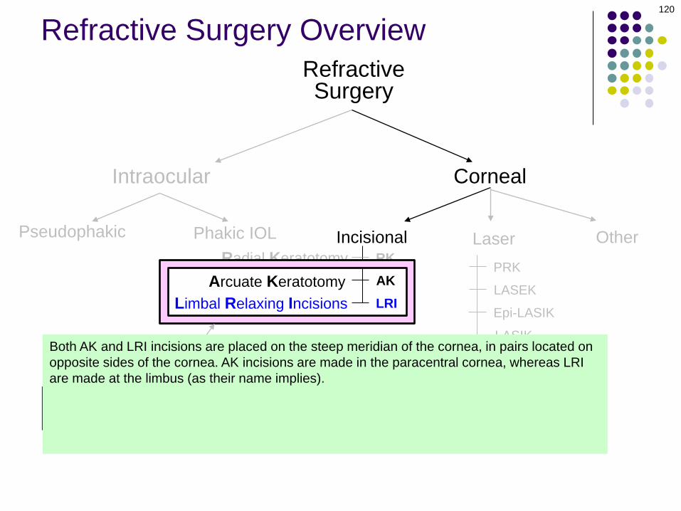
Arcuate KeratotomyLimbal Relaxing Incisions
OtherRKAKLRI
PRK
LASEK
LASIK
SMILE
Epi-LASIK
Specifically, this refers to AK and LPI
OTOH, peripheral incisional procedures to offset astigmaticrefractive error are an important and oft-used surgical technique
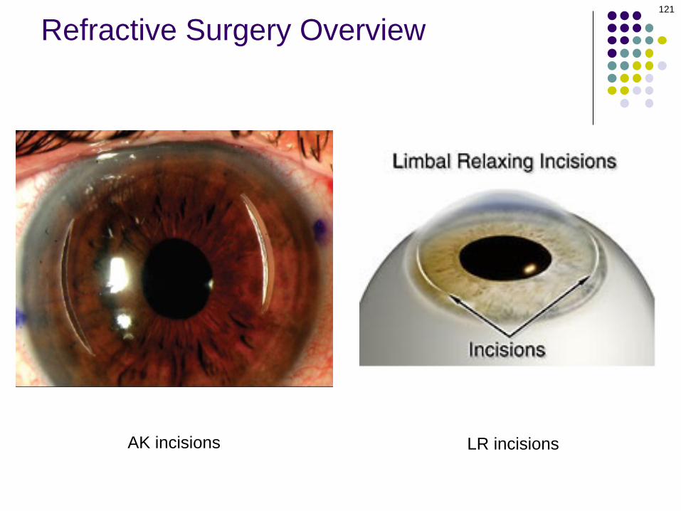
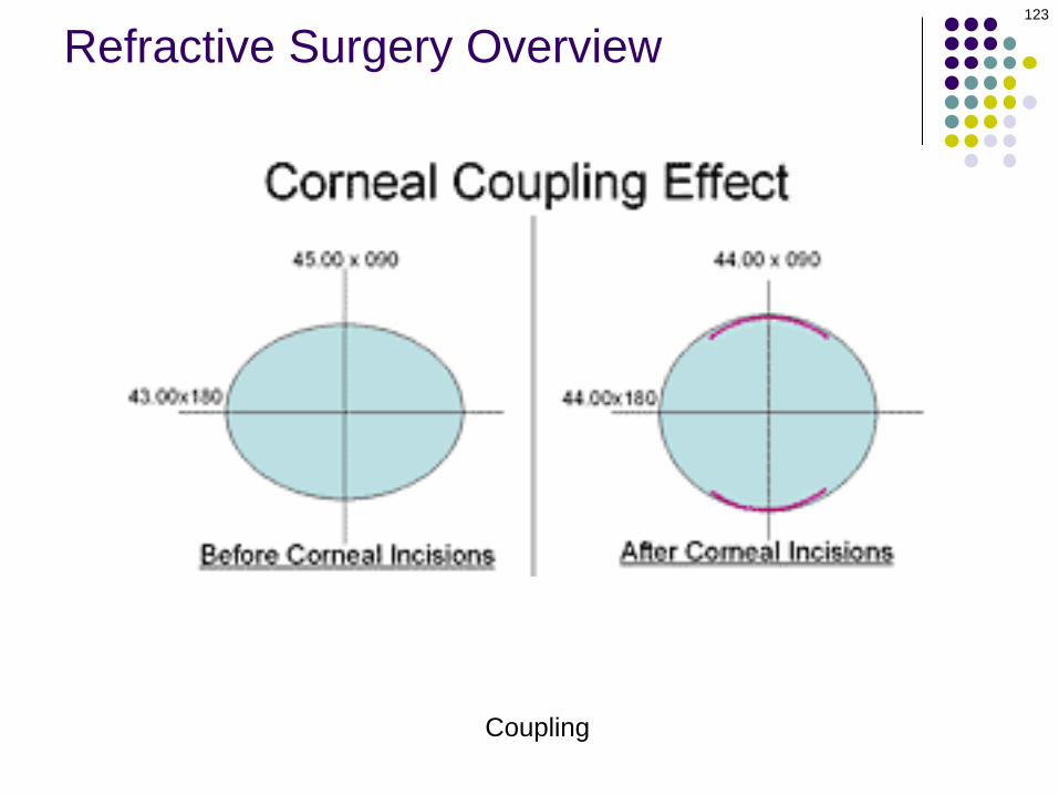
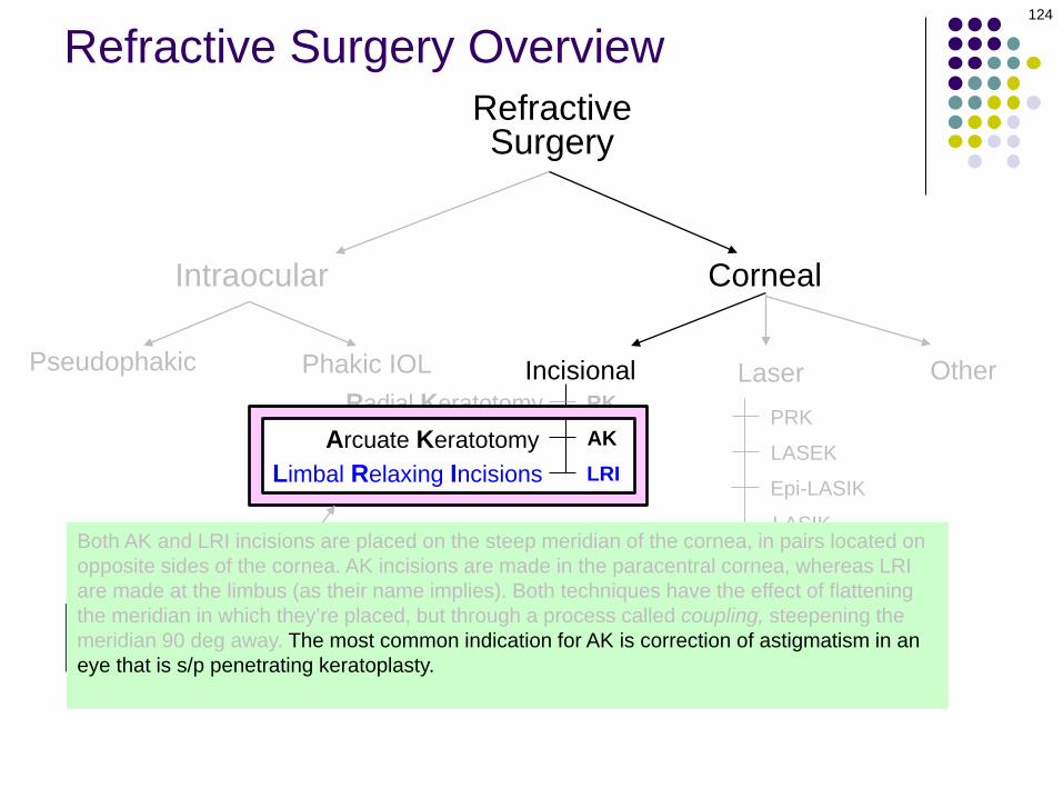
Both AK and LRI incisions are placed on the steep meridian of the cornea, in pairs located on opposite sides of the cornea. AK incisions are made in the paracentral cornea, whereas LRI are made at the limbus (as their name implies). Both techniques have the effect of flattening the meridian in which they’re placed, but (through a process called coupling) steepening the meridian 90 deg away. The most common indication for AK is to correct astigmatism in an eye that is s/p penetrating keratoplasty; LRI are typically placed at the time of cataract surgery to correct corneal astigmatism in hopes of producing better UCVA post-op.
Refractive Surgery Overview
121
Refractive Surgery Overview
AK incisions LR incisions

Radial Keratotomy
RefractiveSurgery
Corneal
Incisional Laser
122
Intraocular
Pseudophakic Phakic IOL
Arcuate KeratotomyLimbal Relaxing Incisions
OtherRKAKLRI
PRK
LASEK
LASIK
SMILE
Epi-LASIK
Specifically, this refers to AK and LPI
OTOH, peripheral incisional procedures to offset astigmaticrefractive error are an important and oft-used surgical technique
Both AK and LRI incisions are placed on the steep meridian of the cornea, in pairs located on opposite sides of the cornea. AK incisions are made in the paracentral cornea, whereas LRI are made at the limbus (as their name implies). Both techniques have the effect of flattening the meridian in which they’re placed, but through a process called coupling, steepening the meridian 90 deg away. The most common indication for AK is to correct astigmatism in an eye that is s/p penetrating keratoplasty; LRI are typically placed at the time of cataract surgery to correct corneal astigmatism in hopes of producing better UCVA post-op.
Refractive Surgery Overview
123
Refractive Surgery Overview
Coupling
Radial Keratotomy
RefractiveSurgery
Corneal
Incisional Laser
124
Intraocular
Pseudophakic Phakic IOL
Arcuate KeratotomyLimbal Relaxing Incisions
OtherRKAKLRI
PRK
LASEK
LASIK
SMILE
Epi-LASIK
Specifically, this refers to AK and LPI
OTOH, peripheral incisional procedures to offset astigmaticrefractive error are an important and oft-used surgical technique
Both AK and LRI incisions are placed on the steep meridian of the cornea, in pairs located on opposite sides of the cornea. AK incisions are made in the paracentral cornea, whereas LRI are made at the limbus (as their name implies). Both techniques have the effect of flattening the meridian in which they’re placed, but through a process called coupling, steepening the meridian 90 deg away. The most common indication for AK is correction of astigmatism in an eye that is s/p penetrating keratoplasty. LRI are typically placed at the time of cataract surgery to correct corneal astigmatism in hopes of producing better UCVA post-op.
Refractive Surgery Overview

Radial Keratotomy
RefractiveSurgery
Corneal
Incisional Laser
125
Intraocular
Pseudophakic Phakic IOL
Arcuate KeratotomyLimbal Relaxing Incisions
OtherRKAKLRI
PRK
LASEK
LASIK
SMILE
Epi-LASIK
Specifically, this refers to AK and LPI
OTOH, peripheral incisional procedures to offset astigmaticrefractive error are an important and oft-used surgical technique
Both AK and LRI incisions are placed on the steep meridian of the cornea, in pairs located on opposite sides of the cornea. AK incisions are made in the paracentral cornea, whereas LRI are made at the limbus (as their name implies). Both techniques have the effect of flattening the meridian in which they’re placed, but through a process called coupling, steepening the meridian 90 deg away. The most common indication for AK is correction of astigmatism in an eye that is s/p penetrating keratoplasty. LRI are typically placed at the time of cataract surgery to correct corneal astigmatism in hopes of producing better UCVA post-op.
Refractive Surgery Overview

Conductive Keratoplasty
RefractiveSurgery
Corneal
Incisional Laser
126
Refractive Surgery Overview
Intraocular
Pseudophakic Phakic IOL
PRK
LASEK
LASIK
SMILE
Epi-LASIK
RK
AK
LRI
CK
Other
These are the ‘other’ procedures covered in the BCSC book

Conductive Keratoplasty
RefractiveSurgery
Corneal
Incisional Laser
127
Refractive Surgery Overview
Intraocular
Pseudophakic Phakic IOL
PRK
LASEK
LASIK
SMILE
Epi-LASIK
RK
AK
LRI
CK
SAISmall Aperture Inlay
Other
These are the ‘other’ procedures covered in the BCSC book

Conductive Keratoplasty
RefractiveSurgery
Corneal
Incisional Laser
128
Refractive Surgery Overview
Intraocular
Pseudophakic Phakic IOL
PRK
LASEK
LASIK
SMILE
Epi-LASIK
RK
AK
LRI
CK
SAI
CRI
Small Aperture InlayCorneal Reshaping Inlay
Other
These are the ‘other’ procedures covered in the BCSC book
Conductive Keratoplasty
RefractiveSurgery
Corneal
Incisional Laser
129
Refractive Surgery Overview
Intraocular
Pseudophakic Phakic IOL
PRK
LASEK
LASIK
SMILE
Epi-LASIK
RK
AK
LRI
CK
CXL
SAI
CRI
Small Aperture Inlay
Corneal CROSS LinkingCorneal Reshaping Inlay
Other
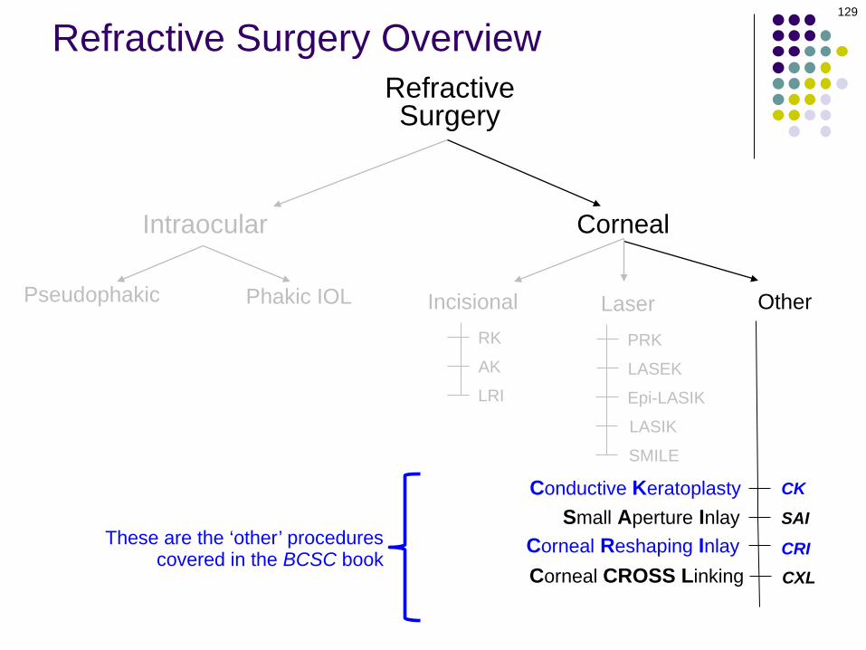
These are the ‘other’ procedures covered in the BCSC book
Conductive Keratoplasty
RefractiveSurgery
Corneal
Incisional Laser
130
Refractive Surgery Overview
Intraocular
Pseudophakic Phakic IOL
PRK
LASEK
LASIK
SMILE
Epi-LASIK
RK
AK
LRI
CK
ICRSCXL
SAI
CRI
Intrastromal Corneal Ring Segments
Small Aperture Inlay
Corneal CROSS LinkingCorneal Reshaping Inlay
Other
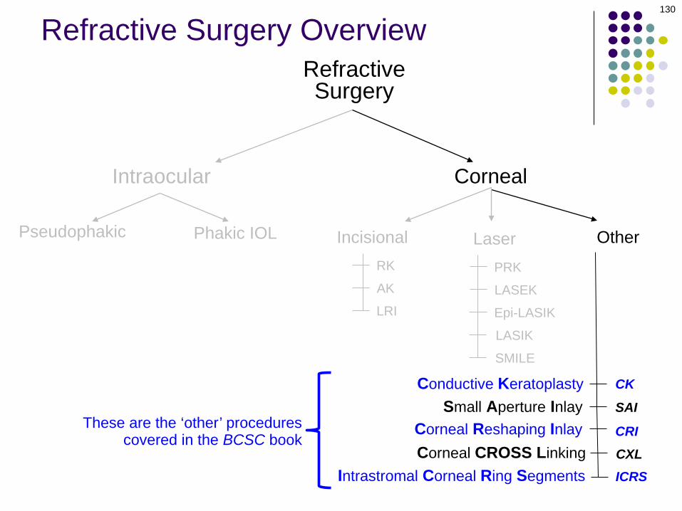
These are the ‘other’ procedures covered in the BCSC book

Conductive Keratoplasty
RefractiveSurgery
Corneal
Incisional Laser
131
Refractive Surgery Overview
Intraocular
Pseudophakic Phakic IOL
PRK
LASEK
LASIK
SMILE
Epi-LASIK
RK
AK
LRI
CK
ICRSCXL
SAI
CRI
Intrastromal Corneal Ring Segments
Small Aperture Inlay
Corneal CROSS LinkingCorneal Reshaping Inlay
Other
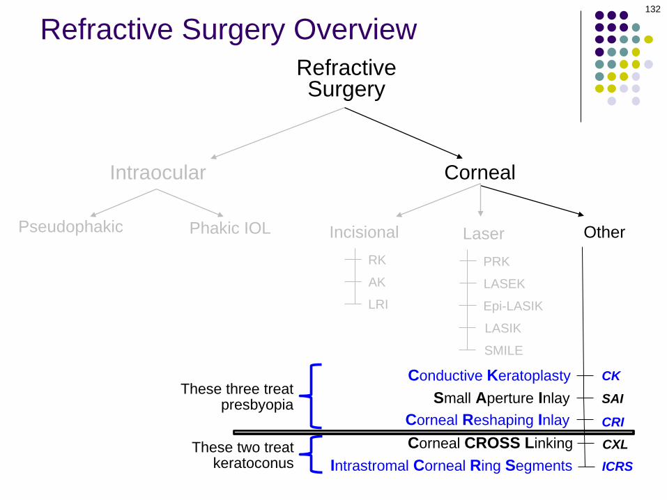
These three treat presbyopia
Conductive Keratoplasty
RefractiveSurgery
Corneal
Incisional Laser
132
Refractive Surgery Overview
Intraocular
Pseudophakic Phakic IOL
PRK
LASEK
LASIK
SMILE
Epi-LASIK
RK
AK
LRI
CK
ICRSCXL
SAI
CRI
Intrastromal Corneal Ring Segments
Small Aperture Inlay
Corneal CROSS LinkingCorneal Reshaping Inlay
Other
These three treat presbyopia
These two treat keratoconus
Conductive Keratoplasty
RefractiveSurgery
Corneal
Incisional Laser
133
Refractive Surgery Overview
Intraocular
Pseudophakic Phakic IOL
PRK
LASEK
LASIK
SMILE
Epi-LASIK
RK
AK
LRI
CK
ICRSCXL
SAI
CRI
Intrastromal Corneal Ring Segments
Small Aperture Inlay
Corneal CROSS LinkingCorneal Reshaping Inlay
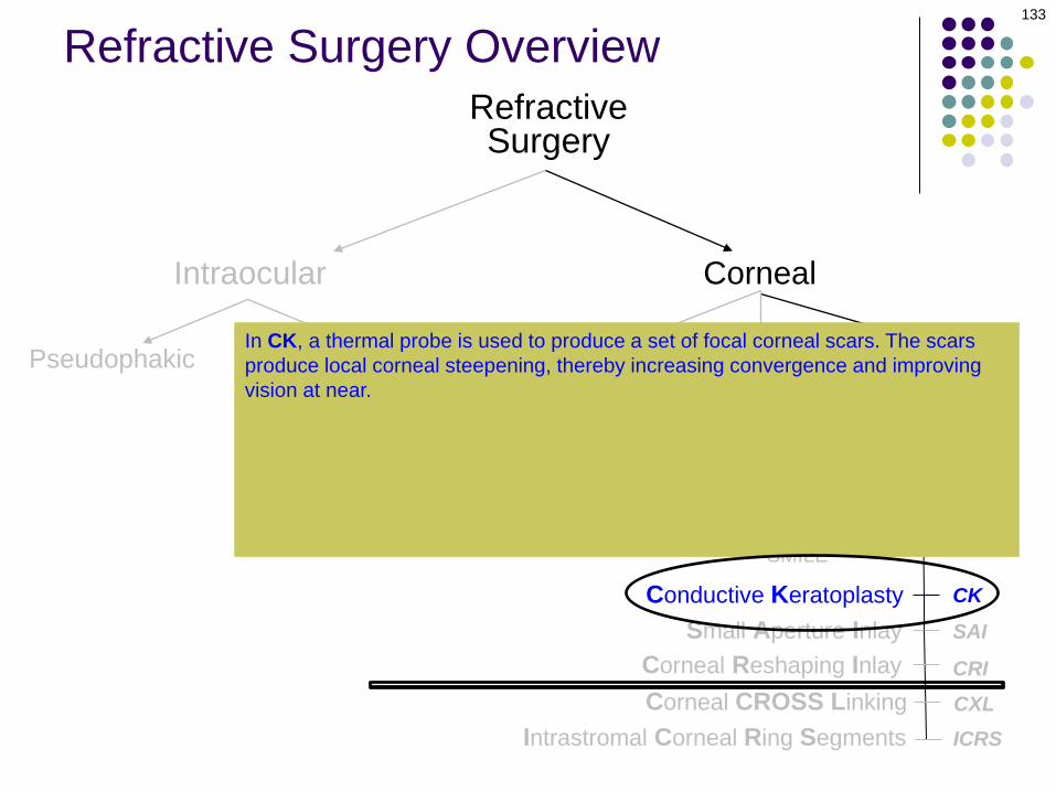
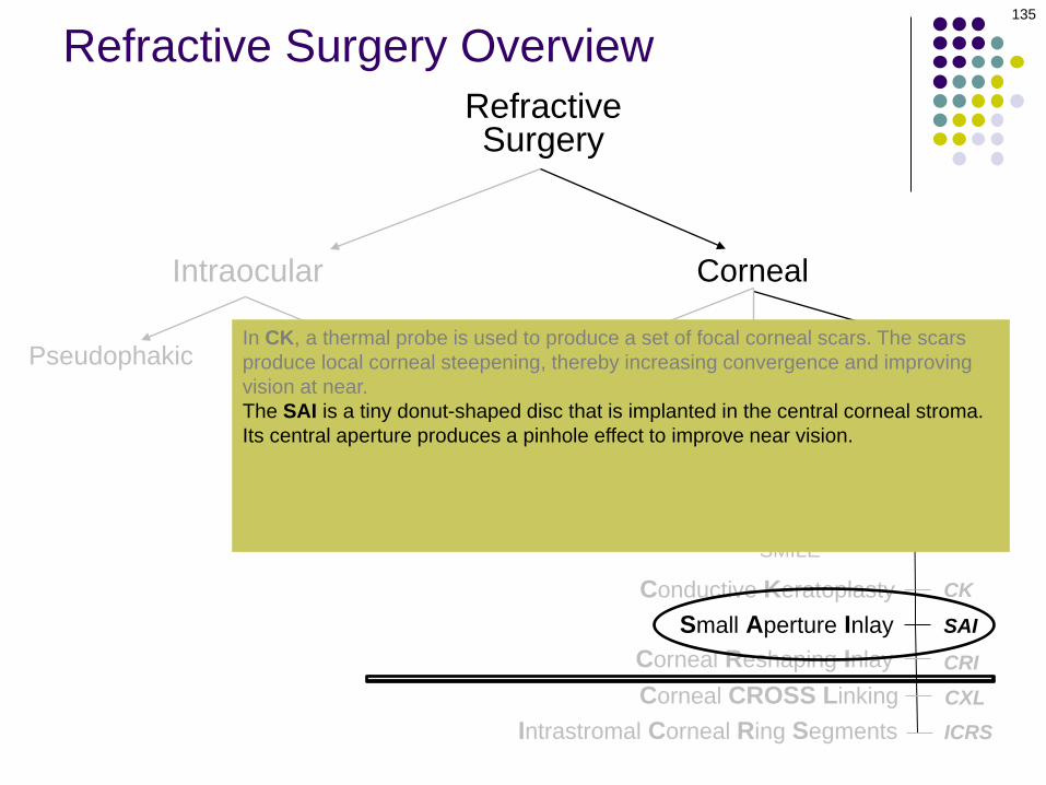
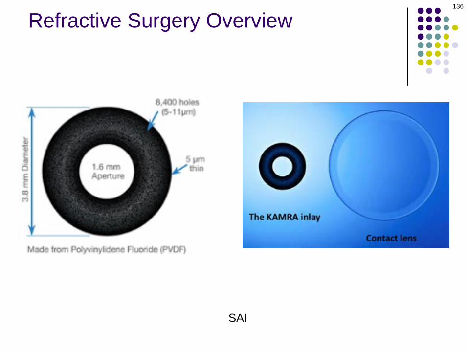
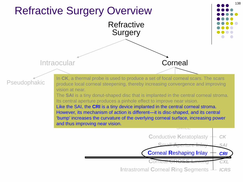
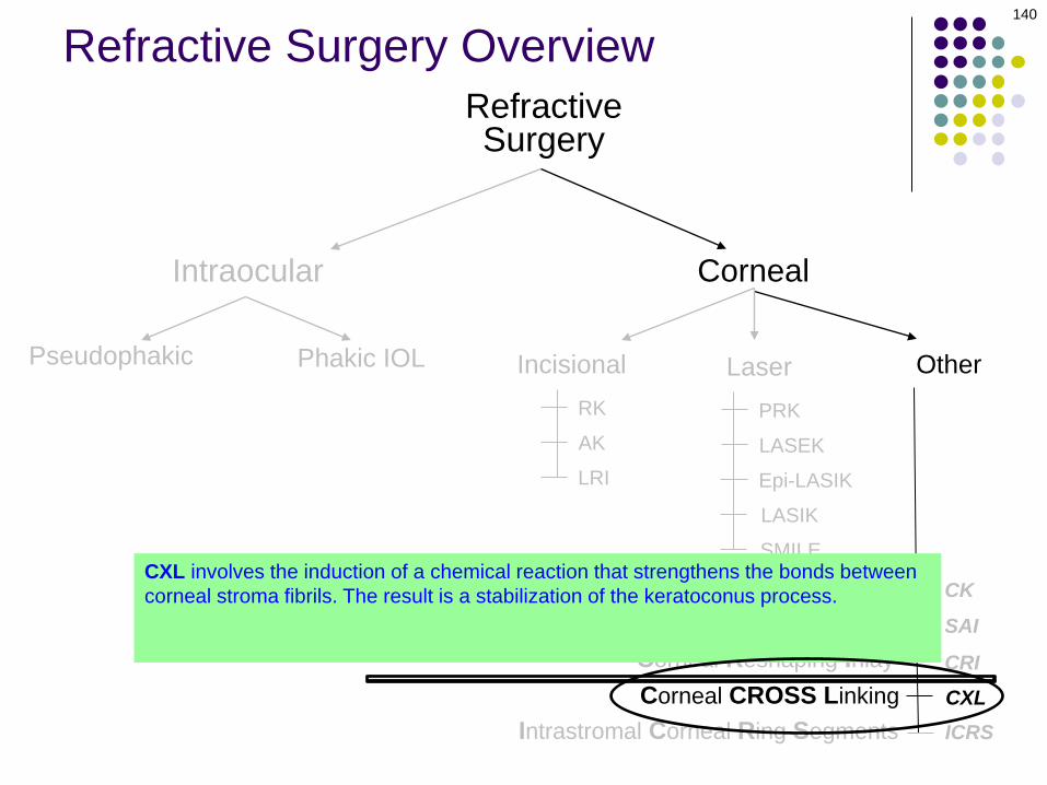
OtherIn CK, a thermal probe is used to produce a set of focal corneal scars. The scars produce local corneal steepening, thereby increasing convergence and improving vision at near. The SAI is a tiny donut-shaped disc that is implanted in the central corneal stroma. Its central aperture produces a pinhole effect to improve near vision.Like the SAI, the CRI is a tiny device implanted in the central corneal stroma. However, its mechanism of action is significantly different—it is disc-shaped, and its central ‘bump’ increases the curvature of the overlying corneal surface, increasing power and thus improving near vision.

134
Refractive Surgery Overview
CK probe CK in action
CK scars
Conductive Keratoplasty
RefractiveSurgery
Corneal
Incisional Laser
135
Refractive Surgery Overview
Intraocular
Pseudophakic Phakic IOL
PRK
LASEK
LASIK
SMILE
Epi-LASIK
RK
AK
LRI
CK
ICRSCXL
SAI
CRI
Intrastromal Corneal Ring Segments
Small Aperture Inlay
Corneal CROSS LinkingCorneal Reshaping Inlay
OtherIn CK, a thermal probe is used to produce a set of focal corneal scars. The scars produce local corneal steepening, thereby increasing convergence and improving vision at near. The SAI is a tiny donut-shaped disc that is implanted in the central corneal stroma. Its central aperture produces a pinhole effect to improve near vision.Like the SAI, the CRI is a tiny device implanted in the central corneal stroma. However, its mechanism of action is significantly different—it is disc-shaped, and its central ‘bump’ increases the curvature of the overlying corneal surface, increasing power and thus improving near vision.
136
Refractive Surgery Overview
SAI

137
Refractive Surgery Overview
SAI
Conductive Keratoplasty
RefractiveSurgery
Corneal
Incisional Laser
138
Refractive Surgery Overview
Intraocular
Pseudophakic Phakic IOL
PRK
LASEK
LASIK
SMILE
Epi-LASIK
RK
AK
LRI
CK
ICRSCXL
SAI
CRI
Intrastromal Corneal Ring Segments
Small Aperture Inlay
Corneal CROSS LinkingCorneal Reshaping Inlay
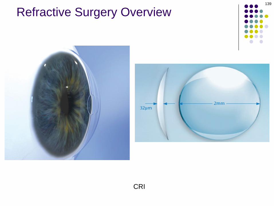
OtherIn CK, a thermal probe is used to produce a set of focal corneal scars. The scars produce local corneal steepening, thereby increasing convergence and improving vision at near. The SAI is a tiny donut-shaped disc that is implanted in the central corneal stroma. Its central aperture produces a pinhole effect to improve near vision.Like the SAI, the CRI is a tiny device implanted in the central corneal stroma. However, its mechanism of action is different—it is disc-shaped, and its central ‘bump’ increases the curvature of the overlying corneal surface, increasing power and thus improving near vision.
139
Refractive Surgery Overview
CRI
CK
SAI
CRI
Conductive Keratoplasty
RefractiveSurgery
Corneal
Incisional Laser
140
Refractive Surgery Overview
Intraocular
Pseudophakic Phakic IOL
PRK
LASEK
LASIK
SMILE
Epi-LASIK
RK
AK
LRI
ICRSCXL
Intrastromal Corneal Ring Segments
Small Aperture Inlay
Corneal CROSS LinkingCorneal Reshaping Inlay
Other
CXL involves the induction of a chemical reaction that strengthens the bonds between corneal stroma fibrils. The result is a stabilization of the keratoconus process. ICRS employs semicircular segments of PMMA. These segments are placed in the peripheral corneal stroma, where they produce local flattening.
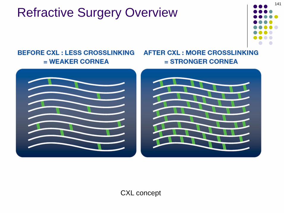
141
Refractive Surgery Overview
CXL concept
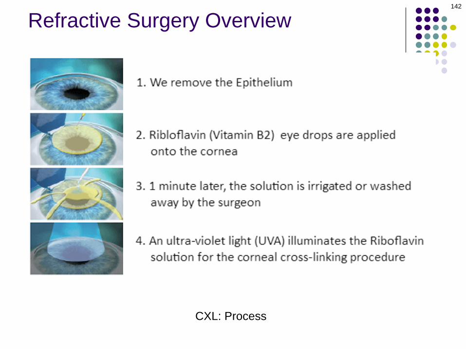
142
Refractive Surgery Overview
CXL: Process

143
Refractive Surgery Overview
CXL: Process

CK
SAI
CRI
Conductive Keratoplasty
RefractiveSurgery
Corneal
Incisional Laser
144
Refractive Surgery Overview
Intraocular
Pseudophakic Phakic IOL
PRK
LASEK
LASIK
SMILE
Epi-LASIK
RK
AK
LRI
ICRSCXL
Intrastromal Corneal Ring Segments
Small Aperture Inlay
Corneal CROSS LinkingCorneal Reshaping Inlay
Other
CXL involves the induction of a chemical reaction that strengthens the bonds between corneal stroma fibrils. The result is a stabilization of the keratoconus process. ICRS employs semicircular segments of PMMA. These segments are placed in the peripheral corneal stroma, where they produce local flattening.

145
Refractive Surgery Overview
Intrastromal ring segments

146
Refractive Surgery Overview
Intrastromal ring segments in situ

147
Refractive Surgery Overview
Intrastromal ring segments placed for KCN