Refraction Training - teamtei.com · Pseudophakia •An intraocular lens (IOL) is an implanted lens...
19
02/20/2013 1 Refraction Training June 11-12, 2007 Topcon Medical Systems, Inc. Paramus, NJ Aaron J. Graham Refraction In ophthalmology and optometry, refraction (also known as refractometry) is a clinical test in which a phoropter is used to determine the eye's refractive error and the best corrective lenses to be prescribed. A series of test lenses in graded optical powers or focal lengths are presented to determine which provide the sharpest, clearest vision. • Refraction can be seen when looking into a glass of water. Air has a refractive index of about 1.0003nm, and water has a refractive index of about 1.33nm. • If a person looks at a straight object, such as a pencil, which is placed at a slant, partially in the water, the object appears to bend at the water's surface. This is due to the bending of light rays as they move from the water to the air. • Once the rays reach the eye, the eye traces them back as straight lines (lines of sight). The lines of sight (shown as dashed lines) intersect at a higher position than where the actual rays originated. • This causes the pencil to appear higher and the water to appear shallower than it really is. The depth that the water appears to be when viewed from above is known as the apparent depth. History of Glasses • The most primitive form of glasses were invented by Roger Bacon in the 13th century, although similar devices are believed to have existed in ancient times in China and in the Mediterranean civilizations. • Early forms of glasses were crude and clumsy and not improved until the 18th century when the grinding of lenses was first based upon the principles of light refraction. • Lenses are made of clear or rock crystal glass or plastic ground to suit the defect of the eye.
Transcript of Refraction Training - teamtei.com · Pseudophakia •An intraocular lens (IOL) is an implanted lens...

02/20/2013
1
Refraction
Training
June 11-12, 2007
Topcon Medical Systems, Inc.
Paramus, NJ
Aaron J. Graham
Refraction
In ophthalmology and optometry, refraction (also known as refractometry) is a clinical test in which a phoropter is used to determine the eye's refractive error and the best corrective lenses to be prescribed. A series of test lenses in graded optical powers or focal lengths are presented to determine which provide the sharpest, clearest vision.
• Refraction can be seen when looking into a glass of water. Air has a refractive index of about 1.0003nm, and water has a refractive index of about 1.33nm.
• If a person looks at a straight object, such as a pencil, which is placed at a slant, partially in the water, the object appears to bend at the water's surface. This is due to the bending of light rays as they move from the water to the air.
• Once the rays reach the eye, the eye traces them back as straight lines (lines of sight). The lines of sight (shown as dashed lines) intersect at a higher position than where the actual rays originated.
• This causes the pencil to appear higher and the water to appear shallower than it really is. The depth that the water appears to be when viewed from above is known as the apparent depth.
History of Glasses
• The most primitive form of glasses were invented by Roger Bacon in the 13th century, although similar devices are believed to have existed in ancient times in China and in the Mediterranean civilizations.
• Early forms of glasses were crude and clumsy and not improved until the 18th century when the grinding of lenses was first based upon the principles of light refraction.
• Lenses are made of clear or rock crystal glass or plastic ground to suit the defect of the eye.

02/20/2013
2
Roger Bacon (1240 A.D.)
A Bit of American Eyeglass History
• Benjamin Franklin is credited with the creation of
the first pair of bifocals in the early 1760's.
Common Visual Abnormalities
• Myopia
• Hyperopia
• Presbyopia
• Diplopia
• Amblyopia
Myopia
• Myopia, also called nearsightedness or short
sightedness, is a refractive defect of the eye in
which light produces image focus in front of the
retina.
• Those with myopia typically can see nearby
objects clearly but distant objects appear
blurred.
• Myopia is a result of a ―long eye‖.

02/20/2013
3
Visual Example of Myopia
Hyperopia
• Hyperopia, also known as hypermetropia or colloquially as farsightedness or long-sightedness, is a defect of vision caused by an imperfection in the eye (often when the eyeball is too short or when the lens cannot become round enough), causing inability to focus on near objects.
• Light rays are focused behind the retina.
• Hyperopia is a result of a ―short eye‖.
Visual Example of Hyperopia
• Presbyopia is the eye's diminished ability to
focus that occurs with age.
• Presbyopia is not a disease as such, but a
condition that affects everyone at a certain age.
• The first symptoms are usually noticed between
the ages of 40-50, though in fact the ability to
focus declines throughout life
Presbyopia

02/20/2013
4
Visual Example of Presbyopia
Diplopia
• Diplopia, commonly known as ―double vision‖, is
the perception of two images from a single
object. The images may be horizontal, vertical,
or diagonal .
• When the eyes are misaligned and aimed at
different targets, two non-matching images are
sent to the viewer's brain. When the viewer's
brain accepts and uses two non-matching
images simultaneously, double vision results.
• Diplopia contributes to loss of depth perception
and binocular vision.
Visual Example of Diplopia

02/20/2013
5
Amblyopia
• Amblyopia, or lazy eye, is a disorder of the eye that is characterized by poor or blurry vision in an eye that is otherwise physically normal
• The problem is caused by either no transmission or poor transmission of the visual image to the brain for a sustained period of dysfunction or during early childhood.
• The condition will only arise at this young age because most of the visual system's development is complete and "locked in" by 8 to 10 years of age.
• Amblyopia normally only affects one eye, but it is possible to be amblyopic in both eyes if both are similarly deprived of a good, clear visual image.
Visual Example of AmblyopiaCommon Visual Impairments
Making Refraction Difficult
• Astigmatism
• Keratoconus
• Cataract
• Macular Degeneration
• Pseudophakia
• Corneal Grafts
• Diabetic Retinopathy
Astigmatism
• Type of faulty vision caused by a irregular curvature in the refractive surfaces, usually the cornea.
• Light rays do not all come to a single focal point on the retina.
• Some light rays focus on the retina while others focus in front of or behind it.
• A special cylindrical lens is placed in the out-of-focus axis to correct the condition.
• In many cases contact lenses are the most effective means of correcting astigmatism.

02/20/2013
6
Visual Example of Astigmatism Keratoconus
• A degenerative non-inflammatory disorder of the eye in which structural changes within the cornea cause it to thin and change to a more conical shape than its normal gradual curve.
• Keratoconus can cause substantial distortion of vision, with multiple images, streaking and sensitivity to light.
• Keratoconus is the most common dystrophy of the cornea, affecting around one person in a thousand, and it seems to occur in all ethnic groups worldwide .
• It is typically diagnosed in the patient's adolescent years and attains its most severe state in the twenties and thirties.
Visual Example of Keratoconus
Cataract
• Opacity of the lens of the eye, which impairs vision.
• In the young, cataracts are generally congenital or hereditary; later they are usually the result of degenerative changes brought on by aging or systemic disease (diabetes).
• Cataracts brought on by aging are most common; most individuals over 60 exhibit some degree of lens opacity.
• Injury, extreme heat, ultraviolet light, X rays, nuclear radiation, inflammatory disease, and toxic substances also cause cataracts.
• There is growing concern that further disintegration of the ozone layer will increase the incidence of cataracts.

02/20/2013
7
Visual Example of Cataract Macular Degeneration• Eye disorder causing loss of central vision.
• The affected area, the macula, lies at the back of the retina and is the part that produces the sharpest vision.
• The most serious visual impairment occurs when abnormal blood vessels form and leak fluid or bleed into the tissue of the macula, ultimately producing scar tissue.
• Peripheral (side) vision is unaffected.
• Onset may be acute with hemorrhage but usually is gradually progressive.
• Although some vision is retained, the ability to read, recognize faces, and drive a motor vehicle is greatly reduced.
• The condition is painless.
• Macular degeneration is a major cause of vision impairment among elderly people.
Visual Example of Macular
Degeneration
Pseudophakia• An intraocular lens (IOL) is an implanted lens in
the eye, usually replacing the existing crystalline lens because it has been clouded over by a cataract, or as a form of refractive surgery to change the eye's optical power.
• Insertion of an intraocular lens is the most commonly performed eye surgical procedure; cataracts are the most common eye disease.
• The procedure can be done under local anesthesia with the patient awake throughout the operation which usually takes less than 30 minutes in the hands of an experienced ophthalmologist.
• The recovery period is about 2-3 weeks.

02/20/2013
8
Corneal Grafts/Transplant
• Corneal transplantation, also known as corneal grafting or penetrating keratoplasty, is a surgical procedure where a damaged or diseased cornea is replaced by donated corneal tissue which has been removed from a recently deceased individual having no known diseases which might affect the viability of the donated tissue.
• The surgical procedure is performed by ophthalmologists, medical doctors who specialize in eyes, and are often done on an outpatient basis (the patient goes home following surgery).
Diabetic Retinopathy
• Diabetic retinopathy (damage to the retina) is caused by complications of diabetes mellitus, which could eventually lead to blindness. It is an ocular manifestation of systemic disease which affects up to 80% of all diabetics who have had diabetes for 15 years or more.
• In some cases, the vision will get better or worse during the day.
• Small blood vessels – such as those in the eye – are especially vulnerable to poor blood glucose control.
• The lack of oxygen (ischemia) in the retina causes fragile, new, blood vessels to grow along the retina and in the clear, gel-like vitreous that fills the inside of the eye.

02/20/2013
9
Visual Example of Diabetic
RetinopathyRefractive Error
• Refractive errors are frequently categorized as spherical errors and cylindrical errors.
• Spherical errors occur when the optical power of the eye is either too large or too small to focus light on the retina.
• People with refraction error frequently have blurry vision.
• When the optics are too powerful for the length of the eyeball (this can arise from a cornea with too much curvature or an eyeball that is too long), one has myopia.
• When the optics are too weak for the length of the eyeball (this can arise from a cornea with not enough curvature or an eyeball that is too short), one has Hyperopia.
Refractive Error Continued
• Cylindrical errors occur when the optical power of the eye is too powerful or too weak across one meridian of the optics.
• It is as if the overall lens tends towards a cylindrical shape along that meridian.
• People with this refraction error see contours of a particular orientation as blurred, but see contours with orientations at right angles as clear.
• When one has a cylindrical error, one has astigmatism.
Accommodation
• Accommodation is the process by which the eye increases optical power to maintain a clear image (focus) on the retina.
• This varies from a maximum of over 15 diopters in an infant to only about 1.5 diopters in a person 70 years old, as the lens becomes less flexible with age.
• A near object (for example, a computer screen) appears large in the field of vision, and the eye receives light from wide angles. When moving focus from a distant to a near object, the eyes converge. The ciliary muscle contracts making the lens more convex, shortening its focal length. The pupil constricts in order to prevent diverging light rays from hitting the periphery of the retina and resulting in a blurred image.

02/20/2013
10
Eye ―Evolution‖
• The common origin of all animal eyes is now widely accepted as fact based on shared anatomical and genetic features of all eyes; that is, all modern eyes, varied as they are, have their origins in a proto-eye evolved some 540 million years ago. The majority of the advancements in early eyes are believed to have taken only a few million years to develop, as the first predator to gain true imaging would have touched off an "arms race". Prey animals and competing predators alike would be forced to rapidly match or exceed any such capabilities to survive. Hence multiple eye types and subtypes developed in parallel.
Eye ―Evolution‖ Cont.
• Eyes in various animals show adaptation to their requirements. For example, birds of prey have much greater visual acuity than humans, and some can see ultraviolet light. The different forms of eyes in, for example, vertebrates and mollusks are often cited as examples of parallel evolution, despite their distant common ancestry.
• The earliest eyes, called "eyespots", were simple patches of photoreceptor cells, physically similar to the receptor patches for taste and smell. These eyespots could only sense ambient brightness: they could distinguish light and dark, but not the direction of the light source.
Eye Facts
• Estimates of the resolution of the human eye are some where around 576 mega pixels (24000 x 24000 pixels) for a 120 degree field of view.
• However, it must be noted that the human eye itself has only a small spot of sharp vision in the middle of the retina, the fovea centralis, the rest of the field of view being blurry.
• The angle of the sharp vision being just few degrees in the middle of the view, the sharp area thus barely achieves even a single mega pixel resolution.
• The experience of wide sharp human vision is in fact based on turning the eyes towards the current point of interest in the field of view, the brain thus perceiving an observation of a wide sharp field of view.
• The narrow beam of sharp vision is easy to test by putting a fingertip on a newspaper and trying to read the text while staring at the finger tip—it is very difficult to read text that's just some centimeters away from the finger tip.
02/20/2013
11
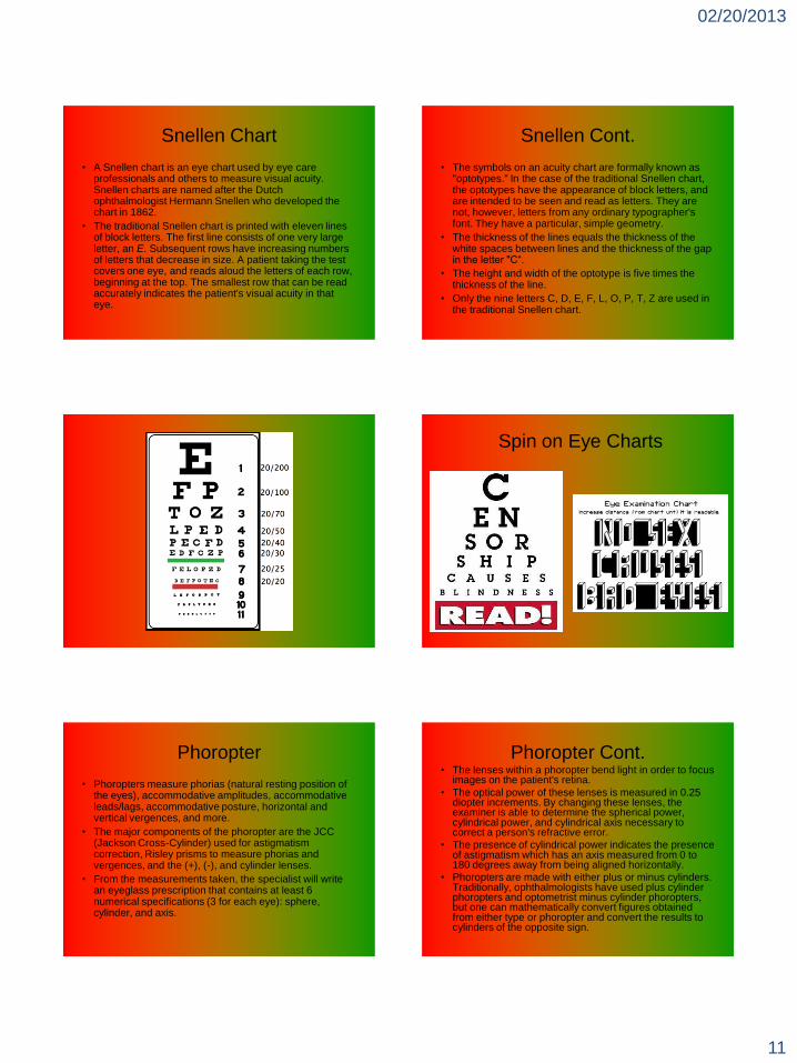
Snellen Chart
• A Snellen chart is an eye chart used by eye care professionals and others to measure visual acuity. Snellen charts are named after the Dutch ophthalmologist Hermann Snellen who developed the chart in 1862.
• The traditional Snellen chart is printed with eleven lines of block letters. The first line consists of one very large letter, an E. Subsequent rows have increasing numbers of letters that decrease in size. A patient taking the test covers one eye, and reads aloud the letters of each row, beginning at the top. The smallest row that can be read accurately indicates the patient's visual acuity in that eye.
Snellen Cont.
• The symbols on an acuity chart are formally known as "optotypes." In the case of the traditional Snellen chart, the optotypes have the appearance of block letters, and are intended to be seen and read as letters. They are not, however, letters from any ordinary typographer's font. They have a particular, simple geometry.
• The thickness of the lines equals the thickness of the white spaces between lines and the thickness of the gap in the letter "C―.
• The height and width of the optotype is five times the thickness of the line.
• Only the nine letters C, D, E, F, L, O, P, T, Z are used in the traditional Snellen chart.
Spin on Eye Charts
Phoropter
• Phoropters measure phorias (natural resting position of the eyes), accommodative amplitudes, accommodative leads/lags, accommodative posture, horizontal and vertical vergences, and more.
• The major components of the phoropter are the JCC (Jackson Cross-Cylinder) used for astigmatism correction, Risley prisms to measure phorias and vergences, and the (+), (-), and cylinder lenses.
• From the measurements taken, the specialist will write an eyeglass prescription that contains at least 6 numerical specifications (3 for each eye): sphere, cylinder, and axis.
Phoropter Cont.• The lenses within a phoropter bend light in order to focus
images on the patient's retina.
• The optical power of these lenses is measured in 0.25 diopter increments. By changing these lenses, the examiner is able to determine the spherical power, cylindrical power, and cylindrical axis necessary to correct a person's refractive error.
• The presence of cylindrical power indicates the presence of astigmatism which has an axis measured from 0 to 180 degrees away from being aligned horizontally.
• Phoropters are made with either plus or minus cylinders. Traditionally, ophthalmologists have used plus cylinder phoropters and optometrist minus cylinder phoropters, but one can mathematically convert figures obtained from either type or phoropter and convert the results to cylinders of the opposite sign.

02/20/2013
12
Retinoscopy
• Retinoscopy is a technique to obtain an objective measurement of the refractive condition of a patient's eye. The examiner uses a retinoscope to shine light into the patient's eye and observes the reflection (reflex) off the patient's retina. While moving the streak or spot of light across the pupil the examiner observes the relative movement of the reflex then uses a phoropter or manually places lenses over the eye to "neutralize" the reflex.
• Retinoscopy is especially useful in prescribing corrective lenses for patients who are unable to undergo a subjective refraction that requires a judgement and response from the patient.
• Retinoscopy is also used to evaluate accommodative ability of the eye and detect latent Hyperopia.
Retinoscopy Cont.
• Retinoscope works on a principle called Foucault's principle. Basically it indicates that the examiner should simulate the infinity to obtain the correct refractive power. Hence a power corresponding to the working distance is subtracted from the gross retinoscope value.
• Static retinoscopy is performed when the patient has relaxed accommodative status viewing a distance target; dynamic retinoscopy is performed when the patient has active accommodation from viewing a near target.
Cycloplegic Refraction
• Cycloplegia is the paralysis of the ciliary muscle,
resulting in a loss of accommodation, resulting in
pupil dilation.
• Most commonly performed on infants and
adolescents.

02/20/2013
13
Auto Refraction• An auto-refractor or automated refractor is a computer-
controlled machine used during an eye examination to provide an objective measurement of a person's refractive error and prescription for glasses or contact lenses. This is achieved by measuring how light is changed as it enters a person's eye.
• The automated refraction technique is quick, simple and painless. The patient takes a seat and places their chin on a rest. One eye at a time, they look into the machine at a picture inside. The picture moves in and out of focus as the machine takes readings to determine when the image is on the retina. Several readings are taken which the machine averages to form a prescription.
• No feedback is required from the patient during this process.
Auto Refraction Cont.
• Within seconds an approximate measurement of a person's prescription can be made by the machine and printed out.
• In some offices this is used to provide the starting point for the optometrist in subjective refraction tests. Here, lenses are switched in and out of a phoropter and the patient is asked "which looks better" while looking at a chart. This feedback refines the prescription to one which provides the patient with the best vision.
• Automated refraction is particularly useful when dealing with non-communicative people such as young children or those with disabilities.
• Retinoscopy performed by an experienced clinician has been found to provide a more accurate estimation of refractive error than auto-refraction.
Subjective Refraction
• Subjective Refraction is the process through which the clinician can determine the best refractive power for the patient, with the aid of the patient.
• Different lenses are placed in front of the eyes of the patient in order to determine which lens best suits the patient (handheld lenses and lenses contained in the phoropter).
• ―Better one or better two.‖
• The patient is able to determine which lens they appreciate.
• Subjective Refraction is often used in conjunction with Retinoscopy for refinement.
02/20/2013
14
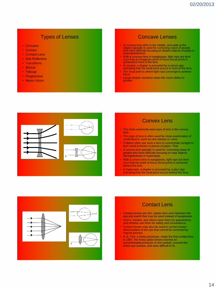
Types of Lenses
• Concave
• Convex
• Contact Lens
• Anti-Reflective
• Transitions
• Bifocal
• Trifocal
• Progressive
• Mono-Vision
Concave Lenses
• A concave lens (thin in the middle, and wide at the edges) typically is used for correcting vision of people who have difficulty focusing on distant objects (myopia or nearsightedness).
• With a concave lens in eyeglasses, light rays are bent such that an imaginary point of focus (focal point) is achieved in front of the lens.
• In myopia, a diopter is preceded by a minus sign indicating that the focal point occurs in front of the lens.
• The focal point is where light rays converge to achieve focus.
• Larger diopter numbers mean the vision defect is greater.
Convex Lens
• The most commonly-seen type of lens is the convex lens.
• This type of lens is often used for close examination of small objects, such as rare stamps or coins.
• Children often use such a lens to concentrate sunlight to burn small pinholes in pieces of paper. That
• A convex lens typically is used for correcting vision of people who have difficulty focusing on near objects (farsightedness or hyperopia).
• With a convex lens in eyeglasses, light rays are bent such that the point of focus (focal point) is achieved behind the lens.
• In hyperopia, a diopter is preceded by a plus sign indicating that the focal point occurs behind the lens.
Contact Lens
• Contact lenses are thin, plastic lens worn between the eye and eyelid that may be used instead of eyeglasses.
• Actors, models, and others wear them for appearance, and athletes use them for safety and convenience.
• Contact lenses may also be used to correct certain abnormalities of the eye that cannot be corrected by regular glasses.
• A. E. Fick, a Swiss physician, made the first contact lens in 1887. His heavy glass lenses exerted an uncomfortable pressure on the eyeball, covered the entire eye surface, and were difficult to fit.

02/20/2013
15
Contact Lens Cont.
• In 1938, the first plastic contact lens was made by Theodore E. Obrig, using a newly discovered methylmethacrylate plastic, known as Plexiglas or Lucite, that could be molded into the proper shape.
• The major drawback was that a solution placed between the lens and eye had to be changed every few hours, because the wearer's tears could not circulate beneath the lens.
• In 1950, the corneal contact lens was introduced. It covered only the cornea of the eye, floated on the tears of the wearer, and could be worn all day without difficulty.
Contact Lens Concluded
• Recent improvements include flexible lenses
that shorten the initial period of adjustment for
the wearer and porous lenses that do not have
to be removed each day.
• Today, contact lenses that "breathe" have
become popular. They allow oxygen to get to the
cornea, preventing blurred vision due to the
corneal exhaustion syndrome.
Anti Reflective Lenses
• Clinicians prescribe "antireflection lenses" because the decreased reflection makes them look better, and they produce less glare, which is particularly noticeable when driving at night or working in front of a computer monitor.
• The decreased glare means that wearers often find their eyes are less tired, particularly at the end of the day.
• Allowing more light to pass through the lens also increases contrast and therefore increases visual acuity.
Transitions
• Photochromic lenses are lenses that darken on exposure to UV radiation. Once the UV is removed (for example by walking indoors), the lenses will gradually return to their clear state. Photochromic lenses may be made of either glass or plastic.
• Because photochromic compounds fade back to their clear state by a thermal process, the higher the temperature, the less dark photochromic lenses will be. This thermal effect is called "temperature dependency" and prevents these devices from achieving true sunglass darkness in very hot weather. Conversely, photochromic lenses will get very dark in cold weather conditions.

02/20/2013
16
Bifocal Lenses
• Bifocals are eyeglasses whose corrective lenses each contain regions with two distinct optical powers. Bifocals are most commonly prescribed to people with Presbyopia who also require a correction for myopia, Hyperopia, and/or astigmatism.
• Bifocals' division of the field of vision has been known to cause headaches and even dizziness in some users. Acclimation to the small field of view offered by the reading segment of bifocals can take some time, as the user learns to move either the head or the reading material rather than the eyes. Computer monitors are generally placed directly in front of users and can lead to muscle fatigue due to the unusual angle and constant movement of the head. This trouble is mitigated by the use of trifocal lenses.
Trifocal Lenses• Trifocals are eyeglasses where the lenses have 3 regions to correct
for distance, intermediate (arm's length), and near vision. They are mostly used by people with advanced Presbyopia who have been prescribed 2 diopters or more of reading addition.
• The intermediate addition is normally half the reading addition. So, for someone with a distance prescription of -4 diopters and a reading addition of +3, the reading portion of their trifocals would have a net power of -1, and the intermediate segment would be -2.5 diopters.
• Trifocal lenses are made in similar styles to bifocals, but with an additional segment for intermediate vision above the reading section. A common style is the 7x28 flat-top or D-shaped segment, 28 mm wide, with a 7 mm high intermediate segment. Larger intermediate segments are available, and are particularly useful for people who spend a lot of time using computers.
• Trifocals are becoming rarer as more people choose to wear progressive lenses
Progressive Lenses
• Progressive lenses, also called progressive addition lenses, progressive power lenses, graduated lenses and varifocal lenses, are corrective lenses used in eyeglasses to correct Presbyopia and other disorders of accommodation.
• A gradient of increasing lens power is added to the correction for the other refraction error, going from a minimum or nothing at the top of the lens to maximum magnification at the bottom of the lens. A wearer can then adjust the lens power required for clear vision at different viewing distances by tilting his or her head to place the line of sight through different parts of the lens.
• Progressive addition lenses avoid the discontinuities in the visual field created by bifocal and trifocal lenses. The lenses are also more cosmetically attractive. The lenses suffer the disadvantage of creating regions of aberration away from the optic axis, yielding poor visual resolution.
• Although manufacturers are constantly striving to minimize these distortions, some wearers cannot tolerate the lenses.

02/20/2013
17
Mono-Vision
• Generally created post cataract surgery.
• One eye is refracted for near vision and the
other for distance to allow the person to have
both near and far vision without the need for
glasses or contacts.
• Has a mild effect on stereopsis (depth
perception).
Almost
Finished!
I Promise!
Refractive Surgery
• Refractive eye surgery is any eye surgery used to improve the refractive state of the eye and decrease dependency on glasses or contact lenses.
• The most common methods today use lasers to reshape the cornea.
• Successful refractive eye surgery can help to reduce such common vision disorders as myopia, hyperopia and astigmatism.
• According to surveys of members of the American Society of Cataract and Refractive Surgery, approximately 948,266 refractive surgery procedures were performed in the United States during 2004 and 928,737 in 2005.
Types of Refractive Surgery
Flap procedures
o Automated lamellar keratoplasty (ALK)
o Laser Assisted In-Situ Keratomileusis (LASIK)
o Laser Assisted Sub-Epithelium Keratomileusis
(LASEK)
o EPI-LASIK
Photoablation procedures
Photorefractive keratectomy (PRK)

02/20/2013
18
Types of Refractive Surgery cont.
Corneal incision procedures
Radial keratotomy (RK)
Arcuate keratotomy (AK)
Other Procedures
Laser thermal keratoplasty (LTK)
Conductive keratoplasty (CK)
Intra-Stromal corneal rings (Intacs)
Clear Lensectomy
Flap Procedures
• Automated lamellar keratoplasty (ALK) commonly abbreviated to ALK uses a device called a microkeratome to separate a thin layer of the cornea and create a flap. The flap is then folded back, and the microkeratome removes a thin disc of corneal stroma below. The thickness and diameter of this disc determines the change in refractive error. The surgeon then places the flap back into position. This procedure can correct large amounts of myopia and hyperopia. However, the resultant change is not as predictable as with other procedures.
• Laser Assisted In-Situ Keratomileusis (LASIK) is the most commonly performed refractive surgery procedure in 2005. It is performed for a wide range of nearsightedness. The surgeon uses an instrument called a microkeratome to cut a flap of corneal tissue, opens the flap like a hinged door, removes the targeted tissue in the corneal stroma beneath it with the excimer laser, and then replaces the flap. Some variations don't use a microkeratome but cut the flap with a laser (intralase).
Flap Procedures cont.
• Laser Assisted Sub-Epithelium Keratomileusis (LASEK) is a procedure that permanently changes the shape of the cornea using an excimer laser to ablate a small amount of tissue from the front of the eye, just under the eye's skin or epithelium which is kept and replaced to act as a natural bandage.
• EPI-LASIK is a new technique similar to LASEK, that uses an epi-keratome (rather than a trephine blade and alcohol) to remove the top layer of the cornea.
Flap
Procedures
Photoablation
• Photorefractive keratectomy (PRK) is an
outpatient procedure generally performed with
local anesthetic eye drops. It is a type of
refractive surgery which reshapes the cornea by
destroying microscopic amounts of tissue from
the outer surface with a cool, computer-
controlled ultraviolet beam of light (an excimer
laser).
PRK

02/20/2013
19
Corneal incision procedures
• Radial keratotomy (RK) uses spoke-shaped incisions (usually made with a diamond knife) to alter the shape of the cornea and reduce myopia; this technique has now been largely superseded by other methods.
• Arcuate keratotomy (AK) is similar to radial keratotomy, but the incisions on the cornea are done parallel to the edge of the cornea. Arcuate keratotomy is used to correct astigmatism. Although most incisional procedures are replaced nowadays by Lasik, AK is still used in correction of residual astigmatism after a keratoplasty procedure.
Other Procedures• Thermal keratoplasty is used to correct hyperopia by
putting a ring of 8 or 16 small burns surrounding the pupil, and steepen the cornea with a ring of collagen constriction. It can also be used to treat selected types of astigmatism.
• Laser thermal keratoplasty (LTK) is a no-touch thermal keratoplasty performed with a Holmium laser.
• Conductive keratoplasty (CK) is thermal keratoplasty performed with a high-frequency electric probe. Thermal keratoplasty can also be used to improve presbyopia or reading vision after age 40.
• Intra-Stromal corneal rings (Intacs) are approved by FDA for treatment of low degrees of myopia.
• Clear lensectomy is the romoval of the natural lens for refractive improvement. Occurs generally in 30’s to 50’s or prior to significant visual loss from a cataract.
The End!!!!
Q and A Time???
References/Bibliography
• Joint Commission on Allied Health Personnel in Ophthalmology– http://www.jcahpo.org/newsite/index.htm
• Topcon Medical Systems– http://global.topcon.com/
• The Macula Society:– http://www.maculasociety.org/
• Google images:– http://images.google.com/
• Reference:– http://www.reference.com/
• American Academy of Ophthalmology:– http://www.aao.org/
• WebMD:– http://www.webmd.com/
• American Society of Cataract and Refractive Surgeons:– http://www.ascrs.org/