Refining Views of Co-morbidities and Vascular Responsiveness in Pulmonary Arterial Hypertension
58
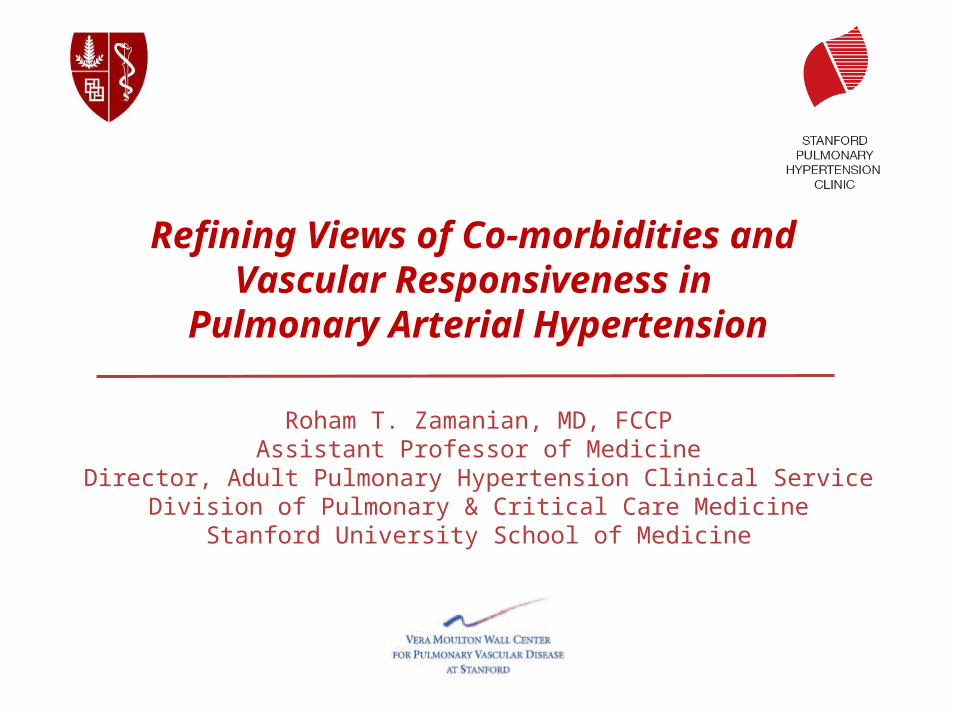
Refining Views of Co-morbidities and Vascular Responsiveness in Pulmonary Arterial Hypertension Roham T. Zamanian, MD, FCCP Assistant Professor of Medicine Director, Adult Pulmonary Hypertension Clinical Service Division of Pulmonary & Critical Care Medicine Stanford University School of Medicine
-
Upload
george-foster -
Category
Documents
-
view
36 -
download
2
description
Refining Views of Co-morbidities and Vascular Responsiveness in Pulmonary Arterial Hypertension. Roham T. Zamanian, MD, FCCP Assistant Professor of Medicine Director, Adult Pulmonary Hypertension Clinical Service Division of Pulmonary & Critical Care Medicine - PowerPoint PPT Presentation
Transcript of Refining Views of Co-morbidities and Vascular Responsiveness in Pulmonary Arterial Hypertension
Refining Views of Co-morbidities and Vascular Responsiveness in
Pulmonary Arterial Hypertension
Roham T. Zamanian, MD, FCCPAssistant Professor of Medicine
Director, Adult Pulmonary Hypertension Clinical ServiceDivision of Pulmonary & Critical Care Medicine
Stanford University School of Medicine


Personal Financial Relationships with Commercial interests relevant to medicine, within the past 3 years:
Consultant: Gilead, United Therapeutics, Ikaria, Bayer, ActelionIndustry-Sponsor Research: United Therapeutics, Gilead, Actelion
Personal Financial Relationships with Non-Commercial interests relevant to medicine, within the past 3 years:
Research Grants: - Vera Moulton Wall Center for Pulmonary Vascular Disease- NIH/NHLBI- NIH/NIAID
Personal relationships with tobacco industry entities within the past 3 years:
No relationships to disclose
No “off-label” discussions in this presentation.
Disclosures:

Pulmonary Hypertension - Diagnostic Definition:
Diagnostic gold standard = hemodynamics
Rest:• Mean PAP >25 mmHg (normal 10-15 mmHg)
Exercise:• Mean PAP > 30 mmHg
PAH = above + PCWP or LVEDP <15 mmHg
(normal 8-10 mmHg)
Associated with adverse changes• In the pulmonary vasculature (arteriopathy)
• At the level of the right ventricle (hypertrophy)
Courtesy Marlene Rabinovitch

Dana Point Classification of Pulmonary Hypertension
1. Pulmonary Arterial Hypertension 1.1 Idiopathic PAH1.2 Heritable
1.2.1. BMPR21.2.2. ALK1, endoglin (with or without hereditary hemorrhagic telangiectasia )1.2.3 Unknown.
1.3 Drug- and toxin-induced1.4 Associated with
1.4.1. Connective tissue diseases1.4.2 HIV infection1.4.3 Portal hypertension1.4.4 Congenital heart diseases1.4.5 Schistosomiasis1.4.6 Chronic hemolytic anemia
1.5 Persistent pulmonary hypertension of the newborn1’. Pulmonary veno-occlusive disease (PVO) and/or pulmonary
capillary hemangiomatosis (PCH)
2. Pulmonary hypertension due to left heart disease 2.1 Systolic dysfunction
2.2 Diastolic dysfunction2.3 Valvular disease
3. Pulmonary hypertension due to lung diseases and/or hypoxia
3.1 Chronic obstructive pulmonary disease
3.2 Interstitial lung disease
3.3 Other pulmonary diseases with mixed restrictive and obstructive pattern
3.4 Sleep-disordered breathing
3.5 Alveolar hypoventilation disorders
3.6 Chronic exposure to high altitude
3.7 Developmental abnormalities
4. Chronic thromboembolic pulmonary hypertension (CTEPH)
5. PH with unclear multifactorial mechanisms
5.1 Hematologic disorders: myeloproliferative disorders splenectomy.
5.2 Systemic disorders, sarcoidosis, pulmonary Langerhans cell histiocytosis, lymphangioleiomyomatosis, neurofibromatosis, vasculitis
5.3 Metabolic disorders: glycogen storage disease, Gaucher disease, thyroid disorders
5.4 Others: tumoral obstruction, fibrosing mediastinitis, chronic renal failure on dialysis.
Simonneau et al, JACC 2009

Outcomes in the Current Era: Stanford Adult & Pediatric Experience
Zamanian et al 2011

Benza RL et al. Circulation. 2010; 122:164-172.
REVEAL: Parameters Independently Associated With Survival (Cox Proportional Hazard Estimates)
APAH-CTDAPAH-PoPH
FPAH
Renal insufficiencyMales age ≥60 yrs
IIIIIV
Heart rate >92 bpmSystolic BP <110 mm Hg
≥440 m<165 m
<50 pg/mL>180 pg/mL
Pericardial effusion: any
% predicted DLCO ≥80% predicted DLCO ≤32
mRAP >20 mm HgPVR >32 Wood Units
WHO Group I PAH Subgroups
Demographics and Comorbidities
NYHA/WHO Functional Class
Vitals
6MWD
BNP
ECHO
DLCO
RHC
p-value
<0.001<0.001
0.012
<0.001<0.001
0.0390.008
<0.001
0.005<0.001
0.008<0.001
0.003<0.001
0.014
0.0310.018
0.043<0.001
Hazard ratios and 95% CI
1/8 1/4 1/2 1 2 4 8
Reduced risk Increased risk
HR
1.593.602.17
1.902.18
0.421.413.13
1.391.67
0.581.68
0.501.97
1.35
0.591.46
1.794.08

REVEAL: Observed 1-year Survival From Time of Enrollment According to Predicted Risk Strata
Benza RL et al. Circulation. 2010;122:164-172.
Months from enrollment
Survival (%)
0
60
100
80
40
0 3 6 9 122 5 8 111 4 7 10
Risk strataLowAverageModerately highHighVery high
No. at risk: Low Average Mod. high High Very high
1374665280295102
1368659277293100
136465727429196
135965326928489
135664826427781
135264726327074
135164026026372
134662825925569
134162525524761
133661825424159
131160424923855
130460224423352
130359624322549

What Is the Optimal Treatment Strategy?
McLaughlin VV et al. J Am Coll Cardiol. 2009;53:1573-1619.
Anticoagulate ± Diuretics ± Oxygen ± Digoxin
Negative
No LOWER RISKLOWER RISK DETERMINANTS OF RISKDETERMINANTS OF RISK HIGHER RISKHIGHER RISK
No Clinical evidence of RV failure Yes
Gradual Progression of symptoms Rapid
II, III WHO class IV
Longer (>400 m) 6MWD Shorter (<300 m)
Peak VO2 >10.4 mL/kg/min CPET Peak VO2 <10.4 mL/kg/min
Minimal RV dysfunction Echocardiography
Pericardial effusion, significant RV
enlargement/dysfunction;RA enlargement
RAP <10 mm Hg;CI >2.5 L/min/m2 Hemodynamics
RAP >20 mm Hg;CI <2.0 L/min/m2
Minimally elevated BNP Significantly elevated
Acute Vasoreactivity Testing
Sustained
Response
Continue CCB
Yes
Positive
Oral CCB

Therapeutic Options in USA for PAH – September 2012
Traditional Tx
• Supplemental O2
• Diuretics• Oral vasodilators
• (CCB)
• Anticoagulants• warfarin
• Inotropic agents• Digitalis
FDA Approved for PAH
• Prostanoids• Epoprostenol• Treprostinil
(IV/SQ/Inhaled)• Inhaled Iloprost
• ERA’s• Bosentan • Ambrisentan
• PDE-5 Inhibitors• Sildenafil• Tadalafil
Investigational Tx
• Prostanoids• Oral Treprostinil
• ERA’s• Actelion-1
• Tyrosine Kinase Inhib’s• Rho-Kinase Inhib’s• Elastase Inhib’s• Dicholoroacetate (DCA)• TetrahydroBiopterin• S. Guanylate Cyclase Act’s• FK-506• LTA4 Hydrolase Inhib’s• Stem Cell Tx

A Paradigm Shift in PAH Clinical Research
• Although we realize that WHO Group I PAH has numerous etiologies, we have
assumed to be “homogeneous” in its biology/physiology.
• Over the last decade there has been an explosion of clinical research in PAH /
PH focused on therapeutics.
• A call for clinical-translational research
• It is time for a Paradigm Shift: New Tools and Topics
• Identification of Novel (modifiable?) Risk Factors / Co-morbidities
• Refining our understanding of acute and chronic vascular responsiveness
• Recognition of potentially novel therapeutics

Overview


Insulin Resistance in PAH:
• Pulmonary Arterial Hypertension (PAH) is a vascular disease characterized by inflammation, proliferation, and vasoconstriction.• Multi-factorial & Complex
• Unlikely that a single factor, pathway, mutation in etiology
• Obesity, hyperlipidemia, and insulin resistance (IR) are known risk factors for systemic cardiovascular diseases (CVD).
• Impact of obesity or IR in PAH instigation or progression have not been validated.
• Several lines of evidence though, suggest a link between insulin resistance and PAH…..

IR in PAH: Suggestive Links
• Obesity is associated with Insulin resistance• Obesity + Inactivity IR
• Obesity is common in PAH and maybe an overlooked risk factor.• Exercise intolerance is a feature of severe PAH
• Insulin resistance has been linked to congestive heart failure and idiopathic cardiomyopathies
• Elevation of “factors” and inflammatory cytokines which have been implicated in PAH are also involved in pathogenesis of IR • IL-6, ET-1, ADMA, MCP-1
Abenhaim et al. NEJM 1996
Rich et al. Chest 2000
Taraseviciute et al. Eur J Med Res 2006
Humbert el al. AJRCCM 1995
Ikeda et al. Am J Physiol Heart Circ Physiol 2002
Stuhlinger et al. JAMA 2002
Yudkin el al. Lancet 2005

PPARActivation Reverses PAH in Insulin Resistant ApoE Deficient Mice
IR
Hansmann et al Circulation 2007
5 4 6 6
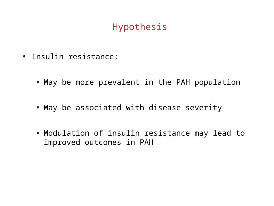
Hypothesis
• Insulin resistance:
• May be more prevalent in the PAH population
• May be associated with disease severity
• Modulation of insulin resistance may lead to improved outcomes in PAH

Design & Methods
• Prospective screening of patients with pulmonary arterial hypertension at SUMC PH Clinic between 2004-2006.
• The National Health and Nutrition Examination Survey (NHANES) as control population.
• Identification of TG/HDL-C ratio as a surrogate marker for insulin resistance.
• Excluded: Overt Diabetics, Secondary PH forms including those with pulmonary parenchymal disease and left heart failure
• Data Collection (PAH): detailed demographic, functional, hemodynamics, and event-free survival.

Receiver-operating Characteristic Curves for Metabolic Markers of Insulin Resistance
McLaughlin & Reaven et al. Ann Int Med. 2003

Zamanian et al, ERJ 2009
Higher Prevalence of IR in PAH

Zamanian et al, ERJ 2009
Unlike Healthy Controls Insulin Resistance in PAH is NOT Associated with Age or BMI

Zamanian et al, ERJ 2009

Zamanian et al, ERJ 2009
IR is a Predictor of Short-term Worse Outcomes
Log-rank p = 0.043
58%
79%

Zamanian et al, ERJ 2009
Age & BMI Adjusted Univariate Analysis
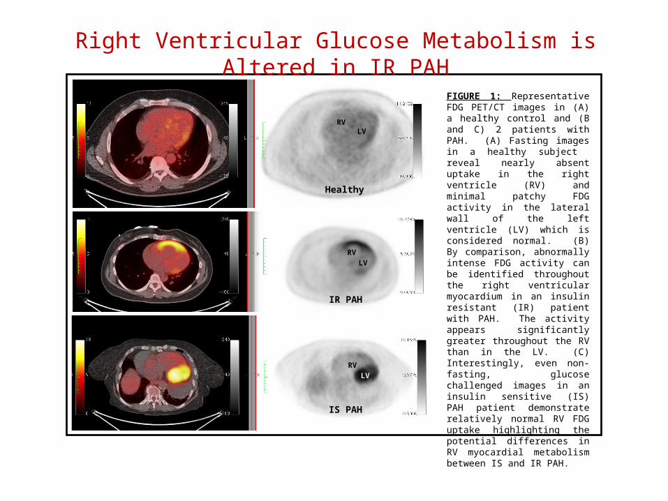
FIGURE 1: Representative FDG PET/CT images in (A) a healthy control and (B and C) 2 patients with PAH. (A) Fasting images in a healthy subject reveal nearly absent uptake in the right ventricle (RV) and minimal patchy FDG activity in the lateral wall of the left ventricle (LV) which is considered normal. (B) By comparison, abnormally intense FDG activity can be identified throughout the right ventricular myocardium in an insulin resistant (IR) patient with PAH. The activity appears significantly greater throughout the RV than in the LV. (C) Interestingly, even non-fasting, glucose challenged images in an insulin sensitive (IS) PAH patient demonstrate relatively normal RV FDG uptake highlighting the potential differences in RV myocardial metabolism between IS and IR PAH.
RVLV
RV
LV
RV
LV
Healthy
IR PAH
IS PAH
Right Ventricular Glucose Metabolism is Altered in IR PAH

Zamanian et al ATS 2012 A3453
Impact of IR on RV Structure & Function: MESA-RV Study
• The Multi-Ethnic Study of Atherosclerosis (MESA) performed interpretable cardiac MRIs on 5,004 participants without clinical cardiovascular disease at six field centers.
• 4168 non-diabetic healthy controls were evaluated categorized as IR or IS and Cardiac MRI data analyzed.
• IR in healthy cohort is associated with higher BMI, systolic and diastolic blood pressure, and higher CRP.
ParameterInsulin
SensitiveInsulin
Resistantp
RVEDM, g 21.48±0.11 20.86±0.11 <0.0001RVEDV, mL 125.01±0.55 123.19±0.55 0.0077RVESV, mL 37.69±0.33 36.74±0.33 0.0269RVEF, % 70.66±0.19 70.61±0.19 0.9728RVSV, mL/min 90.02±0.55 85.67±0.55 <0.0001

Hemodynamic Data IS (n=59) IR (n=25) pmRAP (mmHg) 8+/-3.9 8.2+/-4.3 >0.05mPAP (mmHg) 51+/-13.5 51.5+/-15 >0.05RVEDP (mmHg) 12.5+/-4.5 12.5+/-6 >0.05PCWP (mmHg) 10.8+/-3.5 10.5+/-3.3 >0.05CO (L/min) 3.9+/-1.2 4.1+/-1.6 >0.05SV (mL) 53+/- 20.5 55.5 +/- 25 >0.05PVR (WU) 11.7+/-6 12.7+/-7.9 >0.05
Echocardiography IS n=19 IR n=11 pMV E (cm/sec) 81.5+/-17 64.5+/-19 <0.05MV A (cm/sec) 72.5+/-23.5 78.5+/-18 >0.05Lat E‘ (cm/sec) 13.9+/-3.5 10.4+/-2.2 <0.01 E/A 1.18+/-0.4 0.8+/-0.2 <0.01 E/E' 6.1+/-1.6 6.3+/-1.9 >0.05TAPSE (cm) 2+/-0.4 2.1+/-0.3 >0.05
LA maximum volume index (cm2/m2) 19.5+/-7 17+/-4.5 >0.05
RA maximum volume index (cm2/m2) 45+/-25 40+/-18 >0.05
RV-MPI 0.6+/-0.18 0.65+/-0.18 >0.05
TR max PG (mmHg) 82.4+/-16 75.8+/-15 >0.05RV FAC (%) 26+/-10 24+/-11 >0.05IVC (cm) 1.7+/-0.5 1.6+/-0.6 >0.05TR max PG (mmHg) 82.4+/-16 75.8+/-15 >0.05RV FAC (%) 26+/-10 24+/-11 >0.05IVC (cm) 1.7+/-0.5 1.6+/-0.6 >0.05
Impact of IR on RV Function in PAH
Skhiri et al ATS 2012 A3463

Early Experience with Pioglitazone in PAH
Bosentan Pioglitazone

Interim Summary
• Insulin resistance is more prevalent in women with PAH than the general female population.
• Although obesity may be a link between IR and PAH, our results do not support the idea that obesity alone is the cause of insulin resistance in pulmonary arterial hypertension.
• Though IR confers a poorer prognosis, Insulin resistance in PAH does not appear to correlate with functional class or disease severity.
• IR may be linked to subtle changes in diastolic ventricular function and right ventricular metabolism.

• 35 yo French female (mother of 2)
• Historically very active 3-5 sets of tennis daily
• Over last year with profound dyspnea• Can’t garden
• 1 episode of LOC picking up child
Clinical Case Scenario

RPAP

RPCWP
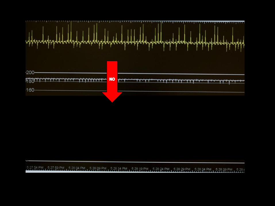

RPAP after 5 minutes NO @ 20 ppm

Vasoreactivity Traditionally Defined
• Right Heart Catheterization:• Reduction in mPAP by at least 10 mmHg
• Must reduce to a mPAP of <40 mmHg.
• Cardiac output must be maintained or improved as a result.
• Agents used:• Nitric Oxide
• Adenosine
• Epoprostenol
• Standard of Care in 1st time RHC for iPAH.

French Registry:Response to Acute Vasodilator Challenge
Res
po
nse
(%
)
Idiopathic
N=649.Challenge with vasodilator at time of right heart
catheterization.
10.3%
Humbert M, et al. Am J Respir Crit Care Med. 2006;173(9):1023-1030.
0%
2.6%
0% 0%
3.3%
1.6%
6.8%
Familial ConnectiveTissue
CongenitalHeart
PortalHypertension
Anorexigens HIV >2 Factors

Survival in IPAH on Oral Calcium Channel Blocker Therapy
Sitbon O, et al. Circulation. 2005;111(23):3105-3111.
Survival endpoint included those who received transplants or were lost to follow-up. Acute response defined as defined by a fall in both mean pulmonary artery pressure (PAP) and pulmonary vascular
resistance (PVR) >20%.
1.0
0.8
0.6
0.4
0.2
0
0 2 4 6 8 10 12 14 16 18
38 33 30 22 13 8 3 3 2 1Years
Failures
Cu
mu
lati
ve S
urv
ival Responders
19 12 7 4 0Subjects
at Risk (n)
RespondersFailures
(Long-term Calcium Channel Blocker Therapy)

Thinking of the Right Ventricle – PA Compliance
Lankhaar J-W et al. European Heart Journal (2008)29,1688-1695
• Pulmonary arterial compliance (SV/Ps-Pd) is being recognized as an important contributor to right ventricular afterload and has been shown to be a strong predictor of survival.
• Vasoreactivity or milder degrees of vascular responsiveness have not yet been correlated with pulmonary vascular compliance.

1. Vasoreactivity is found not only in IPAH but also other forms of PAH and can change over time.
2. Even a mild degree of vasoresponsiveness is of prognostic value.
3. Correlating changes in PVR, mPAP and PAC during vasoreactivity testing could help identify additional patients with a reactive vascular bed.
Hypotheses

Methods
• Retrospective study (220 patients PAH Group 1) presented to Stanford Medical Center between 2000 - 2010.
• Diagnosed at the time of RHC with vasodilator testing with 20ppm NO
• Demographics, functional status, medication as well as hemodynamic parameters were evaluated.
• A positive vasoreactivity was defined by a reduction in pulmonary artery mean pressure (PAPm) 10 mmHg to reach an absolute value of PAPm ≤ 40mmHg with an increased or unchanged cardiac output after challenge with NO (20 PPM)
• Previous definitions of actue vasoreactivity (as defined by changes in pulmonary vascular resistance (PVR) or PAPm by > 20%) was also evaluated
Spiekerkoetter et al, ATS 2011

Female:Male 168:59 (74%,26%)
Age: 44.8 ± 0.9
PAH etiology:
IPAH 38 (17 %)
Drugs and toxins 54 (24 %)
CTD 45 (20 %)
Portal hypertension 12 (5 %)
Congenital heart disease 41 (18 %)
multifactorial 37 (16 %)
NYHA
I 4%
II 29%
III 45%
IV 22%
6-min walk (m) (n=204) 381 ± 10
NT-pro BNP pg/mL (n=110) 1276 ± 168
Therapies
None 96 (42%)
Prostanoids
Epoprostenol 20 (9%)
Iloprost 9 (4%)
Treprostinil 7 (3%)
ERA 38 (17%)
PDE-I 54 (24%)
* 27% on CCB held for VR testing, 28 (12%) on multiple therapies
Overall Demographics and Clinical Characteristics
Baseline hemodynamics
NO challenge p
mRA 9.3 ± 0.3 - N/A
mPAP 56.1 ± 1.0 51.8 ± 1.1 < 0.001
PCWP 9.7 ± 0.3 - N/A
CI 2.13 ± 0.04 2.18 ± 0.05 < 0.005
SV 48.5 ± 1.3 52.8 ± 1.5 < 0.001
PVRI 23.8 ± 0.8 20.0 ± 0.8 < 0.0001
HR 81.6 ± 1.2 78.2 ± 1.2 < 0.0001
MAP 83.3 ± 1.4 83.8 ± 1.1 NS
PAC 0.96 ± 0.04 1.16 ± 0.05 < 0.001
Spiekerkoetter et al, ATS 2011
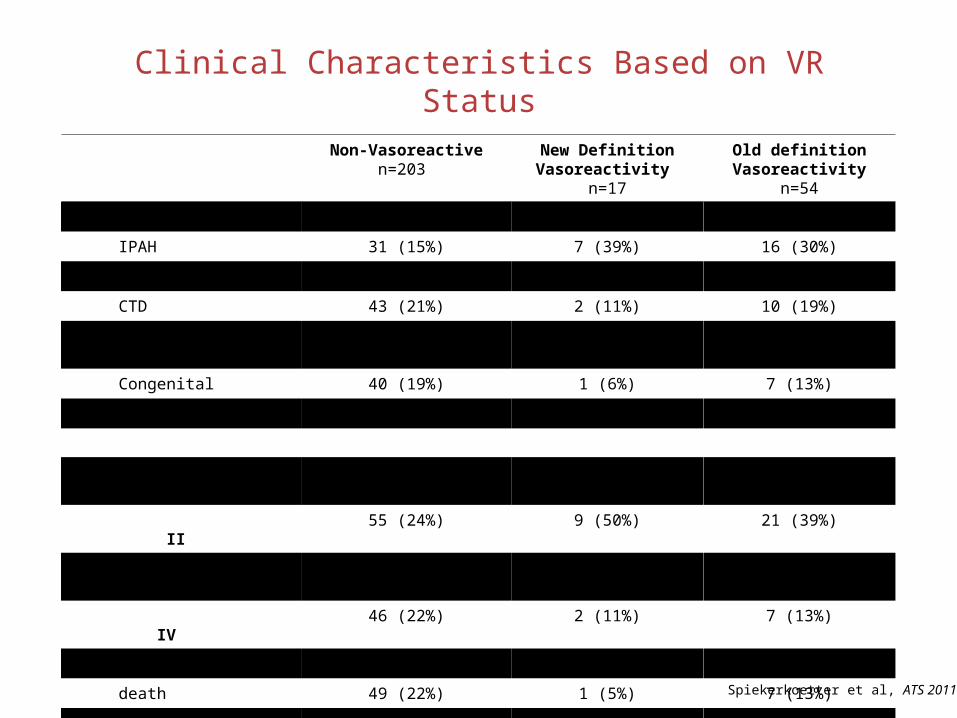
Non-Vasoreactiven=203
New Definition Vasoreactivity
n=17
Old definition Vasoreactivity
n=54
PAH etiology
IPAH 31 (15%) 7 (39%) 16 (30%)
D&T 49 (23%) 5 (28%) 5 (9%)
CTD 43 (21%) 2 (11%) 10 (19%)
Portal hypertension 12 (6%) 0 1 (2%)
Congenital 40 (19%) 1 (6%) 7 (13%)
Multifactorial 34 (16%) 3 (16%) 15 (27%)
NYHA class I 8 (4%) 0 1 (2%)
II 55 (24%) 9 (50%) 21 (39%)
III 91 (44%) 7 (39%) 25 (46%)
IV 46 (22%) 2 (11%) 7 (13%)
events
death 49 (22%) 1 (5%) 7 (13%)
transplant 9 (4%) 0 2 (4%)
Clinical Characteristics Based on VR Status
Spiekerkoetter et al, ATS 2011

Spiekerkoetter et al, ATS 2011


Acute Vasoreactivity isn’t “the” determinant to outcomes in PAH
Spiekerkoetter et al, ATS 2011

2007 2009
Arterial Phase
Capillary Blush
CHRONIC VASCULAR REACTIVITY
VASCULAR REMODELING?
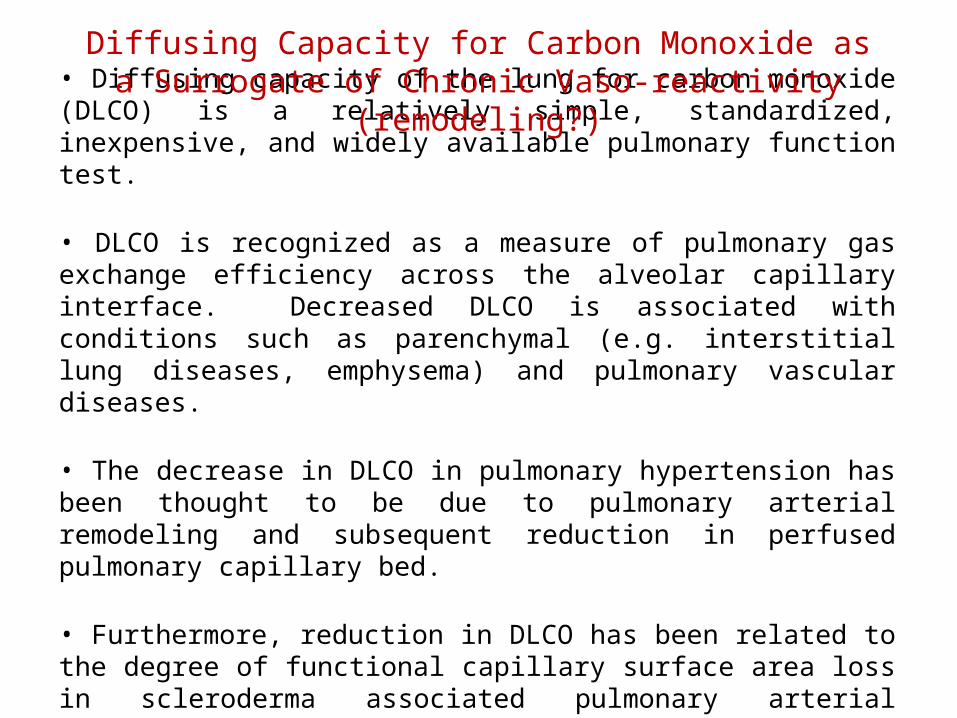
• Diffusing capacity of the lung for carbon monoxide (DLCO) is a relatively simple, standardized, inexpensive, and widely available pulmonary function test.
• DLCO is recognized as a measure of pulmonary gas exchange efficiency across the alveolar capillary interface. Decreased DLCO is associated with conditions such as parenchymal (e.g. interstitial lung diseases, emphysema) and pulmonary vascular diseases.
• The decrease in DLCO in pulmonary hypertension has been thought to be due to pulmonary arterial remodeling and subsequent reduction in perfused pulmonary capillary bed.
• Furthermore, reduction in DLCO has been related to the degree of functional capillary surface area loss in scleroderma associated pulmonary arterial hypertension (PAH), suggesting that DLCO is also a marker of endothelial cell function.
Diffusing Capacity for Carbon Monoxide as a Surrogate of Chronic Vaso-reactivity (remodeling?)

• Numerous recent studies have demonstrated the clinical utility of DLCO in PAH. Baseline DLCO can predict long-term survival in PAH.
• Decreasing DLCO over time can also predict the development of pulmonary hypertension in patients at risk, such as ones with limited scleroderma.
• While changes in DLCO over time have been demonstrated to be more powerful in predicting prognosis in idiopathic pulmonary fibrosis than single-point DLCO measurement, there are currently no studies evaluating the extent and utility of delta DLCO in pulmonary hypertension.
DLCO Cont
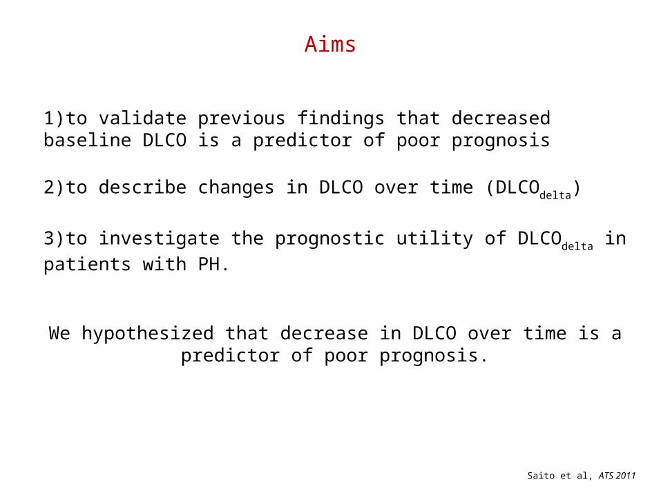
1)to validate previous findings that decreased baseline DLCO is a predictor of poor prognosis
2)to describe changes in DLCO over time (DLCOdelta)
3)to investigate the prognostic utility of DLCOdelta in patients with PH.
We hypothesized that decrease in DLCO over time is a predictor of poor prognosis.
Aims
Saito et al, ATS 2011

PAH or CTEPH (n=313)
“Total Cohort” DLCOadj available
(n=246)
No DLCO (n=18)
Included
Legend:
Excluded
Subjects with DLCO(n=295)
TLC
FVC
TLC < 60% (n=16)
TLC unavailable(n=29)
FVC < 60% (n=11)
TLC > = 60%(n=250)
FVC > = 60%(n=250)
DLCO available(n=268)
“Follow-up Cohort” DLCOadj available @ 1yr
(n=34)
DLCOadjDLCOadj unavailable
(n=22)
DLCOadj @ follow up unavailable
(n=22)
Figure 1

Figure 2
0 365 730 1095 1460 1825 2190 2555 29200
25
50
75
100
DLCOadj <40%
DLCOadj 40-80%
DLCOadj >80%
Log-rank p = 0.0035
Days
Su
rviv
al (
%)

Figure 3

Figure 4
0 365 730 1095 1460 1825 2190 2555 29200
25
50
75
100
Absolute increase in DLCO<10% or loss
Absolute increase in DLCO>=10%
Days
Su
rviv
al (
%)
Log-rank p=0.029

Figure 1: Change in DLCO & Survival – Kaplan Meier analysis demonstrates best survival for patients who had improvement of DLCO over time.
* DLCOdec vs DLCOstab Log rank p=0.041. ** DLCOinc vs DLCOstab Log rank p=0.007.
DLCOdec *
DLCOstab
DLCOinc **
External Validation Cohort

Conclusions
• Advancement in understanding of co-morbidities in a rare disorder such as PAH may have profound clinical and therapeutic implications.• Insulin resistance is modifiable (pharmacologic versus non-pharm)
• Improvement in clinical phenotyping of the acute vasoreactive pulmonary hypertension patient is needed.• Gain of Vaso-responsiveness maybe suggestive of “reverse
remodelling”
• Potentially informative of novel clinical endpoints and mechanisms of action.

Mark NicollsMark KrasnowMarlene RabinovitchJeffery FeinsteinFrancois HaddadVinicio De Jesus PerezEdda SpiekerkoetterKristina KudelkoLorinda Chung
Ramona DoyleSteven Kawut
Juliana LiuSherrie Jones
Angela HerreraDarlene FrieYuwen Liao
Val ScottAndrew Hsi
Patricia Del RosarioAllyson Rupp
Nathan BrunnerKrithika Ramachandran
Acknowledgements:
Work Supported by: NIH/NHLBI NHLBI-HV-10-05, 1U01HL107393-01, PAR-09-185, N01-HV-00242
Vera Moulton Wall Center for Pulmonary Vascular Disease