Reduced Skeletal Muscle Protein Turnover and Thyroid ...
10
ORIGINAL RESEARCH published: 28 February 2019 doi: 10.3389/fendo.2019.00119 Frontiers in Endocrinology | www.frontiersin.org 1 February 2019 | Volume 10 | Article 119 Edited by: Nigel Turner, University of New South Wales, Australia Reviewed by: Andrew Philp, Garvan Institute of Medical Research, Australia Assunta Lombardi, University of Naples Federico II, Italy *Correspondence: Abdul G. Dulloo [email protected] Specialty section: This article was submitted to Obesity, a section of the journal Frontiers in Endocrinology Received: 01 November 2018 Accepted: 08 February 2019 Published: 28 February 2019 Citation: Calonne J, Isacco L, Miles-Chan J, Arsenijevic D, Montani J-P, Guillet C, Boirie Y and Dulloo AG (2019) Reduced Skeletal Muscle Protein Turnover and Thyroid Hormone Metabolism in Adaptive Thermogenesis That Facilitates Body Fat Recovery During Weight Regain. Front. Endocrinol. 10:119. doi: 10.3389/fendo.2019.00119 Reduced Skeletal Muscle Protein Turnover and Thyroid Hormone Metabolism in Adaptive Thermogenesis That Facilitates Body Fat Recovery During Weight Regain Julie Calonne 1 , Laurie Isacco 1,2,3 , Jennifer Miles-Chan 1 , Denis Arsenijevic 1 , Jean-Pierre Montani 1 , Christelle Guillet 2 , Yves Boirie 2 and Abdul G. Dulloo 1 * 1 Department of Endocrinology, Metabolism and Cardiovascular System, Faculty of Sciences and Medicine, University of Fribourg, Fribourg, Switzerland, 2 Université Clermont Auvergne, INRA, UNH, Unité de Nutrition Humaine, CHU Clermont-Ferrand, Service de Nutrition Clinique, CRNH Auvergne, Clermont-Ferrand, France, 3 EA3920 and EPSI Platform, Bourgogne Franche-Comté Université, Besançon, France Objective: The recovery of body composition after weight loss is characterized by an accelerated rate of fat recovery (preferential catch-up fat) resulting partly from an adaptive suppression of thermogenesis. Although the skeletal muscle has been implicated as an effector site for such thrifty (energy conservation) metabolism driving catch-up fat, the underlying mechanisms remain to be elucidated. We test here the hypothesis that this thrifty metabolism driving catch-up fat could reside in a reduced rate of protein turnover (an energetically costly “futile” cycle) and in altered local thyroid hormone metabolism in skeletal muscle. Methods: Using a validated rat model of semistarvation-refeeding in which catch-up fat is driven solely by suppressed thermogenesis, we measured after 1 week of refeeding in refed and control animals the following: (i) in-vivo rates of protein synthesis in hindlimb skeletal muscles using the flooding dose technique of 13 C-labeled valine incorporation in muscle protein, (ii) ex-vivo muscle assay of net formation of thyroid hormone tri-iodothyronine (T3) from precursor hormone thyroxine (T4), and (iii) protein expression of skeletal muscle deiodinases (type 1, 2, and 3). Results: We show that after 1 week of calorie-controlled refeeding, the fractional protein synthesis rate was lower in skeletal muscles of refed animals than in controls (by 30–35%, p < 0.01) despite no between-group differences in the rate of skeletal muscle growth or whole-body protein deposition—thereby underscoring concomitant reductions in both protein synthesis and protein degradation rates in skeletal muscles of refed animals compared to controls. These differences in skeletal muscle protein turnover during catch-up fat were found to be independent of muscle type and fiber composition, and were associated with a slower net formation of muscle T3 from precursor hormone T4, together with increases in muscle protein expression of deiodinases which convert T4 and T3 to inactive forms.
Transcript of Reduced Skeletal Muscle Protein Turnover and Thyroid ...
ORIGINAL RESEARCHpublished: 28 February 2019
doi: 10.3389/fendo.2019.00119
Frontiers in Endocrinology | www.frontiersin.org 1 February 2019 | Volume 10 | Article 119
Edited by:
Nigel Turner,
University of New South Wales,
Australia
Reviewed by:
Andrew Philp,
Garvan Institute of Medical Research,
Australia
Assunta Lombardi,
University of Naples Federico II, Italy
*Correspondence:
Abdul G. Dulloo
Specialty section:
This article was submitted to
Obesity,
a section of the journal
Frontiers in Endocrinology
Received: 01 November 2018
Accepted: 08 February 2019
Published: 28 February 2019
Citation:
Calonne J, Isacco L, Miles-Chan J,
Arsenijevic D, Montani J-P, Guillet C,
Boirie Y and Dulloo AG (2019)
Reduced Skeletal Muscle Protein
Turnover and Thyroid Hormone
Metabolism in Adaptive
Thermogenesis That Facilitates Body
Fat Recovery During Weight Regain.
Front. Endocrinol. 10:119.
doi: 10.3389/fendo.2019.00119
Reduced Skeletal Muscle ProteinTurnover and Thyroid HormoneMetabolism in AdaptiveThermogenesis That Facilitates BodyFat Recovery During Weight Regain
Julie Calonne 1, Laurie Isacco 1,2,3, Jennifer Miles-Chan 1, Denis Arsenijevic 1,
Jean-Pierre Montani 1, Christelle Guillet 2, Yves Boirie 2 and Abdul G. Dulloo 1*
1Department of Endocrinology, Metabolism and Cardiovascular System, Faculty of Sciences and Medicine, University of
Fribourg, Fribourg, Switzerland, 2Université Clermont Auvergne, INRA, UNH, Unité de Nutrition Humaine, CHU
Clermont-Ferrand, Service de Nutrition Clinique, CRNH Auvergne, Clermont-Ferrand, France, 3 EA3920 and EPSI Platform,
Bourgogne Franche-Comté Université, Besançon, France
Objective: The recovery of body composition after weight loss is characterized by an
accelerated rate of fat recovery (preferential catch-up fat) resulting partly from an adaptive
suppression of thermogenesis. Although the skeletal muscle has been implicated as an
effector site for such thrifty (energy conservation) metabolism driving catch-up fat, the
underlying mechanisms remain to be elucidated. We test here the hypothesis that this
thrifty metabolism driving catch-up fat could reside in a reduced rate of protein turnover
(an energetically costly “futile” cycle) and in altered local thyroid hormone metabolism in
skeletal muscle.
Methods: Using a validated rat model of semistarvation-refeeding in which catch-up
fat is driven solely by suppressed thermogenesis, we measured after 1 week of
refeeding in refed and control animals the following: (i) in-vivo rates of protein synthesis
in hindlimb skeletal muscles using the flooding dose technique of 13C-labeled valine
incorporation in muscle protein, (ii) ex-vivo muscle assay of net formation of thyroid
hormone tri-iodothyronine (T3) from precursor hormone thyroxine (T4), and (iii) protein
expression of skeletal muscle deiodinases (type 1, 2, and 3).
Results: We show that after 1 week of calorie-controlled refeeding, the fractional
protein synthesis rate was lower in skeletal muscles of refed animals than in controls
(by 30–35%, p < 0.01) despite no between-group differences in the rate of skeletal
muscle growth or whole-body protein deposition—thereby underscoring concomitant
reductions in both protein synthesis and protein degradation rates in skeletal muscles of
refed animals compared to controls. These differences in skeletal muscle protein turnover
during catch-up fat were found to be independent of muscle type and fiber composition,
and were associated with a slower net formation of muscle T3 from precursor hormone
T4, together with increases in muscle protein expression of deiodinases which convert
T4 and T3 to inactive forms.

Calonne et al. Muscle Metabolism During Weight Regain
Conclusions: These results suggest that diminished skeletal muscle protein turnover,
together with altered local muscle metabolism of thyroid hormones leading to diminished
intracellular T3 availability, are features of the thrifty metabolism that drives the rapid
restoration of the fat reserves during weight regain after caloric restriction.
Keywords: thermogenesis, obesity, catch-up growth, thrifty metabolism, caloric restriction, deiodinase
INTRODUCTION
The recovery of body weight after substantial weight loss ordiminished growth rate is accompanied by a high efficiency offat deposition (1–7). This in part results from an adaptivesuppression of thermogenesis which contributes to thepreferential catch-up fat phenomenon (8) whereby body fatis recovered at a disproportionately faster rate than that of leanbody mass. Such thrifty (energy conservation) metabolism forpreferential catch-up fat probably had evolutionary survivalvalue as it contributes to the rapid restoration of survivalcapacity conferred by the rapid recovery of the fat reserves inpreparation for the next period of food scarcity. Nowadays,however, it is contributing to the “metabolic adaptation” thatfacilitates obesity recidivism after therapeutic slimming (9), andhas also been implicated as a component of the “imprinted”thrifty phenotype in the link between early growth perturbations,excessive fat deposition during subsequent catch-up growth andlater development of obesity and cardiometabolic diseases (10).
The effector mechanisms underlying this thrifty metabolismdriving catch-up fat remain elusive. However, as the skeletalmuscle is a major site for thermogenesis, glucose utilizationand lipid oxidation, the possibility arises that a reduction inthermogenesis in this tissue could result in the redirection ofspared fuel toward fat storage in adipose tissue (8). Whichthermogenic effectors in skeletal muscle are suppressed to spareenergy for catch-up fat are, however, far from being understood.A role for the uncoupling protein homologs UCP2 and UCP3—which have been proposed as potential uncouplers of oxidativephosphorylation (11)—is unlikely on the basis that their patternsof expressions in skeletal muscle do not fit with diminishedthermogenesis in response to starvation and refeeding (12).By contrast, a number of findings suggest a role for alteredperipheral action of the main active thyroid hormone, tri-iodothyronine (T3) for which skeletal muscle is a major target(13–15). Indeed, the circulating levels of T3, which is well-knownto be diminished during caloric restriction, tend to remain lower(albeit marginally) in refed animals than in controls during thephase of catch-up fat (16, 17). More recently, it was shown thatthe net local synthesis of T3 in the gastrocnemius muscle, whichis diminished during semistarvation, persists during the dynamicphase of catch-up fat, and is associated with several features ofdiminished intracellular availability of T3, in particular delayedcontraction-relaxation kinetics and increased proportion of slowat the expense of fast muscle fibers (18). Taken together, thesealterations in thyroid hormone-dependent properties of skeletalmuscle constitute mechanisms that could underlie diminishedskeletal muscle thermogenesis during weight loss and which
persist during weight regain for the purpose of acceleratingfat recovery.
As protein synthesis and protein turnover (an energeticallycostly substrate cycling) is also under the control of thyroidhormones, with protein turnover in skeletal muscle estimated tocontribute to as much as 20% of whole body protein turnover(19–23), we investigated here (i) the extent to which the processesof protein synthesis and protein turnover may be diminishedduring the dynamic phase of catch-up fat in various muscle typesvarying widely in fiber composition, and (ii) their associationswith altered skeletal muscle thyroid hormone metabolism andchanges in the levels of the deiodinases (DIO1, DIO2, andDIO3) that modulate the local metabolism and intracellularavailability of T3.
MATERIALS AND METHODS
AnimalsSprague-Dawley male rats (Elevage Janvier, Le Genest-Saint-Isle, France), 6 weeks-old, were adapted to room and cageenvironments for 5–7 days prior to the start of each experiment.They were caged singly in a controlled room (22 ± 1◦C)with a 12-h light-dark cycle, and maintained on a commercialpelleted chow diet (Provimi-Kliba SA, Kaiseraugst, Switzerland)consisting, by energy, of 24% protein, 66% carbohydrate, and10% fat, and had free access to tap water. Animals weremaintained in accordance with the regulations and guidelines ofthe Department of Medicine, University of Fribourg, for the careand use of laboratory animals; all experimental procedures wereperformed under conditions approved by the Ethical Committeeof the State of Fribourg Veterinary Office.
Experimental DesignExperiments were performed according to our previouslyreported design of semistarvation-refeeding that established a ratmodel for studying adjustments in energy expenditure specificfor accelerating fat deposition during refeeding (3, 4, 16, 17). Inall experiments, the semistarved rats were caloric restricted at50% of ad libitum energy intake for 2 weeks, after which theywere refed for periods of either 1 or 2 weeks, and comparisonsmade with control rats having similar body weight at the onsetof refeeding. Both refed and control groups were provided with(and consumed) the same amount of a semisynthetic diet, whichcorresponded to that consumed during spontaneous food intakeon pelleted chow; the details of composition of this semisynthetic(low-fat) diet and assessments of metabolizable energy (ME)intake have been reported previously (4).
Frontiers in Endocrinology | www.frontiersin.org 2 February 2019 | Volume 10 | Article 119

Calonne et al. Muscle Metabolism During Weight Regain
Body Composition AnalysisAfter the animals were killed by decapitation, the whole carcasseswere dried to a constant weight in an oven maintained at 70◦Cand were subsequently homogenized for analysis of total fatcontent by the Soxhlet extraction method (24). The dry fat-freemass (dry FFM) was determined by subtracting total body fatand body water content from body weight, and the protein masswas calculated as follow: Protein mass (g) = dry FFM (g)∗0.8; asdetailed previously (4).
Energy Balance and Energetic EfficiencyCalculationsEnergy balance measurements were conducted during refeedingby the comparative carcass technique over periods during whichME intake was monitored continuously, and energy expenditureover 2 weeks was determined as the difference between energygain and ME intake. Body energy gain, fat gain, and proteingain during the 2 weeks of refeeding were obtained as thedifference between the final and initial values (with the lattervalues estimated from values obtained from the group killed atthe onset of refeeding). Total body energy content, and 1Bodyenergy can be calculated from a general formula relating the totalenergy value of the carcass, energy derived from fat, and energyderived from protein (4).
Determination of Protein Turnover in-vivoTracer AdministrationProtein synthesis rate was measured by incorporation of astable isotope in the form of labeled amino acid (13C-valine)into the protein pool using the flooding dose method (25–27).Reagents were obtained from Sigma Chemical (St Louis, MO,USA) and L-[13C]-valine (99 atom percent excess) was obtainedfrom Eurisotop France (Saint-Aubin, France). Muscle proteinsynthesis rates were assessed in hindlimb skeletal muscles byusing the flooding-dose method. Food was removed early in themorning (07:00 h). At 6–7 h later, i.e., in the postabsorptive phase,rats were injected subcutaneously with L-[13C]-valine [300 µmol(100 g body)−1]. Fifty minutes after the injection of L-[13C]-valine (incorporation time), the animals were sacrificed, andskeletal muscles (gastrocnemius, soleus, tibialis anterior) werequickly excised, weighed, frozen in liquid nitrogen and storedat −80◦C until further analyses. The contralateral muscles werealso dissected intact, blotted and weighed, and frozen in liquidnitrogen for later total protein determination by the methodof Lowry (28).
Analytical MethodMuscle samples (50mg) were homogenized in an ice-coldbuffer using Polytron homogenizer (PT1200C, Kinematica,Switzerland). After precipitation of the homogenate,centrifugations, and protein hydrolysis, amino acids werederivatized, and measurements of L-[13C]-valine enrichment inhydrolyzed proteins were performed by gas chromatography-combustion-isotope ratio mass spectrometry (Gas system,Fisons Instruments, VG isotech, Middlewich, UK). L-[13C]-valine enrichments in tissue fluid were assessed using gaschromatography-mass spectrometer (GC-MS) (HP5890,
Hewlett-Packard, Paris, France) and used as precursor poolenrichment for the calculations of the fractional synthesis rates.
Calculations of Fractional Synthesis Rate (Ksyn)This is calculated as previously described (25, 26). Basalsubgroups (n = 4) are used for the determination of naturalisotopic abundance in proteins in the muscles, as follows:Fractional synthetic rate (Ksyn) = (Ei × 100)/(Ep × t), whereEi represents the enrichment as atom percentage excess of [13C]derived from valine in muscle proteins at time t (minus basalenrichment); Ep is the mean enrichment in the precursor pool(tissue fluid L-[13C]-valine); t is the incorporation time (fromtime of tracer injection to sacrifice) expressed per day; data onKsyn are expressed as percentage per day (%/d).
Calculations of Fractional Growth Rate (Kgrowth)For each tissue, Kgrowth (expressed as %/d) is determined as theaverage Kgrowth over 48 h immediately before the measurementof protein synthesis as described by Samuels et al. (29), and iscalculated as follows:
Kgrowth = (1body mass/1t) × (1tissue protein mass/1bodymass)× (100/tissue protein mass), where(i) (1body mass/1t) is the body growth rate of individual
animals during the 48 h before measurement of proteinsynthesis,
(ii) (1tissue protein mass/1body mass) is the x-coefficient of alinear regression of tissue protein mass against body weightof all animals in the same treatment group, and
(iii) tissue protein mass is the mass of protein in the individualdissected tissues from each animal when synthesis ismeasured.
Calculations of fractional degradation rate (Kdeg)For each individual rat muscle, Kdeg is obtained by subtracting
Kgrowth from Ksyn;
i.e., Kdeg (%/d) = Ksyn (%/d)—Kgrowth (%/d).
Inherent in this calculation that provides an estimate of proteindegradation are the following assumptions: (i) over the daysinterval at which the growth of the rat was measured, thegrowth of the protein mass in the muscle was proportional tothat of the whole body, and (ii) the rate of muscle proteinsynthesis measured over 50min (incorporation time periodbetween injection of L-[13C]-valine and animal sacrifice), issimilar to the average rate for the days period over which timethe growth rate is measured. These assumptions have beenvalidated in actively growing rats (30–32), and this method ofin-vivo determination of protein turnover in skeletal muscle hasbeen utilized under a variety of nutritional and environmentalconditions (29–36).
Net T3 Neogenesis AssayThe kinetics of thyroid hormone metabolism in skeletal musclewere assessed in vitro as described previously (18), usingthe method of Kaplan and Utiger (37) by incubating musclehomogenates in Tris buffer at 37◦C. The T3 neogenesis reactionwas started by adding T4 (1.3µM) dissolved in PBS containing
Frontiers in Endocrinology | www.frontiersin.org 3 February 2019 | Volume 10 | Article 119

Calonne et al. Muscle Metabolism During Weight Regain
0.25% BSA. Aliquots of the homogenate were removed after 0,5, 10, 15, and 30min, the reaction was stopped by adding 95%ethanol, and the samples were stored at 4◦C until assayed forthyroid hormone content using T3/T4 enzyme immunoassaykits (from Diagnostic System Laboratories, Webster,Texas, USA).
Protein Extraction and Western BlottingThe expression levels of skeletal muscle DIO proteins weredetermined by Western blots according to standard proceduresdescribed in details elsewhere (38, 39). Hindlimb skeletalmuscles (gastrocnemius, soleus, and tibialis anterior) wereharvested and immediately put in liquid nitrogen. Frozentissues (30mg) were rapidly weighed and homogenized inliquid nitrogen. Muscle proteins were extracted in 9 volumesof Guba-Straub buffer (300mM NaCl, 100mM NaH2PO4,10mM Na2HPO4, 10mM Na4P2O7, 1mM MgCl2, 10mMEDTA (pH 6.5) containing 0.1% 2-mercaptoethanol and 0.2%protease and phosphatase inhibitor cocktail. After incubation for45min at 4◦C, samples were sonicated for 10 s, Triton X-100(Applichem, Axon Lab AG, Le Mont-sur-Lausanne, Switzerland)was added to a final concentration of 1% and extracts werecentrifuged at 12,000 g for 15min at 4◦C. The supernatants werecollected, and protein content was determined using Bradfordmethod (BioRad, California, USA). Extracts were first dilutedin Guba-Straub buffer before the addition of Laemmli bufferat a final concentration of 3 mg/mL before being used forimmunoblotting. Thirty micro gram of protein extract wasseparated by SDS-PAGE and blotted on PVDF membranes.Membranes were incubated first with primary antibodies (detailsin Table 1), and then secondary antibody LI-COR anti-rabbit(dilution 1/15,000) or anti-goat (dilution 1/15,000) were usedto detect bands. The signals were visualized and quantified withthe use of Odyssey Infrared Imaging System (Li-Cor Biosciences,Bad Homburg, Germany), and normalized with Ponceau Red.Validation of the antibodies used for detecting and quantifyingDIO1, DIO2, and DIO3 has been detailed as supplementarymaterial in a previous report (18). For each DIOs, between-group comparisons were performed separately at each of thefollowing two time-points: (i) at the end of semistarvation(SS group) vs. the controls (Css group) and (ii) after 7 daysof refeeding (RF group) vs. their controls (CRF group). Eachbetween-group comparison (SS vs. Css or RF vs. CRF) for a givenmuscle was thus made on the same gel and under the sameconditions (sample preparation, exposure conditions towardantibodies, etc.).
Data Analysis and StatisticsAll data are presented as means± SE. Unpaired t-test was used toassess the effects of semistarvation (semistarved vs. control rats)and refeeding (refed vs. control rats) on the various parameters;statistical significance of differences are indicated as follows: ∗p< 0.05; ∗∗p < 0.01; ∗∗∗p < 0.001. The statistical treatment of datawas performed using the computer software STATISTIX, version8.0 (Analytical Software, St. Paul, MN).
TABLE 1 | Primary antibodies and specific conditions used for analysis of DIO
protein levels.
DIO1 DIO2 DIO3
Supplier Proteintech
Europe
Santa-Cruz
biotechnology
Novus biological
Catalog number 11790-1-AP sc-98716 NBP1-05767
Dilution used 1/1,000 1/200 1/1,000
Blocking buffer BSA Milk Milk
Gel 0.8mm 0.8mm 0.8 mm
Stacking 4% 4% 4 %
Resolving 12% 12% 12 %
Running 50V for 30min 50V for 30min 50V for 30 min
conditions 150V for 2h 150V for 2h 150V for 2h
Transfer conditions 400mA for 1 h
30
400mA for 1 h 30 400mA for 1 h 30
RESULTS
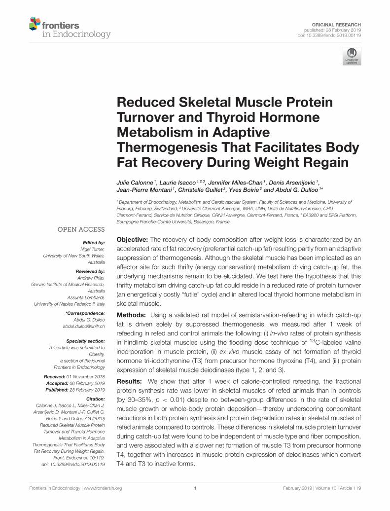
Body Weight and Body CompositionThe results on body weight and body composition are shown inFigures 1A–D. As previously reported (3, 4), the refed animalsgained body fat faster than the controls during both week 1 and2 of isocaloric refeeding (Figure 1B), whereas the gain in lean(protein) mass was not different (Figure 1D). The data on energybalance, body energy gain and total energy expenditure, shown inFigure 1E (as bar charts) indicate that over the 2-week period ofrefeeding, the total energy expenditure was lower in refed animalsthan in the controls (−14%, p < 0.001); the latter underlying thephenomenon of energy conservation directed at accelerating fatdeposition during weight recovery.
Skeletal Muscle Protein Synthesis andProtein TurnoverThe results of fractional synthesis rate (Ksyn) of proteins assessedin vivo in the hindlimb skeletal muscles are shown in Figure 2A.Ksyn was significantly lower in all three skeletal muscles fromrefed animals than from controls, namely by 33% (p < 0.001) inthe gastrocnemius, by 28% (p < 0.001) in the soleus, and by 31%(p < 0.001) in the tibialis anterior. By contrast, in all 3 skeletalmuscles studied, there was no difference in the fractional growthrate (Kgrowth) between the refed and control groups (Figure 2B).From the data on Ksyn and Kgrowth, the calculated fractionalprotein degradation rate (Kdeg) was found to be significantlylower in the refed than the control animals in all three muscles(Figure 2C). Thus, in the absence of between-group differencesin fractional growth rate, the lower fractional protein synthesis aswell as degradation rates in the refed animals than in the controlssuggest that the refed animals show diminished rate of proteinturnover in all the three muscles studied during the phase ofcatch-up fat.
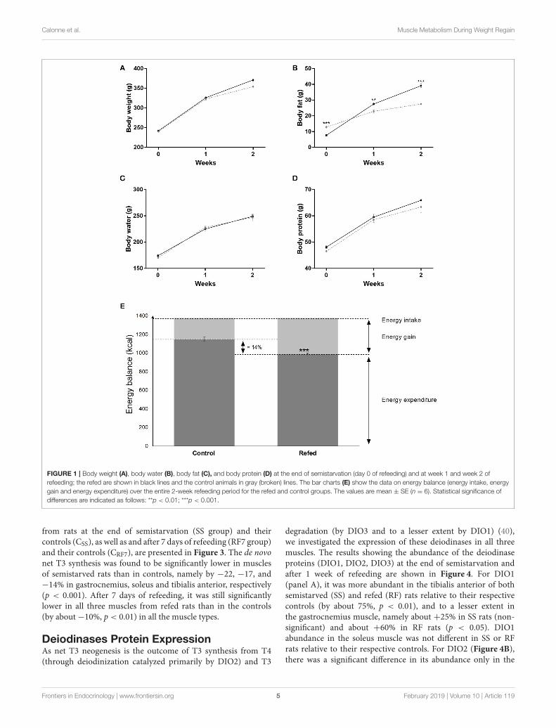
Net T3 NeogenesisThe data on the rate of net T3 neogenesis, assessed ex vivo inextracts of the gastrocnemius, soleus and tibialis anterior muscles
Frontiers in Endocrinology | www.frontiersin.org 4 February 2019 | Volume 10 | Article 119
Calonne et al. Muscle Metabolism During Weight Regain
FIGURE 1 | Body weight (A), body water (B), body fat (C), and body protein (D) at the end of semistarvation (day 0 of refeeding) and at week 1 and week 2 of
refeeding; the refed are shown in black lines and the control animals in gray (broken) lines. The bar charts (E) show the data on energy balance (energy intake, energy
gain and energy expenditure) over the entire 2-week refeeding period for the refed and control groups. The values are mean ± SE (n = 6). Statistical significance of
differences are indicated as follows: **p < 0.01; ***p < 0.001.
from rats at the end of semistarvation (SS group) and theircontrols (CSS), as well as and after 7 days of refeeding (RF7 group)and their controls (CRF7), are presented in Figure 3. The de novonet T3 synthesis was found to be significantly lower in musclesof semistarved rats than in controls, namely by −22, −17, and−14% in gastrocnemius, soleus and tibialis anterior, respectively(p < 0.001). After 7 days of refeeding, it was still significantlylower in all three muscles from refed rats than in the controls(by about−10%, p < 0.01) in all the muscle types.
Deiodinases Protein ExpressionAs net T3 neogenesis is the outcome of T3 synthesis from T4(through deiodinization catalyzed primarily by DIO2) and T3
degradation (by DIO3 and to a lesser extent by DIO1) (40),we investigated the expression of these deiodinases in all threemuscles. The results showing the abundance of the deiodinaseproteins (DIO1, DIO2, DIO3) at the end of semistarvation andafter 1 week of refeeding are shown in Figure 4. For DIO1(panel A), it was more abundant in the tibialis anterior of bothsemistarved (SS) and refed (RF) rats relative to their respectivecontrols (by about 75%, p < 0.01), and to a lesser extent inthe gastrocnemius muscle, namely about +25% in SS rats (non-significant) and about +60% in RF rats (p < 0.05). DIO1abundance in the soleus muscle was not different in SS or RFrats relative to their respective controls. For DIO2 (Figure 4B),there was a significant difference in its abundance only in the
Frontiers in Endocrinology | www.frontiersin.org 5 February 2019 | Volume 10 | Article 119

Calonne et al. Muscle Metabolism During Weight Regain
FIGURE 2 | Fractional rates of (A) protein synthesis (Ksyn), (B) protein growth (Kgrowth ), and (C) protein degradation (Kdeg) in the Gastrocnemius, Soleus and Tibialis
anterior muscles of rats after 7 days of refeeding in refed and control groups. The values are mean ± SE (n = 6–7). Statistical significance of differences are indicated
as follows: **p < 0.01; ***p < 0.001.
gastrocnemius muscle of SS rats than in controls (lower by35%, p < 0.05), but not in the RF rats relative to controls.Furthermore, the abundance of DIO2 in the two other muscles(soleus and tibialis anterior) was not different in SS and RFrats relative to their respective controls. By contrast, DIO3(panel C) was more abundant in all three muscles from theSS than in the controls, namely by 2.2- and 2.5-fold higher ingastrocnemius and tibialis anterior, respectively, and by about60% higher in soleus. Although less pronounced than duringsemistarvation, the abundance of DIO3 in all three skeletalmuscles after 1 week of refeeding was also higher in refed animalsthan in controls: namely+53,+35, and+63% in gastrocnemius,soleus, and tibialis anterior, respectively, with the differencebeing statistically significant in the gastrocnemius and soleusmuscles (p < 0.05).
DISCUSSION
The results presented here suggest a role for diminishedprotein turnover in skeletal muscle, associated with alteredmuscle thyroid hormone metabolism resulting in diminished T3availability, in the high efficiency with which body fat is recoveredafter substantial body fat depletion.
Diminished Rate of Protein Synthesis andTurnoverUsing the stable isotope flooding dose technique of incorporationof 13C-labeled valine in protein for in-vivo measurements ofprotein synthesis in skeletal muscle, the fractional proteinsynthesis rate (Ksyn) is shown to be lower in skeletal muscle ofrefed animals than in controls assessed on day 7 of refeeding,
Frontiers in Endocrinology | www.frontiersin.org 6 February 2019 | Volume 10 | Article 119
Calonne et al. Muscle Metabolism During Weight Regain
FIGURE 3 | Net T3 formation from its T4 precursor in Gastrocnemius, Soleus
and Tibialis anterior muscles from (i) rats semistarved (SS) for 14 days and
their controls (CSS), and (ii) rats refed for 7 days (RF7) and their controls
(CRF7). The values are mean ±SE (n = 6). Statistical significance of differences
are indicated as follows: **p < 0.01; ***p < 0.001.
i.e., at about mid-point in the dynamic phase of catch-up fat.The lower muscle Ksyn in refed animals was observed in all threehindlimb skeletal muscles studied, namely the gastrocnemius
which is predominantly fast-oxidative glycolytic, soleus which ispredominantly slow-oxidative and the tibialis anterior which ispredominantly glycolytic, thereby suggesting that the lower Ksyn
during catch-up fat occurs independently of skeletal muscle fibercomposition. As the rate of skeletal muscle growth (Kgrowth) wassimilar in refed and control animals (in line with similar ratesin total body protein deposition), it follows that the reducedrate of muscle protein synthesis is accompanied by reduced rateof muscle protein degradation (Kdeg), thereby underscoring arole for diminished protein turnover in skeletal muscle in thesuppressed thermogenesis that drives catch-up fat.
Diminished skeletal muscle Ksyn in the postabsorptive statehas previously been shown in obese humans (41) and in ratsmadeobese on a high-fat high-sucrose diet (26), and is consideredto reside in impaired amino acid incorporation into proteinsattributed to lipid infiltration and insulin resistance in the skeletalmuscle (42). These authors have proposed that the mechanismsunderlying such reductions in muscle protein synthesis mayreside in an inhibitory effect of lipid derivative species on insulinsignaling which would result in diminished protein translation,and that insulin resistance in skeletal muscle not only impairsglucose metabolism but also protein metabolism (42). Althoughskeletal muscle insulin resistance has been shown to be an earlyevent that is sustained throughout the dynamic phase of catch-upfat in our rat model (43, 44), the lower Ksyn in skeletal muscle isreported here at a time-point of refeeding (day 7) when (i) skeletalmuscle lipid content was not found to be higher in muscles fromrefed animals than in controls (17, 43), and also (ii) when totaland regional body fat content of the refed animals had not yetexceeded those of controls (17, 43, 44). Furthermore, the findingshere of diminished Ksyn in all muscle types varying widely infiber composition during catch-up fat contrast with past reportsof muscle fiber-type dependency of the lower Ksyn observed inobese rats, namely only in glycolytic muscles but not in thesoleus (slow-oxidative) muscle of diet-induced obese rats (26),or only in red oxidative fibers and not in white glycolytic fibersof the gastrocnemius of the genetic (leptin receptor deficient)obese Zucker rats (45). Taken together, therefore, the lower Ksyn
observed here in skeletal muscle during catch-up fat is unlikelyto be a consequence of excess whole body, regional or lipidinfiltration in skeletal muscle, but rather reflects a diminished rateof substrate cycling between protein synthesis and degradation(i.e., reduced protein turnover) for the purpose of sparing energyfor catch-up fat.
Altered Skeletal Muscle Thyroid HormoneMetabolismAs the thyroid hormone T3 is known to play an importantrole in the control of basal metabolism and thermogenesis (13–15), we have in past studies examined the extent to which thewell-known reduction in circulating levels of T3 during caloricrestriction is restored during refeeding in the rat model of catch-up fat (16, 17). In particular, we found that while the bloodconcentrations of TSH, T4 and T3 are all markedly lower atthe end of caloric restriction, refeeding resulted in differences intheir restoration kinetics. Indeed, whereas plasma TSH and T4
Frontiers in Endocrinology | www.frontiersin.org 7 February 2019 | Volume 10 | Article 119

Calonne et al. Muscle Metabolism During Weight Regain
FIGURE 4 | Protein expression of the deiodinases: (A) DIO1, (B) DIO2, and (C) DIO3 in Gastrocnemius (GA), Soleus and Tibialis anterior (TA) muscles from (i) rats
semistarved (SS) for 14 days and their controls (CSS), and (ii) rats refed for 7 days (RF7) and their controls (CRF7). The values are mean ±SE (n = 4). Statistical
significance of differences are indicated as follows: *p < 0.05; **p < 0.01.
were completely restored to control levels by day 5 of refeeding,plasma T3 remained lower, albeit marginally, in the refed animalsthan in controls up to day 10 day of refeeding (16), andcould hence contribute to the diminished thermogenesis drivingcatch-up fat.
However, circulating levels of thyroid hormones maynot necessarily reflect tissue thyroid hormone levels as thebioavailability at the tissue and cellular level is dependentupon local thyroid hormone metabolism (14, 15, 40). The netformation of T3 from T4 (i.e., net T3 neogenesis) is to alarge extent controlled by an interplay of deiodinase enzymesthat catalyze activation or inactivation of T4 and T3. In theskeletal muscle, the conversion of T4 into the active hormoneT3 is believed to be primarily catalyzed by DIO2 and theinactivation of T4 and T3 to be catalyzed by DIO3 (andpossibly also by DIO1) to rT3 and T2 (3,3′diiodothyronine)(40). In a recent study from our laboratory (18) investigatingpossible alterations in thyroid hormone metabolism in skeletalmuscle during catch-up fat, it was reported that the in-vitrokinetics of T3 generation in the T4-incubated gastrocnemius
muscle of semistarved and refed rats are significantly lower thanin their respective controls. Explanations based upon altereddeiodinase activities were reinforced by the findings that theprotein expression DIO2 was reduced while that of DIO3 wasincreased in this hindlimb muscle both during caloric restrictionand refeeding (18).
In the present study, we have extended these investigationsto other muscle types varying widely in fiber composition.Using the same in-vitro kinetic assay of T3 generated by T4 inincubated skeletal muscle, we show that the reduction in netT3 neogenesis in muscle during semistarvation and refeeding isobserved not only in the gastrocnemius muscle, but also in thesoleus and tibialis anterior, thereby suggesting that the reductionin muscle T3 availability during caloric restriction and persistingduring the catch-up fat phase occurs independently of skeletalmuscle fiber composition, and may involve the whole skeletalmuscle mass. By contrast, alterations in the abundance of thethree deiodinases are found to vary according to muscle type.In response to semistarvation and after 1 week of refeeding,DIO2 which is considered to be primarily responsible for T3
Frontiers in Endocrinology | www.frontiersin.org 8 February 2019 | Volume 10 | Article 119
Calonne et al. Muscle Metabolism During Weight Regain
production from T4 in skeletal muscle was less abundant in thegastrocnemius muscle, but not in the soleus or tibialis anterior.The abundance of DIO1 (which may limit T3 availability bydiverting T4 and T3 to inactive rT3 and T2) was higher inthe gastrocnemius and tibialis anterior but not in the soleus.The most striking feature in the analysis of these data ondeiodinases is the robust upregulation of DIO3 observed inall 3 muscle types during semistarvation, which persisted after1 week of refeeding in two of these 3 muscles, namely inthe gastrocnemius and soleus. Interestingly, the abundanceof DIO1 in the tibialis anterior, which was increased duringsemistarvation, also persisted during refeeding (+70% relativeto controls, p < 001). Thus, during refeeding, in the absence ofa robust increase in the abundance of the T3 inactivator DIO3in the tibialis anterior, the increased DIO1 in this muscle mayassume a greater importance thanDIO3 upregulation in reducingT3 availability. Taken together, our results suggest that the lowernet T3 neogenesis in all three muscles of varied fiber compositionstudied during semistarvation and catch-up fat seems to resideprimarily in the upregulation of the thyroid hormone inactivatingenzymes DIO1 and/or DIO3 (i.e., deiodinases that catalyze theconversion of T4 and T3 to biologically inactive rT3 and T2)rather than in the downregulation of the thyroid hormoneactivating enzyme DIO2 which catalyzes the conversion of T4 toT3. A better understanding of how the muscle-type dependentchanges in these DIOs are co-ordinated to result in diminishedT3 availability during semistarvation and refeeding will need tobe addressed in future studies involving the use of sensitive assaysto detect the changes in the activity of these three deiodinases inrat skeletal muscle.
Whatever, the mechanisms controlling the upregulation ofthe T3 inactivating deiodinase enzymes in the various skeletalmuscle types, our findings here indicate that, the kineticsof T3 generation in skeletal muscle homogenates incubatedwith T4 were lower in semistarved and refed rats. Thisunderscores the possibility that a lower T3 availability inskeletal muscle during semistarvation and refeeding could
be contributed not only from a lower plasma T3 level (16,17), but also from altered muscle deiodinase activities. Giventhe role of T3 in controlling many inter-related aspects ofskeletal muscle energetics that include the maintenance ofionic equilibrium through Na/K ATPase, calcium cycling, fibercomposition, contraction-relaxation kinetics, protein synthesis,and protein turnover, the relative hypothyroidism in skeletalmuscle during semistarvation and persisting during refeedingmay thus contribute to the suppression of thermogenesis duringcaloric restriction and subsequent high efficiency for catch-up fat.
CONCLUSION
The results presented here suggest that diminished skeletalmuscle protein turnover, together with altered local musclemetabolism of thyroid hormones leading to diminishedintracellular T3 availability, are features of the thrifty metabolismthat drive the rapid restoration of the fat reserves during weightregain after caloric restriction.
AUTHOR CONTRIBUTIONS
AD, JC, YB, and CG conceived and designed the experiments.JC, LI, JM-C, DA, and CG performed the experiments. JC,AD, DA, CG, and YB analyzed the data. AD, J-PM, CG, andYB contributed reagents, materials, and analysis tools. JC andAD wrote the paper. LI, JM-C, DA, J-PM, CG, and YB editedthe manuscript.
FUNDING
This study was supported by a grant from the Swiss NationalScience Foundation (grant # 310030_152870). It was presentedat 24th European Congress on Obesity (ECO2017) that was heldin Porto, Portugal, May 17-20, 2017, and published by S. KargerGmbh as an Abstract in Obesity Facts 2017;10 (suppl. 1): AbstractTIP115 (doi: 10.1159/000468958).
REFERENCES
1. Boyle PC, Storlein LH, Keesey RE. Increased efficiency of food
utilization following weight loss. Physiol Behav. (1978) 21:261–4.
doi: 10.1016/0031-9384(78)90050-1
2. Hill JO, Fried SK, DiGirolamoM. Effects of fasting and restricted refeeding on
utilization of ingested energy in rats. Am J Physiol. (1984) 247:R318–27.
3. Dulloo AG, Girardier L. Adaptive changes in energy expenditure during
refeeding following low-calorie intake: evidence for a specific metabolic
component favoring fat storage. Am J Clin Nutr. (1990) 52:415–20.
doi: 10.1093/ajcn/52.3.415
4. Crescenzo R, Samec S, Antic V, Rohner-Jeanrenaud F, Seydoux J, Montani
JP, et al. A role for suppressed thermogenesis favoring catch-up fat
in the pathophysiology of catch-up growth. Diabetes. (2003) 52:1090–7.
doi: 10.2337/diabetes.52.5.1090
5. MacLean PS, Higgins JA, Johnson GC, Fleming-Elder BK, Donahoo
WT, Melanson EL, et al. Enhanced metabolic efficiency contributes to
weight regain after weight loss in obesity-prone rats. Am J Physiol
Regul Integr Comp Physiol. (2004) 287:R1306–15. doi: 10.1152/ajpregu.
00463.2004
6. Evans SA, Messina MM, Knight WD, Parsons AD, Overton JM. Long-Evans
and Sprague-Dawley rats exhibit divergent responses to refeeding after caloric
restriction. Am J Physiol Regul Integr Comp Physiol. (2005) 288:R1468–76.
doi: 10.1152/ajpregu.00602.2004
7. Crescenzo R, Lionetti L, Mollica MP, Ferraro M, D’Andrea E, Mainieri D,
et al. Altered skeletal muscle subsarcolemmal mitochondrial compartment
during catch-up fat after caloric restriction. Diabetes. (2006) 55:2286–93.
doi: 10.2337/db06-0312
8. Dulloo AG, Jacquet J. An adipose-specific control of thermogenesis in body
weight regulation. Int J Obes Relat Metab Disord. (2001) 25(Suppl. 5):S22–9.
doi: 10.1038/sj.ijo.0801907
9. Dulloo AG, Schutz Y. Adaptive thermogenesis in resistance to obesity
therapies: issues in quantifying thrifty energy expenditure phenotypes in
humans. Curr Obes Rep. (2015) 4:230–40. doi: 10.1007/s13679-015-0156-9
10. Dulloo AG, Jacquet J, Seydoux J, Montani JP. The thrifty ’catch-up fat’
phenotype: its impact on insulin sensitivity during growth trajectories to
obesity and metabolic syndrome. Int J Obes. (2006) 30(Suppl. 4):S23–35.
doi: 10.1038/sj.ijo.0803516
11. Hesselink MK, Mensink M, Schrauwen P. Human uncoupling protein-3 and
obesity: an update. Obes Res. (2003) 11:1429–43. doi: 10.1038/oby.2003.192
Frontiers in Endocrinology | www.frontiersin.org 9 February 2019 | Volume 10 | Article 119
Calonne et al. Muscle Metabolism During Weight Regain
12. Dulloo AG, Samec S. Uncoupling proteins: their roles in adaptive
thermogenesis and substrate metabolism reconsidered. Br J Nutr. (2001)
86:123–39. doi: 10.1079/BJN2001412
13. Simonides WS, van Hardeveld C. Thyroid hormone as a determinant of
metabolic and contractile phenotype of skeletal muscle. Thyroid. (2008)
18:205–16. doi: 10.1089/thy.2007.0256
14. Salvatore D, Simonides WS, Dentice M, Zavacki AM, Larsen PR. Thyroid
hormones and skeletal muscle–new insights and potential implications. Nat
Rev Endocrinol. (2014) 10:206–14. doi: 10.1038/nrendo.2013.238
15. Huang SA, Bianco AC. Reawakened interest in type III iodothyronine
deiodinase in critical illness and injury. Nat Clin Pract Endocrinol Metab.
(2008) 4:148–55. doi: 10.1038/ncpendmet0727
16. Mainieri D, Summermatter S, Seydoux J, Montani JP, Rusconi S, Russell
AP, et al. A role for skeletal muscle stearoyl-CoA desaturase 1 in control of
thermogenesis. Faseb J. (2006) 20:1751–3. doi: 10.1096/fj.06-5934fje
17. Summermatter S, Mainieri D, Russell AP, Seydoux J, Montani JP, Buchala A,
et al. Thrifty metabolism that favors fat storage after caloric restriction: a role
for skeletal muscle phosphatidylinositol-3-kinase activity and AMP-activated
protein kinase. FASEB J. (2008) 22:774–85. doi: 10.1096/fj.07-8972com
18. De Andrade PB, Neff LA, Strosova MK, Arsenijevic D, Patthey-Vuadens O,
Scapozza L, et al. Caloric restriction induces energy-sparing alterations in
skeletal muscle contraction, fiber composition and local thyroid hormone
metabolism that persist during catch-up fat upon refeeding. Front Physiol.
(2015) 6:254. doi: 10.3389/fphys.2015.00254
19. Waterlow JC. Protein turnover with special reference to man. Q J Exp Physiol.
(1984) 69:409–38. doi: 10.1113/expphysiol.1984.sp002829
20. Millward DJ. The hormonal control of protein turnover. Clin Nutr. (1990)
9:115–26. doi: 10.1016/0261-5614(90)90042-Q
21. YoungVR,Marchini JS.Mechanisms and nutritional significance ofmetabolic
responses to altered intakes of protein and amino acids, with reference
to nutritional adaptation in humans. Am J Clin Nutr. (1990) 51:270–89.
doi: 10.1093/ajcn/51.2.270
22. Schutz Y. Protein turnover, ureagenesis and gluconeogenesis. Int J Vitam Nutr
Res. (2011) 81:101–7. doi: 10.1024/0300-9831/a000064
23. Rolfe DF, Brown GC. Cellular energy utilization and molecular origin
of standard metabolic rate in mammals. Physiol Rev. (1997) 77:731–58.
doi: 10.1152/physrev.1997.77.3.731
24. Entenman C. [55] General procedures for separating lipid components of
tissue.Methods Enzymol. (1957) 3:299–317.
25. Chanseaume E, Giraudet C, Gryson C, Walrand S, Rousset P, Boirie
Y, et al. Enhanced muscle mixed and mitochondrial protein synthesis
rates after a high-fat or high-sucrose diet. Obesity. (2007) 15:853–9.
doi: 10.1038/oby.2007.582
26. Masgrau A, Mishellany-Dutour A, Murakami H, Beaufrere AM, Walrand
S, Giraudet C, et al. Time-course changes of muscle protein synthesis
associated with obesity-induced lipotoxicity. J Physiol. (2012) 590:5199–210.
doi: 10.1113/jphysiol.2012.238576
27. Boirie Y, Short KR, Ahlman B, CharltonM, Nair KS. Tissue-specific regulation
of mitochondrial and cytoplasmic protein synthesis rates by insulin. Diabetes.
(2001) 50:2652–8. doi: 10.2337/diabetes.50.12.2652
28. Lowry OH, Rosebrough NJ, Farr AL, Randall RJ. Protein measurement with
the Folin phenol reagent. J Biol Chem. (1951) 193:265–75.
29. Samuels SE, Thompson JR, Christopherson RJ. Skeletal and cardiac muscle
protein turnover during short-term cold exposure and rewarming in young
rats. Am J Physiol. (1996) 270:R1231–9.
30. Millward DJ, Garlick PJ, Stewart RJ, Nnanyelugo DO, Waterlow JC.
Skeletal-muscle growth and protein turnover. Biochem J. (1975) 150:235–43.
doi: 10.1042/bj1500235
31. Waterlow JC, Garlick PJ, Millward DJ. Protein Turnover in Mammalian
Tissues and in the Whole Body, Chapter 17, 18. North-Holland
Amsterdam:Elsevier, (1978).
32. Bates PC, Millward DJ. Characteristics of skeletal muscle growth and
protein turnover in a fast-growing rat strain. Br J Nutr. (1981) 46:7–13.
doi: 10.1079/BJN19810004
33. Giugliano R, Millward DJ. The effects of severe zinc deficiency on
protein turnover in muscle and thymus. Br J Nutr. (1987) 57:139–55.
doi: 10.1079/BJN19870017
34. Jepson MM, Pell JM, Bates PC, Millward DJ. The effects of endotoxaemia on
protein metabolism in skeletal muscle and liver of fed and fasted rats. Biochem
J. (1986) 235:329–36. doi: 10.1042/bj2350329
35. McAllister TA, Thompson JR, Samuels SE. Skeletal and cardiac muscle protein
turnover during cold acclimation in young rats. Am J Physiol Regul Integr
Comp Physiol. (2000) 278:R705–11. doi: 10.1152/ajpregu.2000.278.3.R705
36. Samuels SE, Knowles AL, Tilignac T, Debiton E, Madelmont JC, Attaix
D. Higher skeletal muscle protein synthesis and lower breakdown after
chemotherapy in cachectic mice. Am J Physiol Regul Integr Comp Physiol.
(2001) 281:R133–9. doi: 10.1152/ajpregu.2001.281.1.R133
37. Kaplan MM, Utiger RD. Iodothyronine metabolism in rat liver homogenates.
J Clin Invest. (1978) 61:459–71. doi: 10.1172/JCI108957
38. Reutenauer-Patte J, Boittin FX, Patthey-Vuadens O, Ruegg UT, Dorchies
OM. Urocortins improve dystrophic skeletal muscle structure and function
through both PKA- and Epac-dependent pathways. Am J Pathol. (2012)
180:749–62. doi: 10.1016/j.ajpath.2011.10.038
39. Dorchies OM, Reutenauer-Patte J, Dahmane E, Ismail HM, Petermann O,
Patthey- Vuadens O, et al. The anticancer drug tamoxifen counteracts the
pathology in a mouse model of duchenne muscular dystrophy. Am J Pathol.
(2013) 182:485–504. doi: 10.1016/j.ajpath.2012.10.018
40. van der Spek AH, Fliers E, Boelen A. The classic pathways of
thyroid hormone metabolism. Mol Cell Endocrinol. (2017) 458:29–38.
doi: 10.1016/j.mce.2017.01.025
41. Guillet C, Delcourt I, RanceM, Giraudet C,Walrand S, BeduM, et al. Changes
in basal and insulin and amino acid response of whole body and skeletal
muscle proteins in obese men. J Clin Endocrinol Metab. (2009) 94:3044–50.
doi: 10.1210/jc.2008-2216
42. Guillet C, Masgrau A, Walrand S, Boirie Y. Impaired protein metabolism:
interlinks between obesity, insulin resistance and inflammation.
Obes Rev. (2012) 13(Suppl. 2):51–7. doi: 10.1111/j.1467-789X.2012.
01037.x
43. Cettour-Rose P, Samec S, Russell AP, Summermatter S, Mainieri D, Carrillo-
Theander C, et al. Redistribution of glucose from skeletal muscle to adipose
tissue during catch-up fat: a link between catch-up growth and later metabolic
syndrome. Diabetes. (2005) 54:751–6. doi: 10.2337/diabetes.54.3.751
44. Marcelino H, Veyrat-Durebex C, Summermatter S, Sarafian D, Miles-Chan J,
Arsenijevic D, et al. A role for adipose tissue de novo lipogenesis in glucose
homeostasis during catch-up growth: a Randle cycle favoring fat storage.
Diabetes. (2013) 62:362–72. doi: 10.2337/db12-0255
45. Nilsson MI, Greene NP, Dobson JP, Wiggs MP, Gasier HG, Macias
BR, et al. Insulin resistance syndrome blunts the mitochondrial anabolic
response following resistance exercise. Am J Physiol Endocrinol Metab. (2010)
299:E466–74. doi: 10.1152/ajpendo.00118.2010
Conflict of Interest Statement: The authors declare that the research was
conducted in the absence of any commercial or financial relationships that could
be construed as a potential conflict of interest.
Copyright © 2019 Calonne, Isacco, Miles-Chan, Arsenijevic, Montani, Guillet, Boirie
and Dulloo. This is an open-access article distributed under the terms of the Creative
Commons Attribution License (CC BY). The use, distribution or reproduction in
other forums is permitted, provided the original author(s) and the copyright owner(s)
are credited and that the original publication in this journal is cited, in accordance
with accepted academic practice. No use, distribution or reproduction is permitted
which does not comply with these terms.
Frontiers in Endocrinology | www.frontiersin.org 10 February 2019 | Volume 10 | Article 119