Red-Brown Patches in the Groin - MDedge
3
DERMATOPATHOLOGY DIAGNOSIS 416 I CUTIS ® WWW.MDEDGE.COM/CUTIS A 66-year-old man presented with reddish arciform patches in the inguinal area. THE BEST DIAGNOSIS IS: a. candidiasis b. erythrasma c. pitted keratolysis d. tinea cruris e. tinea versicolor Red-Brown Patches in the Groin Dong Chen, MD, PhD; Tammie C. Ferringer, MD Dr. Chen is from the Department of Pathology and Anatomical Sciences, University of Missouri, Columbia. Dr. Ferringer is from the Departments of Dermatology and Laboratory Medicine, Geisinger Medical Center, Danville, Pennsylvania. The authors report no conflict of interest. Correspondence: Dong Chen, MD, PhD, Department of Pathology and Anatomical Sciences, University of Missouri, One Hospital Dr, MA204, DC018.00, Columbia, MO 65212 ([email protected]). H&E, original magnification ×600. Eligible for 1 MOC SA Credit From the ABD This Dermatopathology Diagnosis article in our print edition is eligible for 1 self-assessment credit for Maintenance of Certification from the American Board of Dermatology (ABD). After completing this activity, diplomates can visit the ABD website (http://www.abderm.org) to self-report the credits under the activity title “Cutis Dermatopathology Diagnosis.” You may report the credit after each activity is completed or after accumu- lating multiple credits. PLEASE TURN TO PAGE 419 FOR THE DIAGNOSIS Copyright Cutis 2018. No part of this publication may be reproduced, stored, or transmitted without the prior written permission of the Publisher.
Transcript of Red-Brown Patches in the Groin - MDedge

DERMATOPATHOLOGY DIAGNOSIS
416 I CUTIS® WWW.MDEDGE.COM/CUTIS
A 66-year-old man presented with reddish arciform patches in the inguinal area.
THE BEST DIAGNOSIS IS: a. candidiasisb. erythrasmac. pitted keratolysisd. tinea crurise. tinea versicolor
Red-Brown Patches in the GroinDong Chen, MD, PhD; Tammie C. Ferringer, MD
Dr. Chen is from the Department of Pathology and Anatomical Sciences, University of Missouri, Columbia. Dr. Ferringer is from the Departments of Dermatology and Laboratory Medicine, Geisinger Medical Center, Danville, Pennsylvania. The authors report no conflict of interest.Correspondence: Dong Chen, MD, PhD, Department of Pathology and Anatomical Sciences, University of Missouri, One Hospital Dr, MA204, DC018.00, Columbia, MO 65212 ([email protected]).
H&E, original magnification ×600.
Eligible for 1 MOC SA Credit From the ABDThis Dermatopathology Diagnosis article in our print edition is eligible for 1 self-assessment credit for Maintenance of Certification from the American Board of Dermatology (ABD). After completing this activity, diplomates can visit the ABD website (http://www.abderm.org) to self-report the credits under the activity title “Cutis Dermatopathology Diagnosis.” You may report the credit after each activity is completed or after accumu-lating multiple credits.
PLEASE TURN TO PAGE 419 FOR THE DIAGNOSIS
Copyright Cutis 2018. No part of this publication may be reproduced, stored, or transmitted without the prior written permission of the Publisher.
CUTIS D
o no
t cop
y

DERMATOPATHOLOGY DIAGNOSIS DISCUSSION
VOL. 101 NO. 6 I JUNE 2018 419WWW.MDEDGE.COM/CUTIS
E rythrasma usually involves intertriginous areas (eg, axillae, groin, inframammary area). Patients present with well-demarcated, minimally scaly, red-
brown patches. The interdigital web space of the toes also can be involved with macerated white plaques, often with coexistent dermatophyte infection. Corynebacterium minutissimum, the bacteria responsible for erythrasma, produces coproporphyrin type III, which emits coral red fluorescence under Wood lamp examination.1 Bathing may result in removal of the porphyrin and result in a false-negative finding. Potassium hydroxide preparation of skin scrapings can show chains of bacilli. Biopsy appears relatively normal at low power but reveals compact ortho-keratosis with coccobacilli and filamentous organisms in the superficial stratum corneum (quiz image). When not obvious on hematoxylin and eosin–stained sections, the organisms are Gram-positive and also are seen with periodic acid–Schiff (PAS) and methenamine silver stains. Unlike fungal hyphae, these organisms are thinner and nonrefractile. Inflammation typically is minimal. Due to the subtle histologic findings at low power, erythrasma is considered one of the invisible dermatoses.2 The dif-ferential diagnosis of these inconspicuous dermatoses that appear normal at first glance can be approached in a stepwise fashion starting in the stratum corneum, followed by the granular layer, basal layer, dermal papillae, dermal inflammatory cells, dermal connective tissue, and eccrine glands, and should consider each of the following diag-noses: candidiasis, dermatophytosis, ichthyosis vulgaris, vitiligo, macular amyloid, urticaria, telangiectasia macularis eruptiva perstans, connective tissue nevus, and argyria.2
Candidiasis, most commonly caused by Candida albicans, usually involves the oral cavity (eg, thrush, median rhomboid glossitis, angular cheilitis), intertriginous zones, nail fold (paronychia), genital areas (eg, vulvovaginitis, balanitis), and diaper area.3 The web space between the third and fourth fingers (erosio interdigitalis blastomycetica) can be involved in patients whose hands are frequently in water. Intertriginous candidiasis presents with bright red, sometimes erosive patches with satellite lesions. Spores and mycelia (filamentous forms) are noted on potassium hydroxide preparation of skin scrapings. Histologically, the epidermis often is acanthotic, mildly spongiotic, and contains groups of neutrophils in the superficial layers. The mnemonic device for diseases with clusters of neutrophils in the stratum corneum is PTICSS (psoriasis, tinea, impetigo, candida, seborrheic dermatitis, syphilis).2 Yeast, pseudohyphae, and even true hyphae can be seen in the stratum corneum with hematoxylin and eosin–stained sections and PAS. The filamentous forms tend to be vertically oriented in relation to the skin
surface (Figure 1) compared to dermatophyte hyphae that tend to be parallel to the surface.2
Pitted keratolysis is a superficial bacterial infection involving the soles of the feet. The classic clinical findings are shallow 1- to 2-mm pits in clusters that can coalesce on pressure-bearing areas. Hyperhidrosis, malodor, and maceration commonly are associated. Microscopic exami-nation reveals clusters of small cocci and filamentous bacteria located in the dell or pit of a thick compact ortho-keratotic stratum corneum of acral skin with no notable inflammatory infiltrate (Figure 2).2 Special stains such as Gram, methenamine silver, or PAS can assist in visu-alization of the organisms. Pitted keratolysis is caused
THE DIAGNOSIS:
Erythrasma
FIGURE 2. Pitted keratolysis histopathology shows clusters of small cocci and filamentous bacteria in the dell or pit of acral stratum cor-neum with no notable inflammatory infiltrate (H&E, original magnifica-tion ×200).
FIGURE 1. Candidiasis histopathology shows round yeast (arrow-heads) and vertically oriented pseudohyphae (arrow) in a stratum cor-neum containing neutrophils (H&E, original magnification ×600).
Copyright Cutis 2018. No part of this publication may be reproduced, stored, or transmitted without the prior written permission of the Publisher.
CUTIS D
o no
t cop
y
DERMATOPATHOLOGY DIAGNOSIS DISCUSSION
420 I CUTIS® WWW.MDEDGE.COM/CUTIS
by Dermatophilus congolensis and Kytococcus sedentarius (formerly Micrococcus sedentarius), which produce keratino-lytic enzymes causing the defect in the stratum corneum.3
Tinea cruris, also known as jock itch and ringworm of the groin, presents with advancing pruritic, circinate, erythematous, scaling patches with central clearing on the inner thighs and crural folds. Similar to tinea pedis, Trichophyton rubrum is the most common dermatophyte to cause tinea cruris.4 Potassium hydroxide preparation of skin scrapings from the advancing border show fungal hyphae that cross the keratin cell borders. The histopa-thology of dermatophyte infections can be subtle and resemble normal skin before close inspection of the stra-tum corneum, which can show compact orthokeratosis, neutrophils, or “sandwich sign” where hyphae are sand-wiched between an upper basket weave layer and a lower compact cornified layer (orthokeratotic or parakeratotic)(Figure 3).1 The presence of these patterns in the stratum corneum should result in performance of PAS to highlight obscure hyphae.
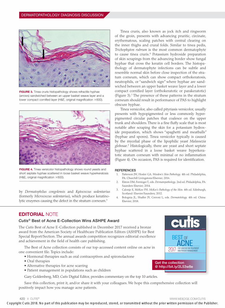
Tinea versicolor, also called pityriasis versicolor, usually presents with hypopigmented or less commonly hyper-pigmented circular patches that coalesce on the upper trunk and shoulders. There is a fine fluffy scale that is most notable after scraping the skin for a potassium hydrox-ide preparation, which shows “spaghetti and meatballs” (hyphae and spores). Tinea versicolor typically is caused by the mycelial phase of the lipophilic yeast Malassezia globosae.3 Histologically, there are yeast and short septate hyphae scattered in a loose basket weave hyperkera-totic stratum corneum with minimal or no inflammation (Figure 4). On occasion, PAS is required for identification.
REFERENCES 1. Patterson JW, Hosler GA. Weedon’s Skin Pathology. 4th ed. Philadelphia,
PA: Churchill Livingstone/Elsevier; 2016. 2. Elston DM, Ferringer T, eds. Dermatopathology. 2nd ed. Philadelphia, PA:
Saunders Elsevier; 2014. 3. Calonje E, McKee PH. McKee’s Pathology of the Skin. 4th ed. Edinburgh,
Scotland: Elsevier/Saunders; 2012. 4. Bolognia JL, Shaffer JV, Cerroni L, eds. Dermatolology. 4th ed. China:
Elsevier; 2018.
FIGURE 3. Tinea cruris histopathology shows refractile hyphae (arrows) sandwiched between an upper basket weave layer and a lower compact cornified layer (H&E, original magnification ×600).
FIGURE 4. Tinea versicolor histopathology shows round yeasts and short septate hyphae scattered in loose basket weave hyperkeratosis (H&E, original magnification ×600).
EDITORIAL NOTECutis® Best of Acne E-Collection Wins ASHPE Award
The Cutis Best of Acne E-Collection published in December 2017 received a bronze award from the American Society of Healthcare Publication Editors (ASHPE) for Best Special Report/Section. The annual awards competition recognizes editorial excellence and achievement in the field of health care publishing.
The Best of Acne collection consists of our top-accessed content online on acne in one convenient file. Topics include:
• Hormonal therapies such as oral contraceptives and spironolactone• Oral therapies• Alternative therapies for acne scarring• Patient management in populations such as children
Gary Goldenberg, MD, Cutis Digital Editor, provides commentary on the top 10 articles.
Save this collection, print it, and/or share it with your colleagues. We hope this comprehensive collection will positively impact how you manage acne patients.
Best OF ACNE
REFERENCED IN INDEX MEDICUS
CUTANEOUS MEDICINE FOR THE PRACTITIONER cutis.com
Top content on treatment and patient management2017
Get the collection @ http://bit.ly/2L53w8e
Copyright Cutis 2018. No part of this publication may be reproduced, stored, or transmitted without the prior written permission of the Publisher.
CUTIS D
o no
t cop
y