Reconstruction of the lower eye lid with a rotation ...
17
Reconstruction of the lower eye lid with a rotation – advancement tarso-conjunctival cheek flap by William Louis Fick Wessels December 2010 Thesis presented in partial fulfilment of the requirements for the degree Master of Medicine in Plastic & Reconstructive Surgery at the University of Stellenbosch Supervisor: Prof FR Graewe Co-supervisor: Dr PV van Deventer Faculty of Health Sciences Department of Surgical Sciences
Transcript of Reconstruction of the lower eye lid with a rotation ...
Reconstruction of the lower eye lid with a rotation – advancement tarso-conjunctival
cheek flap
by William Louis Fick Wessels
December 2010
Thesis presented in partial fulfilment of the requirements for the degree Master of Medicine in Plastic & Reconstructive Surgery at the University
of Stellenbosch
Supervisor: Prof FR Graewe Co-supervisor: Dr PV van Deventer
Faculty of Health Sciences Department of Surgical Sciences

2 WLF Wessels
Manuscript Submission
Reconstruction of the lower eyelid with a rotation-advancement tarso-conjunctival
cheek flap.
Abstract
The repair of full-thickness defects of the lower eyelids poses a challenge because a
graft in combination with a flap is typically used to replace either the posterior or
anterior lamella. This often results in aesthetically and functional unsatisfactory
outcomes. A rotation-advancement tarso-conjunctival cheek flap, which reconstructs
both posterior and anterior lamella with vascularized tissue similar to the native eyelid,
is described.
Nine patients underwent reconstruction with a rotation-advancement tarso-conjunctival
cheek flap. The indications, complications and outcomes were evaluated. The follow-up
time ranged from 6 to 60 months with an average of twenty three months.
The main indication for use of this flap is full-thickness defects of the lower eyelid
between 25 – 75 %, typically after tumour ablation. All the patients had a functional and
aesthetically satisfactory outcome. One patient underwent a revision canthoplasty.
The rotation-advancement tarso-conjunctival cheek flap adheres to basic plastic surgery
principles resulting in a satisfactory outcome; (a) Vascularized tissue is used to
reconstruct the defect. (b)The flap composition is similar to the native eyelid i.e. replace
like with like. (c) The flap makes use of tissue that is excess and therefore limits donor
morbidity.
Key Words
Tarso-conjunctival flap, cheek flap, lower eyelid reconstruction.

3 WLF Wessels
Introduction
Reconstruction of full-thickness defects of the lower eyelid poses multiple challenges.
This is reflected in the numerous methods of reconstruction that have been described
(1-11), none with universal acceptance. The main aim of lower eyelid reconstruction is
to produce a functional and aesthetically pleasing lower eyelid with minimum morbidity.
This should preferably be accomplished in one stage procedure, utilizing robust well
vascularised tissue similar to that which is being replaced; tissue which is in adequate
supply with minimal associated donor morbidity.
Two specialized layers, namely the posterior lamella (tarso-conjunctival layer) and the
anterior lamella (skin and orbicularis oculi muscle) needs to be reconstructed to
accomplish these goals. Many methods of reconstruction rely on replacing one of the
layers with a graft and the other with a vascularized flap. Grafts commonly used for the
posterior lamella include chondromucosal (1, 11), palatal mucosa (12), conchal cartilage
(13) and less commonly fascia (14,15) , venous wall (3) and tarsal grafts (6) . Using the
combination of vascularized posterior lamella and skin grafts is less popular (2, 5, 16).
This is because the posterior lamella is in short supply compared to the anterior lamella.
The tarso-conjunctival layer from the upper lid is usually utilized as a vascularized flap
(2, 16). However, donor complications involving the upper lid can arise from these
procedures, which may be more difficult to repair than the original procedure (1).
The combination of a flap and graft often leads to unpredictable results due to the
unpredictable nature of grafts or unacceptable donor morbidity (2, 16)
Replacing both the anterior and the posterior lamellae with vascularized tissue should
therefore be preferable. Unfortunately, these procedures are often technically difficult
(17), have limited use or result in unacceptable donor morbidity (8)

4 WLF Wessels
We describe and share our experience with a rotation-advancement tarso-conjunctival
cheek flap, which reconstructs both posterior and anterior lamella with vascularized
tissue similar to that of the native eyelid. This method of reconstruction addresses many
of the afore-mentioned challenges.
Materials and Methods
Nine patients, presenting to us between January 2003 and December 2008, underwent
reconstruction with a rotation-advancement tarso-conjunctival cheek flap. We reviewed
the general data, indications, functional and aesthetic outcomes as well as the acute
and late complications. Follow-up ranged from six to sixty months with an average of
twenty three months (Table 1). This retrospective study received ethical approval and
was conducted in accordance to ethical guidelines as prescribed by the Ethical
Committee of University of Stellenbosch, South Africa.
Surgical Technique
All patients were operated under general anaesthesia. The planned surgical excisions
were then marked in a pentagonal or triangular fashion (Figure I). Tumours were
excised with adequate margins according to oncological principals as full-thickness
specimens and were immediately evaluated by frozen-section examination. The
reconstruction proceeded after confirmation of tumour-free margins. The surgical
defects ranged from 33% to 80% (mean 64%) of the lower eyelid width.
The design of the advancement cheek flap is quite similar to the original description by
Mustarde (b, a). It is important to follow the natural curve of the eyelid, the most
noticeable difference is starting at the lateral border of the upper eyelid, resulting in a
high take off over the temporal area (Figure I). This flap is then raised in a subcutaneous
plane. Careful attention is paid to avoid damage to the temporal branches of the facial
nerve as it crosses the zygomatic arch in a superficial plane (Figure II). At the level of the
orbital rim, the dissection plane deepens to include the orbicularis oculi muscle into the
flap. The dissection is usually stopped when the remaining lateral eyelid margin is

5 WLF Wessels
encountered, although this is not essential. The size of the cheek flap is quite large in
relationship to the defect. The extent of the flap can easily be increased until adequate
flap mobilization has taken place. This constitutes the “advancement cheek flap” to
reconstruct the anterior lamella.
The rotation tarso-conjunctival flap is now elevated. Again, this flap can be progressively
elevated until adequate mobilization has taken place. Beginning at the lateral lid margin,
joining the incision of the cheek flap, an incision is made in the grey line of the
remaining lateral upper eyelid continuing into the upper eyelid. This incision travels in a
medial direction and usually involves the lateral quarter of the upper eyelid. The tarso-
conjunctival layer is then dissected free from the lateral fornix and part of the lateral
aspect of the upper eyelid. This tarso-conjunctival layer is rotated around the lateral
canthal point. This creates a “new’ lateral canthus. This principle is similar to Estlander
and Abbé lip flap (18, 19). A lateral cantholysis is performed, to facilitate mobilization of
the tarso-conjunctival flap. Obviously as the dissection is extended medially to mobilize
more tissue, the more blunted the lateral canthal area will appear. This constitutes the
“rotation tarso-conjunctival flap” to reconstruct the posterior lamella.
As the cheek flap is advanced the tarso-conjunctival flap rotates into position, (Figure I,
II) allowing the residual medial and lateral lower eyelid margins to be approximated in
multiple layers. The rotated tarso-conjunctival flap is now sutured to the advancing
cheek flap creating the new canthus. A braided absorbable suture is used with care
taken not to include the conjunctiva since this can lead to corneal irritation. It is
important to secure the advancing flap to a fixed point on the lateral superior orbital
rim with a permanent suture to negate gravitation to prevent inferior descent of the
flap with late ectropion. This should be overdone slightly. An antibiotic eye ointment is
applied and an eye patch worn for 2 days. The skin sutures are removed at day 5.

6 WLF Wessels
If excessive blunting occurs at the lateral canthus, this can simply be divided at a later
stage (this was performed in one case).
Results
Our study group comprised of six males and three females with a combined average age
of sixty-nine. All the patients were Caucasian. The histological diagnosis was basal cell
carcinoma in five patients and squamous cell carcinoma in four patients. All the patients
had signs and gave a history of chronic sun exposure. A biopsy confirmed the diagnosis
prior to ablation.
Tumour specimens were evaluated with formal histology following intra-operative
frozen section histology. All excision margins were declared adequate. All the flaps
survived and healed well without any major complication. There was no incidence of
temporal nerve injury in this series of patients. In one case, a stitch used to repair the
posterior lamella, caused corneal irritation on day three. This was removed which
alleviated the irritation. There were no other acute complications.
All patients were satisfied with their functional results. One patient underwent a
revision canthoplasty to improve the cosmetic appearance. No other late complications
appeared.
Follow-up was conducted every four months by clinical examination for the two years
and then on a six monthly basis thereafter. The longest follow-up was 60 months and
the shortest follow-up was 6 months. The average follow up was 23 months. No relapse
of tumours has occurred in any patient during this time (Figure III)
Discussion
The goals of lower eyelid reconstruction should adhere to basic principles. One should
aim to restore the normal anatomy and attention should be directed toward providing
adequate skin cover, support and lining for the newly constructed eyelid. Only 75% of
the original width needs to be reconstructed (1), especially in older patients. This does
7 WLF Wessels
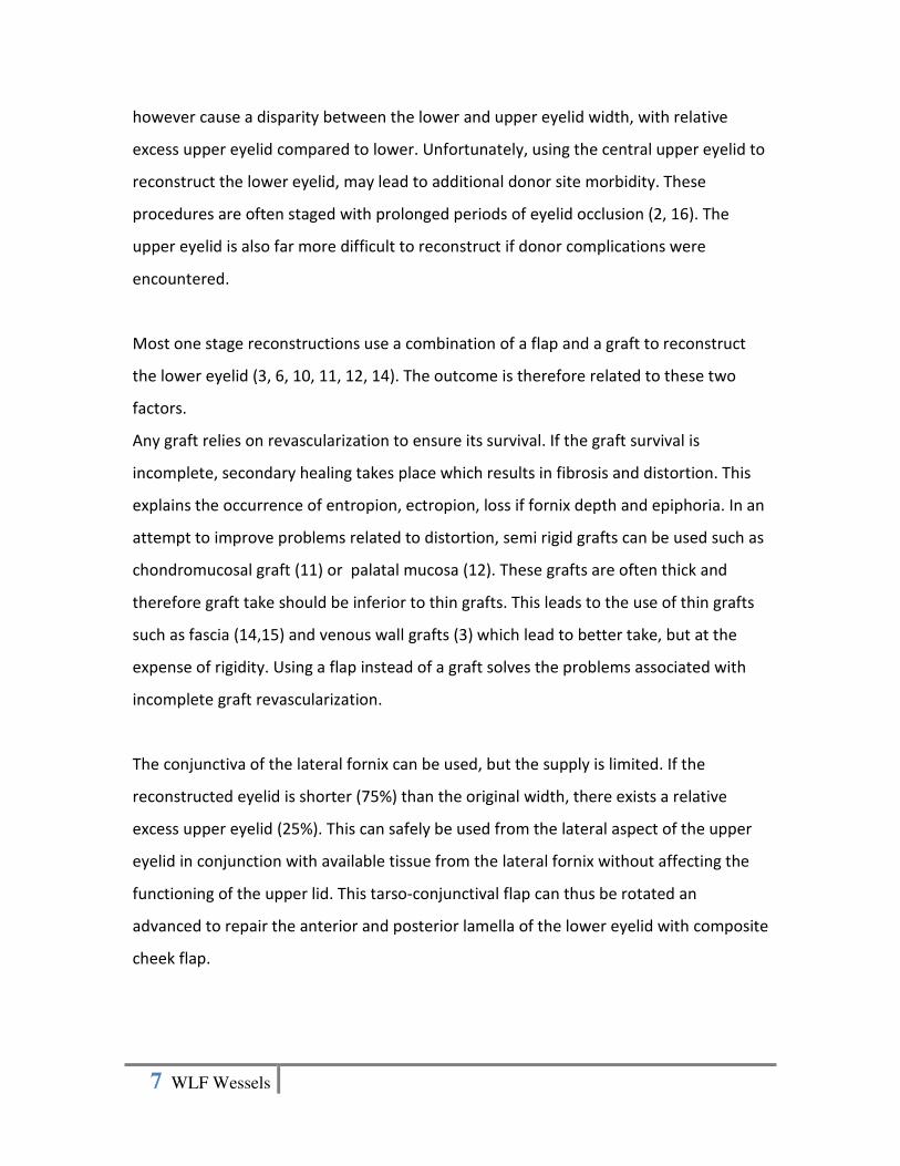
however cause a disparity between the lower and upper eyelid width, with relative
excess upper eyelid compared to lower. Unfortunately, using the central upper eyelid to
reconstruct the lower eyelid, may lead to additional donor site morbidity. These
procedures are often staged with prolonged periods of eyelid occlusion (2, 16). The
upper eyelid is also far more difficult to reconstruct if donor complications were
encountered.
Most one stage reconstructions use a combination of a flap and a graft to reconstruct
the lower eyelid (3, 6, 10, 11, 12, 14). The outcome is therefore related to these two
factors.
Any graft relies on revascularization to ensure its survival. If the graft survival is
incomplete, secondary healing takes place which results in fibrosis and distortion. This
explains the occurrence of entropion, ectropion, loss if fornix depth and epiphoria. In an
attempt to improve problems related to distortion, semi rigid grafts can be used such as
chondromucosal graft (11) or palatal mucosa (12). These grafts are often thick and
therefore graft take should be inferior to thin grafts. This leads to the use of thin grafts
such as fascia (14,15) and venous wall grafts (3) which lead to better take, but at the
expense of rigidity. Using a flap instead of a graft solves the problems associated with
incomplete graft revascularization.
The conjunctiva of the lateral fornix can be used, but the supply is limited. If the
reconstructed eyelid is shorter (75%) than the original width, there exists a relative
excess upper eyelid (25%). This can safely be used from the lateral aspect of the upper
eyelid in conjunction with available tissue from the lateral fornix without affecting the
functioning of the upper lid. This tarso-conjunctival flap can thus be rotated an
advanced to repair the anterior and posterior lamella of the lower eyelid with composite
cheek flap.

8 WLF Wessels
The ‘correct’ rigidity and opposition between eyelid and eye is obtained for three
reasons: 1) Tissue composition is the same as the normal lower eyelid. 2) Being well
vascularized, less fibrotic changes and less distortion should occur. 3) The direction the
flap originates from and is transferred to, results in a natural tendency for the flap to
oppose toward the eye. Utilizing the relative excess of posterior lamella from the lateral
portion of the upper eyelid not only provides tissue for reconstruction, but it also
balances the width relationship between upper and lower eyelids. This is a common
principle used for lip reconstruction (18, 19). Complications related to graft harvest,
such as nasal septum perforations, are prevented.
Flaps are commonly used for the anterior lamella. Local flaps are preferred because they
tend be readily available, thin and provide a good colour match. Any flap that is elevated
inferior to the lower eyelid and then moved in a superior direction, is at high risk for
inferior migration due to gravity and the natural contraction associated with healing.
This leads to ectropion, loss of fornix depth and epiphoria. Unfortunately, alternative
flaps that move and originate in a more ideal direction, have other negative factors to
consider which make them less attractive to use. Forehead flaps (1) tend to be bulky,
the Fricke flap (20) leaves an unsightly donor scar and the Tripier (21) flap is limited in
size especially in a vertical direction and often involves two stages.
The cheek flap has several advantages. The colour match, thickness and reliability is
excellent, the donor scar heals well and there is an abundance of tissue to work with.
The biggest drawback is related to the direction the flap originates from and direction of
advancement. If the flap moves from inferior to superior, descent of the flap is
inevitable which will lead to unsatisfactory results. It is therefore important to design
the flap along the natural curve of the eyelid in a superior direction with a high take-off
from the lateral upper eyelid toward the temple before curving inferiorly (Figure I). This
also ensures that the flap mimics the movement of the tarso-conjunctival flap which
facilitates easy approximation. The flap should be anchored to the periostium of the

9 WLF Wessels
lateral orbit with a permanent suture in a slightly overcorrected position (Figure II).
These precautions will minimize the inferior descent and minimize associated
complications.
It is important to recognize the indications and limitations of the rotation-advancement
tarso-conjunctival cheek flap. This technique is ideally suited for defects measuring
between twenty five and eighty percent of the lower eyelid width. The limiting factor is
the tarso-conjunctival flap. The bigger the defect, the more tarso-conjunctival layer
needs to be mobilized. The dissection of this layer should be limited to the lateral
quarter of the upper eyelid. Excessive mobilization will potentially jeopardize the upper
eyelid function. It may also cause excessive blunting of the lateral canthal area and
obstruct vision during lateral gaze or decrease the palpebral horizontal aperture which
will lead to a noticeable asymmetry with a poor cosmesis . This happened in one of our
cases, which had to be corrected with a revision canthoplasty.
The ideal shape of the defect should be triangular or pentagonal (wedge excision
defect). Defects that are wide in a horizontal plane and narrow in a vertical plane are
less suited and will need to be converted to a more favourable defect, which will mean
enlarging the original defect.
Conclusively, the rotation-advancement tarso-conjunctival cheek flap adheres to basic
plastic surgery principles. (a) Vascularized tissue is used to reconstruct the defect. (b)
The flap composition is similar to the native eyelid i.e. replace like with like. (c) The flap
makes use of tissue that is abundant and therefore limits donor morbidity. These
principles ensure that this is a long-lasting, reliable and reproducible method for the
reconstruction of lower eyelid defects.

10 WLF Wessels
References
1. Mustardé JC. Repair and Reconstruction in the Orbital region. Edinburgh,
Scotland: Churchill Livingston, 1991.
2. Hughes WL. A new method for rebuilding a lower lid. Report of a case. Arch
Opthalmol 1937;17:1008-17.
3. Barbera C, Manzoni R, Dodaro L, et al. Reconstruction of the Tarsus-conjunctival
Layer Using a Venous Wall Graft. Ophthal Plast Reconstr Surg 2008;24(5):352-
356
4. Vayvada H, Manderes A, Tan Ö, et al. Total lower eyelid reconstruction using
paranasal flap. J Craniofacial Surg 2006;17(5):1020-1026.
5. Scuderi N, Rubino C. Islanded chondromucosal flap and skin graft: A new
technique in eyelid reconstruction. Br J Plast Surg 1994;47:57-59.
6. Zinkernagel MS, Catalano E, Ammann-Rauch D. Free tarsal graft combined with
skin transposition flap for full-thickness lower eyelid reconstruction. Ophthal
Plast Reconstr Surg 2007;23(3):228-231.
7. Matsumoto K, Nakanishi H, Urano Y, et al. Lower Eyelid reconstruction with a
cheek flap supported by fascia lata. Plast Reconstr Surg 1999;103:1650-4.
8. Papp C, MaurerH, Geroldinger E. Lower eyelid reconstruction with the upper
eyelid rotation flap. Plast Reconstr Surg 1990;86:563-8.
11 WLF Wessels
9. Heywood AJ, Quaba AA. A cheek island flap for the lower eyelid. Br J Plast Surg
1991;44:183.
10. Nakajima T, Yoshimura Y. One stage reconstruction of full-thickness lower eyelid
defects using a subcutaneous pedicle flap lined by a palatal mucosal graft. Br J
Plast Surg 1996;49:183.
11. Mehrotra ON. Repairing defects of the lower eyelid with a free chondromucosal
graft. Plast Reconstr Surg 1977;59(5):689-693.
12. Siegel RJ. Palatal grafts for eyelid reconstruction. Plast Reconstr Surg
1985;76:411-414.
13. Smith B, Lisman RD. Preparation of split thickness auricular cartilage for use in
ophthalmic plastic surgery. Ophthalmic Surg 1982;13:1018-23.
14. Holt JE, Holt GR, Van kirk m. Use of temporalis fascia in eyelid reconstruction.
Ophthalmology 1984;91:89-93.
15. Cullen KW, Meyer M. Eyelid reconstruction using temporalis fascia. Ophtal Plast
Reconstr Surg 1989;5:271-273.
16. Hughes WL. Total lower lid reconstruction: technical details. Trans Am Opthalmol
Soc 1976;74:321-9.
17. Sasaki K, Nozaki M, Katahira J, et al. A Nasolabial composite free flap with buccal
mucosa: reconstruction of full-thickness lower eyelid defects. Plast Reconstr Surg
1998;102(2):464-472.
12 WLF Wessels
18. Estlander JA. En ny operations metod att atersrall en forstord lapp ellekkind.
Finsak Lak-Sall SK Handl 1872;14:1.
19. Abbé R. A new plastic operation for the relief of the deformity due to double
harelip. Med Rec 1898;53:477.
20. Fricke JCG. Die Bildung neuer Augenlider (Blepheroplastik) nach Zerstorungen
und dedurch hervorgebrachten Auswartswendungen derselben. Hamburg:
Perthes & Brasser, 1829.
21. Tripier L. Lambeau musculo-cutané en forme de point. Gaz Hopitaux
1889;62:1124.

13 WLF Wessels
Figure legends
Figure I
Top left: Squamous cell carcinoma of lower eyelid.
Top right: Design of triangular excision and cheek flap.
Middle left: defect after excision.
Middle right: Tarso-conjunctival flap is dissected.
Bottom left: As the cheek flap is advanced, the tarso-conjunctival flap rotates into
position creating a “new” lateral canthus.
Bottom right: Post-operative appearance after two weeks.
Figure II
Top left: Basel cell carcinoma of right lower eyelid.
Top right: Cheek flap elevated and tarso-conjunctival flap dissected from lateral aspect
of upper eyelid. The temporal branches of the facial nerve, as pointed out, are very
superficial, and therefore the dissection lateral to the orbital rim is a sub-cutaneous
plane.
Middle left: As the cheek flap is advanced, the tarso-conjunctival flap rotates into
position creating a “new” lateral canthus.
Middle right: The cheek flap is anchored to lateral orbital wall with a permanent suture
to prevent descent.
Bottom left: Note slightly overcorrected position of lower eyelid.
Bottom right: Post-operative appearance after nine months.
Figure III
Left column: Pre-operative appearance.
Right column: Post-operative appearance after scars has settled.

14 WLF Wessels
Tables
Table 1
General and Surgical Data
Description Range Mean Number
n 9
Age 54 – 81 years 69 years
Male 6
Female 3
Basel Cell Carcinoma 5
Squamous Cell
Carcinoma
4
Defect size (% of lower
eyelid width)
33 – 80% 64%
Follow-up 6 - 60 months 23 months
Complications
• Corneal
irritation due
to suture.
• Excessive
blunting of
lateral canthus
• Other
1
1
0

15 WLF Wessels

16 WLF Wessels

17 WLF Wessels

![Real-Time Intensity-Image Reconstruction for Event Cameras ... · Cook et al . [7] propose a biologically inspired network that simultaneously estimates cam-era rotation, image gradients](https://static.fdocuments.in/doc/165x107/5f97bb8109292435bd626cf5/real-time-intensity-image-reconstruction-for-event-cameras-cook-et-al-7.jpg)