Recommendations for Clinical Exercise Laboratories: A Scientific...
19
ISSN: 1524-4539 Copyright © 2009 American Heart Association. All rights reserved. Print ISSN: 0009-7322. Online 72514 Circulation is published by the American Heart Association. 7272 Greenville Avenue, Dallas, TX DOI: 10.1161/CIRCULATIONAHA.109.192520 2009;119;3144-3161; originally published online Jun 1, 2009; Circulation Council on Cardiovascular Nursing Cardiology, the Council on Nutrition, Physical Activity, and Metabolism, and the on Exercise, Cardiac Rehabilitation, and Prevention of the Council on Clinical McInnis, Gary J. Balady and on behalf of the American Heart Association Committee Jonathan Myers, Ross Arena, Barry Franklin, Ileana Pina, William E. Kraus, Kyle From the American Heart Association Recommendations for Clinical Exercise Laboratories: A Scientific Statement http://circ.ahajournals.org/cgi/content/full/119/24/3144 located on the World Wide Web at: The online version of this article, along with updated information and services, is http://www.lww.com/reprints Reprints: Information about reprints can be found online at [email protected] 410-528-8550. E-mail: Fax: Kluwer Health, 351 West Camden Street, Baltimore, MD 21202-2436. Phone: 410-528-4050. Permissions: Permissions & Rights Desk, Lippincott Williams & Wilkins, a division of Wolters http://circ.ahajournals.org/subscriptions/ Subscriptions: Information about subscribing to Circulation is online at by on November 26, 2009 circ.ahajournals.org Downloaded from
Transcript of Recommendations for Clinical Exercise Laboratories: A Scientific...

ISSN: 1524-4539 Copyright © 2009 American Heart Association. All rights reserved. Print ISSN: 0009-7322. Online
72514Circulation is published by the American Heart Association. 7272 Greenville Avenue, Dallas, TX
DOI: 10.1161/CIRCULATIONAHA.109.192520 2009;119;3144-3161; originally published online Jun 1, 2009; Circulation
Council on Cardiovascular Nursing Cardiology, the Council on Nutrition, Physical Activity, and Metabolism, and the
on Exercise, Cardiac Rehabilitation, and Prevention of the Council on ClinicalMcInnis, Gary J. Balady and on behalf of the American Heart Association Committee
Jonathan Myers, Ross Arena, Barry Franklin, Ileana Pina, William E. Kraus, Kyle From the American Heart Association
Recommendations for Clinical Exercise Laboratories: A Scientific Statement
http://circ.ahajournals.org/cgi/content/full/119/24/3144located on the World Wide Web at:
The online version of this article, along with updated information and services, is
http://www.lww.com/reprintsReprints: Information about reprints can be found online at
[email protected]. E-mail:
Fax:Kluwer Health, 351 West Camden Street, Baltimore, MD 21202-2436. Phone: 410-528-4050. Permissions: Permissions & Rights Desk, Lippincott Williams & Wilkins, a division of Wolters
http://circ.ahajournals.org/subscriptions/Subscriptions: Information about subscribing to Circulation is online at
by on November 26, 2009 circ.ahajournals.orgDownloaded from

Recommendations for Clinical Exercise LaboratoriesA Scientific Statement From the American Heart Association
Jonathan Myers, PhD, FAHA, Chair; Ross Arena, PhD, FAHA; Barry Franklin, PhD, FAHA;Ileana Pina, MD, FAHA; William E. Kraus, MD, FAHA; Kyle McInnis, PhD; Gary J. Balady, MD, FAHA;
on behalf of the American Heart Association Committee on Exercise, Cardiac Rehabilitation,and Prevention of the Council on Clinical Cardiology, the Council on Nutrition, Physical Activity, and
Metabolism, and the Council on Cardiovascular Nursing
The present statement provides a guide to initiating andmaintaining a high-quality clinical exercise testing lab-
oratory for administering graded exercise tests to adults.Pediatric testing has been addressed separately.1 It is arevision of the 1995 American Heart Association (AHA)“Guidelines for Clinical Exercise Testing Laboratories”2 andis designed to complement several other AHA documentsrelated to exercise testing, including the AHA/AmericanCollege of Cardiology (ACC) guidelines for exercise testing,3
the AHA’s “Exercise Standards for Testing and Training,”4
the AHA’s “Clinical Competence Statement on Stress Test-ing,”5 and the AHA’s “Assessment of Functional Capacity inClinical and Research Settings.”6
Exercise testing is a noninvasive procedure that providesdiagnostic and prognostic information and evaluates an indi-vidual’s capacity for dynamic exercise. Exercise testing facilitiesrange from the sophisticated research setting to more conven-tional equipment in the family practitioner’s or internist’soffice. Regardless of the range of testing procedures per-formed in any given laboratory, basic equipment, personnel,and protocol criteria are necessary to ensure the comfort andsafety of the patient and to conduct a meaningful test.
Testing RoomEnvironmentExercise testing equipment varies in size. The testing roomshould be large enough to accommodate all the equipmentnecessary, including emergency equipment and a defibrilla-
tor, while maintaining walking areas and allowing adequateaccess to the patient in emergency situations. It is alsoimportant that the laboratory comply with local fire standardsand with procedures for other types of emergencies (eg,earthquake, hurricane).
The laboratory should be well lighted, clean, and wellventilated, with temperature and humidity control. A wall-mounted clock with a sweep second hand or a digital counteris useful. The examining table should have space for towels,tape, and other items needed for patient preparation andtesting. A curtain for privacy during patient preparation isuseful. Minimization of interruptions and maintenance ofprivacy allow the patient and laboratory personnel to concen-trate on the testing procedure and add comfort to the patient.
To assess the level of effort, a large-print scale of perceivedexertion7 (Table 1) should be mounted on the wall in clearview of the patient. Either the original (category) scale, whichrates intensity on a scale of 6 to 20, or the revised (category-ratio) scale of 1 to 10 is appropriate as a subjective tool forexercise testing. Simpler 1-to-4 scales are preferable toquantify symptoms of angina or dyspnea.8,9 Dyspnea can alsobe measured by means of a visual analog scale that is validand reliable.10 Handheld scales are useful during cardiopul-monary testing when the mouthpiece or mask may preventspeech. These scales should be clearly explained to thepatient before testing is initiated.
In laboratories that perform ventilatory gas exchange, athermometer, barometer, and hygrometer should be kept in
The American Heart Association makes every effort to avoid any actual or potential conflicts of interest that may arise as a result of an outsiderelationship or a personal, professional, or business interest of a member of the writing panel. Specifically, all members of the writing group are requiredto complete and submit a Disclosure Questionnaire showing all such relationships that might be perceived as real or potential conflicts of interest.
This statement was approved by the American Heart Association Science Advisory and Coordinating Committee on March 26, 2009. A copy of thestatement is available at http://www.americanheart.org/presenter.jhtml?identifier�3003999 by selecting either the “topic list” link or the “chronologicallist” link (No. LS-2092). To purchase additional reprints, call 843-216-2533 or e-mail [email protected].
The American Heart Association requests that this document be cited as follows: Myers J, Arena R, Franklin B, Pina I, Kraus WE, McInnis K, BaladyGJ; on behalf of the American Heart Association Committee on Exercise, Cardiac Rehabilitation, and Prevention of the Council on Clinical Cardiology,the Council on Nutrition, Physical Activity, and Metabolism, and the Council on Cardiovascular Nursing. Recommendations for clinical exerciselaboratories: a scientific statement from the American Heart Association. Circulation. 2009;119:3144–3161.
Expert peer review of AHA Scientific Statements is conducted at the AHA National Center. For more on AHA statements and guidelines development,visit http://www.americanheart.org/presenter.jhtml?identifier�3023366.
Permissions: Multiple copies, modification, alteration, enhancement, and/or distribution of this document are not permitted without the expresspermission of the American Heart Association. Instructions for obtaining permission are located at http://www.americanheart.org/presenter.jhtml?identifier�4431. A link to the “Permission Request Form” appears on the right side of the page.
(Circulation. 2009;119:3144-3161.)© 2009 American Heart Association, Inc.
Circulation is available at http://circ.ahajournals.org DOI: 10.1161/CIRCULATIONAHA.109.192520
3144
AHA Scientific Statement
by on November 26, 2009 circ.ahajournals.orgDownloaded from

the room. Maintenance of an appropriate temperature isimportant because heart rate and perceived exertion rise withan increase in ambient temperature. In addition, cardiovascu-lar responses become variable when humidity exceeds 60%,and the combination of heat and humidity will lower maxi-mum performance. In general, a temperature range of 20°C to22°C (68°F to 71.6°F) is considered comfortable for exercise.A cool, dry environment (50% humidity) enhances cutaneousheat exchange or loss and serves to dissipate excessive heatprovoked by exercise. Circulating fans can assist in control-ling room temperature and ventilation. If gas exchangemeasurements are being performed, barometric pressure andtemperature should be measured, because gases expand withheat or with low barometric pressures and contract with coldor with high barometric pressures. Most commercially avail-able automated cardiopulmonary testing systems will makeadjustments for ambient conditions.
Equipment
ElectrocardiogramA suitable electrocardiographic recording system is essentialfor continuous monitoring of heart rhythm and evaluation ofischemic electrocardiographic changes during exercise and re-covery. Equipment ranges from more sophisticated and costlycomputerized systems to simpler, more conventional types.Nonetheless, the instrument should meet the specifications setby the AHA.11
When purchasing a highly specialized computer system,care must be taken to ensure that the frequency responseaccurately reflects ST-segment changes. It is necessary,therefore, to compare raw analog data with computer-generated data for validity. Continuous oscilloscopic moni-toring of a minimum of 3 leads is recommended to optimallyidentify arrhythmia patterns; however, the ability to producea 12-lead printed copy will enhance interpretation and is
highly recommended.12 The 12-lead electrocardiogram(ECG) is essential for the accurate interpretation of particulararrhythmias, such as to distinguish ventricular tachycardiafrom supraventricular tachycardia with aberrancy. In addi-tion, on rare occasions, significant ST-segment changes maybe isolated to a particular lead set, such as the inferior leads.The Mason-Likar adaptation of the 12-lead ECG has beencommonly used in the clinical setting.3,4,12 A standard 12-leadECG should be performed before placement of the final limbleads, because lead placement with the Mason-Likar systemmay alter the inferior lead complexes to either mimic or hideprevious Q waves. Electrocardiographic systems with built-inautomatic arrhythmia sensing, which alerts the user to theoccurrence of arrhythmias, are available commercially. Al-though not essential in every laboratory, these automaticarrhythmia detectors may be practical when the populationbeing tested is at high risk.
Silver–silver chloride electrodes are recommended as themost dependable for minimizing motion artifact. Commer-cially available disposable electrodes vary in size and adhe-sive preparation. However, the importance of adequate skinpreparation cannot be overlooked, regardless of the size ortype of electrode used. Extra time spent on skin preparationwill result in more stable recordings. Careful, accurate, andreproducible lead placement by staff will help to ensurestandardization and reproducibility of ECGs. Lightweight,shielded cables will lessen motion artifact. In addition, cablesystems that arise from a central box can be worn around thewaist and further stabilize the electrocardiographic signal. Flex-ible knit “tube” shirts for stabilizing the electrodes and cables arealso available.
Blood Pressure MonitoringManual auscultation is still the most feasible method ofmonitoring blood pressure during exercise and the easiest touse. A variety of automated blood pressure units are avail-able, but these devices are expensive and may performerratically at high exercise intensities because of motion. Inaddition, diastolic blood pressure may not be accurate whenmeasured with these devices.13 If such systems are used, theirreliability should be validated against manual cuff measure-ments within each respective laboratory before routine use,and distinctly abnormal hypertensive or hypotensive bloodpressure recordings during exercise should be corroboratedby manual recordings. A staff member should check abnor-mally high or low blood pressure readings.
The laboratory should have cuffs of various sizes, includ-ing large and pediatric.13 AHA recommendations for bloodpressure cuff bladder sizes are presented in Table 2. Envi-ronmental and safety concerns have led to the replacement ofmost mercury manometers with digital or aneroid devices,and this has raised concerns about accuracy.14 The cuffshould be placed at the level of the patient’s heart, the propercuff size should be used, and the equipment should becalibrated. Sphygmomanometers and cuffs, along with othertesting equipment, should be cleaned and inspected on aregular schedule. As with any equipment that comes incontact with patients on a repeated basis, blood pressure cuffs
Table 1. Rating of Perceived Exertion
Borg Modified Borg
6 0–Nothing at all
7–Very, very light 0.5–Very, very weak
8 1–Very weak
9–Very light 2–Weak
10 3–Moderate
11–Fairly light 4–Somewhat strong
12 5–Strong
13–Somewhat hard 6
14 7–Very strong
15–Hard 8
16 9
17–Very hard 10–Very, very strong (almost maximum)
18
19–Very, very hard –Maximum
20
The Borg RPE Scale�. From Borg GAV. Borg’s Scales of Perceived Exertion.Champaign, Ill: Human Kinetics; 1999. Scales with updated instructions can beobtained from: [email protected].
Myers et al Recommendations for Clinical Exercise Laboratories 3145
by on November 26, 2009 circ.ahajournals.orgDownloaded from

should wiped with a cleaning solution each time they areused.
Ergometry
TreadmillA treadmill should be electrically driven and should accom-modate a variety of body weights up to at least 157.5 kg (350lb). In addition, it should have a wide range of speeds, froma low of 1.6 km (1 mph) to a high of at least 12.8 km (8 mph).Elevation should be controlled electronically and should offera variety of settings, from no elevation to 20% elevation. Adedicated 220-V outlet may be required along with heavyelectrical cables that meet electrical safety standards. Thetreadmill platform should be a minimum of 127 cm (50 in) inlength and 40.64 cm (16 in) in width. Models are availablewith side platforms to allow the patient to adapt to the movingbelt before fully stepping onto it. For patient safety andstability, a padded front rail and at least 1 side rail arerecommended. As much as possible, patients should bediscouraged from holding the handrails, because doing sodecreases the metabolic cost of the work rate. An emergencystop button should be easily visible and readily accessible tothe staff and the patient when needed.
The metabolic cost of the treadmill work rate can beestimated from speed and grade by use of standardizedequations (Table 3).8 Although this is common clinically, it is
associated with a significant degree of error,15 and theselimitations should be considered when a patient’s exercisecapacity is estimated.
BicycleCycle ergometry is an alternative to treadmill testing for thosepatients who have orthopedic, peripheral vascular, or neuro-logical limitations that restrict weight bearing. It also remainsthe standard for testing in much of Europe. The cycleergometer can also serve as a less expensive, portablesubstitute for treadmill testing. Work intensity can be ad-justed by varying the resistance and cycling rate. Work ratecan be calculated in watts or kilopond-meters per minute(kpm/min).
Two types of stationary bicycles are used for testing:Mechanically braked and electronically braked. Mechanicallybraked ergometers require that a specified cycling rate bemaintained to keep the work rate constant. Electronicallybraked ergometers are more expensive and less portable butautomatically adjust internal resistance to maintain specifiedwork rates according to the cycling rate. These have becomethe standard for clinical testing when a cycle ergometer isused. Regardless of the type of stationary bicycle used, theergometer must have the capability to adjust the work rate inincrements either automatically or manually.
The cycle ergometer must include handlebars and a seatthat adjusts for height. At the ideal seat height, the kneeshould be slightly flexed at full extension. For safety pur-poses, adaptable pedal grips should be included. In addition,meters, dials, or digital displays should be appropriately sizedand placed for easy reading.
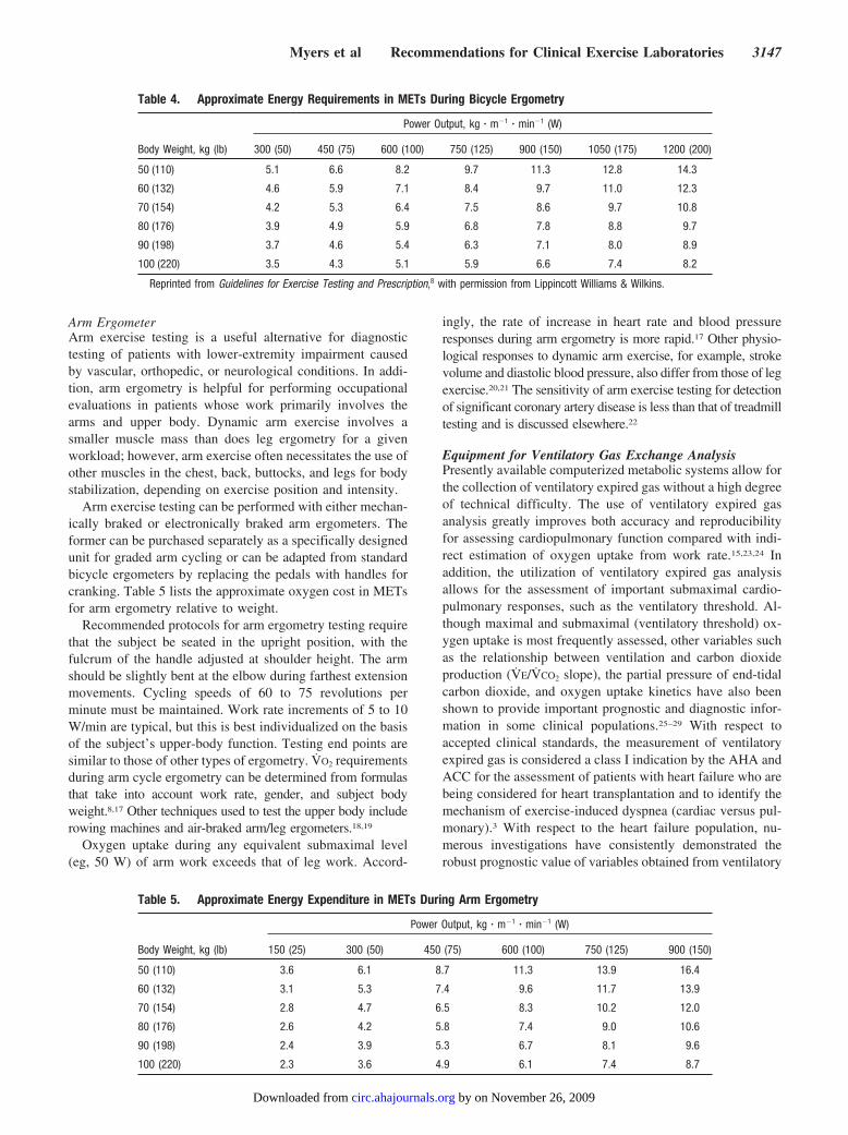
Physiological responses to exercise on a cycle ergometerdiffer from those obtained on a treadmill.8,15,16 Moreover,maximum oxygen uptake is 5% to 20% lower on a cycleergometer than on the treadmill. Table 4 lists the approximateoxygen cost in metabolic equivalents (METs) for cycleergometry relative to weight. As is the case with treadmilltesting, there is a significant degree of error that must beconsidered when exercise capacity is estimated from thecycle ergometer work rate.
Table 2. AHA Recommendations for Blood Pressure CuffBladder Dimensions for Arms of Different Sizes
CuffArm Circumference
Range at Midpoint, cmArm Circumference
Range at Midpoint, in
Adult 27–34 Up to 13.38
Large adult 35–44 13.7–17.3
Adult thigh cuff 45–52 17.7–20.4
There is some overlapping of the recommended range for arm circumfer-ences; The AHA generally recommends that the larger cuff be used forborderline measurements.
Adapted from Perloff et al,13 with permission from Lippincott Williams &Wilkins. Copyright 1993, American Heart Association.
Table 3. Approximate Energy Requirements in METs for Horizontal and Grade Walking
% Grade1.7 mph,
45.6 m/min2.0 mph,
53.6 m/min2.5 mph,
67.0 m/min3.0 mph,
80.4 m/min3.4 mph,
91.2 m/min3.75 mph,
100.5 m/min
0 2.3 2.5 2.9 3.3 3.6 3.9
2.5 2.9 3.2 3.8 4.3 4.8 5.2
5.0 3.5 3.9 4.6 5.4 5.9 6.5
7.5 4.1 4.6 5.5 6.4 7.1 7.8
10.0 4.6 5.3 6.3 7.4 8.3 9.1
12.5 5.2 6.0 7.2 8.5 9.5 10.4
15.0 5.8 6.6 8.1 9.5 10.6 11.7
17.5 6.4 7.3 8.9 10.5 11.8 12.9
20.0 7.0 8.0 9.8 11.6 13.0 14.2
22.5 7.6 8.7 10.6 12.6 14.2 15.5
25.0 8.2 9.4 11.5 13.6 15.3 16.8
METs indicates metabolic equivalents.Reprinted from Guidelines for Exercise Testing and Prescription,8 with permission from Lippincott Williams & Wilkins.
3146 Circulation June 23, 2009
by on November 26, 2009 circ.ahajournals.orgDownloaded from
Arm ErgometerArm exercise testing is a useful alternative for diagnostictesting of patients with lower-extremity impairment causedby vascular, orthopedic, or neurological conditions. In addi-tion, arm ergometry is helpful for performing occupationalevaluations in patients whose work primarily involves thearms and upper body. Dynamic arm exercise involves asmaller muscle mass than does leg ergometry for a givenworkload; however, arm exercise often necessitates the use ofother muscles in the chest, back, buttocks, and legs for bodystabilization, depending on exercise position and intensity.
Arm exercise testing can be performed with either mechan-ically braked or electronically braked arm ergometers. Theformer can be purchased separately as a specifically designedunit for graded arm cycling or can be adapted from standardbicycle ergometers by replacing the pedals with handles forcranking. Table 5 lists the approximate oxygen cost in METsfor arm ergometry relative to weight.
Recommended protocols for arm ergometry testing requirethat the subject be seated in the upright position, with thefulcrum of the handle adjusted at shoulder height. The armshould be slightly bent at the elbow during farthest extensionmovements. Cycling speeds of 60 to 75 revolutions perminute must be maintained. Work rate increments of 5 to 10W/min are typical, but this is best individualized on the basisof the subject’s upper-body function. Testing end points aresimilar to those of other types of ergometry. V̇O2 requirementsduring arm cycle ergometry can be determined from formulasthat take into account work rate, gender, and subject bodyweight.8,17 Other techniques used to test the upper body includerowing machines and air-braked arm/leg ergometers.18,19
Oxygen uptake during any equivalent submaximal level(eg, 50 W) of arm work exceeds that of leg work. Accord-
ingly, the rate of increase in heart rate and blood pressureresponses during arm ergometry is more rapid.17 Other physio-logical responses to dynamic arm exercise, for example, strokevolume and diastolic blood pressure, also differ from those of legexercise.20,21 The sensitivity of arm exercise testing for detectionof significant coronary artery disease is less than that of treadmilltesting and is discussed elsewhere.22
Equipment for Ventilatory Gas Exchange AnalysisPresently available computerized metabolic systems allow forthe collection of ventilatory expired gas without a high degreeof technical difficulty. The use of ventilatory expired gasanalysis greatly improves both accuracy and reproducibilityfor assessing cardiopulmonary function compared with indi-rect estimation of oxygen uptake from work rate.15,23,24 Inaddition, the utilization of ventilatory expired gas analysisallows for the assessment of important submaximal cardio-pulmonary responses, such as the ventilatory threshold. Al-though maximal and submaximal (ventilatory threshold) ox-ygen uptake is most frequently assessed, other variables suchas the relationship between ventilation and carbon dioxideproduction (V̇E/V̇CO2 slope), the partial pressure of end-tidalcarbon dioxide, and oxygen uptake kinetics have also beenshown to provide important prognostic and diagnostic infor-mation in some clinical populations.25–29 With respect toaccepted clinical standards, the measurement of ventilatoryexpired gas is considered a class I indication by the AHA andACC for the assessment of patients with heart failure who arebeing considered for heart transplantation and to identify themechanism of exercise-induced dyspnea (cardiac versus pul-monary).3 With respect to the heart failure population, nu-merous investigations have consistently demonstrated therobust prognostic value of variables obtained from ventilatory
Table 4. Approximate Energy Requirements in METs During Bicycle Ergometry
Body Weight, kg (lb)
Power Output, kg � m�1 � min�1 (W)
300 (50) 450 (75) 600 (100) 750 (125) 900 (150) 1050 (175) 1200 (200)
50 (110) 5.1 6.6 8.2 9.7 11.3 12.8 14.3
60 (132) 4.6 5.9 7.1 8.4 9.7 11.0 12.3
70 (154) 4.2 5.3 6.4 7.5 8.6 9.7 10.8
80 (176) 3.9 4.9 5.9 6.8 7.8 8.8 9.7
90 (198) 3.7 4.6 5.4 6.3 7.1 8.0 8.9
100 (220) 3.5 4.3 5.1 5.9 6.6 7.4 8.2
Reprinted from Guidelines for Exercise Testing and Prescription,8 with permission from Lippincott Williams & Wilkins.
Table 5. Approximate Energy Expenditure in METs During Arm Ergometry
Power Output, kg � m�1 � min�1 (W)
Body Weight, kg (lb) 150 (25) 300 (50) 450 (75) 600 (100) 750 (125) 900 (150)
50 (110) 3.6 6.1 8.7 11.3 13.9 16.4
60 (132) 3.1 5.3 7.4 9.6 11.7 13.9
70 (154) 2.8 4.7 6.5 8.3 10.2 12.0
80 (176) 2.6 4.2 5.8 7.4 9.0 10.6
90 (198) 2.4 3.9 5.3 6.7 8.1 9.6
100 (220) 2.3 3.6 4.9 6.1 7.4 8.7
Myers et al Recommendations for Clinical Exercise Laboratories 3147
by on November 26, 2009 circ.ahajournals.orgDownloaded from

expired gas analysis.25,30,31 In addition, these techniquesallow for accurate quantification of the effects of pharmaco-logical, surgical, and lifestyle interventions.32–34
For these reasons, ventilatory expired gas analysis is beingused more frequently in clinical and research settings. Al-though metabolic systems have become more user-friendly,the additional accuracy and information provided by thistechnology are dependent on some basic skills required of theclinician and technician responsible for calibration and main-tenance of the system, as well as performance of the exercisetest. In addition, the healthcare professional responsible forinterpretation of the ventilatory expired gas data must have anunderstanding of the diagnostic and prognostic implicationseach variable provides, both independently and in combina-tion. Adequate interpretation of the test requires knowledgeof testing procedures as well. Consensus statements address-ing the utility of ventilatory expired gas analysis have beenpublished recently by several well-respected national andinternational organizations.35–39
Ancillary ImagingStress echocardiography and cardiac radionuclide imagingimprove the sensitivity and specificity of the standard exer-cise ECG in patients with suspected myocardial ischemia andallow for visualization of ventricular function. These imagingtechniques are particularly valuable in patients with baselineST segment abnormalities (left bundle-branch block, leftventricular hypertrophy, Wolff-Parkinson-White syndrome,paced rhythms, or digoxin use). In addition to the need forqualified personnel and the substantial cost of the equipment,other factors must be considered if ancillary imaging is to beperformed in conjunction with exercise or pharmacologicalstress. The use of a gamma camera for radionuclide images ora cardiac ultrasound machine for stress echocardiograms willrequire increased space to accommodate this equipment. Forexercise echocardiography, the bed or gurney should be inclose proximity to the treadmill or cycle ergometer andparallel to it lengthwise to facilitate transfer of the patient.Dedicated electrical outlets or a 220-V line may be necessaryas well. Institutional radiation safety committee guidelinesmust be followed carefully. If a large volume of stressechocardiograms are to be performed, a platform or bed witha cutaway mattress for easier imaging can be helpful. De-tailed guidelines for stress echocardiography and cardiacradionuclide imaging are available elsewhere.40,41
Blood AnalysisArterial blood samples allow for the direct measurement ofSaO2, PaO2, PaCO2, pH, and lactate, as well as an accurateassessment of physiological dead space ventilation whenexpired gas analysis is used. Arterial blood gases typically aresampled from the radial artery, whereas lactate can bemeasured rapidly with a handheld unit with a finger-stickblood sample. Pulse oximetry (SpO2) permits a reasonablyaccurate estimation of SaO2, which decreases the need forarterial blood analysis in patients with pulmonary diseasewho are undergoing an exercise test for the assessment ofdyspnea with exertion. Recent data suggest that B-typenatriuretic peptide measurements during exercise testing may
improve diagnostic and prognostic accuracy42–44; however,more data are needed before this can be recommended forclinical use. Handheld units that require a finger-stick bloodsample are available for the rapid measurement of B-typenatriuretic peptide.
Cardiac OutputThere are currently several commercially available systemsthat estimate cardiac output noninvasively at rest and duringexercise. Widespread use of these systems is not common intoday’s testing environment. These systems present an inter-esting approach to the assessment of stroke volume, cardiacoutput, and other measures of cardiac performance and aresometimes used in clinical research; however, their accuracy,their diagnostic and prognostic utility, and the patient popu-lations in which they are most useful require further study.
Emergency PreparationProceduresAlthough exercise testing is a very safe procedure, the risk oftesting varies with the patient population being tested. Studiesdocumenting the safety of exercise testing are outlined below.Because the majority of diagnostic laboratories perform exercisetests in a population with a higher prevalence of coronarydisease, all testing facilities must have equipment, drugs, andpersonnel trained to deliver appropriate emergency care.
In some cases, exercise testing is performed outside of thehospital or clinical setting. For instance, maximal or submaxi-mal exercise tests may be performed in the preventiveexercise setting (eg, a health club) to develop individualizedexercise prescriptive guidelines or to evaluate change incardiorespiratory fitness over time. In such cases, guidelinesfor emergency readiness and supervision by qualified health-care professionals such as those stated in this document andin the joint position papers from the AHA and the AmericanCollege of Sports Medicine45,46 should be followed. Ingeneral, these guidelines call for the following: (1) Riskstratification of persons to be tested to determine the appro-priate level of medical supervision needed during testing; (2)a written emergency plan that is rehearsed quarterly or withenough regularity that it runs effectively if needed; the planshould also describe evacuation of unstable patients by aspecified route for rapid transfer to hospital emergencyfacilities; (3) the presence of an automated external defibril-lator; and (4) trained staff who are well versed in recognizingabnormal hemodynamic responses and/or signs and symp-toms of ischemic heart disease. Regardless of the setting, allexercise laboratories should have a written emergency planappropriate to the individual facility. Moreover, AHA proto-cols for basic and advanced life support should be followed asappropriate.47
Equipment and DrugsTable 6 lists the minimum emergency equipment necessary inan exercise testing laboratory. If intubation becomes neces-sary, suction equipment, a laryngoscope with blades ofvarious sizes, and intubation equipment should be readilyavailable. If a more extensive equipment cart is located in an
3148 Circulation June 23, 2009
by on November 26, 2009 circ.ahajournals.orgDownloaded from

area other than the testing area, a specific plan for rapidaccessibility to the cart should be clearly defined. A defibril-lator should be in every exercise laboratory and should betested on a daily basis and recorded in a log for qualitycontrol. The AHA’s classification of drugs most commonlyused in a life-threatening emergency is listed in Table 7.47
Patient PreparationIn the current practice environment, a written referral forexercise testing should be provided by the physician request-ing the assessment with a brief description of the diagnosis(confirmed or suspected), the reason for testing, a list of thepatient’s medications, medical history, and patient contactinformation. With regard to medications, information aboutthe dose and time taken should be made available. Ideally,after the appointment is made, detailed written instructionsshould be given to the patient before the day of the exercisetest. Written material should state the date and time of theexercise test, directions to the laboratory, and contact infor-mation. Abstinence from food for 3 hours before regulartesting and 8 hours before a radionuclide imaging study issuggested. Clothing should be comfortable and loose, andfootwear should be sturdy and comfortable. If the patient isprescribed medications, the instructions should include arequest for a list of drugs and dosing to be brought to thetesting center. Instructions regarding medication use on theday of the exercise test should also be included. Medicationstaken the day of the test should be confirmed. Certainantiischemic pharmacological agents may decrease the sen-sitivity of exercise tests used to detect ischemia. Althoughtapering of medications for several hours or days beforetesting is no longer considered necessary for most patients,3 itmay be appropriate for some patients to withdraw certainmedications, using an appropriate tapering regimen, beforethe exercise test. Conversely, if the intent of the exercise testis to determine the effectiveness of pharmacological therapyor functional capacity assessment, the patient should continuetaking all medications before the exercise test.48 If the patientis not provided with written instructions, he or she should becontacted by telephone to discuss the aforementioned issues.Involvement of the referring physician is critical in situationsin which plans may require the withholding of daily medica-tions. The AHA provides patient education materials withdetails of exercise testing procedures and rationale.
The Day of Exercise Testing: Patient Instructionsand DiscussionTo optimize the value of diagnostic testing, patient coopera-tion is essential. In most cases, an adequately informedpatient will give a maximum of effort and thus provide themost information for an optimum interpretation. A compre-hensive dialogue between the patient and healthcare profes-sional may be particularly important for individuals unaccus-tomed to exercise and diagnosed with cardiovascular orpulmonary disease. Concern that high levels of physicalexertion will precipitate an adverse event may cause thepatient to put forth a submaximal effort. Discussion regardingthe safety of exercise testing, the monitoring procedures used,and the importance of the exercise data obtained may allevi-ate any apprehension the patient may have. Specific instruc-tions should also be given on how to perform the exercisetest, with a brief demonstration of the test procedure. Lastly,all of the patient’s questions must be answered thoroughlybefore the test.
As a part of patient instructions/discussion, written in-formed consent should be obtained and witnessed by person-nel who can accurately describe the test and potential risks.Translation should be provided for non–English-speakingpatients. The informed consent should be included in theexercise test record. An example of an informed consent ispresented in Appendix 1. Language regarding protection ofpatient health information, both specific to an institution and
Table 7. Emergency Drugs and Solutions
Required drugs
Atropine
Lidocaine
Adenosine
Sublingual nitroglycerin
Diltiazem
Intravenous metoprolol
Diltiazem
Epinephrine
Amiodarone
Dobutamine
Verapamil
Dopamine
Vasopressin
Chewable aspirin 325 mg
Intravenous fluids
Normal saline solution (0.9%)
D5W bolus
For laboratories performing dipyridamole (adenosine) testing
Theophylline
For laboratories performing contrast echocardiography, the following shouldbe available to treat acute allergic reactions
Diphenhydramine
Dexamethasone
Albuterol
EpiPen
Table 6. Emergency Equipment
Defibrillator (portable); automated external defibrillator recommended
Oxygen tank (portable, if possible, for transport)
Nasal cannula, Ventimask, nonrebreathing mask, O2 mask
Airways: oral (OPA) and nasal (NPA)
Bag-valve-mask hand respirator (Ambu bag)
Syringes and needles
Intravenous tubing, solutions, and stand
Adhesive tape
Suction apparatus and supplies (eg, gloves, tubing)*
OPA indicates oropharyngeal airway; NPA, nasopharyngeal airway.*May be immediately available when a team arrives in certain centers.
Myers et al Recommendations for Clinical Exercise Laboratories 3149
by on November 26, 2009 circ.ahajournals.orgDownloaded from

for the Health Information Portability and Accountability Act(HIPAA), is required. The language used in the consent formcan be modified to fit the requirements of a particularlaboratory.
The Day of Exercise Testing: Patient PreparationA brief history and physical examination should be con-ducted before testing to determine medical/surgical his-tory, cardiovascular risk factors, and prior diagnostic testsand to perform a brief cardiovascular and pulmonaryexamination. This is important because the diagnosticaccuracy of the test is directly related to the pretestprobability of disease.3 The last dosing of cardiovasculardrugs should be noted on the record. Conditions that wouldpotentially preclude or limit performance of the exercisetest (ie, orthopedic/neurological restrictions, signs/symp-toms of unstable angina, decompensated heart failure, orpsychiatric conditions such as anxiety and depression)should also be determined. A sample pretest history andphysical form is shown in Appendix 2. In addition,information about usual physical activity habits and per-ceived functional capacity will aid the laboratory staff inselecting an appropriate testing protocol. In general, foryounger individuals with a history of exercise training,more aggressive testing protocols may be preferred (moreaggressive increase in workload from 1 stage to the next).For older and/or sedentary individuals, as well as patientswith moderate to severe cardiovascular or pulmonarydisease, more conservative testing protocols should beconsidered. Ideally, the exercise test should result in amaximal level of exertion within 8 to 12 minutes.3,8,35
Assessment of a subject’s perceived functional capacity byan activity-specific questionnaire, in conjunction withother baseline variables, has been shown to accuratelypredict peak exercise capacity.49,50 This approach may beused to individualize a subject’s exercise protocol andincrease the likelihood of completing the test within anappropriate timeframe.
Skin PreparationNo electrocardiographic recorder can replace good skinpreparation as part of electrode placement. For the interfacebetween the skin and electrode to be optimal, skin resistanceshould be reduced to 5000 � or less. Fortunately, mostcommercially available exercise electrocardiographic rec-orders have an AC-impedance meter built into their interfaceto test for this. Proper skin preparation requires that asuperficial layer of skin be removed. This is even moreimportant in older patients, who often have thinner, morefragile skin. To accomplish this, the areas where electrodeswill be applied should be shaved if significant hair is present.Alcohol-saturated gauze should be used to clean and removeoil from the skin. When the alcohol has evaporated, the exactelectrode-placement areas should be marked with a felt-tipped pen. These marks can then be rubbed with finesandpaper or commercially prepared abrasive tape to removethe superficial layer of skin. It is critical that electrodes forexercise testing possess a metal interface and are sunken,creating a column that is typically filled with an electrolyte
solution. Many disposable electrodes that fulfill these require-ments are available. After electrode placement, the techniciancan lightly tap the electrode to assess adequacy of skinpreparation (the tap should not create noise on the ECG). Inaddition, efforts should be taken to minimize motion at theelectrode-cable interface. This may be achieved by creatingstress loops with precut tape strips or securing the cablescentrally with an elastic belt worn around the waist. Dispos-able mesh vests placed on the upper torso can help secure theelectrodes.
Resting DataA standard resting 12-lead ECG, heart rate, and bloodpressure should be recorded in both the supine and standing(or sitting for cycle ergometry) positions before testing. Thisis necessary to determine the presence of any ECG and/orhemodynamic abnormalities that might contraindicate the testand to determine any changes that occur due to body position.If gas exchange analysis is being performed, resting valuesshould be recorded with the patient at rest for at least 2minutes or until a stable baseline is achieved (ie, approximateoxygen uptake of 3.5 mL O2 · kg�1 · min�1 and respiratoryexchange ratio �0.80). Should the respiratory exchange ratioexceed 1.0, a stable baseline can usually be achieved after thepatient sits for a few minutes while breathing quietly and isgiven reassurance by the testing personnel.
Exercise DataHeart rate, ECG, and blood pressure should be monitoredcontinuously throughout the exercise test. Additionally,heart rate, ECG, blood pressure, and the patient’s per-ceived exertion and symptoms (dyspnea/angina) should berecorded at regular intervals throughout the exercise test.The recommended recording intervals during the exercisetest for these variables are as follows: (1) Heart rate andECG—the last 5 to 10 seconds of each minute; (2) bloodpressure—the last 30 seconds of each stage for intervalprotocols or the last 30 seconds of each 2-minute intervalfor ramping protocols; and (3) perceived exertion—the last5 seconds of each minute. Dyspnea or angina should berecorded when they initially occur, and their progressionshould be recorded with 1-to-4 scales (Table 8).8,9 Each ofthese responses should be recorded at peak exercise. It maybe preferable to record the ECG just after the bloodpressure is taken to reduce electrocardiographic signal
Table 8. Angina and Dyspnea Scales
Angina scale
1� Onset of discomfort
2� Moderate, bothersome
3� Moderately severe
4� Severe; most pain ever experienced
Dyspnea scale
1� Mild, noticeable to patient but not observer
2� Mild, some difficulty, noticeable to observer
3� Moderate difficulty but can continue
4� Severe difficulty, patient cannot continue
3150 Circulation June 23, 2009
by on November 26, 2009 circ.ahajournals.orgDownloaded from

artifact. A copy of the ECG should either be stored (as partof a digital workstation) or a hard copy should be printedat the recommended intervals during the exercise test.Abnormal electrocardiographic responses (ie, arrhythmias,ST-segment shifts) should be recorded as they occur. Inaddition, the patient’s appearance should be monitored forchanges in skin color, alertness, coordination, and respon-siveness during exercise. Termination criteria for exercisetesting include patient request, moderate to severe dyspneaor angina, specific electrocardiographic changes (ie, ar-rhythmias or ST-segment shifts), and abnormal bloodpressure responses. A detailed description of absolute andrelative exercise test termination criteria is given in theACC/AHA’s guidelines for exercise testing.3 A sampledata recording form is shown as Appendix 3. Heart rate,blood pressure, and electrocardiographic responses duringrecovery are included because abnormalities that occurduring the postexercise period provide valuable diagnosticand prognostic information.51–55 The patient should bemonitored carefully until heart rate, blood pressure, andthe ECG have returned to near-baseline levels. If theexercise test was terminated because of subject request, theclinician should inquire as to the exact reason in anonleading fashion. Moreover, if the patient has experi-enced any discomfort, monitoring should continue untilsignificant symptoms have resolved. If symptoms and/orabnormal signs persist beyond 15 minutes in recovery, thesupervising physician should evaluate the patient andrecommend further observation or treatment. Having thepatient recover in a supine position may accentuate ST-segment changes, and it is important that such changes berecorded for diagnostic purposes. If ST-segment changesoccur only in recovery or worsen in recovery, this obser-vation must be included in the report to the referringphysician.
PersonnelHealthcare professionals from several disciplines may pos-sess the training and experience required to perform compe-tently in an exercise testing laboratory. Staff members mayinclude exercise physiologists, nurses, nurse practitioners,physicians’ assistants, and medical technicians. Appropriatetraining and information about the cognitive and performanceskills necessary to competently supervise exercise tests areavailable in published guidelines.5 The attainment of ad-vanced training/certifications, such as the American Collegeof Sports Medicine Exercise Specialist or Registered ClinicalExercise Physiologist, should be considered by exercisetesting laboratory personnel. All staff members must havereceived training in basic life support,45 and training inadvanced cardiac life support is encouraged. Recommenda-tions for exercise test supervision are discussed in the sectionon “Supervision/Staffing of Exercise Testing Laboratories”below.
The medical director of the exercise laboratory is respon-sible for the organization and supervision of the laboratoryand its policies. The physician should also ensure that thelaboratory is properly equipped and that the staff is appropri-ately qualified and trained. The medical director should make
sure that the staff is well prepared for response to emergen-cies and that a regular emergency practice with documenta-tion is conducted. The documentation of emergency practicesshould be kept in a file for later review. The physician isresponsible for interpreting the data, suggesting further eval-uation or additional techniques for testing, if needed, anddelivering appropriate emergency care when necessary. Re-quirements for physician competency in this area have beenoutlined by the ACC and AHA.5 In certain cases, the patientshould remain in the testing laboratory until the supervisingphysician reviews the test, determines appropriate follow-up,and counsels the patient. Accurate and timely written inter-pretation of the exercise test results should be available,ideally in 72 hours or less. A preliminary test reading,however, should be available immediately. If the test resultsare highly abnormal, the referring physician should be noti-fied as soon as possible.
Supervision/Staffing of ExerciseTesting LaboratoriesIt is the consensus of this writing group that the use ofspecially trained nonphysician healthcare professionals isappropriate to supervise clinical exercise testing, if theindividual supervising the test meets competency require-ments for exercise test supervision,5 is fully trained incardiopulmonary resuscitation, and is supervised by a physi-cian skilled in exercise testing who is immediately availableand later reads over the test results. These allied healthprofessionals typically include exercise physiologists, nurses,nurse practitioners, and physician assistants but may includeother health professionals. Because this issue has been thetopic of significant debate in the past, it is addressed in somedetail in this section.
Diagnostic exercise testing necessarily involves a boutof very hard to maximal physical exertion, performedthrough a progressive or incremental protocol to volitionalfatigue or until the occurrence of abnormal clinical signs orsymptoms that may dictate test termination. Pathophysio-logical evidence suggests that in selected individuals withknown or occult coronary artery disease, the increasedcardiac demands may precipitate plaque rupture and acutecoronary thrombosis or myocardial ischemia and threaten-ing ventricular arrhythmias, which, in extreme cases, maybe harbingers of ventricular tachycardia or fibrillation.Thus, the recommendation for direct medical supervisionof peak or symptom-limited exercise testing probablystems, at least in part, from the putative increased risk ofcardiovascular events during the procedure. A contempo-rary survey on practice patterns of exercise testing withinthe Veterans Affairs Health Care system revealed thatconsiderable variation exists in terms of whether directphysician presence is required during exercise testing. Aphysician was present during 73% of all exercise tests;however, 21% of the respondents reported that physicianpresence was required “only for high risk patients”(p 253).56 Although the most conservative approach wouldemploy the physical presence of a physician during allexercise tests, this recommendation requires a criticalevaluation of complication rates with supervision by
Myers et al Recommendations for Clinical Exercise Laboratories 3151
by on November 26, 2009 circ.ahajournals.orgDownloaded from

highly trained allied health personnel versus direct physi-cian monitoring, especially in high-risk patient subsets, aswell as a review of state-specific regulations regarding thescope of practice of nonphysicians.
Complication Rates of Exercise TestingAlthough exercise testing is considered a safe procedure,associated cardiovascular events have been reported. Aprevious review summarizing 8 studies of estimates ofsudden cardiac death during exercise testing revealed ratesranging from zero to 5 per 100 000 tests (0.005%).57
Multiple contemporary surveys indicate that the risks of acomplication that requires hospitalization (including seri-ous arrhythmias), acute myocardial infarction, or suddencardiac death during or immediately after an exercise testare �0.2%, 0.04%, and 0.01%, respectively.8 On the basisof these surveys, the event rate (often defined as death oran event serious enough to require hospitalization) isusually considered to be approximately 1/10 000.
Expanded Role for Allied Health ProfessionalsHistorically, exercise tests were directly supervised by phy-sicians more than 90% of the time58; however, over the past3 decades, cost-containment issues and time constraints onphysicians have encouraged the use of specially trainednurses, nurse practitioners, clinical exercise physiologists,exercise technicians, physician assistants, and physical ther-apists to administer selected exercise tests, with a physicianimmediately available for patient assessment or emergenciesthat may arise. Indeed, numerous reports and empiricalexperience suggest that many hospitals, medical centers, andprivate physician practices now use highly trained and/orcertified nonphysicians in the direct supervision of stresstesting, with and without concomitant myocardial perfusionimaging.
In 1979, the AHA first endorsed the concept thatexercise testing may, in some instances, be delegated to“experienced paramedical personnel” (p 426A).59 Thisrecommendation has evolved so that today, the degree ofsubject supervision needed during a test is generally madeby the supervising laboratory physician or his or herdesignated staff member on the basis of the subject’sclinical status, medical history, and standard 12-lead ECGperformed immediately before testing.4 A defibrillator,other life support equipment, medications, and personneltrained to provide advanced cardiopulmonary resuscitationmust be readily available. Moreover, guidelines from theAmerican College of Sports Medicine suggest that highlytrained allied health professionals may provide exercisetest supervision for patients at increased risk if a physicianis in close proximity and readily available should there bean emergent need.8 The rationale for this recommendationstems from several lines of evidence highlighting the lowcomplication rate associated with nonphysician supervi-sion of exercise stress testing.
A summary of the complication rates of exercise testingfrom 1969 to 1995, which included 12 different reportsinvolving nearly 2 million exercise tests, addressed the
morbidity and mortality rates associated with physician ver-sus nonphysician supervision.60 Subjects included individualswith and without a history of cardiovascular disease, athletes,and patients with a history of malignant ventricular arrhyth-mias. Complications were defined as the occurrence of acutecoronary thrombosis or threatening arrhythmias during exercisetesting (eg, ventricular fibrillation, ventricular tachycardia, orbradycardia) that mandated immediate medical treatment (car-dioversion, use of intravenous drugs, or closed-chest com-pression). One study evaluated the safety of exercise testingby physical therapists for patients with and without docu-mented coronary artery disease.61 The associated morbidityand mortality rates were 3.8 and 0.9 per 10 000 tests,respectively. Using a nurse-supervised exercise laboratory,Lem and associates62 reported 12 complications and nodeaths during 4050 tests; however, another series reported 3deaths among more than 12 000 exercise tests conducted byspecially trained nurses.63 In a summary of their 13-yearexperience using exercise physiologists for the supervisionof exercise testing, Knight and associates64 reported nofatalities, 4 myocardial infarctions, and 5 episodes ofventricular fibrillation in 28 133 tests. Franklin et al60
reviewed 18 years (1978 –1995) of exercise testing expe-rience by nonphysicians (highly trained nurses and certi-fied exercise physiologists credentialed in basic cardiopul-monary resuscitation and advanced cardiac life support)who performed 58 047 tests that included a significantsubset (�15% to 20%) of higher-risk patients. There were14 complications (including 2 fatalities), for an event rateof 1 per 4146 exercise tests.
More recently, others have reported similarly low compli-cation rates in populations tested by experienced technicians,exercise physiologists, physician assistants, and registerednurses, including stress echocardiography.65–69 In a prospec-tive pilot investigation of nonphysician supervision of pa-tients with severe chronic heart failure (left ventricularejection fraction �0.35), Squires et al70 reported only 1serious complication (an episode of ventricular fibrillationthat was successfully reverted without adverse sequelae)during 289 cardiopulmonary exercise tests. Another prelimi-nary report68 noted that exercise and pharmacological stresstesting could be conducted safely by specially trained nursesand exercise physiologists in selected high-risk patients withimplantable cardioverter defibrillators. Because pharmaco-logical stress testing is generally relegated to physicians orselected medical staff (eg, registered nurses), this practiceshould be compatible with institutional policy and stateregulations. Thus, there is ample evidence to support contem-porary guidelines that state that exercise testing “can beperformed safely by properly trained allied health profes-sionals working directly under the supervision of a physi-cian, who should be in the immediate vicinity” (p 264).70a
Moreover, several reports now suggest that highly trainedphysiologists and technicians can also provide an accuratepreliminary interpretation of the exercise ECG, in excellentagreement with senior physician and/or cardiologist consul-tant overreads.65–67,71
3152 Circulation June 23, 2009
by on November 26, 2009 circ.ahajournals.orgDownloaded from

Competency ConsiderationsAlthough the cardiovascular complications associated withexercise testing appear to be infrequent (�0.2%),8 theability to maintain a high degree of safety depends onknowing and identifying relative and absolute contraindi-cations, knowing when to terminate the test, and beingprepared for any emergency that may arise. These compe-tencies can be achieved by highly trained allied healthprofessionals provided that physician staff is readily avail-able for patient assessment, consultation, and managementof emergencies that may arise. Nonphysicians can accu-rately review the medical history and baseline ECG andprovide a timely interpretation of exercise-induced signsand symptoms, terminating an exercise test at an appro-priate intensity level.5 This premise is supported by com-parative data and recent reports showing that the incidenceof cardiovascular complications is no higher with experi-enced paramedical personnel than with physician supervi-sion of exercise tests.60
Although the use of specially trained and/or certifiedhealthcare professionals for supervision of clinical exer-cise testing can be a safe and cost-effective alternative formany hospitals and medical centers, this practice should becompatible with state licensure regulations and statutorydefinitions for the practice of medicine before it is imple-mented.72 Otherwise, there may be the potential for detri-mental consequences, raising medical-legal risks for thephysician/institution as a function of delegation of licensedauthority.
For details regarding the minimum education, training,experience, and cognitive and procedural skills necessary forcompetent performance and interpretation of exercise testing,the reader should refer to the ACC/AHA/American Collegeof Physicians’ “Clinical Competence Statement on StressTesting.”5 For internal medicine and family practice resi-dency programs that provide training in exercise testing, oftenas an elective, a minimum of 4 weeks or the equivalent shouldbe devoted to this rotation to achieve competence in bothsupervision and interpretation. It has been suggested that suchtrainees perform at least 50 exercise stress tests to qualify fordiagnostic exercise testing in private practice and that physi-cians perform at least 25 exercise tests per year to maintaintheir competence.5
Report of Test ResultsThe written report should be completed in a timely fashionand should contain the information necessary to assist thephysician in clinical decision making. Reporting the testresults as positive or negative should be avoided; it ispreferable to use the terms normal or abnormal” and theprecise responses should be reported. Abnormalities shouldinclude data that place the patient outside his or her expectedperformance, including impaired functional capacity, whichis a common but frequently ignored test result. The demo-graphic data, date of test, reason for the test, and protocolused should be clearly identifiable. The report should includethe peak work rate achieved by the patient in METs or V̇O2,peak heart rate and blood pressure, and any abnormal signs orsymptoms that occurred during or after the test. Whether
exercise capacity was determined by estimated METs or bydirectly measured V̇O2 should be specified. If it was necessaryfor the patient to hold the treadmill handrails, the reportshould indicate this. Because the referring physician may notbe familiar with normal standards, appropriate referencevalues for age and gender should be provided. The perceivedexertion level achieved should be recorded. The electrocar-diographic data should consist of rest, abnormal exercisechanges, and return to baseline. Occurrence of arrhythmiasmust be noted as well. If ischemia was demonstrated byelectrocardiographic changes, the time and heart rate at whichthe changes initially occurred should be specified. If gasexchange measurements were made, peak oxygen uptake (inmL of O2 per minute and mL of O2 · kg�1 · min�1) should berecorded, and the ventilatory threshold (if achieved) shouldbe determined. With modern ECG and gas exchange testingsystems, detailed summary reports can be issued that allowthe physician to add comments as needed. An overall inte-grated score (such as the Duke Treadmill Score) should beincluded as part of the report.3 A summary impression of thefindings must be included and should be made part of thepatient’s permanent record. Any recommendations for furtherdiagnostic testing can be included as well. If pharmacologicalor imaging agents or echocardiography is added to theexercise test, appropriate reports should include findings,prognostic characteristics, comparisons with the most recenttest, etc. An example of an exercise test summary report isshown in Appendix 4.
Quality ControlExercise laboratories must have an active quality assuranceplan that should address and enforce practice for emergencysituations. The plan should also assess staff performanceduring emergencies. Other quality control issues include theneed to keep laboratory records that indicate regular mainte-nance and nonscheduled repairs of testing equipment, cali-bration logs, defibrillator testing, and review of emergencymedications and their expiration dates. Staff education,whether formal (eg, in the form of continuing educationcredits or American College of Sports Medicine certification)or informal, should be documented. Hospital-based laborato-ries will need to review quality control issues for certificationby the Joint Commission on Accreditation of HealthcareOrganizations.
Treadmill, Cycle Ergometer, and MetabolicSystem Calibration
Calibration of the treadmill and cycle ergometer (both leg andarm) should be performed on a monthly basis. Specificdirections for calibration and preventive maintenance aretypically included in the treadmill or ergometer operationmanual provided by the manufacturer. Each laboratory shouldrecord dates that calibrations are performed. These recordsare an important part of quality assurance procedures.
Treadmill SpeedCalibration of treadmill speed requires knowledge of the beltlength, which can be obtained from the manufacturer or
Myers et al Recommendations for Clinical Exercise Laboratories 3153
by on November 26, 2009 circ.ahajournals.orgDownloaded from

measured with a tape. The treadmill speed can be calibratedby counting the number of rotations of the treadmill belt perunit of time. Using a mark on the treadmill belt as a reference,the number of belt revolutions in 1 minute can be counted,and, knowing the length of the belt, the actual miles per hourcalculated by:
Belt length (inches) � number of revolutions per minute
1056
(1056�conversionofinchesperminutetomilesperhour).
The value obtained is the treadmill speed in miles perhour. If the speed indicator does not agree with this value,adjust the meter to the proper setting. On some treadmills,a calibration adjustment screw is found in a small openingin the front of the control panel. If not available, themanufacturer should be contacted. The calibration proce-dure should be repeated for several different speeds toascertain accuracy across commonly used protocols in agiven laboratory.
Treadmill ElevationTreadmill elevation is calibrated by measuring a fixed dis-tance on the floor and determining the difference in height ofthe treadmill over the fixed distance. The following specificprocedures are performed:
1. With the use of a carpenter’s level, ensure that thetreadmill is resting on a level surface. Set the treadmillelevation to 0% grade. If the elevation does not read 0%when it is level, adjust the potentiometer until it does.
2. Mark 2 points 50 cm (20 in) apart along the length ofthe treadmill.
3. Elevate the treadmill to its metered reading of 20%grade and measure the distance of each of the 2 pointsto the floor.
4. Divide the difference between the 2 heights by 20. Theresults should be 0.20 or 20% when the elevation isproperly calibrated. If the result is not 20%, adjust theelevation meter potentiometer so that it displays thecalculated elevation percentage. A check of 5%, 10%,and 15% grade readings is recommended to ensure thevalidity of intermediate positions.
Speed and elevation should be calibrated without a subjecton the treadmill. It is recommended, however, that a moder-ately heavy subject (75 to 100 kg) walk on the treadmill aftercalibration to ensure that the calibrations remain accuratewhen the treadmill is in use. Speed should remain unchangedregardless of the weight of the individual on the treadmill.
After approximately 1000 hours of use, the treadmillshould be serviced, which should include lubricating themotor bearings, checking the variable speed belt for wear,centering the belt, greasing the chain drive, and cleaning andlubricating the gears. Service and maintenance schedulesshould be available from the manufacturer.
Bicycle Ergometer CalibrationBecause the work rate on a mechanically braked ergometerdepends not only on the resistance but also on the cycling rate
in revolutions per minute, it is essential that a counterquantify this factor. It is also important that the belt tension beadjusted appropriately and that the flywheel be cleaned toensure smooth operation. Electronically braked ergometersare more difficult to calibrate and require special instrumentsgenerally not available to the individual purchaser, so cali-bration is usually provided by the manufacturer or by theinstitutional biomedical engineering department.
To check the calibration on a mechanically braked cycleergometer, the belt should be removed from the wheel. Themark on the pendulum weight should be set at “0,” and aweight that is known to be accurate should be attached to thebelt. The weight should hang freely. A reading of that weightshould be given accurately on the scale. If all conditions aremet and the scale continues to show an incorrect reading forthe known weight, the adjusting screw should be turned untilthe scale reads the appropriate weight.
When calibrating an ergometer that is braked by a lateralfriction device, the ergometer is placed on 2 chairs so thatthe brake scale plate is vertical. After releasing the brakeregulator knob on the handlebar, a known metric weight ishung on the brake arm using a wire S-hook. Afterloosening the fastening screw of the shock absorber at 1end, the scale should read, in kiloponds, the exact amountof the weight attached to the brake arm. The pointer shouldalways be read from directly above. If the scale does notread the weight accurately, the regulating nut should beturned. When the pointer indicates the same figure as theweight attached, the ergometer is calibrated correctly.
Mechanically braked ergometers can be delicate and maylose adjustment from frequent use or if they are transported.Before an ergometer is used, the chain should be checked fortightness and lubrication, and the braking surfaces of theflywheel should be free of any dirt that has gathered. A finesandpaper pressed against the braking surface while pedalingthe ergometer will smooth the surface.
Arm ErgometryMechanically braked arm ergometers should be manuallycalibrated routinely as recommended for bicycle ergometers.Electronically braked models require periodic calibration byexperienced biomedical technicians as recommended by thespecific equipment manufacturer.
Automated Blood Pressure SystemsAlthough manually determined blood pressure is recom-mended during exercise, several automated systems havebeen shown to provide reasonably accurate measurementsof blood pressure during exercise.73–75 Calibration andmaintenance procedures should be followed according to themanufacturer’s specifications. Additionally, values obtainedby the automated system should be compared periodically(monthly) with manual blood pressure measurements.
Gas Exchange SystemsThe metabolic system should be calibrated immediatelybefore each exercise test. This should include calibration ofairflow and both the oxygen (O2) and carbon dioxide (CO2)analyzers. Gas analyzers and flow meters are prone to drift,
3154 Circulation June 23, 2009
by on November 26, 2009 circ.ahajournals.orgDownloaded from

which can lead to serious errors. Today, nearly all commer-cially available systems have convenient calibration proce-dures controlled by a microprocessor. Validation studies havebeen performed on a number of the computerized sys-tems.76–78 Because ambient conditions affect the concentra-tion of O2 in the inspired air, it is necessary to account fortemperature, barometric pressure, and humidity. Some pres-ently available metabolic systems automatically quantifythese conditions and make appropriate adjustments in theinspired O2 concentration. If this feature is not available,these atmospheric conditions should be measured by anexternal device and input into the metabolic system. A copyof the calibration report should be printed before each testand should be attached to the test report. Valid interpretationof test results is possible only if calibration values areappropriate.
Modern metabolic systems use a wide variety of automatedreports, data sampling techniques, calibration methods, andgraphics. Although this has facilitated ease of use, it has alsoled to some confusion regarding which variables to considerand how the data should be expressed and interpreted. Forexample, many algorithms are available that automaticallychoose the ventilatory threshold or provide a diagnosis, and itis important that these be overread by a person experienced incardiopulmonary exercise testing. In addition, differences insampling (eg, breath-by-breath, an averaged number ofbreaths, or by time intervals such as 10, 15, or 30 seconds)can have a profound effect on test results. Because there is aneed for consistency in this area that appropriately balanceshigh precision but high variability (breath-by-breath) withimprecision but low variability (long sampling intervals), forroutine clinical use, it is recommended that rolling 30-secondaverages be used that are printed frequently (eg, 10 seconds).
Gas exchange measurements are highly reproduciblewithin a given subject if testing methods are consistent.One method often used to validate a system’s performanceis to test laboratory staff members at a matched submaxi-mal workload on a periodic basis. It is recommended thatseveral staff members participate in this process, eachusing a slightly different steady-state submaximal work-load. Wasserman et al79 suggest that oxygen consumptionvalues should be within 5% to 10% of previous measure-ments after 3 minutes of steady-state submaximal exercise
at a given workload. Others80,81 have proposed acceptablelimits of variation for oxygen uptake and for carbondioxide production and ventilatory and hemodynamic datafor a given steady-state workload (Table 9). The followingspecific calibration procedures should be performed toensure that valid data are obtained:
1. Room air should read 20.93�0.03% O2 at 0% humidity;however, the precise fraction is dependent on humidityand should be adjusted accordingly. A calibrationsource containing 100% nitrogen should read 0% O2.The analyzer should be checked further by simulatingthe fraction of expired O2 (FEO2) during the test, ie,approximately 16% O2. The exercise laboratory shouldbe well ventilated to ensure a representative fraction ofinspired O2; a fan is helpful for this purpose.
2. The CO2 analyzer should read a room air fraction of0.03�0.02% and should not change when the 100% N2
or 16% O2 fractions are sampled from the calibrationtanks. The CO2 analyzer should be checked further bysimulating the fraction of expired CO2 (FECO2) duringexercise, ie, approximately 4% CO2.
3. For breath-by-breath O2 and CO2 analyzers, it is alsopreferable to check the analyzer response time or delay.It is important that the system meet the specificationsoutlined by the manufacturer; this feature is available inmost systems.
4. Measurement of ventilatory volume can now be easilyachieved with 1 of several devices, including pneumota-chometers, mass flow sensors, pitot tube flow meters, andturbine volume transducers. All can be validated beforetesting by ascertaining a stable baseline (0 L/min) andinjecting a known volume (usually 3 or 4 L) from asyringe. It is preferable to perform several injections atdifferent flow rates to ensure stability; the average errorshould be within �3% of the known volume.35
Table 9. Limits of Variation in Gas Exchange VariablesObtained by Repeated Study of the Same Subject at a GivenSubmaximal Workload
Variable Variation
O2 uptake �5.0%
CO2 output �6.0%
V̇E �5.5%
Respiratory exchange ratio �3.0%
Heart rate �4.5%
Systolic blood pressure �6.0%
Diastolic blood pressure �8.0%
Cardiac output �6.5%
Data derived from Wasserman et al79 and Jones.80
Appendix 1Example of Informed Consent for Exercise Testing
Myers et al Recommendations for Clinical Exercise Laboratories 3155
by on November 26, 2009 circ.ahajournals.orgDownloaded from

Appendix 2Sample Pretest Data Summary
3156 Circulation June 23, 2009
by on November 26, 2009 circ.ahajournals.orgDownloaded from

Appendix 3Sample Exercise Test Data Recording Form
Myers et al Recommendations for Clinical Exercise Laboratories 3157
by on November 26, 2009 circ.ahajournals.orgDownloaded from

Appendix 4Sample Exercise Test Summary Report
3158 Circulation June 23, 2009
by on November 26, 2009 circ.ahajournals.orgDownloaded from
References1. Washington RL, Bricker JT, Alpert BS, Daniels SR, Deckelbaum RJ,
Fisher EA, Gidding SS, Isabel-Jones J, Kavey RE, Marx GR. Guidelinesfor exercise testing in the pediatric age group: from the Committee onAtherosclerosis and Hypertension in Children, Council on CardiovascularDisease in the Young, the American Heart Association. Circulation.1994;90:2166–2179.
2. Pina IL, Balady GJ, Hanson P, Labovitz AJ, Madonna DW, Myers J.Guidelines for clinical exercise testing laboratories: a statement for healthcareprofessionals from the Committee on Exercise and Cardiac Rehabilitation,American Heart Association. Circulation. 1995;91:912–921.
3. Gibbons RJ, Balady GJ, Timothy BJ, Chaitman BR, Fletcher GF, FroelicherVF, Mark DB, McCallister BD, Mooss AN, O’Reilly MG, Winters WL,Gibbons RJ, Antman EM, Alpert JS, Faxon DP, Fuster V, Gregoratos G,
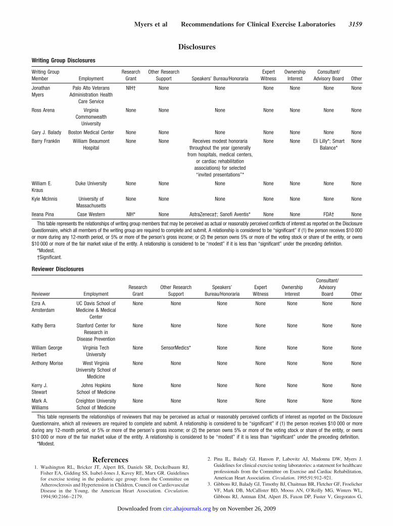
Disclosures
Writing Group Disclosures
Writing GroupMember Employment
ResearchGrant
Other ResearchSupport Speakers’ Bureau/Honoraria
ExpertWitness
OwnershipInterest
Consultant/Advisory Board Other
JonathanMyers
Palo Alto VeteransAdministration Health
Care Service
NIH† None None None None None None
Ross Arena VirginiaCommonwealth
University
None None None None None None None
Gary J. Balady Boston Medical Center None None None None None None None
Barry Franklin William BeaumontHospital
None None Receives modest honorariathroughout the year (generally
from hospitals, medical centers,or cardiac rehabilitation
associations) for selected“invited presentations”*
None None Eli Lilly*; SmartBalance*
None
William E.Kraus
Duke University None None None None None None None
Kyle McInnis University ofMassachusetts
None None None None None None None
Ileana Pina Case Western NIH* None AstraZeneca†; Sanofi Aventis* None None FDA† None
This table represents the relationships of writing group members that may be perceived as actual or reasonably perceived conflicts of interest as reported on the DisclosureQuestionnaire, which all members of the writing group are required to complete and submit. A relationship is considered to be “significant” if (1) the person receives $10 000or more during any 12-month period, or 5% or more of the person’s gross income; or (2) the person owns 5% or more of the voting stock or share of the entity, or owns$10 000 or more of the fair market value of the entity. A relationship is considered to be “modest” if it is less than “significant” under the preceding definition.
*Modest.†Significant.
Reviewer Disclosures
Reviewer EmploymentResearch
GrantOther Research
SupportSpeakers’
Bureau/HonorariaExpert
WitnessOwnership
Interest
Consultant/AdvisoryBoard Other
Ezra A.Amsterdam
UC Davis School ofMedicine & Medical
Center
None None None None None None None
Kathy Berra Stanford Center forResearch in
Disease Prevention
None None None None None None None
William GeorgeHerbert
Virginia TechUniversity
None SensorMedics* None None None None None
Anthony Morise West VirginiaUniversity School of
Medicine
None None None None None None None
Kerry J.Stewart
Johns HopkinsSchool of Medicine
None None None None None None None
Mark A.Williams
Creighton UniversitySchool of Medicine
None None None None None None None
This table represents the relationships of reviewers that may be perceived as actual or reasonably perceived conflicts of interest as reported on the DisclosureQuestionnaire, which all reviewers are required to complete and submit. A relationship is considered to be “significant” if (1) the person receives $10 000 or moreduring any 12-month period, or 5% or more of the person’s gross income; or (2) the person owns 5% or more of the voting stock or share of the entity, or owns$10 000 or more of the fair market value of the entity. A relationship is considered to be “modest” if it is less than “significant” under the preceding definition.
*Modest.
Myers et al Recommendations for Clinical Exercise Laboratories 3159
by on November 26, 2009 circ.ahajournals.orgDownloaded from

Hiratzka LF, Jacobs AK, Russell RO, Smith SC. ACC/AHA 2002 guidelineupdate for exercise testing: summary article: a report of the AmericanCollege of Cardiology/American Heart Association Task Force on PracticeGuidelines (Committee to Update the 1997 Exercise Testing Guidelines)[published correction appears in J Am Coll Cardiol. 2006;48:1731]. J AmColl Cardiol. 2002;40:1531–1540.
4. Fletcher GF, Balady GJ, Amsterdam EA, Chaitman B, Eckel R, Fleg J,Froelicher VF, Leon AS, Piña IL, Rodney R, Simons-Morton DA,Williams MA, Bazzarre T. Exercise standards for testing and training: astatement for healthcare professionals from the American Heart Asso-ciation. Circulation. 2001;104:1694–1740.
5. Rodgers GP, Ayanian JZ, Balady G, Beasley JW, Brown KA, GervinoEV, Paridon S, Quinones M, Schlant RC, Winters WL Jr, Achord JL,Boone AW, Hirshfeld JW Jr, Lorell BH, Rodgers GP, Tracy CM, WeitzHH. American College of Cardiology/American Heart Associationclinical competence statement on stress testing: a report of the AmericanCollege of Cardiology/American Heart Association/American College ofPhysicians–American Society of Internal Medicine Task Force onClinical Competence. J Am Coll Cardiol. 2000;36:1441–1453.
6. Arena R, Myers J, Williams MA, Gulati M, Kligfield P, Balady GJ,Collins E, Fletcher G. Assessment of functional capacity in clinical andresearch settings: a scientific statement from the American Heart Asso-ciation Committee on Exercise, Rehabilitation, and Prevention of theCouncil on Clinical Cardiology and the Council on CardiovascularNursing. Circulation. 2007;116:329–343.
7. Borg GAV. Borg’s Scales of Perceived Exertion. Champaign, Ill: HumanKinetics; 1999.
8. American College of Sports Medicine. ACSM’s Guidelines for ExerciseTesting and Prescription. 7th ed. Philadelphia, Pa: Lippincott Williams &Wilkins; 2006.
9. Myers JN. Perception of chest pain during exercise testing in patients withcoronary artery disease. Med Sci Sports Exerc. 1994;26:1082–1086.
10. Mahler DA, Horowitz MB. Clinical evaluation of exertional dyspnea.Clin Chest Med. 1994;15:259–269.
11. Kligfield P, Gettes LS, Bailey JJ, Childers R, Deal BJ, Hancock EW, vanHerpen G, Kors JA, Macfarlane P, Mirvis DM, Pahlm O, Rautaharju P,Wagner GS. Recommendations for the standardization and interpretationof the electrocardiogram: part l: the electrocardiogram and its technology:a scientific statement from the American Heart Association Electrocar-diography and Arrhythmias Committee, Council on Clinical Cardiology;the American College of Cardiology Foundation; and the Heart RhythmSociety. Circulation. 2007;115:1306–1324.
12. Froelicher VF, Myers J. Exercise and the Heart. 5th ed. Philadelphia, Pa:WB Saunders; 2006.
13. Perloff D, Grim C, Flack J, Frohlich ED, Hill M, McDonald M, Mor-genstern BZ. Human blood pressure determination by sphygmoma-nometry. Circulation. 1993;88(pt 1):2460–2470.
14. Jones DW, Appel LJ, Sheps SG, Roccella EJ, Lenfant C. Measuringblood pressure accurately: new and persistent challenges. JAMA. 2003;289:1027–1030.
15. Myers J. Essentials of Cardiopulmonary Exercise Testing. Champaign,Ill: Human Kinetics; 1996:59–81.
16. Hambrecht R, Schuler GC, Muth T, Grunze MF, Marburger CT, NiebauerJ, Methfessel SM, Kübler W. Greater diagnostic sensitivity of treadmillversus cycle exercise testing of asymptomatic men with coronary arterydisease. Am J Cardiol. 1992;70:141–146.
17. Balady GJ, Weiner DA, Rose L, Ryan TJ. Physiologic responses to armergometry exercise relative to age and gender. J Am Coll Cardiol. 1990;16:130–135.
18. Hagan RD, Gettman LR, Upton SJ, Duncan JJ, Cummings JM. Cardio-respiratory responses to arm, leg, and combined arm and leg work on anair-braked ergometer. J Cardiac Rehabil. 1983;3:689–695.
19. Glaser RM. Arm exercise training for wheelchair users. Med Sci SportsExerc. 1989;21(suppl):S149–S157.
20. Secher NH, Volianitis S. Are the arms and legs in competition for cardiacoutput? Med Sci Sports Exerc. 2006;38:1797–1803.
21. Pendergast DR. Cardiovascular, respiratory, and metabolic responses toupper body exercise. Med Sci Sports Exerc. 1989;21(suppl):S121–S125.
22. Balady GJ, Weiner DA, Rothendler JA, Ryan TJ. Arm exercise-thalliumimaging testing for the detection of coronary artery disease. J Am CollCardiol. 1987;9:84–88.
23. Sullivan M, Genter F, Savvides M, Roberts M, Myers J, Froelicher V.The reproducibility of hemodynamic, electrocardiographic, and gasexchange data during treadmill exercise in patients with stable anginapectoris. Chest. 1984;86:375–382.
24. Marburger CT, Brubaker PH, Pollock WE, Morgan TM, Kitzman DW.Reproducibility of cardiopulmonary exercise testing in elderly patientswith congestive heart failure. Am J Cardiol. 1998;82:905–909.
25. Arena R, Myers J, Aslam SS, Varughese EB, Peberdy MA. Peak VO2 andVE/VCO2 slope in patients with heart failure: a prognostic comparison.Am Heart J. 2004;147:354–360.
26. Koike A, Itoh H, Kato M, Sawada H, Aizawa T, Fu LT, Watanabe H.Prognostic power of ventilatory responses during submaximal exercise inpatients with chronic heart disease. Chest. 2002;121:1581–1588.
27. Reindl I, Wernecke KD, Opitz C, Wensel R, König D, Dengler T, Schimke I,Kleber FX. Impaired ventilatory efficiency in chronic heart failure: possible roleof pulmonary vasoconstriction. Am Heart J. 1998;136:778–785.
28. Matsumoto A, Itoh H, Eto Y, Kobayashi T, Kato M, Omata M, WatanabeH, Kato K, Momomura S. End-tidal CO2 pressure decreases duringexercise in cardiac patients: association with severity of heart failure andcardiac output reserve. J Am Coll Cardiol. 2000;36:242–249.
29. Arena R, Peberdy MA, Myers J, Guazzi M, Tevald M. Prognostic valueof resting end-tidal carbon dioxide in patients with heart failure. IntJ Cardiol. 2006;109:351–358.
30. Francis DP, Shamim W, Davies LC, Piepoli MF, Ponikowski P, AnkerSD, Coats AJ. Cardiopulmonary exercise testing for prognosis in chronicheart failure: continuous and independent prognostic value fromVE/VCO2 slope and peak VO2. Eur Heart J. 2000;21:154–161.
31. Myers J, Gullestad L, Vagelos R, Do D, Bellin D, Ross H, Fowler MB.Cardiopulmonary exercise testing and prognosis in severe heart failure:14 mL/kg/min revisited. Am Heart J. 2000;139(pt 1):78–84.
32. Guazzi M, Palermo P, Pontone G, Susini F, Agostoni P. Synergisticefficacy of enalapril and losartan on exercise performance and oxygenconsumption at peak exercise in congestive heart failure. Am J Cardiol.1999;84:1038–1043.
33. Malfatto G, Facchini M, Branzi G, Brambilla R, Fratianni G, Tortorici E,Balla E, Perego GB. Reverse ventricular remodeling and improved func-tional capacity after ventricular resynchronization in advanced heartfailure. Ital Heart J. 2005;6:578–583.
34. Myers J, Dziekan G, Goebbels U, Dubach P. Influence of high-intensityexercise training on the ventilatory response to exercise in patients withreduced ventricular function. Med Sci Sports Exerc. 1999;31:929–937.
35. American Thoracic Society/American College of Chest Physicians.ATS/ACCP statement on cardiopulmonary exercise testing [publishedcorrection appears in Am J Respir Crit Care Med. 2003;167:1451–1452].Am J Respir Crit Care Med. 2003;167:211–277.
36. Task Force of the Italian Working Group on Cardiac RehabilitationPrevention; Working Group on Cardiac Rehabilitation and Exercise Phys-iology of the European Society of Cardiology; Piepoli MF, Corrà U,Agostoni PG, Belardinelli R, Cohen-Solal A, Hambrecht R, Vanhees L.Statement on cardiopulmonary exercise testing in chronic heart failuredue to left ventricular dysfunction: recommendations for performance andinterpretation, part I: definition of cardiopulmonary exercise testing pa-rameters for appropriate use in chronic heart failure. Eur J CardiovascPrev Rehabil. 2006;13:150–164.
37. Task Force of the Italian Working Group on Cardiac Rehabilitation andPrevention (Gruppo Italiano di Cardiologia Riabilitativa e Prevenzione,GICR); Working Group on Cardiac Rehabilitation and Exercise Physiologyof the European Society of Cardiology; Piepoli MF, Corrà U, Agostoni PG,Belardinelli R, Cohen-Solal A, Hambrecht R, Vanhees L. Statement oncardiopulmonary exercise testing in chronic heart failure due to left ventric-ular dysfunction: recommendations for performance and interpretation, partII: how to perform cardiopulmonary exercise testing in chronic heart failure.Eur J Cardiovasc Prev Rehabil. 2006;13:300–311.
38. Task Force of the Italian Working Group on Cardiac Rehabilitation andPrevention (Gruppo Italiano di Cardiologia Riabilitativa e Prevenzione,GICR); Working Group on Cardiac Rehabilitation and Exercise Physiologyof the European Society of Cardiology; Piepoli MF, Corrà U, Agostoni PG,Belardinelli R, Cohen-Solal A, Hambrecht R, Vanhees L. Statement oncardiopulmonary exercise testing in chronic heart failure due to left ventriculardysfunction: recommendations for performance and interpretation, part III: inter-pretation of cardiopulmonary exercise testing in chronic heart failure and futureapplications. Eur J Cardiovasc Prev Rehabil. 2006;13:485–494.
39. ERS Task Force; Palange P, Ward SA, Carlsen KH, Casaburi R, Gal-lagher CG, Gosselink R, O’Donnell DE, Puente-Maestu L, Schols AM,Singh S, Whipp BJ. Recommendations on the use of exercise testing inclinical practice. Eur Respir J. 2007;29:185–209.
40. Klocke FJ, Baird MG, Lorell BH, Bateman TM, Messer JV, Berman DS,O’Gara PT, Carabello BA, Russell RO Jr, Cerqueira MD, St John SuttonMG, DeMaria AN, Udelson JE, Kennedy JW, Verani MS, Williams KA,
3160 Circulation June 23, 2009
by on November 26, 2009 circ.ahajournals.orgDownloaded from

Antman EM, Smith SC Jr, Alpert JS, Gregoratos G, Anderson JL, Hiratzka LF,Faxon DP, Hunt SA, Fuster V, Jacobs AK, Gibbons RJ, Russell RO. ACC/AHA/ASNC guidelines for the clinical use of cardiac radionuclide imaging:executive summary: a report of the American College of Cardiology/AmericanHeart Association Task Force on Practice Guidelines (ACC/AHA/ASNCCommittee to Revise the 1995 Guidelines for the Clinical Use of CardiacRadionuclide Imaging). Circulation. 2003;108:1404–1418.
41. Cheitlin MD, Armstrong WF, Aurigemma GP, Beller GA, Bierman FZ,Davis JL, Douglas PS, Faxon DP, Gillam LD, Kimball TR, KussmaulWG, Pearlman AS, Philbrick JT, Rakowski H, Thys DM, Antman EM,Smith SC Jr, Alpert JS, Gregoratos G, Anderson JL, Hiratzka LF, HuntSA, Fuster V, Jacobs AK, Gibbons RJ, Russell RO. ACC/AHA/ASE2003 guideline update for the clinical application of echocardiography:summary article: a report of the American College of Cardiology/American Heart Association Task Force on Practice Guidelines (ACC/AHA/ASE Committee to Update the 1997 Guidelines for the ClinicalApplication of Echocardiography). Circulation. 2003;108:1146–1162.
42. Foote RS, Pearlman JD, Siegel AH, Yeo KT. Detection of exercise-induced ischemia by changes in B-type natriuretic peptides. J Am CollCardiol. 2004;44:1980–1987.
43. Yeo KT, Lee HK, Wong KC, Foote RS. Can exercise-induced changes inB-type natriuretic peptides be used to detect cardiac ischemia? J CardFail. 2005;11:S59–S64.
44. Sabatine MS, Morrow DA, de Lemos JA, Omland T, Desai MY,Tanasijevic M, Hall C, McCabe CH, Braunwald E. Acute changes incirculating natriuretic peptide levels in relation to myocardial ischemia.J Am Coll Cardiol. 2004;44:1988–1995.
45. American College of Sports Medicine/American Heart Association JointPosition Statement. American College of Sports Medicine Position Standand American Heart Association: recommendations for cardiovascularscreening, staffing, and emergency policies at health/fitness facilities.Med Sci Sports Exerc 1998;30:1009–1018.
46. American College of Sports Medicine and American Heart Associationjoint position statement: automated external defibrillators in health/fitnessfacilities. Med Sci Sports Exerc. 2002;34:561–564.
47. Ali B, Zafari AM. Narrative review: cardiopulmonary resuscitation andemergency cardiovascular care: review of the current guidelines [pub-lished correction appears in Ann Intern Med. 2007;147:592]. Ann InternMed. 2007;147:171–179.
48. Sicari R. Anti-ischemic therapy and stress testing: pathophysiologic, diagnosticand prognostic implications. Cardiovasc Ultrasound. 2004;2:14.
49. Hlatky MA, Boineau RE, Higginbotham MB, Lee KL, Mark DB, CaliffRM, Cobb FR, Pryor DB. A brief self-administered questionnaire todetermine functional capacity (the Duke Activity Status Index).Am J Cardiol. 1989;64:651–654.
50. Myers J, Bader D, Madhavan R, Froelicher V. Validation of a specificactivity questionnaire to estimate exercise tolerance in patients referredfor exercise testing. Am Heart J. 2001;142:1041–1046.
51. Abe K, Tsuda M, Hayashi H, Hirai M, Sato A, Tsuzuki J, Saito H.Diagnostic usefulness of postexercise systolic blood pressure response fordetection of coronary artery disease in patients with electrocardiographicleft ventricular hypertrophy. Am J Cardiol. 1995;76:892–895.
52. Michaelides AP, Fourlas CA, Giannopoulos N, Aggeli K, AndrikopoulosGK, Tsioufis K, Massias SS, Stefanadis CI. Significance of QRS durationchanges in the evaluation of ST-segment depression presenting exclu-sively during the postexercise recovery period. Ann Noninvasive Electro-cardiol. 2006;11:241–246.
53. Pitsavos CH, Chrysohoou C, Panagiotakos DB, Kokkinos P, Skoumas J,Papaioannou I, Michaelides AP, Singh S, Stefanadis CI. Exercisecapacity and heart rate recovery as predictors of coronary heart diseaseevents, in patients with heterozygous familial hypercholesterolemia.Atherosclerosis. 2004;173:347–350.
54. Bjurö T, Gullestad L, Endresen K, Nordlander M, Malm A, Höglund L,Wahlqvist I, Pernow J. Evaluation of ST-segment changes during andafter maximal exercise tests in one-, two- and three-vessel coronary arterydisease. Scand Cardiovasc J. 2004;38:270–277.
55. Gullestad L, Jorgensen B, Bjuro T, Pernow J, Lundberg JM, Dota CD,Hall C, Simonsen S, Ablad B. Postexercise ischemia is associated withincreased neuropeptide Y in patients with coronary artery disease.Circulation. 2000;102:987–993.
56. Myers J, Voodi L, Umann T, Froelicher VF. A survey of exercise testing:methods, utilization, interpretation, and safety in the VAHCS. J CardiopulmRehabil Prev. 2000;20:251–258.
57. Gordon NF, Kohl HW. Exercise testing and sudden cardiac death. J Car-diopulm Rehabil. 1993;13:381–386.
58. Stuart RJ Jr, Ellestad MH. National survey of exercise stress testingfacilities. Chest. 1980;77:94–97.
59. Ellestad MH, Blomqvist CG, Naughton JP; American Heart AssociationSubcommittee on Rehabilitation Target Activity Group. Standards foradult exercise testing laboratories. Circulation. 1979;59:421A–430A.
60. Franklin BA, Gordon S, Timmis GC, O’Neill WW. Is direct physician super-vision of exercise stress testing routinely necessary? Chest. 1997;111:262–265.
61. Cahalin LP, Blessey RL, Kummer D, Simard M. The safety of exercisetesting performed independently by physical therapists. J CardiopulmRehabil Prev. 1987;7:269–276.
62. Lem V, Krivokapich J, Child JS. A nurse-supervised exercise stresstesting laboratory. Heart Lung. 1985;14:280–284.
63. De Busk RF. Exercise test supervision: time for a reassessment. ExercStand Malpractice Rep. 1988;2:65–70.
64. Knight JA, Laubach CA Jr, Butcher RJ, Menapace FJ. Supervision of clinicalexercise testing by exercise physiologists. Am J Cardiol. 1995;75:390–391.
65. Davis G, Ortloff S, Reed A, Worthington G, Roberts D. Evaluation oftechnician supervised treadmill exercise testing in a cardiac chest painclinic. Heart. 1998;79:613–615.
66. Muir DF, Jummun M, Stewart DJ, Clark AL. Diagnostic accuracy of techniciansupervised and reported exercise tolerance tests. Heart. 2002;87:381–382.
67. Madriago E, Baker L, Amsterdam EA. Supervision and interpretation ofexercise treadmill tests by physician assistants. Crit Pathw Cardiol.2005;4:182–184.
68. Chinnaiyan KM, Trivax J, Franklin BA, Williamson B, Kahn JK. Stresstesting in patients with implantable cardioverter-defibrillators: a pre-liminary report. Prev Cardiol. 2007;10:92–95.
69. Kane GC, Hepinstall MJ, Kidd GM, Kuehl CA, Murphy AT, NelsonJM, Schneider L, Stussy VL, Warmsbecker JA, Miller FA Jr, PellikkaPA, McCully RB. Safety of stress echocardiography supervised byregistered nurses: results of a 2-year audit of 15,404 patients. J Am SocEchocardiogr. 2008;21:337–341.
70. Squires RW, Allison TG, Johnson BD, Gau GT. Non-physician supervisionof cardiopulmonary exercise testing in chronic heart failure: safety and results ofa preliminary investigation. J Cardiopulm Rehabil. 1999;19:249–253.
70a.Gibbons RJ, Balady GJ, Beasley JW, Bricker JT, Duvernoy WFC, Fro-elicher VF, Mark DB, Marwick TH, McAllister BD, Thompson PD,Winters WL, Yanowitz FG. ACC/AHA guidelines for exercise testing: areport of the American College of Cardiology/American Heart Asso-ciation Task Force on Practice Guidelines (Committee on ExerciseTesting). J Am Coll Cardiol. 1997;30:260–315.
71. Maier E, Jensen L, Sonnenberg B, Archer S. Interpretation of exercisestress test recordings: concordance between nurse practitioner and cardi-ologist. Heart Lung. 2008;37:144–152.
72. Herbert DL, Herbert WG, Collum JJ. Legal considerations. In: Ehrman JK,Gordon PM, Visich PS, Keteyian SJ, eds. Clinical Exercise Physiology.Champaign, Ill: Human Kinetics; 2003:25–40.
73. Griffin SE, Robergs RA, Heyward VH. Blood pressure measurementduring exercise: a review. Med Sci Sports Exerc. 1997;29:149–159.
74. Cameron JD, Stevenson I, Reed E, McGrath BP, Dart AM, Kingwell BA.Accuracy of automated auscultatory blood pressure measurement duringsupine exercise and treadmill stress electrocardiogram-testing. BloodPress Monit. 2004;9:269–275.
75. MacRae HS, Allen PJ. Automated blood pressure measurement at restand during exercise: evaluation of the motion tolerant CardioDyne NBP2000. Med Sci Sports Exerc. 1998;30:328–331.
76. Crouter SE Antczak A, Hudak JR, Della Valle DM, Haas JD. Accuracyand reliability of the ParvoMedics TrueOne 2400 and MedGraphicsVO2000 metabolic systems. Eur J Appl Physiol. 2006;98:139–151.
77. Carter J, Jeukendrup AE. Validity and reliability of three commerciallyavailable breath-by-breath respiratory systems. Eur J Appl Physiol. 2002;86:435–441.
78. Foss Ø, Hallén J. Validity and stability of a computerized metabolicsystem with mixing chamber. Int J Sports Med. 2005;26:569–575.
79. Wasserman K, Hansen JE, Sue DY, Stringer W, Whipp B. Clinical exercisetesting. In: Wasserman K, ed. Principles of Exercise Testing and Interpretation.4th ed. Philadelphia, Pa: Lippincott Williams & Wilkins; 2005:133–159.
80. Jones N. Quality control in exercise studies. In: Jones N. Clinical ExerciseTesting. 4th ed. Philadelphia, Pa: WB Saunders; 1997:164–166.
81. Wilmore JH, Stanforth PR, Turley KR, Gagnon J, Daw EW, Leon AS,Rao DC, Skinner JS, Bouchard C. Reproducibility of cardiovascular,respiratory, and metabolic responses to submaximal exercise: theHERITAGE Family Study. Med Sci Sports Exerc. 1998;30:259–265.
KEY WORDS: AHA Scientific Statments � exercise test � laboratories
Myers et al Recommendations for Clinical Exercise Laboratories 3161
by on November 26, 2009 circ.ahajournals.orgDownloaded from