Recombinant Proteins for Bioassay - BioLegend · Recombinant Proteins for Bioassay ... (green and...
28
Toll-Free Tel: (US & Canada): 1.877.BIOLEGEND (246.5343) Tel: 858.768.5800 biolegend.com 02-0007-02 Recombinant Proteins for Bioassay Research Products BioLegend is ISO 9001:2008 and ISO 13485:2003 Certified World-Class Quality | Superior Customer Support | Outstanding Value
Transcript of Recombinant Proteins for Bioassay - BioLegend · Recombinant Proteins for Bioassay ... (green and...
Toll-Free Tel: (US & Canada): 1.877.BIOLEGEND (246.5343)Tel: 858.768.5800
biolegend.com
02-0007-02
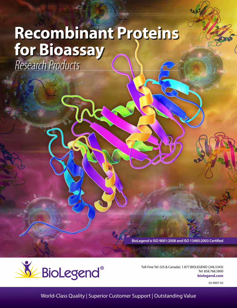
Recombinant Proteins for Bioassay Research Products
BioLegend is ISO 9001:2008 and ISO 13485:2003 Certi�ed
World-Class Quality | Superior Customer Support | Outstanding Value
Customer Service: 858-768-5800
2
Table of Contents
BioLegend Recombinant Proteins.....................................................................................................................................3
Stringent Quality Testing ......................................................................................................................................................4
Recombinant Protein Services ...........................................................................................................................................4
Cytokines ....................................................................................................................................................................................6
Chemokines ..............................................................................................................................................................................8
Growth Factors .........................................................................................................................................................................10
Enzymes and Regulators ......................................................................................................................................................12
Adhesion Molecules ...............................................................................................................................................................14
Soluble Receptors ...................................................................................................................................................................15
Other Proteins ..........................................................................................................................................................................16
Animal-Free Recombinant Proteins .................................................................................................................................17
ELISA Standard Recombinant Proteins ............................................................................................................................18
Protocols for Bioassay ............................................................................................................................................................19
Researcher Spotlight ..............................................................................................................................................................24
Frequently Asked Questions ...............................................................................................................................................26
References Using BioLegend Recombinant Proteins ................................................................................................27

3
BioLegend Recombinant Proteins
Why choose BioLegend?Our recombinant proteins are:
• >95% pure
• Validated in-house through bioassays to ensure reproducibility and activity
• Biologically active and compare favorably against competitors' products
• Competitively priced
• Discounted for bulk orders
• Flexible packaging size
Elevate your Research with BioLegend Recombinant ProteinsOur expanding catalog includes cytokines, chemokines, growth factors, enzymes, adhesion molecules, and more.
• Expressed with mammalian, E.coli, or insect protein expression system
• Both in vivo and in vitro functional assay applications
• Flexible, custom recombinant protein production
• Carrier-free formats for functional assays
• Animal-free formats to avoid animal pathogens and experimental variability
Common uses of our Recombinant Proteins:
• Standard Cell Culture
• Cell Activation
• Cell Expansion
• Di�erentiation
• Enzymatic Cleavage
• Polarization
• Cytokine Production
• Growth and Proliferation
• Inhibition
• ELISA standards
• Stem Cell Di�erentiation
• Cytotoxicity
• Chemotaxis
• Cell Signaling Activation
• WB controls
biolegend.com
Learn more at: biolegend.com/recombinant_proteins
BioLegend’s growing portfolio of recombinant proteins now contains over 600 functional proteins, for human, mouse, and rat, that can be used for bioassays in several research areas including Immunology, Neurobiology, Stem Cell research, Cancer research, Glycobiology, and Cell Biology research.
Customer Service: 858-768-5800
4
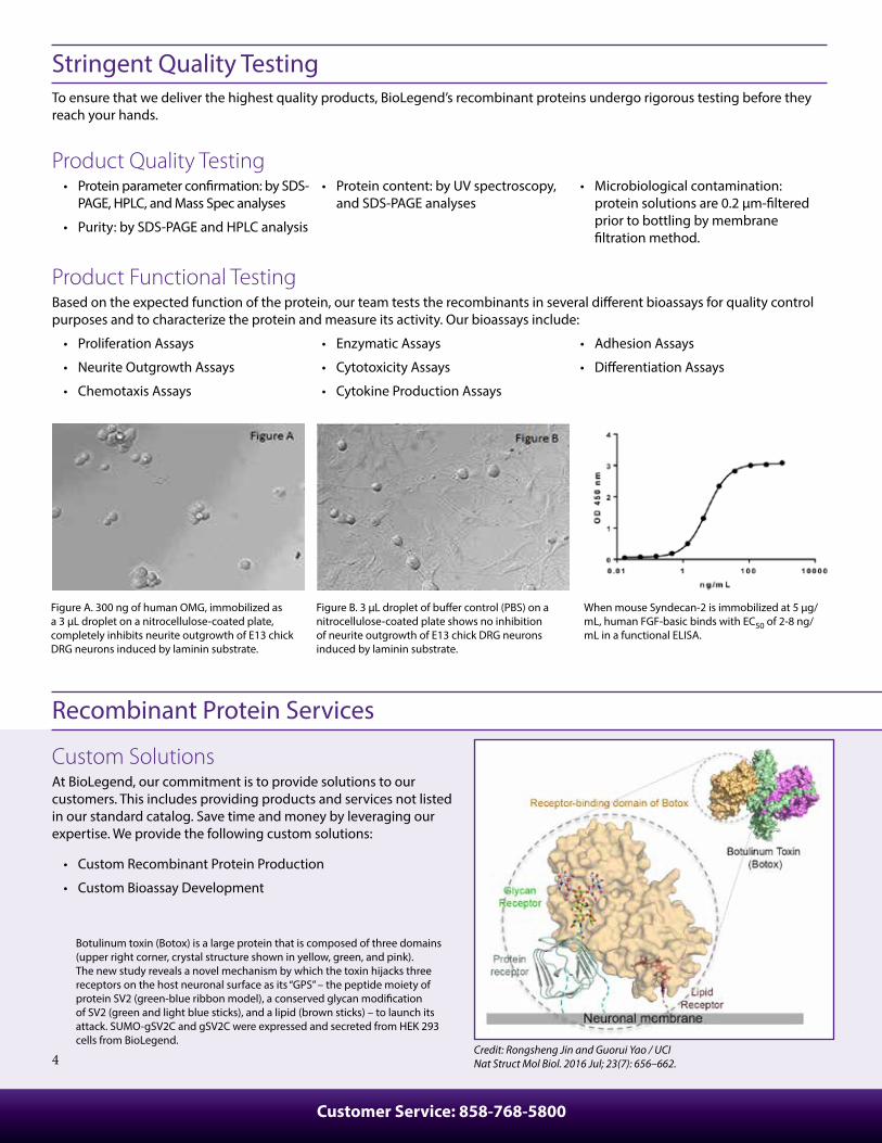
Stringent Quality TestingTo ensure that we deliver the highest quality products, BioLegend’s recombinant proteins undergo rigorous testing before they reach your hands.
Product Quality Testing• Protein parameter con�rmation: by SDS-
PAGE, HPLC, and Mass Spec analyses
• Purity: by SDS-PAGE and HPLC analysis
• Protein content: by UV spectroscopy, and SDS-PAGE analyses
• Microbiological contamination: protein solutions are 0.2 μm-�ltered prior to bottling by membrane �ltration method.
Product Functional TestingBased on the expected function of the protein, our team tests the recombinants in several di�erent bioassays for quality control purposes and to characterize the protein and measure its activity. Our bioassays include:
• Proliferation Assays
• Neurite Outgrowth Assays
• Chemotaxis Assays
• Enzymatic Assays
• Cytotoxicity Assays
• Cytokine Production Assays
• Adhesion Assays
• Di�erentiation Assays
When mouse Syndecan-2 is immobilized at 5 µg/mL, human FGF-basic binds with EC50 of 2-8 ng/mL in a functional ELISA.
Botulinum toxin (Botox) is a large protein that is composed of three domains (upper right corner, crystal structure shown in yellow, green, and pink). The new study reveals a novel mechanism by which the toxin hijacks three receptors on the host neuronal surface as its “GPS” – the peptide moiety of protein SV2 (green-blue ribbon model), a conserved glycan modi�cation of SV2 (green and light blue sticks), and a lipid (brown sticks) – to launch its attack. SUMO-gSV2C and gSV2C were expressed and secreted from HEK 293 cells from BioLegend.
Figure B. 3 µL droplet of bu�er control (PBS) on a nitrocellulose-coated plate shows no inhibition of neurite outgrowth of E13 chick DRG neurons induced by laminin substrate.
Figure A. 300 ng of human OMG, immobilized as a 3 µL droplet on a nitrocellulose-coated plate, completely inhibits neurite outgrowth of E13 chick DRG neurons induced by laminin substrate.
Custom SolutionsAt BioLegend, our commitment is to provide solutions to our customers. This includes providing products and services not listed in our standard catalog. Save time and money by leveraging our expertise. We provide the following custom solutions:
• Custom Recombinant Protein Production
• Custom Bioassay Development
Recombinant Protein Services
Credit: Rongsheng Jin and Guorui Yao / UCINat Struct Mol Biol. 2016 Jul; 23(7): 656–662.
biolegend.com
5
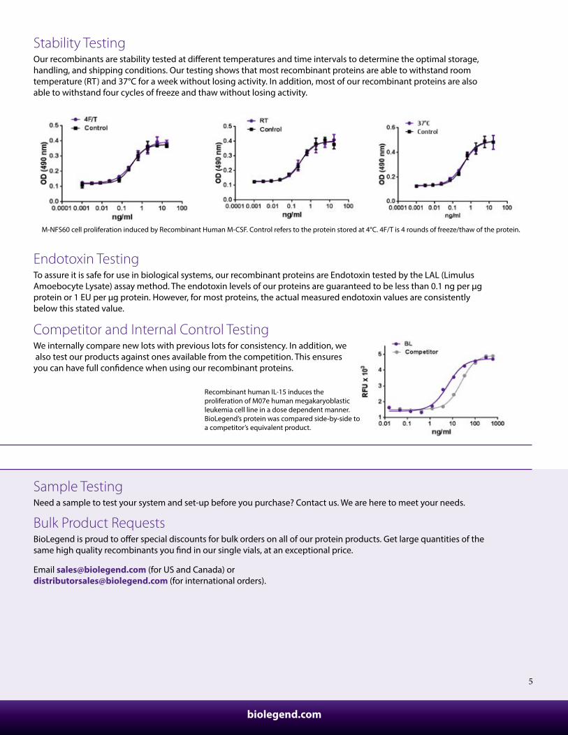
Stability TestingOur recombinants are stability tested at di�erent temperatures and time intervals to determine the optimal storage, handling, and shipping conditions. Our testing shows that most recombinant proteins are able to withstand room temperature (RT) and 37°C for a week without losing activity. In addition, most of our recombinant proteins are also able to withstand four cycles of freeze and thaw without losing activity.
M-NFS60 cell proliferation induced by Recombinant Human M-CSF. Control refers to the protein stored at 4°C. 4F/T is 4 rounds of freeze/thaw of the protein.
Endotoxin TestingTo assure it is safe for use in biological systems, our recombinant proteins are Endotoxin tested by the LAL (Limulus Amoebocyte Lysate) assay method. The endotoxin levels of our proteins are guaranteed to be less than 0.1 ng per µg protein or 1 EU per µg protein. However, for most proteins, the actual measured endotoxin values are consistently below this stated value.
Competitor and Internal Control TestingWe internally compare new lots with previous lots for consistency. In addition, we also test our products against ones available from the competition. This ensures you can have full con�dence when using our recombinant proteins.
Recombinant human IL-15 induces the proliferation of M07e human megakaryoblastic leukemia cell line in a dose dependent manner. BioLegend’s protein was compared side-by-side to a competitor’s equivalent product.
Recombinant Protein Services
Sample TestingNeed a sample to test your system and set-up before you purchase? Contact us. We are here to meet your needs.
Bulk Product RequestsBioLegend is proud to o�er special discounts for bulk orders on all of our protein products. Get large quantities of the same high quality recombinants you �nd in our single vials, at an exceptional price.
Email [email protected] (for US and Canada) or [email protected] (for international orders).
Customer Service: 858-768-5800
6
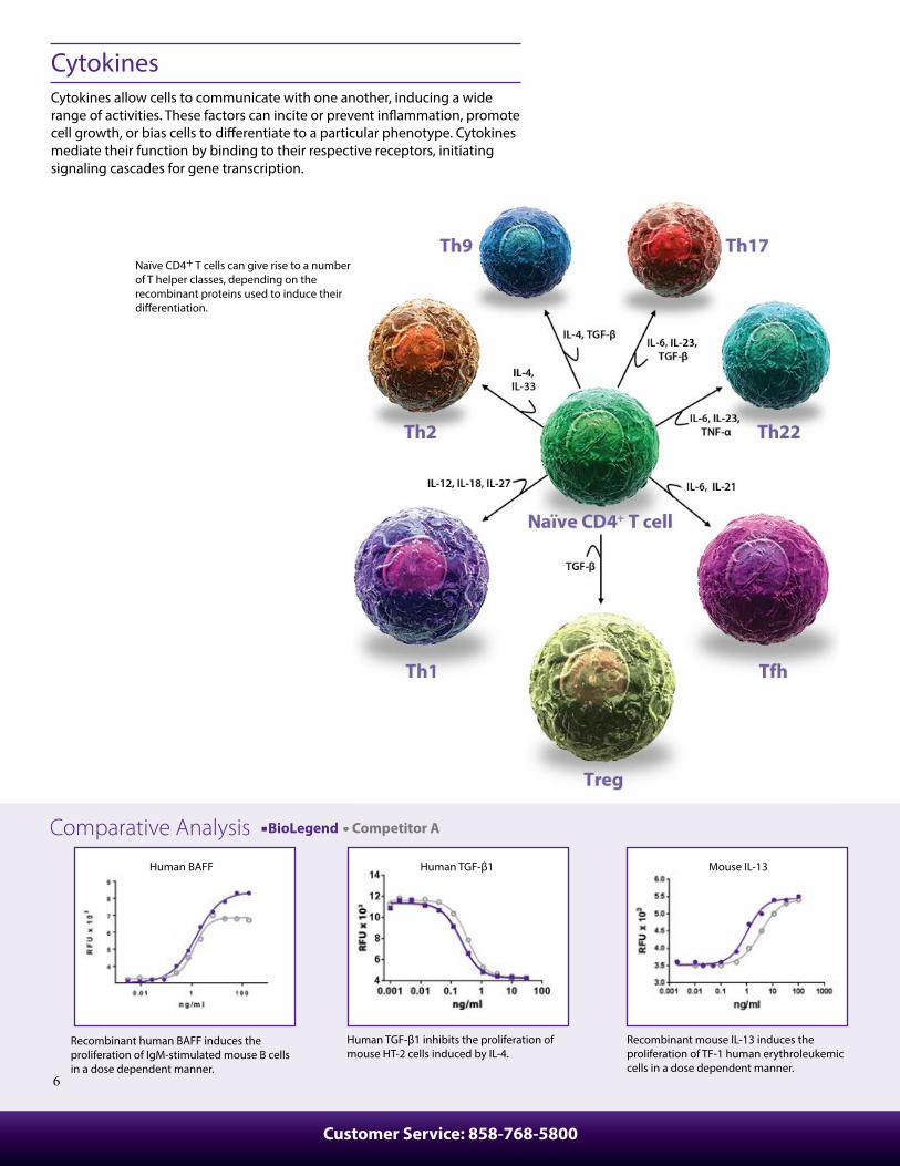
CytokinesCytokines allow cells to communicate with one another, inducing a wide range of activities. These factors can incite or prevent in�ammation, promote cell growth, or bias cells to di�erentiate to a particular phenotype. Cytokines mediate their function by binding to their respective receptors, initiating signaling cascades for gene transcription.
Recombinant human BAFF induces the proliferation of IgM-stimulated mouse B cells in a dose dependent manner.
Human TGF-β1 inhibits the proliferation of mouse HT-2 cells induced by IL-4.
Recombinant mouse IL-13 induces the proliferation of TF-1 human erythroleukemic cells in a dose dependent manner.
Naïve CD4+ T cells can give rise to a number of T helper classes, depending on the recombinant proteins used to induce their di�erentiation.
Human BAFF Human TGF-β1 Mouse IL-13
Comparative Analysis BioLegend Competitor A
7
104
103
102
101
100
100 101 102 103 104
IL-1
7A
CD4
104
103
102
101
100
100 101 102 103 104
IFN
-γ
CD4
104
103
102
101
100
100 101 102 103 104
IL-4
CD4
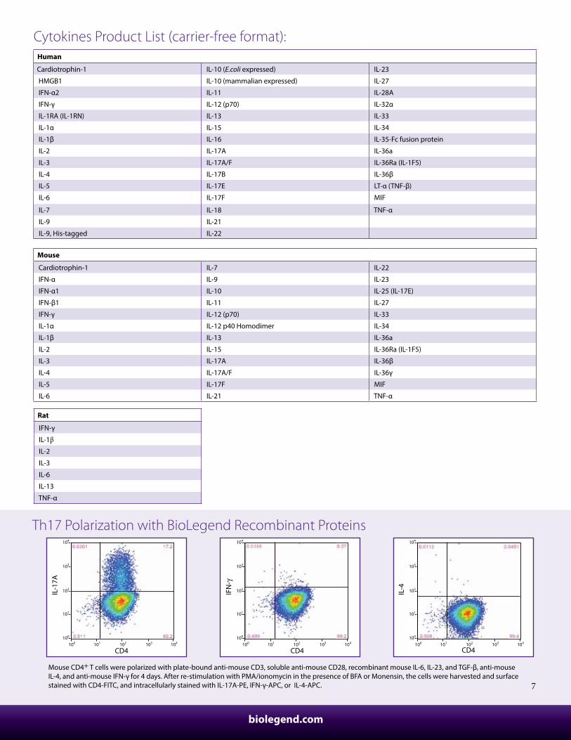
Th17 Polarization with BioLegend Recombinant Proteins
Mouse CD4+ T cells were polarized with plate-bound anti-mouse CD3, soluble anti-mouse CD28, recombinant mouse IL-6, IL-23, and TGF-β, anti-mouse IL-4, and anti-mouse IFN-γ for 4 days. After re-stimulation with PMA/ionomycin in the presence of BFA or Monensin, the cells were harvested and surface stained with CD4-FITC, and intracellularly stained with IL-17A-PE, IFN-γ-APC, or IL-4-APC.
Rat
IFN-γ
IL-1β IL-2
IL-3
IL-6
IL-13
TNF-α
Cytokines Product List (carrier-free format):Human
Cardiotrophin-1 IL-10 (E.coli expressed) IL-23
HMGB1 IL-10 (mammalian expressed) IL-27
IFN-α2 IL-11 IL-28A
IFN-γ IL-12 (p70) IL-32α
IL-1RA (IL-1RN) IL-13 IL-33
IL-1α IL-15 IL-34
IL-1β IL-16 IL-35-Fc fusion protein
IL-2 IL-17A IL-36a
IL-3 IL-17A/F IL-36Ra (IL-1F5)
IL-4 IL-17B IL-36β
IL-5 IL-17E LT-α (TNF-β)
IL-6 IL-17F MIF
IL-7 IL-18 TNF-α
IL-9 IL-21
IL-9, His-tagged IL-22
Mouse
Cardiotrophin-1 IL-7 IL-22
IFN-α IL-9 IL-23
IFN-α1 IL-10 IL-25 (IL-17E)
IFN-β1 IL-11 IL-27
IFN-γ IL-12 (p70) IL-33
IL-1α IL-12 p40 Homodimer IL-34
IL-1β IL-13 IL-36a
IL-2 IL-15 IL-36Ra (IL-1F5)
IL-3 IL-17A IL-36β
IL-4 IL-17A/F IL-36γ
IL-5 IL-17F MIF
IL-6 IL-21 TNF-α
biolegend.com
Customer Service: 858-768-5800
8
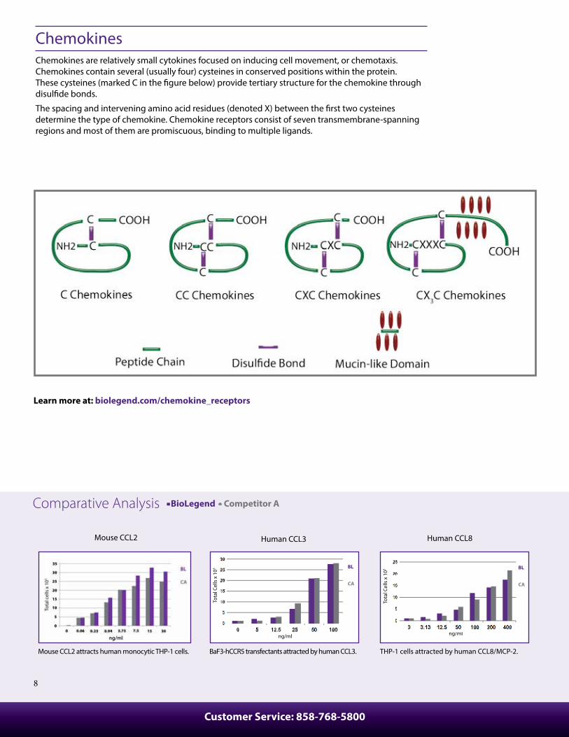
ChemokinesChemokines are relatively small cytokines focused on inducing cell movement, or chemotaxis. Chemokines contain several (usually four) cysteines in conserved positions within the protein. These cysteines (marked C in the �gure below) provide tertiary structure for the chemokine through disul�de bonds.
The spacing and intervening amino acid residues (denoted X) between the �rst two cysteines determine the type of chemokine. Chemokine receptors consist of seven transmembrane-spanning regions and most of them are promiscuous, binding to multiple ligands.
Tota
l Cel
ls x
103
ng/ml
BL
CA
Tota
l Cel
ls x
103
ng/ml
BL
CA
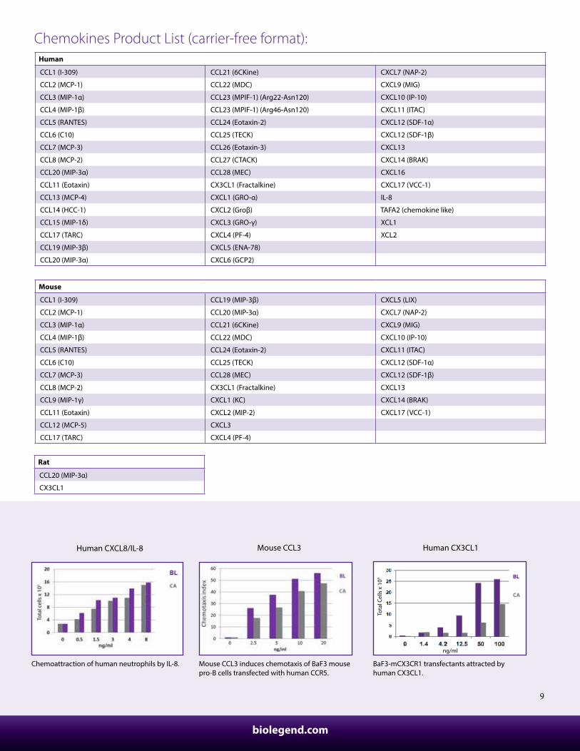
Mouse CCL2 attracts human monocytic THP-1 cells. BaF3-hCCR5 transfectants attracted by human CCL3. THP-1 cells attracted by human CCL8/MCP-2.
Mouse CCL2 Human CCL3 Human CCL8
Learn more at: biolegend.com/chemokine_receptors
Comparative Analysis BioLegend Competitor A
9
Human CX3CL1
Tota
l Cel
ls x
103
ng/ml
BL
CA
Chemoattraction of human neutrophils by IL-8. Mouse CCL3 induces chemotaxis of BaF3 mouse pro-B cells transfected with human CCR5.
BaF3-mCX3CR1 transfectants attracted by human CX3CL1.
Human CXCL8/IL-8 Mouse CCL3 Human CX3CL1
Rat
CCL20 (MIP-3α)
CX3CL1
Chemokines Product List (carrier-free format):Human
CCL1 (I-309) CCL21 (6CKine) CXCL7 (NAP-2)
CCL2 (MCP-1) CCL22 (MDC) CXCL9 (MIG)
CCL3 (MIP-1α) CCL23 (MPIF-1) (Arg22-Asn120) CXCL10 (IP-10)
CCL4 (MIP-1β) CCL23 (MPIF-1) (Arg46-Asn120) CXCL11 (ITAC)
CCL5 (RANTES) CCL24 (Eotaxin-2) CXCL12 (SDF-1α)
CCL6 (C10) CCL25 (TECK) CXCL12 (SDF-1β)
CCL7 (MCP-3) CCL26 (Eotaxin-3) CXCL13
CCL8 (MCP-2) CCL27 (CTACK) CXCL14 (BRAK)
CCL20 (MIP-3α) CCL28 (MEC) CXCL16
CCL11 (Eotaxin) CX3CL1 (Fractalkine) CXCL17 (VCC-1)
CCL13 (MCP-4) CXCL1 (GRO-α) IL-8
CCL14 (HCC-1) CXCL2 (Groβ) TAFA2 (chemokine like)
CCL15 (MIP-1δ) CXCL3 (GRO-γ) XCL1
CCL17 (TARC) CXCL4 (PF-4) XCL2
CCL19 (MIP-3β) CXCL5 (ENA-78)
CCL20 (MIP-3α) CXCL6 (GCP2)
Mouse
CCL1 (I-309) CCL19 (MIP-3β) CXCL5 (LIX)
CCL2 (MCP-1) CCL20 (MIP-3α) CXCL7 (NAP-2)
CCL3 (MIP-1α) CCL21 (6CKine) CXCL9 (MIG)
CCL4 (MIP-1β) CCL22 (MDC) CXCL10 (IP-10)
CCL5 (RANTES) CCL24 (Eotaxin-2) CXCL11 (ITAC)
CCL6 (C10) CCL25 (TECK) CXCL12 (SDF-1α)
CCL7 (MCP-3) CCL28 (MEC) CXCL12 (SDF-1β)
CCL8 (MCP-2) CX3CL1 (Fractalkine) CXCL13
CCL9 (MIP-1γ) CXCL1 (KC) CXCL14 (BRAK)
CCL11 (Eotaxin) CXCL2 (MIP-2) CXCL17 (VCC-1)
CCL12 (MCP-5) CXCL3
CCL17 (TARC) CXCL4 (PF-4)
biolegend.com
Customer Service: 858-768-5800
10
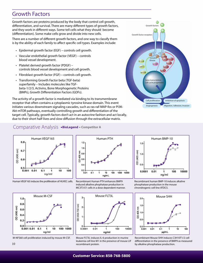
Growth FactorsGrowth factors are proteins produced by the body that control cell growth, di�erentiation, and survival. There are many di�erent types of growth factors, and they work in di�erent ways. Some tell cells what they should become (di�erentiation). Some make cells grow and divide into new cells.
There are a number of di�erent growth factors, and one way to classify them is by the ability of each family to a�ect speci�c cell types. Examples include:
• Epidermal growth factor (EGF) – controls cell growth.
• Vascular endothelial growth factor (VEGF) – controls blood vessel development.
• Platelet derived growth factor (PDGF) – controls blood vessel development and cell growth.
• Fibroblast growth factor (FGF) – controls cell growth.
• Transforming Growth Factor beta (TGF-beta) superfamily – Includes molecules like TGF- beta-1/2/3, Activins, Bone Morphogenetic Proteins (BMPs), Growth Di�erentiation Factors (GDFs).
The activity of a growth factor is mediated via binding to its transmembrane receptor that often contains a cytoplasmic tyrosine kinase domain. This event initiates various downstream signaling cascades, such as ras-raf-MAP-fos or PI3K-Akt-mTOR pathways, eventually controlling growth and di�erentiation of the target cell. Typically, growth factors don’t act in an autocrine fashion and act locally, due to their short half-lives and slow di�usion through the extracellular matrix.
Mouse M-CSF
M-NFS60 cell proliferation induced by mouse M-CSF.
Mouse FLT3L
Mouse FLT3L induces IL‐6 production in murine leukemia cell line M1 in the presence of mouse LIF recombinant protein.
Mouse SHH
Recombinant Mouse SHH induces C3H10T1/2 cell di�erentiation in the presence of BMP9 as measured by alkaline phosphatase production.
Human PTH
Recombinant Human PTH enhances BMP9 induced alkaline phophatase production in MC3T3-E1 cells in a dose dependent manner.
Human BMP-10
Recombinant human BMP-10 induces alkaline phosphatase production in the mouse chrondrogenic cell line ATDC5.
Human VEGF165
Human VEGF165 induces the proliferation of HUVEC cells.
Comparative Analysis BioLegend Competitor A
11
Rat
EGF Oncostatin M
Erythropoietin (EPO) Prolactin
GM-CSF SCF
IGF-I Thrombopoietin (TPO)
M-CSF VEGF-164
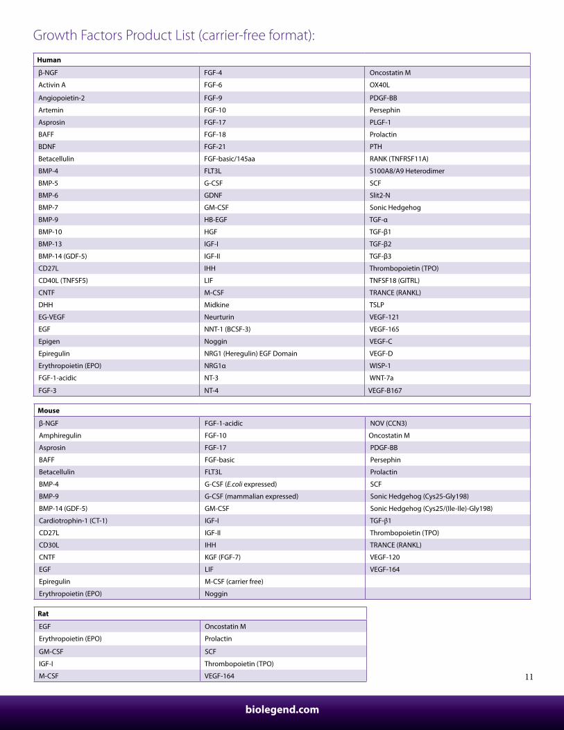
Growth Factors Product List (carrier-free format):
Human
β-NGF FGF-4 Oncostatin M
Activin A FGF-6 OX40L
Angiopoietin-2 FGF-9 PDGF-BB
Artemin FGF-10 Persephin
Asprosin FGF-17 PLGF-1
BAFF FGF-18 Prolactin
BDNF FGF-21 PTH
Betacellulin FGF-basic/145aa RANK (TNFRSF11A)
BMP-4 FLT3L S100A8/A9 Heterodimer
BMP-5 G-CSF SCF
BMP-6 GDNF Slit2-N
BMP-7 GM-CSF Sonic Hedgehog
BMP-9 HB-EGF TGF-α
BMP-10 HGF TGF-β1
BMP-13 IGF-I TGF-β2
BMP-14 (GDF-5) IGF-II TGF-β3
CD27L IHH Thrombopoietin (TPO)
CD40L (TNFSF5) LIF TNFSF18 (GITRL)
CNTF M-CSF TRANCE (RANKL)
DHH Midkine TSLP
EG-VEGF Neurturin VEGF-121
EGF NNT-1 (BCSF-3) VEGF-165
Epigen Noggin VEGF-C
Epiregulin NRG1 (Heregulin) EGF Domain VEGF-D
Erythropoietin (EPO) NRG1α WISP-1
FGF-1-acidic NT-3 WNT-7a
FGF-3 NT-4 VEGF-B167
Mouse
β-NGF FGF-1-acidic NOV (CCN3)
Amphiregulin FGF-10 Oncostatin M
Asprosin FGF-17 PDGF-BB
BAFF FGF-basic Persephin
Betacellulin FLT3L Prolactin
BMP-4 G-CSF (E.coli expressed) SCF
BMP-9 G-CSF (mammalian expressed) Sonic Hedgehog (Cys25-Gly198)
BMP-14 (GDF-5) GM-CSF Sonic Hedgehog (Cys25/(Ile-Ile)-Gly198)
Cardiotrophin-1 (CT-1) IGF-I TGF-β1
CD27L IGF-II Thrombopoietin (TPO)
CD30L IHH TRANCE (RANKL)
CNTF KGF (FGF-7) VEGF-120
EGF LIF VEGF-164
Epiregulin M-CSF (carrier free)
Erythropoietin (EPO) Noggin
biolegend.com
Customer Service: 858-768-5800
12
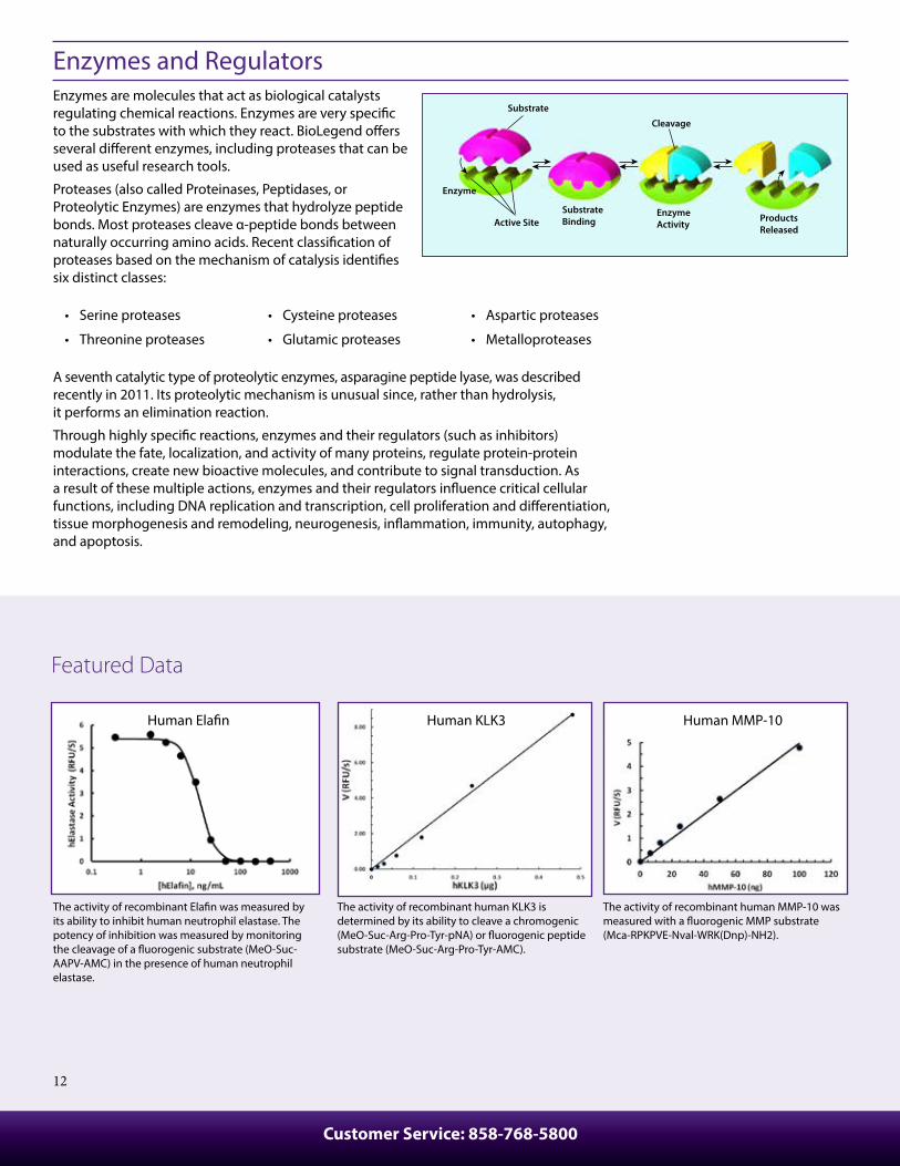
Enzymes and RegulatorsEnzymes are molecules that act as biological catalysts regulating chemical reactions. Enzymes are very speci�c to the substrates with which they react. BioLegend o�ers several di�erent enzymes, including proteases that can be used as useful research tools.
Proteases (also called Proteinases, Peptidases, or Proteolytic Enzymes) are enzymes that hydrolyze peptide bonds. Most proteases cleave α-peptide bonds between naturally occurring amino acids. Recent classi�cation of proteases based on the mechanism of catalysis identi�es six distinct classes:
• Serine proteases
• Threonine proteases
• Cysteine proteases
• Glutamic proteases
• Aspartic proteases
• Metalloproteases
A seventh catalytic type of proteolytic enzymes, asparagine peptide lyase, was described recently in 2011. Its proteolytic mechanism is unusual since, rather than hydrolysis, it performs an elimination reaction.
Through highly speci�c reactions, enzymes and their regulators (such as inhibitors) modulate the fate, localization, and activity of many proteins, regulate protein-protein interactions, create new bioactive molecules, and contribute to signal transduction. As a result of these multiple actions, enzymes and their regulators in�uence critical cellular functions, including DNA replication and transcription, cell proliferation and di�erentiation, tissue morphogenesis and remodeling, neurogenesis, in�ammation, immunity, autophagy, and apoptosis.
Substrate
Active Site
Enzyme
SubstrateBinding
EnzymeActivity
Products Released
Cleavage
The activity of recombinant Ela�n was measured by its ability to inhibit human neutrophil elastase. The potency of inhibition was measured by monitoring the cleavage of a �uorogenic substrate (MeO-Suc-AAPV-AMC) in the presence of human neutrophil elastase.
The activity of recombinant human KLK3 is determined by its ability to cleave a chromogenic (MeO-Suc-Arg-Pro-Tyr-pNA) or �uorogenic peptide substrate (MeO-Suc-Arg-Pro-Tyr-AMC).
The activity of recombinant human MMP-10 was measured with a �uorogenic MMP substrate (Mca-RPKPVE-Nval-WRK(Dnp)-NH2).
Featured Data
Human Ela�n Human KLK3 Human MMP-10
13
Comparative Analysis
Mouse
Cathepsin E (CTSE) MMP-2 Serpin A12
Cathepsin B MMP-3 TIMP-1
Granzyme B MMP-9 (Gelatinase B)
KLK7 PCSK9
Human
Arginase I KLK7 Serpin A12 (Vaspin)
Cathepsin A (CTSA) MMP-1 Serpin E1 (PAI-1)
Cathepsin B MMP-2 Serpin F1
Cathepsin D MMP-3 SLPI
Cathepsin E MMP-7 ST8SIA1
Cystatin C MMP-8 TIMP-1
Ela�n MMP-9 TIMP-2
GALNT2 MMP-9 (dimer) t-Plasminogen Activator (t-PA)
Granzyme A MMP-10 u-Plasminogen Activator (Urokinase)
Granzyme B PCSK9 Visfatin
KLK3 PLA2G7
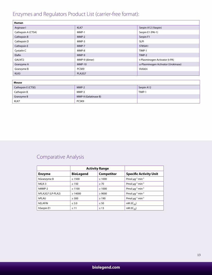
Enzymes and Regulators Product List (carrier-free format):
Activity Range
Enzyme BioLegend Competitor Speci�c Activity UnithGranzyme B ≥ 1500 ≥ 1000 Pmol µg-1 min-1
hKLK-3 ≥ 150 ≥ 70 Pmol µg-1 min-1
hMMP-2 ≥ 1100 ≥ 1000 Pmol µg-1 min-1
hPLA2G7 (LP-PLA2) ≥ 14000 ≥ 9000 Pmol µg-1 min-1
hPLAU ≥ 300 ≥ 190 Pmol µg-1 min-1
hELAFIN ≤ 3.0 ≤ 50 nM (IC50)
hSerpin E1 ≤ 11 ≤ 13 nM (IC50)
biolegend.com
When human EphA2 is immobilized, human Ephrin-A1 binds with EC50 of 3-12 ng/mL in a functional ELISA.
Mouse Ephrin-A1 binds to immobilized human EphA2 with an EC50 of 3-12 ng/mL in a functional ELISA.
Human EphA2 Mouse Ephrin-A1
Customer Service: 858-768-5800
14
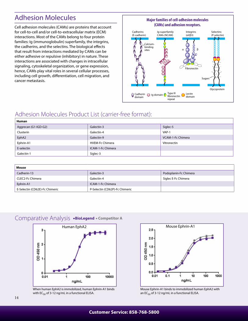
Adhesion MoleculesCell adhesion molecules (CAMs) are proteins that account for cell-to-cell and/or cell-to-extracellular matrix (ECM) interactions. Most of the CAMs belong to four protein families: Ig (immunoglobulin) superfamily, the integrins, the cadherins, and the selectins. The biological e�ects that result from interactions mediated by CAMs can be either adhesive or repulsive (inhibitory) in nature. These interactions are associated with changes in intracellular signaling, cytoskeletal organization, or gene expression, hence, CAMs play vital roles in several cellular processes, including cell growth, di�erentiation, cell migration, and cancer metastasis.
Cadherins(E-cadherin)
Ig-superfamilyCAMs (NCAM)
Integrins(αVβ3)
α β
Selectins(P-selectin)
Sugars
Glycoprotein
Lectindomain
Ig domainCadherindomain
Type III�bronectinrepeat
Fibronectin
Calcium-bindingsites
Major families of cell-adhesion molecules(CAMs) and adhesion receptors.
Adhesion Molecules Product List (carrier-free format):
Mouse
Cadherin-13 Galectin-3 Podoplanin-Fc Chimera
CLEC2-Fc Chimera Galectin-4 Siglec E-Fc Chimera
Ephrin-A1 ICAM-1-Fc Chimera
E-Selectin (CD62E)-Fc Chimeric P-Selectin (CD62P)-Fc Chimeric
Human
Aggrecan (G1-IGD-G2) Galectin-3 Siglec-5
Clusterin Galectin-4 VAP-1
EphA2 Galectin-9 VCAM-1-Fc Chimera
Ephrin-A1 HVEM-Fc Chimera Vitronectin
E-selectin ICAM-1-Fc Chimera
Galectin-1 Siglec-3
Comparative Analysis BioLegend Competitor A
Recombinant mouse BAFFR inhibits the proliferation of mouse B cells induced by BAFF in a dose dependent manner. BioLegend’s protein was compared side-by-side to the leading competitor’s equivalent product.
Mouse TNFRSF13B (TACI) inhibits mouse B cell proliferation induced by mouse BAFF (2.5 ng/mL). BioLegend’s protein was compared side-by-side to the leading competitor’s equivalent product.
Mouse BAFFR Mouse TNFRSF13B
15
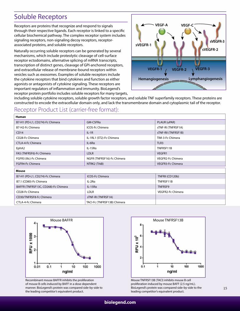
Soluble ReceptorsReceptors are proteins that recognize and respond to signals through their respective ligands. Each receptor is linked to a speci�c cellular biochemical pathway. The complex receptor system includes signaling receptors, non-signaling decoy receptors, receptor-associated proteins, and soluble receptors.
Naturally occurring soluble receptors can be generated by several mechanisms, which include proteolytic cleavage of cell-surface receptor ectodomains, alternative splicing of mRNA transcripts, transcription of distinct genes, cleavage of GPI-anchored receptors, and extracellular release of membrane-bound receptors within vesicles such as exosomes. Examples of soluble receptors include the cytokine receptors that bind cytokines and function as either agonists or antagonists of cytokine signaling. These receptors are important regulators of in�ammation and immunity. BioLegend’s receptor protein portfolio includes soluble receptors for many targets, including soluble cytokine receptors, soluble growth factor receptors, and soluble TNF superfamily receptors. These proteins are constructed to encode the extracellular domain only, and lack the transmembrane domain and cytoplasmic tail of the receptor.
VEGF-A VEGF-C
sVEGFR-1
VEGFR-1
sVEGFR-2
VEGFR-2
sVEGFR-3
VEGFR-3
Hemangiogenesis Lymphangiogenesis
Receptor Product List (carrier-free format):
Mouse
B7-H1 (PD-L1, CD274)-Fc Chimera ICOS-Fc Chimera TNFRII (CD120b)
B7.1 (CD80)-Fc Chimera IL-2Rα TNFRSF11B
BAFFR (TNFRSF13C, CD268)-Fc Chimera IL-15Rα TNFRSF9
CD28-Fc Chimera LDLR VEGFR2-Fc Chimera
CD30/TNFRSF8-Fc Chimera sTNF-RI (TNFRSF1A)
CTLA-4-Fc Chimera TACI-Fc (TNFRSF13B) Chimera
Human
B7-H1 (PD-L1, CD274)-Fc Chimera GM-CSFRα PLAUR (uPAR)
B7-H2-Fc Chimera ICOS-Fc Chimera sTNF-RI (TNFRSF1A)
CD14 IL-1R sTNF-RII (TNFRSF1B)
CD28-Fc Chimera IL-1RL1 (ST2)-Fc Chimera TIM-3-Fc Chimera
CTLA-4-Fc Chimera IL-6Rα TLR3
EphA2 IL-15Rα TNFRSF11B
FAS (TNFRSF6)-Fc Chimera LDLR VEGFR1
FGFR3 (IIIc)-Fc Chimera NGFR (TNFRSF16)-Fc Chimera VEGFR2-Fc Chimera
FGFR4-Fc Chimera NTRK2 (TrkB) VEGFR3-Fc Chimera
biolegend.com
Customer Service: 858-768-5800
16
Recombinant Mouse DKK-1 enhances BMP9 induced alkaline phosphatase production in MC3T3-E1 cells with EC50 of 15 - 60 ng/mL.
Recombinant mouse LIGHT's cytotoxic e�ect on HT-29 human colon adenocarcinoma cells.
When human Glypican-3 is immobilized at 2 µg/mL, human FGF-basic binds with EC50 of 3-12 ng/mL in a functional ELISA.
Other ProteinsIn addition to the aforementioned categories, we have other proteins that can be used in multi-functional assays and have been shown to play important cellular roles including cell migration, survival, regulation of immune responses, apoptosis, neuroin�ammation, Alzheimer's disease, control of tumor cell phenotypes, and autoimmune diseases.
Rat
Nogo-A/Nogo-66-Fc Chimera
Other Proteins Product List (carrier-free format):
Mouse DKK-1Mouse LIGHTHuman Glypican-3
Mouse
DKK-1 IGFBP-6 Syndecan-2
Endostatin Isthmin1 TACI-Fc Chimera
Glypican-1 LIGHT (TNFSF14) TNFRSF17-Fc Chimera
Glypican-3 Mesothelin TNFSF9 (4-1BBL)
IGFBP-1 NGAL (Lipocalin-2) TNFSF15
IGFBP-2 OMG TNFSF18 (GITRL)
IGFBP-4 Osteopontin
IGFBP-5 RBP4
Human
APCS (PTX2) IGFALS Osteopontin
Clusterin IGFBP-1 RBP4
CRP IGFBP-3 Resistin
DLL1 IGFBP-4 SAA1
Endostatin IGFBP-6 TFPI-2
FASL (TNFSF6) IGFBP-7 TNFSF9 (4-1BBL)
GASP-1 LIGHT (TNFSF14) TNFSF15
Glypican-1 Mesothelin TRAIL (TNFSF10)
Glypican-3 NGAL (Lipocalin-2) TWEAK (CD255)
Gremlin-1 OMG Twisted Gastrulation (TSG)
17
Animal-Free Product List (carrier-free format):
Learn more at: biolegend.com/recombinant_proteins
Rat
GM-CSF
M-CSF
SCF
Thrombopoietin (TPO)
Animal-Free Recombinant Proteins BioLegend's line of animal-free recombinant proteins greatly minimize the variables and potential contamination of mammalian pathogens during the production process. All of these proteins are produced in animal-free media, and the puri�cation equipment itself is also animal component-free. The animal-free versions of these proteins function in a similar manner to their animal-derived counterparts. BioLegend provides the animal-free proteins in lyophilized format. Treat your cells to animal-free recombinant proteins and see how they prosper!
Mouse
FGF-basic IL-2 Noggin
G-CSF IL-3 Thrombopoietin (TPO)
GM-CSF IL-4 TNF-α
IFN-γ IL-6 VEGF-164
Human
β-NGF Heregulin-β1 IL-22
Activin A IFN-γ IL-33
BMP-4 IFN-λ1 IL-36γ
CCL2 (MCP-1) IGF-1 KGF (FGF-7)
CCL5 (RANTES) IGF-II LIF
CNTF IL-1RA M-CSF
EGF IL-3 NT-3
FGF-1-acidic IL-4 NT-4
FGF-4 IL-6 Oncostatin M
FGF-8 IL-7 PDGF-AA
FGF-9 IL-8 PlGF-1
FGF-10 IL-9 sCD40L
FGF-basic (146 aa) IL-10 TGF-α
FGF-basic (154 aa) IL-11 Thrombopoietin (TPO)
Flt-3 Ligand IL-15 TNF-α
G-CSF IL-16 TRANCE (RANKL)
GDF-3 IL-17A TWEAK
GDNF IL-17E VEGF-165
GM-CSF IL-21
biolegend.com
Customer Service: 858-768-5800
18
ELISA Standard Recombinant ProteinsHaving the correct standards is critical for the accuracy and dependability of an ELISA assay. BioLegend provides lyophilized recombinant protein standards, optimized for use with our ELISA antibody pairs. Our ELISA standards are formulated with carrier proteins to ensure stability and to prevent the product from sticking to the walls of the vial.
Rat
IFN-γ
Mouse
CCL28 (MEC) IL-5 IL-27
GM-CSF IL-6 IL-33
IFN-β IL-10 IL-34
IFN-γ IL-12 (p70) Lactadherin
IL-1α IL-12/IL-23 (p40) Latent TGF-β
IL-1β IL-17A MCP-1
IL-2 IL-17A/F TNF-α
IL-3 IL-22 TSLP
IL-4 IL-23
Human
APRIL (TNFSF13) IL-2 IL-32α
CCL5 (RANTES) IL-4 IL-33
CCL8 (MCP-2) IL-5 IL-34
CCL17 (TARC) IL-6 LAP (TGF-β1)
CCL28 (MEC) IL-8 Latent TGF-β
CTLA-4 IL-9 LIF
CXCL5 (ENA-78) IL-10 Mesothelin
CXCL10 (IP-10) IL-12 (p70) MIF
FGF-basic/145aa IL-12/IL-23 (p40) Nogo-B
GM-CSF IL-13 SCF
Granulysin IL-15 TGF-β1
IFN-γ IL-17A TNF-α
IL-1α IL-21 TSLP
IL-1β IL-22
ELISA Standard Product List (with carrier protein):
biolegend.com
19
Protocols for BioassayThe biological activity of our recombinant proteins is routinely measured using a bioassay and indicated with a speci�c activity range/ED50 on our datasheets. The speci�c activity of proteins is very much dependent on the cell type and the bioassay used for testing. Our bioassay team, consisting of expert scientists with extensive manufacturing and assay development experience, set-up the right experimental conditions based on the biological function of the protein and test every lot of our recombinant proteins in at least one out of over 20 di�erent types of dose-response bioassays. Here are some example bioassay protocols and ED50 values for of our proteins (this is not an exhaustive list):
1. Proliferation Assay: Proliferation of cells induced by increasing concentrations of recombinant proteins is measured by metabolic �uorescence assay using our Deep Blue Cell Viability™ Kit (Cat. No. 424701).
Human
Target Cytokine Target Cell Line/Binding Partner Cell Number Top Concentration Incubation Time ED50
Artemin SHSY5Y cells 3K/well 300 ng/mL 5 days 4-16 ng/mL
BAFF Mouse B cells 200K/well 180 ng/mL 72 hr 0.3-2.0 ng/mL
BDNF C6 glioma cells 2K/well 30 µg/mL 4 days 1-3 µg/mL
Betacellulin BalbC/3T3 cells 2K/well 3 ng/mL 48 hr 0.04-0.24 ng/mL
CD40L Human B Cells 100K/well 2000 ng/mL 72 hr 20-100 ng/mL
CNTF TF-1 cells 25K/well 12 µg/mL 72 hr 30-180 ng/mL
EGF A431 cells 1.2K/well 100 ng/mL 5 days 1-2 ng/mL
EG-VEGF MIA Paca 2 cells 1K/well 50 µg/mL 72 hr 1-4 µg/mL
EPO TF-1 cells 25K/well 10 ng/mL 48 hr 0.1-0.6 ng/mL
FGF basic NIH/3T3 cells 1.5K/well 60 ng/mL 48 hr 1-4 ng/mL
GM-CSF TF-1 cells 10K/well 2 ng/mL 72 hr 0.10- 0.30 ng/mL
HB-EGF BalbC/3T3 cells 2K/well 30 ng/mL 48 hr 0.2-1.2 ng/mL
hVEGF165 HUVEC 3.6K/well 300 ng/mL 48 hr 1-6 ng/mL
IL-1α D10.G4.1 cells 25K/well 900 pg/mL 48 hr 5-15 pg/mL
IL-1β D10.G4.1 cells 20K/well 1 ng/mL 48 hr 5-15 pg/mL
IL-1RA D10.G4.1 cells 25K/well 5000 ng/mL 48 hr 7-35 ng/mL
IL-2 CTLL2 cells 25K/well 20 ng/mL 48 hr 0.05-0.3 ng/mL
IL-3 TF-1 cells 25K/well 10 ng/mL 48 hr ≤ 0.1 ng/mL
IL-4 TF-1 cells 25K/well 40 ng/mL 72 hr 0.2-0.6 ng/mL
IL-6 7TD1 cells 8K/well 3 ng/mL 48 hr 4-20 pg/mL
IL-7 PHA activated PBL 100K/well 100 ng/mL 72 hr 0.1-0.5 ng/mL
IL-9 M07e cells 25K/well 16.67 ng/mL 72 hr 0.1-0.5 ng/mL
IL-11 7TD1 cells 8K/well 1000 ng/mL 48 hr 4-12 ng/mL
IL-13 TF-1 cells 20K/well 300 ng/mL 48 hr 1.5-3 ng/mL
IL-15 M07e cells 25K/well 600 ng/mL 72 hr 2-10 ng/mL
IL-33 D10.G4.1 cells 25K/well 12 ng/mL 48 hr 0.05-0.25 ng/mL
LIF TF-1 cells 20K/well 50 ng/mL 48 hr 0.03-0.12 ng/mL
M-CSF M-NFS-60 cells 20K/well 100 ng/mL 48 hr 0.5-2 ng/mL
Oncostatin M TF-1 cells 15K/well 60 ng/mL 72 hr 0.5-2.5 ng/mL
PDGF-BB NIH/3T3 cells 2.5K/well 450 ng/mL 48 hr 10-20 ng/mL
S100A8/A9 Astrocytes 1K/well 10 µg/mL 4 days 0.15-0.6 µg/mL
SCF TF-1 cells 20K/well 600 ng/mL 72 hr 3 -12 ng/mL
VEGF-121 HUVEC 5K/well 90 ng/mL 72 hr 0.5-2.5 ng/mL
Customer Service: 858-768-5800
20
1. Proliferation Assay: (Continued)Mouse
Target Cytokine Target Cell Line/Binding Partner Cell Number Top Concentration Incubation Time ED50
BAFF Mouse B cells 200K/well 100 ng/mL 72 hr 0.5-3 ng/mL
EPO TF-1 cells 25K/well 60 ng/mL 48 hr 0.5-2.5 ng/mL
FGF basic NIH/3T3 cells 1.5K/well 60 ng/mL 48 hr 0.3-2 ng/mL
G-CSF M-NFS-60 cells 25K/well 30 ng/mL 48 hr 0.5-3 ng/mL
IL-1α D10.G4.1 cells 25K/well 900 pg/mL 48 hr 1- 5 pg/mL
IL-1β D10.G4.1 cells 25K/well 1000 pg/mL 48 hr 1-5 pg/mL
IL-2 HT-2 cells 10K/well 20 ng/mL 48 hr 01-0.4 ng/mL
IL-3 M-NFS-60 cells 20K/well 30 ng/mL 48 hr 20-100 pg/mL
IL-4 CTLL-2 cells 25K/well 400 ng/mL 48 hr 0.3-1.8 ng/mL
IL-5 BCL-1 cells 7.5K/well 10 ng/mL 72 hr 0.03-0.15 ng/mL
IL-6 7TD1 cells 6.5K/well 1000 pg/mL 72 hr < 0.01 ng/mL
IL-7 PHA activated PBL 100K/well 100 ng/mL 72 hr 1.0-5 ng/mL
IL-9 M07e cells 25K/well 50 ng/mL 72 hr 0.04-0.16 ng/mL
IL-10 MC/9 cells 20K/mL 10 ng/mL 92 hr 0.1-0.5 ng/mL
IL-21 CTLL-2 cells 25K/well 3000 ng/mL 48 hr 15-60 ng/mL
IL-33 D10.G4.1 cells 25K/well 10 ng/mL 48 hr 0.1-0.6 ng/mL
IL-34 M-NFS-60 cells 20K/well 4000 ng/mL 48 hr 20-30 ng/mL
M-CSF M-NFS-60 cells 20K/well 300 ng/mL 48 hr 2-6 ng/mL
SCF TF-1 cells 20K/well 400 ng/mL 72 hr 5-20 ng/mL
VEGF-120 HUVEC 5K/well 90 ng/mL 72 hr 1-4 ng/mL
VEGF-164 HUVEC 5K/well 300 ng/mL 72 hr 1-4 ng/mL
Rat
Target Cytokine Target Cell Line/Binding Partner Cell Number Top Concentration Incubation Time ED50
EPO TF-1 cells 20K/well 60 ng/mL 48 hr 0.5-2.5 ng/mL
IL-1β D10.G4.1 cells 10K/well 10 ng/mL 48 hr ≤ 0.1 ng/mL
IL-2 CTLL-2 cells 25K/well 30 ng/mL 48 hr 0.102-0.295 ng/mL
IL-3 M-NFS-60 cells 25K/well 400 ng/mL 48 hr 1-5 ng/mL
IL-13 TF-1 cells 20K/well 300 ng/mL 72 hr 0.8-5.0 ng/mL
M-CSF M-NFS-60 cells 20K/well 400 ng/mL 48 hr 2.5-12.5 ng/mL
SCF TF-1 cells 20K/well 4000 ng/mL 72 hr 10-40 ng/mL
TPO M07e cells 20K/well 14.16 ng/mL 72 hr 0.08-0.4 ng/mL
VEGF HUVEC 3.6K/well 800 ng/mL 48 hr 0.6-3.6 ng/mL
2. Inhibition of Proliferation Assay: This assay detects the inhibition of cell proliferation with increasing concentration of the recombinant protein using Deep Blue Cell Viability™ Kit (Cat. No. 424701).
Human
Target Cytokine Target Cell Line/Binding Partner Cell Number Top Concentration Incubation Time ED50
TGF-β1 HT-2 cells 25K/well 30 ng/mL 48 hr 0.05-0.2 ng/mL
TGF-β2 HT-2 cells 25K/well 270 ng/mL 48 hr 1-4 ng/mL
TGF-β3 HT-2 cells 25K/well 6 ng/mL 48 hr 0.10-0.4 ng/mL
Mouse
Target Cytokine Target Cell Line/Binding Partner Cell Number Top Concentration Incubation Time ED50
EGF A431 cells 1.2K/well 100 ng/mL 5 days 1-2 ng/mL
LIF Myeloid leukemia M1 cells 10K/well 10 ng/mL 72 hr < 0.05 ng/mL
21
3. Cytotoxicity Assay: Cytotoxic e�ects of increasing concentration of the recombinant protein is measured by �uorescence assay using our Deep Blue Cell Viability™ Kit (Cat. No. 424701).
Human
Target Cytokine Target Cell Line/Binding Partner Cell Number Top Concentration Incubation Time ED50
IFN-γ HT-29 cells 3K/well 500 ng/mL 72 hr 0.3-2.0 ng/mL
TNF-α L929 cells 6K/well 10 ng/mL 24 hr 0.020-0.10 ng/mL
TNF-β L929 cells 6K/well 10 ng/mL 24 hr ≤0.05 ng/mL
TRAIL L929 cells 6K/well 300 ng/mL 24 hr 4-16 ng/mL
Mouse
Target Cytokine Target Cell Line/Binding Partner Cell Number Top Concentration Incubation Time ED50
TNF-α L929 cells 6K/well 3 ng/mL 24 hr 0.004-0.020 ng/mL
Rat
Target Cytokine Target Cell Line/Binding Partner Cell Number Top Concentration Incubation Time ED50
EGF A431 cells 1.2K/well 100 ng/mL 5 days 0.4-2 ng/mL
TNF-α L929 cells 6K/well 3 ng/mL 24 hr 5- 15 pg/mL
4. Cytokine Induction Assay: Detection of induced cytokines in the cell by increasing concentrations of the recombinant protein is detected by cytokine-speci�c ELISA assay.
Human
Target Cytokine Readout Target Cell Line/Binding Partner
Cell Number Top Concentration
Incubation Time ED50
HMGB1 Induction of mTNF-α RAW264.7 cells 6K/well 40 µg/mL 4 days 3.0-15 µg/mL
IL-12 Induction of IFN-γ Activated PBMC 100K/well 10 ng/mL 48 hr 0.05-0.25 ng/mL
IL-17A Induction of IL-6 Human skin �broblasts 2.5K/well 100 ng/mL 48 hr 2-4 ng/mL
IL-17A/F Induction of IL-6 Human neonate �broblasts 2.5K/well 3200 ng/mL 48 hr 15 -25 ng/mL
IL-17F Induction of CXCL1 Human skin �broblasts 2.5K/well 10000 ng/mL 48 hr 400-800 ng/mL
IL-21 Induction of IFN-γ NK-92 cells 100K/well 40 ng/mL 18 hr 0.3-0.5 ng/mL
IL-22 Induction of IL-10 Colo-205 cells 20K/well 6 ng/mL 48 hr 0.062-0.177 ng/mL
IL-23 Induction of mouse IL-17A Mouse splenocytes 200K/well 180 ng/mL 72 hr 0.4-2.0 ng/mL
IL-27 Inhibition of IL-2 production Activated PBMC 200K/well 1000 ng/mL 48 hr 30-150 ng/mL
IL-34 Induction of MCP-1 PBMC 200K/well 400 ng/mL 24 hrs 20 -80 ng/mL
Mouse
Target Cytokine Readout Target Cell Line/Binding Partner
Cell Number Top Concentration
Incubation Time ED50
FLT3L Induction of IL-6 Myeloid leukemia M1 cells 100K/well 1000 ng/mL 48 hr 5.0-25.0 ng/mL
IL-12 Induction of mIFN-γ Mouse splenocytes 100K/well 10 ng/mL 48 hr 0.10-0.20 ng/mL
IL-17A Induction of IL-6 Fetal mouse skin �broblasts 2.5K/well 100 ng/mL 48 hr 0.25-1 ng/mL
IL-17A/F Induction of IL-6 Fetal mouse skin �broblasts 2.5K/well 3750 ng/mL 48 hr 125-175 ng/mL
IL-17F Induction of CXCL1 Fetal mouse skin �broblasts 2.5K/well 5000 ng/mL 48 hr 300-600 ng/mL
IL-22 Induction of hIL-10 Colo-205 cells 20K/well 6 ng/mL 48 hr 0.062-0.177 ng/mL
IL-23 Induction of mIL-17A Mouse splenocytes 200K/well 100 ng/mL 72 hr ≤1.5 ng/mL
5. Inhibition of Cytokine Production Assay: Inhibition of cytokine production by the cells is measured by ELISA in the presence of increasing concentrations of the recombinant protein.
Human
Target Cytokine Readout Target Cell Line/Binding Partner
Cell Number Top Concentration
Incubation Time ED50
IL-10 Inhibition of IFN-γ PHA activated PBMC 200K/well 10 ng/mL 72 hr 0.1-0.3 ng/mL
IL-27 Inhibition of IL-2 production Activated PBMC 200K/well 1000 ng/mL 48 hr 30-150 ng/mL
biolegend.com
Customer Service: 858-768-5800
22
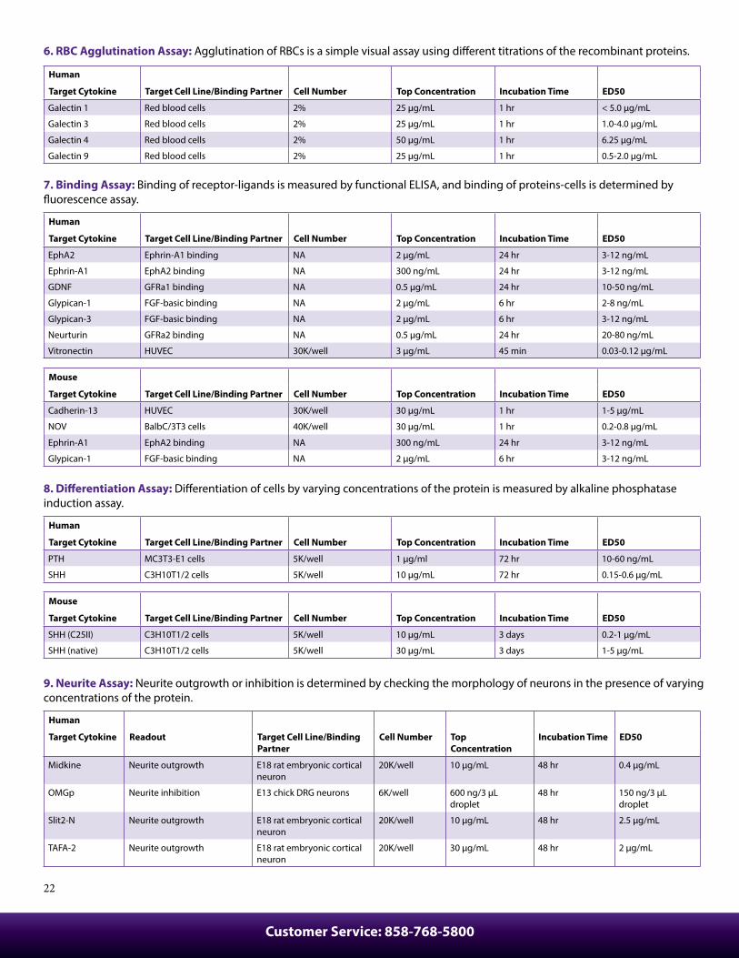
6. RBC Agglutination Assay: Agglutination of RBCs is a simple visual assay using di�erent titrations of the recombinant proteins.
Human
Target Cytokine Target Cell Line/Binding Partner Cell Number Top Concentration Incubation Time ED50
Galectin 1 Red blood cells 2% 25 µg/mL 1 hr < 5.0 µg/mL
Galectin 3 Red blood cells 2% 25 µg/mL 1 hr 1.0-4.0 µg/mL
Galectin 4 Red blood cells 2% 50 µg/mL 1 hr 6.25 µg/mL
Galectin 9 Red blood cells 2% 25 µg/mL 1 hr 0.5-2.0 µg/mL
7. Binding Assay: Binding of receptor-ligands is measured by functional ELISA, and binding of proteins-cells is determined by �uorescence assay.
Human
Target Cytokine Target Cell Line/Binding Partner Cell Number Top Concentration Incubation Time ED50
EphA2 Ephrin-A1 binding NA 2 µg/mL 24 hr 3-12 ng/mL
Ephrin-A1 EphA2 binding NA 300 ng/mL 24 hr 3-12 ng/mL
GDNF GFRa1 binding NA 0.5 µg/mL 24 hr 10-50 ng/mL
Glypican-1 FGF-basic binding NA 2 µg/mL 6 hr 2-8 ng/mL
Glypican-3 FGF-basic binding NA 2 µg/mL 6 hr 3-12 ng/mL
Neurturin GFRa2 binding NA 0.5 µg/mL 24 hr 20-80 ng/mL
Vitronectin HUVEC 30K/well 3 µg/mL 45 min 0.03-0.12 µg/mL
Mouse
Target Cytokine Target Cell Line/Binding Partner Cell Number Top Concentration Incubation Time ED50
Cadherin-13 HUVEC 30K/well 30 µg/mL 1 hr 1-5 µg/mL
NOV BalbC/3T3 cells 40K/well 30 µg/mL 1 hr 0.2-0.8 µg/mL
Ephrin-A1 EphA2 binding NA 300 ng/mL 24 hr 3-12 ng/mL
Glypican-1 FGF-basic binding NA 2 µg/mL 6 hr 3-12 ng/mL
8. Di�erentiation Assay: Di�erentiation of cells by varying concentrations of the protein is measured by alkaline phosphatase induction assay.
Human
Target Cytokine Target Cell Line/Binding Partner Cell Number Top Concentration Incubation Time ED50
PTH MC3T3-E1 cells 5K/well 1 µg/ml 72 hr 10-60 ng/mL
SHH C3H10T1/2 cells 5K/well 10 µg/mL 72 hr 0.15-0.6 µg/mL
Mouse
Target Cytokine Target Cell Line/Binding Partner Cell Number Top Concentration Incubation Time ED50
SHH (C25II) C3H10T1/2 cells 5K/well 10 µg/mL 3 days 0.2-1 µg/mL
SHH (native) C3H10T1/2 cells 5K/well 30 µg/mL 3 days 1-5 µg/mL
9. Neurite Assay: Neurite outgrowth or inhibition is determined by checking the morphology of neurons in the presence of varying concentrations of the protein.
Human
Target Cytokine Readout Target Cell Line/Binding Partner
Cell Number Top Concentration
Incubation Time ED50
Midkine Neurite outgrowth E18 rat embryonic cortical neuron
20K/well 10 µg/mL 48 hr 0.4 µg/mL
OMGp Neurite inhibition E13 chick DRG neurons 6K/well 600 ng/3 µL droplet
48 hr 150 ng/3 µL droplet
Slit2-N Neurite outgrowth E18 rat embryonic cortical neuron
20K/well 10 µg/mL 48 hr 2.5 µg/mL
TAFA-2 Neurite outgrowth E18 rat embryonic cortical neuron
20K/well 30 µg/mL 48 hr 2 µg/mL
biolegend.com
23
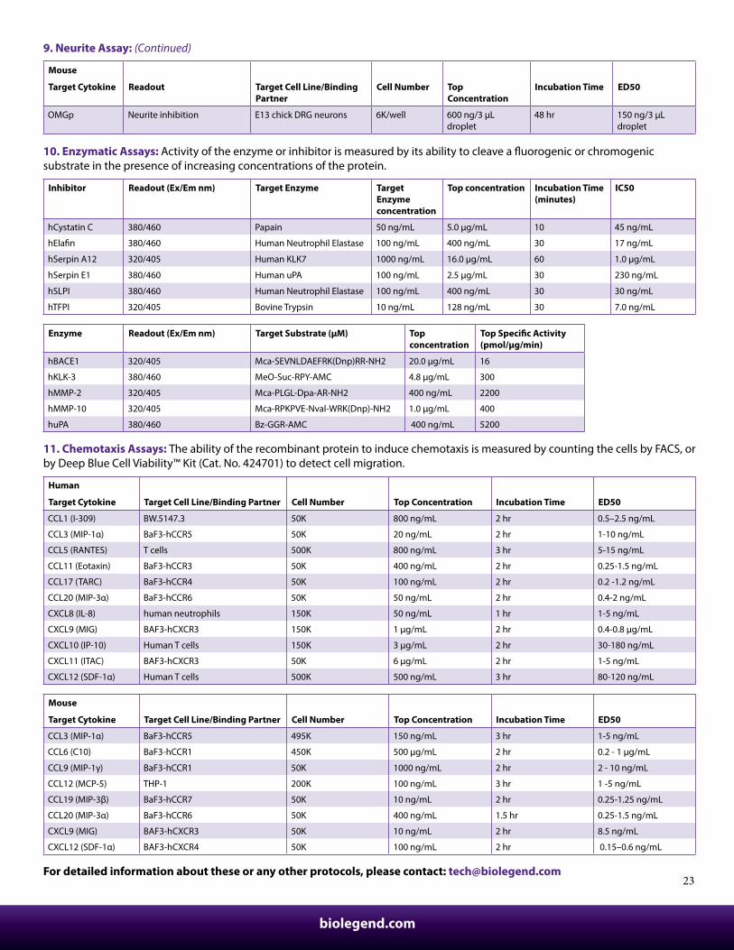
9. Neurite Assay: (Continued)
Mouse
Target Cytokine Readout Target Cell Line/Binding Partner
Cell Number Top Concentration
Incubation Time ED50
OMGp Neurite inhibition E13 chick DRG neurons 6K/well 600 ng/3 µL droplet
48 hr 150 ng/3 µL droplet
10. Enzymatic Assays: Activity of the enzyme or inhibitor is measured by its ability to cleave a �uorogenic or chromogenic substrate in the presence of increasing concentrations of the protein.
Inhibitor Readout (Ex/Em nm) Target Enzyme Target Enzyme concentration
Top concentration Incubation Time (minutes)
IC50
hCystatin C 380/460 Papain 50 ng/mL 5.0 µg/mL 10 45 ng/mL
hEla�n 380/460 Human Neutrophil Elastase 100 ng/mL 400 ng/mL 30 17 ng/mL
hSerpin A12 320/405 Human KLK7 1000 ng/mL 16.0 µg/mL 60 1.0 µg/mL
hSerpin E1 380/460 Human uPA 100 ng/mL 2.5 µg/mL 30 230 ng/mL
hSLPI 380/460 Human Neutrophil Elastase 100 ng/mL 400 ng/mL 30 30 ng/mL
hTFPI 320/405 Bovine Trypsin 10 ng/mL 128 ng/mL 30 7.0 ng/mL
Enzyme Readout (Ex/Em nm) Target Substrate (µM) Top concentration
Top Speci�c Activity (pmol/µg/min)
hBACE1 320/405 Mca-SEVNLDAEFRK(Dnp)RR-NH2 20.0 µg/mL 16
hKLK-3 380/460 MeO-Suc-RPY-AMC 4.8 µg/mL 300
hMMP-2 320/405 Mca-PLGL-Dpa-AR-NH2 400 ng/mL 2200
hMMP-10 320/405 Mca-RPKPVE-Nval-WRK(Dnp)-NH2 1.0 µg/mL 400
huPA 380/460 Bz-GGR-AMC 400 ng/mL 5200
11. Chemotaxis Assays: The ability of the recombinant protein to induce chemotaxis is measured by counting the cells by FACS, or by Deep Blue Cell Viability™ Kit (Cat. No. 424701) to detect cell migration.
Human
Target Cytokine Target Cell Line/Binding Partner Cell Number Top Concentration Incubation Time ED50
CCL1 (I-309) BW.5147.3 50K 800 ng/mL 2 hr 0.5–2.5 ng/mL
CCL3 (MIP-1α) BaF3-hCCR5 50K 20 ng/mL 2 hr 1-10 ng/mL
CCL5 (RANTES) T cells 500K 800 ng/mL 3 hr 5-15 ng/mL
CCL11 (Eotaxin) BaF3-hCCR3 50K 400 ng/mL 2 hr 0.25-1.5 ng/mL
CCL17 (TARC) BaF3-hCCR4 50K 100 ng/mL 2 hr 0.2 -1.2 ng/mL
CCL20 (MIP-3α) BaF3-hCCR6 50K 50 ng/mL 2 hr 0.4-2 ng/mL
CXCL8 (IL-8) human neutrophils 150K 50 ng/mL 1 hr 1-5 ng/mL
CXCL9 (MIG) BAF3-hCXCR3 150K 1 µg/mL 2 hr 0.4-0.8 µg/mL
CXCL10 (IP-10) Human T cells 150K 3 µg/mL 2 hr 30-180 ng/mL
CXCL11 (ITAC) BAF3-hCXCR3 50K 6 µg/mL 2 hr 1-5 ng/mL
CXCL12 (SDF-1α) Human T cells 500K 500 ng/mL 3 hr 80-120 ng/mL
Mouse
Target Cytokine Target Cell Line/Binding Partner Cell Number Top Concentration Incubation Time ED50
CCL3 (MIP-1α) BaF3-hCCR5 495K 150 ng/mL 3 hr 1-5 ng/mL
CCL6 (C10) BaF3-hCCR1 450K 500 µg/mL 2 hr 0.2 - 1 µg/mL
CCL9 (MIP-1γ) BaF3-hCCR1 50K 1000 ng/mL 2 hr 2 - 10 ng/mL
CCL12 (MCP-5) THP-1 200K 100 ng/mL 3 hr 1 -5 ng/mL
CCL19 (MIP-3β) BaF3-hCCR7 50K 10 ng/mL 2 hr 0.25-1.25 ng/mL
CCL20 (MIP-3α) BaF3-hCCR6 50K 400 ng/mL 1.5 hr 0.25-1.5 ng/mL
CXCL9 (MIG) BAF3-hCXCR3 50K 10 ng/mL 2 hr 8.5 ng/mL
CXCL12 (SDF-1α) BAF3-hCXCR4 50K 100 ng/mL 2 hr 0.15–0.6 ng/mL
For detailed information about these or any other protocols, please contact: [email protected]
Customer Service: 858-768-5800
24
Researcher Spotlight
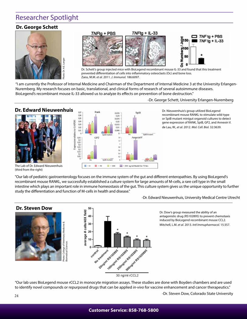
“I am currently the Professor of Internal Medicine and Chairman of the Department of Internal Medicine 3 at the University Erlangen-Nuremberg. My research focuses on basic, translational, and clinical forms of research of several autoimmune diseases. BioLegend’s recombinant mouse IL-33 allowed us to analyze its e�ects on prevention of bone destruction.”
-Dr. George Schett, University Erlangen-Nuremberg
Phot
o: D
r. H
.E. L
ange
rPh
oto:
Col
lege
of V
eter
inar
y M
edic
ine
&
Biom
edic
al S
cien
ces
Dr. George Schett
Dr. Edward Nieuwenhuis
Dr. Steven Dow
Dr. Schett’s group injected mice with BioLegend recombinant mouse IL-33 and found that this treatment prevented di�erentiation of cells into in�ammatory osteoclasts (Oc) and bone loss. Zaiss, M.M. et al. 2011. J. Immunol. 186:6097.
“Our lab uses BioLegend mouse rCCL2 in monocyte migration assays. These studies are done with Boyden chambers and are used to identify novel compounds or repurposed drugs that can be applied in-vivo for vaccine enhancement and cancer therapeutics.”
-Dr. Steven Dow, Colorado State University
Dr. Dow’s group measured the ability of an antagonistic drug (RS102895) to prevent chemotaxis induced by BioLegend recombinant mouse CCL2.Mitchell, L.M. et al. 2013. Intl Immupharmacol. 15:357.
The Lab of Dr. Edward Nieuwenhuis (third from the right)
Dr. Nieuwenhuis’s group utilized BioLegend recombinant mouse RANKL to stimulate wild-type or SpiB mutant minigut organoid cultures to detect gene expression of RANK, SpiB, GP2, and Annexin V.de Lau, W., et al. 2012. Mol. Cell. Biol. 32:3639.
"Our lab of pediatric gastroenterology focuses on the immune system of the gut and di�erent enteropathies. By using BioLegend's recombinant mouse RANKL, we successfully established a culture system for large amounts of M-cells, a rare cell type in the small intestine which plays an important role in immune homeostasis of the gut. This culture system gives us the unique opportunity to further study the di�erentiation and function of M-cells in health and disease."
-Dr. Edward Nieuwenhuis, University Medical Centre Utrecht
biolegend.com
25
Phot
o : F
ranç
ois
L. D
elag
rave
Dr. Xian C. Li
Dr. Tatiana Scorza
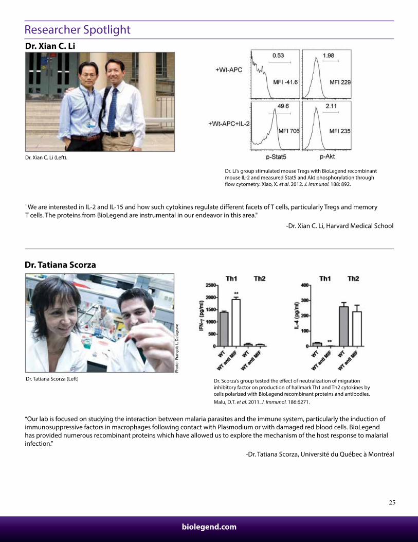
Dr. Tatiana Scorza (Left) Dr. Scorza’s group tested the e�ect of neutralization of migration inhibitory factor on production of hallmark Th1 and Th2 cytokines by cells polarized with BioLegend recombinant proteins and antibodies. Malu, D.T. et al. 2011. J. Immunol. 186:6271.
“Our lab is focused on studying the interaction between malaria parasites and the immune system, particularly the induction of immunosuppressive factors in macrophages following contact with Plasmodium or with damaged red blood cells. BioLegend has provided numerous recombinant proteins which have allowed us to explore the mechanism of the host response to malarial infection.”
-Dr. Tatiana Scorza, Université du Québec à Montréal
Researcher Spotlight
Dr. Xian C. Li (Left).
Dr. Li’s group stimulated mouse Tregs with BioLegend recombinant mouse IL-2 and measured Stat5 and Akt phosphorylation through �ow cytometry. Xiao, X. et al. 2012. J. Immunol. 188: 892.
"We are interested in IL-2 and IL-15 and how such cytokines regulate di�erent facets of T cells, particularly Tregs and memory T cells. The proteins from BioLegend are instrumental in our endeavor in this area."
-Dr. Xian C. Li, Harvard Medical School
Customer Service: 858-768-5800
26
Frequently Asked Questions
How does the activity of your recombinant proteins compare to competitors?We quality control each and every lot of recombinant protein. Not only do we check its bioactivity, but we also compare it against other commercially available recombinant proteins. We make sure each recombinant protein’s activity is at least as good as or better than the competitor's. In order to provide you with the best possible product, we ensure that our testing process is rigorous and thorough. If you’re curious and eager to make the switch to BioLegend recombinants, contact [email protected] today!
What is the speci�c activity or ED50 of my recombinant protein?The speci�c activity range of the protein is indicated on the product datasheets. Because the exact activity values on a per unit basis can largely �uctuate depending on a number of factors, including the nature of the assay, cell density, age of cells/passage number, culture media used, and end user technique, the speci�c activity is best de�ned as a range and we guarantee the speci�c activity of all our lots will be within the range indicated on the datasheet.
Does speci�c activity of a recombinant protein vary between lots?Speci�c activity will vary for each lot and for the type of experiment that is done to validate it, but all passed lots will have activity within the established ED50 range for the product and we guarantee that our products will have lot-to-lot consistency. Please do your own experiment speci�c validations to �nd out the optimal ED50 for your speci�c system.
How do you convert activity as an ED50 in ng/mL to a speci�c activity in Units/mg?Use the formula Speci�c activity (Units/mg) = 106/ED50 (ng/mL)
What is the di�erence between carrier-free and non carrier-free recombinant proteins?All our carrier-free and animal-free formats of recombinant proteins do not have any additional carrier proteins such as BSA in the formulation. Typically our ELISA standard recombinants have carrier proteins added to the formulation for added stability and to avoid the product from sticking to the wall of the vial. When the presence of carrier is not desirable (e.g., in-vivo applications), carrier-free proteins can be used directly. When carrier proteins do not a�ect the outcome in a study, the customer can decide what type of carrier protein they would like to use and whether it is necessary to add it to their stock.
How are BioLegend’s recombinant proteins shipped?All our recombinants are shipped on blue ice. These products have been validated to maintain activity after shipping using blue ice.
What is the di�erence between carrier-free and animal-free categories of recombinant proteins?Our animal-free products go through the entire production process without touching any animal-containing components. This includes using animal-free media and puri�cation equipment. Studies which are particularly sensitive to contamination by mammalian pathogens may require the use of animal-free products. Our carrier-free products do not contain any carrier protein, but they are produced using animal-containing components. Both versions are expected to have similar activity and function, though speci�c activity is lot-dependent.
What should I reconstitute the protein with? What do you recommend for its long-term storage?Most of our carrier-free recombinants are shipped in liquid form, so no need for reconstitution. Our animal-free recombinant proteins are shipped in lyophilized form and protein reconstitution information is indicated on the respective datasheets. If you need to make dilutions, refer to the formulation on the product data sheet. Stock solutions should be prepared at 50-100 μg/mL in bu�er containing carrier protein such as 1% BSA or HSA or 10% FBS (for chemokines, use either BSA or HSA). For long-term storage, aliquot into polypropylene vials and store in a manual defrost freezer. Avoid repeated freeze/thaw cycles.
Do you test the bioactivity of your recombinant proteins with in-vivo assays?We typically validate the activity of the proteins with in-vitro assays as described on the data sheet and not with in-vivo testing. However, all our carrier-free and animal-free formats can be used for in-vivo applications as is evident from many customers who have successfully used it for this purpose.
Are BioLegend’s recombinant proteins suitable for in-vitro and in-vivo bioassays?BioLegend’s recombinant protein solutions are 0.2 μm-�ltered prior to bottling by membrane �ltration method. In addition, our recombinants are endotoxin tested and are guaranteed to have levels less than 0.1 ng per μg protein. All our recombinants can be safely used for both in-vitro and in-vivo bioassays.
What is the di�erence between laboratory (observed) units and international units?There is no direct relationship between International Units and the units that are calculated using the inverse of the speci�c activity because we do not use the International Standard provided by WHO (National Institute for Biological Standards and control). The best way to compare the activity of two sources of recombinants is by doing the bioassay side by side using the same system.
biolegend.com
27
BAFF1. Han S, et al. 2016. Arthritis Res Ther. 0.975.GM-CSF1. Bretscher, P., et al. 2015. EMBO Mol Med. 7:593.2. Ahn, J., et al. 2012. PNAS. 109:19386. 3. Verhagen, J., et al. 2012. PNAS. 110:221. IFN-β1. Gries CM, et al. 2016. Infect. Immun. 84(12):3564-3574. 2. Mounce, B.C., et al. 2014. J. Virol. 88:2688.IFN-γ1. Inoue M, et al. 2016. Nat Neurosci. 19:1599-1609.2. Greer R, et al. 2016. Nat Commun. 7:13329.3. Ho J, et al. 2016. PLoS Biol. 14:e2000117.4. Bremser A, et al. 2015. PLOS ONE. 10: 0137393.IL-1α 1. Lee, P.Y., et al. 2011. J. Immunol. 186:1747.IL-1β1. O’ Sullivan, B.J., et al. 2006. J. Immunol. 176:7278.IL-21. Guo Z, et al. 2016. Nat Commun. 7:10307.2. Bao K, et al. 2016. J. Immunol. 197(11):4371-4381.3. Hrdinka M, et al. 2016. PLoS One. 11: 0162863.IL-41. Bao K, et al. 2016. J. Immunol. 197(11):4371-4381.2. Malu, D.T., et al. 2011. J. Immunol. 186:6271. 3. Ong, Y.C., et al. 2010. J. Biol. Chem. 285:28731.IL-61. Lin C, et al. 2016. J. Exp. Med. 213: 251 - 271.2. Bremser A, et al. 2015. PLOS ONE. 10: 0137393.3. Jeannie, M.A., et al. 2014. J. Nutr. 144:1306. 4. Alcaide, P., et al. 2012. J. Immunol. 188:1421. 5. Hilberath, J.N., et al. 2011. FASEB J. 25:1827.IL-101. Perkins D, et al. 2015. J. Immunol. 195: 2461-2471. 2. Poonam, K., et al. 2014. J. Immunol. 192:349. 3. Nguyen, H.H., et al. 2012. J. Immunol. 189:3112.4. Schaefer, J.S., et al. 2011. J. Immunol. 187:5834.IL-12p40 Homodimer1. Yabu, M., et al. 2010. Int. Immunol. 23:29. IL-17A1. Darling, A.R., et al. 2014. Clin Immunol. 150:153. 2. Cho, K.A., et al. 2012. Int. Immunol. 24:147. 3. Saha, A., et al. 2012. Hum. Mol. Genet. 21:2233.IL-331. Gao, X., et al. 2015. J. Immunol. 194:435. 2. Rosen, M.J., et al. 2013. J. Immunol. 190:1849. 3. Barlow, J.L., et al. 2012. J. Allergy. Clin. Immunol. 129:151.TNF-α1. Bradley W, et al. 2016. Infect. Immun. 84: 998-1015. 2. Alcaide, P., et al. 2012. J. Immunol. 188:1421. 3. Azcutia, V., et al. 2012. J. Immunol. 189:2553. 4. Theiss, A.L., et al. 2009. Mol. Biol. Cell. 20:4412. TRANCE (RANKL)1. de Lau, W., et al. 2012. Mol. Cell. Biol. 32:3639.VEGF-A1. Yamauchi F, et al. 2016. Cancer Res. 76: 5266 - 5276.2. Schwarz KA, et al. 2016. Nat Chem Biol. 13:202-209.3. Mangani D, et al. 2016. Neuro Oncology. 18(12):1610-1621.
CXCL101. Sek A, et al. 2015. PLOS ONE. 10: 0133266.2. Ramirez, L. A., et al. 2014 J. Leukoc. Biol 96:1055.GM-CSF1. Chalubinsk, M., et al. 2014. Food Chem Toxicol. 69:289.2. Byrd, D., et al. 2014. J. Virol. 88:6819. FGF-basic/145aa1. Wang, J., et al. 2013. Biochem Biophys Res Commun. 41:143.IFN-γ1. Meissner, T.B., et al. 2012. J. Immunol. 188:4951.2. Meissner, T.B., et al. 2010. PNAS. 107:13794.IL-21. Wingender, G., et al. 2011. J. Exp. Med. 208:1151. 2. Wang, N., et al. 2010. J. Immunol. 185:5683.IL-41. Chalubinsk, M., et al. 2014. Food Chem Toxicol. 69:289.IL-81. Popova T, et al. 2016. PLoS One. 11: 0163163.2. DuMont A, et al. 2015. Infect. Immun. 83: 3825 - 3837.M-CSF1. Nijaguna M, et al. 2015. J. Biol. Chem. 290: 23401-23415. 2. Lou, J., et al. 2014. J. Cell Sci. 127:5228.PDGF-BB1. Banerjee, P., et al. 2011. J. Biol. Chem. 286:33580.TGF-β1. Guo W, et al. 2016. Mol. Cell. Biol. 36: 2141.2. Benlhabib H, et al. 2015. J. Biol. Chem. 290: 22409-22422.3. Lin C, et al. 2016. J. Exp. Med. 213: 251 - 271.4. Alcaide, P., et al. 2012. J. Immunol. 188:1421.TWEAK (CD255)1. Novoyatleva, T., et al. 2010. Cardiovasc. Res. 85:681.VEGF-1651. Phay M, et al. 2015. PLOS ONE. 10: 0137344.2. Lee K, et al. 2015. J. Biol. Chem. 290: 28438 - 28445.3. Basavarajappa, H.D., et al. 2014. PLoS One. 9:95694.
References Using BioLegend Recombinant Proteins
Human
Rat IL-21. Xiao, X., et al. 2012. J. Immunol. 188:892.
Mouse
Rat
For complete list of publications, visit: biolegend.com/publications
Contact BioLegend
Customer Service:
US & Canada Toll-Free: 1.877.246.5343 (877-BIOLEGEND)
International: 1.858.768.5800
Fax: 1.877.455.9587
email: [email protected]
Technical Service:
US & Canada Toll-Free: 1.877.273.3103
International: 1.858.768.5801
email: [email protected]
Headquarters:
BioLegend
9727 Paci�c Heights Blvd.
San Diego, CA 92121
USA
International O�ces
Europe:BioLegend4B Highgate Business Centre 33 Greenwood PlaceLondon NW5 1LBUnited KingdomTel: +44 (0) 20 3475 3880 Fax: +44 (0) 20 3318 3271email Inquiries: [email protected] Technical Support: [email protected]
Japan:BioLegend8F, SB bldg., 1-4-6, Nezu, Bunkyo-ku, Tokyo113-0031, JapanTel: +81-3-3823-9071Fax: +81-3-3823-9072email: [email protected] biolegend.com/jp
For complete worldwide ordering details, visit: biolegend.com
BioLegend is ISO 9001:2008 and ISO 13485:2003 Certi�ed