Tracing Determinants of Dual Substrate Specificity in Glycoside Hydrolase Family 5* S

1
Recognition and specificity determinants of the human Cbx chromodomains Lilia Kaustov1, Hui Ouyang2, Maria Amaya2, Alexander Lemak1, Nataliya Nady1, Shili Duan1, Greg Wasney2, Zhihong Li2, Masoud Vedadi2, Matthieu Schapira2, Jinrong Min2, and Cheryl H. Arrowsmith1
Ontario Cancer Institute, Campbell Family Cancer Research Institute and Department of Medical Biophysics, University of Toronto1, Ontario M5G 1L7, Canada. Structural Genomics Consortium2, University of Toronto, 101 College Street, Toronto, Ontario M5G 1L5. Address correspondence to: Cheryl Arrowsmith, University of Toronto, 101 College Street, Toronto, Ontario M5G 1L7; Email: [email protected] Running head: Binding specificity of the chromodomains of Cbx protein family Abstract The eight mammalian Cbx proteins are chromodomain containing proteins involved in regulation of heterochromatin, gene expression and developmental programs. They are evolutionarily related to the Drosophila HP1 (dHP1) and Pc (dPc) proteins that are key components of chromatin associated complexes capable of recognizing repressive marks such as trimethylated Lys9 and Lys27, respectively on histone H3. However, the binding specificity and function of the human homologs, Cbx1-8, remain unclear. To this end, we employed structural, biophysical and mutagenic approaches to characterize the molecular determinants of sequence contextual methyllysine binding to human Cbx1-8 proteins. While all three human HP1 homologs (Cbx1,3,5) replicate the structural and binding features of their dHP counterparts, the five Pc homologs (Cbx2,4,6,7,8) bind with lower affinity to H3K9me3 or H3K27me3 peptides and are unable to distinguish between these two marks. Additionally, peptide permutation arrays revealed a greater sequence tolerance within the Pc family and suggest alternative nonhistone sequences as potential binding targets for this class of chromodomains. Our structures explain the divergence of peptide binding selectivity in the Pc subfamily and highlight previously unrecognized features of the chromodomain that influence binding and specificity.
The heterochromatin protein HP1 and Polycomb proteins are two distinct regulators of chromatin structure in Drosophila melanogaster involved in epigenetic repression of gene expression. The chromodomains of dHP1 and dPc
direct the localization of their respective complexes via specific recognition of distinct, but sequence related repressive marks on histone H3 trimethylated at Lys9 (H3K9me3) and Lys27 (H3K27me3), respectively (1,2). Both of these marks reside within the identical sequence motif, ARKS. These properties in the fly have served as an important paradigm for understanding chromatin dynamics and gene regulation through development (3,4) as well as fundamental structure-function and specificity properties of the chromodomains in general (1,2,5-7).
In mammals the dPc and dHP1 homologs have expanded to five (Cbx2,4,6,7,8) and three (Cbx1,3,5) proteins, respectively. Each human/mouse Cbx contains one chromodomain with ~55-65% sequence identity to dHP1 or dPc (8). A characteristic feature of the chromodomain is the positioning of the mono-, di- and tri-methylammonium moiety within a pocket lined with aromatic residues, often supplemented by one or more acidic side chains (9). Despite the similarity in overall structure, the peptide-binding grooves of the dHP1 and dPc chromodomains show distinct features. Each protein reveals clear discrimination for its cognate site via recognition of different residues upstream of each ARKS motif in H3 which are complementary to differences in the binding grooves of the chromodomains (1).
Although the exact cellular role of each Cbx protein has not been defined, many of the Cbx’s play important roles in human development and disease. Cbx1,3,5 are important for formation of, and gene repression in heterochromatin. Cbx2,4,6,7,8 are components of polycomb repressive complex 1 (PRC1), a key regulator of developmental genes. Mutation of Cbx2 was
http://www.jbc.org/cgi/doi/10.1074/jbc.M110.191411The latest version is at JBC Papers in Press. Published on November 3, 2010 as Manuscript M110.191411
Copyright 2010 by The American Society for Biochemistry and Molecular Biology, Inc.
by guest on June 12, 2018http://w
ww
.jbc.org/D
ownloaded from
2
shown to affect human sexual development (10). Finally, Cbx7 has been implicated in the development of leukemia (11) and Cbx7 and 8 contribute to repression of the INK4A locus (12-14).
The chromodomains of Cbx proteins are thought to, at least in part, localize the proteins and their respective complexes to appropriately marked sites of the epigenome via recognition of histone H3 trimethylated at either Lys9 or Lys27. A number of studies have been conducted to decipher the specificity within the Cbx family of proteins. Bernstein et al. have investigated five mouse dPc homologs (cbx2,4,6,7,8) and showed that despite a high degree of conservation, the chromodomains display significant differences in histone peptide binding preferences. Not all dPc homologs bind preferentially to H3K27me3 as might be expected based on dPc specificity (15). Cbx2 and Cbx7 recognized both H3K9me3 and H3K27me3, while Cbx4 preferred H3K9me3. Recently, Vincenz et al. studied the Pc family of Cbx proteins in ES cells and fibroblasts and observed that the ability of various Cbx proteins to bind chromatin was mediated by non-conserved regions of the protein (16). Using Bimolecular fluorescence complementation experiments they demonstrated that neither the chromodomains nor H3K27me3 were required for targeting of Cbx proteins to chromatin in vivo. More recently, Yap et al. showed that Cbx7 employs overlapping yet distinct regions within its chromodomain for binding H3K27me3 and RNA (17). Collectively, these studies show that related chromodomain-binding motifs have differences in binding characteristics and highlight the need to carefully analyze the differences between mammalian and fly chromodomain-containing proteins as well as differences within mammalian chromodomain subfamilies.
In the present study, we sought to understand in detail the biochemical and structural basis for histone mark recognition and discrimination amongst the human Cbx chromodomain protein family. We measured the binding affinities of all human Cbx chromodomains for H3K9me3 and H3K27me3 and determined the 3D structures (by NMR or X-ray crystallography) of several chromodomains in complex with the modified histone peptides to elucidate the structural determinants for the
specificity. The human HP1 homologs Cbx1, 3 and 5 preferentially recognize H3K9me3 in a manner similar to their dHP1 counterpart with dissociation constants in the low micromolar range. On the other hand, chromodomains from the human Pc homologs all have lower affinities for H3K9me3 and H3K27me3 peptides, do not distinguish well between these two marks and are less sensitive to mutations within the peptide sequence. Our structural data show that the primary origin of the difference between human HP1 and Pc chromodomains lies in their respective electrostatic surfaces. Cbx1,3,5 all have a large electronegative peptide binding surface that complements the basic histone peptides, whereas Cbx2,4,6,7,8 have a much more hydrophobic surface. Further peptide array and binding studies suggest that at least some of the human Pc chromodomains bind nonhistone sequences in a methylation dependent manner. These data suggest that Pc-class Cbx proteins may use their chromodomains to couple PRC1 complexes with novel binding partners or chromatin sites in the mammalian genome.
Experimental Procedures
Cloning, Expression and Purification - Chromodomains of Cbx1(20-73), Cbx2(9-66), Cbx3 (29-81), Cbx4(8-65), Cbx5(18-75), Cbx6(8-65), Cbx7(8-62) and Cbx8(8-61) were overexpressed as N-terminal His6-tagged proteins at 15ºC using E. coli BL21 (DE3) Codon plus RIL (Stratagene) as a host organism. For purification, cells were lysed by passing through Microfluidizer (Microfluidics Corporation) at 18,000 psi. The purification procedure comprises of two chromatographic steps, an affinity chromatography on a Ni-NTA Chelating column (Qiagen) and a gel filtration Superdex 75 column (26/60, GE Healthcare). The His6-tag was cleaved with TEV protease (except for Cbx6 which has a Thrombin cutting site) after the affinity column step, while dialyzing against 20 mM Tris pH 8.0, 0.5 M NaCl, and 1 mM TCEP. Protein for NMR samples was prepared as follows. The bacteria were grown in M9-defined medium supplemented with 15N-ammonium chloride (0.8 g/L) and 13C6-D-glucose (0.4 g/L) for 15N/13C-labeled samples at RT. These highly expressed
by guest on June 12, 2018http://w
ww
.jbc.org/D
ownloaded from

3
proteins were purified by Talon (BD) affinity chromatography under native conditions and eluted with buffer containing 500mM Imidazole. The proteins were treated with TEV protease and further purified by size exclusion chromatography using a HiLoad 26/60 Superdex-75 column (GE Helathcare). The proteins were monomeric in solution as determined by size exclusion chromatography. The final NMR samples were prepared in buffer containing 20 mM Sodium Phosphate (pH 7.4), 200 mM NaCl, 2mM DTT, 1mM TCEP, 1 mM Benzamidine, 0.5 mM PMSF for Cbx3 complexes and 10 mM Sodium Phosphate (pH 7.4), 300 mM NaCl, 0.5mM TCEP, 1 mM Benzamidine, 0.5 mM PMSF for Cbx7 complexes. Protein-peptide complexes were prepared for NMR by titrating aliquots of unlabeled peptide into the labeled chromodomains of Cbx3 and Cbx7 in molar ratios 1:1, 1:3 (for Cbx3/K9me3) and 1:5 (for Cbx7/K27me3 and Cbx7/K9me3) until no further changes in chemical shifts were detected in the 1H-15N HSQC spectrum. Fluorescence Polarization - H3K9Me3 (ARTKQTARK(me3)STGGKA), H3K9Me3T6A (ARTKQAARK(me3)STGGKA), H3K27Me3 (QLATKAARK(me3)SAPATG), and H3K27Me3A24T (QLATKTARK(me3)SAPATG) were synthesized, N-terminally labeled with fluorescein and purified by Tufts University Core Services (Boston, MA, USA). Binding assays were performed in 10 µl volume at a constant labeled peptide concentration of 40 nM, and Cbx protein concentrations at saturation ranging from 800 to 1300 µM in buffer containing 20 mM Tris pH 8.0, 250 mM NaCl, 1 mM DTT, 1mM Benzamidine, 1 mM PMSF, 0.01% Tween-20. Fluorescence polarization assays were performed in 384 well plates using Synergy 2 microplate reader (BioTek, Vermont, USA). The excitation wavelength of 485nm and the emission wavelength of 528 nm were used. The data was corrected for background of the free labeled peptides. To determine Kd values, the data were fit to a hyperbolic function using Sigma Plot software (Systat Software, Inc., CA, USA). The Kd values represent averages ± standard errors for at least three independent experiments. Histone Peptide Array Screen- Peptides were synthesized directly on a modified cellulose
membrane with a polyethylglycol linker using the peptide synthesizer MultiPep (Intavis). The degenerate screening peptide library consisted of 520 membrane-immobilized peptides corresponding to control peptides and 13 residue-long stretches of histone H3 sequences corresponding to residues 3-15 and 21-33. The lysine at positions K9 and K27 was always trimethylated, while flanking residues were systematically mutated to each of the twenty L-amino acids. The binding assay was performed as described previously (18). Briefly, the membrane was extensively blocked with skim milk, incubated over night with 5μM 6xHIS-tagged protein of interest, washed with PBS/T, and visualized via Western blot analysis with an anti-His antibody (Abcam). NMR Spectroscopy and Data Analysis- All complex structures have been determined using the ABACUS approach (19) from NMR data collected at high resolution from nonlinearly sampled spectra and processed using multidimensional decomposition (20,21). NMR spectra were recorded at 250C on Varian INOVA 500-MHz spectrometer equipped with triple resonance cold probe and Bruker Avance 600 and 800-MHz spectrometers equipped with cryoprobes. The sequences specific assignment of 1H, 13C and 15N resonances were assigned using the ABACUS protocol (19) from peaks lists derived from manually peak picked spectra. The restraints for backbone φ and ψ torsion angles were derived from chemical shifts of backbone atoms using TALOS (22). Hydrogen bond constraints were used within regular secondary structure elements. Automated NOE assignment (23) and structure calculations were performed using CYANA (version 2.1) according to its standard protocol. The resonances of free and bound histone peptides were assigned from two-dimensional total correlation spectroscopy (TOCSY) and NOESY experiments. Intermolecular NOEs were assigned from a 3D 13C/15N-filtered, edited NOESY spectrum (39) of 13C/15N-labeled protein bound to unlabeled peptide. A subset of unambiguous NOEs between peptide and protein were initially assigned manually, and were sufficient to dock each peptide to the respective protein in CYANA calculations. Next, additional intermolecular
by guest on June 12, 2018http://w
ww
.jbc.org/D
ownloaded from

4
NOEs were assigned based on knowledge of the residues expected to be in close proximity between protein and peptide (Figure S7). The final 20-lowest-energy structures were refined within CNS (24) by a short constrained molecular dynamics simulation in explicit solvent (25) (Table S1). After refinement 99.4% and 0.6% residues in Cbx3/K9me3, 95.7% and 4.3% residues in Cbx7/K9me3 and 94.4% and 5.6% residues in Cbx7/K27me3 were in favorable and allowed regions of the Ramachandran plot. All NOE distance and angular restraints are deposited with the atomic coordinates in the PDB. Figures were prepared using MOLMOL (26), iSee (27) and PyMOL (DeLano Scientific). Crystallization, Data Collection and Structure Determination- Crystals of the Cbx chromodomains were grown at 18°C using the sitting-drop method by mixing equal volumes of the mother liquor with protein solutions. Crystallization conditions for individual structures are shown in Table S2. All diffraction data were collected at 100K and reduced with the HKL2000 suite of programs. The crystal structures of these Cbx chromodomains and their complexes with histone peptides were solved by molecular replacement using MOLREP. COOT (28), REFMAC (29), and MOLPROBITY (30) were used for interactive model building, refinement and validation, respectively. Crystal diffraction data and refinement statistics are displayed in Table S3. Accession Codes- Protein Data Bank: Coordinates for the Cbx2/H3K27me3, Cbx5/H3K9me3, Cbx6/H3K9me3, Cbx6/H3K27me3, Cbx3/H3K9me3, Cbx7/H3K9me3, Cbx7/H3K27me3 and Cbx8/H3K9me3 have been deposited with accession codes 3H91, 3FDT, 3GV6, 3I90, 2L11, 2L12, 2L1B and 3I91, respectively.
RESULTS
Binding preferences of human Cbx proteins diverge from those of Drosophila
In order to elucidate the binding specificity for the chromodomains of human Cbx proteins we measured the affinity of the recombinant chromodomains for both
trimethylated K9 and K27 marks on histone H3 peptides using fluorescence polarization. Modified and mutant peptide sequences were assayed with each chromodomain (Table 1). Similar to dHP1, the human HP1 homologs (Cbx1,3,5) showed significant preference for H3K9me3 peptides (31), and were sensitive to mutation of T6 to an alanine confirming that these chromodomains can distinguish H3K9me3 and H3K27me3 sequences via the third residue preceding the methyllysine. In contrast, most human Pc homologs (Cbx2,4,6,7,8) had a wide range of affinities towards both marks without a distinct selectivity for one, similar to their mouse homologs (15,31). Cbx4 and Cbx7 bound to both methylated marks but behaved more like a HP1 protein with modest binding to H3K9me3 and 2-3 fold weaker binding to H3K27me3. In fact, Cbx2 was the only Pc-like chromodomain with a clear preference for the K27me3 mark, however the affinity was rather low. Furthermore, Cbx2,4 and 7 were insensitive to mutation of the third residue upstream of methyllysine (T6, or A24). Finally, neither Cbx6 nor Cbx8 bound to K9me3 or K27me3 very weakly with Kd values over 500μM.
Because the human Cbx binding specificity does not correspond closely to the prototypical Drosophilia specificities of HP1 and Pc proteins (2,5,18,31) we sought to identify the spectrum of peptide sequences to which human Cbx chromodomains would bind using a SPOT-blot assay (18). Peptide arrays were synthesized for two sets of peptides from histone H3 covering residues 3-15 and 21-33 with trimethylated lysine (Kme3) at position K9 or K27, respectively. These two sequences were systematically mutated to each of the twenty amino acids (Fig. 1, vertical series) at each position of the peptide (Fig. 1, horizontal series). Binding was then assessed for representative HP1-like (Cbx3) and Pc-like (Cbx7 and Cbx8) chromodomains.
As expected, Cbx3 chromodomain bound to H3K9me3- but not to H3K27me3-related peptides while Cbx7 showed binding to both with an absolute dependence on trimethylation of K9 or K27 for both chromodomains (Fig. 1A,B). Cbx8 showed binding to H3K27me3-related peptides only (Fig. 1C). Variations of the ARKS motif shared by both sequences were not tolerated for either protein except for mutations of the arginine residue to hydrophobic residues. This suggests that
by guest on June 12, 2018http://w
ww
.jbc.org/D
ownloaded from

5
the hydrophobic side chain of arginine within ARKS contributes to the binding energy as opposed to its guanidino group which points away from the interaction surface. Consistent with its similarity to dHP1, and the data in Table 1, Cbx3 binding was disrupted by most mutations of Q5 and T6 residues upstream of the ARKS motif of H3 (5). On the other hand, many but not all mutations of Q5 and T6 were tolerated by Cbx7, again demonstrating less sensitivity to residues in this region of the peptide. These screens confirmed that the binding event is driven primarily by the ARKS motif with little or no contribution from the C-terminal residues for either protein.
Structural analysis of human Cbx chromodomain family: conserved binding features In order to better understand how human Cbx chromodomains recognize their substrates we determined the structures of Cbx2,3,5,6,7 and 8 with a combination of solution NMR and X-ray crystallography including (where possible) complexes with H3K9me3 and H3K27me3 peptides. Analysis of the structures confirmed that they have a common overall fold, and bind trimethyllysine containing peptides in a manner similar to their Drosophila homologs. In all Cbx structures, the trimethylammonium moiety is positioned within an aromatic cage consisting of three aromatic residues supplemented by one or two acidic residues and the remaining peptide binds with a surface-groove recognition mode (9). Two key elements of the peptide binding groove that are conserved among all Cbx proteins are (1) the extended β-strand conformation of the peptide forming a continuous β-sheet with the protein, and (2) a conserved binding pocket for the Ala residue at the -2 position in the peptide substrate relative to the trimethyllysine. For all Cbx histone peptide complexes, this Ala (histone residues A7 or A25) is buried in a small hydrophobic pocket, the size of which is sufficient to accommodate an alanine sidechain, but nothing larger. This explains the absolute requirement for Ala at the -2 position of the peptide as seen for the Drosophila proteins. Structural basis for Cbx1, 3, and 5 selectivity
As previously reported for dHP1(5,7) and shown above for the human homologs, T6 of the H3 peptide is an important determinant of the binding and selectivity of the HP1 class of
chromodomains. T6V is the only mutation at peptide residue K-3 that is capable of interacting with Cbx3 (Fig. 1A). Comparison of the complex structures of Cbx3 and Cbx5 suggest an important role of two conserved negatively charged residues or “polar fingers”. The fingers are formed by a pair of residues Glu29/19 (top) and Asp68/58 (bottom) of Cbx3 and Cbx5 chromodomains, respectively, whose β and γ methylene groups are sandwiched by the sidechains of T6 and R8 of the peptide (Fig. 2C and Fig. 3). Importantly, only valine, which is similar in size to threonine but has a methyl instead of hydroxyl group, would allow the acidic “polar fingers” to adopt a similar conformation. This is noteworthy because the position of the “top” finger (i.e. Glu29/19) is important for the conformation of the subsequent residue (Phe30/20) which contributes to the formation of the methylysine binding aromatic cage. Mutation of the polar clasp residues of Cbx3 to the corresponding residues found in the human Pc homologs (Glu29 to Val, and Asp68 to Leu) dramatically reduced binding of Cbx3 to both H3K9me3 and H3K27me3 peptides (Table S4). Thus, a key feature of the HP1 class-peptide interactions is not a specific interaction with T6, per se, but rather the favorable polar clasp that only Thr (or Val) in this position allows.
In addition, Q5 also seems to contribute to the binding specificity albeit to a lesser extent than T6 (Fig. 1A). This residue makes main-chain interactions with Val32 of Cbx3 and interacts with the N terminus of the chromodomain α-helix. At this position only medium sized polar residues such as Asn, Cys, Gln and His are tolerated.
Overall, our binding data indicate that Cbx1, 3 and 5 bind with greater affinity to H3K9me3 than the Pc orthologs bind to either peptides. We attribute the stronger binding affinity of human HP1 homologs to the stark difference in electrostatic surface charge distribution as shown in Fig. 2A,B and Fig. 4. The HP1 class chromodomains are highly negatively charged and this charge complimentarity with respect to the basic histone peptides is likely an important driving force for the interaction.
Structural basis for loss of selectivity for human Pc homologs
In contrast to the HP1-like proteins, the four human Pc-like proteins (Cbx2,6,7,8) are less
by guest on June 12, 2018http://w
ww
.jbc.org/D
ownloaded from

6
acidic. This very likely contributes to their lower binding affinities for basic histone peptides. Another important difference is a conserved “hydrophobic clasp” formed by Val10 and Leu49 (Cbx7 numbering) in place of the “polar clasp” seen in the HP1 class of chromodomains (Fig. 2B). A24 of the H3K27me3 peptide interacts with this “hydrophobic clasp” in all four Pc-class complex structures. However, our peptide binding and array data indicate that many other amino acids including threonine, and especially hydrophobic residues at this -3 position of the peptide are favorable for binding to Cbx7 (Fig. 1B). Thus, the “hydrophobic clasp” is an important contributor for the lack of selectivity of human dPc homologs towards the methyl lysine marks, just as the “polar fingers” are pivotal for selectivity within the HP1 homologs. Mutation of the “hydrophobic clasp” residues of Cbx7 (Val10 and Leu49) to the corresponding “polar fingers” residues in Cbx3 (Glu/Asp) abrogated the binding to both histone peptide sequences (Table S4). NMR spectra indicated that both Cbx3 and Cbx7 clasp mutants were folded, but may have local or long-range perturbations of their structure (Fig. S1 and Fig. S2). Thus, the “clasp” residues are not a simple element that can be swapped between HP1/Pc classes to switch binding selectivity, but are context dependent within each chromodomain.
Cbx6 and Cbx8 have functional aromatic cages and hydrophobic fingers very similar to those of Cbx2,4 and 7, but the former bind to H3K9me3 and H3K27me3 peptides with much lower affinity. We attribute this to an overall greater electropositive charge of Cbx6 and Cbx8 in the region that accommodates the RKme3S portion of the peptide. In particular, Cbx6 and Cbx8 are the only two chromodomains with an Arg residue just prior to residue Val10 of the “hydrophobic clasp” (Fig. 2C, Arg residues highlighted in yellow). The N-terminal residues of the chromodomain including this Arg residue is disordered in the absence of peptide, but upon complex formation it forms the upper strand of peptide-interacting β-sheet (Fig. 5). As such, the positive charge from Arg9 – present only in Cbx6 and Cbx8 may contribute to less efficient association with the basic histone peptide. Consistent with this notion, we observe a variable position or this Arg residue in the three different low affinity Cbx6 and Cbx8 complexes (Fig. 4).
Another structural element of dPc that was noted to be important for peptide binding selectivity, is an extended recognition groove that accommodates five extra histone residues (relative to dHP1) via hydrogen bonds to mainchain atoms of the peptide (1). We examined the role of residues in this groove for human Cbx2,3 and 7 by mutagenesis. We swapped pairs of key H-bond mediating residues along the “bottom” of this groove between the two classes of Cbx proteins (Fig. 2C, Table S4 and Fig. S3). For Cbx3 and Cbx7, these “groove” mutants had reduced binding affinities for both histone peptides. While we did not observe a switch in peptide binding specificity, this indicates the importance of these residues for binding affinity in the context of their respective proteins. For Cbx2, the only chromodomain that has a preference for H3K27me3, the “groove-2” mutant (Asp51Cys/Arg53Asp) retained the same binding affinity for H3K27me3, but had improved binding to H3K9me3. Taken together these data indicate that the extended groove likely contributes to peptide binding affinity but may not contribute to selectivity.
Human Cbx’s bind to alternative nonhistone methylated sequences
Based on our peptide array data, a consensus binding motif can be derived for chromodomains of Cbx7 (A-R/I/L/F/Y/V-Kme3-S/T) and for Cbx3 (Q/N-T/V-A-R/I/F/W/V-Kme3-S/T), where ‘/’ separates alternative tolerated amino acids at each peptide residue. We hypothesized that there may be alternative, or higher affinity binding partners for Cbx proteins in the human genome that include aforementioned binding motif. We used the consensus sequence for Cbx7 to search NCBI RefSeq Database for nuclear proteins that matched the array-derived consensus sequences. Numerous candidate Cbx-interacting proteins that contain at least one consensus sequence and are known or are predicted to be nuclear were identified. Then, a peptide array was designed to include these human peptide sequences from non-histone proteins that could bind to Cbx7 in a trimethylation dependent manner (Fig. S4). Many, but not all of the sequences bound to Cbx7 chromodomain. However, if Cbx7 were to interact with one or more of these proteins in vivo it would require for
by guest on June 12, 2018http://w
ww
.jbc.org/D
ownloaded from
7
Cbx7 binding partner to be trimethylated in the cell. Therefore, we searched these candidate interacting proteins for those which could potentially be methylated by virtue of their interaction with, or being a component of, a known methyltransferase protein or complex.
Interestingly, SET Domain Bifurcated-1 (SETDB1) is a histone H3K9 methyltransferase that contains three Cbx7 binding consensus sites in close proximity within the sequence that “bifurcates” the conserved SET domain. In order to elucidate the binding of these consensus sites for Cbx7 we measured the affinity of Cbx7 and Cbx8 chromodomains for di-,tri- and non-methylated peptides corresponding to the three consensus sites within SETDB1 (Table 2). Cbx7/8 chromodomains bound to trimethylated SETDB1 peptides with equal or greater affinity than histone peptides and did not bind to the non-methylated peptides. Mass spectrometry of full length human SETDB1 expressed in insect cells revealed methylation of K1170 (ALKS motif) in di- and tri-methylation states (data not shown) suggesting that SETDB1 may be capable of automethylation. Taken together, these data show that Cbx7/8 and perhaps, other Cbx proteins are likely to have an alternative non-histone binding activity.
Discussion
The present work provides a structural
framework for understanding the recognition and specificity of human Cbx chromodomains. We found a key structural feature of the Cbx chromodomains that mediates interaction with the ARKme3S peptide sequence – the “polar fingers” of Cbx1,3,5 and the “hydrophobic fingers” of Cbx2,4,6,7,8. These fingers encircle the peptide which is in an extended β-strand conformation with the A7 or A2 resting in a selective pocket at the bottom of the peptide binding groove, and the R8 or R26 pointing outward and nestling against the two fingers. The peptide trimethyllysine points into a conserved aromatic cage that is formed, in part by a conserved aromatic residue (Tyr or Phe) immediately adjacent to the “top” finger (as viewed in Fig. 3 and Fig. 5). Thus, the conformation of the finger residues are critical for both peptide sequence recognition and formation of the methyllysine binding aromatic cage. The “left” side of the fingers are adjacent to key
peptide residues, T6 or A24, that distinguish the two important histone marks, H3K9me3 and H3K27me3, respectively.
Our structural, binding and mutagenesis data revealed two key factors that contribute to the selectivity and lack of thereof towards histone peptides by human Cbx family. First, there is a stark difference in surface electrostatics, with the acidic HP1 class having greater affinity for basic histone peptide, and the Pc class having a more hydrophobic surface resulting in lower affinities for even their most optimal histone binding peptides (Fig. 4). Second, the “polar clasp” of the HP1 class of chromodomains is sensitive to the residue immediately upstream from the ARKS motif, tolerating only T or V with the appropriate size and shape, whereas the Pc “hydrophobic clasp” can tolerate most hydrophobic residues. The role of electrostatics in Cbx interactions also likely contributes to the overall weaker absolute affinities measured here for human Cbx proteins compared to those reported for mouse Cbx proteins by Bernstein et al. in a buffer with much lower salt concentration (15). Nevertheless, the overall trend in relative binding affinities among Cbx proteins are the same in both cases.
Of the eight human Cbx proteins, only Cbx2 appears to have a strong preference for the H3K27me3 mark. This suggests that Cbx2 may be the functional ortholog of dPc in humans, although the binding affinity of Cbx2 for H3K27me3 peptide is rather low. We note however, that Cbx2 is also the only Cbx protein to have a DNA-binding domain – an AT-hook domain (8,32). This may increase the affinity of Cbx2 for certain H3K27me3 nucleosomes in vivo due to divalent interaction from the chromo- and AT-hook domains. Additional components of the PRC1 complex also likely contribute to added specificity and affinity in the context of nucleosomes, chromatin and specific gene loci. For example, Yap et al. recently showed that the chromodomain of Cbx7 interacts with both H3K27me3 as well as the noncoding RNA ANRIL in suppression of transcription at the INK4a locus (17).
Accumulating evidence suggests that many of the “readers” and “writers” of histone marks may also be active for non-histone sequences (33-35). For example, the prevalence of the QTARKS and similar sequences in the human proteome (such as those from our peptide
by guest on June 12, 2018http://w
ww
.jbc.org/D
ownloaded from

8
array consensus) suggests that both H3K9 methyltransferases and “reader” domains such as the HP1 class of chromodomains are likely to recognize non-histone proteins as well (36,37). Our data suggest that the Pc class of Cbx proteins which are components of PRC1 and PRC1-like complexes, also interact with nonhistone proteins (Fig. S4). Binding of Cbx2,4,6,7, or 8 to epigenetic or transcription factors in a methyllysine dependent manner would be an attractive mechanism for targeting various Cbx-containing PRC1 complexes to selective sites of the genome occupied by specific factors. This may help explain the expansion of the Pc class of Cbx proteins from insects to mammals, contributing to the greater complexity of mammalian biology.
We identified SETDB1, a H3K9 methyltransferase, as a potential trimethyllysine dependent interacting partner for Cbx7 and Cbx8. Trimethylated SETDB1 peptides have the ability to directly interact with Cbx7/8 with affinities
equal or greater than histone peptides H3K27me3 or H3K9me3. This is significant in light of the recent study of Bilodeau et al. showing that shRNA knockdown SETDB1, Cbx7 or Cbx8 were among the most effective regulators of embryonic stem cell fate in a large scale screen of chromatin regulatory factors (38). This study reported that SETDB1 and H3K9me3 nucleosomes are both found at a subset of genes encoding key developmental regulators in ES cells. These SETDB1-occupied genomic sites are among those previously shown to be repressed by PcG proteins and are “bivalent”, having both H3K4me3 and H3K27me3 marks. Given the relative binding affinities observed in our experiments it is tempting to summarize that Cbx7 and/or Cbx8 may be involved in recruiting PRC1 complexes to SETDB1 occupied sites by virtue of their chromodomain interaction with SETDB1 instead of H3K27me3.
by guest on June 12, 2018http://w
ww
.jbc.org/D
ownloaded from
9
References 1. Fischle, W., Wang, Y., Jacobs, S. A., Kim, Y., Allis, C. D., and Khorasanizadeh, S. (2003) Genes
Dev 17, 1870-1881 2. Jacobs, S. A., Taverna, S. D., Zhang, Y., Briggs, S. D., Li, J., Eissenberg, J. C., Allis, C. D., and
Khorasanizadeh, S. (2001) Embo J 20, 5232-5241 3. Martin, C., and Zhang, Y. (2005) Nat Rev Mol Cell Biol 6, 838-849 4. Hublitz, P., Albert, M., and Peters, A. H. (2009) Int J Dev Biol 53, 335-354 5. Jacobs, S. A., and Khorasanizadeh, S. (2002) Science 295, 2080-2083 6. Min, J., Zhang, Y., and Xu, R. M. (2003) Genes Dev 17, 1823-1828 7. Nielsen, P. R., Nietlispach, D., Mott, H. R., Callaghan, J., Bannister, A., Kouzarides, T., Murzin,
A. G., Murzina, N. V., and Laue, E. D. (2002) Nature 416, 103-107 8. Whitcomb, S. J., Basu, A., Allis, C. D., and Bernstein, E. (2007) Trends Genet 23, 494-502 9. Taverna, S. D., Li, H., Ruthenburg, A. J., Allis, C. D., and Patel, D. J. (2007) Nat Struct Mol Biol
14, 1025-1040 10. Biason-Lauber, A., Konrad, D., Meyer, M., DeBeaufort, C., and Schoenle, E. J. (2009) Am J Hum
Genet 84, 658-663 11. Scott, C. L., Gil, J., Hernando, E., Teruya-Feldstein, J., Narita, M., Martinez, D., Visakorpi, T.,
Mu, D., Cordon-Cardo, C., Peters, G., Beach, D., and Lowe, S. W. (2007) Proc Natl Acad Sci U S A 104, 5389-5394
12. Maertens, G. N., El Messaoudi-Aubert, S., Racek, T., Stock, J. K., Nicholls, J., Rodriguez-Niedenfuhr, M., Gil, J., and Peters, G. (2009) PLoS One 4, e6380
13. Dietrich, N., Bracken, A. P., Trinh, E., Schjerling, C. K., Koseki, H., Rappsilber, J., Helin, K., and Hansen, K. H. (2007) Embo J 26, 1637-1648
14. Bernard, D., Martinez-Leal, J. F., Rizzo, S., Martinez, D., Hudson, D., Visakorpi, T., Peters, G., Carnero, A., Beach, D., and Gil, J. (2005) Oncogene 24, 5543-5551
15. Bernstein, E., Duncan, E. M., Masui, O., Gil, J., Heard, E., and Allis, C. D. (2006) Mol Cell Biol 26, 2560-2569
16. Vincenz, C., and Kerppola, T. K. (2008) Proc Natl Acad Sci U S A 105, 16572-16577 17. Yap, K. L., Li, S., Munoz-Cabello, A. M., Raguz, S., Zeng, L., Mujtaba, S., Gil, J., Walsh, M. J.,
and Zhou, M. M. Mol Cell 38, 662-674 18. Nady, N., Min, J., Kareta, M. S., Chedin, F., and Arrowsmith, C. H. (2008) Trends Biochem Sci
33, 305-313 19. Lemak, A., Steren, C. A., Arrowsmith, C. H., and Llinas, M. (2008) J Biomol NMR 41, 29-41 20. Gutmanas, A., Jarvoll, P., Orekhov, V. Y., and Billeter, M. (2002) J Biomol NMR 24, 191-201 21. Orekhov, V. Y., Ibraghimov, I., and Billeter, M. (2003) J Biomol NMR 27, 165-173 22. Cornilescu, G., Delaglio, F., and Bax, A. (1999) J Biomol NMR 13, 289-302 23. Guntert, P., Mumenthaler, C., and Wuthrich, K. (1997) J Mol Biol 273, 283-298 24. Brunger, A. T., Adams, P. D., Clore, G. M., DeLano, W. L., Gros, P., Grosse-Kunstleve, R. W.,
Jiang, J. S., Kuszewski, J., Nilges, M., Pannu, N. S., Read, R. J., Rice, L. M., Simonson, T., and Warren, G. L. (1998) Acta Crystallogr D Biol Crystallogr 54, 905-921
25. Linge, J. P., Williams, M. A., Spronk, C. A., Bonvin, A. M., and Nilges, M. (2003) Proteins 50, 496-506
26. Koradi, R., Billeter, M., and Wuthrich, K. (1996) J Mol Graph 14, 51-55, 29-32 27. Raush, E., Totrov, M., Marsden, B. D., and Abagyan, R. (2009) PLoS One 4, e7394 28. Emsley, P., and Cowtan, K. (2004) Acta Crystallogr D Biol Crystallogr 60, 2126-2132 29. Vagin, A. A., Steiner, R. A., Lebedev, A. A., Potterton, L., McNicholas, S., Long, F., and
Murshudov, G. N. (2004) Acta Crystallogr D Biol Crystallogr 60, 2184-2195 30. Davis, I. W., Murray, L. W., Richardson, J. S., and Richardson, D. C. (2004) Nucleic Acids Res
32, W615-619
by guest on June 12, 2018http://w
ww
.jbc.org/D
ownloaded from

10
31. Vermeulen, M., Eberl, H. C., Matarese, F., Marks, H., Denissov, S., Butter, F., Lee, K. K., Olsen, J. V., Hyman, A. A., Stunnenberg, H. G., and Mann, M. Cell 142, 967-980
32. Senthilkumar, R., and Mishra, R. K. (2009) BMC Genomics 10, 549 33. Couture, J. F., Collazo, E., Hauk, G., and Trievel, R. C. (2006) Nat Struct Mol Biol 13, 140-146 34. Feng, L., Lin, T., Uranishi, H., Gu, W., and Xu, Y. (2005) Mol Cell Biol 25, 5389-5395 35. Chuikov, S., Kurash, J. K., Wilson, J. R., Xiao, B., Justin, N., Ivanov, G. S., McKinney, K.,
Tempst, P., Prives, C., Gamblin, S. J., Barlev, N. A., and Reinberg, D. (2004) Nature 432, 353-360
36. Sampath, S. C., Marazzi, I., Yap, K. L., Krutchinsky, A. N., Mecklenbrauker, I., Viale, A., Rudensky, E., Zhou, M. M., Chait, B. T., and Tarakhovsky, A. (2007) Mol Cell 27, 596-608
37. Chin, H. G., Esteve, P. O., Pradhan, M., Benner, J., Patnaik, D., Carey, M. F., and Pradhan, S. (2007) Nucleic Acids Res 35, 7313-7323
38. Bilodeau, S., Kagey, M. H., Frampton, G. M., Rahl, P. B., and Young, R. A. (2009) Genes Dev 23, 2484-2489
39. Zwahlen, C., Pascale, L., Vincent, S.J.F., Greenblatt, J., Konrat, R. & Kay, L.E. (1997). Methods for Measurement of Intermolecular NOEs by Multinuclear NMR Spectroscopy: Application to a Bacteriophage λ N-peptide/boxB RNA Complex. J. Am. Che. Soc. 119: 6711-21.
by guest on June 12, 2018http://w
ww
.jbc.org/D
ownloaded from

11
FOOTNOTES
This research was funded in part by the Ontario Ministry of Health and Long Term Care. The views expressed do not necessarily reflect those of the OMOHLTC. We are grateful to Antoine Peters for fruitful discussions. We thank Chen Chen, Brett Larsen and Andrew James (Dr. Pawson lab, UofT) for their help with MS data acquisition and analysis. N.N. is supported by a Doctoral Research Award, CIHR. The Structural Genomics Consortium is a registered charity (number 1097737) that receives funds from the Canadian Institutes of Health Research, the Canadian Foundation for Innovation, Genome Canada through the Ontario Genomics Institute, GlaxoSmithKline, Karolinska Institutet, the Knut and Alice Wallenberg Foundation, the Ontario Innovation Trust, the Ontario Ministry for Research and Innovation, Merck & Co., Inc., the Novartis Research Foundation, the Swedish Agency for Innovation Systems, the Swedish Foundation for Strategic Research and the Wellcome Trust. This work was also funded by the Canadian Cancer Society (CCS). CHA holds a Canada Research Chair in Structural Proteomics. The abbreviations used are: dHP1, drosophila Heterochromatin Protein 1, dPc, drosophila Polycomb protein, PRC1, polycomb repressive complex 1, Cbx, chromobox proteins.
FIGURE LEGENDS Figure 1. SPOT-blot binding assay of chromodomain from human (A) Cbx3, (B) Cbx7 and (C) Cbx8. Two sets of peptides from histone H3 covering regions aa3-15 and aa21-33 with trimethylated lysine at position K9 and K27 (K*), respectively, were systematically mutated to each of the twenty amino acids (vertical) at every position (top) as indicated on the figure. While Cbx7 showed binding to both peptides (see also Table 1) with an absolute dependence on ARKS motif, Cbx3 bound to H3K9me3 peptide only and Cbx8 bound to H3K27me3. Low panel numbering corresponds H3 residues downstream (negative) or upstream (positive) to the methyl Lysine. Figure 2. Recognition and specificity determinants in human Cbx protein family. (A)”Polar fingers”of human dHP1 homologs, (B)”hydrophobic clasp” of human dPc homologs, and (C) sequence alignment of Drosophila and human chromodomains. Surface representations of the binding grooves: (A) Cbx3/K9 has a tighter and (B) Cbx7/K27 adopt wider groove as a result of a conformational change of the turn bearing key residues. The groove contributes to peptide binding affinity, but not to selectivity. Surfaces with positive electrostatic potential are blue, and negative ones are red. The arginine residues specific to Cbx6 and Cbx8 are highlighted in yellow. Figure 3. Selectivity of human HP1 homologs is driven by “polar fingers”. Surface representations of the ”polar fingers” for (A) Cbx3 and (B) Cbx5 chromodomains in complex with H3K9me3 peptide. T6 and R8 residues of the peptide are shown in the binding cleft. The β and γ methylene groups of conserved “polar fingers” are sandwiched by sidechains of T6 and R8. The right panel is rotated by 90o with respect to the left panel. Figure 4. Electrostatic surface depiction of the human Pc (top panel) and HP1 (bottom panel) chromodomain homologs. Surfaces with positive electrostatic potential are blue, and negative ones are red. Arginines present only in Cbx6 and Cbx8 are indicated by an yellow arrow. Figure 5. Structural characteristics of chromodomain me-Lys binding. Superimposition of apo (red) and peptide-bound (blue) conformations of Cbx7 reveals a dramatic rearrangement of the N-terminal extremity that closes onto the histone peptide (violet). The interaction is stabilized by anti-parallel
by guest on June 12, 2018http://w
ww
.jbc.org/D
ownloaded from

12
backbone hydrogen bonds and placement of a conserved aromatic residue (Phe11 of Cbx7) in the aromatic system that reads the methyl mark (blue and gray mesh). The same trend is observed for other apo (Cbx4:3I8Z, Cbx2:2D9U, Cbx7:2K1B) and peptide-bound structures within the chromodomains.
TABLE LEGENDS
Table 1. Peptide binding specificity of human Cbxs. Dissociation constant values (Kd) for all chromodomains from human Cbx proteins with methylated peptides and their mutants are shown. Interactions were specified as no binding (N/B) when the FP data was close to background levels and designated as >500 μM when the values were too weak to accurately estimate a Kd value. The Kd values obtained from fitting the data points are listed. Values represent averages ± standard errors for at least three independent experiments. Yellow color specifies dHP1 human homologs. Table 2. Peptide binding affinity of SETDB1 peptides. Dissociation constant values (Kd) for Cbx7 and Cbx8 chromodomains with consensus SETDB1 methylated peptides are shown. The Kd values obtained from fitting the data points are listed. Values represent averages ± standard errors for at least two independent experiments.
by guest on June 12, 2018http://w
ww
.jbc.org/D
ownloaded from

Table 1
>500 >500 N/B >500 CBX8
210 ± 50 110 ±17 140 ± 15 55 ± 5 CBX7
>500 >500 N/B >500 CBX6
N/B N/B N/B 30 ± 5 CBX5
250 ± 50 205 ± 20 80 ± 8 70 ± 7 CBX4
N/B N/B N/B 15 ± 8 CBX3
330 ± 50 185 ± 20 N/B >500 CBX2
N/B N/B 45 ± 20 5 ± 2 CBX1
H3K27me3A24T H3K27me3 H3K9me3T6A H3K9me3
Kdpeptide (µM) Protein
by guest on June 12, 2018http://w
ww
.jbc.org/D
ownloaded from
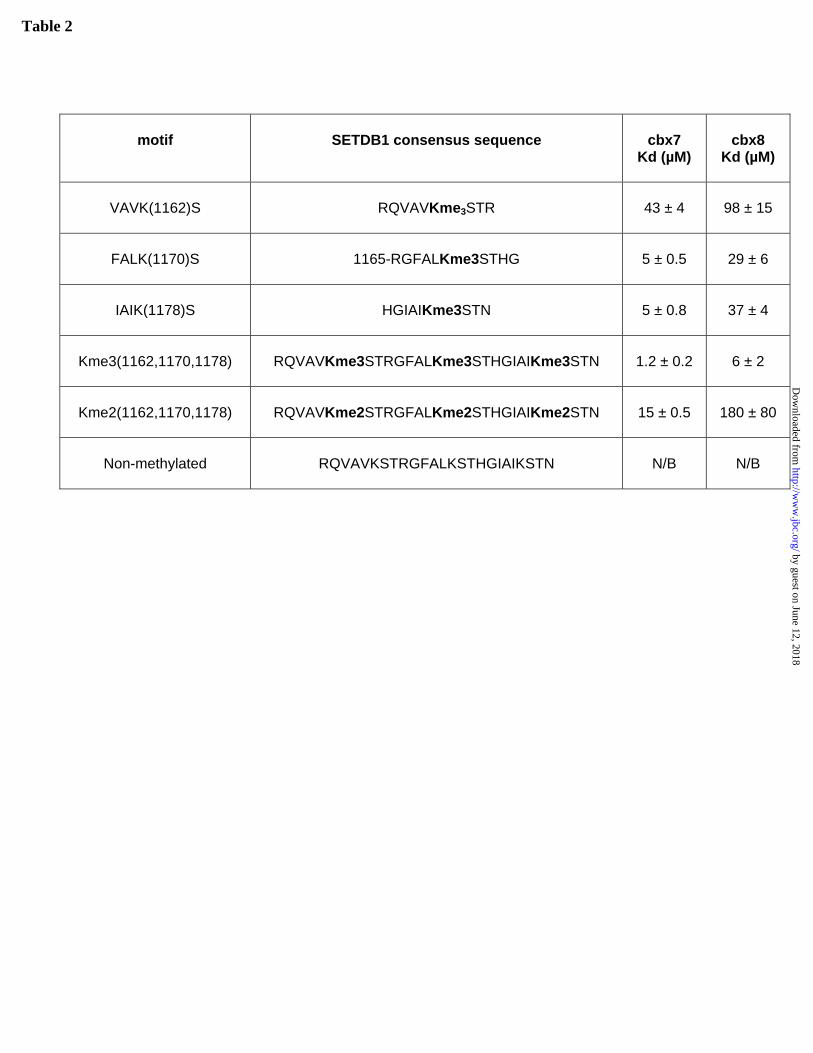
Table 2
motif
SETDB1 consensus sequence
cbx7 Kd (µM)
cbx8
Kd (µM)
VAVK(1162)S
RQVAVKme3STR
43 ± 4
98 ± 15
FALK(1170)S
1165-RGFALKme3STHG
5 ± 0.5
29 ± 6
IAIK(1178)S
HGIAIKme3STN
5 ± 0.8
37 ± 4
Kme3(1162,1170,1178)
RQVAVKme3STRGFALKme3STHGIAIKme3STN
1.2 ± 0.2
6 ± 2
Kme2(1162,1170,1178)
RQVAVKme2STRGFALKme2STHGIAIKme2STN
15 ± 0.5
180 ± 80
Non-methylated
RQVAVKSTRGFALKSTHGIAIKSTN
N/B
N/B
by guest on June 12, 2018http://w
ww
.jbc.org/D
ownloaded from

Figure 1.
(A) ARNDCEQGHILKMFPSTWYV
T K Q T A R K*S T G G K A
H3 aa3-15
-6 -5 -4 -3 -2 -1 K* 1 2 3 4 5 6
A T K A A R K* S A P A T G
H3 aa21-33
-6 -5 -4 -3 -2 -1 K* 1 2 3 4 5 6
(C)ARNDCEQGHILKMFPS
TWY
V
(B)A T K A A R K* S A P A T G
H3 aa21-33
-6 -5 -4 -3 -2 -1 K* 1 2 3 4 5 6
ARNDC
EQGH
ILKMFPS
TWY
V
T K Q T A R K* S T G G K A
H3 aa3-15
-6 -5 -4 -3 -2 -1 K* 1 2 3 4 5 6
by guest on June 12, 2018 http://www.jbc.org/ Downloaded from

Ala24
Thr6(B)
(C)
(A)
: 76: 73: 81: 75: 66: 65: 65: 62: 61: 73
Clasp residues Groove mutants1 2
Figure 2.
by guest on June 12, 2018 http://www.jbc.org/ Downloaded from

90°
Glu29
Asp68
K9me3Phe30
Glu29
Asp68 R8
T6
Glu19
Asp58
K9me3Phe20Glu19
Asp58R8
T6
Figure 3
by guest on June 12, 2018 http://www.jbc.org/ Downloaded from

Cbx6/H3K9me3
Cbx6/H3K27me3
Cbx8/H3K9 me3
dHP1/H3K9me3
dPc/H3K27me3
Cbx3/H3K9me3 Cbx5/H3K9me3
Cbx7/H3K9me3
Cbx7/H3K27me3Cbx2/H3K27me3
Arg9
Arg9 Arg9
Figure 4
by guest on June 12, 2018 http://www.jbc.org/ Downloaded from

Phe11
Apo conformation
H3K27me3
Figure 5
by guest on June 12, 2018 http://www.jbc.org/ Downloaded from
ArrowsmithGreg Wasney, Zhihong Li, Masoud Vedadi, Matthieu Schapira, Jinrong Min and Cheryl H. Lilia Kaustov, Hui Quyang, Maria Amaya, Alexander Lemak, Nataliya Nady, Shili Duan,
Recognition and specificity determinants of the human Cbx chromodomains
published online November 3, 2010J. Biol. Chem.
10.1074/jbc.M110.191411Access the most updated version of this article at doi:
Alerts:
When a correction for this article is posted•
When this article is cited•
to choose from all of JBC's e-mail alertsClick here
by guest on June 12, 2018http://w
ww
.jbc.org/D
ownloaded from