Receptor-Mediated Sorting of Typhoid Toxin during Its ... Host... · Cell Host & Microbe Short...
9
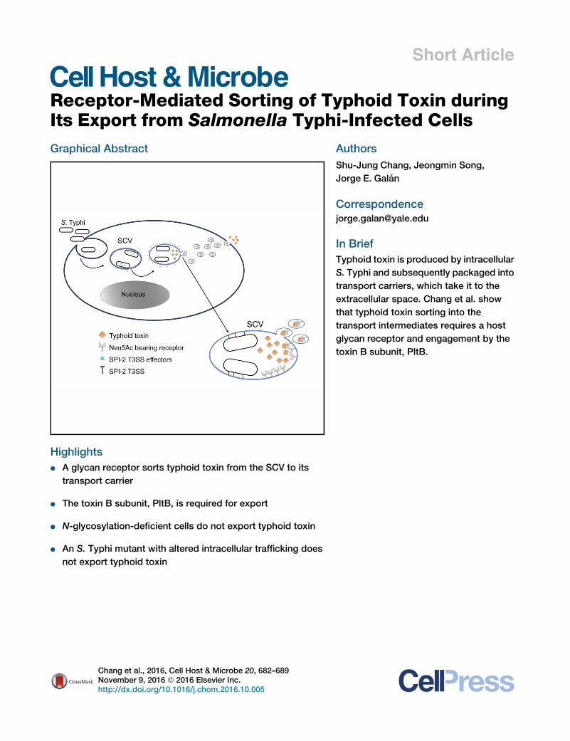
Short Article Receptor-Mediated Sorting of Typhoid Toxin during Its Export from Salmonella Typhi-Infected Cells Graphical Abstract Highlights d A glycan receptor sorts typhoid toxin from the SCV to its transport carrier d The toxin B subunit, PltB, is required for export d N-glycosylation-deficient cells do not export typhoid toxin d An S. Typhi mutant with altered intracellular trafficking does not export typhoid toxin Authors Shu-Jung Chang, Jeongmin Song, Jorge E. Gala ´n Correspondence [email protected] In Brief Typhoid toxin is produced by intracellular S. Typhi and subsequently packaged into transport carriers, which take it to the extracellular space. Chang et al. show that typhoid toxin sorting into the transport intermediates requires a host glycan receptor and engagement by the toxin B subunit, PltB. Chang et al., 2016, Cell Host & Microbe 20, 682–689 November 9, 2016 ª 2016 Elsevier Inc. http://dx.doi.org/10.1016/j.chom.2016.10.005
Transcript of Receptor-Mediated Sorting of Typhoid Toxin during Its ... Host... · Cell Host & Microbe Short...
Short Article
Receptor-Mediated Sorting of Typhoid Toxin during
Its Export from Salmonella Typhi-Infected CellsGraphical Abstract
Highlights
d A glycan receptor sorts typhoid toxin from the SCV to its
transport carrier
d The toxin B subunit, PltB, is required for export
d N-glycosylation-deficient cells do not export typhoid toxin
d An S. Typhi mutant with altered intracellular trafficking does
not export typhoid toxin
Chang et al., 2016, Cell Host & Microbe 20, 682–689November 9, 2016 ª 2016 Elsevier Inc.http://dx.doi.org/10.1016/j.chom.2016.10.005
Authors
Shu-Jung Chang, Jeongmin Song,
Jorge E. Galan
In Brief
Typhoid toxin is produced by intracellular
S. Typhi and subsequently packaged into
transport carriers, which take it to the
extracellular space. Chang et al. show
that typhoid toxin sorting into the
transport intermediates requires a host
glycan receptor and engagement by the
toxin B subunit, PltB.
Cell Host & Microbe
Short Article
Receptor-Mediated Sortingof Typhoid Toxin during Its Exportfrom Salmonella Typhi-Infected CellsShu-Jung Chang,1 Jeongmin Song,1,2 and Jorge E. Galan1,3,*1Department of Microbial Pathogenesis, Yale University School of Medicine, New Haven, CT 06536, USA2Present address: Department of Microbiology & Immunology, Cornell University College of Veterinary Medicine, Ithaca,
NY 14853-6401, USA3Lead Contact
*Correspondence: [email protected]
http://dx.doi.org/10.1016/j.chom.2016.10.005
SUMMARY
Typhoid toxin is an essential virulence factor ofSalmonellaTyphi, the causeof typhoid fever. Typhoidtoxin is secreted into the lumen of Salmonella-containing vacuole (SCV), after which it is packagedinto vesicle carrier intermediates and released extra-cellularly through incompletely understood mecha-nisms. Following export, the toxin targets cells byinteracting with human-specific Neu5Ac-terminatedglycan receptors. We show that typhoid toxin issorted from the SCV into vesicle carrier intermediatesvia interactions of its B subunit, PltB, with specificlumenal sialylated glycan packaging receptors.Cells deficient in N-glycosylation or the synthesis ofspecific gangliosides or displaying Neu5Gc-termi-nated, as opposed to Neu5Ac-terminated, glycansdo not support typhoid toxin export. Additionally,typhoid toxin packaging requires the specific SCVenvironment, as toxin produced by an S. Typhimutantwith impaired trafficking is notproperly sortedinto vesicles. These results reveal how the exotoxin ofan intracellular pathogen engages host pathways forpackaging and release.
INTRODUCTION
Salmonella enterica serovars Typhi (S. Typhi) is an exclusive hu-
man pathogen and the cause of typhoid fever, a major global
public health concern (Crump and Mintz, 2010; Dougan and
Baker, 2014; Parry et al., 2002; Raffatellu et al., 2008; Wain
et al., 2015). A related illness caused by S. enterica serovar Para-
typhi A (S. Paratyphi) is becoming increasingly prevalent in some
parts of the world (Baker et al., 2014; Fangtham and Wilde,
2008). Unlike illnesses caused by other S. enterica serovars,
such as S. Typhimurium, which are usually associated with
limited gastroenteritis (Grassl and Finlay, 2008; Ohl and Miller,
2001), typhoid fever is a systemic disease (House et al., 2001;
Parry et al., 2002; Raffatellu et al., 2008; Wain et al., 2015) that
results in �200,000 annual deaths (Buckle et al., 2012; Crump
682 Cell Host & Microbe 20, 682–689, November 9, 2016 ª 2016 Els
and Mintz, 2010; House et al., 2001; Parry et al., 2002; Raffatellu
et al., 2008; Wain et al., 2015). Both S. Typhi and S. Paratyphi
encode typhoid toxin, an unusual AB family exotoxin, which is
largely absent from non-typhoidal Salmonella enterica serovars
(Haghjoo and Galan, 2004; Spano et al., 2008). Previous studies
have shown that administration of purified typhoid toxin to
experimental animals can reproducemost of the pathognomonic
symptoms of typhoid fever, thus placing this toxin at the center of
the pathogenesis of this disease (Song et al., 2013). Unlike all
known AB toxin family members (Beddoe et al., 2010; Merritt
and Hol, 1995), typhoid toxin exhibits a unique A2B5 organiza-
tion with two enzymatically active ‘‘A’’ subunits, PltA and CdtB,
linked to a homopentameric ‘‘B’’ subunit made up of PltB
(Song et al., 2013). Typhoid toxin is targeted to cells by its PltB
B subunit, which interacts with specific glycans on the surface,
glycoproteins podocalyxin 1 (on epithelial cells) or CD45 (on
myelocytic cells) (Song et al., 2013). Recent studies have shown
that typhoid toxin has unique binding specificity for human gly-
cans (Deng et al., 2014), which is consistent with the observation
that typhoid fever occurs only in humans. In contrast to most
mammals, whose sialylated glycans are terminated in N-glyco-
lylneuraminic acid (Neu5Gc), human sialoglycans are primarily
terminated in N-acetylneuraminic acid (Neu5Ac) because of
the absence of CMP-N-acetylneuraminic acid hydroxylase
(CMAH), whose coding gene has been deleted by an Alu-medi-
ated event (Varki et al., 2011). Cells or animals express-
ing Neu5Gc-terminated glycans are resistant to typhoid toxin
(Deng et al., 2014).
Typhoid toxin is expressed only by intracellular S. Typhi
(Haghjoo and Galan, 2004; Spano et al., 2008), and once synthe-
sized, the toxin is secreted into the S. Typhi-containing vacuole
(SCV) by a unique transport mechanism (Hodak and Galan,
2013). The secreted toxin is then packaged into vesicle carriers
that transport to the extracellular space from where it reaches
its target cells (Spano et al., 2008). Notably, intoxication can
only occur via autocrine or paracrine pathways. Toxin transport
within infected cells is therefore central to the biology of typhoid
toxin. Although some Rab GTPases required for toxin transport
have been identified (Spano et al., 2011), the mechanism by
which the toxin is packaged into vesicle carriers and transported
to the extracellular space remains unknown. We show here that
typhoid toxin sorting into vesicle carriers requires its B subunit
and that the interaction of PltB with sialylated glycans on the
evier Inc.
Figure 1. PltB Is Required for the Packaging
of Typhoid Toxin into Vesicle Carrier Inter-
mediates
(A) Immunostaining of typhoid toxin in infected
cells. Infected cells were stained with antibodies
against the FLAG epitope (green) andS. Typhi LPS
(red). Scale bar, 5 mm.
(B) Quantification of the intensity of typhoid
toxin-associated fluorescent puncta, a measure
of typhoid toxin carrier intermediates, in infected
cells. Values represent relative fluorescence in-
tensity and are the mean ± SEM of three inde-
pendent experiments in which at least 70 images
were analyzed. ****p < 0.0001.
(C) Western immunoblot analysis of CdtB-3xFLAG
expression in Salmonella-infected cell lysates. The
levels of the S. Typhi protein RecA were used as
loading control.
See also Figure S1.
SCV is essential for the formation of toxin carrier intermediates.
We also show that toxin packaging requires the specific envi-
ronment of the Salmonella-containing vacuole since the toxin
produced by an S. Typhi mutant that does not traffic properly
is not sorted into the vesicle carrier intermediates. These re-
sults demonstrate a remarkable adaptation of an exotoxin to
the biology of an intracellular pathogen.
RESULTS
PltB Is Required for the Packaging of Typhoid Toxin intoVesicle Carrier IntermediatesWe hypothesized that the sorting of typhoid toxin into vesicle
export carriers must involve a packaging receptor that may
interact with a component(s) of the holotoxin and that PltB may
be such a component. To investigate this hypothesis, we exam-
ined the formation of toxin carrier intermediates in cells infected
with an S. Typhi DpltB mutant strain expressing FLAG-epitope-
tagged CdtB. We have previously shown that these vesicle car-
riers can be visualized by immunofluorescence microscopy as
discrete puncta irradiating from the SCV that can be quantified
by image analysis (Spano et al., 2008, 2011). We found that cells
infected with the DpltB mutant strain lacked detectable CdtB
puncta although the expression level of CdtB in the mutant
strain was indistinguishable from wild-type (Figures 1A–1C). As
expected from previous results (Spano et al., 2008), addition of
a typhoid toxin neutralizing antibody to the infection media
blocked intoxication but did not alter the number and the distri-
bution of the puncta, consistent with the notion that the observed
puncta represent toxin export carrier intermediates (Figure S1). A
critical residue on PltB (Ser35) is strictly required for its interac-
tion with the typhoid-toxin glycan receptors (Song et al., 2013).
To explore the potential role of the glycan-binding ability of
Cell Host & Mic
PltB on typhoid toxin sorting, we exam-
ined the formation of toxin carrier inter-
mediates in cells infected with an S. Typhi
mutant strain expressing PltBS35A. We
found no detectable CdtB puncta in cells
infected with the S. Typhi pltBS35Amutant
strain even though the levels of expression of CdtB in this strain
were indistinguishable from wild-type (Figures 1A–1C). These
results demonstrate that PltB orchestrates the packaging of
typhoid toxin into vesicle carrier intermediates and suggest
that a glycan receptor on the lumen of the SCV is required for
typhoid toxin sorting into vesicle carrier intermediates.
Disruption of Protein N-glycosylation Impairs TyphoidToxin Export from S. Typhi-Infected CellsTo further investigate the potential requirement of a glycan re-
ceptor for the sorting of typhoid toxin from the SCV into vesicle
carrier intermediates, we used CRISPR/Cas9 genome editing
to generate a cell line defective in alpha-1,3-mannosyl-glyco-
protein 2-b-N-acetylglucosaminyltransferase (MGAT1) (Fig-
ure S2). This enzyme is required for the initiation of complex
and hybrid N-glycan synthesis, and therefore it is essential for
protein N-glycosylation (Schachter, 2010) (Figure 2A). We veri-
fied the phenotype of theMGAT1-deficient cell line by examining
the mobility in SDS-PAGE of the heavily glycosylated protein
LAMP1. In the parent HEK293T cells, and consistent with its
heavy glycosylation, LAMP1 migrated as a broadly diffuse spe-
cies with a lower mobility than that of its predicted molecular
weight. In contrast, in MGAT1-deficient cells, LAMP1 migrated
as a tight band of the predicted molecular weight of its un-
modified form (Figure 2B). Furthermore, binding of exogenously
applied, fluorescently labeled typhoid toxin to MGAT1-deficient
cells was significantly reduced as predicted by the role of glyco-
sylated receptor proteins on typhoid toxin binding (Song et al.,
2013) (Figure S3). To assess the integrity of the endosomal sys-
tem in the mutant cell line, we examined its ability to take up flu-
orescently labeled dextran and secrete alkaline phosphatase.
We found that both these activities in the MGAT1-deficient cell
line were indistinguishable from the parent cell line (Figure S3),
robe 20, 682–689, November 9, 2016 683
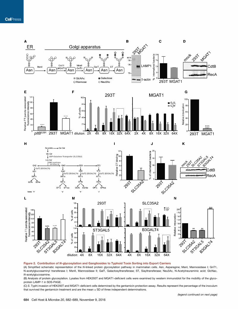
Figure 2. Contribution of N-glycosylation and Gangliosides to Typhoid Toxin Sorting into Export Carriers
(A) Simplified schematic representation of the N-linked protein glycosylation pathway in mammalian cells. Asn, Asparagine; ManI, Mannosidase I; GnT1,
N-acetylglucosaminyl transferase I; MsnII, Mannosidase II; GalT, Galactosyltransferase; ST, Siayltransferase; Neu5Ac, N-Acetylneuraminic acid; GlcNac,
N-acetylglucosamine.
(B) Analysis of protein glycosylation. Lysates from HEK293T and MGAT1-deficient cells were examined by western immunoblot for the mobility of the glyco-
protein LAMP-1 in SDS-PAGE.
(C) S. Typhi invasion of HEK293T and MGAT1-deficient cells determined by the gentamicin protection assay. Results represent the percentage of the inoculum
that survived the gentamicin treatment and are the mean ± SD of three independent determinations.
(legend continued on next page)
684 Cell Host & Microbe 20, 682–689, November 9, 2016
indicating that introduction of the mutation did not grossly alter
the cell’s endosomal system. We then infected the parent and
MGAT1-deficient cells with S. Typhi expressing FLAG-epitope-
tagged CdtB (to track typhoid toxin) and examined the formation
of vesicle carrier intermediates by fluorescence microscopy.
We found a markedly reduced number of fluorescent puncta
associated with typhoid toxin vesicle carrier intermediates in
MGAT1-deficient cells relative to the parent cell line despite
indistinguishable levels of S. Typhi invasion and typhoid toxin
expression in both cell lines (Figures 2C–2E; Figure S4). Since
the formation of typhoid toxin carrier intermediates is reduced
in MGAT1-deficient cells, we reasoned that the levels of typhoid
toxin exported to the extracellular medium should also be
reduced. We therefore quantified typhoid toxin export in the
parent and MGAT1-deficient cells infected with S. Typhi by
examining the amount of toxin activity in the infection media.
We found that, consistent with the observed deficiency in the
formation of export carrier intermediates, the level of typhoid
toxin activity in the infection medium of MGAT1-deficient cells
was significantly reduced in comparison to the parent cell line
(Figures 2F and 2G). Taken together, these results indicate
that typhoid toxin requires an N-glycosylated protein packaging
receptor for its PltB-mediated sorting from the SCV into vesicle
export carrier intermediates.
Contribution of Gangliosides to Typhoid Toxin Sortinginto Export CarriersAlthough the elimination of N-glycosylation significantly disrup-
ted typhoid toxin export, the phenotype was less severe than
that of a PltB mutant unable to bind glycans, suggesting the ex-
istence of an alternative route for typhoid toxin sorting. We have
previously shown that typhoid toxin can also bind other moieties
containing sialic acid, such as gangliosides (Song et al., 2013).
Furthermore, gangliosides have been implicated in the vesicle
traffic of viruses and some bacterial toxins (Cho et al., 2012;
Ravindran et al., 2013). Therefore, we investigated the poten-
tial contribution of gangliosides to the sorting of typhoid toxin
into vesicle export carrier. To this end, using CRISPR/Cas9
genome editing, we generated a cell line defective in SLC35A2,
an enzyme required for the synthesis of lactosylceramide, a
(D) Western immunoblot analysis of CdtB-3xFLAG expression in the indicated un
(E) Quantification of the intensity of typhoid toxin-associated fluorescent punct
represent relative fluorescence intensity and are the mean ± SEM of three indepe
(F and G) Quantification of typhoid toxin export into the infection medium. Infect
indicated and applied to HEK293T cells. 48 hr after treatment, the cell cycle profi
cells in G2M (ameasure of typhoid toxin toxicity) was determined. Values aremean
(G) was obtained by comparing the dilutions of the infection media preparations (
cells in G0G1 and G2M that, when compared across samples, showed statistical
(H) Simplified schematic representation of the major ganglioside synthesis pathw
(I) Binding of fluorescently labeled cholera toxin B subunit to the indicated cells w
the mean ± SEM of three independent determinations. **p < 0.01.
(J) S. Typhi invasion of HEK293T and SLC35A2-deficient cells determined by the
that survived the gentamicin treatment and are the mean ± SD of three independ
(K) Western immunoblot analysis of CdtB-3xFLAG expression in the indicated un
(L) Quantification of the intensity of typhoid toxin-associated fluorescent puncta
and are the mean ± SEM of three independent experiments in which at least 90
(M and N) Quantification of typhoid toxin export into the infectionmedium (see abo
of the relative toxicity (N) determined as indicated in (G). **p < 0.01; ***p < 0.001
See also Figures S1–S4.
common precursor for the synthesis of all gangliosides (Schnaar,
2016) (Figure 2H; Figure S2). To verify the phenotype of the
SLC35A2-deficient cell line, we tested the binding of fluores-
cently labeled cholera toxin, which is known to recognize GM1
gangliosides as its main receptor at the plasmamembrane (Fish-
man et al., 1993). Consistent with the predicted depletion of gan-
gliosides, the binding of cholera toxin was significantly impaired
in the SLC35A2-deficient cell line relative to its binding to the
parent cell line (Figure 2I). Furthermore, the binding of exoge-
nously applied, fluorescently labeled typhoid toxin was also
reduced (Figure S3). Assessment of the ability of the mutant
cell lines to take up dextran or secrete alkaline phosphatase indi-
cated that introduction of the mutation did not grossly alter the
endocytic and secretorymachineries of themutant cell lines (Fig-
ure S3). We then tested the efficiency of the formation of typhoid
toxin export carrier intermediates by examining the CdtB-asso-
ciated fluorescent puncta and typhoid toxin export to the extra-
cellular milieu in S. Typhi-infected mutant cells. We found a
reduced number of typhoid toxin export carriers and lower levels
of typhoid toxin in the infection media in the SLC35A2-deficient
cells, although the relative reduction was less substantial than
that observed in N-glycosylation-deficient and MGAT1-deficient
cells (Figures 2L–2N; Figure S4). Levels of S. Typhi invasion and
typhoid toxin expression in the SLC35A2-deficient cells were
indistinguishable from those observed in the parent cell line (Fig-
ures 2J and 2K). Similar results were obtained in cells rendered
deficient in the GM3 synthase encoded by the ST3GAL5 gene
(Figures 2L–2N; Figure S4). This enzyme encodes the synthesis
of GM3, which is the precursor for the synthesis of all glycosy-
lated gangliosides (Schnaar, 2016) (Figure 2H). Interestingly,
disruption of B3GALT4, which encodes a b-1,3-galactosyltrans-
ferase 4 involved in the synthesis of complex, highly glycosy-
lated gangliosides (Schnaar, 2016) (Figure 2H), resulted in
increased typhoid toxin export (Figures 2L–2N; Figure S4).
We hypothesize that the impairment of the synthesis of these
gangliosides may result in the increase of other less complex
gangliosides that may be more efficient at typhoid toxin sorting.
Taken together, these results indicate that gangliosides can
also contribute to typhoid toxin sorting into export carriers
intermediates.
infected (mock) or Salmonella-infected cell lysates.
a, a measure of typhoid toxin carrier intermediates, in infected cells. Values
ndent experiments in which at least 70 images were analyzed. ****p < 0.0001.
ion media obtained from the indicated S. Typhi-infected cells were diluted as
le of the different cells was analyzed by flow cytometry, and the percentage of
± SD of three independent determinations (F). Ameasure of the relative toxicity
marked with dashed rectangles) that resulted in a ratio between the number of
ly insignificant differences. ****p < 0.0001.
ay (Svennerholm, 1964). Cer, ceramide; Gal, galactose; Glu, glucose.
as measured by flow cytometry. Values represent relative fluorescence and are
gentamicin protection assay. Results represent the percentage of the inoculum
ent determinations.
infected (mock) or Salmonella-infected cell lysates.
in the indicated infected cells. Values represent relative fluorescence intensity
images were analyzed. ****p < 0.0001.
ve) (M). Values are mean ± SD of three independent determinations. Ameasure
.
Cell Host & Microbe 20, 682–689, November 9, 2016 685
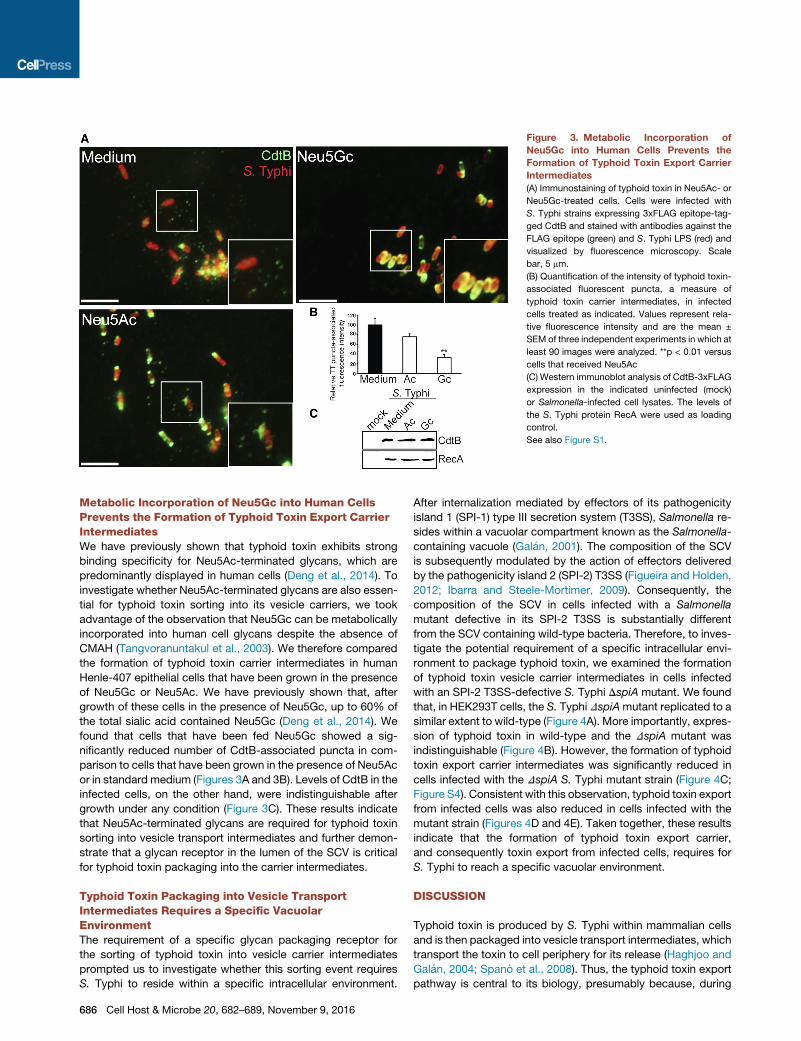
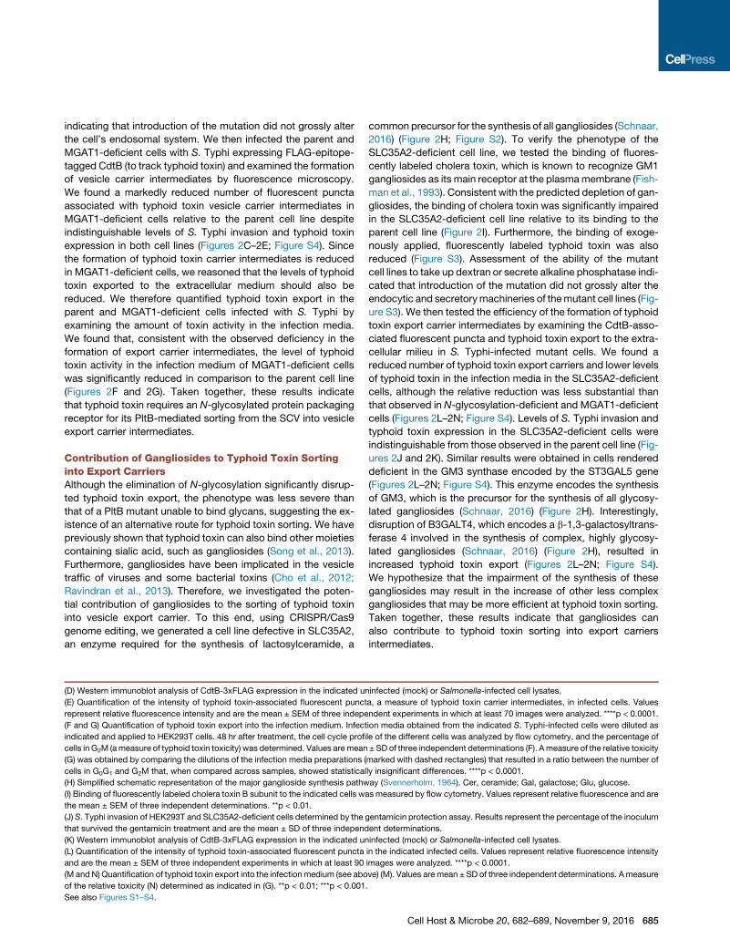
Figure 3. Metabolic Incorporation of
Neu5Gc into Human Cells Prevents the
Formation of Typhoid Toxin Export Carrier
Intermediates
(A) Immunostaining of typhoid toxin in Neu5Ac- or
Neu5Gc-treated cells. Cells were infected with
S. Typhi strains expressing 3xFLAG epitope-tag-
ged CdtB and stained with antibodies against the
FLAG epitope (green) and S. Typhi LPS (red) and
visualized by fluorescence microscopy. Scale
bar, 5 mm.
(B) Quantification of the intensity of typhoid toxin-
associated fluorescent puncta, a measure of
typhoid toxin carrier intermediates, in infected
cells treated as indicated. Values represent rela-
tive fluorescence intensity and are the mean ±
SEM of three independent experiments in which at
least 90 images were analyzed. **p < 0.01 versus
cells that received Neu5Ac
(C) Western immunoblot analysis of CdtB-3xFLAG
expression in the indicated uninfected (mock)
or Salmonella-infected cell lysates. The levels of
the S. Typhi protein RecA were used as loading
control.
See also Figure S1.
Metabolic Incorporation of Neu5Gc into Human CellsPrevents the Formation of Typhoid Toxin Export CarrierIntermediatesWe have previously shown that typhoid toxin exhibits strong
binding specificity for Neu5Ac-terminated glycans, which are
predominantly displayed in human cells (Deng et al., 2014). To
investigate whether Neu5Ac-terminated glycans are also essen-
tial for typhoid toxin sorting into its vesicle carriers, we took
advantage of the observation that Neu5Gc can be metabolically
incorporated into human cell glycans despite the absence of
CMAH (Tangvoranuntakul et al., 2003). We therefore compared
the formation of typhoid toxin carrier intermediates in human
Henle-407 epithelial cells that have been grown in the presence
of Neu5Gc or Neu5Ac. We have previously shown that, after
growth of these cells in the presence of Neu5Gc, up to 60% of
the total sialic acid contained Neu5Gc (Deng et al., 2014). We
found that cells that have been fed Neu5Gc showed a sig-
nificantly reduced number of CdtB-associated puncta in com-
parison to cells that have been grown in the presence of Neu5Ac
or in standard medium (Figures 3A and 3B). Levels of CdtB in the
infected cells, on the other hand, were indistinguishable after
growth under any condition (Figure 3C). These results indicate
that Neu5Ac-terminated glycans are required for typhoid toxin
sorting into vesicle transport intermediates and further demon-
strate that a glycan receptor in the lumen of the SCV is critical
for typhoid toxin packaging into the carrier intermediates.
Typhoid Toxin Packaging into Vesicle TransportIntermediates Requires a Specific VacuolarEnvironmentThe requirement of a specific glycan packaging receptor for
the sorting of typhoid toxin into vesicle carrier intermediates
prompted us to investigate whether this sorting event requires
S. Typhi to reside within a specific intracellular environment.
686 Cell Host & Microbe 20, 682–689, November 9, 2016
After internalization mediated by effectors of its pathogenicity
island 1 (SPI-1) type III secretion system (T3SS), Salmonella re-
sides within a vacuolar compartment known as the Salmonella-
containing vacuole (Galan, 2001). The composition of the SCV
is subsequently modulated by the action of effectors delivered
by the pathogenicity island 2 (SPI-2) T3SS (Figueira and Holden,
2012; Ibarra and Steele-Mortimer, 2009). Consequently, the
composition of the SCV in cells infected with a Salmonella
mutant defective in its SPI-2 T3SS is substantially different
from the SCV containing wild-type bacteria. Therefore, to inves-
tigate the potential requirement of a specific intracellular envi-
ronment to package typhoid toxin, we examined the formation
of typhoid toxin vesicle carrier intermediates in cells infected
with an SPI-2 T3SS-defective S. Typhi DspiA mutant. We found
that, in HEK293T cells, the S. Typhi DspiAmutant replicated to a
similar extent to wild-type (Figure 4A). More importantly, expres-
sion of typhoid toxin in wild-type and the DspiA mutant was
indistinguishable (Figure 4B). However, the formation of typhoid
toxin export carrier intermediates was significantly reduced in
cells infected with the DspiA S. Typhi mutant strain (Figure 4C;
Figure S4). Consistent with this observation, typhoid toxin export
from infected cells was also reduced in cells infected with the
mutant strain (Figures 4D and 4E). Taken together, these results
indicate that the formation of typhoid toxin export carrier,
and consequently toxin export from infected cells, requires for
S. Typhi to reach a specific vacuolar environment.
DISCUSSION
Typhoid toxin is produced by S. Typhi within mammalian cells
and is then packaged into vesicle transport intermediates, which
transport the toxin to cell periphery for its release (Haghjoo and
Galan, 2004; Spano et al., 2008). Thus, the typhoid toxin export
pathway is central to its biology, presumably because, during
Figure 4. Typhoid Toxin Packaging into
Vesicle Transport Intermediates Requires a
Specific Vacuolar Environment
(A) Intracellular survival of S. Typhi wild-type (WT)
and the SPI-2 TTSS-defective spiA mutant in
HEK293T cells. Values represent percentage of
colony-forming units (CFUs) 24 hr post-infection
relative to the CFUs 2 hr after infection and have
been standardized to the CFUs of wild-type, which
was considered to be 100%. Data are mean ± SEM
from three independent experiments.
(B) Western immunoblot analysis of CdtB-3xFLAG
expression in HEK293T cells infected with the indi-
cated S. Typhi strains. The levels of the S. Typhi
protein RecA were used as loading control.
(C) Quantification of the intensity of typhoid toxin-
associated fluorescent puncta, ameasure of typhoid
toxin carrier intermediates, in HEK293T cells in-
fected with the indicated S. Typhi strains. Values
represent relative fluorescence intensity and are the
mean ± SEM of three independent experiments in
which at least 60 images were analyzed. **p < 0.01
(D and E) Quantification of typhoid toxin export into
the infection medium. Infection media obtained from
HEK293T cells infected with the indicated S. Typhi
strains were diluted as indicated and applied to un-
infected HEK293T cells. 48 hr after treatment, the
cell cycle profile of the different cells was analyzed
by flow cytometry, and the percentage of cells in
G2M (a measure of typhoid toxin toxicity) was
determined. Values are mean ± SD of three inde-
pendent determinations (D). A measure of the rela-
tive toxicity (E) was obtained by comparing the di-
lutions of the infection media preparations (marked
with dashed rectangles) that resulted in a ratio be-
tween the number of cells in G0G1 and G2M that
when compared across samples showed statisti-
cally insignificant differences. ****p < 0.0001.
See also Figures S1 and S4.
infection, its cellular targets may not be the cells that harbor the
bacteria but rather other uninfected cells, perhaps those of the
immune system. In fact, previous studies in humanized mice
are consistent with this hypothesis since they have indicated
that typhoid toxin may be important for S. Typhi’s persistent
infection, a process that may require targeting immune cells
(Song et al., 2010). Therefore, understanding the mechanisms
by which the toxin is exported from infected cells could lead to
the development of therapeutic strategies that may help the
eradication of S. Typhi from chronic carriers.
Cargo selection during vesicle transport is a complex process
that requires specific cellular machinery (Dancourt and Barlowe,
2010). Although the components of this machinery vary depend-
ing on the cellular compartment or the cargo to be packaged,
the packaging mechanisms usually involve transmembrane
coat proteins that can select transmembrane protein cargo,
additional coat components that deform the membrane and
orchestrate the budding process, and specific small GTP-bind-
ing proteins that regulate the entire process. In addition, soluble
cargo requires specific membrane protein receptors for their
recruitment into the budding vesicle. The Salmonella-containing
vacuole is a highly specialized compartment built through the
action of numerous effectors of its two type III secretions sys-
tems (Steele-Mortimer, 2008). Consequently, it is likely that the
machinery involved in the packaging of typhoid toxin is different
from the packaging machineries that have been described in
specific cellular compartments. For example, we have previously
identified two Rab-family GTPases, Rab40b and Rab29, that are
required for efficient packaging of typhoid toxin (Spano et al.,
2011). Although neither of these GTPases has been previously
implicated in endocytic sorting, it is intriguing that Rab29 can
be seen decorating the SCV (Spano et al., 2011), where it may
play a regulatory role in cargo selection and/or budding. Here
we report another important element for the selection of typhoid
toxin for packaging into vesicle carrier intermediates. We found
that typhoid toxin packaging is orchestrated by a sialylated
glycan receptor on the SCV that specifically interacts with PltB,
the B subunit of typhoid toxin. We showed that the glycan recep-
tor must be terminated in Neu5Ac, which is most abundant in hu-
man cells but largely absent in glycans from other mammalian
cells. In fact, we found that feeding Neu5Gc, which is incorpo-
rated into human cell glycans, prevents typhoid toxin packaging
into vesicle carrier intermediates. These results are consistent
with our previous observations that indicate that typhoid toxin
Cell Host & Microbe 20, 682–689, November 9, 2016 687
is exquisitely adapted to humans. Our results also showed that
the glycan receptor(s) must be preferentially associated with a
glycoprotein, since a cell line unable to perform protein N-glyco-
sylation is defective in the formation of typhoid toxin carriers
for its export from infected cells. However, we also found that
gangliosides also contribute to typhoid toxin sorting, since cell
lines defective in the synthesis of gangliosides were defective
at packaging and exporting typhoid toxin. Interestingly, ganglio-
sides have been previously implicated in the endocytic sorting
of various toxins and viruses during their retrograde transport
from the plasma membrane to their final destination within the
endocytic network (Cho et al., 2012; Ravindran et al., 2013).
Consistent with the requirement of specific machinery to sort
and package typhoid toxin into export carriers, we found that
the nature of the Salmonella-containing vacuole profoundly im-
pacts the packaging process. More specifically, we found that
typhoid toxin expressed and secreted by an S. Typhi mutant
defective in the SPI-2-encoded T3SS is inefficiently packaged
into vesicle carrier intermediates. Effectors delivered by this
T3SS are essential for the formation of the SCV; therefore, these
findings indicate that typhoid toxin has evolved to be packaged
specifically within this specific environment.
In summary, our results depict a remarkable adaptation of an
exotoxin for the specific biology of an intracellular pathogen.
Furthermore, given the importance of typhoid toxin export in its
biology, these findings may provide the bases for the develop-
ment of potential therapeutic strategies that may help with the
treatment of typhoid fever.
EXPERIMENTAL PROCEDURES
Bacterial Strains and Plasmids
All the S. Typhi strains were derived from strain ISP2825 (Galan and Curtiss,
1991) and are listed in Table S1, and all plasmids are listed in Table S2.
Cell Culture, Bacterial Infections, and Quantification of Typhoid
Toxin Transport Intermediates and Export
Cell culture, metabolic incorporation of Neu5Ac or Neu5Gc, and bacterial
infections were carried out as previously described (Deng et al., 2014). The
visualization of typhoid toxin vesicle carrier intermediates was carried out as
previously described (Spano et al., 2008) and quantified using the open source
software ImageJ (https://imagej.nih.gov/ij/). S. Typhi internalization and repli-
cation within cultured cells was carried out using the gentamicin protection
assay as previously described (Galan and Curtiss, 1989). The supernatants
from infected cells were collected and filtered through 0.2 mm syringe filters
at 24 or 48 hr post-infection, diluted as indicated in each experiment, and
applied to fresh cells. Toxicity of the different preparations was measured as
previously described (Spano et al., 2008).
Cell Culture and CRISPR/Cas9 Gene Inactivation in Cultured
Human Cells
CRISPR/Cas9 genome editing was carried out following standard protocols
(Ran et al., 2013) (Figure S2).
Dextran Internalization and Secreted Alkaline Phosphatase Assays
Cells were incubated in the presence of Alexa 488-conjugated dextran for
40 min (molecular weight 10,000; ThermoFisher Scientific) and dextran uptake
quantified by fluorescence-activated cell sorting (FACS). To assay, we trans-
fected secreted alkaline phosphatase cells with a plasmid encoding the
secreted alkaline phosphatase or co-transfected them as a negative control
with a plasmid encoding pArf1-T31N. Next day, the levels of secreted alkaline
phosphatase in the culture medium were determined with the fluorescent
substrate DiFMUP (6,8-difluoro-4-methylumbelliferyl phosphate). The total
688 Cell Host & Microbe 20, 682–689, November 9, 2016
level of cell-associated alkaline phosphatase was measured after lysis in
0.2% Triton X-100.
SUPPLEMENTAL INFORMATION
Supplemental Information includes Supplemental Experimental Procedures,
four figures, and three tables and can be found with this article online at
http://dx.doi.org/10.1016/j.chom.2016.10.005.
AUTHOR CONTRIBUTIONS
S.-J.C. conducted all experiments shown; J.S. made some important initial
observations; J.E.G. was involved in the design, interpretation, and super-
vision of this study; and S.-J.C. and J.E.G. wrote the paper.
ACKNOWLEDGMENTS
We thank members of the J.E.G. laboratory for careful review of this manu-
script. S.-J.C. was supported in part by a Postdoctoral Fellowship from the
Government of Taiwan, Ministry of Science and Technology, Taiwan, R. O.
C. 104-2917-I-564-017. This work was supported by National Institute of
Allergy and Infectious Diseases grant AI079022 (to J.E.G.).
Received: July 23, 2016
Revised: September 16, 2016
Accepted: October 9, 2016
Published: November 9, 2016
REFERENCES
Baker, S., Karkey, A., and Parry, C. (2014). Are we adequately prepared for the
emergence of Salmonella enterica serovar Paratyphi A? Lancet Glob. Health 2,
e195–e196.
Beddoe, T., Paton, A.W., Le Nours, J., Rossjohn, J., and Paton, J.C. (2010).
Structure, biological functions, and applications of the AB5 toxins. Trends
Biochem. Sci. 35, 411–418.
Buckle, G.C., Walker, C.L., and Black, R.E. (2012). Typhoid fever and paraty-
phoid fever: systematic review to estimate global morbidity and mortality for
2010. J. Glob. Health 2, 010401.
Cho, J.A., Chinnapen, D.J., Aamar, E., te Welscher, Y.M., Lencer, W.I., and
Massol, R. (2012). Insights on the trafficking and retro-translocation of glyco-
sphingolipid-binding bacterial toxins. Front. Cell. Infect. Microbiol. 2, 51–60.
Crump, J.A., and Mintz, E.D. (2010). Global trends in typhoid and paratyphoid
fever. Clin. Infect. Dis. 50, 241–246.
Dancourt, J., and Barlowe, C. (2010). Protein sorting receptors in the early
secretory pathway. Annu. Rev. Biochem. 79, 777–802.
Deng, L., Song, J., Gao, X., Wang, J., Yu, H., Chen, X., Varki, N., Naito-Matsui,
Y., Galan, J.E., and Varki, A. (2014). Host adaptation of a bacterial toxin from
the human pathogen Salmonella Typhi. Cell 159, 1290–1299.
Dougan, G., and Baker, S. (2014). Salmonella enterica serovar Typhi and the
pathogenesis of typhoid fever. Annu. Rev. Microbiol. 68, 317–336.
Fangtham,M., andWilde, H. (2008). Emergence of Salmonella paratyphi A as a
major cause of enteric fever: need for early detection, preventive measures,
and effective vaccines. J. Travel Med. 15, 344–350.
Figueira, R., and Holden, D.W. (2012). Functions of the Salmonella pathoge-
nicity island 2 (SPI-2) type III secretion system effectors. Microbiology 158,
1147–1161.
Fishman, P.H., Pacuszka, T., and Orlandi, P.A. (1993). Gangliosides as recep-
tors for bacterial enterotoxins. Adv. Lipid Res. 25, 165–187.
Galan, J.E. (2001). Salmonella interactions with host cells: type III secretion at
work. Annu. Rev. Cell Dev. Biol. 17, 53–86.
Galan, J.E., and Curtiss, R., III (1989). Cloning and molecular characterization
of genes whose products allow Salmonella typhimurium to penetrate tissue
culture cells. Proc. Natl. Acad. Sci. USA 86, 6383–6387.
Galan, J.E., and Curtiss, R., III (1991). Distribution of the invA, -B, -C, and -D
genes of Salmonella typhimurium among other Salmonella serovars: invA
mutants of Salmonella typhi are deficient for entry into mammalian cells.
Infect. Immun. 59, 2901–2908.
Grassl, G.A., and Finlay, B.B. (2008). Pathogenesis of enteric Salmonella infec-
tions. Curr. Opin. Gastroenterol. 24, 22–26.
Haghjoo, E., and Galan, J.E. (2004). Salmonella typhi encodes a functional cy-
tolethal distending toxin that is delivered into host cells by a bacterial-internal-
ization pathway. Proc. Natl. Acad. Sci. USA 101, 4614–4619.
Hodak, H., and Galan, J.E. (2013). A Salmonella Typhi homologue of bacterio-
phage muramidases controls typhoid toxin secretion. EMBO Rep. 14, 95–102.
House, D., Bishop, A., Parry, C., Dougan, G., and Wain, J. (2001). Typhoid fe-
ver: pathogenesis and disease. Curr. Opin. Infect. Dis. 14, 573–578.
Ibarra, J.A., and Steele-Mortimer, O. (2009). Salmonella—the ultimate
insider. Salmonella virulence factors that modulate intracellular survival. Cell.
Microbiol. 11, 1579–1586.
Merritt, E.A., and Hol, W.G. (1995). AB5 toxins. Curr. Opin. Struct. Biol. 5,
165–171.
Ohl, M.E., and Miller, S.I. (2001). Salmonella: a model for bacterial pathogen-
esis. Annu. Rev. Med. 52, 259–274.
Parry, C.M., Hien, T.T., Dougan, G., White, N.J., and Farrar, J.J. (2002).
Typhoid fever. N. Engl. J. Med. 347, 1770–1782.
Raffatellu, M., Wilson, R.P., Winter, S.E., and B€aumler, A.J. (2008). Clinical
pathogenesis of typhoid fever. J. Infect. Dev. Ctries. 2, 260–266.
Ran, F.A., Hsu, P.D., Wright, J., Agarwala, V., Scott, D.A., and Zhang, F. (2013).
Genome engineering using the CRISPR-Cas9 system. Nat. Protoc. 8, 2281–
2308.
Ravindran, M.S., Tanner, L.B., and Wenk, M.R. (2013). Sialic acid linkage in
glycosphingolipids is a molecular correlate for trafficking and delivery of extra-
cellular cargo. Traffic 14, 1182–1191.
Schachter, H. (2010). Mgat1-dependent N-glycans are essential for the normal
development of both vertebrate and invertebrate metazoans. Semin. Cell Dev.
Biol. 21, 609–615.
Schnaar, R.L. (2016). Gangliosides of the vertebrate nervous system. J. Mol.
Biol. 428, 3325–3336.
Song, J., Willinger, T., Rongvaux, A., Eynon, E.E., Stevens, S., Manz, M.G.,
Flavell, R.A., and Galan, J.E. (2010). A mouse model for the human pathogen
Salmonella typhi. Cell Host Microbe 8, 369–376.
Song, J., Gao, X., and Galan, J.E. (2013). Structure and function of the
Salmonella Typhi chimaeric A(2)B(5) typhoid toxin. Nature 499, 350–354.
Spano, S., Ugalde, J.E., and Galan, J.E. (2008). Delivery of a Salmonella Typhi
exotoxin from a host intracellular compartment. Cell Host Microbe 3, 30–38.
Spano, S., Liu, X., and Galan, J.E. (2011). Proteolytic targeting of Rab29 by an
effector protein distinguishes the intracellular compartments of human-adapt-
ed and broad-host Salmonella. Proc. Natl. Acad. Sci. USA 108, 18418–18423.
Steele-Mortimer, O. (2008). The Salmonella-containing vacuole: moving with
the times. Curr. Opin. Microbiol. 11, 38–45.
Svennerholm, L. (1964). The Gangliosides. J. Lipid Res. 5, 145–155.
Tangvoranuntakul, P., Gagneux, P., Diaz, S., Bardor, M., Varki, N., Varki, A.,
and Muchmore, E. (2003). Human uptake and incorporation of an immuno-
genic nonhuman dietary sialic acid. Proc. Natl. Acad. Sci. USA 100, 12045–
12050.
Varki, N.M., Strobert, E., Dick, E.J., Jr., Benirschke, K., and Varki, A. (2011).
Biomedical differences between human and nonhuman hominids: potential
roles for uniquely human aspects of sialic acid biology. Annu. Rev. Pathol. 6,
365–393.
Wain, J., Hendriksen, R.S., Mikoleit, M.L., Keddy, K.H., andOchiai, R.L. (2015).
Typhoid fever. Lancet 385, 1136–1145.
Cell Host & Microbe 20, 682–689, November 9, 2016 689