Recent progress on nanoparticle-based drug delivery ...
14
REVIEW Recent progress on nanoparticle-based drug delivery systems for cancer therapy Yanru Xin, Mingming Yin, Liyuan Zhao, Fanling Meng, Liang Luo College of Life Science and Technology, National Engineering Research Center for Nanomedicine, Huazhong University of Science and Technology, Wuhan 430074, China ABSTRACT The development of cancer nanotherapeutics has attracted great interest in the recent decade. Cancer nanotherapeutics have overcome several limitations of conventional therapies, such as nonspecific biodistribution, poor water solubility, and limited bioavailability. Nanoparticles with tuned size and surface characteristics are the key components of nanotherapeutics, and are designed to passively or actively deliver anti-cancer drugs to tumor cells. We provide an overview of nanoparticle-based drug delivery methods and cancer therapies based on tumor-targeting delivery strategies that have been developed in recent years. KEYWORDS Nanoparticles; nanomedicine; drug delivery; tumor targeting; cancer therapy Introduction Nanomedicine involves the development and application of nanometer-scaled materials in the diagnosis, treatment, and prevention of diseases. Its indications range from the medical applications of nanomaterials and biological devices to nanometer-scaled electronic biosensors. Nanoparticles are the key components of nanomedicine and have received extensive interest as promising drug-delivery systems for cancer diagnosis and treatment. 1-3 Nanoparticles applied as drug delivery systems are submicron-sized (100–1,000 nm) particles, devices, or systems synthesized from various materials, including polymers (e.g., polymeric nanoparticles, micelles, vesicles, or dendrimers), 4 lipids (liposomes), 5 viruses (viral nanoparticles), 6 and even inorganics. 7,8 By using either passive or active targeting strategies, nanoparticles can increase the intracellular concentration of drugs in cancer cells while preventing toxicity to normal cells. Nanoparticles are usually enveloped by endosomes via receptor-mediated endocytosis after entering tumor cells, therefore escaping P-glycoprotein recognition, 9 one of the main mechanisms of drug resistance. However, although nanoparticles are promising drug carrier systems, their poor oral bioavailability, instability in circulation, inadequate tissue distribution, and toxicity are some limitations to practical application that remain unresolved. In 2016, Wilhelm et al. 10 surveyed the literature from the past 10 years on nanoparticle-based drug carriers; their review showed that only 0.7% (median) of the administered nanoparticle dose was delivered to a solid tumor. This low delivery efficiency negatively affects the translation of nanotechnology to clinical applications. To overcome these concerns and to satisfy safety, regulatory, and ethical requirements, researchers have continued to develop nanomedicine with improved characteristics. A more recent review by a group of pioneers in nanomedicine has provided a comprehensive overview of developments and current challenges in the field. The review also highlighted the upcoming opportunities in the field and clinical applications 11 of nanomedicine for in vivo diagnosis, in vitro diagnosis, in vivo therapeutics, and implantable materials. Despite of all the problems associated with nanomedicine, nanoparticle-based drug delivery systems remain as a potential strategy for cancer therapy. Some nanoparticle formulations for cancer treatment have been already approved by regulatory agencies. These formulations exert fewer adverse effects than unmodified or bare drugs. Considerable effort has been expended to develop systems with precisely controlled functions and material properties. In this review, we will focus on the recent progress in the development of novel responsive nanoparticle systems with Correspondence to: Liang Luo E-mail: [email protected] Received April 29, 2017; accepted May 24, 2017. Available at www.cancerbiomed.org Copyright © 2017 by Cancer Biology & Medicine Cancer Biol Med 2017. doi: 10.20892/j.issn.2095-3941.2017.0052
Transcript of Recent progress on nanoparticle-based drug delivery ...

REVIEW
Recent progress on nanoparticle-based drug delivery systemsfor cancer therapy
Yanru Xin, Mingming Yin, Liyuan Zhao, Fanling Meng, Liang LuoCollege of Life Science and Technology, National Engineering Research Center for Nanomedicine, Huazhong University ofScience and Technology, Wuhan 430074, China
ABSTRACT The development of cancer nanotherapeutics has attracted great interest in the recent decade. Cancer nanotherapeutics have
overcome several limitations of conventional therapies, such as nonspecific biodistribution, poor water solubility, and limited
bioavailability. Nanoparticles with tuned size and surface characteristics are the key components of nanotherapeutics, and are
designed to passively or actively deliver anti-cancer drugs to tumor cells. We provide an overview of nanoparticle-based drug
delivery methods and cancer therapies based on tumor-targeting delivery strategies that have been developed in recent years.
KEYWORDS Nanoparticles; nanomedicine; drug delivery; tumor targeting; cancer therapy
Introduction
Nanomedicine involves the development and application of
nanometer-scaled materials in the diagnosis, treatment, and
prevention of diseases. Its indications range from the medical
applications of nanomaterials and biological devices to
nanometer-scaled electronic biosensors. Nanoparticles are
the key components of nanomedicine and have received
extensive interest as promising drug-delivery systems for
cancer diagnosis and treatment.1-3 Nanoparticles applied as
drug delivery systems are submicron-sized (100–1,000 nm)
particles, devices, or systems synthesized from various
materials, including polymers (e.g., polymeric nanoparticles,
micelles, vesicles, or dendrimers),4 lipids (liposomes),5
viruses (viral nanoparticles),6 and even inorganics.7,8 By
using either passive or active targeting strategies,
nanoparticles can increase the intracellular concentration of
drugs in cancer cells while preventing toxicity to normal cells.
Nanoparticles are usually enveloped by endosomes via
receptor-mediated endocytosis after entering tumor cells,
therefore escaping P-glycoprotein recognition,9 one of the
main mechanisms of drug resistance.
However, although nanoparticles are promising drug
carrier systems, their poor oral bioavailability, instability in
circulation, inadequate tissue distribution, and toxicity are
some limitations to practical application that remain
unresolved. In 2016, Wilhelm et al.10 surveyed the literature
from the past 10 years on nanoparticle-based drug carriers;
their review showed that only 0.7% (median) of the
administered nanoparticle dose was delivered to a solid
tumor. This low delivery efficiency negatively affects the
translation of nanotechnology to clinical applications.
To overcome these concerns and to satisfy safety,
regulatory, and ethical requirements, researchers have
continued to develop nanomedicine with improved
characteristics. A more recent review by a group of pioneers
in nanomedicine has provided a comprehensive overview of
developments and current challenges in the field. The review
also highlighted the upcoming opportunities in the field and
clinical applications11 of nanomedicine for in vivo diagnosis,
in vitro diagnosis, in vivo therapeutics, and implantable
materials.
Despite of all the problems associated with nanomedicine,
nanoparticle-based drug delivery systems remain as a
potential strategy for cancer therapy. Some nanoparticle
formulations for cancer treatment have been already
approved by regulatory agencies. These formulations exert
fewer adverse effects than unmodified or bare drugs.
Considerable effort has been expended to develop systems
with precisely controlled functions and material properties.
In this review, we will focus on the recent progress in the
development of novel responsive nanoparticle systems with
Correspondence to: Liang LuoE-mail: [email protected] April 29, 2017; accepted May 24, 2017.Available at www.cancerbiomed.orgCopyright © 2017 by Cancer Biology & Medicine
Cancer Biol Med 2017. doi: 10.20892/j.issn.2095-3941.2017.0052

high sensitivity to tumor microenvironments for improved
cancer diagnosis and treatment, as well as on combined
nanoparticle-assisted cancer therapies.
Tumor targeting by nanoparticles
The basis of tumor-targeting drug delivery systems is the
ability of nanoparticles to passively or actively accumulate in
the desired tissues or cells. In passive targeting, nanoparticles
are designed for transport through leaky vessels and the
unique intra-organ pressures of tumors. In active targeting,
nanoparticles are designed to adhere to specific biological
structures in tumors via the molecular recognition of surface-
bound ligands. The nanoparticles and loaded drugs thus
escape immune clearance, avoid nonspecific cell uptake, and
specifically accumulate in the targeted tumor cells and tissues.
Enhanced permeability and retention (EPR)effect
EPR effect is the property by which molecules of certain sizes
(normally 100–1,000 nm) preferentially accumulate in tumor
tissues instead of in normal tissues. Given their vigorous
activity, tumor cells require more nutrients than normal cells,
thus leading to the secretion of vascular endothelial growth
factor and other growth factors that promote angiogenesis in
tumors. Compared with that of normal vessels, the
endothelial gap of new blood vessels is larger, thus facilitating
the transport of macromolecular substances through blood
vessels to tumor tissue. In addition, the lack of lymphatic
vessels causes lymph circumfluence to suffocate. Under these
double circumstances, macromolecular substances, or
nanoparticles, accumulate in tumor tissues. EPR-passive
targeting is the basis of tumor drug delivery. By stimulating
tumor vasodilation, reducing lymphocytes, and extending
circulation time, we can maximize the EPR effect of tumor-
targeting delivery systems.
Park et al.12 developed a new type of porous silicon
nanoparticle to decrease the metabolic rate and increase the
circulation time of loaded drugs in the body. To allow the
nanomaterials to accumulate in the tumor site, the group
used silicon and silicon dioxide as the core of the nano-particles and packed dextran outside of the nanoparticles
after drug loading. After loading on the formed luminescent
porous silicon nanoparticles (LPSiNPs), the circulation time
of the drugs in the body was extended. The fluorescence
emitted by the LPSiNPs under near-infrared (NIR) excitation
can be used to trace the metabolism of the drug in the body.
Low toxicity metabolism, extended circulation time, and
fluorescence tracing make LPSiNPs a unique nanomaterial
with broad prospective applications.
Mundra et al.13 covalently conjugated indocyanine green
(ICG)-NH2 to the pendant carboxyl groups of poly (ethylene
glycol)-block-poly(2-methyl-2-carboxyl-propylene car-bonate) copolymer via carbodiimide coupling. The system
self-assembles into micelles with a particle size of 30–50 nm
and high ICG loading. In vivo NIR imaging demonstrated
that ICG-conjugated micelles have prolonged circulation
time and increased tumor accumulation through the EPR
effect. Compared with the control ICG solution, the NIR-
irradiated ICG-conjugated micelles have improved
therapeutic efficacy with complete tumor regression in an
A375 human melanoma tumor model in athymic nude mice.
pH response
The abnormal metabolism and protein regulation of tumor
tissues form an acidic microenvironment that favors the
proliferation of tumor cells. This pH abnormality is widely
exploited in tumor-targeted delivery. In the acidic
microenvironment of tumors, nanocarrier structures can be
changed by chemical bond dissociation or charge reversal for
specific drug release.
To overcome the resistance of breast cancer to doxorubicin
(DOX), which is currently the most widely used anti-cancer
drug, Yu et al.14 constructed pH-sensitive microspheres that
encapsulate DOX. The microspheres are formed by the self-
assembly of polyethylene glycol (PEG)-block-poly[2-
(diisopropylamino)ethyl methacrylate (PEG-b-PDPA)], and
D-a-tocopheryl PEG 1000 succinate (TPGS). DOX is
encapsulated in the cores of the microspheres. The
microspheres are stable under the normal pH level of 7.4 but
are degraded after cellular absorption. The early endosome
and dissolved enzyme in tumor cells provide an acidic
environment, which transforms the protonated PDPA/
TPGS@DOX microspheres into micelles and releases DOX in
the cells. At the same time, TPGS greatly reduces the toxicity
of mitochondria, thus leading to the synergistic effect of the
treatment.
Lee et al.15 designed pH-responsive intelligent micro-spheres Doxorubicin (DOX) loaded P(Asp-g-Im)-PEG
micelles (DPHAIM, Figure 1) for the weakly acidic tumor
microenvironment. With aspartic acid imidazole as the
hydrophobic group and ethylene glycol as the hydrophilic
group, the formed copolymer P(Aspg-Im)-PEG can load up
to 28% DOX. In the pH<7 acid conditions of the tumor
microenvironment, the microspheres are protonated and
dissolved, thus releasing DOX and achieving the goal of
Cancer Biol Med Vol 14, No 3 August 2017 229
targeted DOX delivery with reduced toxicity.
Talelli et al.16 developed a multi-layer package for the
targeted delivery of DOX and to overcome its serious side
effects. DOX derivatives are introduced into the core of the
polymer micelles by covalent coupling. Under weak acidic
conditions, such as the tumor microenvironment and the
Figure 1 Schematic for the proposed in vivo behavior of DPHAIM. Reproduced with permission from Ref. 15.
230 Xin et al. Nanoparticle-based cancer therapy

endosome, covalent bonds are broken and free DOX
derivatives are released. Experiments showed that only
approximately 5% of the drug is released under pH 7.4 but is
fully released within the same duration at pH 5 and 37°C.
This result indicated the good characteristics of targeted drug
delivery and reduced damage to normal tissues.
Numerous pH-responsive, intelligent nanoparticle delivery
examples have been developed. A double-strand pH-sensitive
DNA has been developed to release DOX to the tumor17. Jia
et al.18 also designed a nanovehicle with hydrophilic methoxy
poly(ethylene glycol) (mPEG)-PBA and hydrophobic
HEHDO, which has a pH-labile profile. Torres et al.19
developed a pH-sensitive liposome system that accelerates
cytosolic release at pH values of 5.5–6.0. Seib et al.20
synthesized silk nanoparticles with pH-dependent release
(pH 4.5>6.0>7.4). Battogtokh et al.21 reported an acid-
cleavable nanocarrier with enhanced cellular delivery at pH
6.5 compared with that at pH 7.4; by contrast, the cellular
delivery of drugs by noncleavable nanocarriers showed no
difference at pH 5.5 and pH 7.4. Tail vein injections enhance
the intracellular uptake of the acid-cleavable nanocarrier
relative to the noncleavable nanocarrier into the tumor cells
of tumor-bearing mice, although total tumor accumulation is
not significantly different. Van Butsele et al.22 reported the
preparation and the aqueous solution properties of hybrid
polymeric micelles that consist of a hydrophobic poly (ε-
caprolactone) (PCL) core and a mixed shell of hydrophilic
poly(ethylene oxide) and pH-sens i t ive poly (2-
vinylpyridine).
Temperature response
The temperature of tumor tissues is slightly higher than that
of surrounding normal tissues. This temperature difference
can be applied in temperature-responsive drug delivery to
tumors. Lv et al.23 developed a novel polymer system for
intelligent drug release; the developed system is based on
molecular recognition and phase transition temperature
response (Figure 2). In their nanoparticle drug delivery
system, magnetic Fe3O4 colloidal nanocrystal clusters
(MCNCs) are used as the core, and poly (N-
isopropylacrylamide) is used as a communication bridge
between MCNCs and β-cyclodextrin (β-CD). β-CD can
incorporate a number of hydrophobic groups and can easily
load hydrophobic 8-anilino-1-naphthalenesulfonic acid
ammonium salt (ANS) drugs. The conformation of MCNCs
changes with the change in external magnetic field and
temperature, thus decreasing the drug loadings of the whole
system and consequently releasing ANS. The release rate of
ANS from a drug nanoparticle system is easily controlled by
simply adjusting external temperature. These nanoparticles
have great potential for targeted delivery.
Talelli et al.24 developed biodegradable thermosensitive
polymeric micelles for the stable encapsulation of
hydrophobic oleic-acid-coated superparamagnetic iron oxide
nanoparticles (diameter 5-10 nm). The micelles are
composed of amphiphilic, thermosensitive, and bio-degradable block copolymers of poly (ethylene glycol)-b-
poly[N-(2-hydroxypropyl) methacrylamide dilactate]
[mPEGb-p(HPMAm-Lac2)]. Liu et al.25 developed a
precision cancer nanomedicine based on Bi2S3 nanorods
(NRs) designed specifically for multispectral optoacoustic
tomography (MSOT)/X-ray computed tomography (CT).
The as-prepared Bi2S3 NRs exhibit an ideal photothermal
effect and contrast enhancement in MSOT/CT bimodal
imaging.
Enzymatic response
Some enzymes are only present or are highly concentrated in
tumor cells. Their specific binding with enzyme substrates
can be employed to achieve the intelligent response of
targeting drugs. Mi et al.26 constructed a multistage complex
targeted drug delivery nanosystem enzyme-stimulated
multistage vector (ESMSV) based on the special recognition
between the enzyme and the appropriate substrate. A
metalloproteinases-2 (MMP-2) enzyme substrate is modified
on a polymer poly (lactic-co-glycolic acid) (PLGA)-PEG
nanoparticle surface, followed by loading onto porous
silicon. When ESMSV accumulates in pulmonary tumor
tissues because of MMP2 enzymatic activity, the connecting
structure between polymeric nanoparticles and porous
silicon nanoparticles (SiNP) is destroyed, thus releasing poly
Figure 2 Schematic of the temperature-switched Fe3O4@PNG-
CD nanosystem for controlled drug release. Reproduced with
permission from Ref. 23.
Cancer Biol Med Vol 14, No 3 August 2017 231

nanoparticles from porous silicon. These processes have been
validated both in vivo and in vitro. The results indicated that
ESMSV showed noticeable efficacy in the treatment of
metastatic lung melanoma.
Tang et al.27 reported a general strategy for the direct
delivery of functional proteins to the cytosol by nanoparticle-
stabilized capsules (NPSCs). The effectiveness of these NPSCs
as therapeutic protein carriers has been demonstrated
through the delivery of fully functionalized caspase-3 to HeLa
cells with concomitant apoptosis. The similar delivery of
green fluorescent protein confirmed the cytosolic delivery
and intracellular targeting of the delivered protein, thus
demonstrating the utility of the system for both therapeutic
and imaging applications.
Barve et al.28 developed an enzyme-responsive peptide
drug conjugate for TGX-D1, a promising PI3K inhibitor for
prostate cancer. In this system, the LNCaP-specific KYL
peptide is used as the targeting ligand and the prostate-
specific antigen (PSA)-cleavable peptide (SSKYQSL) is used
as the enzyme-responsive linker. Ruan et al.29 experimentally
proved that pre-incubating G-AuNP-DOX-PEG with MMP-
2 can significantly enhance the penetrating efficiency of the
drug. Yu et al.30 developed surface-engineering nanoparticles
with programmable extended circulation and targeting
ability. The nanoparticles, which are structured with a
targeting middle layer and a hydrophobic internal core, are
constructed by mixing matrix metalloproteinase MMP2,
MMP9-sensitive copolymers (mPEG-Pep-PCL), and folate
receptor targeted copolymers (FA-PEG-PCL). The results
showed the successful combination between enzymatic
stimulus and surface-engineered technology for tumor
targeting.
Antigen response
Similar to enzymes, the special identification and connection
between antigens and antibodies can be exploited for the
design of active targeting drug delivery. Ding et al.31 utilized
the specific binding between a receptor and a ligand to realize
targeted drug delivery. The authors combined immune
nanoparticles containing the death receptor 5 monoclonal
antibody (DR5 mAb) and nanoparticles carrying kappa
oxazine (DTIC) to form the composite nanoparticle DTIC
NP-DR5 mAb. The drug-loading nanoparticles accumulate
in tumor cells due to the specificity of the monoclonal
antibody and DR5 recognition effect in the body. This system
is also a good example of the combined therapy of immune
therapy and chemotherapy. The authors demonstrated that
DTIC-PLA-DR5 mAb nanoparticles are an active targeting
drug delivery system that can specifically target DR5-
overexpressing malignant melanoma cells and can be
efficiently internalized. Most strikingly, compared with
conventional DTIC-NPs, DTIC-NP-DR5 mAb showed
significantly enhanced cytotoxicity and increased cell
apoptosis in DR5-positive malignant melanoma cells.
Saraf et al.32 synthesized cyclic Arg-Gly-Asp (RGD)
micelles, which exhibit better targeting efficacy than linear
RGD micelles as drug delivery vehicles. This improved
targeting efficacy is attributed to the low drug solubilization
and kinetic stability of the micelles. The results from the
study proved the effectiveness of self-assembling low-
molecular-weight RGD amphiphiles as carriers for the
targeted delivery of paclitaxel (PTX).
Nanoparticle-based cancer therapy
Magnetic therapy
Tissues rarely absorb magnetic waves, which penetrate deeper
than visible light and infrared without side effects. Therefore,
magnetic nanomaterials are good candidates for disease
treatment. Magnetic nanoparticles, which mainly contain
iron oxide, can be used for localized heating when triggered
by a magnetic field. Carregal-Romero et al.33 synthesized
multilayer-assembled polyelectrolyte microcapsules with
diameters of 4.6 µm. The walls of the microcapsules are
integrated with 18 nm-diameter iron oxide nanotubes to
supply a magnetic trigger for the release of molecules with
low absorption and deep permeability. An organic
fluorescent polymer (Cascade-Blue-labeled dextran) is then
loaded onto the microcapsules as a model molecular cargo.
Under an alternating magnetic field, the magnetic nanotubes
can heat their surroundings and destroy the walls of the
microcapsules, thus releasing the embedded cargo into the
surrounding solution. The use of magnetic nanoparticles
with high heating properties allows the release of the
magnetic properties of the encapsulated material; moreover,
this use provides the possibility of applying new and exciting
materials in the body.
Fang et al.34 employed core-shell magnetoresponsive virus-
mimetic nanocapsules (VNs) anchored with iron oxide
nanoparticles for rapid and simultaneous drug release. The
VNs produce a strong heating effect under an external high
frequency magnetic field. Furthermore, the VNs can
subsequently infect adjacent cancer cells and deliver
sufficient therapeutic agents to the next target. Kuo et al.35
used Fe3O4 nanoparticles to encapsulate anticancer drugs and
to immobilize the antibody-targeting peptide AP-1 (MPVA-
232 Xin et al. Nanoparticle-based cancer therapy

AP1) to form multifunctional magnetic nanovehicles. The
magnetic nanovehicles can be stably stored and exert
thermotherapeutic (i.e., hyperthermia) and chemo-therapeutic (i.e., anti-cancer drug) effects on tumor tissues
under a high-frequency magnetic field. Lee et al.36 used
magnetoretic-responsive DOX-encapsulated supramolecular
magnetic nanoparticles (DOX-SMNPs) as a unique one-
demand drug release system to enhance therapeutic effects
and attenuate systematic toxicity to the tumor. The SMNPs
self-assemble from four different molecular building blocks:
Ad-grafted polyamidoamine dendrimers, β-CD-grafted
branched polyethylenimine, Ad-PEG, and 6-nm Ad-grafted
Zn0.4Fe2.6O4 superparamagnetic nanoparticles. Li and
coworkers37 designed a magnetothermally responsive
nanocarrier, in which NIPAAm and HMAAm are composed
of thermosensitive copolymers and Mn-Zn ferrite
nanoparticles are embedded in the polymer matrix. Under an
alternating magnetic field, this system demonstrated efficient
therapeutic effects and good biocompatibility.
Photodynamic therapy (PDT)
PDT can kill tumor cells through the transformation of
tumor oxygen into cytotoxic reactive singlet oxygen (1O2)
under light irradiation. PDT is a safe and selective treatment
method for various cancer types. However, the efficacy of
PDT in tumor therapy is limited due to the hypoxic
microenvironment of tumors and oxygen consumption
during PDT. To overcome this problem, Cheng et al.38
developed a novel oxygen self-enriched photodynamic
therapy system by loading IR780 on perfluorohexane (PFH)
nanodroplets. PFH can maintain a high oxygen content and
extend1O2 lifetime because of its high oxygen capacity. The
photosensitizer IR780 is dispersed in a lipid monolayer
composed of lecithin, cholesterol, and DSPE-PEG2000.
Under irradiation by a NIR 808-nm laser, IR780 transfers
energy to the oxygen enriched in the PFH, thus producing
singlet oxygen and exerting a cytotoxic effect, which
significantly improves the photodynamic effect. After
intratumoral injection into tumors, tumor growth is
inhibited in Oxy-PDT-treated mice. Later, Gong et al.39 used
PEGylated chlorine e6 (Ce6)-coupled poly (maleic
anhydridealt-1-octadecene) to form Ce6-containing
nanomicelles in an aqueous solution (Figure 3) and
discovered that hyaluronidase resolves hyaluronan, a major
component of extracellular matrix in tumors. The complex
enhances the efficacy of nanoparticle-based PDT for in vivo
cancer treatment.
Hou et al.40 developed a nanodumbbell model, which
consists of a hydrophobic upconversion nanoparticle (UCN)
core and a hydrophil ic polymersome shel l . UCN
NaYF4:Yb:Er nanocrystals, which were synthesized via the
thermal decomposition method, can be used as transducers
to convert NIR light to visible light to activate the
photosensitizers zinc (II) phthalocyanine (ZnPc) in
photodynamic therapy. The polymersome lipid shell is used
to load ZnPc and to avoid non-specific absorbance or
corrosion during transportation. In addition, folic acid may
combine with hybrid Fe3O4-ZnO (FZ-SFA) nanoparticles in
therapeutic applications associated with PDT41. The photo-
killing effect and photo-induced toxicity are significantly
enhanced under UV irradiation. To increase the production
of reactive oxygen species (ROS) under normoxic and
hypoxic conditions, Usacheva et al.42 used a polymer-
surfactant nanoparticle system to load the photosensitizer
methylene blue. Nanoparticles loaded with methylene blue
can remove cancer stem cells under hypoxic conditions,
therefore providing a novel route for cancer treatment.
Photothermal therapy (PTT)
In PTT, cancer tissues are irradiated with electromagnetic
radiation, which elevates temperature to kill targeted cells. In
this method, the careful selection of laser parameters and
illumination can enable local light penetration for specific
targeting.
One of the most promising directions for PTT is the use of
plasma nanostructures, particularly gold nanostructures,
such as gold nanoparticles (AuNPs) and gold nanoshells, to
take the advantage of localized surface plasmon resonance. In
addition, visible light with a longer wavelength or NIR can be
Figure 3 Scheme showing the effects of HAase on the
modulation of the tumor microenvironment. By improving tumor
oxygenation and promoting the EPR effect, HAase enhances the
efficacy of in vivo PDT cancer treatment. Reproduced with
permission from Ref. 39.
Cancer Biol Med Vol 14, No 3 August 2017 233
used in PTT since they have a deep tissue permeation and
low energy, which causes limited damage to other cells and
tissues.
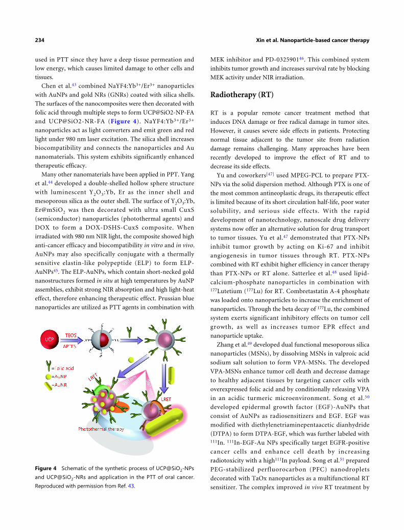
Chen et al.43 combined NaYF4:Yb3+/Er3+ nanoparticles
with AuNPs and gold NRs (GNRs) coated with silica shells.
The surfaces of the nanocomposites were then decorated with
folic acid through multiple steps to form UCP@SiO2-NP-FA
and UCP@SiO2-NR-FA (Figure 4). NaYF4:Yb3+/Er3+
nanoparticles act as light converters and emit green and red
light under 980 nm laser excitation. The silica shell increases
biocompatibility and connects the nanoparticles and Au
nanomaterials. This system exhibits significantly enhanced
therapeutic efficacy.
Many other nanomaterials have been applied in PPT. Yang
et al.44 developed a double-shelled hollow sphere structure
with luminescent Y2O3:Yb, Er as the inner shell and
mesoporous silica as the outer shell. The surface of Y2O3:Yb,
Er@mSiO2 was then decorated with ultra small CuxS
(semiconductor) nanoparticles (photothermal agents) and
DOX to form a DOX-DSHS-CuxS composite. When
irradiated with 980 nm NIR light, the composite showed high
anti-cancer efficacy and biocompatibility in vitro and in vivo.
AuNPs may also specifically conjugate with a thermally
sensitive elastin-like polypeptide (ELP) to form ELP-
AuNPs45. The ELP-AuNPs, which contain short-necked gold
nanostructures formed in situ at high temperatures by AuNP
assemblies, exhibit strong NIR absorption and high light-heat
effect, therefore enhancing therapeutic effect. Prussian blue
nanoparticles are utilized as PTT agents in combination with
MEK inhibitor and PD-032590146. This combined system
inhibits tumor growth and increases survival rate by blocking
MEK activity under NIR irradiation.
Radiotherapy (RT)
RT is a popular remote cancer treatment method that
induces DNA damage or free radical damage in tumor sites.
However, it causes severe side effects in patients. Protecting
normal tissue adjacent to the tumor site from radiation
damage remains challenging. Many approaches have been
recently developed to improve the effect of RT and to
decrease its side effects.
Yu and coworkers[47] used MPEG-PCL to prepare PTX-
NPs via the solid dispersion method. Although PTX is one of
the most common antineoplastic drugs, its therapeutic effect
is limited because of its short circulation half-life, poor water
solubility, and serious side effects. With the rapid
development of nanotechnology, nanoscale drug delivery
systems now offer an alternative solution for drug transport
to tumor tissues. Yu et al.47 demonstrated that PTX-NPs
inhibit tumor growth by acting on Ki-67 and inhibit
angiogenesis in tumor tissues through RT. PTX-NPs
combined with RT exhibit higher efficiency in cancer therapy
than PTX-NPs or RT alone. Satterlee et al.48 used lipid-
calcium-phosphate nanoparticles in combination with177Lutetium (177Lu) for RT. Combretastatin A-4 phosphate
was loaded onto nanoparticles to increase the enrichment of
nanoparticles. Through the beta decay of 177Lu, the combined
system exerts significant inhibitory effects on tumor cell
growth, as well as increases tumor EPR effect and
nanoparticle uptake.
Zhang et al.49 developed dual functional mesoporous silica
nanoparticles (MSNs), by dissolving MSNs in valproic acid
sodium salt solution to form VPA-MSNs. The developed
VPA-MSNs enhance tumor cell death and decrease damage
to healthy adjacent tissues by targeting cancer cells with
overexpressed folic acid and by conditionally releasing VPA
in an acidic turmeric microenvironment. Song et al.50
developed epidermal growth factor (EGF)-AuNPs that
consist of AuNPs as radiosensitizers and EGF. EGF was
modified with diethylenetriaminepentaacetic dianhydride
(DTPA) to form DTPA-EGF, which was further labeled with111In. 111In-EGF-Au NPs specifically target EGFR-positive
cancer cells and enhance cell death by increasing
radiotoxicity with a high111In payload. Song et al.51 prepared
PEG-stabilized perfluorocarbon (PFC) nanodroplets
decorated with TaOx nanoparticles as a multifunctional RT
sensitizer. The complex improved in vivo RT treatment by
Figure 4 Schematic of the synthetic process of UCP@SiO2-NPs
and UCP@SiO2-NRs and application in the PTT of oral cancer.
Reproduced with permission from Ref. 43.
234 Xin et al. Nanoparticle-based cancer therapy

concentrating radiation energy and improving tumor
oxygenation.
Ultrasound (US)
US is a conventional method for cancer diagnosis and
treatment. It can form cavitation bubbles, heat local tissues,
and produce radiation force, which are used to release drugs
from nanocomposites, to purify drugs and/or nanoparticles
from blood vessels in tumors, and to improve drug
penetration to the tumor. Wang et al.52 developed an efficient
strategy to overcome multi-drug resistance (MDR) by
combining negatively charged nanoparticles with US. In their
study, cells are exposed to US with microbubbles prior to
treatment with negatively charged Heparin-Folate-Tat-Taxol
nanoparticles. This work reported that the optimal reversal of
the MDR effect occurs through the enhancement of cell
membrane permeability, the inhibition of cell proliferation
rate, and the downregulation of the genes and proteins
associated with MDR. Furthermore, US enhances the
accumulation of nanoparticles by indirectly inducing the
endosomal escape of negatively charged nanoparticles. The
combined strategy can potentially overcome the MDR
problem in cancer therapy. Sviridov et al.53 prepared SiNPs
by mechanically grinding luminescent porous silicon
decorated with dextran. They found that dextran-coated
SiNPs could significantly decrease cytotoxicity. After SiNP
uptake, cells were treated with therapeutic US for 5–20 min.
The living cells decreased whereas the total cells was almost
unchanged, indicating the potential of the system as a mild
cancer therapy. Brazzale et al.54 used folic acid to decorate
AuNPs surface-coated with PEG to stabilize the colloid and
prevent opsonization in vivo. The nanoparticles, as US
sensitizers, can specifically act on KB and HCT-116 cells and
inhibit cell growth by generating reactive oxygen and
increasing cell necrosis.
Combined cancer therapy
Although nanoparticle-based cancer therapies have already
demonstrated intriguing therapeutic efficacies, researchers
continue to pursue an ideal synergistic effect by combining
two or more treatment methods. Significantly enhanced anti-
tumor efficacy has been achieved by combined treatment
strategies.
PTT and PDT combined therapy
Qiu et al.55 devised a GNR-activated, cell-penetrating
peptide-protoporphyrin (GNR-ACPP-PpIX) nanoplatform
that can be applied for dual imaging and combined
activatable PDT/PTT therapy (Figure 5A). In GNR-ACPI,
GNRs are conjugated with a protoporphyrin (PpIX, a PDT
agent) combined with ACPP. The ACPP comprises a matrix
of the MMP-2-sensitive peptide sequence GPLGLAG. First,
the special “U-type” structure of ACPP facilitates close
contact between PpIX and GNR. As tumor cells can
overexpress MMP-2, the GPLGLAG sequence is hydrolyzed
by MMP-2 as soon as it arrives at the tumor site, leading to
the release of residual CPP bound to PpIX (CPP-PpIX) and
the recovery of the optical activity of PpIX. Moreover, with
the help of CPP, the cellular uptake of PpIX by tumor cells
can be more efficient, greatly enhancing PDT efficacy. GNR
can also be used for photothermal imaging and PTT in the
treatment of tumors. Experimental results show that the
combination of PTT and PDT can greatly enhance the anti-
tumor effect with negligible systemic toxicity.
Other PDT/PTT combined cancer therapy strategies and
applied nanoparticle systems include: cyanine dye-integrated
micelles loaded on photosensitizers as potential theranostic
micelles56; Cit/CuS@Fe3O4-based nanoparticles for PDT/PTT
therapy with magnetic-targeting capability57; collagen-gold
hybrid hydrogels58 ; Au NR-capped and Ce6-doped
mesoporous silica NRs; and Au NR-capped and Ce6-doped
mesoporous silica nanorods59.
Chemotherapy and PTT combined therapy
In malignant glioma, treatment effect and patient compliance
are unsatisfactory due to low efficiency and systemic side
effects of treatment methods. Wang et al.60 combined the
chemo-photothermal targeted therapy of glioma with a novel
multifunctional drug delivery system. A targeting peptide-
modified graphene nanosheet coated with mesoporous silica
(GSPI) was successfully synthesized. The developed DOX-
loaded GSPI system (GSPID) showed good heat-stimulative,
pH-responsive, and sustained release properties. Based on the
results of cytotoxicity experiments, glioma cells treated with
the combined therapy showed higher mortality rates than
those treated with single chemotherapy or PTT alone. In
addition, peptide modification significantly enhanced GSPID
accumulation in glioma cells. GSPID demonstrated advanced
chemo-photothermal synergistic targeted therapy and good
drug release properties, providing an excellent drug delivery
system for the combined therapy of glioma. In addition, this
therapy can effectively avoid frequent and invasive dosing
and improve patient compliance.
Shao et al.61 constructed a polydopamine-functionalized
reduced graphene oxide (pRGO) with mesoporous silica
Cancer Biol Med Vol 14, No 3 August 2017 235

(MS) coating. pRGO was further modified with hyaluronic
acid (HA) to form pRGO@MS (DOX)-HA nanocomposites,
a multimodal therapy system and a multifunctional drug
delivery system. These nanoparticles exhibit efficient
synergistic targeted chemo-PTT. With mussel-inspired
dopamine as the reducing reagent, the functionalized
biomolecule was prepared as biocompatible biopolymer-
coated RGO nanosheets in one step. Mesoporous silica (MS)
was then coated with pRGO to enhance DOX loading and to
provide an active interface for modification with HA, the
targeting moiety. The pRGO@MS (DOX)-HA showed
excellent photothermal properties and good dispersibility
under NIR laser irradiation. Gao et al.62 developed a
thermal/pH dual-sensitive nanocarrier for the treatment of
bladder cancer. Their results revealed that chemo/
photothermal targeted therapy might be more advantageous
than conventional therapeutics for bladder cancer. Meng et
al.63 designed and synthesized a “smart” MEO2MA@
MEO2MA-co-OEGMA-CuS-DOX composite (abbreviated as
G-CuS-DOX). The nanoparticles consisted of ther-
mosensitive MEO2MA@MEO2MA-co-OEGMA nanogels,
with a low critical solution temperature of 42°C, CuS as the
photothermal component, and DOX as the anticancer drug.
Upon the injection of the nanocapsule solution into tumors
in mice, DOX is released and temperature elevation in the
tumor can be switched on/off by an ex vivo NIR laser, thus
exhibiting an efficient synergistic photothermal/chemother-apy for tumors. Shen et al.64 studied the in vivo and in vitro
ablation of the tumor via combined chemotherapy and PTT
with DOX-loaded GNRs@mSiO2. Feng et al.65 designed a
magnetic tumor-targeting and NIR-responsive drug delivery
system. This nanoplatform can generate improved
photothermal transduction efficiency in a short time, and
DOX is spontaneously released due to photohyperthermia
with the spatiotemporal control of NIR irradiation.
Chemo-photodynamic combined therapy
Advanced colorectal cancer is one of the deadliest cancers.
Traditional treatments, such as the inhibition of the PD-
Figure 5 (A) Construction of the GNR-ACPI nanoplatform. (B) Dual-mode imaging and combined PTT/PDT therapy in vivo after the
intravenous injection of GNR-ACPI. (B1) Accumulation of GNR-ACPI in the tumor site through the EPR effect. (B2) Enhanced internalization
of activated CPP-PpIX by tumor cells via the help of CPP and triggered by the combination of PTT/PDT therapy upon laser irradiation.
Reproduced with permission from Ref. 55.
236 Xin et al. Nanoparticle-based cancer therapy

1/PD-L1 axis by antibodies, remain the most promising
immunotherapies, but the sustained response rate of these
therapies is very low. He et al.66 developed nanoscale
coordination polymer (NCP) core-shell nanoparticles, which
carry oxaliplatin in the core and the photosensitizer
pyropheophorbide-lipid conjugate (pyrolipid) in the shell
(NCP@pyrolipid), for PDT, chemotherapy, and PD-L1
checkpoint blockade cancer therapy. NCP@pyrolipid has
high drug loading and long circulation. It can also avoid
being salvaged in the body; hence, it can be enriched in the
tumor site. Research showed that upon subcutaneous
injection in CT26 and HT29 mouse models, the PDT effect of
NCP@pyrolipid effectively inhibits tumor growth. Moreover,
the combination of oxaliplatin and PDT creates an immune
microenvironment in the tumor site, significantly increasing
the efficacy of the PD-L1 checkpoint blockade therapy. This
strategy greatly increases the curative effect of tumor
treatment strategies.
Dong et al.67 introduced UCNs that were surface modified
with bovine serum albumin-poly (ε-caprolactone) BSA-PCL,
and achieved a combined system of tumor cell imaging,
photodynamic therapy, and chemotherapy. The UCN core
comprises a protein-polymer bioconjugate-coated
multifunctional upconversion nanosystem. The antitumor
drugs DOX and ZnPc are co-loaded in the tailored
amphiphilic protein-polymer bioconjugate shell. In this
structure, the UCN core converts NIR light to visible light,
thus allowing for cell fluorescence imaging, whereas activated
ZnPc generates cytotoxic ROS to achieve photodynamic
therapy. The BSA-PCL system exhibits good stability, water
solubility, and low cytotoxicity. Similarly, Ai et al.68
assembled b iocompat ib le core-she l l - she l l UCNs
(NaGdF4:Yb/Nd@NaGdF4:Yb/Er@NaGdF4) loaded with
photosensitizers and Pt (IV) prodrugs for simultaneous PDT
and Pt chemotherapy. Chen et al.69 developed a type of
theranostic micelle based on multifunctional UCN, which
combines chemotherapy and PDT for NIR-controlled and
neuroendocrine tumor targeting. Feng et al.70 prepared a
nanovehicle that can co-deliver the photosensitizer
pyropheophorbide-a (PPa) and the drug PTX through the
synthesis of PPa-conjugated amphiphilic copolymer PPa-
PLA-PEG-PLA-PPa. The nanovehicle has a high drug-
loading capacity for both drugs. Decorating the obtained
nanoparticles with the tumor-targeting and penetrating
peptide F3 achieved tumor-targeting therapy.
Immunotherapy and photothermal combinedtherapy
The immunotherapy and PTT bined therapeutic strategy can
inhibit tumor metastasis and eliminate primary tumors
through the combination of adjuvant nanoparticle-based
PTT with checkpoint-blockade immunotherapy. Chen et al.71
developed a class of PLGA-ICG-R837 nanoparticles, in which
PLGA co-encapsulated imiquimod (R837) serves as a Toll-
like-receptor-7 agonist and ICG serves as a photothermal
agent. When excited by NIR light, the nanoparticles can
generate a photothermal effect to eliminate the main tumor,
and the contained R837 produces tumor-associated antigens
and behaves as a vaccine. Anti-cytotoxic T-lymphocyte
antigen-4 has been used as the checkpoint-blockade, which
produces an immune response in mice to eliminate
remaining tumor cells and to inhibit metastases. Moreover,
this strategy is suitable for various kinds of tumor models
and has the advantage of immune memory (Figure 6).
Whole tumor cell lysate (TCL) has been used as a tumor
antigen for the development of cancer vaccines. Shi et al.72
designed chitosan nanoparticles (CTS NPs) with surface
mannose (Man) moieties (Man-CTS NPs) for the specific
targeting of dendritic cells. The Man-CTS NPs were then
loaded with TCL generated from B16 melanoma cells (Man-
CTS-TCL NPs). TCL improves the efficacy of TCL-based
vaccines as a result of the effective activation of humoral and
cellular immunity. The traditional anti-fibroblast-activation
protein (FAP) treatment is associated with numerous side
effects. Studies have shown that FAP is excessively expressed
on the surface of carcinoma-associated fibroblasts (CAFs);
therefore, FAP can be used as a target antigen in tumor cells.
Zhen et al.73 reported a photoimmunotherapy (PIT)
approach based on nanoparticles. The author applied ferritin,
a compact nanoparticle protein cage, as the photosensitizer
carrier connected with a FAP-specificity single-variable
region (scFv). Under photoirradiation, nanoPIT can
effectively eliminate CAFs in tumors but not harm healthy
tissues due to localized targeting. The nanoPIT effectively
suppresses tumor growth by inhibiting the secretion of C-X-
C motif chemokine ligand (CXCL12), thus enhancing T cell
infiltration and in turn decreasing the number of tumor cells.
Cano-Mejia et al.74 combined Prussian-blue-nanoparticle-
based PTT and anti-CTLA-4 checkpoint inhibition for
neuroblastoma treatment. The strategy can lead to an
immune response and decrease tumor burden. Huo et al.75
proposed a PLGA-PEG-MBA micelle system for the
implementation of the tumor-targeted delivery of a low dose
of sunitinib. The therapy can decrease the number of MCSCs
and Tregs in the tumor microenvironment, increase T-cell
infiltration in the tumor site, and eliminate immu-nosuppression in the tumor site.
Cancer Biol Med Vol 14, No 3 August 2017 237

Diagnosis and PTT
The early detection or diagnosis of disease and the precise
monitoring of therapeutic effects on lesions after treatment
are critical to improve the effectiveness of cancer treatment.
Combined diagnosis and treatment has become an important
cancer therapy strategy. Liu et al.76 prepared ultrasmall
Cu3BiS3 nanodots (NDs), single-phased ternary bimetal
sulfide nanomaterials that are powerful, degradable, and safe
nanomedicine for PTT-guided MSOT and X-ray CT. The
authors demonstrated that Cu3BiS3 NDs could be quickly
degraded through renal clearance. The metabolic behavior of
NDs, which have low toxicity, from biological and chemical
aspects was monitored through CT and X-ray absorption
near-edge spectroscopy. Moon et al.77 reported a system of
reduced graphene oxide-coated gold nanorods (r-GO-
AuNRs) which has a strongly amplified photoacoustic (PA)
performance provided by the excellent NIR light absorption
and photothermal stability of the reduced graphene oxide
layer. Chen et al.78 proposed a nanosystem composed of
human serum albumin (HSA), PTX, and ICG; this system
can self-assemble into stable nanoparticles. ICG acts as a
photothermal agent and a fluorescence imaging probe. The
obtained HSA-ICG-PTX nanoparticles have good stability
and long circulation. The heat generated by ICG under NIR
laser irradiation promotes the uptake of HSA-ICG-PTX
nanoparticles and tumor cell death.
Wang et al.79 designed a Gd-hybridized plasmonic Au-
nanocomposite system, where citrate-Gd complexes and
DOX are co-loaded on mesoporous silica-coated AuNR.
Under NIR laser irradiation, the mild heat produced by
nanoparticles enhances circulation and targets drug delivery
to the tumor site. The nanocomposite materials promote five
imaging or analysis techniques: tumor diagnosis by CT and
MRI, therapeutic responses by PA imaging, synchrotron
radiation (SR), X-ray fluorescence mapping, and SR
scanning-transmission X-ray microscopy imaging. Yang et
al.80 developed a polymer micelle system for dual NIR
fluorescence imaging and the PTT of tumors. The
copolymers, which consist of monomethoxy poly (ethylene
glycol) and alkylamine-grafted poly (L-aspartic acid), form
theranostic micelles by assembly with carbocyanine dyes.
Given their small size, high loading capacity, good stability,
and sustained release, the micelles can enhance the cellular
uptake of micelles and achieve better biological distribution
and the long-term retention of cyanine dye in the tumor site,
thus enhancing NIR fluorescence imaging. In addition, the
stability of subcellular organelles is destroyed under
photoirradiation, resulting in severe photothermal damage to
cancer cells.
Conclusions and perspectives
More and more nanoparticles tuned for diverse
nanomedicine applications have emerged, thus suggesting the
potential development of multifunctional “smart”
nanoparticle drug delivery systems that may facilitate
individualized cancer therapy. Various types of nanoparticles
have been evaluated for their suitability for simultaneous in
vivo imaging and the treatment of cancers with improved
efficacy.
Although nanoparticles have raised exciting expectations
for cancer diagnosis and treatment, challenges continue to
exist and arise, especially in achieving practical application in
Figure 6 Mechanism of anti-tumor immune responses induced by PLGA-ICG-R837-based PTT in combination with checkpoint-blockade.
Reproduced with permission from Ref. 71.
238 Xin et al. Nanoparticle-based cancer therapy

living organisms. Nevertheless, current research studies are
closer than ever to solving these issues and achieving practical
nanotherapy for cancer. Prospective directions for the
development in this field include the specific accumulation of
nanoparticles solely in malignant cells; the real-time imaging
and monitoring of treatment effects in vivo; and killing the
cancer cells with minimal side effects by sparing normal cells.
Conflict of interest statement
No potential conflicts of interest are disclosed.
References
Panyam J, Labhasetwar V. Biodegradable nanoparticles for drug
and gene delivery to cells and tissue. Adv Drug Deliv Rev. 2003; 55:
329-47.
1.
Davis ME, Chen Z, Shin DM. Nanoparticle therapeutics: An
emerging treatment modality for cancer. Nat Rev Drug Discovery.
2008; 7: 771-82.
2.
Brannon-Peppas L, Blanchette JO. Nanoparticle and targeted
systems for cancer therapy. Adv Drug Deliv Rev. 2004; 56: 1649-59.
3.
Kumari A, Yadav SK, Yadav SC. Biodegradable polymeric
nanoparticles based drug delivery systems. Colloids Surf B
Biointerfaces. 2010; 75: 1-18.
4.
Rivera E. Current status of liposomal anthracycline therapy in
metastatic breast cancer. Clin Breast Cancer. 2003; 4 Suppl 2: S76-
83.
5.
Manchester M, Singh P. Virus-based nanoparticles (VNPs):
Platform technologies for diagnostic imaging. Adv Drug Deliv Rev.
2006; 58: 1505-22.
6.
Ghosh P, Han G, De M, Kim CK, Rotello VM. Gold nanoparticles
in delivery applications. Adv Drug Deliv Rev. 2008; 60: 1307-15.
7.
Mornet S, Vasseur S, Grasset F, Duguet E. Magnetic nanoparticle
design for medical diagnosis and therapy. J Mater Chem. 2004; 14:
2161-75.
8.
Larsen AK, Escargueil AE, Skladanowski A. Resistance mechanisms
associated with altered intracellular distribution of anticancer
agents. Pharmaco Ther. 2000; 85: 217-29.
9.
Wilhelm S, Tavares AJ, Dai Q, Ohta S, Audet J, Dvorak HF, et al.
Analysis of nanoparticle delivery to tumours. Nat Rev Mater. 2016;
1: 16014
10.
Pelaz B, Alexiou C, Alvarez-Puebla RA, Alves F, Andrews AM,
Ashraf S, et al. Diverse applications of nanomedicine. ACS Nano.
2017; 11: 2313-81.
11.
Park JH, Gu L, Von Maltzahn G, Ruoslahti E, Bhatia SN, Sailor MJ.
Biodegradable luminescent porous silicon nanoparticles for in vivo
applications. Nat Mat. 2009; 8: 331-6.
12.
Mundra V, Peng Y, Rana S, Natarajan A, Mahato RI. Micellar
formulation of indocyanine green for phototherapy of melanoma. J
Control Release. 2015; 220: 130-40.
13.
Yu PC, Yu HJ, Guo CY, Cui ZR, Chen XZ, Yin Q, et al. Reversal of14.
doxorubicin resistance in breast cancer by mitochondria-targeted
pH-responsive micelles. Acta Biomater. 2015; 14: 115-24.
Lee ES, Kim JH, Sim T, Youn YS, Lee BJ, Oh YT, et al. A feasibility
study of a ph sensitive nanomedicine using doxorubicin loaded
poly (aspartic acid-graft-imidazole)-block-poly (ethylene glycol)
micelles. J Mater Chem B. 2014; 2: 1152-9.
15.
Talelli M, Iman M, Varkouhi AK, Rijcken CJ, Schiffelers RM,
Etrych T, et al. Core-crosslinked polymeric micelles with controlled
release of covalently entrapped doxorubicin. Biomaterials. 2010; 31:
7797-804.
16.
Zhang WJ, Wang FH, Wang Y, Wang JN, Yu YN, Guo SR, et al. Ph
and near-infrared light dual-stimuli responsive drug delivery using
DNA-conjugated gold nanorods for effective treatment of
multidrug resistant cancer cells. J Control Release. 2016; 232: 9-19.
17.
Jia HZ, Zhu JY, Wang XL, Cheng H, Chen G, Zhao YF, et al. A
boronate-linked linear-hyperbranched polymeric nanovehicle for
pH-dependent tumor-targeted drug delivery. Biomaterials. 2014;
35: 5240-9.
18.
Torres E, Mainini F, Napolitano R, Fedeli F, Cavalli R, Aime S, et al.
Improved paramagnetic liposomes for MRI visualization of pH
triggered release. J Control Release. 2011; 154: 196-202.
19.
Seib FP, Jones GT, Rnjak-Kovacina J, Lin YN, Kaplan DL. pH-
dependent anticancer drug release from silk nanoparticles. Adv
Healthc Mater. 2013; 2: 1606-11.
20.
Battogtokh G, Ko YT. Self-assembling micelle-like nanoparticles
with detachable envelopes for enhanced delivery of nucleic acid
therapeutics. Mol Pharmaceutics. 2014; 11: 904-12.
21.
Van Butsele K, Sibret P, Fustin CA, Gohy JF, Passirani C, Benoit JP,
et al. Synthesis and pH-dependent micellization of diblock
copolymer mixtures. J Colloid Interf Sci. 2009; 329: 235-43.
22.
Lv SN, Cheng CJ, Song YY, Zhao ZG. Temperature-switched
controlled release nanosystems based on molecular recognition and
polymer phase transition. RSC Adv. 2015; 5: 3248-59.
23.
Talelli M, Rijcken CJF, Lammers T, Seevinck PR, Storm G, Van
Nostrum CF, et al. Superparamagnetic iron oxide nanoparticles
encapsulated in biodegradable thermosensitive polymeric micelles:
Toward a targeted nanomedicine suitable for image-guided drug
delivery. Langmuir. 2009; 25: 2060-7.
24.
Liu J, Zheng XP, Yan L, Zhou LJ, Tian G, Yin WY, et al. Bismuth
sulfide nanorods as a precision nanomedicine for in vivo
multimodal imaging-guided photothermal therapy of tumor. ACS
Nano. 2015; 9: 696-707.
25.
Mi Y, Wolfram J, Mu CF, Liu XW, Blanco E, Shen HF, et al.
Enzyme-responsive multistage vector for drug delivery to tumor
tissue. Pharmacol Res. 2016; 113: 92-9.
26.
Tang R, Kim CS, Solfiell DJ, Rana S, Mout R, Velázquez-Delgado
EM, et al. Direct delivery of functional proteins and enzymes to the
cytosol using nanoparticle-stabilized nanocapsules. ACS Nano.
2013; 7: 6667-73.
27.
Barve A, Jain A, Liu H, Jin W, Cheng K. An enzyme-responsive
conjugate improves the delivery of a pi3k inhibitor to prostate
cancer. Nanomed: Nanotechnol, Biol Med. 2016; 12: 2373-81.
28.
Ruan SB, Cao X, Cun XL, Hu GL, Zhou Y, Zhang YJ, et al. Matrix29.
Cancer Biol Med Vol 14, No 3 August 2017 239

metalloproteinase-sensitive size-shrinkable nanoparticles for deep
tumor penetration and ph triggered doxorubicin release.
Biomaterials. 2015; 60: 100-10.
Yu HL, Chen J, Liu S, Lu Q, He J, Zhou ZY, et al. Enzyme sensitive,
surface engineered nanoparticles for enhanced delivery of
camptothecin. J Control Release. 2015; 216: 111-20.
30.
Ding BY, Zhang W, Wu X, Wang J, Xie C, Huang X, et al. Dr5
mab-conjugated, dtic-loaded immuno-nanoparticles effectively and
specifically kill malignant melanoma cells in vivo. Oncotarget. 2016;
7: 57160-70.
31.
Saraf P, Li XL, Wrischnik L, Jasti B. In vitro and in vivo efficacy of
self-assembling rgd peptide amphiphiles for targeted delivery of
paclitaxel. Pharm Res. 2015; 32: 3087-101.
32.
Carregal-Romero S, Guardia P, Yu X, Hartmann R, Pellegrino T,
Parak WJ. Magnetically triggered release of molecular cargo from
iron oxide nanoparticle loaded microcapsules. Nanoscale. 2015; 7:
570-6.
33.
Fang JH, Lee YT, Chiang WH, Hu SH. Magnetoresponsive virus-
mimetic nanocapsules with dual heat-triggered sequential-infected
multiple drug-delivery approach for combinatorial tumor therapy.
Small. 2015; 11: 2417-28.
34.
Kuo CY, Liu TY, Chan TY, Tsai SC, Hardiansyah A, Huang LY, et
al. Magnetically triggered nanovehicles for controlled drug release
as a colorectal cancer therapy. Colloids Surf B Biointerf. 2016; 140:
567-73.
35.
Lee JH, Chen KJ, Noh SH, Garcia MA, Wang H, Lin WY, et al. On-
demand drug release system for in vivo cancer treatment through
self-assembled magnetic nanoparticles. Angew Chem Int Ed Engl.
2013; 52: 4384-8.
36.
Li JB, Qu Y, Ren J, Yuan WZ, Shi DH. Magnetocaloric effect in
magnetothermally-responsive nanocarriers for hyperthermia-
triggered drug release. Nanotechnology. 2012; 23: 505706
37.
Cheng YH, Cheng H, Jiang CX, Qiu XF, Wang KK, Huan W, et al.
Perfluorocarbon nanoparticles enhance reactive oxygen levels and
tumour growth inhibition in photodynamic therapy. Nat
Commun. 2015; 6: 8785
38.
Gong H, Chao Y, Xiang J, Han X, Song GS, Feng LZ, et al.
Hyaluronidase to enhance nanoparticle-based photodynamic
tumor therapy. Nano Lett. 2016; 16: 2512-21.
39.
Hou BB, Zheng B, Yang WT, Dong CH, Wang HJ, Chang J.
Construction of near infrared light triggered nanodumbbell for
cancer photodynamic therapy. J Colloid Interface Sci. 2017; 494:
363-72.
40.
Patel K, Raj BS, Chen Y, Lou X. Novel folic acid conjugated Fe3O4-
zno hybrid nanoparticles for targeted photodynamic therapy.
Colloids Surf B Biointerf. 2017; 150: 317-25.
41.
Usacheva M, Swaminathan SK, Kirtane AR, Panyam J. Enhanced
photodynamic therapy and effective elimination of cancer stem
cells using surfactant-polymer nanoparticles. Mol Pharmceutics.
2014; 11: 3186-95.
42.
Chen CW, Lee PH, Chan YC, Hsiao M, Chen CH, Wu PC, et al.
Plasmon-induced hyperthermia: Hybrid upconversion NaYF4:
Yb/er and gold nanomaterials for oral cancer photothermal
43.
therapy. J Mater Chem B. 2015; 3: 8293-302.
Yang D, Yang GX, Wang XM, Lv RC, Gai SL, He F, et al. Y2O3: Yb,
Er@mSiO2-CuxS double-shelled hollow spheres for enhanced
chemo-/photothermal anti-cancer therapy and dual-modal
imaging. Nanoscale. 2015; 7: 12180-91.
44.
Sun MM, Peng D, Hao HJ, Hu J, Wang DL, Wang K, et al.
Thermally triggered in situ assembly of gold nanoparticles for
cancer multimodal imaging and photothermal therapy. ACS Appl
Mater Interfaces. 2017; 9: 10453-60.
45.
Sweeney EE, Burga RA, Li CY, Zhu Y, Fernandes R. Photothermal
therapy improves the efficacy of a mek inhibitor in
neurofibromatosis type 1-associated malignant peripheral nerve
sheath tumors. Sci Rep. 2016; 6: 37035
46.
Yu YX, Xu S, You H, Zhang YJ, Yang B, Sun XY, et al. In vivo
synergistic anti-tumor effect of paclitaxel nanoparticles combined
with radiotherapy on human cervical carcinoma. Drug Deliv. 2017;
24: 75-82.
47.
Satterlee AB, Rojas JD, Dayton PA, Huang L. Enhancing
nanoparticle accumulation and retention in desmoplastic tumors
via vascular disruption for internal radiation therapy. Theranostics.
2017; 7: 253-69.
48.
Zhang HL, Zhang W, Zhou Y, Jiang YH, Li SP. Dual functional
mesoporous silicon nanoparticles enhance the radiosensitivity of
vpa in glioblastoma. Transl Oncol. 2017; 10: 229-40.
49.
Song L, Falzone N, Vallis KA. Egf-coated gold nanoparticles
provide an efficient nano-scale delivery system for the molecular
radiotherapy of egfr-positive cancer. Int J Radiat Biol. 2016; 92:
716-23.
50.
Song GS, Ji CH, Liang C, Song XJ, Yi X, Dong ZL, et al. Taox
decorated perfluorocarbon nanodroplets as oxygen reservoirs to
overcome tumor hypoxia and enhance cancer radiotherapy.
Biomaterials. 2017; 112: 257-63.
51.
Wang DX, Luo WX, Wen G, Yang L, Hong SF, Zhang SY, et al.
Synergistic effects of negative-charged nanoparticles assisted by
ultrasound on the reversal multidrug resistance phenotype in breast
cancer cells. Ultrason Sonochem. 2017; 34: 448-57.
52.
Sviridov AP, Osminkina LA, Kharin AY, Gongansky MB, Kargina
JV, Kudryavtsev AA, et al. Cytotoxicity control of silicon
nanoparticles by biopolymer coating and ultrasound irradiation for
cancer theranostic applications. Nanotechnology. 2017; 28: 105102
53.
Brazzale C, Canaparo R, Racca L, Foglietta F, Durando G, Fantozzi
R, et al. Enhanced selective sonosensitizing efficacy of ultrasound-
based anticancer treatment by targeted gold nanoparticles.
Nanomedicine. 2016; 11: 3053-70.
54.
Qiu WX, Liu LH, Li SY, Lei Q, Luo GF, Zhang XZ. Acpi conjugated
gold nanorods as nanoplatform for dual image guided activatable
photodynamic and photothermal combined therapy in vivo. Small.
2017; doi: 10.1002/smll.201603956.
55.
Guo M, Mao HJ, Li YL, Zhu AJ, He H, Yang H, et al. Dual imaging-
guided photothermal/photodynamic therapy using micelles.
Biomaterials. 2014; 35: 4656-66.
56.
Zhu XL, Huang HQ, Zhang YJ, Zhang HJ, Hou L, Zhang ZZ.
Cit/CuS@Fe3O4-based and enzyme-responsive magnetic
57.
240 Xin et al. Nanoparticle-based cancer therapy
nanoparticles for tumor chemotherapy, photothermal, and
photodynamic therapy. J Biomater Appl. 2017; 31: 1010-25.
Sun JJ, Guo Y, Xing RR, Jiao TF, Zou QL, Yan XH. Synergistic in
vivo photodynamic and photothermal antitumor therapy based on
collagen-gold hybrid hydrogels with inclusion of photosensitive
drugs. Colloids Surf A: Physicochem and Eng Asp. 2017; 514: 155-
60.
58.
Sun Q, You Q, Pang XJ, Tan XX, Wang JP, Liu L, et al. A
photoresponsive and rod-shape nanocarrier: Single wavelength of
light triggered photothermal and photodynamic therapy based on
AuNRs-capped & Ce6-doped mesoporous silica nanorods.
Biomaterials. 2017; 122: 188-200.
59.
Wang Y, Wang KY, Zhao JF, Liu XG, Bu J, Yan XY, et al.
Multifunctional mesoporous silica-coated graphene nanosheet used
for chemo-photothermal synergistic targeted therapy of glioma. J
Am Chem Soc. 2013; 135: 4799-804.
60.
Shao LH, Zhang RR, Lu JQ, Zhao CY, Deng XW, Wu Y.
Mesoporous silica coated polydopamine functionalized reduced
graphene oxide for synergistic targeted chemo-photothermal
therapy. ACS Appl Mater Interfaces. 2017; 9: 1226-36.
61.
Gao H, Bi Y, Chen J, Peng LR, Wen KK, Ji P, et al. Near-infrared
light-triggered switchable nanoparticles for targeted
chemo/photothermal cancer therapy. ACS Appl Mater Interfaces.
2016; 8: 15103-12.
62.
Meng ZQ, Wei F, Wang RH, Xia MG, Chen ZG, Wang HP, et al.
NIR-laser-switched in vivo smart nanocapsules for synergic
photothermal and chemotherapy of tumors. Adv Mater. 2016; 28:
245-53.
63.
Shen S, Tang HY, Zhang XT, Ren JF, Pang ZQ, Wang DG, et al.
Targeting mesoporous silica-encapsulated gold nanorods for
chemo-photothermal therapy with near-infrared radiation.
Biomaterials. 2013; 34: 3150-8.
64.
Feng QH, Zhang YY, Zhang WX, Hao YW, Wang YC, Zhang HL, et
al. Programmed near-infrared light-responsive drug delivery system
for combined magnetic tumor-targeting magnetic resonance
imaging and chemo-phototherapy. Acta Biomater. 2017; 49: 402-
13.
65.
He CB, Duan XP, Guo NN, Chan C, Poon C, Weichselbaum RR, et
al. Core-shell nanoscale coordination polymers combine
chemotherapy and photodynamic therapy to potentiate checkpoint
blockade cancer immunotherapy. Nat Commun. 2016; 7: 12499
66.
Dong CH, Liu ZY, Wang S, Zheng B, Guo WS, Yang WT, et al. A
protein-polymer bioconjugate-coated upconversion nanosystem
for simultaneous tumor cell imaging, photodynamic therapy, and
chemotherapy. ACS Appl Mater Interfaces. 2016; 8: 32688-98.
67.
Ai FJ, Sun TY, Xu ZF, Wang ZG, Kong W, To MW, et al. An
upconversion nanoplatform for simultaneous photodynamic
therapy and pt chemotherapy to combat cisplatin resistance. Dalton
Trans. 2016; 45: 13052-60.
68.
Chen GJ, Jaskula-Sztul R, Esquibel CR, Lou I, Zheng QF,
Dammalapati A, et al. Neuroendocrine tumor-targeted
upconversion nanoparticle-based micelles for simultaneous NIR-
controlled combination chemotherapy and photodynamic therapy,
69.
and fluorescence imaging. Adv Funct Mat. 2017; 27: 1604671
Feng XF, Jiang D, Kang T, Yao JH, Jing YX, Jiang TZ, et al. Tumor-
homing and penetrating peptide-functionalized photosensitizer-
conjugated peg-pla nanoparticles for chemo-photodynamic
combination therapy of drug-resistant cancer. ACS Appl Mater
Interfaces. 2016; 8: 17817-32.
70.
Chen Q, Xu LG, Liang C, Wang C, Peng R, Liu Z. Photothermal
therapy with immune-adjuvant nanoparticles together with
checkpoint blockade for effective cancer immunotherapy. Nat
Commun. 2016; 7: 13193
71.
Shi GN, Zhang CN, Xu R, Niu JF, Song HJ, Zhang XY, et al.
Enhanced antitumor immunity by targeting dendritic cells with
tumor cell lysate-loaded chitosan nanoparticles vaccine.
Biomaterials. 2017; 113: 191-202.
72.
Zhen ZP, Tang W, Wang MZ, Zhou SY, Wang H, Wu ZH, et al.
Protein nanocage mediated fibroblast-activation protein targeted
photoimmunotherapy to enhance cytotoxic T cell infiltration and
tumor control. Nano Lett. 2017; 17: 862-9.
73.
Cano-Mejia J, Burga RA, Sweeney EE, Fisher JP, Bollard CM,
Sandler AD, et al. Prussian blue nanoparticle-based photothermal
therapy combined with checkpoint inhibition for photothermal
immunotherapy of neuroblastoma. Nanomed: Nanotechnol, Biol
Med. 2017; 13: 771-81.
74.
Huo MR, Zhao Y, Satterlee AB, Wang YH, Xu Y, Huang L. Tumor-
targeted delivery of sunitinib base enhances vaccine therapy for
advanced melanoma by remodeling the tumor microenvironment.
J Control Release. 2017; 245: 81-94.
75.
Liu J, Wang PY, Zhang X, Wang LM, Wang DL, Gu ZJ, et al. Rapid
degradation and high renal clearance of Cu3BiS3 nanodots for
efficient cancer diagnosis and photothermal therapy in vivo. ACS
Nano. 2016; 10: 4587-98.
76.
Moon H, Kumar D, Kim H, Sim C, Chang JH, Kim JM, et al.
Amplified photoacoustic performance and enhanced photothermal
stability of reduced graphene oxide coated gold nanorods for
sensitive photo acoustic imaging. ACS Nano. 2015; 9: 2711-9.
77.
Chen Q, Liang C, Wang C, Liu Z. An imagable and photothermal
"abraxane-like" nanodrug for combination cancer therapy to treat
subcutaneous and metastatic breast tumors. Adv Mater. 2015; 27:
903-10.
78.
Wang J, Liu J, Liu Y, Wang LM, Cao MJ, Ji YL, et al. Gd-hybridized
plasmonic au-nanocomposites enhanced tumor-interior drug
permeability in multimodal imaging-guided therapy. Adv Mater.
2016; 28: 8950-8.
79.
Yang H, Mao HJ, Wan ZH, Zhu AJ, Guo M, Li YL, et al. Micelles
assembled with carbocyanine dyes for theranostic near-infrared
fluorescent cancer imaging and photothermal therapy.
Biomaterials. 2013; 34: 9124-33.
80.
Cite this article as: Xin Y, Yin M, Zhao L, Meng F, Luo L. Recent progress on
nanoparticle-based drug delivery systems for cancer therapy. Cancer Biol
Med. 2017; 14: 228-41. doi: 10.20892/j.issn.2095-3941.2017.0052
Cancer Biol Med Vol 14, No 3 August 2017 241