Real-Time Telerobotic 3D Ultrasound for Soft-Tissue Guidance
11
1 Real-Time Telerobotic 3D Ultrasound for Soft-Tissue Guidance Concurrent with Beam Delivery Dimitre Hristov Radiation Oncology Stanford University The team • Stanford Bio-robotics – Ken Salisbury – Jeff Schlosser Philips Ultrasound Investigations Vijay Shamdasani Steven Metz Disclosure: Dimitre Hristov is a recipient of pass-through royalties from technology licensed to Resonant Medical (Elekta) and research support from Philips. Stanford Radiation Oncology Can Kirmizibayrak The state of art in EBRT image-guidance Add-on, real-time, volumetric, soft-tissue guidance during radiation beam delivery is unmet challenge
Transcript of Real-Time Telerobotic 3D Ultrasound for Soft-Tissue Guidance

1
Real-Time Telerobotic 3D Ultrasound
for Soft-Tissue Guidance Concurrent
with Beam Delivery
Dimitre Hristov
Radiation Oncology
Stanford University
The team
• Stanford Bio-robotics
– Ken Salisbury
– Jeff Schlosser
� Philips Ultrasound
Investigations
� Vijay Shamdasani
� Steven Metz
Disclosure: Dimitre Hristov is a recipient of pass-through royalties from technology licensed to Resonant Medical (Elekta) and research support from Philips.
� Stanford Radiation Oncology
� Can Kirmizibayrak
The state of art in EBRT image-guidance
Add-on, real-time, volumetric, soft-tissue guidance during radiation beam delivery is unmet challenge

2
Ultrasound soft-tissue imaging
Left
Kidney
Liver
Prostate
Cervix
Previous work on US guidance
� A. Hsu, N. R. Miller, P. M. Evans et al., "Feasibility of using ultrasound for real-time tracking during radiotherapy," Medical physics 32 (6), 1500-1512
(2005).
� Q. Xu and R. J. Hamilton, "A novel respiratory detection method based on automated analysis of ultrasound diaphragm video," Medical physics 33 (4),
916-921 (2006).
� A. Sawada, K. Yoda, M. Kokubo et al., "A technique for noninvasive
respiratory gated radiation treatment system based on a real time 3D ultrasound image correlation: a phantom study," Medical physics 31 (2),
245-250 (2004).
� F. Jacso, A. Kouznetsov, and W. L. Smith, "Development and evaluation of
an ultrasound-guided tracking and gating system for hepatic radiotherapy," Med Phys 36 (12), 5633-5640 (2009).
� Bell MA, Byram BC, Harris EJ, Evans PM, Bamber JC. In vivo liver tracking with a high volume rate 4D ultrasound scanner and a 2D matrix array probe.
Phys Med Biol. 2012 Mar 7;57(5):1359-74.
The tough questions
� How to reliably acquire US images during beam
delivery?
� How to accommodate telerobotic imaging in
treatment designs?
� Is robust ultrasound monitoring/tracking of actual
human anatomy feasible?

3
3D US image stream
Haptic Interface
US-guidance workstation computer
Linear Accelerator
Patient
4D US probe
US Robot
US imagingsystem
Robot Control
Novel image guidance solution
Accelerator control console
Tre
atm
en
t In
terv
en
tio
n
Probe position data (6 DOF)
Opticaltracker
Telerobotic system to enable remote probe control
Telerobotic imaging
Remote Haptic Interface Robot
Schlosser J, Salisbury K, Hristov D, Telerobotic system concept for real-time soft-tissue imaging during radiotherapy beam delivery, Med Phys. 2010 Dec;37(12):6357-67.
Robot interference with the LINAC?
(a) (b)
(c) (d)
Compact design to avoids gantry collisions.
Schlosser J, Salisbury K, Hristov D, Med Phys. 2010 Dec;37(12):6357-67.

4
10
Telerobotic Imaging for multiple sites
Prostate
Volunteer
Liver
Volunteer
Kidney
Volunteer
0 100 200 300 400 500 600
0
0.5
1
Time [sec]
0 100 200 300 400 500 600
0
0.5
1
Time [sec]
0 100 200 300 400 500 600
0
0.5
1
Time [sec]
PitchForce
Image quality remotely maintained over 10 minutes
100 200 300 400 500 600Time [sec] 100 200 300 400 500 600Time [sec]
Probe
position (6 DOF)
IWSInterventional Workstation
UIS
1 GB/s EthernetDigital Navigation Link
2D/3D
From 2D to Bi-plane to 4D
X6-1 matrix array
2D+time
Bi-plane+time(X-plane)
3D+time(4D)
From 2D to 4D: 2nd generation robot
US probe
robot

5
Large portion
of probe is available for
operator to
grip by hand
Probe quickly
snaps in and out for easy
replacement
with “dummy” probe during
CT
From 2D to 4D: 2nd generation robot
The tough questions
� How to reliably acquire US images during beam
delivery?
� How to accommodate telerobotic imaging in
treatment designs?
� Is robust ultrasound monitoring/tracking of actual
human anatomy feasible?
Treatment Plan Impact
Plans are nearly identical. Potential margin reduction from real-time guidance is beneficial.
Clinical prostate IMRT planRe-optimized IMRT plan with restricted beam angles to avoid US
probe and robot linksRe-optimized plan with 2mm margin reduction as potentially
enabled by real-time image guidance
Rectum
Bladder
PTV
GTV
Rectum
Bladder
PTV
GTV

6
Treatment impact: evaluation tool
Simulation environment incorporating exact Linac, patient, robot 3D models
The tough questions
� How to reliably acquire US images during beam
delivery?
� How to accommodate telerobotic imaging in
treatment designs?
� Is robust ultrasound monitoring/tracking of actual
human anatomy feasible?
Online Internal Displacement Monitoring
Tissue Displacement Parameters (TDP):d - in-plane displacementR - max correlation value
Trigger signal is activated if a TDP exceeds threshold.
0 2 4 6 8 10
0
5
10
Prostate A/P Trajectory [mm]
thresholdthreshold
trigger signal
0 2 4 6 8 10
0.9
0.95
1
Prostate M/L Trajectory [mm]
max
thresholdthreshold
trigger signal
J Schlosser, K Salisbury, D Hristov, Online Image-based Monitoring of Soft-tissue Displacements for Radiation Therapy of the Prostate, IJROBP, 3(5), 08/2012
d R

7
Motion Detection: Experimental Evaluations
� Trans-abdominal robotic prostate imaging in 5 volunteers for ~ 12 minutes at different probe pressure levels
• Determine TDP inter- and intra- subject variability over 20 second periods
• Establish TDP thresholds for acceptable false positive rates across all subjects
J Schlosser, K Salisbury, D Hristov, Online Image-based Monitoring of Soft-tissue Displacements for Radiation Therapy of the Prostate, IJROBP, 3(5), 08/2012
Selection of TDP threshold values
d=1.4 mm R=0.963
~1 False Positive per 7 min
Motion Detection: Experimental Evaluations
� Simulate prostate displacements by manually moving the tracked probe with respect to prostate
• Evaluate detected displacements at the TDP thresholds
• Determine range of detected displacements at TDP thresholds

8
TDP sensitivity to in-vivo displacements
TDP sensitivity to in-vivo displacements
For TDP thresholds of d=1.4 mm and R=0.963, and with 95% confidence, in vivo prostate
translations were detected before exceeding 2.3, 2.5, and 2.8 mm in the AP, SI, and ML directions.
J Schlosser, K Salisbury, D Hristov, Online Image-based Monitoring of Soft-tissue Displacements for Radiation Therapy of the Prostate, IJROBP, 3(5), 08/2012
Demonstration of on-line monitoring

9
From 2D to 4D: pick your 3
field-of-view (FOV)
temporal sampling
spatial resolution
High-resolution real-time imaging with adequate FOV is possible!
26
Managing motion in dynamic targets
Beam On
Beam Off
Experimental Method
0 2 4 6 8 100
2
4
6
8
10
External/Internal Signal [mm]
Actu
al
Ta
rget
Dis
pla
cem
ent
[mm
]
External Model
Internal Model
Ground truth target
Nearby US feature
Gro
un
d T
ruth
Targ
et
Dis
pla
cem
en
t [m
m]
Motion of ground truth target predicted using nearby US feature and external IR marker
10
28
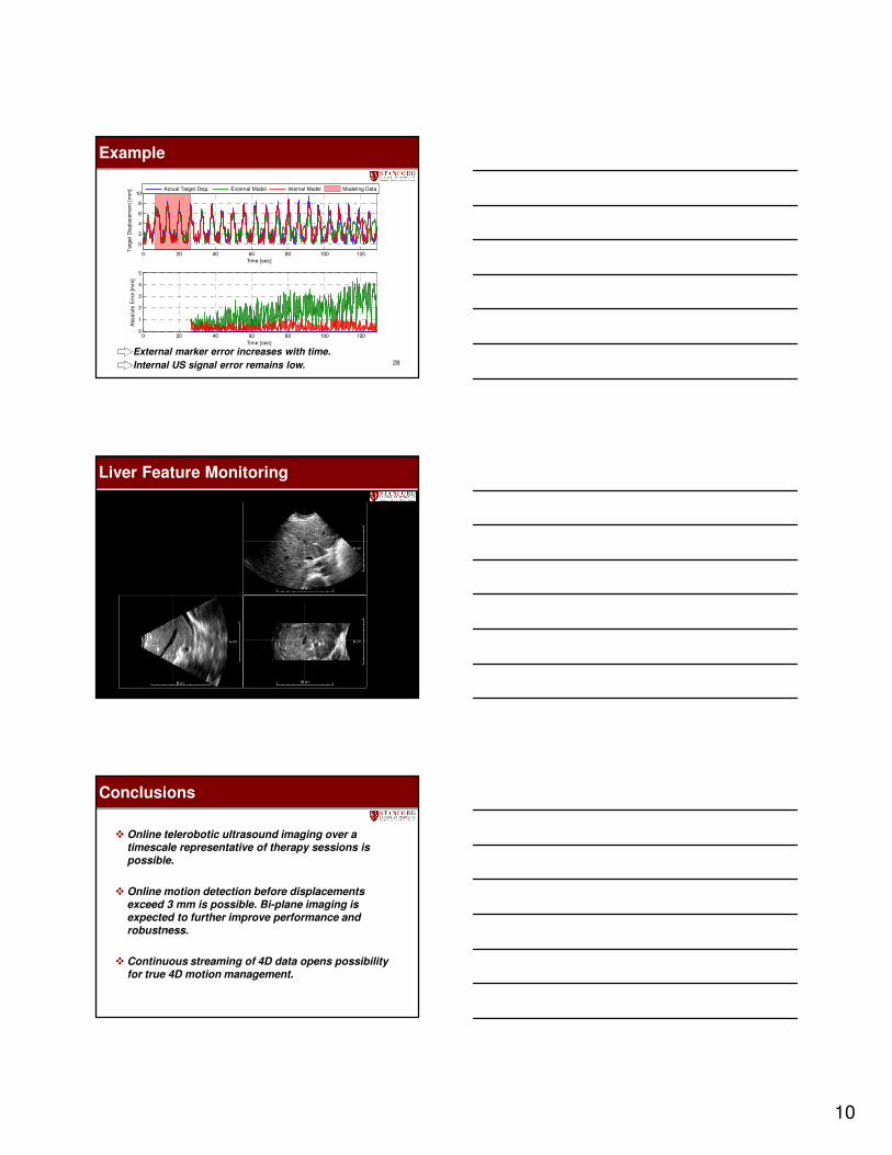
Example
External marker error increases with time.
Internal US signal error remains low.
0 20 40 60 80 100 120
0
2
4
6
8
10
Time [sec]
Targ
et
Dis
pla
cem
en
t [m
m]
Actual Target Disp. External Model Internal Model Modeling Data
0 20 40 60 80 100 1200
1
2
3
4
5
Abso
lute
Err
or
[mm
]
Time [sec]
29
Liver Feature Monitoring
Conclusions
� Online telerobotic ultrasound imaging over a timescale representative of therapy sessions is possible.
� Online motion detection before displacements exceed 3 mm is possible. Bi-plane imaging is expected to further improve performance and robustness.
� Continuous streaming of 4D data opens possibility for true 4D motion management.
11
Conclusions
� Simulation tools are expected to enable comprehensive studies on treatment planning strategies to account for the manipulator.
� Evaluation of long term effects of radiation on the transducer performance is required.
� Cross-validation against other modalities (radiographic imaging of fiducial markers) is ultimately necessary.
Questions ?