REAKSI HIPERSENSITIVITAS
37
م ي ح ر ل ا ن م ح ر ل ا ة ل ل م ا س ب م ي ح ر ل ا ن م ح ر ل ا ة ل ل م ا س ب
-
Upload
metta-sari -
Category
Documents
-
view
24 -
download
5
description
REAKSI HIPERSENSITIVITAS
Transcript of REAKSI HIPERSENSITIVITAS

الرحمن اللة الرحمن بسم اللة بسمالرحيمالرحيم

REAKSI REAKSI HIPERSENSITIVITASHIPERSENSITIVITAS
dr Atik Sutisna, SpAndr Atik Sutisna, SpAn Vice Dean for Academic affairVice Dean for Academic affair Lecturer of Anaesthesiology and Reanimation, Lecturer of Anaesthesiology and Reanimation,
Faculty of Medicine, Unswagati , CirebonFaculty of Medicine, Unswagati , Cirebon
Vice Director for Medical services and NursingVice Director for Medical services and Nursing Head of Anaesthesia and Intensive Care Unit Head of Anaesthesia and Intensive Care Unit
Putera Bahagia Hospital, CirebonPutera Bahagia Hospital, Cirebon

Immunologic Mechanisms Immunologic Mechanisms of Tissue Damageof Tissue Damage
(Immuopathology)(Immuopathology)
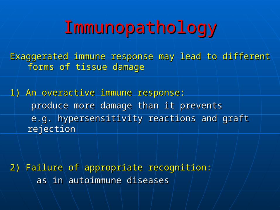
ImmunopathologyImmunopathology
Exaggerated immune response may lead to different Exaggerated immune response may lead to different forms of tissue damageforms of tissue damage
1) An overactive immune response:1) An overactive immune response:
produce more damage than it preventsproduce more damage than it prevents
e.g. hypersensitivity reactions and graft rejectione.g. hypersensitivity reactions and graft rejection
2) Failure of appropriate recognition:2) Failure of appropriate recognition:
as in autoimmune diseasesas in autoimmune diseases

Hypersensitivity ReactionHypersensitivity Reaction
Hypersensitivity or allergyHypersensitivity or allergy * An immune response results in exaggerated * An immune response results in exaggerated
reactions harmful to the hostreactions harmful to the host
* There are * There are four typesfour types of hypersensitivity reactions: of hypersensitivity reactions:
Type I, Type II, Type III, Type IV Type I, Type II, Type III, Type IV
* Types I, II and III are * Types I, II and III are antibody mediatedantibody mediated
* Type IV is * Type IV is cell mediatedcell mediated


Type I: Immediate hypersensitivityType I: Immediate hypersensitivity
* An antigen* An antigen reacts with cell fixed antibody (Ig E) reacts with cell fixed antibody (Ig E)
leading to release of leading to release of soluble moleculessoluble molecules
An antigenAn antigen (allergen) (allergen)
soluble moleculessoluble molecules (mediators) (mediators)
* Soluble molecules cause the manifestation of disease* Soluble molecules cause the manifestation of disease
* Systemic life threatening; * Systemic life threatening; anaphylactic shockanaphylactic shock
* Local atopic allergies; * Local atopic allergies; bronchial asthma, hay fever bronchial asthma, hay fever
and food allergiesand food allergies

Pathogenic mechanismsPathogenic mechanisms* First exposure to allergen* First exposure to allergen
Allergen stimulates formation of antibody (Ig E type)Allergen stimulates formation of antibody (Ig E type)
Ig E fixes, by its Fc portion to mast cells and basophilesIg E fixes, by its Fc portion to mast cells and basophiles
* Second exposure to the same allergen* Second exposure to the same allergen
It bridges between Ig E molecules fixed to mast It bridges between Ig E molecules fixed to mast cellsleading to activation and degranulation of mast cellsleading to activation and degranulation of mast cells and release of mediatorscells and release of mediators

Pathogenic mechanismsPathogenic mechanisms
* Three classes of mediators derived from mast cells:* Three classes of mediators derived from mast cells: !) Preformed mediators stored in granules !) Preformed mediators stored in granules (histamine)(histamine)
2) Newly sensitized mediators:2) Newly sensitized mediators: leukotrienes, prostaglandins, platelets activating factorleukotrienes, prostaglandins, platelets activating factor
3) Cytokines produced by activated mast cells, basophils 3) Cytokines produced by activated mast cells, basophils e.g. e.g. TNF, IL3, IL-4, IL-5 IL-13, chemokinesTNF, IL3, IL-4, IL-5 IL-13, chemokines
* These mediators cause:* These mediators cause: smooth muscle contraction, smooth muscle contraction, mucous secretion and bronchial spasm, vasodilatation, mucous secretion and bronchial spasm, vasodilatation, vascular permeability and edema vascular permeability and edema


AnaphylaxisAnaphylaxis* Systemic form of * Systemic form of Type I hypersensitivityType I hypersensitivity
* * Exposure to allergenExposure to allergen to which a person is previously sensitized to which a person is previously sensitized
* Allergens:* Allergens:
Drugs: Drugs: penicillinpenicillin
Serum injection : Serum injection : anti-diphtheritic or ant-tetanic serumanti-diphtheritic or ant-tetanic serum
anesthesia or insect venomanesthesia or insect venom
* Clinical picture:* Clinical picture:
ShockShock due to due to sudden decrease of blood pressuresudden decrease of blood pressure, , respiratory respiratory distress due to bronhospasmdistress due to bronhospasm, cyanosis, edema, urticaria, cyanosis, edema, urticaria
* Treatment: * Treatment: corticosteroids injection, epinephrine, antihistaminescorticosteroids injection, epinephrine, antihistamines

AtopyAtopy* * Local form of type I hypersensitivityLocal form of type I hypersensitivity
* * Exposure to certain allergensExposure to certain allergens that induce production of that induce production of specific Ig Especific Ig E
* * Allergens :Allergens : Inhalants:Inhalants:dust mite faeces, tree or pollens, mould spor.dust mite faeces, tree or pollens, mould spor. Ingestants:Ingestants: milk, egg, fish, choclate milk, egg, fish, choclate Contactants:Contactants: wool, nylon, animal fur wool, nylon, animal fur Drugs:Drugs: penicillin, salicylates, anesthesia insect venom penicillin, salicylates, anesthesia insect venom
* There is a strong * There is a strong familial predispositionfamilial predisposition to atopic allergy to atopic allergy
* The predisposition is * The predisposition is genetically determined genetically determined

Methods of diagnosisMethods of diagnosis
1) History taking1) History taking for determining the allergen involved for determining the allergen involved
2) Skin tests:2) Skin tests:
Intradermal injection of battery of different allergens Intradermal injection of battery of different allergens
A wheal and flare (erythema) develop at the site ofA wheal and flare (erythema) develop at the site of
allergen to which the person is allergicallergen to which the person is allergic
3) Determination of total serum Ig E level3) Determination of total serum Ig E level
4) Determination of specific Ig E levels4) Determination of specific Ig E levels to the different to the different allergens allergens
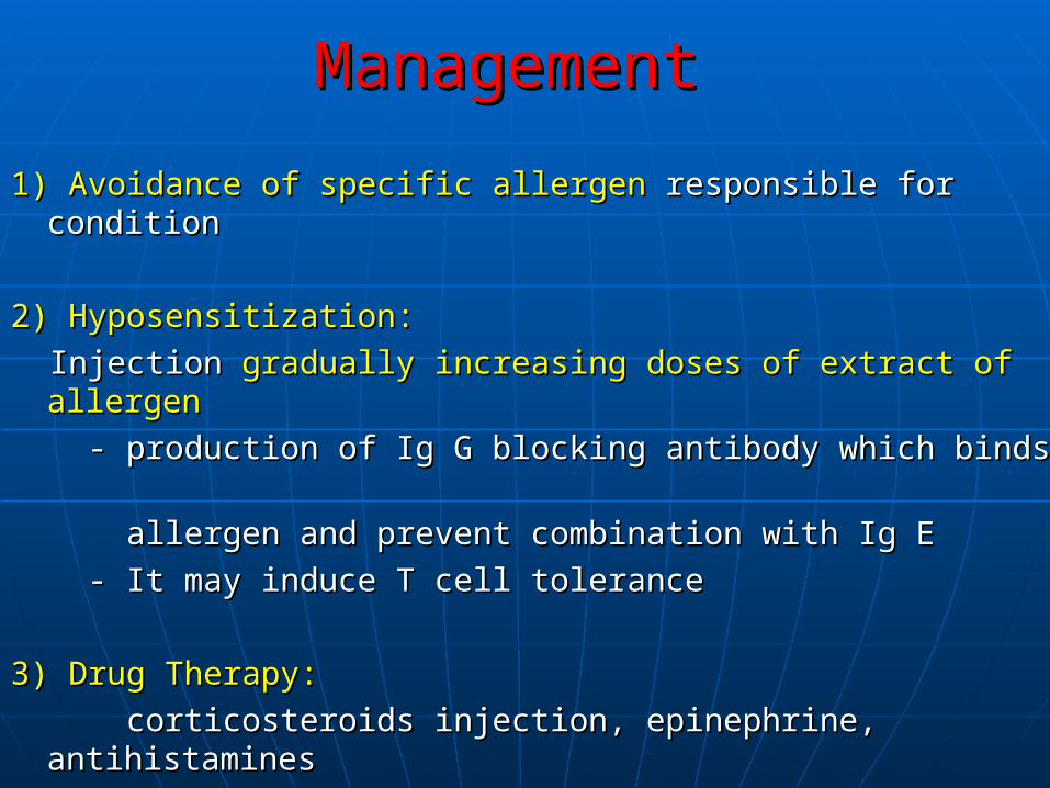
ManagementManagement
1) Avoidance of specific allergen1) Avoidance of specific allergen responsible for condition responsible for condition
2) Hyposensitization:2) Hyposensitization:
Injection Injection gradually increasing doses of extract of allergengradually increasing doses of extract of allergen
- production of Ig G blocking antibody which binds - production of Ig G blocking antibody which binds
allergen and prevent combination with Ig E allergen and prevent combination with Ig E
- It may induce T cell tolerance- It may induce T cell tolerance
3) Drug Therapy:3) Drug Therapy:
corticosteroids injection, epinephrine, antihistaminescorticosteroids injection, epinephrine, antihistamines

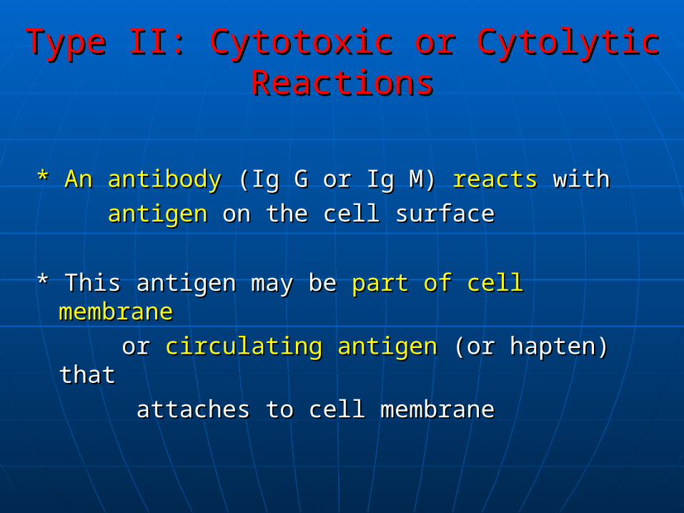
Type II: Cytotoxic or Cytolytic ReactionsType II: Cytotoxic or Cytolytic Reactions
* An antibody* An antibody (Ig G or Ig M) (Ig G or Ig M) reactsreacts with with
antigenantigen on the cell surface on the cell surface
* This antigen may be * This antigen may be part of cell membranepart of cell membrane
or or circulating antigencirculating antigen (or hapten) that (or hapten) that
attaches to cell membrane attaches to cell membrane

Mechanism of CytolysisMechanism of Cytolysis
* Cell lysis* Cell lysis results due to : results due to :
1) Complement fixation to antigen antibody 1) Complement fixation to antigen antibody complexcomplex on cell surface on cell surface
The activated complement will lead to cell lysisThe activated complement will lead to cell lysis
2) Phagocytosis is enhanced by the antibody2) Phagocytosis is enhanced by the antibody (opsinin) bound to cell antigen leading to (opsinin) bound to cell antigen leading to opsonization of the target cellopsonization of the target cell

Mechanism of cytolysisMechanism of cytolysis3) Antibody depended cellular cytotoxicity (ADCC):3) Antibody depended cellular cytotoxicity (ADCC):
- Antibody coated cells- Antibody coated cells
e.g. tumour cells, graft cells or infected cells can e.g. tumour cells, graft cells or infected cells can
be killed by cells possess Fc receptorsbe killed by cells possess Fc receptors
- The process - The process differentdifferent from from phagocytosisphagocytosis and and
independent of complementindependent of complement
- Cells most active in ADCC are:- Cells most active in ADCC are:
NK, macrophages, neutrophils and eosinophils NK, macrophages, neutrophils and eosinophils

Clinical ConditionsClinical Conditions
1) Transfusion1) Transfusion reaction due to ABO incompatibility reaction due to ABO incompatibility
2) Rh-incompatability2) Rh-incompatability (Haemolytic disease of the newborn)(Haemolytic disease of the newborn)
3) Autoimmune diseases3) Autoimmune diseases The mechanism of tissue damage is cytotoxic reactionsThe mechanism of tissue damage is cytotoxic reactions e.g. SLE, autoimmune haemolytic anaemia, idiopathic e.g. SLE, autoimmune haemolytic anaemia, idiopathic
thrombocytopenic purpura, myasthenia gravis, nephrotoxic thrombocytopenic purpura, myasthenia gravis, nephrotoxic nephritis, Hashimoto’s thyroiditisnephritis, Hashimoto’s thyroiditis
44) A non-cytotoxic Type II hypersensitivity) A non-cytotoxic Type II hypersensitivity is Graves’s disea is Graves’s disea It is a form of thyroditits in which antibodies are produced It is a form of thyroditits in which antibodies are produced
against TSH surface receptoragainst TSH surface receptor This lead to mimic the effect of TSH and stimulate cells to over- This lead to mimic the effect of TSH and stimulate cells to over-
produce thyroid hormonesproduce thyroid hormones

Clinical ConditionsClinical Conditions5- Graft rejection cytotoxic reactions:5- Graft rejection cytotoxic reactions: In hyperacute rejection the recipient already has In hyperacute rejection the recipient already has
performed antibody against the graftperformed antibody against the graft
6- Drug reaction:6- Drug reaction: PenicillinPenicillin may attach as haptens to RBCs and may attach as haptens to RBCs and
induce antibodies which are cytotoxic for the induce antibodies which are cytotoxic for the
cell-drug complex leading to haemolysiscell-drug complex leading to haemolysis
QuinineQuinine may attach to platelets and the antibodies may attach to platelets and the antibodies
cause platelets destruction and thrombocytopenic cause platelets destruction and thrombocytopenic
purpurapurpura

Type III HypersensitivityType III Hypersensitivity
Immune Complex Mediated ReactionImmune Complex Mediated Reaction


Type III: Immune Complex Mediated ReactionType III: Immune Complex Mediated Reaction
*When *When antibodiesantibodies (Ig G or Ig M) and (Ig G or Ig M) and antigenantigen coexist coexist immune complexesimmune complexes are formed are formed
*Immune complexes are *Immune complexes are removedremoved by by reticuloendoth. syst.reticuloendoth. syst.
*Some *Some immune complexesimmune complexes escape phagocytosisescape phagocytosis
**Immune complexes depositedImmune complexes deposited in tissues on the basement in tissues on the basement
membrane of blood vessels and cause membrane of blood vessels and cause tissue injurytissue injury

1- Arthus Reaction1- Arthus Reaction
* This is a local immune complex deposition phenomenon* This is a local immune complex deposition phenomenon e.g. e.g. diabetic patients receiving insulin subcutaneouslydiabetic patients receiving insulin subcutaneously
edemaedema* Local reactions in the form of erythema * Local reactions in the form of erythema necrosisnecrosis
depositeddeposited* Immune complexes in small blood vessels * Immune complexes in small blood vessels
vasculitisvasculitis leading toleading to microthrombi formation microthrombi formation vascular occlusion vascular occlusion necrosis necrosis
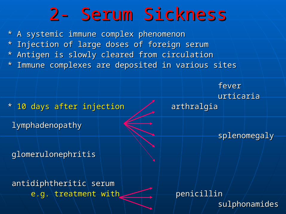
2- Serum Sickness2- Serum Sickness * A systemic immune complex phenomenon* A systemic immune complex phenomenon * Injection of large doses of foreign serum* Injection of large doses of foreign serum * Antigen is slowly cleared from circulation* Antigen is slowly cleared from circulation * Immune complexes are deposited in various sites* Immune complexes are deposited in various sites
feverfever urticariaurticaria * * 10 days after injection10 days after injection arthralgia arthralgia lymphadenopathylymphadenopathy splenomegalysplenomegaly glomerulonephritisglomerulonephritis
antidiphtheritic serum antidiphtheritic serum
e.g. treatment withe.g. treatment with penicillin penicillin sulphonamidessulphonamides

Clinical conditions of Type III HypersensitivityClinical conditions of Type III Hypersensitivity
DiseasesDiseases produced by produced by immune complexesimmune complexes are those in are those in
which antigens persists without being eliminated as:which antigens persists without being eliminated as:
a- Repeated exposure to extrinsic antigena- Repeated exposure to extrinsic antigen
b- injection of large amounts of antigensb- injection of large amounts of antigens
c- Persistent infectionsc- Persistent infections
d- Autoimmunity to self componentsd- Autoimmunity to self components

Mechanism Of Tissue InjuryMechanism Of Tissue Injury Immune complexes trigger inflammatory processes:Immune complexes trigger inflammatory processes:
activate releaseactivate release
1) Immune complexes the complement anaphylatoxins 1) Immune complexes the complement anaphylatoxins C3a, C5aC3a, C5a
stimulate releasestimulate release
degranulation of basophiles and mast cells histamine degranulation of basophiles and mast cells histamine
Histamine vascular permeability and help deposition of immune complexesHistamine vascular permeability and help deposition of immune complexes
2) Neutrophils are attracted to the site by immune complexes and release2) Neutrophils are attracted to the site by immune complexes and release
lysosomal enzymes which damage tissues and intensify the inflammat. Pro. lysosomal enzymes which damage tissues and intensify the inflammat. Pro.
3) Platelets are aggregated with two consequences 3) Platelets are aggregated with two consequences
a- release of histaminea- release of histamine
b- form of microthrombi which lead to ischemiab- form of microthrombi which lead to ischemia

Type IV Type IV
Cell Mediated Cell Mediated Delayed Type HypersensitivityDelayed Type Hypersensitivity


Type IV: Cell Mediated Type IV: Cell Mediated Delayed Type HypersensitivityDelayed Type Hypersensitivity
triggering DTH reactions by TH1triggering DTH reactions by TH1
* T-cells cause tissue injury by or* T-cells cause tissue injury by or
directly killing target cells by CD8directly killing target cells by CD8
* TH1 and CD8 T cells secrete cytokines (IFN-* TH1 and CD8 T cells secrete cytokines (IFN-γγ and TNF) and TNF)
attract lymphocytesattract lymphocytes
* Cytokines activate macrophages* Cytokines activate macrophages
induce inflammation induce inflammation
* Tissue damage results from products of activated macrophages* Tissue damage results from products of activated macrophages

Tuberculin –Type HypersensitivityTuberculin –Type Hypersensitivity
* When * When PPD is injectedPPD is injected intradermally in intradermally in sensitizedsensitized person person
* Local * Local indurated areaindurated area appears appears injection siteinjection site (48-72 hs) (48-72 hs)
* Indurations due to * Indurations due to accumulationaccumulation Of: Of:
macrophages and lymphocytesmacrophages and lymphocytes
* Similar reactions observed in diseases * Similar reactions observed in diseases
e.g. e.g. brucellosis, lepromin test in leprosy, Frei’s test in brucellosis, lepromin test in leprosy, Frei’s test in
lymphogranuloma venereumlymphogranuloma venereum

Granulomatous lesionsGranulomatous lesions
* * In chronic diseasesIn chronic diseases : T.B., Leprosy, schistosomiases : T.B., Leprosy, schistosomiases
* * Intracellular organisms resist destructionIntracellular organisms resist destruction by macrophag. by macrophag.
* * Persistent antigenPersistent antigen in tissues in tissues stimulate local DTHstimulate local DTH reaction reaction
* Continuous * Continuous release of cytokinesrelease of cytokines leads to leads to accumulationaccumulation of of macrophagesmacrophages which give rise to which give rise to epitheloidal and giant epitheloidal and giant cell granuloma cell granuloma

Contact DermatitisContact Dermatitis* * Contact of skinContact of skin with with chemical chemical substances orsubstances or drugs drugs e.g. poison, hair dyes, cosmetics, soaps, neomycine.g. poison, hair dyes, cosmetics, soaps, neomycin
* These substances * These substances enter skinenter skin in small molecules in small molecules
* They are * They are haptenshaptens that that attachedattached to body to body proteinsproteins, form, form immunogenic substancesimmunogenic substances
* * DTH reactionDTH reaction to these to these immunogenicimmunogenic subst. lead to: subst. lead to:
eczymaeczyma inflammtory reaction of skin ininflammtory reaction of skin in rash rash vesicular eruptionvesicular eruption

Type IV Hypersensitivity Clinical ConditionsType IV Hypersensitivity Clinical Conditions
1)1) AutoAuto immune diseases immune diseases andand graft rejection graft rejection are due to are due to in part to delayed hypersensitivity reactionsin part to delayed hypersensitivity reactions
2)2) Insulin dependant diabetes mellitusInsulin dependant diabetes mellitus
T-cells invadeT-cells invade the the pancreatic isletspancreatic islets and specifically and specifically destroydestroy insulin secreting insulin secreting beta cellsbeta cells

Thanks

![HIPERSENSITIVITAS [Autosaved]](https://static.fdocuments.in/doc/165x107/577c80b81a28abe054a9e3f3/hipersensitivitas-autosaved.jpg)