Re-evaluation of the WHO (2010) formaldehyde indoor air quality ... · production of plastics,...
27
1 3 Arch Toxicol (2017) 91:35–61 DOI 10.1007/s00204-016-1733-8 REVIEW ARTICLE Re‑evaluation of the WHO (2010) formaldehyde indoor air quality guideline for cancer risk assessment Gunnar Damgård Nielsen 1 · Søren Thor Larsen 1 · Peder Wolkoff 1 Received: 5 March 2016 / Accepted: 27 April 2016 / Published online: 21 May 2016 © The Author(s) 2016. This article is published with open access at Springerlink.com credibility of the WHO guideline has not been challenged by new studies. Keywords Formaldehyde · World Health Organization · Indoor air guideline · Cancer · Risk assessment Introduction Formaldehyde (FA; 1 ppm = 1.23 mg/m 3 at 1 atm and 25 °C) is a high-volume chemical, which is used for dis- infection purposes and as a preservative. Also, it is used in the production of resins and binders, which are used in wood-products (e.g. particle board and plywood), pulp and paper, and mineral wool. Furthermore, FA is used in the production of plastics, coatings and paints, flooring materi- als, for textile finishing, for synthesis of chemicals, and it is a component of combustion products (Salthammer et al. 2010; IARC 2012). Additionally, FA is a major compound derived from ozone-initiated reactions with alkenes, e.g. terpenes (Atkinson and Arey 2003). Due to its ubiquitous use, FA is a common indoor air pollutant. The majority of the studies showed that indoor air con- centrations in Europe and the USA were below 100 µg/m 3 and the median, geometric mean or arithmetic means ranged between 5 and 60 µg/m 3 (Salthammer et al. 2010; Sarigiannis et al. 2011). Overall, these levels are supported by recent studies in industrialized countries. Thus, nurs- ing homes for elderly people in seven countries in Europa had a mean (8 h) FA concentration of 7 µg/m 3 and a maxi- mum concentration of 21 µg/m 3 (Bentayeb et al. 2015). In a study in French dwellings, the range of FA concentrations varied from 18 to 26 µg/m 3 (Brown et al. 2015). In another study in French dwellings, the mean, 90th percentile and the maximum concentration was 29, 46 and 113 µg/m 3 , Abstract In 2010, the World Health Organization (WHO) established an indoor air quality guideline for short- and long-term exposures to formaldehyde (FA) of 0.1 mg/ m 3 (0.08 ppm) for all 30-min periods at lifelong expo- sure. This guideline was supported by studies from 2010 to 2013. Since 2013, new key studies have been published and key cancer cohorts have been updated, which we have evaluated and compared with the WHO guideline. FA is genotoxic, causing DNA adduct formation, and has a clas- togenic effect; exposure–response relationships were non- linear. Relevant genetic polymorphisms were not identified. Normal indoor air FA concentrations do not pass beyond the respiratory epithelium, and therefore FA’s direct effects are limited to portal-of-entry effects. However, systemic effects have been observed in rats and mice, which may be due to secondary effects as airway inflammation and (sensory) irritation of eyes and the upper airways, which inter alia decreases respiratory ventilation. Both secondary effects are prevented at the guideline level. Nasopharyn- geal cancer and leukaemia were observed inconsistently among studies; new updates of the US National Cancer Institute (NCI) cohort confirmed that the relative risk was not increased with mean FA exposures below 1 ppm and peak exposures below 4 ppm. Hodgkin’s lymphoma, not observed in the other studies reviewed and not considered FA dependent, was increased in the NCI cohort at a mean concentration ≥0.6 mg/m 3 and at peak exposures ≥2.5 mg/ m 3 ; both levels are above the WHO guideline. Overall, the * Gunnar Damgård Nielsen [email protected] 1 National Research Centre for the Working Environment, Lersø Parkallé 105, 2100 Copenhagen, Denmark
-
Upload
truongtuyen -
Category
Documents
-
view
213 -
download
0
Transcript of Re-evaluation of the WHO (2010) formaldehyde indoor air quality ... · production of plastics,...
1 3
Arch Toxicol (2017) 91:35–61DOI 10.1007/s00204-016-1733-8
REVIEW ARTICLE
Re‑evaluation of the WHO (2010) formaldehyde indoor air quality guideline for cancer risk assessment
Gunnar Damgård Nielsen1 · Søren Thor Larsen1 · Peder Wolkoff1
Received: 5 March 2016 / Accepted: 27 April 2016 / Published online: 21 May 2016 © The Author(s) 2016. This article is published with open access at Springerlink.com
credibility of the WHO guideline has not been challenged by new studies.
Keywords Formaldehyde · World Health Organization · Indoor air guideline · Cancer · Risk assessment
Introduction
Formaldehyde (FA; 1 ppm = 1.23 mg/m3 at 1 atm and 25 °C) is a high-volume chemical, which is used for dis-infection purposes and as a preservative. Also, it is used in the production of resins and binders, which are used in wood-products (e.g. particle board and plywood), pulp and paper, and mineral wool. Furthermore, FA is used in the production of plastics, coatings and paints, flooring materi-als, for textile finishing, for synthesis of chemicals, and it is a component of combustion products (Salthammer et al. 2010; IARC 2012). Additionally, FA is a major compound derived from ozone-initiated reactions with alkenes, e.g. terpenes (Atkinson and Arey 2003). Due to its ubiquitous use, FA is a common indoor air pollutant.
The majority of the studies showed that indoor air con-centrations in Europe and the USA were below 100 µg/m3 and the median, geometric mean or arithmetic means ranged between 5 and 60 µg/m3 (Salthammer et al. 2010; Sarigiannis et al. 2011). Overall, these levels are supported by recent studies in industrialized countries. Thus, nurs-ing homes for elderly people in seven countries in Europa had a mean (8 h) FA concentration of 7 µg/m3 and a maxi-mum concentration of 21 µg/m3 (Bentayeb et al. 2015). In a study in French dwellings, the range of FA concentrations varied from 18 to 26 µg/m3 (Brown et al. 2015). In another study in French dwellings, the mean, 90th percentile and the maximum concentration was 29, 46 and 113 µg/m3,
Abstract In 2010, the World Health Organization (WHO) established an indoor air quality guideline for short- and long-term exposures to formaldehyde (FA) of 0.1 mg/m3 (0.08 ppm) for all 30-min periods at lifelong expo-sure. This guideline was supported by studies from 2010 to 2013. Since 2013, new key studies have been published and key cancer cohorts have been updated, which we have evaluated and compared with the WHO guideline. FA is genotoxic, causing DNA adduct formation, and has a clas-togenic effect; exposure–response relationships were non-linear. Relevant genetic polymorphisms were not identified. Normal indoor air FA concentrations do not pass beyond the respiratory epithelium, and therefore FA’s direct effects are limited to portal-of-entry effects. However, systemic effects have been observed in rats and mice, which may be due to secondary effects as airway inflammation and (sensory) irritation of eyes and the upper airways, which inter alia decreases respiratory ventilation. Both secondary effects are prevented at the guideline level. Nasopharyn-geal cancer and leukaemia were observed inconsistently among studies; new updates of the US National Cancer Institute (NCI) cohort confirmed that the relative risk was not increased with mean FA exposures below 1 ppm and peak exposures below 4 ppm. Hodgkin’s lymphoma, not observed in the other studies reviewed and not considered FA dependent, was increased in the NCI cohort at a mean concentration ≥0.6 mg/m3 and at peak exposures ≥2.5 mg/m3; both levels are above the WHO guideline. Overall, the
* Gunnar Damgård Nielsen [email protected]
1 National Research Centre for the Working Environment, Lersø Parkallé 105, 2100 Copenhagen, Denmark

36 Arch Toxicol (2017) 91:35–61
1 3
respectively, in children’s bedrooms (Dallongeville et al. 2015). Another study compared apartments in Finland and in Lithuania (Du et al. 2015). The mean and maximum concentrations were 17.5 and 40 µg/m3 and 23 and 51 µg/m3, respectively. A Spanish study showed that homes in a Spanish city had a mean, 75th percentile and maximum FA concentration of 55, 74 and 91 µg/m3, respectively (Vil-lanueva et al. 2015). In another Spanish study, no differ-ence was found in FA concentrations in indoor air concen-trations in the bedrooms, living rooms and non-industrial workplaces, mainly offices, where the mean concentration was about 25 µg/m3 and ranged from 6 to 48 µg/m3 (Rovira et al. 2016). Mullen et al. (2016) showed that in Californian homes, the 25th and 75th percentiles were 12 and 25 µg/m3, respectively, with a maximum of 50 µg/m3. In another Californian study, the mean FA concentration was 34 µg/m3 in homes built with low-emitting materials and 46 µg/m3 in conventional homes at an air exchange rate of 0.35 h−1 (Hult et al. 2015). Furthermore, 40 early childhood edu-cation facilities were studied in California; the arithmetic mean FA concentration was 19 µg/m3 with a range from 0.7 to 49 µg/m3 (Bradman et al. 2016). In houses inhabited by asthmatics in the Boston area, the geometric mean FA con-centration was 43 µg/m3. The concentrations ranged from 6 to 162 µg/m3, and 6 % of the houses had a FA concentration exceeding 122 µg/m3 (Dannemiller et al. 2013). In homes in Australia, the mean and maximum FA concentration was 15 and 46 µg/m3, respectively (Lazenby et al. 2012). In homes in Japan, the mean and maximum concentration was 13 and 58 µg/m3, respectively, in the winter and in the sum-mer 34 and 220 µg/m3, respectively, with 0.7 % exceed-ing 100 µg/m3 (Uchiyama et al. 2015). In Korea, in newly built apartments at the pre-occupancy stage, the mean, the 95th percentile and the maximum FA concentration was 61, 110 and 160 µg/m3, respectively (Shin and Jo 2012). In apartments in a Chinese city, the mean (range) concentra-tion was 100 (80–130) µg/m3 in living rooms (Zhu and Liu 2014). In Beijing, even higher concentrations were found in dwellings and offices that had been remodelled within the past year. Thus, the mean (±SD) was 131 ± 90 µg/m3 in dwellings with a maximum concentration of 800 and 85 ± 56 µg/m3 in offices with a maximum concentration of 300 µg/m3 (Huang et al. 2013).
Many countries have set guideline values for indoor air FA (Salthammer et al. 2010). The World Health Organiza-tion (2010) set an indoor air quality guideline (IAQG) for FA at 0.1 mg/m3 (0.08 ppm), which applies to all 30-min periods lifelong. The guideline was further supported by extended literature reviews (Nielsen and Wolkoff 2010; Wolkoff and Nielsen 2010). Shortly after the WHO pre-sented its recommendation, Golden (2011) also analysed the FA data and proposed an indoor air guideline value of 0.1 ppm (0.12 mg/m3). A recent update by IARC (2012)
classified FA as “carcinogenic to humans (Group 1)” on the basis that FA may cause cancer of the nasopharynx and leukaemia, whereas there was limited evidence for associa-tion with sinonasal cancer. However, a consistent finding is the observed occurrence of nasal cancer in rats and mice at high FA exposures.
Many new key studies have been published that have been used in this re-evaluation of the WHO (2010) IAQG. Previous conclusions have been summarized from the eval-uations (WHO 2010; Nielsen and Wolkoff 2010; Wolkoff and Nielsen 2010; Golden 2011; RAC 2012; Nielsen et al. 2013; NRC 2014). The focus of this review is recent stud-ies (mainly years ≥ 2013); however, for transparency rea-sons, earlier key studies have also been included when con-sidered appropriate. We have excluded new occupational studies that do not include measured FA concentrations, studies where the exposure–response relationships could not be evaluated (e.g. Attia et al. 2014; Santovito et al. 2014), and studies with complex environmental exposures (e.g. Vilavert et al. 2014) where no measured health effect was included or where a low-level environmental FA expo-sure was a proxy of an exposure to a complex outdoor air mixture (e.g. Marcon et al. 2014) as such exposures do not allow disentangling of the effects of FA. It should be noted that this does not mean that such studies are not useful for risk management purposes. Also, we excluded animal stud-ies with mixtures where effects of FA could not be disen-tangled (e.g. Wang et al. 2013a) and studies with exposures to FA aerosols (e.g. Lima et al. 2015) as the WHO IAQG is set for gaseous FA. Although the specific purpose is the evaluation of the WHO IAQG for FA, the evaluated studies are also relevant for setting other guidelines or standards for FA, for example, occupational exposure limits.
Absorption, distribution, metabolism and elimination
Due to its high water solubility and reactivity, airborne FA is absorbed mainly (~90 %) in the upper airways (c.f. WHO 2010; Nielsen et al. 2013). In the aqueous tissue phase, FA adds water, forming methandiol (methylene glycol, CH2(OH)2), accounting for more than 99.9 % of total FA in the aqueous phase. CH2(OH)2 is in equilibrium with free FA (<0.1 %) in the water phase, where CH2(OH)2 serves as a FA liberator. CH2(OH)2 itself may have a low toxic-ity (Golden and Valantini 2014). In the tissue, FA forms adducts and cross-links with RNA, DNA and proteins, including DNA–protein cross-links (DPX). In rat nasal tis-sue, DPX increases disproportionately at exposure levels above 2–3 ppm. FA is an endogenous metabolite, and its blood concentration is about 2–3 mg FA/L. The half-life of FA in blood is about 1–1.5 min. FA is metabolized to

37Arch Toxicol (2017) 91:35–61
1 3
formate, which is incorporated in tissue components via the one-carbon pool, excreted in the urine or oxidized to carbon dioxide (c.f. WHO 2010; Nielsen et al. 2013). Using a specific (unbiased) method, the FA concentration in air exhaled through the mouth was found at levels up to 1.7 ppb; this figure may be higher shortly after smoking a cigarette. However, the concentration was below 0.5 ppb in most cases (Riess et al. 2010). The exhaled FA concentra-tion may be higher in air breathed through the nose (Spanel et al. 2013). Estimated FA deposition in the upper airways and DPX formation were similar in children and adults (c.f. WHO 2010; Nielsen and Wolkoff 2010; Nielsen et al. 2013).
Uptake of FA in the nose of rats, monkeys and humans was estimated by means of an anatomically accurate com-putational fluid dynamics model. At ≥0.1 ppm, the nasal uptake was about 99, 87 and 85 %, respectively. The uptake was nonlinear, especially at lower concentrations (<0.1 ppm), and thus resulted in a lower nasal uptake frac-tion due to the effects of endogenous FA. Also, the higher fluxes were predicted to occur in regions located in the more anterior sections of the nose (Schroeter et al. 2014).
A mechanistic model was developed to study the uptake of airborne FA and transport into the surrounding lung tissue at 1 mg/m3 in humans. Disregarding the scrub-bing effects of the nasal and oral tissue, it was predicted that FA would be quickly absorbed (~97 %) by the mucus membranes with a very high uptake in the trachea (airway generation 0), and that no FA would pass beyond airway generation 8. Thus, no FA would reach the deep airways, including the alveoli, and no FA was predicted to pass to the blood compartment (Asgharian et al. 2012).
In the mucus layer, CH2(OH)2 diffuses into the epithe-lial cells and liberates FA, which reacts with glutathione (GSH), proteins, DNA and RNA. The GSH adduct (GS-FA) is oxidized by the FA dehydrogenase to the formate adduct. After hydrolysis, GSH and formate are released. Rats were exposed to 0 (control), 0.7, 2, 6, 10 and 15 ppm FA 6 h/day for 1, 4 or 13 weeks. Nasal tissue concentra-tions of CH2(OH)2, GSH, GS-FA and DPX were assessed as were histological effects, epithelial cell proliferation and gene expression. The data were analysed by means of a pharmacokinetic model, taking into account the back-ground CH2(OH)2 and GSH levels. The cellular levels of CH2(OH)2 and DPX only showed a minor increase with exposures at 0.7 and 2 ppm FA. At these levels, GSH decreased slightly. Several ppm FA would be required to achieve significant changes. Above 4 ppm, the changes were more conspicuous. Histopathology showed nasal lesions at 2 ppm and epithelial cell proliferation at higher concentrations. The lowest benchmark dose for change of gene expression approximated 1 ppm. The authors con-cluded that genomic changes at 0.7–2 ppm likely reflected
changes in extracellular CH2(OH)2 and GSH levels and that FA levels below 1 or 2 ppm would not affect FA homeosta-sis within the epithelial cells (Andersen et al. 2010).
A major advance was the differentiation between FA-induced DNA damage from the (normal) endogenous FA (CH2O) level in blood and tissue and from the inhaled (exogenous) FA, using isotope-labelled FA (13CD2O) for the airborne exposure; exposure in rats was to 10 ppm labelled FA for 1 or 5 days at 6 h/day. Inhaled FA induced labelled mono-adducts (N2–HO–13CD2-deoxyguanosine; labelled FA-dG), DNA–13CD2–DNA cross-links (labelled dG–FA–dG) and labelled DPX in the nasal tissue. Both at 1 or 5 days of exposure, the labelled FA-dG adduct was about 10 times more common than labelled dG–CH2–dG in the nasal tissue. The labelled FA-dG adduct on day 1 and day 5 was 32 and 46 %, respectively, and labelled dG–FA–dG was 45 and 59 %, respectively, of the respective adduct type. Neither labelled FA–dG nor labelled dG–FA–dG was detected in the liver, lungs, thymus, bone marrow, spleen and the blood lymphocytes. In contrast, high amounts of endogenous FA adducts were detected in all tissues. This indicated that exogenous FA exposures only had access to the portal-of-entry area (Lu et al. 2010). The dominating FA adduct to DNA is the FA–dG, which can be used as a sensitive biomarker of FA exposure (Lu et al. 2012a).
A single 6-h exposure to 0.7, 2, 6, 9 or 15 ppm in rats showed that the ratio between exogenous FA–dG and endogenous FA–dG was 0.01, 0.03, 0.2, 0.6 and 2.8, respectively, indicating a strongly nonlinear relationship in the nasal tissue. No exogenous FA–dG adduct was found in the bone marrow at the 15-ppm exposure concentration (Lu et al. 2011). Also in monkeys exposed to 2 or 6 ppm, 6 h/day for 2 days, the external FA–dG adduct was only detected in the nose and not in the bone marrow. At 6 ppm, the FA–dG adduct level was lower in the monkeys than in rats with a single 6-h exposure, suggesting a lower geno-toxic effect in primates than in rats (Moeller et al. 2011).
FA is a major source of N6-formyllysine (FA-Lys) adducts in cell proteins. In rats, exposure to isotope-labelled FA (13CD2O) at 0.7, 2, 6 and 9 ppm for 6 h was used to differentiate between adducts from exogenous and endogenous FA-Lys adducts in the total amount, the cyto-plasmic, the membrane and the nuclear proteins. After pro-teolysis and analysis of FA-Lys, the ratio between exoge-nous and endogenous adducts was shown to increase with increasing exposures; for example, for the total amount nasal epithelial proteins, the ratio was 0.035, 0.14, 0.15 and 0.40, respectively. At each FA exposure, the ratios were in the order cytoplasmic ≈ membrane > soluble nuclear > chromatin protein bound, indicating a decrease in the exogenous FA concentration from the cytoplasmic to the nuclear proteins. In contrast, the endogenous FA-Lys adducts were similar at all exposure concentrations in all

38 Arch Toxicol (2017) 91:35–61
1 3
cellular compartments. Moreover, this indicated that the exogenous FA exposure did not influence the endogenous FA production. No external FA-Lys adducts were detected in the lungs, liver and bone marrow, and thus the results paralleled studies on FA-dG adducts, confirming that direct exogenous FA effects are limited to the nasal epithelium (Edrissi et al. 2013).
In rats, absorption of inhaled FA into the blood was studied with (13C) labelled FA for a single 6-h exposure to 10 ppm; this allows differentiation between endogenous FA and FA from external exposure. The background blood FA levels were from 1.9 to 5.4 mg/L. Inhalation of FA did not increase the blood FA level nor was inhaled (13C labelled) FA detected in the blood above the natural background level (Kleinnijenhuis et al. 2013). These findings provide further support for the finding that the airway epithelium in rats is an efficient barrier against even high FA concentra-tion and its transport into the blood.
In a recent rat study, the exposure period was extended to 28 days with 2 ppm (13CD2)-labelled FA for 6 h per day and 7 days per week. Exogenous and endogenous FA–DNA adducts were obtained from the labelled and unla-belled FA–dG biomarker. The biomarker was considered to represent both mono-adducts and DPX-adducts as the DPX cross-links hydrolysed spontaneously to the mono-adduct. The exogenous adduct accumulated during the 28-day period and reached quasi-steady state after 28 days, at which point the ratio between the exogenous and endog-enous adducts was 0.37 in the nasal tissue; this value was higher than the ratio after a few exposures. In the first 6 h post-exposure, there was a rapid initial loss of nearly 20 % of the adducts in the nasal tissue that was followed by a phase with a longer half-life of 7.1 days. This was consid-ered to reflect DNA repair and/or spontaneous hydrolysis. No consistent exogenous adducts were found in internal organs, including the white blood cells, trachea, tracheal bronchial lymph nodes and lungs. This is in agreement with results from previous studies with fewer exposures conducted by the research group. Also, monkeys (cynomol-gus macques) were exposed to 13CD2-FA at 6 ppm for 6 h per day for 2 days. The exogenous biomarker was only observed in the nasal tissue and not in the tracheal carina, proximal trachea, white blood cells and the bone marrow (Yu et al. 2015).
Furthermore, DPX formation has been studied by an ultrasensitive and selective liquid chromatography-mass spectrometry method, where monkeys and rats were exposed to (13CD2)-labelled FA. This allowed differentia-tion between DPX from inhaled FA and DPX from endog-enous (normal) FA. Monkeys were exposed at 6 ppm, 6 h per day for 2 days. Labelled DPX was detected in the nasal tissue, but not in the peripheral blood mononuclear cells, bone marrow and the liver. Endogenously generated DPX
was detected in all investigated tissues. In the nasal tis-sue, endogenous DPX was about threefold higher than the exogenously generated DPX. Different tissues had differ-ent endogenous DPX levels. Thus, endogenous DPX was almost threefold higher in the liver than in the nasal tissue. Rats were exposed at 15 ppm, 6 h per day up to 4 days. Also in rats, exogenous DPX was only detected in the nasal tissue. Furthermore, the decay of exogenous DPX was studied in rats, which were exposed at 2 ppm, 6 h per day for 7 and 28 days, respectively, with a post-exposure period up to 7 days. In the post-exposure period, the exog-enous DPX decreased slowly (~10 %). In the nasal tissue, exogenous DPX increased with the number of exposures in both rat studies (Lai et al. 2016). It is noted that inhaled FA only caused DPX formation in the nasal tissue and DPX formation in internal organs cannot be explained by a direct transport of FA to the internal organs.
Overall, the recent studies have demonstrated that air-borne FA does not reach internal organs. Thus, if systemic effects occur, they have to be explained by secondary effects from portal-of-entry toxicity, which includes sen-sory-irritation-induced hypoxia (Nielsen et al. 2013) and airway inflammation. Other important findings are that the recent studies confirm that the external-induced FA-DNA adducts increase disproportionately in the nasal tissue at high FA concentrations that is similar to the exposure–response relationship for nasal cancer in rats. Furthermore, rats had more exogenous induced DNA adducts in the nasal tissue than monkeys.
The WHO (2010) IAQG accepts that direct internal organ effects may occur if the metabolic capacity of the upper airways is overloaded; this may begin at ≥2 ppm. Overall, the WHO (2010) evaluation constitutes a conserv-ative approach.
Genotoxicity
Formaldehyde is genotoxic due to its covalent binding to DNA, causing DNA mono-adducts, DNA–DNA cross-links, DPX and DNA glutathione cross-links that can cause mutations and clastogenic effects such as DNA strand breaks, chromosomal aberration (CA), micronu-cleus (MN) formation and sister chromatid exchange (SCE) as reviewed (IARC 2006; RAC 2012; NRC 2014; Kawanishi et al. 2014; Yu et al. 2015). Repair of the FA–DNA mono-adducts may include the base excision repair (BER) pathway, and the intra-strand cross-links may be by the nucleotide excision repair (NER) pathway (Kawanishi et al. 2014). FA-induced DPX may be repaired by the NER repair and by the homologous recombination (HR) path-ways (de Graaf et al. 2009; Kawanishi et al. 2014; McHale et al. 2014). Furthermore, DPX may partly be broken

39Arch Toxicol (2017) 91:35–61
1 3
down by specific proteolytic enzymes, allowing transle-sion synthesis polymerases (a potentially mutagenic path-way) to replicate across DNA-peptide lesions. Addition-ally, a tolerance pathway also exists, allowing replication across unrepaired DPX lesions that may include strand breaks (potentially causing genomic rearrangements) fol-lowed by strand ligation (Stingele et al. 2015). Not least, the Fanconi anaemia pathway is important in the repair of inter-strand DNA cross-links and DPX (Ren et al. 2013; Kirsch-Volders et al. 2014; McHale et al. 2014; Schneider et al. 2015).
Endogenously generated FA and toxicity
As FA is an endogenously generated compound, it may play a role in induction of diseases. Thus, a recent experi-mental study showed that elevation of the endogenous (natural) FA concentration in tissues can cause cell dam-age and destruction, as well as genetic damage and can-cer (Pontel et al. 2015). As FA is detoxified to formate by the alcohol dehydrogenase 5 (ADH5), mice without the gene (Aldh5−/−) had elevated FA-dG adducts in the bone marrow (1.7-fold), kidney (1.7-fold) and liver (2.3-fold) compared with the wild-type (Ald5+/+) mice. In Aldh5−/− mice, administration of methanol [a FA precursor (Lu et al. 2012b)] further increased the level of FA–dG adducts. As the FANCD2 protein is involved in the repair of FA–DNA cross-links, Fancd2−/− mice were also studied. The double deletion (Aldh5−/− Fancd2−/−) caused a profound decrease in survival, induced blood pancytopenia, reduced bone marrow cellularity (including hematopoietic stem and progenitor cells) and colony formation at cultivation of spleen hematopoietic stem cells. Cultivated spleen B cells stimulated with lipopolysaccharide showed a high level of chromosome breakages. Additionally, liver and kid-ney dysfunction with DNA damages were also observed. In contrast, no or limited effects were observed on the mentioned endpoints in the wild-type mice or mice with a single deletion of Aldh5−/− or Fancd2−/−, indicating a profound synergistic interaction between deletion of both the Aldh5−/− and Fancd2−/− genes. Transplantation of bone marrow from the wild-type mice to the double-defi-cient (Aldh5−/− Fancd2−/−) mice increased survival time and decreased kidney toxicity, but these animals devel-oped hepatocellular- and cholangiocarcinoma as well as T-lymphoblastic leukaemia. The authors concluded that FA is an important source of endogenous DNA damage that is counteracted in mammals by conserved protection mechanisms. It is noted that FA can cause serious damage at the place of contact. However, to observe these effects, external FA has to reach the blood and afterwards the internal organs, and this has not been observed in compre-hensive toxicokinetic studies.
Genotoxicity in human epithelial and blood cells
Previous reviews have shown that occupational exposures, either to mean or to peak FA concentrations from about 1 ppm and above, were associated with single strand break, MN formation, SCE and chromosomal aberration in buc-cal and nasal epithelial cells, and in peripheral lymphocytes (Nielsen and Wolkoff 2010; Nielsen et al. 2013). This indi-cates that an IAQG has to be below 1 ppm.
Ladeira et al. (2013) studied genotoxicity in buccal mucosa cells and showed an increase in MN frequency in employees exposed to FA in six histopathology hospital laboratories in Portugal; mean exposure for 8-h periods was 0.16 ppm (range 0.04–0.51 ppm) with a mean peak exposure of 1.14 ppm (range 0.18–2.93 ppm). The buc-cal MN effect may be a high-level effect. This finding was supported by a previous study with exposure for 4 h per day for 10 working days, where the daily background (constant) FA exposures ranged from 0.15 to 0.5 ppm with added peak exposures up to 1 ppm. At these exposure lev-els, no increase was observed in buccal MN compared to the pre-exposure MN level (Speit et al. 2007). Similarly in a controlled chamber study with FA exposure for 4 h for 5 days, where the FA concentrations ranged from 0.3 to 0.7 ppm with peaks up to 0.8 ppm, FA exposure had no effect on MN occurrence in the nasal epithelium (Zeller et al. 2011).
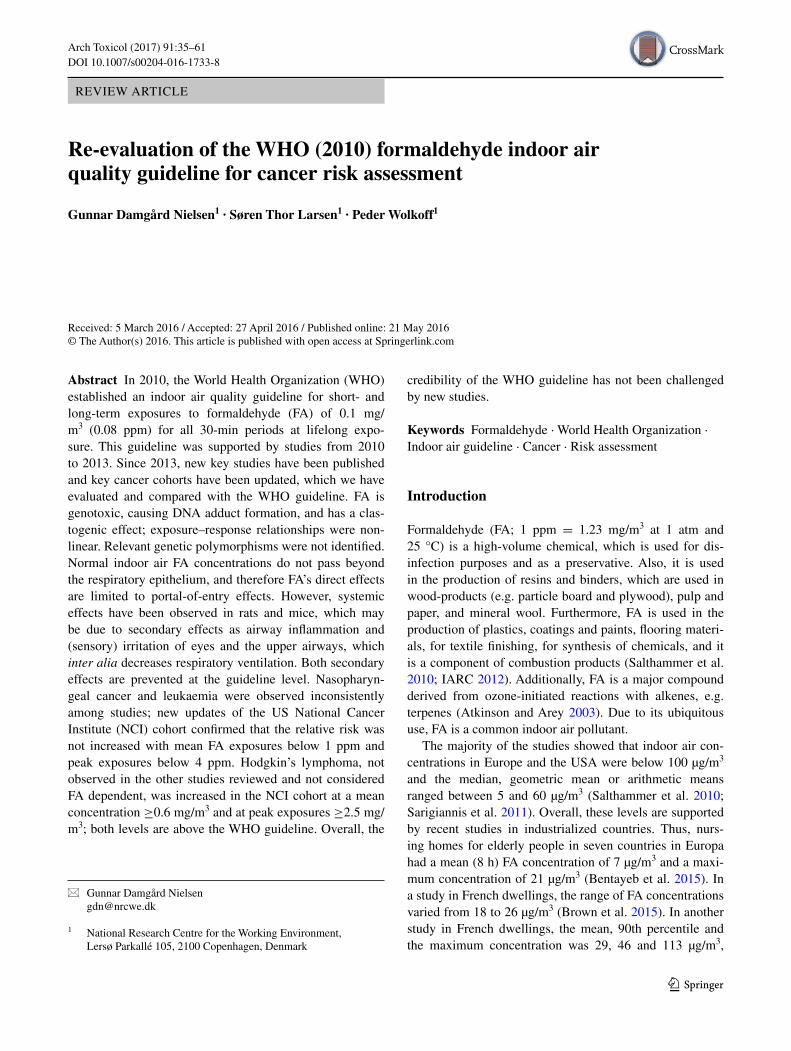
The recent studies on genotoxic effects in blood lym-phocytes are listed in Table 1. Studies in pathology lab-oratories confirm the previous association between FA exposure and genotoxicity in lymphocytes, where expo-sures included mean FA concentrations or peak concen-trations above 1 ppm. Where peak exposures were not reported, it can reasonably be assumed that exposures included peak concentrations above 1 ppm, as suggested from the studies where peak exposures were measured and in agreement with previous evaluations (Nielsen and Wolkoff 2010; Nielsen et al. 2013). Furthermore, this is supported by a study in pathology laboratory workers, who were exposed during successive decanting opera-tions, where they manually emptied and filled tissue processor reagent reservoirs (Persoons et al. 2012). The measured 15-min average concentration was 1.17 mg/m3 (1.0 ppm), whereas the estimated concentration was 1.7 mg/m3 (1.4 ppm), and the upper 95th percentile was 4.32 mg/m3 (3.5 ppm). The mean instantaneous peak con-centration was 19.5 mg/m3 (16 ppm), and the upper 95th percentile was 43.4 mg/m3 (35 ppm). In another study, plywood workers had exposures to high mean concentra-tions (Lin et al. 2013), and the peak concentrations may reasonably have been considerably higher than the mean concentrations. In contrast, workers in a medium density fibreboard plant (Aydin et al. 2013) had a stable exposure
40 Arch Toxicol (2017) 91:35–61
1 3
Tabl
e 1
Rec
ent s
tudi
es o
n cy
toge
netic
eff
ects
in p
erip
hera
l blo
od ly
mph
ocyt
es in
for
mal
dehy
de (
FA)-
expo
sed
empl
oyee
s
CA
chr
omos
omal
abe
rrat
ion,
Com
et c
omet
ass
ay a
nd p
os p
ositi
ve f
or g
enot
oxic
ity, D
PX
DN
A–p
rote
in c
ross
-lin
ks, M
N m
icro
nucl
eus,
NB
UD
nuc
lear
bud
s, N
PB
nuc
leop
lasm
ic b
ridg
es, N
S no
t si
gnifi
cant
, SC
E s
iste
r ch
rom
atid
exc
hang
e, T
WA
tim
e-w
eigh
ted
aver
age
expo
sure
, ? u
nkno
wn
conc
entr
atio
n
Exp
osur
eN
umbe
r of
par
ticip
ants
; exp
osed
(E
),
non-
expo
sed
cont
rols
(C
), in
tern
al
cont
rol g
roup
[C
(in
t)]
and
smok
ers
(S %
)
FA e
xpos
ure
in y
ears
, mea
n (r
ange
) or
as
indi
cate
dE
xpos
ure
in p
pm:
mea
n (M
) (r
ange
)pe
ak (
P)
(ran
ge)
or a
s in
dica
ted
Stat
istic
ally
sig
nific
ant a
ssoc
iatio
n w
ith F
A e
xpos
ure
Path
olog
y la
bora
tori
es
(Cos
ta e
t al.
2015
)E
: 84
(S: 2
5)C
: 87
(S: 2
5)12
(SD
8.2
)M
: 0.3
8 (0
.08–
1.39
) (8
h T
WA
)P
: (0.
3–3.
2)C
A: i
ncre
ased
Ane
uplo
idy:
incr
ease
dC
omet
: pos
Ana
tom
y an
d Fo
rens
ic m
edic
ine
labo
rato
ries
(So
uza
and
Dev
i 201
4)E
: 30
(S: 5
0)C
: 30
(S: 3
3)10
.7 (
1–30
)M
: ?P
: ?M
N: i
ncre
ased
Path
olog
y la
bora
tory
(B
oura
oui e
t al.
2013
)E
: 31
(S: 1
0)C
: 31
(S: 1
3)15
.7 (
SD 1
0.1)
M: ?
(0.
2–3.
4)P
: ?M
N: i
ncre
ased
Ane
uplo
idy:
incr
ease
d
Path
olog
y la
bora
tori
es
(Cos
ta e
t al.
2013
)E
: 35
(S: 2
0)C
: 35
(S: 2
0)12
.5 (
1–30
)M
: 0.3
6 (0
.23–
0.69
)P
: ?M
N: i
ncre
ased
SCE
: inc
reas
ed
His
topa
thol
ogy
labo
rato
ries
(L
adei
ra e
t al.
2013
)E
: 54
(S: 2
0)C
: 82
(S: 3
1)?
M: 0
.16
(0.0
4–0.
51)
(8 h
TW
A)
P: 1
.14
(0.1
8–2.
93)
MN
: inc
reas
edN
PB: i
ncre
ased
NB
UD
: inc
reas
ed
Path
olog
ical
dep
artm
ents
(M
usak
et a
l. 20
13)
E: 1
05 (
S: 2
8)C
: 250
(S:
19)
14.7
(SD
10.
4)M
: 0.3
2 (0
.14–
0.66
)P
: ?C
A: i
ncre
ased
Plyw
ood
wor
kers
(L
in e
t al.
2013
)E
(hi
gh):
38
(S: 3
2)2.
52 (
SD 2
.0)
for
all i
n th
e hi
gh, l
ow
and
int.
grou
psM
: 1.2
0 (0
.74–
1.66
) (8
h T
WA
)P
: ?C
omet
: pos
DPX
: NS
MN
: NS
E (
low
): 5
8 (S
: 29)
M: 0
.55
(0.3
7–0.
64)
(8 h
TW
A)
P: ?
Com
et: p
osD
PX: N
SM
N: N
S
C (
int)
: 82
(S:4
0)M
: 0.1
1 (0
.015
–0.2
0) (
8 h
TW
A)
P: ?
E: 6
2 (S
: 18)
Eff
ect a
cros
s an
8 h
wor
kday
M: 0
.22
(0.0
1–0.
54)
(8 h
TW
A)
P: ?
Com
et: p
osD
PX: i
ncre
ased
, but
not
FA
dep
ende
ntM
N: N
S
Med
ium
den
sity
fibe
rboa
rd p
lant
s (A
ydin
et a
l. 20
13)
E: 4
6 (S
: 39)
C: 4
6 (S
: 50)
7.3
(0.3
3–30
)M
: 0.2
(0.
10–0
.33)
P: ≤
0.3
5C
omet
: sig
nific
antly
low
er th
an in
the
cont
rols

41Arch Toxicol (2017) 91:35–61
1 3
concentration as the mean and the peak concentrations were of the same order of magnitude (≤0.3 ppm). How-ever, the interpretation of the study by Aydin et al. (2013) is complicated by the fact that 57 % of the workers used dust masks, suggesting a considerable and unmeasured dust concentration. It is also noted that positive comet assay tests with increased DNA migration were observed in some studies (Lin et al. 2013; Costa et al. 2015), which is difficult to reconcile with a direct FA effect as FA should induce DPX, causing a decrease in DNA migration (e.g. Speit et al. 2009).
A cross-sectional study was performed in 43 FA-exposed workers and 51 matched controls (Zhang et al. 2010). The 8-h time-weighted average (TWA) FA concentration was 1.28 (10th, 90th percentile: 0.63, 2.51) and 0.026 (0.009, 0.026) ppm, respectively. The FA-exposed workers had a significantly lower white and red blood cell, lympho-cyte, granulocyte and platelet count, but not of monocyte count. Blood mononuclear cells were cultivated to granu-locyte–macrophage colony-forming progenitor (CFU-GM) cells, which were 20 % lower in the FA-exposed workers. However, this was not statistically significant (p = 0.10). A small subset of 10 workers with a high TWA-FA con-centration [2.14 (1.38–4.14) ppm] was compared with 12 controls [0.026 (0.015–0.026) ppm]. In the CFU-GM cells, monosomy for chromosome 7 increased from about 5 % in the controls to about 10 % in the FA-exposed, and tri-somy for chromosome 8 increased from about 4 % to about 12 %, respectively. As all participants had cultivation to CFU-GM cells, this allowed investigation of aneuploidy and structural chromosome aberrations in an expanded sub-set of 29 workers and 23 controls with a TWA-FA concen-tration of 1.38 (0.78, 2.61) ppm and 0.026 (0.015, 0.026) ppm, respectively. Monosomy was significantly increased for 16 of 24 chromosomes with the highest significance for chromosomes 1, 5, 7, 4 and 19, shown in decreasing order of significance. Trisomy was significantly increased for 6 of 24 chromosomes, which were chromosomes 5, 19, 21, 1, 20 and 16. Tetrasomy was significantly increased for 10 of 24 chromosomes, with the highest significance for chromosomes 4, 15, 17, 14 and 3. Structural chromosome aberration was only detected for chromosome 5 (Lan et al. 2015). It is noted that the two studies are subsets from the same cross-sectional study with high FA exposures. It is a reasonable assumption that the peak exposures may have been much higher. Further, it is unlikely that FA reaches the internal organs, including the bone marrow and the bone marrow progenitor cells. Also, the studies are not consist-ent with in vitro effects of FA as reviewed below.
Blood from healthy young non-smoking volunteers were used to derive CFU-GM cells, which were investigated for monosomy and trisomy from chromosomes 5, 7 and 8. The
frequency of aneuploidy metaphases was similar and low for the three chromosomes and not increased by exposure to 10–50 µM FA during the cultivation. In contrast, vincris-tine (an aneugen) increased monosomies for all three chro-mosomes, but caused no clear increase in trisomy (Kueh-ner et al. 2012). This was further supported by an in vitro study in FA-exposed TK6 cells, where gene expression was analysed using a whole-genome microarray. This showed that the gene expression profile in FA-exposed cells was closer to the two clastogens, methyl methanesulfonate and ethyl methanesulfonate, than to the two aneugens, colcemid and vincristine (Kuehner et al. 2013). Additionally, human peripheral blood mononuclear cells were cultivated to derive erythropoietic progenitor cells, which were exposed to 0 (control group), 50 or 100 µM FA in vitro. The FA exposure did not significantly increase monosomy or tri-somy for chromosome 7 and 8, respectively, even though a high number of cells (~7000) were investigated. Never-theless, combining monosomies for chromosomes 7 and 8 increased the frequency from 1.45 % in the control group to 1.93 % (statistically significant) in the 50 µM group, but the frequency was decreased to 1.24 % in the 100 µM group. Combining trisomies for chromosomes 7 and 8 resulted in a frequency of 0.03 %, 0.13 % (statistically sig-nificant) and 0.09 % (not significant), respectively (Ji et al. 2014). It is noted that the frequencies of aneuploidies were much lower than in the Zhang et al. (2010) and Lan et al. (2015) studies.
Overall, several recent studies confirm a genotoxic effect of FA in blood lymphocytes; the genotoxic effects indicate risk of developing malignant diseases (e.g. Norppa et al. 2006; Kirsch-Volders et al. 2014). However, with regard to assessing risk, previously conducted studies are still the most informative for setting an IAQG for FA. Thus, in a controlled chamber study with volunteers exposed to FA for 4 h per day for 5 days, where the FA concentrations ranged from 0.3 to 0.7 ppm with peaks up to 0.8 ppm, no relevant genotoxic effect was found in blood lymphocytes (Zeller et al. 2011). In contrast to the results from human studies, rats exposed for 6 h per day, 5 days per week for 4 weeks at FA concentrations up to 15 ppm showed no gen-otoxic effects in peripheral blood cells in the Comet, MN and SCE tests (Speit et al. 2009). Overall, an IAQG has to be below 1 ppm for both mean and peak FA exposures as previously suggested (Nielsen and Wolkoff 2010; Nielsen et al. 2013). Genotoxic effects can be used as a proxy for the risk of malignant diseases, but because comprehensive long-term epidemiological and animal studies are available with the ultimate endpoints, these studies together with the toxicokinetic studies should constitute the appropriate and final basis for evaluation of the WHO guideline value on cancer risk assessment.

42 Arch Toxicol (2017) 91:35–61
1 3
Genetic polymorphisms and genotoxicity in human blood cells
The study by Pontel et al. (2015) identified potential path-ways in which polymorphisms in humans may give rise to especially sensitive individuals that must be considered in the guideline setting. ADH5 (also termed ALD3) is the key FA-metabolizing enzyme (Staab et al. 2008). Expres-sion of mRNA of ADH5 in peripheral leucocytes was not affected by FA exposures in a controlled chamber study with 4 h of FA exposure per day for 5 days with FA con-centrations between 0.3 and 0.7 ppm and with peaks up to 0.8 ppm (Zeller et al. 2011). However, 38 single nucleo-tide polymorphisms (SNPs) were described that are nearly all rare (<1 %) alleles, in agreement with two of the poly-morphisms (rs11568816, investigated in 150 subjects, and 17028487, investigated in 70 subjects) that showed no vari-ant allele. As regards the third polymorphism (rs13832), 41 % of alleles were heterozygous (G/T) and 59 % were homozygous (44 % T/T and 15 % GG). These polymor-phisms had no influence on the expression of blood cell mRNA of ADH5, and FA exposure of cultivated blood cells showed no difference in DPX levels (Just et al. 2011). In a recent study, pathology laboratory employees with a mean peak FA exposure of 1.14 ppm and with a maximum of 2.91 ppm were investigated for genotoxic effects of FA in the cytokinesis-block micronucleus assay. Two ADH5 polymorphisms (Val309Ile and Asp353Glu) did not show biologically relevant effects on FA-induced genotoxicity (Ladeira et al. 2013).
Numerous studies have shown that polymorphisms involved in DNA repair and metabolism influence genetic damage in human peripheral blood lymphocytes (e.g. Dhillon et al. 2011). Costa et al. (2015) investigated poly-morphisms of three genes, XRCC1, PARP1 and MUTYG, which participate in the BER pathway (Dhillon et al. 2011), and XRCC3, which participates in the HR path-way (de Oliveira et al. 2014); mean exposures were up to 1.39 ppm and peak exposures up to 3.2 ppm. The inves-tigated endpoints included CAs, aneuploidies, aberrant cells, multi-aberrant cells and percentage of DNA in the comet tail. The XRCC1 allele rs1799782 (Arg194Trp) was associated with more DNA in the tail (damage) in the heterozygous (Arg/Trp) than in the homozygous (Arg/Arg) wild type; none of the other endpoints showed an association with this allele. The authors mention that the effect was only observed in the heterozygous group and the group contained a small number of FA-exposed individuals. The PART1 allele rs1136410 had lower occurrence (protective effect) of multi-aberrant cells in the heterozygous ((Val/Ala) type than in the homozy-gous (Val/Val) wild type. None of the other investigated alleles (XRCC1 rs25487, MUTYH rs3219489 and XRCC3
rs861539) showed any significant association with the FA-induced effects in the investigated endpoints. It is noted that a high number of statistical tests were con-ducted and that this may have caused mass significance. In previous studies, the XRCC3 allele with the same pol-ymorphisms was investigated in the cytokinesis-block micronucleus assay with MN, nucleoplasmic bridges and nuclear buds (NBUD) as the endpoints (Ladeira et al. 2013), where the Thr241Met had a higher frequency of NBUD formation. It is noted that no increase was seen in the two other endpoints or in any of the endpoints stud-ied in the recent investigation by Costa et al. (2015). In a study by Costa et al. (2008), polymorphisms in ERCC1 allele rs3212986, ERCC4 rs180067, ERCC5 rs17655 and ERCC5 rs2227869 were investigated, which are all genes involved in the NER pathway (Dhillon et al. 2011); mean exposures were up to 1.58 ppm, and peak exposures up to 4.43 ppm. The investigated endpoints were MM, SCE and the comet tail length. The authors did not find any effect in these endpoints.
Several phase I and phase II metabolizing enzymes have also been investigated for effects of polymorphisms on FA-induced genotoxicity. Cytochromes P450 (CYPs) are phase I mono-oxygenase enzymes, where CYP2E1 is involved in metabolism of many carcinogenic and non-carcinogenic compounds (Trafalis et al. 2010). The genotoxicity of FA was investigated in blood lymphocytes of FA-exposed sub-jects with a CYP2E1 polymorphism (rs6413432) with the wild type carrying the T/T allele versus the combined T/A plus A/A allele group. CAs were not affected by the alleles, whereas the T/A plus A/A allele group had a lower amount of DNA in the comet tails; the authors suggested that this represented a protective effect (Costa et al. 2015).
Glutathione S-transferases (GSTs) are phase II enzymes that catalyse conjugations of glutathione to electrophilic centres of reactive compounds. Polymorphisms of the GSTM1, GSTT1 and GSTP1 genes have been associated with lymphohaematopoietic malignancies or predisposition to these (Dahabreh et al. 2010; Bin and Luo 2013; He et al. 2014). Comparing FA-induced genotoxicity in the GSTM1 null versus in the GSTM1 non-null and in the GSTT1 null versus in the GSTT1 non-null polymorphisms, respec-tively, showed no consistent difference between the respec-tive null and non-null genotypes (Costa et al. 2008; Jiang et al. 2010; Santovito et al. 2011; Zeller et al. 2012; Costa et al. 2015). Furthermore, FA-associated genotoxicity was investigated in the GSTP1 gene, where the isoleucine (Ile) amino acid at position 105 in the wild type (Ile/Ile) was substituted with valine (Val) with the heterozygous (Ile/Val) genotype and the mutant (Val/Val) genotype. Whereas FA-associated CAs were lower in the combined Ile/Val plus Val/Val group than in the Ile/Ile group (Costa et al. 2015), no effect was observed in the comet assay (Jiang et al.

43Arch Toxicol (2017) 91:35–61
1 3
2010; Costa et al. 2015), and MN was marginally (~26 %) increased (Jiang et al. 2010).
In conclusion, no major influence of polymorphisms has been identified that constitutes an important risk factor with regard to the genotoxic effects of FA exposure.
Oxidative stress‑associated genotoxicity in animal studies
Reactive oxygen species (ROS) may be caused by air pol-lutants (Azad et al. 2008), for example as a consequence of exposure to inhaled particles and their associated com-pounds (Møller et al. 2014). Reactive oxygen species (ROS), such as superoxide anion, hydrogen peroxide, hydroxyl radical and hydrochlorous acid, and reactive nitrogen species (RNS), such as nitric oxide and peroxyni-trite, are highly reactive towards lipids, proteins and DNA (Azad et al. 2008; Filomeni et al. 2015) that may cause cel-lular adaptation by up-regulation of antioxidant and repair mechanisms or cell death and cancer (Azad et al. 2008; Filomeni et al. 2015).
In vitro exposure of the lung A549 epithelial cell line to FA caused DPX formation, malondialdehyde formation and up-regulation of DNA transcription factors (NF-ĸB and AP-1). Moreover, it also caused a decrease in superoxide dismutase and glutathione peroxidase, which were attenu-ated by antioxidants such as curcumin (Zhang et al. 2013a) and selenium (Shi et al. 2014), indicating that genotoxic-ity and ROS formation can play a direct role in FA-induced cellular toxicity. Recent studies in mice and rats of the association between FA exposures and DNA damage and ROS effects are evaluated below.
Acute 2–24 h of exposure at 0 (control) and 0.1 ppm FA were studied in ICR mice. The marker, 8-hydroxy-2′-deoxyguanosine, of ROS-induced DNA damage was unchanged in urine, plasma, lungs, liver and brain in the FA-exposed animals. NO production was measured after conversion to the stable NO3
− ion. Plasma NO3− levels
were only increased after 24 h of exposure. The lung NO3−
level was unaffected by the exposures. In contrast, the liver and brain NO3
− levels decreased after ≥2 h of exposure, and the urinary NO3
− levels decreased after ≥8 h of expo-sure. Plasma interleukin-6 (IL-6) was unaffected by the exposures, and no tissue damage was observed. However, exposure at 3 ppm for 24 h increased superoxide dismutase in urine and plasma, but not in the liver (Matsuoka et al. 2010). It is noted that the low FA concentration did not show consistent adverse effects, including oxidative stress.
A recent comprehensive study in BALB/c mice evalu-ated DPX formation, decrease in glutathione (GSH), increase in ROS, and increase in malondialdehyde (MDA) formation in bone marrow, peripheral blood mononuclear cells (PBMC), lungs, liver, spleen and testes. Mice were
exposed to 0 (control), 0.5, 1 and 3 mg/m3 FA 8 h/day for 7 consecutive days. The liver was the most sensitive organ for DPX formation, decrease in GSH and increase in ROS (LOAEC: 0.5 mg/m3). The NOAEC for DPX formation in the bone marrow, spleen and testes was 0.5 mg/m3. Least sensitive was the PBMC and the lungs, where no increase in DPX was observed at 3 mg/m3. The lung was highly sensitive to a decrease in GSH with a LOAEC of 0.5 mg/m3. The bone marrow, PBMC and spleen had an interme-diate sensitivity (NOAEC: 0.5 mg/m3). The NOAEC for the testes effect was higher (1 mg/m3). No ROS formation occurred at 0.5 mg/m3 (NOAEC) in the bone marrow, the lungs, spleen and testes. PBMC was less sensitive with a NOAEL at 1 mg/m3. For MDA formation, the NOAEC was 0.5 mg/m3 for the bone marrow, lungs, liver, spleen and tes-tes effect, whereas the NOAEC for the PBMC was 3 mg/m3. To further substantiate the effect of oxidative stress, two groups of mice were exposed to 3 mg/m3 FA, where one group was given an additional oral dose of 100 mg/kg GSH after each FA exposure. In the GSH-treated group, bone marrow, PBMC, lungs, liver, spleen and testes had higher GSH levels than in the GSH-untreated group. Fur-thermore, ROS and MDA formation was lower than in the untreated group. The GSH group had lower DPX forma-tion, except in the lungs, where the level was similar in the GSH-treated and untreated group. It is mentioned that mice reduce their respiratory minute volume, which may induce oxidative stress (Ye et al. 2013). It is noted that the liver was the most sensitive organ (LOAEC: 0.5 mg/m3) and the NOAEC was higher (0.5 mg/m3) for bone marrow effects.
The finding that FA exposures can cause DPX forma-tion at distant sites was supported by two previous stud-ies of Kunming mice that were exposed continuously for 72 h at the above-mentioned concentrations. DPX forma-tion was increased significantly in the bone marrow (Cheng et al. 2010) and the kidney and testes (Peng et al. 2006) at 0.5 mg/m3 (LOAEC), whereas the NOAEC for DPX for-mation in the liver was 0.5 mg/m3 (Peng et al. 2006).
In another recent study, male BALB/c mice were exposed to 0 (control), 0.5 and 3 mg/m3 FA, 8 h/day, 5 days/week for 2 weeks (Zhang et al. 2013b). At the end of the study, the red blood cell counts were decreased con-centration dependently (17 and 27 %, respectively) as were the white blood cell count (43 and 52 %, respectively). The decrease in lymphocytes was similar (~40 %) in the exposed groups. The increase in platelets was 109 and 67 %, respectively. No change was observed in neutrophilic granulocytes and intermediate cells. Bone marrow histol-ogy qualitatively suggested that the number of megakaryo-cytes (producing thrombocytes) increased with increasing FA concentrations. At 3 mg/m3, myofibrosis was observed. Bone marrow ROS increased concentration dependently, and the increased ROS was observed already at 0.5 mg/
44 Arch Toxicol (2017) 91:35–61
1 3
m3. At 3 mg/m3, a significant decrease was observed in bone marrow GSH and glutathione S-transferase theta 1 (GSTT1), whereas a significant increase was observed in cytochrome P450 1A1 (CYP 1A1), NF-ĸB, TNF-α, IL-1β and caspase-3 activity. It is noted that the LOAEC was 0.5 mg/m3 with haematological changes, increased bone marrow megacaryocytes and increased ROS formation.
ICR mice were exposed at 0 (controls), 20, 40 and 80 mg/m3 (0, 16, 33 and 65 ppm, respectively) for 2 h per day for 15 days. White blood cell and platelet counts were decreased at ≥40 mg/m3, whereas the red blood cell count was unaffected at 80 mg/m3. In the bone marrow, the most sensitive endpoints were a decrease in superoxide dismutase and an increase in MDA (both oxidative stress markers), which were significant at 20 mg/m3 (LOAEC). At ≥40 mg/m3, the bone marrow content of Bax and cytochrome c (both pro-apoptotic) increased, whereas the Bcl-2 protein (anti-apoptotic) decreased. In the bone mar-row at 80 mg/m3, a decrease occurred in nucleated cells, in mitochondrial membrane potential and in colony formation at in vitro cultivation. Also at 80 mg/m3, there was arrest in the S phase of the cell cycle (Yu et al. 2014). It is noted that the FA concentrations are at extremes. They are high compared with the mucosal detoxification mechanisms, and an extreme decrease [>60 %, calculated from Nielsen et al. (1999)] in respiratory ventilation is predicted. This study required high FA concentrations (≥40 mg/m3) for change of the blood constituents, which is different from the above-mentioned studies.
Rats were exposed 8 h per day, 5 days per week for 4 or 13 weeks at 10 and 20 ppm FA, respectively. In the heart, the superoxide dismutase activity increased significantly in all exposed groups. The catalase activity decreased sig-nificantly in the 4-week groups, but the decrease was not significant in the 13-week groups. The heart lipid peroxida-tion (thiobarbituric acid reactive) products increased non-significantly in the FA groups. The heart NO levels were not affected by the FA exposures (Güleç et al. 2006). This study applied very high FA concentrations and, at most, it showed a marginal and non-significant increase in ROS formation.
Wistar rats were exposed to 0 (control), 0.5, 1 and 3 mg/m3 FA continuously for 72 h. In the bone marrow, DPX was decreased by 14 % at 0.5 mg/m3, unaltered at 1 mg/m3 and significantly increased by 26 % at 3 mg/m3 when com-pared with the control group. In the comet assay, the per-centage of DNA in the tail increased significantly to 250 % at 0.5 mg/m3 and to 455 % at 1 mg/m3, but no change occurred in the 3 mg/m3 group. The tail moments showed a similar pattern (Wang et al. 2009). The slight increase in the DPX formation in the 3 mg/m3 might support a slight oxidative stress response associated with a decrease in res-piratory ventilation, although rats are less prone to decrease
ventilation compared with mice (Nielsen et al. 2013). As FA causes DPX formation, DNA migration in the comet assay is expected to decrease and not to increase (Speit et al. 2009). It is noted that the bell-shaped response in the comet assay is unexplained, as it is not related to an increased oxidative stress or to a potential systemic absorp-tion of FA as no effect was observed at 3 mg/m3. It is coun-ter-intuitive that the bell-shaped relationships are relevant proxies for carcinogenic effects, as these effects increase monotonously with exposure concentrations in animal and human studies as discussed below.
In a previous study, rats were exposed 6 h per day, 5 days per week for 4 weeks at FA concentrations up to 15 ppm and showed no genotoxic effects in peripheral blood cells in the Comet, MN and SCE tests (Speit et al. 2009).
For risk assessment, it is noted that the observed effects occurred after short-term exposures and effects show high variability within mice studies as well as between mice and rat studies. The Ye et al. (2013) study observed no increase of DXP formation in PBMC at 3 mg/m3. This indicates that FA neither reaches the blood compartment in mice nor can be distributed to the internal organs, in agreement with the isotope-labelled FA studies in rats and monkeys (Lai et al. 2016). Overall, this strongly suggests that the DPX forma-tion and the oxidative stress in distant organs are secondary effects of portal-of-entry effects.
Sensory-irritation-induced effects are candidates for a FA-induced portal-of-entry effect. Thus, BALB/c mice experienced a decrease in their respiratory ventilation due to sensory irritation in the upper airways at ≥0.3 ppm (0.37 mg/m3). The examples below indicate that hypoxia may cause profound physiological changes; in humans, hypoxia is known inter alia from obstructive sleep apnoea. Chronic intermittent hypoxia may cause cardiovascular deterioration in animals and humans that may be due to oxi-dative stress, systemic inflammation, sympathetic activa-tion, decrease of bone marrow-derived endothelial progeni-tor cell mobilization, which decreases repair of endothelial injuries, systemic and pulmonary arterial hypertension, and heart failure (Dumitrascu et al. 2013; Wang et al. 2013b; Yin et al. 2012, 2014). In the lungs, hypoxia can induce oxidative stress and inflammation that can cause bronchial vasoconstriction, pulmonary oedema, vascular remodel-ling and pulmonary hypertension (Araneda and Tuesta 2012). In addition, oxidative stress in the lungs may induce autophagy, which is a catabolic process that regulates turn-over of proteins and eliminates damaged organelles and protein aggregates (Malaviya et al. 2014). Furthermore, hypoxia may cause epigenetic effects due to dysregulation of histone methylation (Chervona and Costa 2012).
Estimated from Nielsen et al. (1999), BALB/c mice decrease their respiratory ventilation between 4 and 15 % at 0.4 ppm (0.5 mg/m3), suggesting that hypoxia-induced

45Arch Toxicol (2017) 91:35–61
1 3
oxidative stress may be a potential secondary effect, also noted by Ye et al. (2013). This agrees with the higher FA concentration needed for DPX formation in rats (Wang et al. 2009). The concentration that depresses the respira-tory rate by 50 % (RD50) in mice is about 4–8 ppm (Chang et al. 1981; Nielsen et al. 1999), but about 30 ppm in rats (Chang et al. 1981); the corresponding decrease in res-piratory ventilation was 47 and 45 %, respectively (Chang et al. 1981). The difference in the FA effect on the respira-tory ventilation has toxicological significance. For exam-ple, the nasal tissue in mice received half the dose per unit area and time at 15 ppm compared with the dose in rats, which explains the lower frequency of nasal cancer in mice (Barrow et al. 1983). For that reason, a systemic effect of a potentially absorbed dose should also be lower in mice than in rats. However, the opposite is the case with regard to DPX formation in the bone marrow; here DPX formation is associated with the decrease in ventilation. Moreover, in the bone marrow, hypoxia and ROS formation play an important role in regulation of the hematopoietic stem cells, where oxygen sensors (hypoxia-inducible factors) regulate numerous genes controlling cell proliferation and survival, angiogenesis, metabolism and haematopoiesis (Zhang and Sadek 2014; Morikawa and Takubo 2016). Although sensory-irritation-induced decrease in respiratory rate and ventilation in rodents is one of the most commonly used endpoints for the study of irritation of chemicals, numer-ous other less well-investigated reflex-induced reactions are also caused by sensory irritation. These reactions include decreased heart rate, increased peripheral vasoconstric-tion, increase in systolic blood pressure, decreased renal blood flow and clearance, and decreased coronary blood flow (Alarie 1973). Additionally, FA induces other sen-sory-irritation effects such as decrease in body temperature and decrease in total metabolism, indicated by a decrease in CO2 production. These effects were more prominent in mice than in rats (Jaeger and Gearhart 1982). On the whole, the observed FA effects are in a concentration range where sensory irritation is present and where one or more of the secondary sensory-irritation effects may play a role in the observed systemic effects; low oxygen supply may cause oxidative stress in humans (Askew 2002), but this may be less important in rats (Nagatomo et al. 2012), suggesting that species differences may exist.
Risk assessment should be based on relevant long-term studies in mice and rats and not on short-term studies in these species. The 2-year study of FA exposure in mice and rats found only nasal cancer at levels up to 14 ppm (17 mg/m3) FA where all organ systems were investigated (Kerns et al. 1983). Thus, speculations about the consequences of the internal organ effects in the short-term studies cannot overrule the findings in the long-term studies. The WHO (2010) guideline prevents sensory irritation; this threshold
is considered precautionary and is not contradicted by the short-term studies discussed above.
Transcriptional regulation by microRNAs
MicroRNAs (miRNAs, miRs) are single-stranded oligonu-cleotides non-coding RNA sequences of about 22 nucleo-tides. miRNAs are important regulators of gene expression at the posttranscriptional level. This may result in transcrip-tional repression, mRNA degradation or up-regulation of gene expression (Vrijens et al. 2015).
Epigenetic changes were studied based on expression profiles of 534 miRNAS in the nasal tissue of nonhuman primates. Thus, cynomolgus macaques were exposed to 0 (controls), 2 and 6 ppm FA for 6 h/day for 2 consecu-tive days. At 2 ppm, three miRNAs (miR-142-3p, miR-145 and miR-203) were significantly decreased. A decrease in miR-142-3p may be related to the expression of genes that increase cell proliferation. At 6 ppm, FA disrupted expres-sion levels of 13 miRNAs, indicating an exposure-depend-ent effect. The miR-125b was the highest up-regulated miRNA, and thus it was associated with a decrease in apop-tosis-related gene expression. The most decreased miRNAs at 6 ppm FA were miR-145 and miR-142-3p (Rager et al. 2013).
miRNA and mRNA expression profiles were also stud-ied in the nasal respiratory epithelium, in white blood cells (WBC), and bone marrow cells in FA-exposed rats. Exposures were with labelled FA (13CD2O) at 0 (control) or 2 ppm FA for 7, 28 or 28 days followed by a 7-day recovery period. Exposure lasted 6 h per day. Two ppm was selected as this concentration altered gene expression, caused DNA adduct formation, but only caused minimal inflammatory cell infiltration in the nose. Alteration of expression of 84, 59 and 0 miRNAs was observed among 695 miRNAs in the nasal tissue in the three FA-exposed groups. In the WBC, the altered miRNA expression was 31, 8 and 3, respectively. The miRNA expression was not altered in the bone marrow. The miRNA expression pro-file showed a strong time-dependent and tissue-specific profile. The expression of miRNAs in the nose was asso-ciated with down-regulation of tumour-suppressor activity. It was predicted that miRNA regulated up to 35 % of FA-induced transcriptional responses. The miRNA expression changes did not persist in the nose after 7 days of recov-ery. Expression of mRNA levels of 27,342 genes was also studied in the nose and WBC. FA exposure caused differ-ential expression of 830 and 42 genes in the nose in the 7-day and 28-day groups, respectively. In the 7-day group, 25 % of the FA-responsive transcripts represent olfac-tory receptors. In WBC, altered gene expression was seen in 96 in the 7-day group and in 130 in the 28-day group. Both in the nose and the WBC, gene expression was time

46 Arch Toxicol (2017) 91:35–61
1 3
dependent. Of the FA-responsive transcripts in the nose, only 2 % were also responsive to FA exposure in the WBC with 1 % in the same direction of altered expression. Path-way analyses of miRNAs and mRNA profiles revealed a total of 45 pathways associated with FA-induced tran-scriptomic changes where enrichment of immune system/inflammation signalling was observed both in the nose and in the WBC. From the sensitive biomarker FA-dG, external FA-dG adducts were detected in the nasal DNA, but no external generated FA-dG adducts were found in the WBC DNA, indicating that external FA did not reach the WBC. This led the authors to hypothesize that FA-induced inflammatory signals originating in the nose drive the effects observed in WBC (Rager et al. 2014). Overall, this study supports that the FA-induced effect on the WBC may be a secondary effect from the upper airways as no FA is absorbed beyond the portal-of-entry mucosa.
In three groups, mixtures of FA, benzene, toluene and xylene, were investigated in mice exposed 2 h/day, 5 days/week for 2 weeks and compared with an unex-posed control group. Group 1: 3 + 3.3 + 6 + 6 mg/m3, respectively; Group 2: 5 + 5.5 + 10 + 10 mg/m3, respec-tively; and Group 3: 10 + 11 + 20 + 20 mg/m3, respec-tively. The bronchoalveolar lavage fluid content of IL-8 increased significantly in the exposed groups with a similar increase (~25 %). In Group 1, lung tissue GSH decreased non-significantly (15 %), whereas a significant increase was observed in the total nitric oxide synthase (25 %) and in the inducible nitric oxide synthase (66 %). In group 2, the values were 29, 72 and 78 %, respectively, and in Group 3, values were 29, 62 and 36 %, respectively. In the lungs in Group 1, 662 miRNAs were down-regulated and 96 were up-regulated, in Group 2, 592 were down-regulated and 68 up-regulated, and in Group 3, 11 were down-regulated and 18 up-regulated. In Group 1, the most significantly up-regulated miRNAs were miR-1187, miR-125a-3p, miR-466c-5p, miR-5105 and miR-3472, whereas the most significantly down-regulated was miR-125b-5p. These miRNAs were biologically linked to cell death, cell adhesion and metal ion transport (Wang et al. 2014). It is noted that the FA concentrations in themselves decrease the respiratory ventilation substantially, which may cause hypoxia. The exposure-dependent effects were limited or absent; the miRNAs were changed minimally at the highest exposure concentration. Additionally, FA is not expected to reach the lungs if inhaled as a gas. Also, it is not possible to deduce effects of gaseous FA due to mixed exposure.
The WHO (2010) IAQG is for gaseous FA exposure with the NOAEL (1.25 mg/m3) for nasal epithelial inflam-mation in rats is used as a critical effect in the setting of the guideline. Thus, there is no contradiction between the guideline value and the new studies, which have been con-ducted at concentrations in the effect-range.
Carcinogenicity
Previous evaluations
The histopathological NOAEL was 1 ppm for damage of the nasal epithelium in rats and monkeys, repeatedly exposed from 6 to 22 h per day (c.f. Nielsen and Wolkoff 2010). This suggested that the FA concentration may be more important for cytotoxicity and cell proliferation than the total FA dose. FA caused nasal squamous cell carci-noma (SCC) that is the critical cancer type in rats. Fischer 344 and Sprague–Dawley rats were more sensitive in devel-oping SCC than Wistar rats, mice and hamsters. Results from four long-term studies with the sensitive rat strains are combined in Table 2, showing an apparent NOAEC for SCC at 2 ppm (WHO 2010; Nielsen and Wolkoff 2010; Nielsen et al. 2013). In rats, epithelial cell damage-induced cell proliferation was shown experimentally to be a key mechanism for development of SCC; in Wistar rats, no SCC could be induced at ≤1 ppm FA, even with induced cell proliferation (Woutersen et al. 1989). The two NOAECs were used in the WHO (2010) risk characteriza-tion. In addition to SCC in the rat nose, FA exposure also induced a lower number of (benign) polypoid adenomas at ≥2 ppm FA (RAC 2012). These tumours were not consid-ered in the WHO (2010) evaluation. This is supported by a recent evaluation that concluded that this type of lesion is unlikely to be a pre-stage to the (malignant) SCC (Gelbke et al. 2014).
In rats and mice, long-term inhalation of FA has not shown convincing development of lymphohaematopoietic malignancies (WHO 2010; Nielsen and Wolkoff 2010; Golden 2011; Rhomberg et al. 2011; RAC 2012). Never-theless, if such an effect was masked by a high mortality in rats (IARC 2012) and mice (WHO 2010; IARC 2012) due
Table 2 Nasal epithelial squamous cell carcinomas (SCC) in com-bined groups of male and female rats from four long-term inhalation studiesa with formaldehyde (FA) exposures
a Kerns et al. (1983), Sellakumar et al. (1985), Monticello et al. (1996) and Kamata et al. (1997). For review of details, see Nielsen and Wolkoff (2010) and Nielsen et al. (2013)
FA (ppm) Rats with SCC/group size (% with SCC)
0 0/453 (0)
0.3 0/32 (0)
0.7 0/90 (0)
2 0/364 (0) (apparent NOAEC)
6 3/325 (0.9) (apparent LOAEC)
10 20/90 (22)
14 103/232 (44)
15 120/278 (43)

47Arch Toxicol (2017) 91:35–61
1 3
to development of nasal SCC at high exposure levels, the incidence of lymphohaematopoietic malignancies would be much lower than that of SCC in rats, why SCC was consid-ered the more sensitive endpoint (WHO 2010).
Meta-analyses were used to identify associations between FA exposures and cancer (WHO 2010; Nielsen and Wolkoff 2010). Except for one meta-analysis, which has been repeated and updated (Schwilk et al. 2010), none of the other meta-analyses showed any clear association between the different types of cancer and FA exposure. The meta-analysis by Schwilk et al. (2010) used 13 cohort studies and one nested case control study. The relative risk (RR) was selected from the highest exposure group in each study; the intention was to evaluate potential effects of high FA exposures. Furthermore, when RRs from dif-ferent exposure metrics were given, the value was selected in the order: peak exposure, average intensity, cumulative exposure, exposure duration, and earlier date of hire. The meta RR (95 % CI) was 1.5 (1.1–2.1) for leukaemia, 2.5 (1.4–4.3) for myeloid leukaemia, and 0.95 (0.6–1.5) for lymphatic leukaemia. Combining all exposed individuals into one group, the mRR was 1.07 (0.86–1.32). This sug-gested that high exposures may cause leukaemia, especially myeloid leukaemia. However, the analysis has been criti-cized for methodological shortcomings such as not using all available information and choosing highest exposure cut points that vary across the combined studies, which may cause heterogeneity; the homogeneity tests used in the study are considered insensitive. Predictive intervals are recommended instead of confidence intervals, and the find-ings of elevated leukaemia and myeloid leukaemia risks are far from significant if using these techniques in the data analyses (Morfeld 2013).
Recently, two comprehensive reviews of epidemiologi-cal studies on lymphohaematopoietic malignancies were published. Thus, eight case–control and 24 cohort studies were evaluated by Rhomberg et al. (2011), and 17 case–control and 22 cohort studies were reviewed by Checkoway et al. (2012). None of these reviews found any consistent or strong evidence that FA was causally related to any of the lymphohaematopoietic malignancies, including myeloid leukaemia.
Recent evaluations by national or international panels
A recent joint EU evaluation of cancer hazards was per-formed by RAC (2012). After long-term inhalation in rats and mice, nasal SCC and benign tumours (papillomas and adenomas) were the key effects. Moreover, RAC evalu-ated a series of epidemiological studies, including their strengths and weaknesses, and found the key effect of exposure to be nasopharyngeal cancer (NPC). Based on the overall consistency within and between species, and
biological plausibility (comprising all genotoxic effects of FA), RAC concluded that there is “limited evidence of carcinogeneity in humans (Car. 1B)”; the human evidence was from nasopharyngeal cancer. With regard to inhalation, RAC did not find evidence of tumours outside the respira-tory tract.
A different conclusion was reached by NRC (2014), which found that there was clear and convincing epidemio-logical evidence (Sufficient evidence) of a causal relation-ship between FA exposure and occurrence of nasopharyn-geal and sinonasal cancer, and myeloid leukaemia; the carcinogenic effect at any additional sites does not meet the requirement of limited evidence. Sufficient evidence was accepted if at least two strong or moderately strong stud-ies with different study design and populations showed an association between FA exposure and a specific cancer type and for which chance, bias and confounding could reason-ably be ruled out. An epidemiological study was considered strong if it comprised a large population with long dura-tion of exposure and sufficient follow-up for latency, had an appreciable FA gradient, and the FA exposure was well characterized. Accept of a systemic carcinogenic effect does not require that the mechanism is known or FA is systemically available. Also, the negative findings did not necessarily negate positive findings. It is mentioned that the evaluation is hazard based and not a risk assessment. It is noted that limitations of the key studies were not addressed, although they have been discussed intensively in the scien-tific literature. The different conclusions between the two evaluations are due to differences in evaluation criteria. All the recent studies that were considered to be strong by NRC (Beane Freeman et al. 2009, 2013; Hauptmann et al. 2009; Meyers et al. 2013) are considered below.
Identification of studies with quantitative exposure response relationships
Quantitative FA exposures and associations with different types of cancer are available from three major and recently updated occupational cohorts: the National Cancer Insti-tute (NCI) cohort (Beane Freeman et al. 2009, 2013), the British (UK) factory cohort with exposures to FA (Cog-gon et al. 2014), and the US NIOSH Garment Industry cohort (Meyers et al. 2013). Moreover, data were also available from a case–control study of cancer among US embalmers (Hauptmann et al. 2009). The NCI and the UK cohorts are considered to have the best exposure assess-ments (Checkoway et al. 2012) and thus to be the key stud-ies for establishing exposure–response relationships. The NIOSH cohort had limitations in the exposure assessment (Checkoway et al. 2012), but due to the size of the cohort, it is considered valuable for hazard identification (Table 3). The US embalmer study reported an increase in myeloid
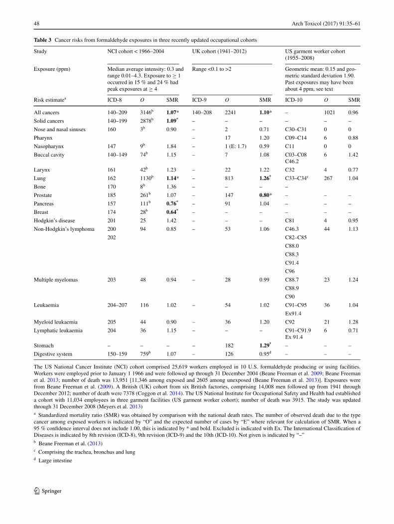
48 Arch Toxicol (2017) 91:35–61
1 3
Table 3 Cancer risks from formaldehyde exposures in three recently updated occupational cohorts
The US National Cancer Institute (NCI) cohort comprised 25,619 workers employed in 10 U.S. formaldehyde producing or using facilities. Workers were employed prior to January 1 1966 and were followed up through 31 December 2004 (Beane Freeman et al. 2009; Beane Freeman et al. 2013; number of death was 13,951 [11,346 among exposed and 2605 among unexposed (Beane Freeman et al. 2013)]. Exposures were from Beane Freeman et al. (2009). A British (UK) cohort from six British factories, comprising 14,008 men followed up from 1941 through December 2012; number of death were 7378 (Coggon et al. 2014). The US National Institute for Occupational Safety and Health had established a cohort with 11,034 employees in three garment facilities (US garment worker cohort); number of death was 3915. The study was updated through 31 December 2008 (Meyers et al. 2013)a Standardized mortality ratio (SMR) was obtained by comparison with the national death rates. The number of observed death due to the type cancer among exposed workers is indicated by “O” and the expected number of cases by “E” where relevant for calculation of SMR. When a 95 % confidence interval does not include 1.00, this is indicated by * and bold. Excluded is indicated with Ex. The International Classification of Diseases is indicated by 8th revision (ICD-8), 9th revision (ICD-9) and the 10th (ICD-10). Not given is indicated by “–”b Beane Freeman et al. (2013)c Comprising the trachea, bronchus and lungd Large intestine
Study NCI cohort < 1966–2004 UK cohort (1941–2012) US garment worker cohort (1955–2008)
Exposure (ppm) Median average intensity: 0.3 and range 0.01–4.3. Exposure to ≥ 1 occurred in 15 % and 24 % had peak exposures at ≥ 4
Range <0.1 to >2 Geometric mean: 0.15 and geo-metric standard deviation 1.90. Past exposures may have been about 4 ppm, see text
Risk estimatea ICD-8 O SMR ICD-9 O SMR ICD-10 O SMR
All cancers 140–209 3146b 1.07* 140–208 2241 1.10* – 1021 0.96
Solid cancers 140–199 2878b 1.09* – – – – – –
Nose and nasal sinuses 160 3b 0.90 – 2 0.71 C30–C31 0 0
Pharynx – 17 1.20 C09–C14 6 0.88
Nasopharynx 147 9b 1.84 – 1 (E: 1.7) 0.59 C11 0 0
Buccal cavity 140–149 74b 1.15 – 7 1.08 C03–C08C46.2
6 1.42
Larynx 161 42b 1.23 – 22 1.22 C32 4 0.77
Lung 162 1130b 1.14* – 813 1.26* C33–C34c 267 1.04
Bone 170 8b 1.36 – – – –
Prostate 185 261b 1.07 – 147 0.80* – – –
Pancreas 157 111b 0.76* – 91 1.04 – – –
Breast 174 28b 0.64* – – – – – –
Hodgkin’s disease 201 25 1.42 – – – C81 4 0.95
Non-Hodgkin’s lymphoma 200 94 0.85 – 53 1.06 C46.3 44 1.13
202 C82–C85
C88.0
C88.3
C91.4
C96
Multiple myelomas 203 48 0.94 – 28 0.99 C88.7 23 1.24
C88.9
C90
Leukaemia 204–207 116 1.02 – 54 1.02 C91–C95 36 1.04
Ex91.4
Myeloid leukaemia 205 44 0.90 – 36 1.20 C92 21 1.28
Lymphatic leukaemia 204 36 1.15 – – – C91–C91.9Ex 91.4
6 0.71
Stomach – – – – 182 1.29* – – –
Digestive system 150–159 759b 1.07 – 126 0.95d – – –
49Arch Toxicol (2017) 91:35–61
1 3
leukaemia [OR (95 % CI) 11.2 (1.3–95.6)] where the refer-ent group contained one myeloid leukaemia case who had never been exposed to FA. No increase was observed in the lymphoid malignancies, including Hodgkin’s lymphoma [0.5 (0.1–2.6)]. The study has severe limitations, including lack of exposure–response relationship within FA exposure groups, unstable estimates due to only one case in the refer-ent group, or where more reliable estimates were present, no appropriate statistical testing was provided (WHO 2010; Cole et al. 2010; Golden 2011; Rhomberg et al. 2011). Due to these limitations, the US embalmer study is not consid-ered a key study for risk assessment.
In the WHO (2010) evaluation, nasal cancers and lym-phohaematopoietic malignancies were the main focus. Additionally, standardized mortality ratios were evalu-ated for other types of cancer in the three comprehensive occupational cohorts, which have been updated recently (Table 3). A key approach by the WHO (2010) in the evalu-ations of cancer effects was whether a nonlinear expo-sure–response relationship could reasonably be identified with a plausible NOAEC. This approach is justified from the nonlinear FA–DNA adduct formation and the assump-tion that an increase in cancer incidence would occur if the cellular level of FA is increased above the normal back-ground to a level where the detoxification mechanisms are overwhelmed. This causes cytotoxicity and regenera-tive cell proliferation, during which DNA lesions may be fixed as mutations due to a decrease of time available for DNA repair. Transmission from the normal homeostasis, determining a NOAEC, to a gradual increase in cancer incidence could also be expected to show a nonlinear expo-sure response relationship. And, importantly, SCC showed a clear, nonlinear exposure–response relationship in rats (Table 2).
Portal‑of‑entry cancers in humans
Airway cancers associated with FA exposures were stud-ied in a Finnish cohort with 1.2 million employees. All men born between 1906 and 1945 who were in employ-ment during 1970 were included. The follow-up study was in the Finnish Cancer Register for nasal cancer (292 cases), cancer of the nasopharynx (149 cases) and lung cancer (30,137 cases) during the period 1971–1995. The Finnish job-exposure matrix was used to estimate cumula-tive exposures. Duration of exposure was estimated from census data. A latency period of 20 years was accepted. Number of exposed cases (N), RR obtained by comparison with unexposed, and 95 % confidence intervals were esti-mated [N; RR (95 % CI)]. The RR for FA exposure was adjusted for smoking, socioeconomic status, and exposure to wood dust. The risk of nasal cancer [17; 1.1 (0.6–1.9)], nasal squamous cell carcinoma [9; 1.0 (0.4–2.0)] and
nasopharyngeal cancer [5; 0.9 (0.3–2.2)] was not increased. The risk was slightly increased for lung cancer [1831; 1.2 (1.1–1.3)], which was adjusted for asbestos and silica dust exposures. However, the risk in the highest exposure group (FA ≥ 1 ppm) was not increased. Thus, the authors consid-ered the increased risk to be due to residual confounding effects of smoking and co-exposures, including asbestos and crystalline silica. FA exposures were below 1 ppm in most occupations. Only floor layers and men who worked with varnish and lacquer had average exposures at 1 ppm (Siew et al. 2012). Overall, this study found no increase in portal-of-entry cancer at low concentrations of FA in occu-pational settings, and only a minor part of the nasal cancer cases were associated with FA exposures.
The US National Cancer Institute (NCI) cohort includes 25,619 workers employed prior to 1 January 1966 in 10 industrial plants. The cohort was followed up until 31 December 2004 (Beane Freeman et al. 2013). At present, it is the second largest industrial cohort on FA exposure. In the cohort, 13,951 had died. The median duration of follow-up was 42 years, and median length of employment was 2.6 years. A lag time of 15 years was applied. The cal-endar year-specific US mortality rates were used to obtain standardized mortality ratios (SMRs), which were stratified for sex, race and age. Internal exposure-dependent trends were obtained for the metrics, peak, average intensity and cumulative exposure, where the RR was set to one in the lowest FA exposure group. SMRs were slightly increased for all cancers, for solid, for respiratory system and for lung cancers (Table 3). For all cancers and solid cancers, the RR was below 1 for peak, average intensity and cumula-tive exposure in the highest exposure groups. In the highest exposure group, the RR was 0.77, 1.01 and 0.79, respec-tively, for cancer of the respiratory system. In the highest exposure category, lung cancer was significantly decreased in the peak exposure metric (RR: 0.77), unchanged in the average intensity metric (RR: 1.0) and significantly decreased in the cumulative exposure metric (RR: 0.78). SMRs were significantly decreased for pancreas and breast cancer. No increase was observed for SMRs for solid can-cer of the buccal cavity, digestive system, liver, nose and nasal sinuses, larynx, bone, skin, female genital, prostate, bladder, kidney, and brain and central nervous system. The only remarkable effect was the non-significant increase in nasopharyngeal cancer [NPC; SMR (95 % CI) 1.84 (0.84–3.49)]; one misclassified oropharyngeal cancer was included in the SMR calculations, but excluded from the calculation of the RRs. For NPC, the trend was only signifi-cant in the peak exposure metric if the non-exposed group was excluded, and the RR was only significantly increased in the average intensity metric in the highest exposure group (Table 4). Overall, this suggests that nasopharyn-geal cancer was the only solid cancer associated with FA
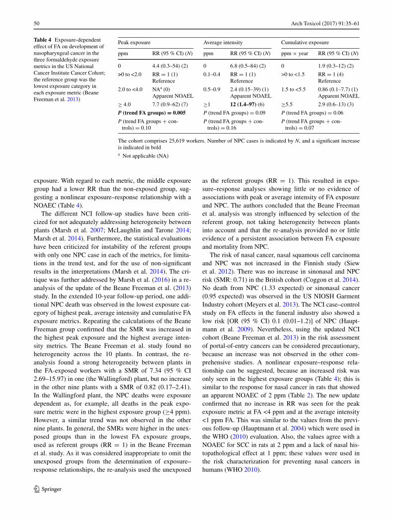
50 Arch Toxicol (2017) 91:35–61
1 3
exposure. With regard to each metric, the middle exposure group had a lower RR than the non-exposed group, sug-gesting a nonlinear exposure–response relationship with a NOAEC (Table 4).
The different NCI follow-up studies have been criti-cized for not adequately addressing heterogeneity between plants (Marsh et al. 2007; McLaughlin and Tarone 2014; Marsh et al. 2014). Furthermore, the statistical evaluations have been criticized for instability of the referent groups with only one NPC case in each of the metrics, for limita-tions in the trend test, and for the use of non-significant results in the interpretations (Marsh et al. 2014). The cri-tique was further addressed by Marsh et al. (2016) in a re-analysis of the update of the Beane Freeman et al. (2013) study. In the extended 10-year follow-up period, one addi-tional NPC death was observed in the lowest exposure cat-egory of highest peak, average intensity and cumulative FA exposure metrics. Repeating the calculations of the Beane Freeman group confirmed that the SMR was increased in the highest peak exposure and the highest average inten-sity metrics. The Beane Freeman et al. study found no heterogeneity across the 10 plants. In contrast, the re-analysis found a strong heterogeneity between plants in the FA-exposed workers with a SMR of 7.34 (95 % CI 2.69–15.97) in one (the Wallingford) plant, but no increase in the other nine plants with a SMR of 0.82 (0.17–2.41). In the Wallingford plant, the NPC deaths were exposure dependent as, for example, all deaths in the peak expo-sure metric were in the highest exposure group (≥4 ppm). However, a similar trend was not observed in the other nine plants. In general, the SMRs were higher in the unex-posed groups than in the lowest FA exposure groups, used as referent groups (RR = 1) in the Beane Freeman et al. study. As it was considered inappropriate to omit the unexposed groups from the determination of exposure–response relationships, the re-analysis used the unexposed
as the referent groups (RR = 1). This resulted in expo-sure–response analyses showing little or no evidence of associations with peak or average intensity of FA exposure and NPC. The authors concluded that the Beane Freeman et al. analysis was strongly influenced by selection of the referent group, not taking heterogeneity between plants into account and that the re-analysis provided no or little evidence of a persistent association between FA exposure and mortality from NPC.
The risk of nasal cancer, nasal squamous cell carcinoma and NPC was not increased in the Finnish study (Siew et al. 2012). There was no increase in sinonasal and NPC risk (SMR: 0.71) in the British cohort (Coggon et al. 2014). No death from NPC (1.33 expected) or sinonasal cancer (0.95 expected) was observed in the US NIOSH Garment Industry cohort (Meyers et al. 2013). The NCI case–control study on FA effects in the funeral industry also showed a low risk [OR (95 % CI) 0.1 (0.01–1.2)] of NPC (Haupt-mann et al. 2009). Nevertheless, using the updated NCI cohort (Beane Freeman et al. 2013) in the risk assessment of portal-of-entry cancers can be considered precautionary, because an increase was not observed in the other com-prehensive studies. A nonlinear exposure–response rela-tionship can be suggested, because an increased risk was only seen in the highest exposure groups (Table 4); this is similar to the response for nasal cancer in rats that showed an apparent NOAEC of 2 ppm (Table 2). The new update confirmed that no increase in RR was seen for the peak exposure metric at FA <4 ppm and at the average intensity <1 ppm FA. This was similar to the values from the previ-ous follow-up (Hauptmann et al. 2004) which were used in the WHO (2010) evaluation. Also, the values agree with a NOAEC for SCC in rats at 2 ppm and a lack of nasal his-topathological effect at 1 ppm; these values were used in the risk characterization for preventing nasal cancers in humans (WHO 2010).
Table 4 Exposure-dependent effect of FA on development of nasopharyngeal cancer in the three formaldehyde exposure metrics in the US National Cancer Institute Cancer Cohort; the reference group was the lowest exposure category in each exposure metric (Beane Freeman et al. 2013)
The cohort comprises 25,619 workers. Number of NPC cases is indicated by N, and a significant increase is indicated in bolda Not applicable (NA)
Peak exposure Average intensity Cumulative exposure
ppm RR (95 % CI) (N) ppm RR (95 % CI) (N) ppm × year RR (95 % CI) (N)
0 4.4 (0.3–54) (2) 0 6.8 (0.5–84) (2) 0 1.9 (0.3–12) (2)
>0 to <2.0 RR = 1 (1)Reference
0.1–0.4 RR = 1 (1)Reference
>0 to <1.5 RR = 1 (4)Reference
2.0 to <4.0 NAa (0)Apparent NOAEL
0.5–0.9 2.4 (0.15–39) (1)Apparent NOAEL
1.5 to <5.5 0.86 (0.1–7.7) (1)Apparent NOAEL
≥ 4.0 7.7 (0.9–62) (7) ≥1 12 (1.4–97) (6) ≥5.5 2.9 (0.6–13) (3)
P (trend FA groups) = 0.005 P (trend FA groups) = 0.09 P (trend FA groups) = 0.06
P (trend FA groups + con-trols) = 0.10
P (trend FA groups + con-trols) = 0.16
P (trend FA groups + con-trols) = 0.07

51Arch Toxicol (2017) 91:35–61
1 3
Laryngeal cancer
The association between laryngeal cancer and FA exposure was studied in a recent meta-analysis, which included six case–control studies [mRR (95 % CI) 1.15 (0.97–1.36)] and five cohort studies [1.10 (0.84–1.44)] with an overall mRR of 1.13 (0.98–1.31). The authors concluded that the meta-analysis did not support that there is an association between FA exposure and laryngeal cancer (Paget-Bailly et al. 2012). There was no increase in the risk in the three updated occupational cohorts (Table 3).
Lung cancer in humans
Combined data on lung cancer were constructed from two population-based case–control studies conducted in 1979–1986 and 1996–2002 in Montreal in Canada. A total of 2060 lung cancer cases were compared with 2046 sex- and age-matched controls. Sociodemographic characteristics, lifestyle-habits (including smoking) and complete work history (including work tasks, work conditions and chemical exposures) were obtained from similar interview questions in the two studies. Chemical exposures were evaluated from exposure concentrations (low, medium and high), frequency of exposures (low, medium and high) and years of exposure. Unconditional logistic regression was used to estimate risks [OR (95 % CI)]; confounders taken into account were age, sex, income, number of years in school, ethnicity, smoking, and exposures to occupational lung carcinogens, includ-ing asbestos and silica. About 25 % of the cases and the controls were exposed to FA. However, FA exposures were low in the majority of the subjects. For men and women combined, the lung cancer risk for ever- versus never-FA-exposed was not significantly increased [1.06 (0.89–1.27)]. For the group of substantially exposed, the risk was low [0.88 (0.63–1.24)]. Moreover, a long dura-tion of exposure (>20 years) did not increase the risk [0.93 (0.69–1.24)]. Similarly, no increase in risk was observed with age at first exposure, time since first expo-sure, and maximum intensity of exposure. The authors concluded that FA exposure was not associated with an increased lung cancer risk, which they found was in agreement with majority of studies reported in the scien-tific literature (Mahboubi et al. 2013). In the NCI cohort, there was no increase in lung cancer risk in the highest exposure category in any of the exposure metrics (Beane Freeman et al. 2013). Neither was an increase observed in the NIOSH garment cohort (Meyers et al. 2013). How-ever, there was an increase in the UK cohort (Coggon et al. 2014), which was not adjusted for smoking. Over-all, there is no consistent association between FA expo-sure and lung cancer (IARC 2006; NRC 2014).
Cancers at distant sites
In epidemiology, a major focus has been on the association between FA exposure and leukaemia, especially myeloid leukaemia (Hauptmann et al. 2003, 2009; Beane Freeman et al. 2009; Schwilk et al. 2010). Of the human carcino-gens identified by IARC, approximately 25 % induce leu-kaemias or lymphomas. Alkylating agents, benzene, topoi-somerase II inhibitors and ionizing radiation induce mainly acute myeloid leukaemia (AML). However, ethylene oxide and 1,3-butadiene are linked primarily with lymphoid can-cers (Eastmond et al. 2014). The industries and occupations with the highest occurrence of leukaemia are not those with high exposures to chemicals. Thus, in the US National Occupational Mortality Surveillance System, the highest proportionality mortality ratios (PMRs) for white males (the largest group) are in the industries and occupations: bank/savings and loan/credit agency (PMR: 139–172), advertising/sales manager (PMR: 138–174) and electrical engineers (PMR: 141–192) in different periods between 1985 and 2007 (Robinson et al. 2015).
The NCI Cohort was also followed up until 31 Decem-ber 2004, for lymphohaematopoietic malignancies (Beane Freeman et al. 2009). Of the 25,619 cohort members, 4359 were classified as never having been exposed to FA. In the follow-up period, 13,951 deaths occurred. The SMRs were calculated using the US mortality rates; they showed no remarkable effect of FA exposure on lymphohaematopoi-etic malignancies (Table 3). The RR (95 % CI) for death from lymphohaematopoietic malignancies was examined for association with peak exposure, average intensity and cumulative exposure. The group with the lowest FA expo-sure was selected as the referent group. There was no sig-nificant association between cumulative exposure and any of the malignancies. Therefore, results are presented only for the peak exposure and the average intensity metrics (Table 5). The most remarkable increase in RR is for Hodg-kin’s lymphoma, apparent from both metrics; this increase was also present in the previous follow-up (Hauptmann et al. 2003), but was not seen in other FA-exposed cohorts as mentioned by the authors. Furthermore, Hodgkin’s lym-phoma has not previously been associated with exposures to chemicals (Nielsen et al. 2013; Checkoway et al. 2015); known risk factors are, for example, socioeconomic status, family size and Epstein–Barr virus infection (c.f. Nielsen et al. 2013). In this study, there was no significant associa-tion with FA exposures and myeloid leukaemia (Table 3). In the peak exposure metrics, there was a tendency of an exposure-dependent increase in leukaemia (ICD-8 codes: 204–207) as the RRs increased in the four groups from 0.59 (0.25–1.36), 1.0 (referent group), 0.98 (0.60–1.62) to 1.42 (0.92–2.18), respectively, with P-trend = 0.02. However, within the three FA groups themselves, the trend was not
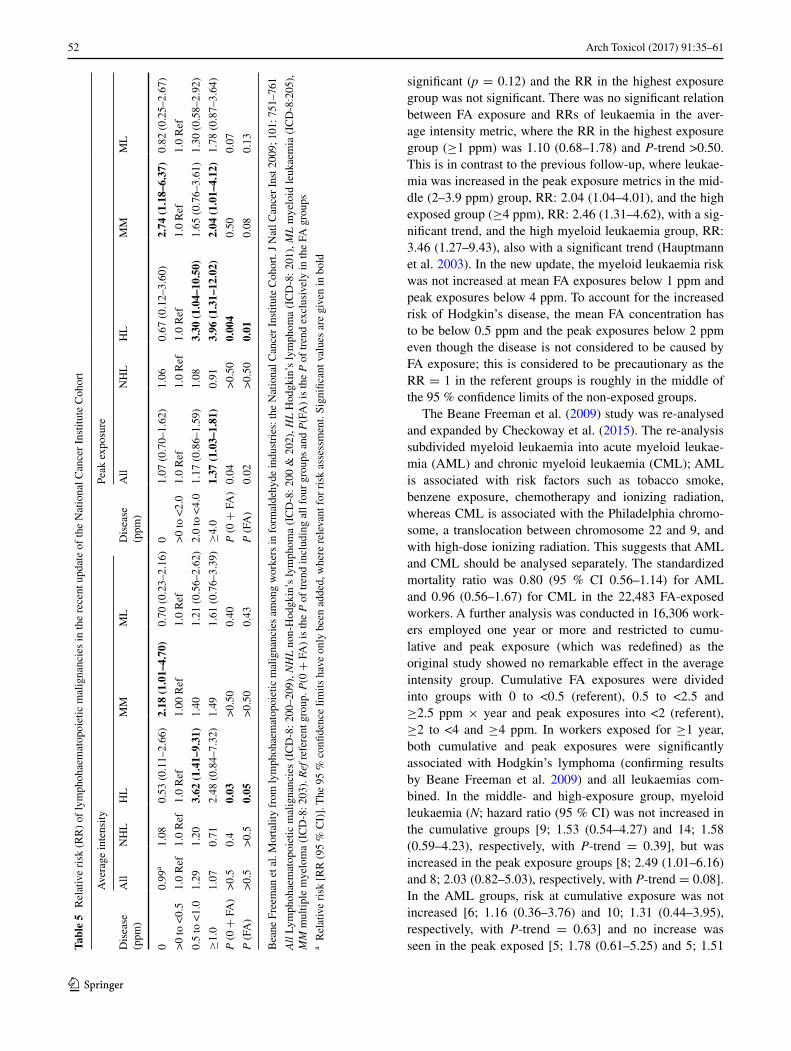
52 Arch Toxicol (2017) 91:35–61
1 3
significant (p = 0.12) and the RR in the highest exposure group was not significant. There was no significant relation between FA exposure and RRs of leukaemia in the aver-age intensity metric, where the RR in the highest exposure group (≥1 ppm) was 1.10 (0.68–1.78) and P-trend >0.50. This is in contrast to the previous follow-up, where leukae-mia was increased in the peak exposure metrics in the mid-dle (2–3.9 ppm) group, RR: 2.04 (1.04–4.01), and the high exposed group (≥4 ppm), RR: 2.46 (1.31–4.62), with a sig-nificant trend, and the high myeloid leukaemia group, RR: 3.46 (1.27–9.43), also with a significant trend (Hauptmann et al. 2003). In the new update, the myeloid leukaemia risk was not increased at mean FA exposures below 1 ppm and peak exposures below 4 ppm. To account for the increased risk of Hodgkin’s disease, the mean FA concentration has to be below 0.5 ppm and the peak exposures below 2 ppm even though the disease is not considered to be caused by FA exposure; this is considered to be precautionary as the RR = 1 in the referent groups is roughly in the middle of the 95 % confidence limits of the non-exposed groups.
The Beane Freeman et al. (2009) study was re-analysed and expanded by Checkoway et al. (2015). The re-analysis subdivided myeloid leukaemia into acute myeloid leukae-mia (AML) and chronic myeloid leukaemia (CML); AML is associated with risk factors such as tobacco smoke, benzene exposure, chemotherapy and ionizing radiation, whereas CML is associated with the Philadelphia chromo-some, a translocation between chromosome 22 and 9, and with high-dose ionizing radiation. This suggests that AML and CML should be analysed separately. The standardized mortality ratio was 0.80 (95 % CI 0.56–1.14) for AML and 0.96 (0.56–1.67) for CML in the 22,483 FA-exposed workers. A further analysis was conducted in 16,306 work-ers employed one year or more and restricted to cumu-lative and peak exposure (which was redefined) as the original study showed no remarkable effect in the average intensity group. Cumulative FA exposures were divided into groups with 0 to <0.5 (referent), 0.5 to <2.5 and ≥2.5 ppm × year and peak exposures into <2 (referent), ≥2 to <4 and ≥4 ppm. In workers exposed for ≥1 year, both cumulative and peak exposures were significantly associated with Hodgkin’s lymphoma (confirming results by Beane Freeman et al. 2009) and all leukaemias com-bined. In the middle- and high-exposure group, myeloid leukaemia (N; hazard ratio (95 % CI) was not increased in the cumulative groups [9; 1.53 (0.54–4.27) and 14; 1.58 (0.59–4.23), respectively, with P-trend = 0.39], but was increased in the peak exposure groups [8; 2.49 (1.01–6.16) and 8; 2.03 (0.82–5.03), respectively, with P-trend = 0.08]. In the AML groups, risk at cumulative exposure was not increased [6; 1.16 (0.36–3.76) and 10; 1.31 (0.44–3.95), respectively, with P-trend = 0.63] and no increase was seen in the peak exposed [5; 1.78 (0.61–5.25) and 5; 1.51 Ta
ble
5 R
elat
ive
risk
(R
R)
of ly
mph
ohae
mat
opoi
etic
mal
igna
ncie
s in
the
rece
nt u
pdat
e of
the
Nat
iona
l Can
cer
Inst
itute
Coh
ort
Bea
ne F
reem
an e
t al.
Mor
talit
y fr
om ly
mph
ohae
mat
opoi
etic
mal
igna
ncie
s am
ong
wor
kers
in f
orm
alde
hyde
indu
stri
es: t
he N
atio
nal C
ance
r In
stitu
te C
ohor
t. J
Nat
l Can
cer
Inst
200
9; 1
01: 7
51–7
61
All
Lym
phoh
aem
atop
oiet
ic m
alig
nanc
ies
(IC
D-8
: 200
–209
), N
HL
non
-Hod
gkin
’s ly
mph
oma
(IC
D-8
: 200
& 2
02),
HL
Hod
gkin
’s ly
mph
oma
(IC
D-8
: 201
), M
L m
yelo
id le
ukae
mia
(IC
D-8
:205
),
MM
mul
tiple
mye
lom
a (I
CD
-8: 2
03).
Ref
ref
eren
t gro
up. P
(0 +
FA
) is
the
P o
f tr
end
incl
udin
g al
l fou
r gr
oups
and
P(F
A)
is th
e P
of
tren
d ex
clus
ivel
y in
the
FA g
roup
sa R
elat
ive
risk
[R
R (
95 %
CI)
]. T
he 9
5 %
con
fiden
ce li
mits
hav
e on
ly b
een
adde
d, w
here
rel
evan
t for
ris
k as
sess
men
t. Si
gnifi
cant
val
ues
are
give
n in
bol
d
Ave
rage
inte
nsity
Peak
exp
osur
e
Dis
ease
(p
pm)
All
NH
LH
LM
MM
LD
isea
se
(ppm
)A
llN
HL
HL
MM
ML
00.
99a
1.08
0.53
(0.
11–2
.66)
2.18
(1.
01–4
.70)
0.70
(0.
23–2
.16)
01.
07 (
0.70
–1.6
2)1.
060.
67 (
0.12
–3.6
0)2.
74 (
1.18
–6.3
7)0.
82 (
0.25
–2.6
7)
>0
to <
0.5
1.0
Ref
1.0
Ref
1.0
Ref
1.00
Ref
1.0
Ref
>0
to <
2.0
1.0
Ref
1.0
Ref
1.0
Ref
1.0
Ref
1.0
Ref
0.5
to <
1.0
1.29
1.20
3.62
(1.
41–9
.31)
1.40
1.21
(0.
56–2
.62)
2.0
to <
4.0
1.17
(0.
86–1
.59)
1.08
3.30
(1.
04–1
0.50
)1.
65 (
0.76
–3.6
1)1.
30 (
0.58
–2.9
2)
≥1.
01.
070.
712.
48 (
0.84
–7.3
2)1.
491.
61 (
0.76
–3.3
9)≥
4.0
1.37
(1.
03–1
.81)
0.91
3.96
(1.
31–1
2.02
)2.
04 (
1.01
–4.1
2)1.
78 (
0.87
–3.6
4)
P (
0 +
FA
)>
0.5
0.4
0.03
>0.
500.
40P
(0 +
FA
)0.
04>
0.50
0.00
40.
500.
07
P (
FA)
>0.
5>
0.5
0.05
>0.
500.
43P
(FA
)0.
02>
0.50
0.01
0.08
0.13

53Arch Toxicol (2017) 91:35–61
1 3
(0.51–4.44), respectively, with P-trend = 0.37]. With regard to CML, the cumulative exposed group showed a non-significant increase [2; 2.91 (0.24–35.64) and 4; 3.81 (0.36–40.44), respectively, with P-trend = 0.27], similar to the increase seen for the groups with peak exposure [2; 4.83 (0.64–36.42) and 3; 5.32 (0.81–34.90), respectively, with P-trend = 0.07]. However, the number of deaths was low. In the full cohort, 13 of the 34 AML deaths were from the group with peak exposures of more than 2 ppm, only four workers had jobs with peaks within the 20 years pre-ceding their death, and only one death occurred within the typical AML window of 2–15 years. Non-Hodgkin’s lymphoma, chronic lymphocytic leukaemia and multiple myeloma showed no increased risk. The authors concluded that the re-analysis does not support that FA causes AML, which they considered the most relevant leukaemia for FA exposure.
A follow-up from the period 1941 through December 2012 was conducted in the British (UK) cohort from six factories comprising 14,008 men (Coggon et al. 2014). In the period, 7378 men had died. A total of 3991 of these men had been highly exposed to FA. In the cohort, the standardized mortality ratio [SMRs (95 % CI)] for all can-cers [1.10 (1.06–1.15)], for stomach cancer [1.29 (1.11–1.49)], for rectum cancer [1.23 (1.01–1.49)], for lung can-cer [1.26 (1.17–1.35)] was significantly increased based on the national death rate for England and Wales; other values are given in Table 3. No significant increase was seen for cancer in the pharynx, larynx, nose and nasal sinuses, or for the different hematopoietic malignancies. Prostate cancer was significantly decreased [0.80 (0.68–0.94)]. The cohort was stratified for levels of exposure. The exposure in the high-exposure group was more than 2 ppm. A significant increase in SMR in the high-exposure group was observed for all cancers [1.28 (1.20–1.37)], cancer in the oesophagus [1.45 (1.03–1.98)], stomach [1.51 (1.18–1.90)], lungs [1.59 (1.42–1.77)] and the lips [9.98 (1.21–36.04)]; observed/expected: 2/0.2). No increase was seen for non-Hodgkin’s lymphoma [0.90 (0.48–1.55)], multiple myeloma [1.18 (0.57–2.18)], leukaemia [0.82 (0.44–1.41)] and myeloid leukaemia [0.93 (0.40–1.82)]. Exposure in the high-expo-sure group was further stratified for duration of exposure (<1 year, 1–14 years and ≥15 years). For oesophagus and lung cancer, the SMR was highest in the in the group with the shortest exposure (<1 year), and for stomach and rec-tum cancer, the SMRs were independent of the length of the exposure period. Additionally, the authors included a nested case–control analysis of cancer in the upper air-ways, larynx, mouth, pharynx, tongue, and for all leukae-mia and myeloid leukaemia. ORs for these cancers were independent of the duration of the exposure. The authors ascribed the increases in risk estimates to non-occupational confounding factors, which may include smoking and
socioeconomic factors, and they concluded that the study provided no evidence that FA posed an increased hazard of upper airway cancer or of myeloid leukaemia. It is noted that the study was not able to take smoking and socioeco-nomic factors into account. Overall, the NOAEC from this study is approximately 2 ppm.
A new follow-up (1960–2008) was conducted of the US National Institute for Occupational Safety and Health (NIOSH) Garment Industry cohort, which is among the largest prospective cohorts (Meyers et al. 2013). The study comprised 11,043 workers. Causes of death were obtained from 99.7 % (3904) of the identified deaths. The year of first exposure was 1970 or earlier for about 77 % of the workers. In the early 1980s, personal FA sampling was performed among 549 employees. The geometric mean FA concentration was 0.15 ppm with a geometric stand-ard deviation of 1.90. No exposure data were available before this time, but FA concentrations are believed to have decreased over time. Recently, NRC (2014) estimated the FA concentration to be about 4 ppm before 1970. Standard-ized mortality ratios [SMRs (95 % CIs)] and internal com-parisons were made using directly standardized rate ratios [SRRs (95 % CIs)] for “duration of exposure”. The SMRs were similar to that of the US population for all cancers, for lymphohaematopoietic cancers (leukaemias, Hodghin dis-ease, non-Hodgkin’s lymphoma, and multiple myeloma), for buccal cavity and pharyngeal cancers, for respiratory cancers, and for brain cancer and other parts of the nervous system (Table 3).
Stratifying SMRs for “year of first exposure” (<1963, 1963–1970, ≥1971) showed no significant increase for lymphohaematopoietic cancers, for trachea, bronchus and lung cancer, and for brain cancer and other parts of the nervous system. Similarly, no significant increase was observed for SMRs for “time since first exposure” (<10, 10–19, ≥20 years). Association with “duration of FA expo-sures” (<3, 3–9, ≥10 years) was studied with SMRs and SRRs. There was no exposure-dependent increase in risks for lymphohaematopoietic cancers and non-Hodgkin’s lymphoma. The risks increased with the length of the expo-sures for leukaemia, myeloid leukaemia and acute myeloid leukaemia, but the risks were not statistically significant. For multiple myeloma, the SMR for the exposure groups was 1.2 (0.5–2.3), 2.0 (>1.0–3.6) and 0.6 (0.2–1.6), respec-tively, and the SRR was 1.00 (reference), 1.2 (0.5–3.3) and 0.3 (0.08 to <1.0), respectively. For trachea, bronchus and lung cancer, the SMR was 1.2 (>1.0–1.5), 1.1 (0.9–1.4) and 0.7 (0.5–0.9), respectively, and the SRR 1.00 (reference), 1.0 (0.8–1.3) and 0.7 (0.5–1.1), respectively. Thus, where the values were statistically significant, they were not asso-ciated with the length of the exposure period. However, for individuals with ≥10 years of exposure and ≥20 years since first exposures, leukaemia [23 death, SMR: 1.7

54 Arch Toxicol (2017) 91:35–61
1 3
(1.1–2.6)] was significantly increased when multiple causes of death were considered.
Additionally, duration of exposure was studied for leukae-mia (36 cases) and myeloid leukaemia (21 cases) using four multivariate Poisson regression models (adjusted for age, year of birth and years since first exposure), where exposures either were untransformed or transformed (log, square root and categorical [<1.6 (reference), 1.6 to <6.5, 6.5 to <16, 16 to <19 and ≥19 years)]. Only the untransformed model for leukaemia and the categorical model for myeloid leukaemia showed a statistically significant trend. However, for leukae-mia and myeloid leukaemia, the rate ratios in the categori-cal model were significantly increased in the fourth category [4.6 (1.3–16) and 6.4 (1.4–32), respectively], but not for the second, third and fifth categories; in the fifth category (expo-sure > 19 years), no significant increase [2.6 (0.7–10) and 1.7 (0.3–11), respectively] was observed.
The authors concluded that the study showed limited evidence of association between FA exposure and leukae-mia, and little evidence of an increased risk of mortality from buccal cavity, pharyngeal (including nasopharyn-geal), respiratory and brain cancer, and for Hodgkin’s dis-ease. It is noted that the study lacks appropriate FA expo-sure assessments and it was not able to take smoking into account. The importance of smoking is not clear; there were sporadically significantly increased values for chronic obstructive lung disease, but no increase in lung cancer.
Also, an Italian cohort with subjects employed in a fac-tory producing laminate plastic, decorative papers and craft papers, using phenolic and melamine resins, has been established (Pira et al. 2014). The major risk was consid-ered to be FA exposure, but FA concentrations were not measured. The cohort comprised 2750 employees from the period 1947 to 31 May 2011, who had been employed for at least 180 days. Data on survival (80.3 %), death (16.6 %, N = 457) and emigration (3.1 %) were obtained. Cause of death could not be retrieved for 26 out of 457 (5.7 %) deceased employees. Person-years of observation were 70,933 in the analysis. Expected number of deaths (E) and SMRs were obtained by comparison with the regional death rates. Observed deaths (O) and SMR [O, SMR (95 % CI)] for lymphoma [4; 0.74 (0.20–1.90)], myeloma (O/E = 0/2.3), leukaemia [5; 0.92 (0.30–2.15)] and for all lymphohaematopoietic neoplasms [9; 0.69 (0.31–1.30)] were not increased. Neither was an increased risk of cancer observed for all cancers [149; 0.80 (0.68–0.94)]. The risk was non-significantly increased for oral and pharynx can-cer [9; 1.49 (0.68–2.82)] and for bladder cancer [10; 1.51 (0.72–2.77)]. For oesophagus, stomach, colorectal, liver, pancreas, larynx, lung, breast, prostate, kidney, and brain and CNS cancer, the SMRs were below one. The study has a long follow-up period, but a limitation is the lack of quantitative FA exposure data.
Hauptmann et al. (2009) investigated the relationship between mortality and work practices and FA exposure levels among American embalmers in a case–control study. Professionals employed in the American funeral indus-try who died between 1 January 1960 and 1 January 1986 from lymphohaematopoietic malignancies (n = 168), brain tumours (n = 48) or nasopharyngeal cancers (n = 4) were obtained for 6808 who died in the period and compared with deceased matched controls (n = 265) with regard to lifetime work practice. Exposures in the funeral industry were obtained by interviews with next of kin and co-work-ers, and predictive models were used to estimate levels of formaldehyde exposure. Mean peak concentrations were 8.1–10.5 ppm (model predicted as the maximum 15-min-ute average intensity ever experienced in connection with embalming for all years) and the average FA intensity was 1.5–1.8 ppm while embalming. Cases were exposed for about 32 years. With one myeloid leukaemia in the referent group, the odds ratio [OR (95 % CI)] for myeloid leukae-mia was 11.2 (1.3–95.5) in the ever-embalming versus the never-embalming group. Mortality from myeloid leukaemia increased statistically significantly only with an increasing number of years of embalming (P for trend = 0.020) and with an increasing peak FA exposure (P for trend = 0.036). Within the exposure groups themselves (duration of years with embalming, number of embalming, cumulative FA exposure, average FA exposure while embalming, 8-h time-weighted average (TWA) FA intensity and peak FA expo-sure), there were no significant trends in any of the groups (P for trend = 0.58–0.97). There was no exposure-depend-ent effect on monocytic leukaemia, polycythemia vera or myelofibrosis. ORs were roughly about 10 (range 5–15) in exposed groups. To increase stability of the risk estimates, subjects who performed fewer than 500 lifetime embalm-ings were used as the referent group in a second evalua-tion. In this analysis, the ORs for myeloid leukaemia were roughly about 3 (range 0.5–3.9) in the “exposed” groups. No true trend tests were available for this evaluation as the authors without explanation used the results from the trend tests from the first (unstable) analysis in this (more sta-ble) analysis. However, ORs were significantly increased for duration of years with embalming at >20–34 years and >34 years, which was 3.2 (1.0–10.1) and 3.9 (1.2–12.5), respectively, with the highest number of embalmings (>3068), 3.0 (1.0–9.2), and at the highest cumulative FA exposure (ppm × hours: >9253), 3.1 (1.0–9.6). These expo-sures were not related to lymphohaematopoietic malignan-cies of the lymphoid organs (non-Hodgkin’s lymphoma, multiple myeloma, all lymphoma and Hodgkin’s disease), brain cancer or nasopharyngeal cancer [0.1 (0.01–1.2)].
The validity of the Hauptmann et al. (2009) study has been challenged as it relied only on RR estimates. The expected number of subjects with lymphohaematopoietic
55Arch Toxicol (2017) 91:35–61
1 3
malignancies, all myeloid leukaemias and all acute myeloid leukaemias were estimated from the 6808 embalmers (Cole et al. 2010). Comparison between the observed and the estimated number of subjects showed no meaningful eleva-tion. Furthermore, the proportional mortality ratios showed no significant elevation. With regard to risk assessment, it is noted that the study does not provide convincing expo-sure–response relationships within the different FA expo-sure metrics, and therefore it cannot be used for setting an indoor air guideline level (Nielsen et al. 2013). Moreover, the potential hazards addressed in this study are not rele-vant for setting an indoor air guideline level due to the very high exposure levels.
With regard to setting its guideline, the WHO (2010) used the Hauptmann et al. (2003) study as the key study for risk evaluation of lymphohaematopoietic malignancies. The new update (Beane Freeman et al. 2009) weakens the association between FA exposure and leukaemia and mye-loid leukaemia, indicating that the WHO (2010) guideline is based on conservative estimates. Additionally, the guide-line is set below levels associated with increase in Hodg-kin’s lymphoma, although this disease has neither been consistently associated with FA exposures nor with expo-sure to other chemicals.
Evaluation of carcinogenic risk and the WHO (2010) indoor air quality guideline
The two critical effects of FA are nasal cancer and irrita-tion of eyes and upper airways (sensory irritation), where sensory irritation had the lowest NOAEC and is the basis of the WHO IAQG at 0.1 mg/m3 (0.08 ppm), applying to each 30-min period during each day lifelong. It is noted that the NOEAC for sensory irritation is 0.5 ppm and thus an assessment factor of 6 is used when setting the IAQG, which is a conservative approach. Moreover, this guideline value is considered to prevent all portal-of-entry effects, including nasal cancer, and potential systemic cancers. Even though the potential systemic cancer effects are con-sidered not to be relevant with regard to setting an indoor guideline, all endpoints were evaluated to assure that the guideline level was below effect levels to ascertain a con-tradiction-free guideline (WHO 2010; Nielsen and Wolkoff 2010; Nielsen et al. 2013).
Cancer risk assessment of indoor air exposures to FA has been carried out based on two different approaches. One approach is linear extrapolation (e.g. Sarigian-nis et al. 2011; Huang et al. 2013; Du et al. 2014; Rovira et al. 2016), which provides conservative estimates (e.g. Nielsen and Wolkoff 2010; Sarigiannis et al. 2011; Du et al. 2014). As FA is an endogenous metabolite, at a certain low exogenous exposure level, the cellular FA level will be
dominated by the endogenously (naturally) generated FA and exogenous FA contribution will be low or negligible (Andersen et al. 2010; Swenberg et al. 2013). Based on lev-els of endogenous and exogenous FA–DNA adducts, it was shown that the linear extrapolation approach greatly over-estimates the cancer risk (Starr and Swenberg 2013, 2016).
In 2010, the WHO launched a threshold approach for establishing an IAQG for FA. Analysis of cancer exposure–response relationships in experimental animal (e.g. Table 2) and human epidemiological studies showed that the rela-tionships were nonlinear (WHO 2010). Additionally, the mucosal metabolism would prevent systemic access of FA when airborne concentrations were below a few ppm FA.
With regard to experimental animal species, rats were the most sensitive to developing nasal cancer due to life-long exposure with a LOAEC of 6 ppm and an apparent NOAEC of 2 ppm (Table 2). Nasal cancer was driven by cytotoxicity induced cell proliferation (Conolly et al. 2003, 2004). This may cause DNA transcription before DNA lesions were repaired and thereby allow the DNA damages to be fixed as mutations, which in turn may cause cancer. The NOAEC for increased cell proliferation was >2 ppm (Monticello et al. 1996; Andersen et al. 2010). The his-topathological NOAEC (1 ppm) for damage of the nasal epithelium was lower than the NOAEL for cell prolifera-tion and for nasal cancer, for which reason the histopatho-logical NOAEC was accepted as the point of departure for the cancer risk assessment. Also, it was accepted that the histopathological NOAEC was independent of the dura-tion of the daily exposure period and therefore NOAEC was considered to be a full day of exposure. Furthermore, rats were considered more sensitive than humans. Due to the local airway effect, the 1 ppm level was divided by an interspecies assessment factor of 3 (it is noted that rats are a sensitive species) and an assessment factor of 2 for the limit variations within the human population [deposi-tion in the upper airways is similar in children and adults, and the variation in the population is about 1.6-fold (Gar-cia et al. 2009)], resulting in a guideline value of 0.17 ppm (0.2 mg/m3) for protection against nasal cancer. It is noted that the assessment factors are highly conservative. This approach was backed up by a comprehensive biologically motivated computational modelling method, predicting that the 80-year lifetime additional risk is ≤10−6 at 0.2 ppm (0.246 mg/m3) for non-smokers (Conolly et al. 2004). Pre-venting nasal cancer was considered to prevent systemic cancers, including leukaemia.
The credibility of the histopathological endpoint in the risk assessment of cancer is further supported by recent animal studies. For example, the toxicokinetic studies by Andersen et al. (2010) showed that the local airway epi-thelial cell FA level was not markedly affected by FA exposures from 0.7 to 2 ppm, and at 1 ppm, the epithelial

56 Arch Toxicol (2017) 91:35–61
1 3
FA concentration was dominated by the endogenous (natural) FA concentration. At these concentrations, the exogenous FA–DNA adducts were lower than the endog-enous adducts in the rat nasal tissue (Lu et al. 2010, 2011; Moeller et al. 2011; Edrissi et al. 2013;Yu et al. 2015). Moreover, the recent studies have confirmed experimen-tally that apart from the portal of entry no absorption occurs in rats at exposures in the range 0.7–15 ppm (Lu et al. 2010, 2011; Edrissi et al. 2013; Kleinnijenhuis et al. 2013; Yu et al. 2015) and in monkeys up 6 ppm (Moe-ller et al. 2011). Overall, this supports the credibility of the selected point of departure by the WHO for low-level portal-of-entry effects, which if prevented also prevent systemic effects.
The WHO (2010) IAQG was also supported by epidemi-ological studies. In the previous update of the NCI cohort, nasal cancer occurrence had a nonlinear exposure–response relationship where no increased risk was observed below 1 ppm average intensity and below 4 ppm peak exposures (Hauptmann et al. 2004); the exposure–response relation-ship was nonlinear. The Hauptmann et al. (2004) study was used as a key study by the WHO. The cohort was recently updated (Beane Freeman et al. 2013) with similar results. However, no increase in nasal cancer was observed in later comprehensive studies and updates (Siew et al. 2012; Mey-ers et al. 2013; Coggon et al. 2014) and a case–control study (Hauptmann et al. 2009). In the latest update of the NCI cohort (Beane Freeman et al. 2009), myeloid leukae-mia risk was not increased at mean FA exposures below 1 ppm and peak exposures below 4 ppm and thus shows that preventing nasal cancer also prevents leukaemia. Remarkably, the previous follow-up study (Hauptmann et al. 2003) and the most recent follow-up (Beane Freeman et al. 2009) of the NCI cohort both showed an increase in Hodgkin’s lymphoma at the average intensity ≥0.5 ppm and peak exposures ≥2 ppm. Hodgkin’s lymphoma has not previously been associated with exposures to chemicals (Nielsen et al. 2013; Checkoway et al. 2015); known risk factors are, for example, socioeconomic status, family size and Epstein–Barr virus infection (c.f. Nielsen et al. 2013). Therefore, it is considered sufficient that the guideline is below these levels.
On the whole, the nonlinear exposure–response rela-tionships, the epidemiological effects at levels much higher than the WHO IAQG and the lack of consistency across studies indicate that the WHO IAQG is highly precautionary.
Acknowledgments This study was conducted as a research activ-ity at the National Research Centre for the Working Environment (NRCWE), Denmark, a governmental research institute. Additional support was obtained from the framework of the CISBO project (Centre for Indoor Climate and Diseases in Dwellings) supported by Realdania.
Compliance with ethical standards
Conflict of interest The authors declare that they have no conflict of interest.
Open Access This article is distributed under the terms of the Crea-tive Commons Attribution 4.0 International License (http://crea-tivecommons.org/licenses/by/4.0/), which permits unrestricted use, distribution, and reproduction in any medium, provided you give appropriate credit to the original author(s) and the source, provide a link to the Creative Commons license, and indicate if changes were made.
References
Alarie Y (1973) Sensory irritation by airborne chemicals. CRC Crit Rev Toxicol 2:299–363
Andersen ME, Clewell HJ, Bermudez E, Dodd DE, Willson GA, Campbell JL, Thomas RS (2010) Formaldehyde: integrating dosimetry, cytotoxicity, and genomics to understand dose-dependent transitions for an endogenous compound. Toxicol Sci 118:716–731
Araneda OF, Tuesta M (2012) Lung oxidative damage by hypoxia. Oxid Med Cell Longev 2012:856918. doi:10.1155/2012/856918
Asgharian B, Price OT, Schroeter JD, Kimbell JS, Singal M (2012) A lung dosimetry model of vapor uptake and tissue disposition. Inhal Toxicol 24:182–193
Askew EW (2002) Work at high altitude and oxidative stress: antioxi-dant nutrients. Toxicology 180:107–119
Atkinson R, Arey J (2003) Gas-phase tropospheric chemistry of bio-genic volatile organic compounds: a review. Atmos Environ 37(suppl 2):S197–S219
Attia D, Mansour N, Taha F, El Dein AS (2014) Assessment of lipid peroxidation and p53 as a biomarker of carcinogenesis among workers exposed to formaldehyde in cosmetic industry. Toxicol Ind Health. doi:10.1177/0748233714547152
Aydin S, Canpinar H, Ündeğer Ü, Güç D, Çolakoğlu M, Kars A, Basaran N (2013) Assessment of immunotoxicity and geno-toxicity in workers exposed to low concentrations of formalde-hyde. Arch Toxicol 87:145–153
Azad N, Rojanasakul Y, Vallyathan V (2008) Inflammation and lung cancer: roles of reactive oxygen/nitrogen species. J Toxicol Environ Health B Crit Rev 11:1–15
Barrow CS, Steinhagen WH, Chang JCF (1983) Formaldehyde sen-sory irritation. In: Gibson JE (ed) Formaldehyde toxicity. Hemi-sphere Publishing Corporation, Washington, pp 16–25
Beane Freeman LE, Blair A, Lubin JH, Stewart PA, Hayes RB, Hoo-ver RN, Hauptmann M (2009) Mortality from lymphohemat-opoietic malignancies among workers in formaldehyde indus-tries: the National Cancer Institute cohort. J Natl Cancer Inst 101:751–761
Beane Freeman LE, Blair A, Lubin JH, Stewart PA, Hayes RB, Hoo-ver RN, Hauptmann M (2013) Mortality from solid tumors among workers in formaldehyde industries: an update of the NCI Cohort. Am J Ind Med 56:1015–1026
Bentayeb M, Norback D, Bednarek M, Bernard A, Cai G, Cerrai S, Eleftheriou KK, Gratziou C, Holst GJ, Lavaud F, Nasilowski J, Sestini P, Sarno G, Sigsgaard T, Wieslander G, Zielinski J, Viegi G, Annesi-Maesano I, GERIE Study (2015) Indoor air quality, ventilation and respiratory health in elderly residents living in nursing homes in Europe. Eur Respir J 45:1228–1238
Bin Q, Luo J (2013) Role of polymorphisms of GSTM1, GSTT1 and GSTP1 Ile105Val in Hodgkin and non-Hodgkin lymphoma
57Arch Toxicol (2017) 91:35–61
1 3
risk: a Human Genome Epidemiology (HuGE) review. Leuk Lymphoma 54:14–20
Bouraoui S, Mougou S, Brahem A, Tabka F, Ben Khelifa H, Harrabi I, Mrizek N, Elghezal H, Saad A (2013) A combination of micro-nucleus assay and fluorescence in situ hybridization analysis to evaluate the genotoxicity of formaldehyde. Arch Environ Con-tam Toxicol 64:337–344
Bradman A, Gaspar F, Castorina R, Williams J, Hoang T, Jenkins PL, McKone TE, Maddalena R (2016) Formaldehyde and acetal-dehyde exposure and risk characterization in california early childhood education environments. Indoor Air. doi:10.1111/ina.12283
Brown T, Dassonville C, Derbez M, Ramalho O, Kirchner S, Crump D, Mandin C (2015) Relationships between socioeconomic and lifestyle factors and indoor air quality in French dwellings. Environ Res 140:385–396
Chang JCF, Steinhagen WH, Barrow CS (1981) Effect of single or repeated formaldehyde exposure on minute volume of B6C3F1 mice and F-344 rats. Toxicol Appl Pharmacol 61:451–459
Checkoway H, Boffetta P, Mundt DJ, Mundt KA (2012) Critical review and synthesis of the epidemiologic evidence on formal-dehyde exposure and risk of leukemia and other lymphohemat-opoietic malignancies. Cancer Causes Control 23:1747–1766
Checkoway H, Dell LD, Boffetta P, Gallagher AE, Crawford L, Lees PSJ, Mundt KA (2015) Formaldehyde exposure and mortal-ity risks from acute myeloid leukemia and other lymphohe-matopoietic malignancies in the US National Cancer Institute Cohort study of workers in formaldehyde industries. J Occup Environ Med 57:785–794
Cheng W, Li L, Hu C, Wei C, Yang X, Ding S (2010) Gaseous formal-dehyde-induced DNA-protein crosslinks in mouse bone marrow cells. Asian J Ecotoxicol 5:262–267
Chervona Y, Costa M (2012) The control of histone methylation and gene expression by oxidative stress, hypoxia and metals. Free Radic Biol Med 53:1041–1047
Coggon D, Ntani G, Harris EC, Palmer KT (2014) Upper airway can-cer, myeloid leukemia, and other cancers in a cohort of British chemical workers exposed to formaldehyde. Am J Epidemiol 179:1301–1311
Cole P, Adami HO, Trichopoulos D, Mandel J (2010) Formaldehyde and lymphohematopoietic cancers: a review of two recent stud-ies. Regul Toxicol Pharmacol 58:161–166
Conolly RB, Kimbell JS, Janszen D, Schlosser PM, Kalisak D, Pres-ton J, Miller FJ (2003) Biologically motivated computational modeling of formaldehyde carcinogenicity in the F344 rat. Tox-icol Sci 75:432–447
Conolly RB, Kimbell JS, Janszen D, Schlosser PM, Kalisak D, Pres-ton J, Miller FJ (2004) Human respiratory tract cancer risks of inhaled formaldehyde: dose–response predictions derived from biologically-motivated computational modeling of a combined rodent and human dataset. Toxicol Sci 82:279–296
Costa S, Coelho P, Costa C, Silva S, Mayan O, Santos LS, Gaspar J, Teixeira JP (2008) Genotoxic damage in pathology anat-omy laboratory workers exposed to formaldehyde. Toxicology 252:40–48
Costa S, García-Lestón J, Coelho M, Coelho P, Costa C, Silva S, Porto B, Laffon B, Teixeira JP (2013) Cytogenetic and immu-nological effects associated with occupational formaldehyde exposure. J Toxicol Environ Health A 76:217–229
Costa S, Carvalho S, Costa C, Coelho P, Silva S, Santos LS, Gaspar JF, Porto B, Laffon B, Teixeira JP (2015) Increased levels of chromosomal aberrations and DNA damage in a group of work-ers exposed to formaldehyde. Mutagenesis 30:463–473
Dahabreh IJ, Giannouli S, Gota V, Voulgarelis M (2010) GSTT1 and GSTM1 polymorphisms and myelodysplastic syndrome
risk: a systematic review and meta-analysis. Int J Cancer 126:1716–1723
Dallongeville A, Costet N, Zmirou-Navier D, Le Bot B, Chevrier C, Deguen S, Annesi-Maesano I, Blanchard O (2015) Volatile and semi-volatile organic compounds of respiratory health relevance in French dwellings. Indoor Air. doi:10.1111/ina.12225
Dannemiller KC, Dixon SL, Pennell KG, Suuberg EM, Jacobs DE, Sandel M (2013) Formaldehyde concentrations in household air of asthma patients determined using colorimetric detector tubes. Indoor Air 23:285–294
de Graaf B, Clore A, McCullough AK (2009) Cellular pathways for DNA repair and damage tolerance of formaldehyde-induced DNA-protein crosslinks. DNA Repair (Amst) 8:1207–1214
de Oliveira AH, da Silva AE, de Oliveira IM, Henriques JA, Agnez-Lima LF (2014) MutY-glycosylase: an overview on mutagenesis and activities beyond the GO system. Mutat Res 769:119–131
Dhillon VS, Thomas P, Iarmarcovai G, Kirsch-Volders M, Bonassi S, Fenech M (2011) Genetic polymorphisms of genes involved in DNA repair and metabolism influence micronucleus frequen-cies in human peripheral blood lymphocytes. Mutagenesis 26:33–42
Du Z, Mo J, Zhang Y (2014) Risk assessment of population inhalation exposure to volatile organic compounds and carbonyls in urban China. Environ Int 73:33–45
Du L, Prasauskas T, Leivo V, Turunen M, Pekkonen M, Kiviste M, Aaltonen A, Martuzevicius D, Haverinen-Shaughnessy U (2015) Assessment of indoor environmental quality in exist-ing multi-family buildings in North-East Europe. Environ Int 79:74–84
Dumitrascu R, Heitmann J, Seeger W, Weissmann N, Schulz R (2013) Obstructive sleep apnea, oxidative stress and cardiovascular disease: lessons from animal studies. Oxid Med Cell Longev. doi:10.1155/2013/234631
Eastmond DA, Keshava N, Sonawane B (2014) Lymphohemat-opoietic cancers induced by chemicals and other agents and their implications for risk evaluation: an overview. Mutat Res 761:40–64
Edrissi B, Taghizadeh K, Moeller BC, Kracko D, Doyle-Eisele M, Swenberg JA, Dedon PC (2013) Dosimetry of N6-formyllysine adducts following [13C2H2]-formaldehyde exposures in rats. Chem Res Toxicol 26:1421–1423
Filomeni G, De Zio D, Cecconi F (2015) Oxidative stress and autophagy: the clash between damage and metabolic needs. Cell Death Differ 22:377–388
Garcia GJ, Schroeter JD, Segal RA, Stanek J, Foureman GL, Kimbell JS (2009) Dosimetry of nasal uptake of water-soluble and reac-tive gases: a first study of interhuman variability. Inhal Toxicol 21:607–618
Gelbke HP, Gröters S, Morfeld P (2014) Lowest adverse effects con-centrations (LOAECs) for formaldehyde exposure. Regul Toxi-col Pharmacol 70:340–348
Golden R (2011) Identifying an indoor air exposure limit for formal-dehyde considering both irritation and cancer hazards. Crit Rev Toxicol 41:672–721
Golden R, Valantini M (2014) Formaldehyde and methylene glycol equivalence: critical assessment of chemical and toxicological aspects. Regul Toxicol Pharmacol 69:178–186
Güleç M, Songur A, Sahin S, Ozen OA, Sarsilmaz M, Akyol O (2006) Antioxidant enzyme activities and lipid peroxidation products in heart tissue of subacute and subchronic formaldehyde-exposed rats: a preliminary study. Toxicol Ind Health 22:117–124
Hauptmann M, Lubin JH, Stewart PA, Hayes RB, Blair A (2003) Mor-tality from lymphohematopoietic malignancies among workers in formaldehyde industries. J Natl Cancer Inst 95:1615–1623
58 Arch Toxicol (2017) 91:35–61
1 3
Hauptmann M, Lubin JH, Stewart PA, Hayes RB, Blair A (2004) Mortality from solid cancers among workers in formaldehyde industries. Am J Epidemiol 159:1117–1130
Hauptmann M, Stewart PA, Lubin JH, Beane Freeman LE, Hornung RW, Herrick RF, Hoover RN, Fraumeni JF, Blair A, Hayes RB (2009) Mortality from lymphohematopoietic malignancies and brain cancer among embalmers exposed to formaldehyde. J Natl Cancer Inst 101:1696–1708
He HR, Zhang XX, Sun JY, Hu SS, Ma Y, Dong YL, Lu J (2014) Glu-tathione S-transferase gene polymorphisms and susceptibility to chronic myeloid leukemia. Tumour Biol 35:6119–6125
Huang L, Mo J, Sundell J, Fan Z, Zhang Y (2013) Health risk assess-ment of inhalation exposure to formaldehyde and benzene in newly remodeled buildings, Beijing. PLoS One 8(11):e79553
Hult EL, Willem H, Price PN, Hotchi T, Russell ML, Singer BC (2015) Formaldehyde and acetaldehyde exposure mitigation in US residences: in-home measurements of ventilation control and source control. Indoor Air 25:523–535
International Agency for Research on Cancer (IARC) (2006) For-maldehyde, 2-butoxyethanol and 1-tert-butoxypropan-2-ol. In: IARC Monographs on the Evaluation of Carcinogenic Risks to Humans, Volume 88, Lyon. http://monographs.iarc.fr/ENG/Monographs/vol88/. Accessed 23 Oct 2015
International Agency for Research on Cancer (IARC) (2012) Formal-dehyde. In: IARC Monographs on the Evaluation of Carcino-genic Risks to Humans, Volume 100F. IARC, Lyon 2012. http://monographs.iarc.fr/ENG/Monographs/vol100F/index.php. Accessed 26 Jan 2014
Jaeger RJ, Gearhart JM (1982) Respiratory and metabolic response of rats and mice to formalin vapor. Toxicology 25:299–309
Ji Z, Li X, Fromowitz M, Mutter-Rottmayer E, Tung J, Smith MT, Zhang L (2014) Formaldehyde induces micronuclei in mouse erythropoietic cells and suppresses the expansion of human erythroid progenitor cells. Toxicol Lett 224:233–239
Jiang S, Yu L, Cheng J, Leng S, Dai Y, Zhang Y, Niu Y, Yan H, Qu W, Zhang C, Zhang K, Yang R, Zhou L, Zheng Y (2010) Genomic damages in peripheral blood lymphocytes and association with polymorphisms of three glutathione S-transferases in workers exposed to formaldehyde. Mutat Res 695:9–15
Just W, Zeller J, Riegert C, Speit G (2011) Genetic polymorphisms in the formaldehyde dehydrogenase gene and their biological sig-nificance. Toxicol Lett 207:121–127
Kamata E, Nakadate M, Uchida O, Ogawa Y, Suzuki S, Kaneko T, Saito M, Kurokawa Y (1997) Results of a 28-month chronic inhalation toxicity study of formaldehyde in male Fisher-344 rats. J Toxicol Sci 22:239–254
Kawanishi M, Matsuda T, Yagi T (2014) Genotoxicity of formalde-hyde: molecular basis of DNA damage and mutation. Front Environ Sci. doi:10.3389/fenvs.2014.00036
Kerns WD, Pavkov KL, Donofrio DJ, Gralla EJ, Swenberg JA (1983) Carcinogenicity of formaldehyde in rats and mice after long-term inhalation exposure. Cancer Res 43:4382–4392
Kirsch-Volders M, Bonassi S, Knasmueller S, Holland N, Bolog-nesi C, Fenech MF (2014) Commentary: critical questions, misconceptions and a road map for improving the use of the lymphocyte cytokinesis-block micronucleus assay for in vivo biomonitoring of human exposure to genotoxic chemicals—a HUMN project perspective. Mutat Res Rev Mutat Res 759:49–58
Kleinnijenhuis AJ, Staal YCM, Duistermaat E, Engel R, Woutersen RA (2013) The determination of exogenous formaldehyde in blood of rats during and after inhalation exposure. Food Chem Toxicol 52:105–112
Kuehner S, Schlaier M, Schwarz K, Speit G (2012) Analysis of leuke-mia-specific aneuploidies in cultured myeloid progenitor cells
in the absence and presence of formaldehyde exposure. Toxicol Sci 128:72–78
Kuehner S, Holzmann K, Speit G (2013) Characterization of formal-dehyde’s genotoxic mode of action by gene expression analysis in TK6 cells. Arch Toxicol 87:1999–2012
Ladeira C, Viegas S, Carolino E, Gomes MC, Brito M (2013) The influence of genetic polymorphisms in XRCC3 and ADH5 genes on the frequency of genotoxicity biomarkers in workers exposed to formaldehyde. Environ Mol Mutagen 54:213–221
Lai Y, Yu R, Hartwell HJ, Moeller BC, Bodnar WM, Swenberg JA (2016) Measurement of endogenous versus exogenous formal-dehyde-induced DNA-protein crosslinks in animal tissues by stable isotope labeling and ultrasensitive mass spectrometry. Cancer Res. doi:10.1158/0008-5472
Lan Q, Smith MT, Tang X, Guo W, Vermeulen R, Ji Z, Hu W, Hub-bard AE, Shen M, McHale CM, Qiu C, Liu S, Reiss B, Beane-Freeman L, Blair A, Ge Y, Xiong J, Li L, Rappaport SM, Huang H, Rothman N, Zhang L (2015) Chromosome-wide aneuploidy study of cultured circulating myeloid progenitor cells from workers occupationally exposed to formaldehyde. Carcinogen-esis 36:160–167
Lazenby V, Hinwood A, Callan A, Franklin P (2012) Formaldehyde personal exposure measurements and time weighted exposure estimates in children. Chemosphere 88:966–973
Lima LF, Murta GL, Bandeira ACB, Nardeli CR, Lima WG, Bezerra FS (2015) Short-term exposure to formaldehyde promotes oxi-dative damage and inflammation in the trachea and diaphragm muscle of adult rats. Ann Anat 202:45–51
Lin D, Guo Y, Yi J, Kuang D, Li X, Deng H, Huang K, Guan L, He Y, Zhang X, Hu D, Zhang Z, Zheng H, Zhang X, McHale CM, Zhang L, Wu T (2013) Occupational exposure to formaldehyde and genetic damage in the peripheral blood lymphocytes of ply-wood workers. J Occup Health 55:284–291
Lu K, Collins LB, Ru H, Bermudez E, Swenberg JA (2010) Distri-bution of DNA adducts caused by inhaled formaldehyde is consistent with induction of nasal carcinoma but not leukemia. Toxicol Sci 116:441–451
Lu K, Moeller B, Doyle-Eisele M, McDonald J, Swenberg JA (2011) Molecular dosimetry of N2-hydroxymethyl-dG DNA adducts in rats exposed to formaldehyde. Chem Res Toxicol 24:159–161
Lu K, Craft S, Nakamura J, Moeller BC, Swenberg JA (2012a) Use of LC-MS/MS and stable isotopes to differentiate hydroxymethyl and methyl DNA adducts from formaldehyde and nitrosodi-methylamine. Chem Res Toxicol 25:664–675
Lu K, Gul H, Upton PB, Moeller BC, Swenberg JA (2012b) Forma-tion of hydroxymethyl DNA adducts in rats orally exposed to stable isotope labeled methanol. Toxicol Sci 126:28–38
Mahboubi A, Koushik A, Siemiatycki J, Laoué J, Rousseau MC (2013) Assessment of the effect of occupational exposure to for-maldehyde on the risk of lung cancer in two Canadian popula-tion-based case–control studies. Scand J Work Environ Health 39:401–410
Malaviya R, Laskin JD, Laskin DL (2014) Oxidative stress-induced autophagy: role in pulmonary toxicity. Toxicol Appl Pharmacol 275:145–151
Marcon A, Fracassio ME, Marchetti P, Doria D, Girardi P, Guarda L, Pesce G, Pironi V, Ricci P, de Marco R (2014) Outdoor formal-dehyde and NO2 exposures and markers of genotoxicity in chil-dren living near chipboard industries. Environ Health Perspect 122:639–645
Marsh GM, Youk AO, Buchanich JM, Erdal S, Esmen NA (2007) Work in the metal industry and nasopharyngeal cancer mortality among formaldehyde-exposed workers. Regul Toxicol Pharma-col 48:308–319

59Arch Toxicol (2017) 91:35–61
1 3
Marsh GM, Morfeld P, Collins JJ, Symons JM (2014) Issues of meth-ods and interpretation in the National Cancer Institute formal-dehyde cohort study. J Occup Med Toxicol 9:22
Marsh GM, Morfeld P, Zimmerman SD, Liu Y, Balmert LC (2016) An updated re-analysis of the mortality risk from nasopharyn-geal cancer in the National Cancer Institute formaldehyde worker cohort study. J Occup Med Toxicol 11:8. doi:10.1186/s12995-016-0097-6
Matsuoka T, Takaki A, Ohtaki H, Shioda S (2010) Early changes to oxidative stress levels following exposure to formaldehyde in ICR mice. J Toxicol Sci 35:721–730
McHale CM, Smith MT, Zhang L (2014) Application of toxicog-enomic profiling to evaluate effects of benzene and formalde-hyde: from yeast to human. Ann N Y Acad Sci 1310:74–83
McLaughlin JK, Tarone RE (2014) Mortality from solid tumors in the updated NCI formaldehyde cohort. Am J Ind Med 57:486–487
Meyers AR, Pinkerton LE, Hein MJ (2013) Cohort mortality study of garment industry workers exposed to formaldehyde: update and internal comparisons. Am J Ind Med 56:1027–1039
Moeller BC, Lu K, Doyle-Eisele M, McDonald J, Gigliotti A, Swen-berg JA (2011) Determination of N2-hydroxymethyl-dG adducts in the nasal epithelium and bone marrow of nonhuman primates following 13CD2-formaldehyde inhalation exposure. Chem Res Toxicol 24:162–164
Møller P, Danielsen PH, Karottki DG, Jantzen K, Roursgaard M, Klingberg H, Jensen DM, Christophersen DV, Hemmingsen JG, Cao Y, Loft S (2014) Oxidative stress and inflammation gener-ated DNA damage by exposure to air pollution particles. Mutat Res Rev Mutat Res 762:133–166
Monticello TM, Swenberg JA, Gross EA, Leininger JR, Kimbell JS, Seilkop S, Starr TB, Gibson JE, Morgan KT (1996) Cor-relation of regional and nonlinear formaldehyde-induced nasal cancer with proliferating populations of cells. Cancer Res 56:1012–1022
Morfeld P (2013) Formaldehyde and leukemia: missing evidence! Cancer Causes Control 24:203–204
Morikawa T, Takubo K (2016) Hypoxia regulates the hematopoietic stem cell niche. Eur J Physiol 468:13–22
Mullen NA, Li J, Russell ML, Spears M, Less BD, Singer BC (2016) Results of the California Healthy Homes Indoor Air Quality Study of 2011–2013: impact of natural gas appliances on air pollutant concentrations. Indoor Air 26:231–245
Musak L, Smerhovsky Z, Halasova E, Osina O, Letkova L, Vodickova L, Polakova V, Buchancova J, Hemminki K, Vodicka P (2013) Chromosomal damage among medical staff occupationally exposed to volatile anesthetics, antineoplastic drugs, and for-maldehyde. Scand J Work Environ Health 39:618–630
Nagatomo F, Fujino H, Kondo H, Ishihara A (2012) Oxygen concen-tration-dependent oxidative stress levels in rats. Oxid Med Cell Longev 2012:381763. doi:10.1155/2012/381763
National Research Council of the National Academies (NRC) (2014) Review of the formaldehyde assessment in the National Toxi-cology Program, 12th Report on Carcinogens. The National Academies Press, Washington. ISBN-13: 978-0-309-31227-1
Nielsen GD, Wolkoff P (2010) Cancer effects of formaldehyde: a proposal for an indoor air guideline value. Arch Toxicol 84:423–446
Nielsen GD, Hougaard KS, Larsen ST, Hammer M, Wolkoff P, Clausen PA, Wilkins CK, Alarie Y (1999) Acute airway effects of formaldehyde and ozone in BALB/c mice. Hum Exp Toxicol 18:400–409
Nielsen GD, Larsen ST, Wolkoff P (2013) Recent trend in risk assess-ment of formaldehyde exposures from indoor air. Arch Toxicol 87:73–98
Norppa H, Bonassi S, Hansteen IL, Hagmar L, Strömberg U, Rössner P, Boffetta P, Lindholm C, Gundy S, Lazutka J,
Cebulska-Wasilewska A, Fabiánová E, Srám RJ, Knudsen LE, Barale R, Fucic A (2006) Chromosomal aberrations and SCEs as biomarkers of cancer risk. Mutat Res 600:37–45
Paget-Bailly S, Cyr D, Luce D (2012) Occupational exposures and cancer of the larynx–systematic review and meta-analysis. J Occup Environ Med 54:71–84
Peng G, Yang X, Zhao W, Sun J, Cao Y, Xu Q, Yaun J, Ding S (2006) Gaseous formaldehyde-induced DNA-protein crosslinks in liver, kidney and testicle of Kunming mice. Life Sci J 3:82–87
Persoons R, Maitre A, Bicout DJ (2012) Modelling occupational inhalation exposure to concentration peaks of chemicals and associated health risk assessment. Ann Occup Hyg 56:934–947
Pira E, Romano C, Verga F, La Vecchia C (2014) Mortality from lymphohematopoietic neoplasms and other causes in a cohort of laminated plastic workers exposed to formaldehyde. Cancer Causes Control 25:1343–1349
Pontel LB, Rosado IV, Burgos-Barragan G, Garaycoechea JI, Yu R, Arends MJ, Chandrasekaran G, Broecker V, Wei W, Liu L, Swenberg JA, Crossan GP, Patel KJ (2015) Endogenous formal-dehyde is a hematopoietic stem cell genotoxin and metabolic carcinogen. Mol Cell 60:177–188
RAC (Committee for Risk Assessment) (2012) Opinion proposing harmonized classification and labelling at EU level of formal-dehyde, CLH-O-0000003155-80-01/F. European Chemicals Agency, Helsinki
Rager JE, Moeller BC, Doyle-Eisele M, Kracko D, Swenberg JA, Fry RC (2013) Formaldehyde and epigenetic alterations: microRNA changes in the nasal epithelium of nonhuman primates. Environ Health Perspect 121:339–344
Rager JE, Moeller BC, Miller SK, Kracko D, Doyle-Eisele M, Swenberg JA, Fry RC (2014) Formaldehyde-associated changes in microRNAs: tissue and temporal specificity in the rat nose, white blood cells, and bone marrow. Toxicol Sci 138:36–46
Ren X, Ji Z, McHale CM, Yuh J, Bersonda J, Tang M, Smith MT, Zhang L (2013) The impact of FANCD2 deficiency on formal-dehyde-induced toxicity in human lymphoblastoid cell lines. Arch Toxicol 87:189–196
Rhomberg LR, Bailey LA, Goodman JE, Hamade AK, Mayfield D (2011) Is exposure to formaldehyde in air causally associated with leukemia? A hypothesis-based weight-of-evidence analy-sis. Crit Rev Toxicol 41:555–621
Riess U, Tegtbur U, Fauck C, Fuhrmann F, Markewitz D, Salthammer T (2010) Experimental setup and analytical methods for the non-inva-sive determination of volatile organic compounds, formaldehyde and NOx in exhaled human breath. Anal Chim Acta 669:53–62
Robinson CF, Walker JT, Sweeney MH, Shen R, Calvert GM, Schu-macher PK, Ju J, Nowlin S (2015) Overview of the National Occupational Mortality Surveillance (NOMS) System: leuke-mia and acute myocardial infarction risk by industry and occu-pation in 30 US States in 1985–1999, 2003–2004, and 2007. Am J Ind Med 58:123–137
Rovira J, Roig N, Nadal M, Schuhmacher M, Domingo JL (2016) Human health risks of formaldehyde indoor levels: an issue of concern. J Environ Sci Health A Tox Hazard Subst Environ Eng 51:357–363
Salthammer T, Mentese S, Marutzky R (2010) Formaldehyde in the indoor environment. Chem Rev 110:2536–2572
Santovito A, Schilirò T, Castellano S, Cervella P, Bigatti MP, Gilli G, Bono R, DelPero M (2011) Combined analysis of chromosomal aberrations and glutathione S-transferase M1 and T1 polymor-phisms in pathologists occupationally exposed to formaldehyde. Arch Toxicol 85:1295–1302
Santovito A, Cervella P, Delpero M (2014) Chromosomal damage in peripheral blood lymphocytes from nurses occupationally exposed to chemicals. Hum Exp Toxicol 33:897–903

60 Arch Toxicol (2017) 91:35–61
1 3
Sarigiannis DA, Karakitsios SP, Gotti A, Liakos IL, Katsoyiannis A (2011) Exposure to major volatile organic compounds and car-bonyls in European indoor environments and associated health risk. Environ Int 37:743–765
Schneider M, Chandler K, Tischkowitz M, Meyer S (2015) Fanconi anaemia: genetics, molecular biology, and cancer—implica-tions for clinical management in children and adults. Clin Genet 88:13–24
Schroeter JD, Campbell J, Kimbell JS, Conolly RB, Clewell HJ, Andersen ME (2014) Effects of endogenous formaldehyde in nasal tissues on inhaled formaldehyde dosimetry predictions in the rat, monkey, and human nasal passages. Toxicol Sci 138:412–424
Schwilk E, Zhang L, Smith MT, Smith AH, Steinmaus C (2010) For-maldehyde and leukemia: an updated meta-analysis and evalua-tion of bias. J Occup Environ Med 52:878–886
Sellakumar AR, Snyder CA, Solomon JJ, Albert RE (1985) Carcino-genicity of formaldehyde and hydrogen chloride in rats. Toxicol Appl Pharmacol 81:401–406
Shi YQ, Chen X, Dai J, Jiang ZF, Li N, Zhang BY, Zhang ZB (2014) Selenium pretreatment attenuates formaldehyde-induced geno-toxicity in A549 cell lines. Toxicol Ind Health 30:901–909
Shin SH, Jo WK (2012) Volatile organic compound concentrations, emission rates, and source apportionment in newly-built apart-ments at pre-occupancy stage. Chemosphere 89:569–578
Siew SS, Kauppinen T, Kyyrönen P, Heikkilä P, Pukkala E (2012) Occupational exposure to wood dust and formaldehyde and risk of nasal, nasopharyngeal, and lung cancer among Finnish men. Cancer Manag Res 4:223–232
Souza AD, Devi R (2014) Cytokinesis blocked micronucleus assay of peripheral lymphocytes revealing the genotoxic effect of for-maldehyde exposure. Clin Anat 27:308–312
Spanel P, Dryahina K, Smith D (2013) A quantitative study of the influence of inhaled compounds on their concen-trations in exhaled breath. J Breath Res 7(1):017106. doi:10.88/1752-7155/7/1/017106
Speit G, Schmid O, Fröhler-Keller M, Lang I, Triebig G (2007) Assessment of local genotoxic effects of formaldehyde in humans measured by the micronucleus test with exfoliated buc-cal mucosa cells. Mutat Res 627:129–135
Speit G, Zeller J, Schmid O, Elhajouji A, Ma-Hock L, Neuss S (2009) Inhalation of formaldehyde does not induce systemic genotoxic effects in rats. Mutat Res 677:76–85
Staab CA, Hellgren M, Höög JO (2008) Medium- and short-chain dehydrogenase/reductase gene and protein families: dual func-tions of alcohol dehydrogenase 3: implications with focus on formaldehyde dehydrogenase and S-nitrosoglutathione reduc-tase activities. Cell Mol Life Sci 65:3950–3960
Starr TB, Swenberg JA (2013) A novel bottom-up approach to bound-ing low-dose human cancer risks from chemical exposures. Regul Toxicol Pharmacol 65:311–315
Starr TB, Swenberg JA (2016) The bottom-up approach to bounding potential low-dose cancer risks from formaldehyde: an update. Regul Toxicol Pharmacol 77:167–174
Stingele J, Habermann B, Jentsch S (2015) DNA-protein crosslink repair: proteases as DNA repair enzymes. Trends Biochem Sci 40:67–71
Swenberg JA, Moeller BC, Lu K, Rager JE, Fry RC, Starr TB (2013) Formaldehyde carcinogenicity research: 30 years and counting for mode of action, epidemiology, and cancer risk assessment. Toxicol Pathol 41:181–189
Trafalis DT, Panteli ES, Grivas A, Tsigris C, Karamanakos PN (2010) CYP2E1 and risk of chemically mediated cancers. Expert Opin Drug Metab Toxicol 6:307–319
Uchiyama S, Tomizawa T, Tokoro A, Aoki M, Hishiki M, Yamada T, Tanaka R, Sakamoto H, Yoshida T, Bekki K, Inaba Y, Nakagome
H, Kunugita N (2015) Gaseous chemical compounds in indoor and outdoor air of 602 houses throughout Japan in winter and summer. Environ Res 137:364–372
Vilavert L, Figueras MJ, Schuhmacher M, Nadal M, Domingo JL (2014) Formaldehyde: a chemical of concern in the vicinity of MBT plants of municipal solid waste. Environ Res 133:27–35
Villanueva F, Tapia A, Amo-Salas M, Notario A, Cabañas B, Mar-tínez E (2015) Levels and sources of volatile organic com-pounds including carbonyls in indoor air of homes of Puertollano, the most industrialized city in central Iberian Peninsula. Estimation of health risk. Int J Hyg Environ Health 218:522–534
Vrijens K, Bollati V, Nawrot TS (2015) MicroRNAs as potential signatures of environmental exposure or effect: a systematic review. Environ Health Perspect 123:399–411
Wang K, Zhou Y, Wu K, Ding S, Yang X (2009) Studies on forma-tion of liquid and gaseous formaldehyde-induced DNA-protein crosslinks in rat marrow cells. Asian J Ecotoxicol 4:780–785
Wang F, Li C, Liu W, Jin Y (2013a) Oxidative damage and genotoxic effect in mice caused by sub-chronic exposure to low-dose vola-tile organic compounds. Inhal Toxicol 25:235–242
Wang Q, Wu Q, Feng J, Sun X (2013b) Obstructive sleep apnea and endothelial progenitor cells. Patient Prefer Adherence 7:1077–1090
Wang F, Li C, Liu W, Jin Y (2014) Modulation of microRNA expres-sion by volatile organic compounds in mouse lung. Environ Toxicol 29:679–689
Wolkoff P, Nielsen GN (2010) Non-cancer effects of formaldehyde and relevance for setting an indoor air guideline. Environ Int 36:788–799
World Health Organization (WHO) (2010) Formaldehyde. In: Selected pollutants. WHO Guidelines for Indoor Air Quality. WHO, Regional Office for Europe, Copenhagen, Denmark, pp 103-156. ISBN 978 92 890 02134
Woutersen RA, van Garderen-Hoetmer A, Bruijntjes JP, Zwart A, Feron VJ (1989) Nasal tumours in rats after severe injury to the nasal mucosa and prolonged exposure to 10 ppm formaldehyde. J Appl Toxicol 9:39–46
Ye X, Ji Z, Wei C, McHale CM, Ding S, Thomas R, Yang X, Zhang L (2013) Inhaled formaldehyde induces DNA-protein crosslinks and oxidative stress in bone marrow and other distant organs of exposed mice. Environ Mol Mutagen 54:705–718
Yin X, Zheng Y, Liu Q, Cai J, Cai L (2012) Cardiac response to chronic intermittent hypoxia with a transition from adaptation to maladaptation: the role of hydrogen peroxide. Oxid Med Cell Longev 2012:569520. doi:10.1155/2012/569520
Yin X, Zhou S, Zheng Y, Tan Y, Kong M, Wang B, Feng W, Epstein PN, Cai J, Cai L (2014) Metallothionein as a compensatory component prevents intermittent hypoxia-induced cardiomyo-pathy in mice. Toxicol Appl Pharmacol 277:58–66
Yu GY, Song XF, Liu Y, Sun ZW (2014) Inhaled formaldehyde induces bone marrow toxicity via oxidative stress in exposed mice. Asian Pac J Cancer Prev 15:5253–5257
Yu R, Lai Y, Hartwell HJ, Moeller BC, Doyle-Eisele M, Kracko D, Bodnar WM, Starr TB, Swenberg JA (2015) Formation, accumulation and hydrolysis of endogenous and exog-enous formaldehyde-induced DNA damage. Toxicol Sci 146:170–182
Zeller J, Neuss S, Mueller JU, Kühner S, Holzmann K, Högel J, Klingmann C, Bruckner T, Triebig G, Speit G (2011) Assess-ment of genotoxic effects and changes in gene expression in humans exposed to formaldehyde by inhalation under con-trolled conditions. Mutagenesis 26:555–561
Zeller J, Högel J, Linsenmeyer R, Teller C, Speit G (2012) Investiga-tions of potential susceptibility toward formaldehyde-induced genotoxicity. Arch Toxicol 86:1465–1473

61Arch Toxicol (2017) 91:35–61
1 3
Zhang CC, Sadek HA (2014) Hypoxia and metabolic properties of hematopoietic stem cells. Antioxid Redox Signal 20:1891–1901
Zhang L, Tang X, Rothman N, Vermeulen R, Ji Z, Shen M, Qiu C, Guo W, Liu S, Reiss B, Freeman LB, Ge Y, Hubbard AE, Hua M, Blair A, Galvan N, Ruan X, Alter BP, Xin KX, Li S, Moore LE, Kim S, Xie Y, Hayes RB, Azuma M, Hauptmann M, Xiong J, Stewart P, Li L, Rappaport SM, Huang H, Fraumeni JF Jr, Smith MT, Lan Q (2010) Occupational exposure to formal-dehyde, hematotoxicity, and leukemia-specific chromosome changes in cultured myeloid progenitor cells. Cancer Epidemiol Biomarkers Prev 19:80–88
Zhang BY, Shi YQ, Chen X, Dai J, Jiang ZF, Li N, Zhang ZB (2013a) Protective effect of curcumin against formaldehyde-induced genotoxicity in A549 cell lines. J Appl Toxicol 33:1468–1473
Zhang Y, Liu X, McHale C, Li R, Zhang L, Wu Y, Ye X, Zhang X, Ding S (2013b) Bone marrow injury induced via oxidative stress in mice by inhalation exposure to formaldehyde. PLoS ONE 8(9):e74974
Zhu X, Liu Y (2014) Characterization and risk assessment of expo-sure to volatile organic compounds in apartment buildings in Harbin, China. Bull Environ Contam Toxicol 92:96–102


![[XLS] Star... · Web viewSteel Calculation GHG OI IEQ-14 Emission Limits Public Transport DTS Paints & Adhesives OI IEQ-12 Formaldehyde Formaldehyde Emission Limits Chain of Custody](https://static.fdocuments.in/doc/165x107/5b1ecad27f8b9af1328beed8/xls-star-web-viewsteel-calculation-ghg-oi-ieq-14-emission-limits-public.jpg)