Introduction to X-Ray Computed Tomography and Artefact Types
Upload
sumit-chauhanCategory
view
247download
0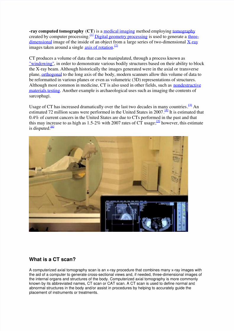
8/3/2019 Ray Computed Tomography
http://slidepdf.com/reader/full/ray-computed-tomography 1/7
-ray computed tomography (CT) is a medical imaging method employing tomography
created by computer processing.[1]
Digital geometry processing is used to generate a three-
dimensional image of the inside of an object from a large series of two-dimensional X-ray
images taken around a single axis of rotation.[2]
CT produces a volume of data that can be manipulated, through a process known as"windowing", in order to demonstrate various bodily structures based on their ability to block
the X-ray beam. Although historically the images generated were in the axial or transverse
plane, orthogonal to the long axis of the body, modern scanners allow this volume of data to
be reformatted in various planes or even as volumetric (3D) representations of structures.
Although most common in medicine, CT is also used in other fields, such as nondestructive
materials testing. Another example is archaeological uses such as imaging the contents of
sarcophagi.
Usage of CT has increased dramatically over the last two decades in many countries.[3]
An
estimated 72 million scans were performed in the United States in 2007.[4]
It is estimated that
0.4% of current cancers in the United States are due to CTs performed in the past and thatthis may increase to as high as 1.5-2% with 2007 rates of CT usage;
[5] however, this estimate
is disputed.[6]
What is a CT scan?
A computerized axial tomography scan is an x-ray procedure that combines many x-ray images withthe aid of a computer to generate cross-sectional views and, if needed, three-dimensional images ofthe internal organs and structures of the body. Computerized axial tomography is more commonlyknown by its abbreviated names, CT scan or CAT scan. A CT scan is used to define normal andabnormal structures in the body and/or assist in procedures by helping to accurately guide theplacement of instruments or treatments.
8/3/2019 Ray Computed Tomography
http://slidepdf.com/reader/full/ray-computed-tomography 2/7
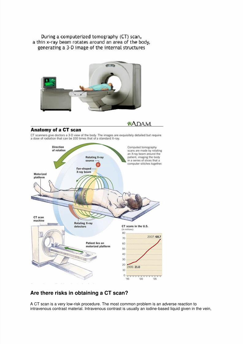
A large donut-shaped x-ray machine takes x-ray images at many different angles around the body.These images are processed by a computer to produce cross-sectional pictures of the body. In eachof these pictures the body is seen as an x-ray "slice" of the body, which is recorded on a film. Thisrecorded image is called a tomogram. "Computerized Axial Tomography" refers to the recordedtomogram "sections" at different levels of the body.
Imagine the body as a loaf of bread and you are looking at one end of the loaf. As you remove eachslice of bread, you can see the entire surface of that slice from the crust to the center. The body isseen on CT scan slices in a similar fashion from the skin to the central part of the body beingexamined. When these levels are further "added" together, a three-dimensional picture of an organ orabnormal body structure can be obtained
Why are CT scans performed?
CT scans are performed to analyze the internal structures of various parts of the body. This includesthe head, where traumatic injuries, (such as blood clots or skull fractures), tumors, and infections canbe identified. In the spine, the bony structure of the vertebrae can be accurately defined, as can theanatomy of the intervertebral discs and spinal cord. In fact, CT scan methods can be used to
accurately measure the density of bone in evaluatingosteoporosis.
Occasionally, contrast material (an x-ray dye) is placed into the spinal fluid to further enhance thescan and the various structural relationships of the spine, the spinal cord, and its nerves. Contrastmaterial is also often administered intravenously or through other routes prior to obtaining a CT scan(see below). CT scans are also used in the chest to identify tumors, cysts, or infections that may besuspected on a chest x-ray. CT scans of the abdomen are extremely helpful in defining body organanatomy, including visualizing the liver, gallbladder, pancreas, spleen, aorta, kidneys, uterus, andovaries. CT scans in this area are used to verify the presence or absence of tumors, infection,abnormal anatomy, or changes of the body from trauma.
The technique is painless and can provide extremely accurate images of body structures in addition toguiding the radiologist in performing certain procedures, such as biopsies of suspected cancers,removal of internal body fluids for various tests, and the draining of abscesses which are deep in thebody. Many of these procedures are minimally invasive and have markedly decreased the need toperform surgery to accomplish the same goal.
8/3/2019 Ray Computed Tomography
http://slidepdf.com/reader/full/ray-computed-tomography 3/7
Are there risks in obtaining a CT scan?
A CT scan is a very low-risk procedure. The most common problem is an adverse reaction tointravenous contrast material. Intravenous contrast is usually an iodine-based liquid given in the vein,
8/3/2019 Ray Computed Tomography
http://slidepdf.com/reader/full/ray-computed-tomography 4/7
which makes many organs and structures, such as the kidneys and blood vessels much more visibleon the CT scan. There may be resulting itching, a rash, hives, or a feeling of warmth throughout thebody. These are usually self-limiting reactions that go away rather quickly. If needed, antihistaminescan be given to help relieve the symptoms. A more serious allergic reaction to intravenous contrast iscalled an anaphylactic reaction
How does a patient prepare for CT scanning, and how is it performed?
In preparation for a CT scan, patients are often asked to avoid food, especially when contrast materialis to be used. Contrast material may be injected intravenously, or administered by mouth or by anenema in order to increase the distinction between various organs or areas of the body. Therefore,fluids and food may be restricted for several hours prior to the examination. If the patient has a historyof allergy to contrast material (such as iodine), the requesting physician and radiology staff should benotified. All metallic materials and certain clothing around the body are removed because they caninterfere with the clarity of the images.
Patients are placed on a movable table, and the table is slipped into the center of a large donut-shaped machine which takes the x-ray images around the body. The actual procedure can take from
a half an hour to an hour and a half. If specific tests, biopsies, or intervention are performed by theradiologist during CT scanning, additional time and monitoring may be required. It is important duringthe CT scan procedure that the patient minimize any body movement by remaining as still and quietas is possible. This significantly increases the clarity of the x-ray images. The CT scan technologisttells the patient when to breathe or hold his/her breath during scans of the chest and abdomen. If anyproblems are experienced during the CT scan, the technologist should be informed immediately. Thetechnologist directly watches the patient through an observation window during the procedure, andthere is an intercom system in the room for added patient safety.
CT scans have vastly improved the ability of doctors to diagnose many diseases earlier in their courseand with much less risk than previous methods. Further refinements in CT scan technology continueto evolve which promise even better picture quality and patient safety. Newer CT scans called "spiral"or "helical" CT scans can provide more rapid and accurate visualization of internal organs. For
example, many trauma centers are using these scans to more rapidly diagnose internal injuries afterserious body trauma. High resolution CT scans (HRCT) are used to accurately assess the lungs forinflammation and scarring.
CT Scan At A Glance
CT scanning adds x-ray images with the aid of a computer to generate cross-sectional viewsof anatomy.
CT scanning can identify normal and abnormal structures and be used to guide procedures.
CT scanning is painless.
Iodine-containing contrast material is sometimes used in CT scanning. Patients with a historyof allergy to iodine or contrast materials should notify their physicians and radiology staff.
http://www.medicinenet.com/cat_scan/discussion-208.htm
Basic Working Principle Of CT Scan
8/3/2019 Ray Computed Tomography
http://slidepdf.com/reader/full/ray-computed-tomography 5/7
It is as simple as passing X-rays through the patient and obtaining information with a detector
on the other side. The X-ray source and the detector are interconnected and rotated around the
patient during scanning period. Digital computers then assemble the data that is obtained and
integrate it to provide a cross sectional image (tomogram) that is displayed on a computer
screen. The image can be photographed or stored for later retrieval and use as the case may
be.
X-rays are electromagnetic waves. The main reason why X-rays is used in diagnosis is
because all substances and tissues differ in their ability to absorb X-rays. Some substances
are more permeable to X-rays while some others impermeable. Owing to this difference,
different tissues seem different when the X-ray film is developed.
Dense tissues such as the bones appear white on a CT film while the soft tissues such as the
brain or kidney appear gray. The cavities filled with air such as the lungs appear black.
CT Scan - What Does The Equipment Look Like
Read more: CT Scan - What Does The Equipment Look Like | Medindia
http://www.medindia.net/patients/patientinfo/CT_Scan_equipment.htm#ixzz1cOIXQ23D
Most of the patients get nervous when they see a CT machine because it seems very huge and
massive. However, there is no reason for worry because a general CT examination is a
relatively safer procedure. The CT scanner is a large machine that has a hole in the center. It
is provided with a sliding patient table, which can move back and front. You will be asked to
lie over the table and would be instructed by the radiographer regarding the procedure. Once
he/she has made sure that you are familiar with the instructions, the examination would start.
You may be required to remain still for a few minutes during the examination. However, this
should not be for long. It usually takes about less than 10 to 15 minutes. You will be alone in
the examination room. The radiologist would be seated in the adjacent room and you can
communicate with them incase you feel any discomfort.
CT Scan - Disadvantages
Need for contrast media for enhanced soft tissue contrast.
Tissue non-specificity i.e. it does have ability to highlight any particular organ/ tissue.
Cost concerns
8/3/2019 Ray Computed Tomography
http://slidepdf.com/reader/full/ray-computed-tomography 6/7
In a conventional X-ray, a broad beam of x-rays passes through the patient to produce an
image on the X-ray film. Whereas, in CT, a thin beam of X-rays is passed through the patient
that is absorbed by a detector.
Read more: Difference Between CT Scan & X-ray | Medindia
http://www.medindia.net/patients/patientinfo/CT_Scan_difference.htm#ixzz1cOJFCe14
What are the risks associated with the procedure
Read more: CT Scan - Risks Associated With The Procedure | Medindia
http://www.medindia.net/patients/patientinfo/Ct_Scan_risks.htm#ixzz1cOJQUQmt
The device uses X-ray radiation as described previously. The amount of radiation received
from a CT examination is less than the amount of background radiation received by a person
in 3 years. Examining the risk Vs benefit ratio, the benefit is considerably greater.
In some cases where a contrast material is injected or swallowed, the radiology department is
well equipped to tackle any emergency situation due to an allergic reaction. Mothers who
breast-feed can usually resume the same after 24 hours of injection of the contrast material.
By this time, the material would be excreted from the body. It is recommended that you
discuss with your doctor regarding the issue for more information.
8/3/2019 Ray Computed Tomography
http://slidepdf.com/reader/full/ray-computed-tomography 7/7
ch
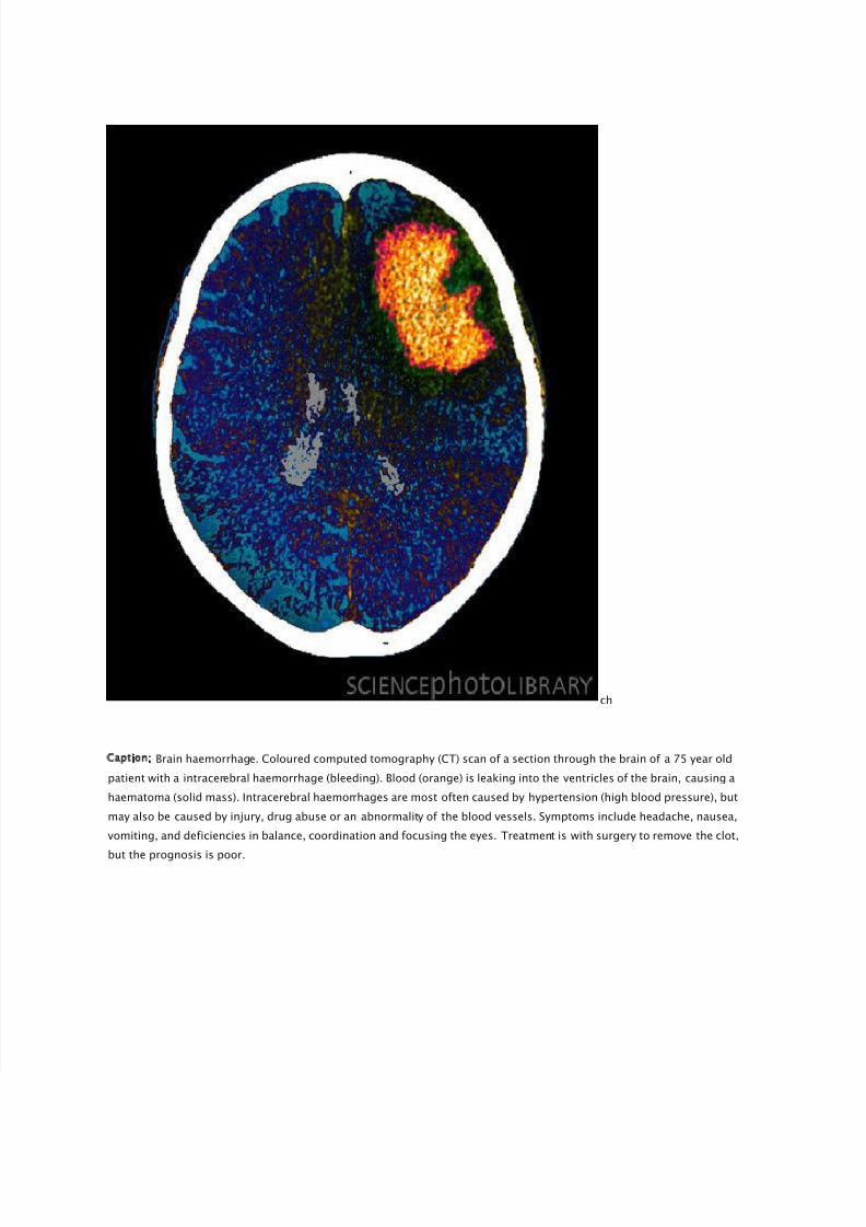
Caption: Brain haemorrhage. Coloured computed tomography (CT) scan of a section through the brain of a 75 year old
patient with a intracerebral haemorrhage (bleeding). Blood (orange) is leaking into the ventricles of the brain, causing a
haematoma (solid mass). Intracerebral haemorrhages are most often caused by hypertension (high blood pressure), but
may also be caused by injury, drug abuse or an abnormality of the blood vessels. Symptoms include headache, nausea,
vomiting, and deficiencies in balance, coordination and focusing the eyes. Treatment is with surgery to remove the clot,
but the prognosis is poor.