Expression of Active Fluorophore Proteins in the Milk of ...

ChemicalScience
EDGE ARTICLE
Ope
n A
cces
s A
rtic
le. P
ublis
hed
on 1
0 O
ctob
er 2
018.
Dow
nloa
ded
on 2
/18/
2022
8:4
2:15
AM
. T
his
artic
le is
lice
nsed
und
er a
Cre
ativ
e C
omm
ons
Attr
ibut
ion-
Non
Com
mer
cial
3.0
Unp
orte
d L
icen
ce.
View Article OnlineView Journal | View Issue
Rational design o
aLaboratory of Molecular Imaging and Nanom
Imaging and Bioengineering (NIBIB), Natio
Maryland 20892, USA. E-mail: shoujun.zhubDepartment of Materials Science & Enginee
Organic Electronics, South University of Sc
518055, China. E-mail: [email protected] of Chemistry, Stanford UniversdState Key Laboratory of Precision Spectro
Science, East China Normal University, ShaeResearch Center for Advanced Materials a
Tsinghua University in Shenzhen, Shenzhen
† Electronic supplementary informationprocess and structure characterization, exDOI: 10.1039/c8sc03751e
‡ These authors contributed equally to th
Cite this: Chem. Sci., 2019, 10, 326
All publication charges for this articlehave been paid for by the Royal Societyof Chemistry
Received 22nd August 2018Accepted 8th October 2018
DOI: 10.1039/c8sc03751e
rsc.li/chemical-science
326 | Chem. Sci., 2019, 10, 326–332
f a super-contrast NIR-IIfluorophore affords high-performance NIR-IImolecular imaging guided microsurgery†
Rui Tian,‡a Huilong Ma,‡b Qinglai Yang,‡be Hao Wan,‡c Shoujun Zhu, *a
Swati Chandra,a Haitao Sun, d Dale O. Kiesewetter,a Gang Niu,a Yongye Liang *b
and Xiaoyuan Chen *a
In vivomolecular imaging in the “transparent” near-infrared II (NIR-II) window has demonstrated impressive
benefits in reaching millimeter penetration depths with high specificity and imaging quality. Previous NIR-II
molecular imaging generally relied on high hepatic uptake fluorophores with an unclear mechanism and
antibody-derived conjugates, suffering from inevitable nonspecific retention in the main organs/skin with
a relatively low signal-to-background ratio. It is still challenging to synthesize a NIR-II fluorophore with
both high quantum yield and minimal liver-retention feature. Herein, we identified the structural design
and excretion mechanism of novel NIR-II fluorophores for NIR-II molecular imaging with an extremely
clean background. With the optimized renally excreted fluorophore–peptide conjugates, superior NIR-II
targeting imaging was accompanied by the improved signal-to-background ratio during tumor detection
with reducing off-target tissue exposure. An unprecedented NIR-II imaging-guided microsurgery was
achieved using such an imaging platform, which provides us with a great preclinical example to
accelerate the potential clinical translation of NIR-II imaging.
Introduction
Biological imaging with uorescence in the NIR-II regionranging from 1000 to 1700 nm (ref. 1–4) benets from reducedbody autouorescence and light scattering, allowing forenhanced sub-centimeter penetration into biologicalsystems.5–11 To achieve NIR-II molecular imaging with highcontrast for preclinical/clinical use, both the uorophore andconjugated targeting probe need to be renally excreted with lowretention in normal tissues.12–14 Compared to antibodies andhormones, peptides provide high binding affinity, low immu-nogenicity and faster body clearance.15,16 So far, many kinds of
edicine, National Institute of Biomedical
nal Institutes of Health (NIH), Bethesda,
@nih.gov; [email protected]
ring, Shenzhen Key Laboratory of Printed
ience & Technology of China, Shenzhen
n
ity, Stanford, CA 94305, USA
scopy, School of Physics and Materials
nghai 200062, China
nd Biotechnology, Research Institute of
518057, China
(ESI) available: Synthesis optimizationcretion study, binding affinity, etc. See
is work.
peptides have been recognized and developed for specic tumorbiomarkers,17 and over 150 peptides in active development haveentered human clinical studies.18 For example, arginylglycylas-partic acid (RGD) motifs have been widely utilized as imaging-guided diagnostic probes,19–21 and radioactive isotope modi-ed targeting peptides can reach high tumor-to-normal tissue(T/NT) ratios in positron emission tomography (PET)imaging.22,23
Fluorescent probes derived from specic peptides have beenused clinically in NIR-I imaging-guided tumor surgery.24 Fluo-rescence imaging possesses an advantage in both spatial andtemporal resolutions,25,26 and the bottleneck caused by pene-tration issues could be improved by imaging at longer wave-lengths.27–30 Renally excreted conjugates in the NIR-II windowprot from both the low uptake of normal tissue/immunesystems and reduced tissue autouorescence/scattering. Asa result, NIR-II peptide conjugated dyes with high quantumyields and renal excretion ability afford great penetrationdepths and high signal/background ratios for preclinical andclinical settings. So far, NIR-II uorophores have expanded toinorganic materials, polymer encapsulated organic dyes andwater-soluble molecular uorophores,29,31–47 whichmostly sufferfrom high uptake and long-term retention in the immunesystem, causing considerable safety concerns in terms ofclinical applications. It is of great importance to exploit brightNIR-II uorophores with both high quantum yields and fastexcretion ability.8,48–51
This journal is © The Royal Society of Chemistry 2019
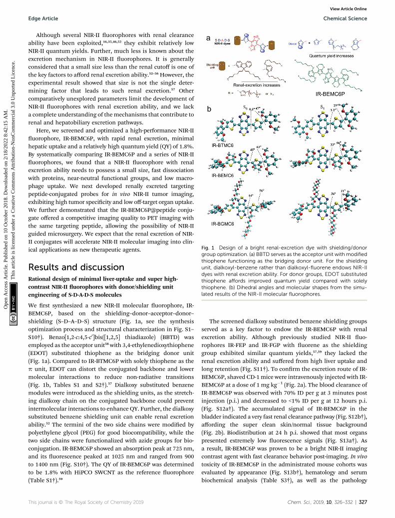
Fig. 1 Design of a bright renal-excretion dye with shielding/donorgroup optimization. (a) BBTD serves as the acceptor unit with modifiedthiophene functioning as the bridging donor unit. For the shieldingunit, dialkoxyl-benzene rather than dialkoxyl-fluorene endows NIR-IIdyes with renal excretion ability. For donor groups, EDOT substitutedthiophene affords improved quantum yield compared with solelythiophene. (b) Dihedral angles and molecular shapes from the simu-lated results of the NIR-II molecular fluorophores.
Edge Article Chemical Science
Ope
n A
cces
s A
rtic
le. P
ublis
hed
on 1
0 O
ctob
er 2
018.
Dow
nloa
ded
on 2
/18/
2022
8:4
2:15
AM
. T
his
artic
le is
lice
nsed
und
er a
Cre
ativ
e C
omm
ons
Attr
ibut
ion-
Non
Com
mer
cial
3.0
Unp
orte
d L
icen
ce.
View Article Online
Although several NIR-II uorophores with renal clearanceability have been exploited,16,35,46,52 they exhibit relatively lowNIR-II quantum yields. Further, much less is known about theexcretion mechanism in NIR-II uorophores. It is generallyconsidered that a small size less than the renal cutoff is one ofthe key factors to afford renal excretion ability.52–56 However, theexperimental result showed that size is not the single deter-mining factor that leads to such renal excretion.57 Othercomparatively unexplored parameters limit the development ofNIR-II uorophores with renal excretion ability, and we lacka complete understanding of themechanisms that contribute torenal and hepatobiliary excretion pathways.
Here, we screened and optimized a high-performance NIR-IIuorophore, IR-BEMC6P, with rapid renal excretion, minimalhepatic uptake and a relatively high quantum yield (QY) of 1.8%.By systematically comparing IR-BEMC6P and a series of NIR-IIuorophores, we found that a NIR-II uorophore with renalexcretion ability needs to possess a small size, fast dissociationwith proteins, near-neutral functional groups, and low macro-phage uptake. We next developed renally excreted targetingpeptide-conjugated probes for in vivo NIR-II tumor imaging,exhibiting high tumor specicity and low off-target organ uptake.We further demonstrated that the IR-BEMC6P@peptide conju-gate offered a competitive imaging quality to PET imaging withthe same targeting peptide, allowing the possibility of NIR-IIguided microsurgery. We expect that the renal excretion of NIR-II conjugates will accelerate NIR-II molecular imaging into clin-ical applications as new therapeutic agents.
Results and discussionRational design of minimal liver-uptake and super high-contrast NIR-II uorophores with donor/shielding unitengineering of S-D-A-D-S molecules
We rst synthesized a new NIR-II molecular uorophore, IR-BEMC6P, based on the shielding–donor–acceptor–donor–shielding (S–D–A–D–S) structure (Fig. 1a, see the synthesisoptimization process and structural characterization in Fig. S1–S10†). Benzo[1,2-c:4,5-c0]bis([1,2,5] thiadiazole) (BBTD) wasemployed as the acceptor unit58with 3,4-ethylenedioxythiophene(EDOT) substituted thiophene as the bridging donor unit(Fig. 1a). Compared to IR-BTMC6P with solely thiophene as thep unit, EDOT can distort the conjugated backbone and lowermolecular interactions to reduce non-radiative transitions(Fig. 1b, Tables S1 and S2†).57 Dialkoxy substituted benzenemodules were introduced as the shielding units, as the stretch-ing dialkoxy chain on the conjugated backbone could preventintermolecular interactions to enhance QY. Further, the dialkoxysubstituted benzene shielding unit can enable renal excretionability.52 The termini of the two side chains were modied bypolyethylene glycol (PEG) for good biocompatibility, while thetwo side chains were functionalized with azide groups for bio-conjugation. IR-BEMC6P showed an absorption peak at 725 nm,and its uorescence peaked at 1025 nm and ranged from 900to 1400 nm (Fig. S10†). The QY of IR-BEMC6P was determinedto be 1.8% with HiPCO SWCNT as the reference uorophore(Table S1†).59
This journal is © The Royal Society of Chemistry 2019
The screened dialkoxy substituted benzene shielding groupsserved as a key factor to endow the IR-BEMC6P with renalexcretion ability. Although previously studied NIR-II uo-rophores IR-FEP and IR-FGP with uorene as the shieldinggroup exhibited similar quantum yields,57,59 they lacked therenal excretion ability and suffered from high liver uptake andlong retention (Fig. S11†). To conrm the excretion route of IR-BEMC6P, shaved CD-1mice were intravenously injected with IR-BEMC6P at a dose of 1 mg kg�1 (Fig. 2a). The blood clearance ofIR-BEMC6P was observed with 70% ID per g at 3 minutes postinjection (p.i.) and decreased to <1% ID per g at 12 hours p.i.(Fig. S12a†). The accumulated signal of IR-BEMC6P in thebladder indicated a very fast renal clearance pathway (Fig. S12b†),affording the super clean skin/normal tissue background(Fig. 2b). Biodistribution at 24 h p.i. showed that most organspresented extremely low uorescence signals (Fig. S13a†). Asa result, IR-BEMC6P was proven to be a bright NIR-II imagingcontrast agent with fast clearance behavior post-imaging. In vivotoxicity of IR-BEMC6P in the administrated mouse cohorts wasevaluated by appearance (Fig. S13b†), hematology and serumbiochemical analysis (Table S3†), as well as the pathology
Chem. Sci., 2019, 10, 326–332 | 327
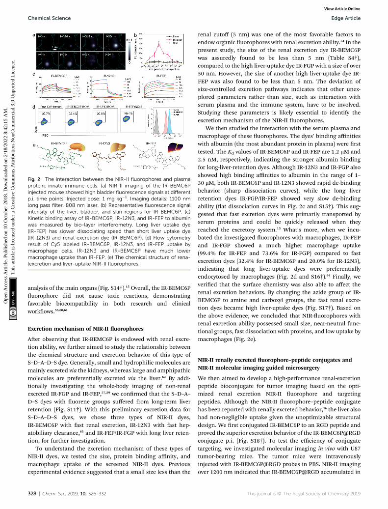
Fig. 2 The interaction between the NIR-II fluorophores and plasmaprotein, innate immune cells. (a) NIR-II imaging of the IR-BEMC6Pinjected mouse showed high bladder fluorescence signals at differentp.i. time points. Injected dose: 1 mg kg�1. Imaging details: 1100 nmlong pass filter, 808 nm laser. (b) Representative fluorescence signalintensity of the liver, bladder, and skin regions for IR-BEMC6P. (c)Kinetic binding assay of IR-BEMC6P, IR-12N3, and IR-FEP to albuminwas measured by bio-layer interferometry. Long liver uptake dye(IR-FEP) has slower dissociating speed than short liver uptake dye(IR-12N3) and renal excretion dye (IR-BEMC6P). (d) Flow cytometryresult of Cy5 labeled IR-BEMC6P, IR-12N3, and IR-FEP uptake bymacrophage cells. IR-12N3 and IR-BEMC6P have much lowermacrophage uptake than IR-FEP. (e) The chemical structure of rena-lexcretion and liver-uptake NIR-II fluorophores.
Chemical Science Edge Article
Ope
n A
cces
s A
rtic
le. P
ublis
hed
on 1
0 O
ctob
er 2
018.
Dow
nloa
ded
on 2
/18/
2022
8:4
2:15
AM
. T
his
artic
le is
lice
nsed
und
er a
Cre
ativ
e C
omm
ons
Attr
ibut
ion-
Non
Com
mer
cial
3.0
Unp
orte
d L
icen
ce.
View Article Online
analysis of the main organs (Fig. S14†).15 Overall, the IR-BEMC6Puorophore did not cause toxic reactions, demonstratingfavorable biocompatibility in both research and clinicalworkows.56,60,61
Excretion mechanism of NIR-II uorophores
Aer observing that IR-BEMC6P is endowed with renal excre-tion ability, we further aimed to study the relationship betweenthe chemical structure and excretion behavior of this type ofS–D–A–D–S dye. Generally, small and hydrophilic molecules aremainly excreted via the kidneys, whereas large and amphipathicmolecules are preferentially excreted via the liver.62 By addi-tionally investigating the whole-body imaging of non-renalexcreted IR-FGP and IR-FEP,57,59 we conrmed that the S–D–A–D–S dyes with uorene groups suffered from long-term liverretention (Fig. S11†). With this preliminary excretion data forS–D–A–D–S dyes, we chose three types of NIR-II dyes,IR-BEMC6P with fast renal excretion, IR-12N3 with fast hep-atobiliary clearance,63 and IR-FEP/IR-FGP with long liver reten-tion, for further investigation.
To understand the excretion mechanism of these types ofNIR-II dyes, we tested the size, protein binding affinity, andmacrophage uptake of the screened NIR-II dyes. Previousexperimental evidence suggested that a small size less than the
328 | Chem. Sci., 2019, 10, 326–332
renal cutoff (5 nm) was one of the most favorable factors toendow organic uorophores with renal excretion ability.54 In thepresent study, the size of the renal excretion dye IR-BEMC6Pwas assuredly found to be less than 5 nm (Table S4†),compared to the high liver-uptake dye IR-FGP with a size of over50 nm. However, the size of another high liver-uptake dye IR-FEP was also found to be less than 5 nm. The deviation ofsize-controlled excretion pathways indicates that other unex-plored parameters rather than size, such as interaction withserum plasma and the immune system, have to be involved.Studying these parameters is likely essential to identify theexcretion mechanism of the NIR-II uorophores.
We then studied the interaction with the serum plasma andmacrophage of these uorophores. The dyes' binding affinitieswith albumin (the most abundant protein in plasma) were rsttested. The Kd values of IR-BEMC6P and IR-FEP are 1.2 mM and2.5 nM, respectively, indicating the stronger albumin bindingfor long-liver-retention dyes. Although IR-12N3 and IR-FGP alsoshowed high binding affinities to albumin in the range of 1–30 mM, both IR-BEMC6P and IR-12N3 showed rapid de-bindingbehavior (sharp dissociation curves), while the long liverretention dyes IR-FGP/IR-FEP showed very slow de-bindingability (at dissociation curves in Fig. 2c and S15†). This sug-gested that fast excretion dyes were primarily transported byserum proteins and could be quickly released when theyreached the excretory system.15 What's more, when we incu-bated the investigated uorophores with macrophages, IR-FEPand IR-FGP showed a much higher macrophage uptake(99.4% for IR-FEP and 73.6% for IR-FGP) compared to fastexcretion dyes (32.4% for IR-BEMC6P and 20.0% for IR-12N3),indicating that long liver-uptake dyes were preferentiallyendocytosed by macrophages (Fig. 2d and S16†).64 Finally, weveried that the surface chemistry was also able to affect therenal excretion behaviors. By changing the azide group of IR-BEMC6P to amine and carboxyl groups, the fast renal excre-tion dyes became high liver-uptake dyes (Fig. S17†). Based onthe above evidence, we concluded that NIR-uorophores withrenal excretion ability possessed small size, near-neutral func-tional groups, fast dissociation with proteins, and low uptake bymacrophages (Fig. 2e).
NIR-II renally excreted uorophore–peptide conjugates andNIR-II molecular imaging guided microsurgery
We then aimed to develop a high-performance renal-excretionpeptide bioconjugate for tumor imaging based on the opti-mized renal excretion NIR-II uorophore and targetingpeptides. Although the NIR-II uorophore–peptide conjugatehas been reported with renally excreted behavior,16 the liver alsohad non-negligible uptake given the unoptimizable structuraldesign. We rst conjugated IR-BEMC6P to an RGD peptide andproved the superior excretion behavior of the IR-BEMC6P@RGDconjugate p.i. (Fig. S18†). To test the efficiency of conjugatetargeting, we investigated molecular imaging in vivo with U87tumor-bearing mice. The tumor mice were intravenouslyinjected with IR-BEMC6P@RGD probes in PBS. NIR-II imagingover 1200 nm indicated that IR-BEMC6P@RGD accumulated in
This journal is © The Royal Society of Chemistry 2019
Edge Article Chemical Science
Ope
n A
cces
s A
rtic
le. P
ublis
hed
on 1
0 O
ctob
er 2
018.
Dow
nloa
ded
on 2
/18/
2022
8:4
2:15
AM
. T
his
artic
le is
lice
nsed
und
er a
Cre
ativ
e C
omm
ons
Attr
ibut
ion-
Non
Com
mer
cial
3.0
Unp
orte
d L
icen
ce.
View Article Online
the U87 tumor in less than 1 h (Fig. 3a), with the T/NT ratioreaching a maximum of �9 at 6 h p.i. (Fig. 3b) and low accu-mulation in other organs (Fig. 3c).46 In contrast, the non-targeted IR-BEMC6P uorophore showed a relatively weakersignal in the U87 tumor and the maximum T/NT ratio was lessthan �4 (Fig. S19†), due to the nonspecic accumulation of thefree IR-BEMC6P.
We further investigated the advantage of NIR-II bioimagingby imaging the brain vessels in C57 mice with intact skin/scalpusing either ICG or IR-BEMC6P, respectively (Fig. 3d–f).53 Todemonstrate the benet of the developed renal excretionconjugate in NIR-II molecular imaging, a glioblastoma braintumor was surgically inoculated through the U87MG cells in thele hemisphere of the mouse brain. By intravenously injectingIR-BEMC6P@RGD, the NIR-II imaging of the brain tumorthrough a non-invasive route was performed through both thescalp and the skull. 6–12 h aer administration of IR-BEMC6P@RGD, the tumor was distinguished with a T/NTratio of �6 with NIR-II whole body imaging (Fig. 3g). By
Fig. 3 NIR-II targeting imaging of the U87 tumor model with IR-BEMC6P@RGD. (a) Imaging of the U87 tumor-bearing mouse intra-venously treated with IR-BEMC6P@RGD in the NIR-II window. Injec-ted dose: 200 mL of 25–75 mM conjugate solution. Imaging details:1200 nm long pass filter, 808 nm laser. (b) CBR and TBR statistics ofU87 tumor signal: contrast-to-background ratio (CBR) ¼ (fluores-cence � background)/background. Tumor-to-background ratio (TBR)¼ CBR of tumor/CBR of normal tissue. (c) Ex vivo scanning of theorgans (tumor, liver, skin) after 24 h post injection of IR-BEMC6P@RGD. (d and e) Brain vessel imaging in C57 mice with shavedheads by injecting either ICG at 850–900 nm or IR-BEMC6P with1300 nm long pass filter. (f) Section profile curve of vessels in both NIR-I and NIR-II windows. (g) High magnification NIR-II fluorescenceimaging showing strong tumor fluorescence detectable through theintact scalp/skull at 12 h post-injection in an orthotopic brain tumormodel. PbS quantum dots were intravenously injected as an additionalchannel over 1500 nm emission to visualize the vessels.65,66
This journal is © The Royal Society of Chemistry 2019
subsequently injecting PbS@PEG quantum dots with over1500 nm emission,65 two-color imaging of both the brain tumorand vessel was obtained at the same time, affording multicolorlive molecular imaging across the NIR-II window in a deeptumor model.
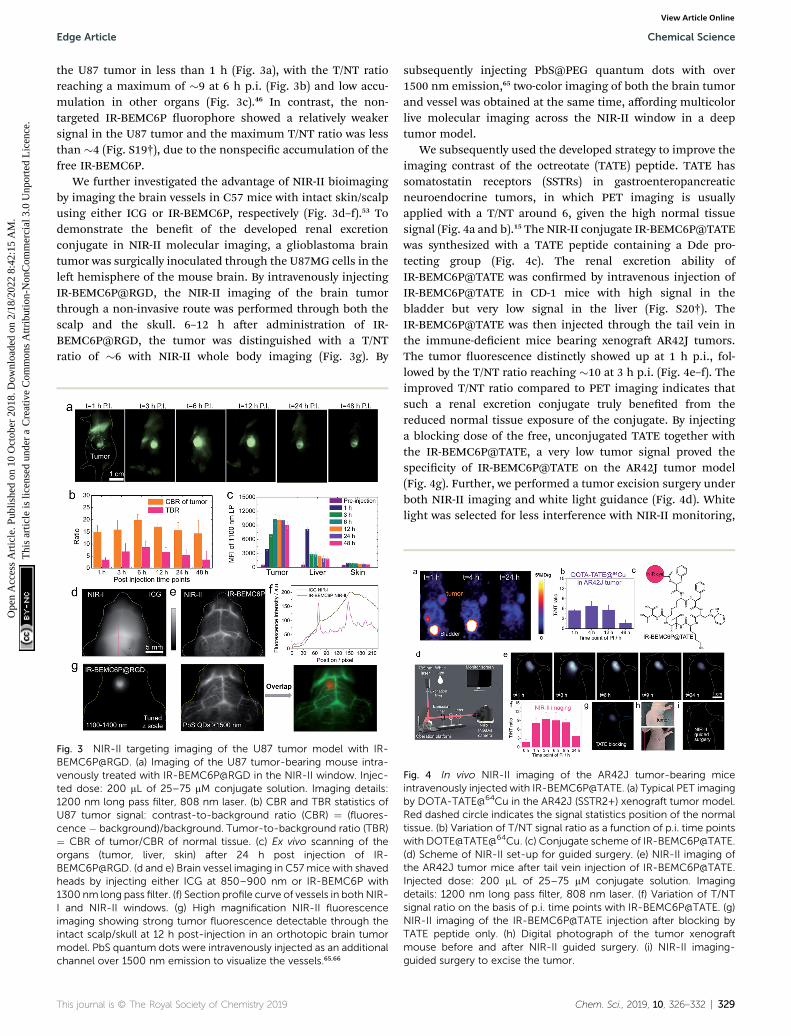
We subsequently used the developed strategy to improve theimaging contrast of the octreotate (TATE) peptide. TATE hassomatostatin receptors (SSTRs) in gastroenteropancreaticneuroendocrine tumors, in which PET imaging is usuallyapplied with a T/NT around 6, given the high normal tissuesignal (Fig. 4a and b).15 The NIR-II conjugate IR-BEMC6P@TATEwas synthesized with a TATE peptide containing a Dde pro-tecting group (Fig. 4c). The renal excretion ability ofIR-BEMC6P@TATE was conrmed by intravenous injection ofIR-BEMC6P@TATE in CD-1 mice with high signal in thebladder but very low signal in the liver (Fig. S20†). TheIR-BEMC6P@TATE was then injected through the tail vein inthe immune-decient mice bearing xenogra AR42J tumors.The tumor uorescence distinctly showed up at 1 h p.i., fol-lowed by the T/NT ratio reaching �10 at 3 h p.i. (Fig. 4e–f). Theimproved T/NT ratio compared to PET imaging indicates thatsuch a renal excretion conjugate truly beneted from thereduced normal tissue exposure of the conjugate. By injectinga blocking dose of the free, unconjugated TATE together withthe IR-BEMC6P@TATE, a very low tumor signal proved thespecicity of IR-BEMC6P@TATE on the AR42J tumor model(Fig. 4g). Further, we performed a tumor excision surgery underboth NIR-II imaging and white light guidance (Fig. 4d). Whitelight was selected for less interference with NIR-II monitoring,
Fig. 4 In vivo NIR-II imaging of the AR42J tumor-bearing miceintravenously injected with IR-BEMC6P@TATE. (a) Typical PET imagingby DOTA-TATE@64Cu in the AR42J (SSTR2+) xenograft tumor model.Red dashed circle indicates the signal statistics position of the normaltissue. (b) Variation of T/NT signal ratio as a function of p.i. time pointswith DOTE@TATE@64Cu. (c) Conjugate scheme of IR-BEMC6P@TATE.(d) Scheme of NIR-II set-up for guided surgery. (e) NIR-II imaging ofthe AR42J tumor mice after tail vein injection of [email protected] dose: 200 mL of 25–75 mM conjugate solution. Imagingdetails: 1200 nm long pass filter, 808 nm laser. (f) Variation of T/NTsignal ratio on the basis of p.i. time points with IR-BEMC6P@TATE. (g)NIR-II imaging of the IR-BEMC6P@TATE injection after blocking byTATE peptide only. (h) Digital photograph of the tumor xenograftmouse before and after NIR-II guided surgery. (i) NIR-II imaging-guided surgery to excise the tumor.
Chem. Sci., 2019, 10, 326–332 | 329
Chemical Science Edge Article
Ope
n A
cces
s A
rtic
le. P
ublis
hed
on 1
0 O
ctob
er 2
018.
Dow
nloa
ded
on 2
/18/
2022
8:4
2:15
AM
. T
his
artic
le is
lice
nsed
und
er a
Cre
ativ
e C
omm
ons
Attr
ibut
ion-
Non
Com
mer
cial
3.0
Unp
orte
d L
icen
ce.
View Article Online
and the NIR-II imaging allowed for clear distinction of thetumor margin (Fig. 4h and i). Moreover, NIR-II guided micro-surgery was additionally performed utilizing an IR-BEMC6P@follicle-stimulating hormone (FSH) conjugate,allowing us to cut single follicles that are hundreds of mm insize (Fig. S21†). Only mild alterations in behavior post-surgerywere observed, indicating the high safety of micro-surgery.Although uorescence-guided surgery was achieved in theNIR-I systems,67 it is still critically important to furtherimprove the detection depth and sensitivity, to yielda comprehensive uorescence-guided platform with adjust-able imaging speed (real-time) and contrast (S/B ratio), andmulti-targeted imaging for visualizing complex diseasessimultaneously.30,68
Conclusions
In summary, we developed a minimal hepatic-uptake NIR-II dye(IR-BEMC6P) with enhanced QY and derived renal excretionpeptide conjugations. The excretion mechanism was exploitedthrough comprehensive factors, and we found that NIR-II u-orophores with renal excretion ability possessed small size,near-neutral functional groups, fast de-binding with proteins,and low uptake by macrophages. This well-established rule forexcretion behavior will guide future NIR-II dye synthesis andenrich the NIR-II uorophore library with the clinical choice ofNIR-II imaging, as one of the important features of potentialNIR uorophores for clinically translatable imaging is timelyclearance from the body with minimum retention in majororgans aer administration.5 The renal excretion uorophorederived peptide–dye conjugate provided efficient tumor target-ing ability in mice with a high T/NT ratio in two different tumormodels, affording great ability for NIR-II guided microsurgery.Importantly, the peptide IR-BEMC6P@(RGD, TATE) probeswere rapidly renally excreted, in sharp contrast to long-termliver accumulation of typical antibody–dye conjugates. Theserapidly excreted, highly safe integrin/somatostatin receptortargeting NIR-II probes will accelerate the clinical translation ofNIR-II molecular imaging in cancer diagnosis and imagingnavigation therapy.
Conflicts of interest
There are no conicts to declare.
Acknowledgements
This work was supported by the Shenzhen Key Lab FundingGrant ZDSYS201505291525382, the Shenzhen Peacock ProgramGrant KQTD20140630160825828, the Shenzhen Basic Researchfunding (JCYJ20170307151634428) and the IntramuralResearch Program of the National Institute of BiomedicalImaging and Bioengineering (NIBIB), the National Institutes ofHealth (NIH). S. Z. thanks Dr Xue Zhang in Jilin AgriculturalUniversity for scheme designing. All animal studies were con-ducted in accordance with the principles and procedures out-lined in the National Institutes of Health (NIH) Guide for the
330 | Chem. Sci., 2019, 10, 326–332
Care and Use of Animals, and under protocols approved by theNIH Clinical Center Animal Care and Use Committee (CC-ACUC, protocol number: NIBIB 16-03).
Notes and references
1 R. Jin, C. Zeng, M. Zhou and Y. Chen, Chem. Rev., 2016, 116,10346–10413.
2 Y. Lyu, Y. Fang, Q. Miao, X. Zhen, D. Ding and K. Pu, ACSNano, 2016, 10, 4472–4481.
3 Y. Lyu, C. Xie, S. A. Chechetka, E. Miyako and K. Pu, J. Am.Chem. Soc., 2016, 138, 9049–9052.
4 B. Shi, K. Jie, Y. Zhou, J. Zhou, D. Xia and F. Huang, J. Am.Chem. Soc., 2016, 138, 80–83.
5 G. Hong, A. L. Antaris and H. Dai, Nature BiomedicalEngineering, 2017, 1, 0010.
6 R. Wang and F. Zhang, J. Mater. Chem. B, 2014, 2, 2422.7 S. He, J. Song, J. Qu and Z. Cheng, Chem. Soc. Rev., 2018, 47,4258–4278.
8 J. Qi, C. Sun, A. Zebibula, H. Zhang, R. T. K. Kwok, X. Zhao,W. Xi, J. W. Y. Lam, J. Qian and B. Z. Tang, Adv. Mater., 2018,30, e1706856.
9 S. J. Woo, S. Park, J. E. Jeong, Y. Hong, M. Ku, B. Y. Kim,I. H. Jang, S. C. Heo, T. Wang, K. H. Kim, J. Yang,J. H. Kim and H. Y. Woo, ACS Appl. Mater. Interfaces, 2016,8, 15937–15947.
10 K. Shou, Y. Tang, H. Chen, S. Chen, L. Zhang, A. Zhang,Q. Fan, A. Yu and Z. Cheng, Chem. Sci., 2018, 9, 3105–3110.
11 J. Zhao, D. Zhong and S. Zhou, J. Mater. Chem. B, 2018, 6,349–365.
12 G. E. Winter, D. L. Buckley, J. Paulk, J. M. Roberts, A. Souza,S. Dhe-Paganon and J. E. Bradner, Science, 2015, 348, 1376–1381.
13 O. S. Woleis, Chem. Soc. Rev., 2015, 44, 4743–4768.14 Y. Dai, C. Xu, X. Sun and X. Chen, Chem. Soc. Rev., 2017, 46,
3830–3852.15 R. Tian, O. Jacobson, G. Niu, D. O. Kiesewetter, Z. Wang,
G. Zhu, Y. Ma, G. Liu and X. Chen, Theranostics, 2018, 8,735–745.
16 W. Wang, Z. Ma, S. Zhu, H. Wan, J. Yue, H. Ma, R. Ma,Q. Yang, Z. Wang, Q. Li, Y. Qian, C. Yue, Y. Wang, L. Fan,Y. Zhong, Y. Zhou, H. Gao, J. Ruan, Z. Hu, Y. Liang andH. Dai, Adv. Mater., 2018, 30, e1800106.
17 T. Saito, H. Nishikawa, H. Wada, Y. Nagano, D. Sugiyama,K. Atarashi, Y. Maeda, M. Hamaguchi, N. Ohkura, E. Sato,H. Nagase, J. Nishimura, H. Yamamoto, S. Takiguchi,T. Tanoue, W. Suda, H. Morita, M. Hattori, K. Honda,M. Mori, Y. Doki and S. Sakaguchi, Nat. Med., 2016, 22, 679.
18 J. L. Lau andM. K. Dunn, Bioorg. Med. Chem., 2018, 26, 2700–2707.
19 H. Chen, G. Niu, H. Wu and X. Chen, Theranostics, 2016, 6,78–92.
20 X. Xu, J. Wu, Y. Liu, M. Yu, L. Zhao, X. Zhu, S. Bhasin, Q. Li,E. Ha, J. Shi and O. C. Farokhzad, Angew. Chem., Int. Ed.,2016, 55, 7091–7094.
21 F. Curnis, M. Fiocchi, A. Sacchi, A. Gori, A. Gasparri andA. Corti, Nano Res., 2016, 9, 1393–1408.
This journal is © The Royal Society of Chemistry 2019

Edge Article Chemical Science
Ope
n A
cces
s A
rtic
le. P
ublis
hed
on 1
0 O
ctob
er 2
018.
Dow
nloa
ded
on 2
/18/
2022
8:4
2:15
AM
. T
his
artic
le is
lice
nsed
und
er a
Cre
ativ
e C
omm
ons
Attr
ibut
ion-
Non
Com
mer
cial
3.0
Unp
orte
d L
icen
ce.
View Article Online
22 M. Aioub and M. A. El-Sayed, J. Am. Chem. Soc., 2016, 138,1258–1264.
23 Q. Zou, M. Abbas, L. Zhao, S. Li, G. Shen and X. Yan, J. Am.Chem. Soc., 2017, 139, 1921–1927.
24 D. Li, J. Zhang, C. Chi, X. Xiao, J. Wang, L. Lang, I. Ali, G. Niu,L. Zhang, J. Tian, N. Ji, Z. Zhu and X. Chen, Theranostics,2018, 8, 2508–2520.
25 Z. Zhang, J. Wang and C. Chen, Adv. Mater., 2013, 25, 3869–3880.
26 C. Xie, X. Zhen, Q. Miao, Y. Lyu and K. Pu, Adv. Mater., 2018,30, e1801331.
27 X. Michalet, F. F. Pinaud, L. A. Bentolila, J. M. Tsay, S. Doose,J. J. Li, G. Sundaresan, A. M. Wu, S. S. Gambhir and S. Weiss,Science, 2005, 307, 538–544.
28 F. Ding, Y. Zhan, X. Lu and Y. Sun, Chem. Sci., 2018, 9, 4370–4380.
29 Y. Feng, S. Zhu, A. L. Antaris, H. Chen, Y. Xiao, X. Lu,L. Jiang, S. Diao, K. Yu, Y. Wang, S. Herraiz, J. Yue,X. Hong, G. Hong, Z. Cheng, H. Dai and A. J. Hsueh,Chem. Sci., 2017, 8, 3703–3711.
30 B. Guo, Z. Sheng, K. Kenry, D. Hu, X. Lin, S. Xu, C. Liu,H. Zheng and B. Liu, Mater. Horiz., 2017, 4, 1151–1156.
31 A. L. Antaris, H. Chen, S. Diao, Z. Ma, Z. Zhang, S. Zhu,J. Wang, A. X. Lozano, Q. Fan and L. Chew, Nat. Commun.,2017, 8, 15269.
32 Y. Zhong, Z. Ma, S. Zhu, J. Yue, M. Zhang, A. L. Antaris,J. Yuan, R. Cui, H. Wan and Y. Zhou, Nat. Commun., 2017,8, 737.
33 S. Zhu, B. C. Yung, S. Chandra, G. Niu, A. L. Antaris andX. Chen, Theranostics, 2018, 8, 4141–4151.
34 G. Hong, S. Diao, A. L. Antaris and H. Dai, Chem. Rev., 2015,115, 10816.
35 A. L. Antaris, H. Chen, K. Cheng, Y. Sun, G. S. Hong, C. R. Qu,S. Diao, Z. X. Deng, X. M. Hu, B. Zhang, X. D. Zhang,O. K. Yaghi, Z. R. Alamparambil, X. C. Hong, Z. Cheng andH. J. Dai, Nat. Mater., 2016, 15, 235.
36 R. Wang, X. Li, L. Zhou and F. Zhang, Angew. Chem., Int. Ed.,2014, 53, 12086–12090.
37 D. Franke, D. K. Harris, O. Chen, O. T. Bruns, J. A. Carr,M.W.Wilson andM.G. Bawendi,Nat. Commun., 2016, 7, 12749.
38 K. Cheng, H. Chen, C. H. Jenkins, G. Zhang, W. Zhao,Z. Zhang, F. Han, J. Fung, M. Yang, Y. Jiang, L. Xing andZ. Cheng, ACS Nano, 2017, 11, 12276–12291.
39 H. Wan, J. Yue, S. Zhu, T. Uno, X. Zhang, Q. Yang, K. Yu,G. Hong, J. Wang, L. Li, Z. Ma, H. Gao, Y. Zhong, J. Su,A. L. Antaris, Y. Xia, J. Luo, Y. Liang and H. Dai, Nat.Commun., 2018, 9, 1171.
40 H. He, Y. Lin, Z. Q. Tian, D. L. Zhu, Z. L. Zhang andD. W. Pang, Small, 2018, 14, e1703296.
41 S. Diao, J. L. Blackburn, G. Hong, A. L. Antaris, J. Chang,J. Z. Wu, B. Zhang, K. Cheng, C. J. Kuo and H. Dai, Angew.Chem., Int. Ed., 2015, 54, 14758–14762.
42 A. Graf, L. Tropf, Y. Zakharko, J. Zaumseil and M. C. Gather,Nat. Commun., 2016, 7, 13078.
43 D. J. Naczynski, M. C. Tan, M. Zevon, B. Wall, J. Kohl,A. Kulesa, S. Chen, C. M. Roth, R. E. Riman andP. V. Moghe, Nat. Commun., 2013, 4, 2199.
This journal is © The Royal Society of Chemistry 2019
44 X. Dang, L. Gu, J. Qi, S. Correa, G. Zhang, A. M. Belcher andP. T. Hammond, Proc. Natl. Acad. Sci. U. S. A., 2016, 113,5179–5184.
45 K. Welsher, Z. Liu, S. P. Sherlock, J. T. Robinson, Z. Chen,D. Daranciang and H. Dai, Nat. Nanotechnol., 2009, 4, 773–780.
46 Y. Sun, M. Ding, X. Zeng, Y. Xiao, H. Wu, H. Zhou, B. Ding,C. Qu, W. Hou, A. Er-Bu, Y. Zhang, Z. Cheng and X. Hong,Chem. Sci., 2017, 8, 3489–3493.
47 Y. Sun, C. Qu, H. Chen, M. He, C. Tang, K. Shou, S. Hong,M. Yang, Y. Jiang, B. Ding, Y. Xiao, L. Xing, X. Hong andZ. Cheng, Chem. Sci., 2016, 7, 6203–6207.
48 E. D. Cosco, J. R. Caram, O. T. Bruns, D. Franke, R. A. Day,E. P. Farr, M. G. Bawendi and E. M. Sletten, Angew. Chem.,Int. Ed., 2017, 56, 13126–13129.
49 B. Li, L. Lu, M. Zhao, Z. Lei and F. Zhang, Angew. Chem., Int.Ed., 2018, 57, 7483–7487.
50 G. Xu, Q. Yan, X. Lv, Y. Zhu, K. Xin, B. Shi, R. Wang, J. Chen,W. Gao, P. Shi, C. Fan, C. Zhao and H. Tian, Angew. Chem.,Int. Ed., 2018, 57, 3626–3630.
51 J. A. Carr, D. Franke, J. R. Caram, C. F. Perkinson, M. Saif,V. Askoxylakis, M. Datta, D. Fukumura, R. K. Jain,M. G. Bawendi and O. T. Bruns, Proc. Natl. Acad. Sci. U. S.A., 2018, 115, 4465–4470.
52 X. D. Zhang, H. Wang, A. L. Antaris, L. Li, S. Diao, R. Ma,A. Nguyen, G. Hong, Z. Ma, J. Wang, S. Zhu,J. M. Castellano, T. Wyss-Coray, Y. Liang, J. Luo andH. Dai, Adv. Mater., 2016, 28, 6872–6879.
53 A. L. Antaris, H. Chen, K. Cheng, Y. Sun, G. Hong, C. Qu,S. Diao, Z. Deng, X. Hu, B. Zhang, X. Zhang, O. K. Yaghi,Z. R. Alamparambil, X. Hong, Z. Cheng and H. Dai, Nat.Mater., 2016, 15, 235–242.
54 H. S. Choi, W. Liu, P. Misra, E. Tanaka, J. P. Zimmer, B. IttyIpe, M. G. Bawendi and J. V. Frangioni, Nat. Biotechnol.,2007, 25, 1165–1170.
55 H. Kang, J. Gravier, K. Bao, H. Wada, J. H. Lee, Y. Baek, G. ElFakhri, S. Gioux, B. P. Rubin, J. L. Coll and H. S. Choi, Adv.Mater., 2016, 28, 8162–8168.
56 M. Yu, J. Liu, X. Ning and J. Zheng, Angew. Chem., Int. Ed.,2015, 54, 15434–15438.
57 Q. Yang, Z. Ma, H. Wang, B. Zhou, S. Zhu, Y. Zhong, J. Wang,H. Wan, A. Antaris, R. Ma, X. Zhang, J. Yang, X. Zhang,H. Sun, W. Liu, Y. Liang and H. Dai, Adv. Mater., 2017, 29,1605497.
58 G. Qian, J. P. Gao and Z. Y. Wang, Chem. Commun., 2012, 48,6426–6428.
59 S. Zhu, Q. Yang, A. L. Antaris, J. Yue, Z. Ma, H. Wang,W. Huang, H. Wan, J. Wang, S. Diao, B. Zhang, X. Li,Y. Zhong, K. Yu, G. Hong, J. Luo, Y. Liang and H. Dai,Proc. Natl. Acad. Sci. U. S. A., 2017, 114, 962–967.
60 H. S. Choi, K. Nasr, S. Alyabyev, D. Feith, J. H. Lee, S. H. Kim,Y. Ashitate, H. Hyun, G. Patonay, L. Strekowski, M. Henaryand J. V. Frangioni, Angew. Chem., Int. Ed., 2011, 50, 6258–6263.
61 C. Zhou, G. Hao, P. Thomas, J. Liu, M. Yu, S. Sun, O. K. Oz,X. Sun and J. Zheng, Angew. Chem., Int. Ed., 2012, 51, 10118–10122.
62 B. Hagenbuch, Clin. Pharmacol. Ther., 2010, 87, 39–47.
Chem. Sci., 2019, 10, 326–332 | 331
Chemical Science Edge Article
Ope
n A
cces
s A
rtic
le. P
ublis
hed
on 1
0 O
ctob
er 2
018.
Dow
nloa
ded
on 2
/18/
2022
8:4
2:15
AM
. T
his
artic
le is
lice
nsed
und
er a
Cre
ativ
e C
omm
ons
Attr
ibut
ion-
Non
Com
mer
cial
3.0
Unp
orte
d L
icen
ce.
View Article Online
63 S. Zhu, Z. Hu, R. Tian, B. C. Yung, Q. Yang, S. Zhao,D. O. Kiesewetter, G. Niu, H. Sun, A. L. Antaris andX. Chen, Adv. Mater., 2018, 30, e1802546.
64 R. Weissleder, M. Nahrendorf and M. J. Pittet, Nat. Mater.,2014, 13, 125–138.
65 S. Zhu, S. Herraiz, J. Yue, M. Zhang, H. Wan, Q. Yang, Z. Ma,Y. Wang, J. He, A. L. Antaris, Y. Zhong, S. Diao, Y. Feng,Y. Zhou, K. Yu, G. Hong, Y. Liang, A. J. Hsueh and H. Dai,Adv. Mater., 2018, 30, e1705799.
332 | Chem. Sci., 2019, 10, 326–332
66 M. Zhang, J. Yue, R. Cui, Z. Ma, H. Wan, F. Wang, S. Zhu,Y. Zhou, Y. Kuang, Y. Zhong, D. W. Pang and H. Dai, Proc.Natl. Acad. Sci. U. S. A., 2018, 115, 6590–6595.
67 C. Chi, Y. Du, J. Ye, D. Kou, J. Qiu, J. Wang, J. Tian andX. Chen, Theranostics, 2014, 4, 1072–1084.
68 W. Wu, D. Mao, F. Hu, S. Xu, C. Chen, C. J. Zhang, X. Cheng,Y. Yuan, D. Ding, D. Kong and B. Liu, Adv. Mater., 2017, 29,1700548.
This journal is © The Royal Society of Chemistry 2019