RATIONAL DESIGN AND THERAPEUTIC POTENTIAL OF...
148
RATIONAL DESIGN AND THERAPEUTIC POTENTIAL OF A NOVEL NOX1 INHIBITOR FOR THE TREATMENT OF PULMONARY HYPERTENSION: IN VITRO AND IN VIVO EFFECTS OF NOX1 INHIBITION by Daniel Jacob Ranayhossaini Bachelor of Science, Penn State University, 2010 Submitted to the Graduate Faculty of The University of Pittsburgh School of Medicine in partial fulfillment of the requirements for the degree of Doctor of Philosophy University of Pittsburgh
Transcript of RATIONAL DESIGN AND THERAPEUTIC POTENTIAL OF...

i
RATIONAL DESIGN AND THERAPEUTIC POTENTIAL OF A NOVEL NOX1
INHIBITOR FOR THE TREATMENT OF PULMONARY HYPERTENSION: IN VITRO
AND IN VIVO EFFECTS OF NOX1 INHIBITION
by
Daniel Jacob Ranayhossaini
Bachelor of Science, Penn State University, 2010
Submitted to the Graduate Faculty of
The University of Pittsburgh School of Medicine
in partial fulfillment of the requirements for the degree of
Doctor of Philosophy
University of Pittsburgh

ii
UNIVERSITY OF PITTSBURGH
SCHOOL OF MEDICINE
This dissertation was presented
by
Daniel Jacob Ranayhossaini
It was defended on
August 18, 2014
and approved by
Committee Chair: Mark T. Gladwin, MD, Director, Vascular Medicine Institute
Aaron Barchowsky, PhD, Department of Environmental and Occupational Health
Donald B. DeFranco, PhD, Department of Pharmacology & Chemical Biology
Guillermo Romero, PhD, Department of Pharmacology & Chemical Biology
Dissertation Advisor:
Patrick J. Pagano, PhD, Department of Pharmacology & Chemical Biology

iii
Copyright © by Daniel Jacob Ranayhossaini
2014

iv
NADPH oxidases (Noxes) represent a family of enzymes who produce reactive oxygen species.
Excessive Nox activity is associated with multiple pathological conditions, including
hypertension. Despite Nox1’s association with morbidity, there is a paucity of specific Nox1
inhibitors. The overarching hypothesis of this project was that Nox1 promotes endothelial
phenotypes contributing to pulmonary hypertension and associated cardiac dysfunction.
Pharmacological Nox1 inhibition testing this hypothesis was performed via designing the first
specific peptidic Nox1 inhibitor (NoxA1ds). Our results show that Nox1 is key to endothelial
O2·- and VEGF-stimulated migration and that Nox1 contributes to left ventricle cardiac
dysfunction.
Functional Nox1 is activated in part by association of Nox1 with one of its essential
cytosolic subunits NOXA1. NoxA1ds was designed to mimic a putative activation domain in
NOXA1 with a single F199A amino acid mutation. NoxA1ds specifically inhibited Nox1 but
not Nox2, Nox4, Nox5 in reconstituted cell-free systems. Mechanistically, we found that
NoxA1ds binds to Nox1 and disrupts Nox1:NOXA1 association and thus enzyme assembly.
To identify the relative roles of Nox1 and Nox2 in human pulmonary artery endothelial
cell (HPAEC) physiology, the relative specificity of Nox2ds for Nox2 vs Nox1 required
validation. In part, this thesis established Nox2ds as specific for Nox2 over canonical, hybrid, or
RATIONAL DESIGN AND THERAPEUTIC POTENTIAL OF A NOVEL NOX1
INHIBITOR FOR THE TREATMENT OF PULMONARY HYPERTENSION: IN
VITRO AND IN VIVO EFFECTS OF NOX1 INHIBITION
Daniel Jacob Ranayhossaini, PhD
University of Pittsburgh, 2014

v
inducible Nox1 and Nox4. NoxA1ds and Nox2ds were then used to establish that Nox1, but not
Nox2, is responsible for hypoxia-induced O2·- in HPAEC and VEGF-stimulated HPAEC
migration. Additional data revealed that VEGF stimulates Nox1:NOXA1 association and
identified fibroblasts as a source for hypoxic VEGF production.
The role of Nox1 in HPAEC O2- and migration suggested that Nox1 may contribute to of
the development of pulmonary arterial hypertension. Treatment of pulmonary hypertensive rats
with aerosolized NoxA1ds improved left ventricular dilation but displayed minimal benefit in the
right ventricle, indicating Nox1 may play a predominant role in the systemic vs pulmonary
vasculature.
Major contributions of this study include the design and characterization a novel Nox1
inhibitor (NoxA1ds), the identification of pulmonary endothelial phenotypes mediated by Nox1
rather than Nox2, and that the contribution of Nox1 to left ventricular dilation in the context of
severe PAH.
vi
TABLE OF CONTENTS
LIST OF TABLES ....................................................................................................................... X
LIST OF FIGURES .................................................................................................................... XI
PREFACE ................................................................................................................................. XIII
1.0 INTRODUCTION ........................................................................................................ 1
1.1 ROLES OF ROS/RNS IN CELLULAR BIOLOGY ........................................ 1
1.1.1 Distinctions between individual ROS/RNS ................................................... 1
1.1.2 Physiological ROS/RNS Dynamics................................................................. 1
1.2 NADPH OXIDASE-DERIVED ROS IN THE VASCULATURE ................... 6
1.2.1 NADPH Oxidase, Roles in Physiology ........................................................... 6
1.2.2 Anatomy and Physiology of the Cardiovascular System ............................. 8
1.2.3 Structure and Regulation of Vascular Nox Isoforms ................................. 13
1.2.4 Roles of Nox in Cardiovascular Physiology/Pathophysiology ................... 16
1.2.5 Existing Nox Inhibitors ................................................................................. 20
1.3 PULMONARY ARTERIAL HYPERTENSION ............................................ 23
1.3.1 Pathophysiology of Pulmonary Arterial Hypertension .............................. 23
1.3.2 Cellular Signaling Pathways Contributing to Pulmonary Arterial
Hypertension ............................................................................................................... 24
1.3.3 In Vitro Cellular Models Applicable to Pulmonary Arterial Hypertension
25
1.3.4 In Vivo Models of Pulmonary Arterial Hypertension ................................ 27
1.3.5 Clinical Treatment Options for Pulmonary Arterial Hypertension ......... 29
1.4 OVERVIEW AND SPECIFIC AIMS .............................................................. 32
vii
1.4.1 Overview ......................................................................................................... 32
1.4.2 Specific Aim 1: Development and Characterization of NoxA1ds ............. 33
1.4.3 Specific Aim 2: To determine the relative contribution of Nox1 vs Nox2 to
endothelial O2·- production and cell migration ........................................................ 34
1.4.4 Specific Aim 3: To investigate the role of Nox1 in Pulmonary Arterial
Hypertension using NoxA1ds .................................................................................... 34
2.0 MATERIALS AND METHODS .............................................................................. 35
2.1.1 Reagents .......................................................................................................... 35
2.1.2 Cell Lines ........................................................................................................ 36
2.1.3 Plasmid Preparation, Amplification and Purification ............................... 37
2.1.4 Detection of Nox1/2/5-derived Superoxide Anion (O2˙-) ............................ 37
2.1.5 Detection of Nox4-derived hydrogen peroxide (H2O2) ............................... 39
2.1.6 O2˙- generating activity in HEK 293 cells .................................................... 40
2.1.7 Xanthine oxidase-derived O2˙- production .................................................. 41
2.1.8 Detection of O2˙- Production by Whole Cells Treated with NoxA1ds ...... 41
2.1.9 Enzyme-Linked Immunosorbent Assay (ELISA) ....................................... 42
2.1.10 Fluorescence Recovery After Photobleaching (FRAP) ............................. 43
2.1.11 Fluorescence Resonance Energy Transfer (FRET) ................................... 44
2.1.12 Cell Migration Assay .................................................................................... 45
2.1.13 Quantification of in vitro VEGF Production .............................................. 46
2.1.14 MTT (Methylthiazolyldiphenyl-tetrazolium bromide) Assay of Cell
Proliferation ................................................................................................................ 46
2.1.15 Rodent Model of Pulmonary Arterial Hypertension (PAH) .................... 47
viii
2.1.16 Measurement of ROS in Tissue Homogenates ........................................... 48
2.1.17 Statistical Analysis ........................................................................................ 48
3.0 DEVELOPMENT AND CHARACTERIZATION OF AN ISOFORM-SPECIFIC
NOX1 INHIBITOR ..................................................................................................................... 49
3.1 INTRODUCTION ............................................................................................. 49
3.2 RESULTS ........................................................................................................... 52
3.2.1 NoxA1ds is a Potent Inhibitor of Nox1-Derived O2˙- in Cell-Free
Preparations ............................................................................................................... 52
3.2.2 NoxA1ds is an Isoform-Specific Nox1 Inhibitor ......................................... 52
3.2.3 NoxA1ds Inhibits O2˙- in Whole Cells .......................................................... 57
3.2.4 NoxA1ds Binds to Nox1 ................................................................................. 60
3.2.5 NoxA1ds Interrupts Nox1 : NOXA1 Association ....................................... 63
3.3 DISCUSSION ..................................................................................................... 66
4.0 DETERMINE RELATIVE ROLES OF NOX1 VS NOX2 IN ENDOTHELIAL
O2·- PRODUCTION AND CELL MIGRATION ..................................................................... 71
4.1 INTRODUCTION ............................................................................................. 71
4.2 RESULTS ........................................................................................................... 73
4.2.1 Nox2ds inhibits O2˙- production from Nox2-oxidase .................................. 73
4.2.2 Nox2ds does not inhibit O2˙- production from Canonical or Hybrid Nox1-
oxidase ......................................................................................................................... 73
4.2.3 p47phox, but not NOXO1, binds to Nox2ds ................................................... 78
4.2.4 Nox1, but not Nox2-derived O2˙- Mediates Endothelial Cell Migration ... 78
4.2.5 Fibroblasts are a Potential in vivo Source for VEGF during Hypoxia ..... 79

ix
4.3 DISCUSSION ..................................................................................................... 86
5.0 INTERROGATE A ROLE FOR NOX1 IN PULMONARY ARTERIAL
HYPERTENSION....................................................................................................................... 90
5.1 INTRODUCTION ............................................................................................. 90
5.1.1 Etiology of Pulmonary Arterial Hypertension ............................................ 90
5.1.2 Pulmonary Vascular Hemodynamics in PAH............................................. 91
5.2 RESULTS ........................................................................................................... 94
5.2.1 Aerosolized NoxA1ds inhibits SUCH induced RV O2˙- production and
insignificantly reduces RV hypertrophy .................................................................. 94
5.2.2 Nox1 inhibition does not improve RV hemodynamic dysfunction in severe
PAH 94
5.2.3 Nox1 inhibition prevents LV dilatory cardiomyopathy ............................. 98
5.3 DISCUSSION ................................................................................................... 100
6.0 GENERAL DISCUSSION AND CONCLUSION ................................................. 105
6.1 GENERAL DISCUSSION .............................................................................. 105
6.1.1 Specific Aim 1: Develop and Characterize an Isoform Specific Nox1
Inhibitor .................................................................................................................... 106
Specific Aim 2: Determine Relative Roles of Nox1 vs Nox2 in Endothelial O2·-
Production and Cell Migration ............................................................................... 107
6.1.2 Specific Aim 3: Interrogate a Role for Nox in Pulmonary Arterial
Hypertension ............................................................................................................. 109
6.2 CONCLUSION ................................................................................................ 111
BIBLIOGRAPHY ..................................................................................................................... 118
x
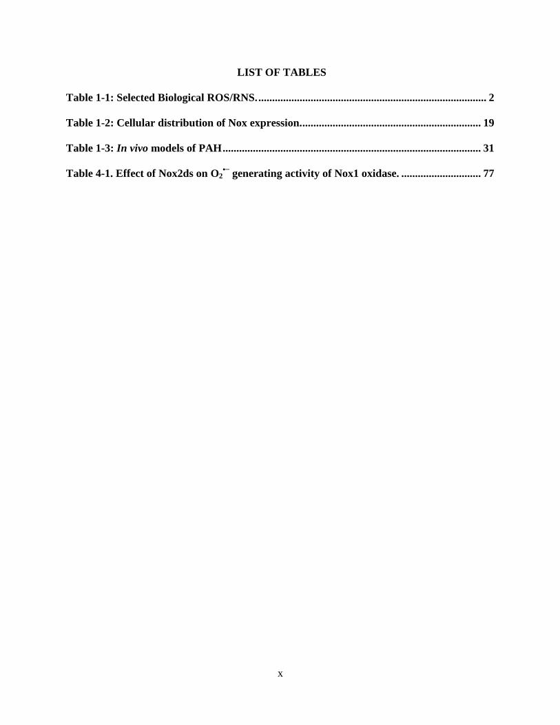
LIST OF TABLES
Table 1-1: Selected Biological ROS/RNS. ................................................................................... 2
Table 1-2: Cellular distribution of Nox expression. ................................................................. 19
Table 1-3: In vivo models of PAH .............................................................................................. 31
Table 4-1. Effect of Nox2ds on O2•− generating activity of Nox1 oxidase. ............................. 77

xi
LIST OF FIGURES
Figure 1-1: Blood flow and anatomy of the heart ...................................................................... 9
Figure 1-2: Schematic of an ideal PV loop................................................................................ 11
Figure 1-3: Relationship of ESPVR and EDPVR to an ideal series of PV loops. ................. 12
Figure 1-4 Structures of Existing Nox1 Inhibitors................................................................... 21
Figure 1-5: Tissue, cellular, and intracellular distribution of vascular Nox isoforms. ........ 22
Figure 3-1: Design of NoxA1ds. ................................................................................................. 51
Figure 3-2: NoxA1ds inhibits Nox1-derived O2˙- but not related enzymes. ........................... 54
Figure 3-3: SCRMB NoxA1ds does not inhibit Nox2, Nox4, Nox5 or XO. ............................ 56
Figure 3-4: NoxA1ds is cell-permeant and effective in whole colon adenocarcinoma cells yet
ineffective in Nox1-/y cells. .......................................................................................................... 58
Figure 3-5: NoxA1ds binds to Nox1 but does not detectably bind Nox2. .............................. 61
Figure 3-6. NoxA1ds disrupts Nox1-NOXA1 interaction. ....................................................... 64
Figure 3-7: Nox1:NOXA1 FRET interaction is not a result of CFP and YFP proximity in
the membrane. ............................................................................................................................. 65
Figure 3-8: Schematic of NoxA1ds mechanism of action. ....................................................... 70
Figure 4-1. Nox2ds dose-dependently inhibits O2˙- production from Nox2-oxidase. ............ 75
Figure 4-2. Nox2ds does not inhibit O2˙- production from Nox1-oxidase. ............................. 76
Figure 4-3: p47phox, but not NOXO1, binds to Nox2ds. ........................................................... 80
Figure 4-4: NoxA1ds attenuates hypoxia-induced O2˙- production. ....................................... 81
Figure 4-5: Nox1, but not Nox2, is responsible for VEGF-stimulated wound healing. ........ 83
Figure 4-6: NoxA1ds disrupts VEGF-stimulated Nox1-NOXA1 interaction. ....................... 84
Figure 4-7: VEGF production by HPAF is enhanced by hypoxia. ......................................... 85
xii
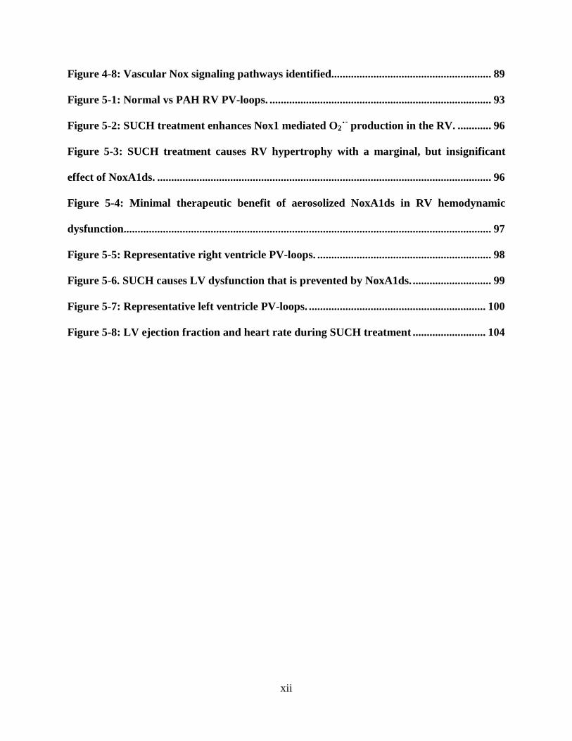
Figure 4-8: Vascular Nox signaling pathways identified......................................................... 89
Figure 5-1: Normal vs PAH RV PV-loops. ............................................................................... 93
Figure 5-2: SUCH treatment enhances Nox1 mediated O2˙- production in the RV. ............ 96
Figure 5-3: SUCH treatment causes RV hypertrophy with a marginal, but insignificant
effect of NoxA1ds. ....................................................................................................................... 96
Figure 5-4: Minimal therapeutic benefit of aerosolized NoxA1ds in RV hemodynamic
dysfunction................................................................................................................................... 97
Figure 5-5: Representative right ventricle PV-loops. .............................................................. 98
Figure 5-6. SUCH causes LV dysfunction that is prevented by NoxA1ds. ............................ 99
Figure 5-7: Representative left ventricle PV-loops. ............................................................... 100
Figure 5-8: LV ejection fraction and heart rate during SUCH treatment .......................... 104

xiii
PREFACE
ACKNOWLEDGEMENTS
This dissertation represents the culmination of my career as a scientist thus far, and I am forever
grateful to those who have contributed to my growth as such. My greatest thanks go to my
parents, who fostered my inherent curiosity of the natural world from the earliest days of my
childhood and have remained eager to hear the progress I make. Second only to my parents are
my brothers, who have always made my life tremendously fun and exciting and provided much
needed breaks from the daily stress in the preparation of my thesis. I expect that we still have
many more adventures to come.
I also extend my thanks to the many scientific mentors I have had over the years. Patty
Griest, Rebecca Finch, and Matthew Delp: your advice and education at the earliest stages of my
career set the stage for my future independent investigations. Margaret Voss, Michael Campbell,
Pamela Silver: when I engaged in my first truly independent study, you gave me the freedom to
pursue whichever direction my curiosity turned and provided invaluable guidance as I stepped
forward in my quest for knowledge. Natalie Fursov and Russell Lingham: you opened my eyes
to entirely new words of research in antibody engineering and gave me an entirely new paradigm
on the role of scientific research in industrial pharmaceutical development. Gabor Csanyi and
Imad Al Ghouleh: you were my brothers in the lab. I don’t think I’ll ever have as much fun
working in a lab and maintaining productivity as I did with you two.
Last, and most certainly not the least, I must thank my mentor for the past four years,
Patrick Pagano. My success in the pursuit of my PhD is largely to your credit and would not
have been possible without your exemplary mentoring. Your guidance as I navigated the world
xiv
of experimental design, data presentation, funding agencies, and manuscript publication all
whilst maintaining lab productivity was invaluable and your patience with my occasionally naïve
and rushed actions has taught me countless invaluable lessons. You have been an outstanding
mentor, and your academic, personal, and intellectual support has set the stage for the rest of my
scientific career.
Parts of this work appear as published manuscripts on which I am an author and would not have
been possible without the contributions of the co-authors:
1. Ranayhossaini DJ, Rodriguez AI, Sahoo S, Chen BB, Mallampalli RK, Kelley EE,
Csanyi G, Gladwin MT, Romero G, Pagano PJ. Selective Recapitulation of Conserved and
Nonconserved Regions of Putative NOXA1 Protein Activation Domain Confers Isoform-specific
Inhibition of Nox1 Oxidase and Attenuation of Endothelial Cell Migration. J Biol Chem. 2013
Dec 20;288(51):36437-50
2. Ranayhossaini D, Pagano PJ. TrACEing Angiotensin II Type 1 to Right Ventricular
Hypertrophy: Are the "Sartans" a Viable Course to Treating Pulmonary Arterial Hypertension?
AJRCCM 2012 Oct 15, 186(8):705-7
3. Csányi G, Cifuentes-Pagano E, Al Ghouleh I, Ranayhossaini DJ, Egaña L, Lopes LR,
Jackson HM, Kelley EE, Pagano PJ. Nox2 B-loop Peptide, Nox2ds, Specifically Inhibits Nox2
Oxidase. FRBM 2011 Sep 15;51(6):1116-25

xv
LIST OF ABBREVIATIONS
ANOVA analysis of variance
CFP cyan fluorescent protein
COS-22 COS-7 cells transfected with p22phox
COS-NOX1 COS-22 cells transfected with Nox1 NOXO1, and NOXA1
COS-NOX2 COS-22 cells transfected with Nox2, p47phox, and p67phox
COS-NOX4 COS-22 cells transfected with Nox4 and p22phox
CPH 1-hydroxy-3-methoxycarbonyl-2,2,5,5-tetramethylpyrrolidine
hydrochloride
DMSO dimethyl sulfoxide
DPI diphenyleneiodonium chloride
Duox dual oxidase
EDPVR end diastole pressure volume relationship
EDV end diastolic volume
EGF epidermal growth factor
EPR electron paramagnetic resonance
eNOS endothelial nitric oxide synthase
ESPVR end systole pressure volume relationship
FAD flavin adenine dinucleotide
FDA United States Food and Drug Administration
FRAP fluorescence recovery after photobleaching
FRET fluorescence resonance energy transfer
H2O2 hydrogen peroxide
xvi
HFpEF heart failure with preserved ejection fraction
HIF1α hypoxia inducible factor 1α
HPAEC human pulmonary artery endothelial cells
HPASMC human pulmonary artery smooth muscle cells
HPAF human pulmonary artery fibroblasts
HX hypoxanthine
LV left ventricle of the heart
mPAP mean pulmonary artery pressure
MTT Methylthiazolyldiphenyl-tetrazolium bromide
NADPH nicotinamide adenine dinucleotide phosphate
NOS nitric oxide synthase
Nox NADPH oxidase
NOXA1 Nox activator subunit 1
NOXO1 Nox organizer subunit 1
O2·- superoxide anion
OAB oxidase assay buffer
PAH pulmonary arterial hypertension
PBS phosphate buffered saline
PMA phorbol myristate acetate
RNS reactive nitrogen species
ROS reactive oxygen species
RV right ventricle of the heart
SEM standard error of the mean

xvii
SOD superoxide dismutase
SUCH SU5416 + chronic hypoxia model of PAH
VEGF vascular endothelial growth factor
VEGFR VEGF receptor
XO xanthine oxidase
YFP yellow fluorescent protein

1
1.0 INTRODUCTION
1.1 ROLES OF ROS/RNS IN CELLULAR BIOLOGY
1.1.1 Distinctions between individual ROS/RNS
The family of reactive oxygen species (ROS) and reactive nitrogen species (RNS) is an
amalgamated collection of numerous distinct molecules that are differentiated by their atomic
composition, charge state, and free vs. paired electron status. The majority of ROS and RNS
share the primary atomic components of oxygen, nitrogen, and hydrogen and as such, the
number and types of ROS/RNS family members is limited only by natural physical and chemical
laws. Despite the great variety of potential ROS/RNS, selected species bear particular
significance in biology. Particularly significant ROS/RNS in biological systems include
hydrogen peroxide (H2O2), hydroxyl radical (OH·), superoxide anion (O2·-), nitric oxide (NO·),
and peroxynitrite (ONOO-) (Table 1). These molecules are derived from enzymatic processes as
well as redox reactions among ROS/RNS and other organic/inorganic molecules such as lipids,
proteins and iron. The presence of charged moieties and free electrons on many of these species
contribute greatly to their dynamic reactions, half-lives, and physiological signaling
consequences.
1.1.2 Physiological ROS/RNS Dynamics
Cellular sources and inorganic catalysts of O2·- include NADPH oxidases (Noxes), uncoupled
endothelial nitric oxide synthase (eNOS), free iron, and xanthine oxidoreductase (XO) [1-3]. To

2
mitigate the inherent reactivity of O2·-, most mammalian cells abundantly express superoxide
dismutase (SOD). The conversion of O2·- to H2O2 by copper/zinc superoxide dismutase (Cu,Zn-
SOD) is an extremely fast reaction occurring with a rate constant of 6.4 x 10⁹ M-1s-1 [4]. Less
efficient Mn-SOD or Fe-SOD catalyze this reaction at approximately 6.4 x 10⁸ M-1s-1 [4]. The
speed of these reactions is necessary to protect the cell from the damaging reactions of O2·- with
cellular enzymes, in particular, aconitase. Aconitase is a critical enzyme in the Krebs cycle for
the conversion of citrate to isocitrate and its iron-sulfur core is rapidly oxidized and inactivated
by O2·- at a reaction rate constant of 107 M-1s-1, only marginally slower than the reaction rate of
SOD, further emphasizing the importance of antioxidant enzymes for cellular protection [5].
The reactive ability of O2·- extends to its potential to oxidize enzymatic cofactors, such as
tetrahydrobiopterin, an essential cofactor of eNOS activation [6-8]. This reaction is also very
rapid occurring at 3.9 x 10⁵ M−1s−1 and is a significant cause of eNOS uncoupling leading to
increased O2- production [8, 9]. Beyond its ability to inactivate enzymatic cofactors, O2·- is
capable of reacting with thiols at reaction rate constants ranging from 1.0 – 5.0 x 10⁵ M−1s−1,
depending on thiol availability [10].
Species Name Biological Sources
H2O2 Hydrogen Peroxide Nox, XO, SOD
OH· Hydroxyl Radical O2·-, Fenton Reaction
O2·- Superoxide Nox, XO, eNOS, free iron
NO· Nitric Oxide NOS, Nitrite reductases incl. myoglobin and neuroglobin
ONOO- Peroxynitrite O2·- interaction with NO·
Table 1-1: Selected Biological ROS/RNS.

3
O2·- very potently reacts with most cysteine containing enzymes and/or enzymatic
cofactors. Through dismutation of O2·- to H2O2, the reactive properties and half-life of O2·- are
significantly changed and provides an alternate route for H2O2-mediated protein modification
and oxidation. H2O2 is arguably the most stable of the reactive oxygen species, with its half-life
extending for days in solutions at room temperature [11]. Like O2·-, H2O2 is capable of oxidizing
cysteines and thus modifying protein function [12]. Kinetically, the oxidation reaction of H2O2
with cysteine occurs slowly at 720 M-1s-1, allowing H2O2 to diffuse away from its source and
expanding its potential cellular targets [13]. The relatively low reaction rate constant of H2O2
with cysteine and its long half-life indicate that this molecule is capable of diffusing throughout
and beyond the cell. This diffusion of H2O2 is limited, however, by the prevalence and
distribution of catalase. Catalase is by far most significant negative contributor to the half-life of
H2O2 by catalyzing the formation of oxygen and water from H2O2 at a rate constant of 6.62 x 10⁷
M-1s-1. Catalase is ubiquitously expressed in most mammalian cells and is responsible for
controlling diffusion of H2O2 beyond its intended target [14, 15]. In addition to catalase, a wide
variety of cellular peroxidases participate in the metabolism of H2O2. Peroxidases utilize the
oxidative capacity of H2O2 to perform a two-step reaction, first oxidizing themselves (reaction I),
before transferring the oxidant to a target protein or porphyrin (reaction II) [16]. Physiologically
relevant oxidation targets of peroxidases include pyridine nucleotides [16]. Peroxidase reaction I
is often the rate limiting step of peroxidase activity, and depending on the class of peroxidase,
target species, the kinetics of reaction I can be as slow as 5.4 x 10⁵ M-1s-1 (lignin peroxidase) or
as fast as 8.0 x 10⁷ M-1s-1 (ascorbate peroxidase) [16, 17]. These kinetics are remarkably stable
over wide pH ranges with horseradish peroxidase retaining its reaction rate constant of 1.8 x 10⁷
M-1s-1 from pH 5-9 [17]. Together, SOD, catalase, and peroxidases perform the majority of

4
cellular ROS catabolism and in doing so, protect the cell from the damaging effects of
dysregulated ROS and maintain targeted ROS activity for physiological signaling.
Perhaps the most widely studied RNS is nitric oxide (NO), which is a ubiquitous
biological signaling molecule primarily produced by nitric oxide synthase (NOS) and nitrite
reductases. While NO confers many physiological effects, it is best known for its vasodilatory
properties [18-20]. A single unpaired electron on NO dictates which molecules it can favorably
react with, these are typically either other free radicals or metals such as heme iron [21]. The
stability of NO and its ability to freely diffuse across and also utilize aquaporin channels allows
it to serve as a paracrine signaling factor between endothelial and smooth muscle cells, lending it
extraordinary control of vasodilation by causing smooth muscle relaxation [18, 19, 22]. The
half-life of physiological NO in aqueous solution is quite high with a half-life of about 5 min in
solution [21]. In the cellular environment, this is greatly reduced to less than 1 minute, which
can be doubled by the addition of SOD [18]. Indeed, the reaction between NO and O2·- to form
ONOO- is diffusion-limited and thus very rapid. Hence, SOD leads to an increase in the half-life
of NO by preventing this reaction [23]. The reaction of NO and O2·- to form ONOO- occurs at
6.7 x 10⁹ M-1s-1[24]. This extremely fast reaction closely competes with SOD for O2·- radicals.
In turn, ONOO- is stable for days in alkaline solutions [25]. In biological systems, only 20% of
ONOO- will be protonated and subsequently degraded to nitrate with the remainder remaining
capable of oxidizing cellular components, with a relatively short half-life of 1 second [25].
Biological protection from ONOO- appears to be achieved through the scavenging properties of
uric acid [26].
The vasoprotective functions of NO are in stark contrast to the ability of ONOO- to
damage the endothelium leading to cardiovascular disease [27]. ONOO—mediated damage to
5
proteins is often observed as a consequence of the nitration of tyrosines, although ONOO-
oxidation can occur on iron, sulfur, or zinc complexes as well as cysteine residues [21, 28].
ONOO- reaction rate constants differ widely based on the target and can proceed as slowly as 103
M–1s–1 or as rapidly as 107 M–1s–1 [3, 29, 30]. Beyond its direct effect on cellular components,
protonation of ONOO- leads to formation of OH· radicals that are capable of reacting with most
organic molecules, including nucleic and amino acids [27]. Reactions between OH· and most
organic molecules are fairly rapid on the order of 108 to 109 M–1s–1 [31]. The speed of this
reaction and the plethora of potential targets makes it extremely difficult to specifically scavenge
OH·, and thus most efforts focus on preventing formation of its precursors.
To further appreciate the intricate relationship of interacting ROS/RNS species and thus
protect cells from their detrimental effects, recent efforts have been focused on defining the exact
mechanisms through which ONOO- and other ROS modify proteins. One mechanism that is
potentially translatable across a wide range of proteins involves the targeting of heme
coordination sites by ONOO-. Indeed, while all tyrosines are susceptible to nitration by ONOO-,
tyrosines proximal to hexameric heme coordination sites are particularly important in protein
function. When these tyrosines become nitrated, the integrity of heme coordination is greatly
reduced and can cause deactivation of catalytic sites [32]. Through this mechanism and others
yet to be delineated, tyrosine nitration by ONOO- can inactivate, activate, or engender new
functions for proteins including SOD, cytochrome c, fibrinogen, and HSP90 [32-35].
While many unknowns remain in ROS/RNS signaling, consistent and sustained effort has
helped to determine functional roles for biologically relevant ROS/RNS including OH·, O2·-,
NO·, ONOO-, and H2O2. Each of these molecules is capable of modifying protein function
through the modification of cysteines, tyrosines, or enzymatic cofactors among others, albeit at
6
significantly different rates. Negative regulators of ROS-mediated protein modification and
protective cellular enzymes include SOD and catalase. SOD converts O2·- to less reactive H2O2
and in turn, catalase converts H2O2 to water. These reactions occur at very high rates of 6.4 x
109 M-1s-1 and 6.62 x 10⁷ M-1s-1, respectively. Despite its very high reaction rate constant, SOD
is incapable of completely preventing the formation of ONOO- from NO· and O2·- (rate = 6.7 x
109 M-1s-1). These reaction rate constants are so similar that a close competition for the fate of
cellular O2-, H2O2, and NO· exists. The outcome of this competition largely depends on the
concentrations and availability of each individual species, target availability, and antioxidant
scavenging enzymes. The reactivity of these agents alone does not determine their influence in
vitro or in vivo as different cell types and different stimuli can result in very different ROS
profiles, in part as a result of different protein expression (e.g. SOD, catalase), subcellular
localization, and available protein targets.
1.2 NADPH OXIDASE-DERIVED ROS IN THE VASCULATURE
1.2.1 NADPH Oxidase, Roles in Physiology
NADPH oxidases (Noxes) are a family of proteins taxonomically characterized by their ability to
harvest electrons from NADPH and transfer these to O2, producing ROS such as O2·- and H2O2.
The Nox family includes the proteins Nox1, Nox2, Nox3, Nox4 Nox5 and the Dual Oxidases 1
and 2 (Duox1 and Duox2). Noxes 1, 2, 3 & 5 produce O2·-, while Nox4 and the Duoxes
reportedly produce H2O2 [36]. Somewhat of a misnomer, Nox2 was the first member of this
family to be discovered and historically, has been referred to as the phagocyte oxidase for its role

7
in bactericidal activity of the neutrophil and other leukocytes [37-39]. Tissue expression of the
Nox family is as diverse as their lineage, with predominant expression of Nox1 in colon
epithelium, uterus, vascular smooth muscle and vascular endothelium; predominant expression
of Nox2 in neutrophils, macrophages, endothelial cells, neurons, and fibroblasts [40-48]. Nox3
seems to be localized primarily to the inner ear, while Nox4 is ubiquitously expressed throughout
the vasculature, with additional significant expression in in the kidney [48-52]. Nox5 is largely
observed in vascular smooth muscle cells, but is also expressed in the endothelium, of higher
mammals while the Duoxes are best known for their expression in the pulmonary epithelium [45,
53-56] (Table 2).
Beyond their differences in tissue expression, the Nox isoforms have distinct subcellular
localization, with Nox expression being differentially expressed in an isoform-dependent manner
in the plasma membrane, endosomes, phagosomes, and the endoplasmic reticulum (Table 2).
These distinct subcellular locales are purported to be inextricably tied to the signal transduction
consequences of the enzyme for two reasons: a) the reactive nature of the ROS produced and
thus its radius of diffusion limits specific protein targeting and b) rapid scavenging by SOD and
catalase prevents distal diffusion of ROS-mediated signal transduction, particularly in the case of
O2·- and moderately less so for H2O2. While physiologically important for normal cardiovascular
homeostasis and immune function, under pathological conditions excessive stimulation of Noxes
often leads to hyperproliferative and migratory phenotypes as well as oxidative stress, often
resulting in end-organ damage [57, 58]. Increasingly the role of individual Nox isoforms are
being appreciated as playing highly-localized, specific, and temporal roles in disease
progression. Broad and nonspecific antioxidant actions of ROS scavengers (i.e., vitamin
antioxidants and antioxidant enzymes) cannot manipulate cellular signaling in the specific and
8
temporal manner necessary to elicit subtle changes in cell physiology and pathophysiology.
Thus, while ROS scavengers/antioxidants are extremely valuable as controls and confirmatory
tools, their utility is superseded by the need for highly-selective inhibitors of Nox-derived ROS
production. Importantly, Noxes are increasingly being appreciated as key players in
cardiovascular physiology through suppression of NO bioavailability, and induction of cell
proliferation and vessel tone, for example, despite their long-emphasized major importance in
the phagocytic oxidative burst.
1.2.2 Anatomy and Physiology of the Cardiovascular System
An essential component of vertebrate physiology is the cardiovascular system, which serves to
provide oxygen and nutrients to tissue beds while removing waste for excretion by the kidneys.
Mammalian hearts are divided into four chambers: the left atrium, left ventricle, right atrium, and
right ventricle. Together, these chambers are the pump that drives the circulation of blood
throughout the body. After tissue perfusion and oxygen depletion, blood returns to the right side
of the heart via the superior vena cava where blood is sent via the pulmonary artery for
oxygenation in the lungs. Via the left and right pulmonary veins, the left side of the heart
receives oxygenated blood from the lungs and distributes it through the body (Figure 1-1) [59,
60]. Control of heart beats/contractions is achieved through sympathetic and parasympathetic
innervation and it is through the sympathetic innervation which synchronous contraction of the
left and right ventricles, or systole, is stimulated [60, 61]. Systolic contractions force ejection of
the blood through the vasculature and is immediately followed by diastole, the relaxation of the
heart muscle allowing its chambers to refill [60-62].
The progress of blood flow can be described by starting at the filling of the left atrium
with blood draining from the pulmonary veins. In turn, contractions of the left atrium fill the

9
relaxed left ventricle (LV). The LV is the largest component of the heart and pumps oxygenated
blood into the aorta, which branches distally to smaller conduit vessels and sequentially smaller
and thinner arteries greater diffusion of oxygen from the vessels to organ systems occurs, with
the greatest diffusion occurring between the smallest of vessels or capillaries. After oxygen
diffusion, the blood returns to the heart via the vena cava as “venous return” and is drained by
the right atrium which, during systole, fills the diastolic right ventricle (RV). Contraction of the
RV sends blood through the pulmonary artery for oxygenation in the lungs before blood is and
then transferred back to the left atrium and LV for subsequent distribution through the body [60,
63].
Figure 1-1: Blood flow and anatomy of the heart
Image from U.S. National Library of Medicine, no copyright restriction.

10
The cycle of blood flow through the heart through systolic contractions and diastolic
relaxations causes substantial changes in pressures and volumes within the heart. Hemodynamic
analysis of these pressures and volumes provides great detail concerning the function of each
component of the heart and is best measured by pressure-volume conductance catheters and the
resultant pressure-volume loops (PV loops) (Figure 1-1) [64]. Critical measures of ventricular
function determined by PV loop analysis are the End Systole Pressure Volume Relationship
(ESPVR) and the End Diastole Pressure Volume Relationship (EDPVR). ESPVR describes the
maximal pressure that can be achieved at any given volume and is critical in determining
ventricular contractility [65-67]. In turn, EDPVR describes the maximal volume that can be
achieved at any given pressure and is a function of ventricular compliance [67-69] (Figure 1-2).
Determining ESPVR/EDPVR by PV loop analysis offers the benefit of being a highly sensitive
and load-independent technique for measuring cardiac states and is a key measure of cardiac
function under any condition [70]. Deviation of either ESPVR or EDPVR from their initial slope
values can indicate any one of a number of cardiomyopathies depending on the direction of the
shift. For example, a decrease in the slope of EDPVR indicates a more compliant heart that fills
more readily while an increase in ESPVR indicates a more contractile heart or one that is
working against abnormally high pressures to move blood [68, 71].
Larger blood vessels that carry blood throughout the body are composed of three distinct
layers and cell types: the innermost intima (endothelial cells), the central media (smooth muscle
cells), and the outermost adventitia (fibroblasts, Figure 1-2) [72]. In the smallest of vessels
including resistance vessels and capillaries, the smooth muscle cells and fibroblasts are replaced
by pericytes [73]. The intima surrounds the interior of the vessel, the lumen, which contains the
flowing blood, including plasma, erythrocytes, and leukocytes. Conduit arteries, which carry
11
blood away from the heart, maintain a significantly thicker media than veins returning blood to
the heart with multiple layers of smooth muscle cells. As one progresses down the vascular tree,
anywhere between 1 through 7 layer(s) of smooth muscle cells are found, with vessels of a larger
diameter having more smooth muscle while the most distal arteries, veins, and capillaries have
lesser to no virtually smooth muscle [60]. Functionally, the adventitia appears to preserve
vascular tone, at least in part, through its production of O2·- and the subsequent destruction of NO
while structurally supporting the vessel via production of matrix proteins including collagen [74-
76]. The smooth muscle cells of the media provide the main contractile force that determines
vessel diameter and consequent vascular resistance [77]. The innermost endothelial cells lining
the vessel lumen counteract the contractile force of the media through primarily the production
of NO and subsequent sGC activation in larger vessels as well as eicosanoids, H2O2, and
adenosine in smaller vessels [18, 78-81].
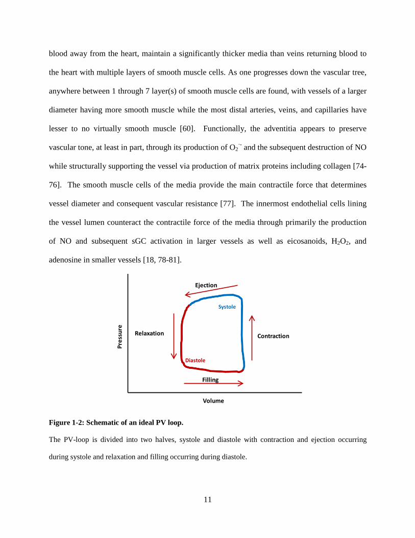
Figure 1-2: Schematic of an ideal PV loop.
The PV-loop is divided into two halves, systole and diastole with contraction and ejection occurring
during systole and relaxation and filling occurring during diastole.
Filling
Contraction
Ejection
Relaxation
Volume
Pres
sure
Systole
Diastole

12
Figure 1-3: Relationship of ESPVR and EDPVR to an ideal series of PV loops.
End Systole Pressure Volume Relationship (ESPVR) indicates the maximum pressure that can be attained
at any given volume while End Diastole Pressure Volume Relationship (EDPVR) indicates the maximum
volume that can be attained at any given pressure. ESPVR and EDPVR are calculated though occlusion
of the vena cava and reducing ventricular volume, leading to sequential PV loops shown. Calculation of
ESPVR and EDPVR through this method contributes to the load independence of this cardiac measure.
Angiogenesis, the growth of new blood vessels, is a tightly-controlled vascular process
that functions to extend the circulatory system to hypoxic growing and damaged tissue beds that
are unable to survive on interstitial fluid alone [82]. Angiogenic signaling is driven primarily by
oxygen gradients and is stimulated by multiple key proteins, including VEGF, HIF1α, and Noxes
have emerged as important players in this process [82-85]. Originally described as a “vascular
permeability factor” for its ability to rearrange endothelial cells and vascular superstructure in
tumors, VEGF was later renamed as “vascular endothelial growth factor” when its mitogenic
properties were being revealed [84, 86-89]. As an extremely potent stimulator of mitogenesis,
endothelial migration, and angiogenic sprouting by vessels, VEGF is also a major contributor to
Volume
Pres
sure
ESPVR
EDPVR

13
malignant tumor growth through its potentiation of angiogenesis [84, 88, 90]. Hypoxia inducible
factor 1α (HIF1α) is the master regulator of oxygen sensitive angiogenic gene transcription,
including the transcript for VEGF and potentially the transcripts for Noxes [85, 91]. HIF1α
activity is tightly controlled by endogenous prolyl hydroxlases that target HIF1α for rapid
proteolytic degradation through post-translational modification [92, 93]. However, under
conditions of low oxygen, prolyl hydroxlases are inhibited, allowing HIF to remain active in the
cell, leading to transcription of genes with HIF1α-responsive elements, including VEGF and Nox
[85, 94]. Noxes are established contributors to angiogenic signaling, in part through oxidative
activation of matrix metalloproteases that permit angiogenic remodeling through degradation of
extracellular matrix proteins [95-97]. Nox1, Nox2, and Nox4 have each been identified as
contributors to angiogenesis, with some evidence indicating that Nox1 may be more influential
in angiogenic signaling [98, 99]. The important role of Nox1 in angiogenesis is likely not only
due to potential ROS-mediated activation of metalloproteases, but also its potentiation of VEGF
transcription [83, 95, 100]. This has generated some interest as to whether Nox inhibition may
be a potential treatment for diseases mediated by angiogenic signaling and vessel remodeling,
including cancer and pulmonary arterial hypertension.
1.2.3 Structure and Regulation of Vascular Nox Isoforms
Beyond their role in preventing bacterial pathogenesis, Nox1 and Nox2 are significant
contributors to vascular cell proliferation and invasion [98, 100-106]. Tissue expression data and
knockout mice have identified Nox1, Nox2, Nox4, and Nox5 as the most prominent Noxes in
vascular physiology [46, 107-109]. These Noxes play key roles in vascular physiology in part
through the destruction of NO via Nox-derived O2·- and subsequent generation of ONOO-.
While critical for normal vascular homeostasis, when this pathway is dysregulated, Nox1 and

14
Nox2 contribute to left heart failure, atherosclerosis and hypertension [101, 110-112]; and
whereas the link between enhanced Nox1 & 2 activity and systemic vascular disease in
experimental models is well established, less clear is the link between Nox4 and cardiovascular
disease progression. Rather, significant controversy surrounds Nox4 as to whether this enzyme
plays a protective or destructive role in cardiovascular disease [99, 113]. Nox5’s role in the
vasculature is less well defined, largely due to the lack of murine Nox5 expression and resulting
lack of appropriate animal models and genetic tools to investigate cardiovascular disease.
However, it is presumed that Nox5 potentiates similar signaling pathways as Nox1 through the
production of O2·- [109, 114]. Additionally, the role of Nox1 and Nox2 in pulmonary vascular
disorders is largely unknown.
As evidence has grown defining the role of Noxes in cardiovascular health and disease,
Nox biochemistry has emerged as an area of great interest. Structurally, the cardiovascular
Noxes1-5 bear significant differences in their amino acid sequence and assembly. Functional
Nox1 and Nox2 oxidases are the most homologous vascular Noxes and, when structurally
complete, are a multimeric protein complex consisting of a transmembrane catalytic core (either
Nox1 or Nox2), a cytoplasmic GTPase (Rac1), a cytoplasmic organizing subunit (NOXO1
organizing Nox1; p47phox organizing Nox1 and 2), a cytoplasmic activating subunit (NOXA1
activating Nox1; p67phox activating Nox2) and the transmembrane stabilizing protein p22phox
[115-123].
The cytoplasmic subunits NOXO1, p47phox, NOXA1, and p67phox are critically important
components of Nox activation in vitro and in vivo. Indeed, mutations in cytoplasmic Nox
subunits can manifest as chronic granulomatous disease in humans, a condition of insufficient
microbial clearance by the macrophage [124]. Through catalyzing enzyme assembly NOXO1

15
and p47phox serve as the organizing subunits of Nox1 and Nox2, respectively. Although both
proteins share significant homology, a major difference between the two is the presence of an
auto-inhibitory domain in p47phox. This domain prevents p47phox interaction with either Nox1 or
Nox2 until p47phox is phosphorylated [125, 126]. NOXA1 and p67phox serve as the key canonical
activating proteins for Nox1 and Nox2, respectively. As testament to the key activating function
of NOXA1/p67phox, in reconstituted cell-free assays containing prenylated p67phox, Nox2 can be
activated independently of the organizing subunits p47phox/NOXO1, at least in an experimental
setting [119]. Within p67phox exists a key activation domain extending through residues 190-210,
that is critical for the successful transfer of electrons from NADPH to FAD in Nox2 [127, 128].
While the homologue of p67phox’s activation domain in NOXA1 remains to be conclusively
characterized as an activation domain, the significant homology between these two proteins, in
particular residues 190-210, as well as their sensitivity to mutation, implies that residues 190-210
in NOXA1 may serve as a domain with a similar function for Nox1 [129].
Recent reports have suggested that NOXO1 and p47phox are interchangeable organizers of
either Nox1 or Nox2 while NOXA1 and p67phox are interchangeable activators of either Nox1 or
Nox2 in smooth muscle cells [46]. It would appear that cellular expression levels of the different
organizing and activating subunits NOXO1, NOXA1, p47phox, and p67phox is the greatest
determinant in whether a “hybrid” Nox system where Nox1 is associated with Nox2 subunits
p47phox and/or p67phox exists. Hybrid Nox systems further complicate the expression profile and
regulation of Noxes. That is, with respect to Nox1 or 2 systems utilizing p47phox, these systems
may be inducible through activation of protein kinase C-dependent p47phox phosphorylation and
subsequent repression of its auto-inhibitory domain [125, 130]. The presence of hybrid Nox1
and/or Nox2 systems complicates the use of pharmacological Nox inhibitors in that drugs
16
specifically targeting any Nox cytosolic organizing or activating component will potentially lack
specificity against a single Nox oxidase complex, be they canonical or hybrid. As such, any drug
targeting cytosolic Nox proteins must be carefully evaluated for its specificity against Nox
hybrid systems.
In contrast to the multimeric protein organization of Nox1 and Nox2, Nox4 and Nox5 are
simpler in nature. It is now generally accepted that the fully-functional Nox4 oxidase is the
aggregate of two polypeptide chains, the first being p22phox and the second being the Nox4
catalytic core [52, 115, 131]. Even simpler yet is the single polypeptide transcript that
constitutes the entire Nox5 oxidase [53]. However, the simplicity in constitution of Nox4 and
Nox5 belies their more complex regulatory mechanisms, such as the stimulation of Nox4 by
polymerase delta interaction protein 2, Poldip2 [132]. Separately, Nox5’s transcript bears two
calcium binding EF hands which serve to activate enzyme activity in response to increasing
intracellular calcium concentrations [54]. As endothelin receptors are known potentiators of
calcium release, it is possible that Nox5 is indirectly activated by endothelin receptor signaling
[133]. The independent expression and regulation of these Noxes can lead to dramatically
different vascular phenotypes in a context-dependent manner, in part due to the different cellular
and intracellular localization of the enzyme.
1.2.4 Roles of Nox in Cardiovascular Physiology/Pathophysiology
A number of studies have provided evidence supporting a major role for Nox1 and Nox2 in
vascular homeostasis and pathophysiological ischemic injury and systemic hypertension [2, 9,
75, 107, 134-136]. Multiple effectors of O2·- are responsible for the downstream signaling of
Noxes in blood flow and vascular tone, with two major culprits being a) the destruction of NO
and b) uncoupling of eNOS through BH4 oxidation, with both resulting in a decreased soluble

17
guanylate cyclase activation and smooth muscle relaxation [8, 9, 23, 137]. Beyond prevention of
smooth muscle relaxation and vasodilation, Nox1- and Nox2-derived O2·- are established
contributors to neointimal and medial proliferation/cell migration and subsequent vessel
occlusion [138, 139]. Intimal/medial structural abnormalities of the microvasculature, i.e. small
arteries, and resistance vessels, have been correlated with hypertension and support the current
paradigm that the majority of pathophysiological changes in arterial walls in hypertension occur
in small resistance vessels [140, 141].
The role of Nox5 in the vasculature is presumed to be similar to that of Nox1 and Nox2,
although substantially less is known about its function due to the Nox5 gene being absent in
murine models. The ensuing lack of appropriate animal models of Nox5 in cardiovascular
disease and its relatively recent discovery in the human vasculature limit clear phenotypic
connections between Nox5 and cardiovascular physiology, with the most detailed studies
suggesting a correlation between Nox5 expression and myocardial infarction and more recent
interest in its role in kidney glomerular filtration barrier damage as well as spermatozoa
maturation and capacitation [109, 142-144]. Furthermore, evidence exists for Nox5’s role in
carcinogenesis and its direct role in cell transformation and indirect potentiation of angiogenesis
by increased ROS [109, 145, 146]. Pathophysiological effects of Nox-derived O2·- effects on
hypertension inherently extend from resistance vessels to the heart where increased resistance
vessel afterload forces cardiac adaptation to maintain adequate tissue perfusion. While multiple
pathways contribute to cardiac compensation in response to afterload, one controversial
pathways is a potentially protective role for Nox4 in myocardial damage by promoting
compensatory cardiac hypertrophy, angiogenesis, and fibroblastmyofibroblast transformation
[99, 113, 147]. In contrast, Nox2 appears to have a less controversial, deleterious role in cardiac
18
dysfunction, causing pathologic remodeling in response to myocardial infarction and
chemotherapeutic agents [148, 149]. To date, most cardiac Nox research has been narrowly
focused on the most highly-expressed cardiac Nox isoforms Nox2 and Nox4 and, as such, very
little is known about Nox1 in cardiac tissue.
Beyond their role in systemic hypertension and left heart failure largely established
through genetic knockouts and knockdowns, Noxes have been also been identified as
contributors to other systemic CVD, including atherosclerosis, ischemia reperfusion injury,
aberrant vessel growth and diabetic vasculopathy [150-154]. Most simply put, Nox1 directly
augments vascular tone via its downstream effects on actin-myosin coupling, thus contributing to
hypertension [136]. Additionally, Nox1 also contributes to intimal occlusion, vessel remodeling,
and neointimal hyperplasia throughout the systemic vasculature [2, 138, 155]. Mechanistically-
speaking, Nox1 contributes to neoplastic growth by uncoupling eNOS, increasing production of
VEGF and the ROS produced by Nox1 are capable of activating matrix metalloproteases (MMP)
[9, 83, 95, 100, 156]. By extension of these biochemical data, in vitro and in vivo data have
shown Nox1 is an angiogenic factor [83, 98]. The contribution of Nox1 to neoplasia,
angiogenesis, and eNOS uncoupling is strongly suggestive of a role for Nox1 in the development
and progression of diseases linked to angiogenesis and vessel remodeling including cancer and
pulmonary arterial hypertension. Specific pharmacological inhibitors of individual Nox isoforms
remain highly sought after for their potential utility in scientific investigation and therapeutic
intervention in a variety of pathologies. Furthermore, despite the well-established role of Noxes
in systemic hypertension, little is known about Noxes in pulmonary hypertension.
19
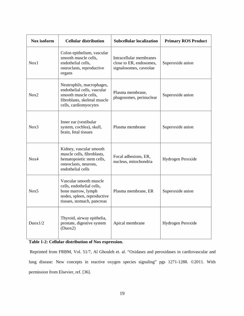
Table 1-2: Cellular distribution of Nox expression.
Reprinted from FRBM, Vol. 51/7, Al Ghouleh et. al. “Oxidases and peroxidases in cardiovascular and
lung disease: New concepts in reactive oxygen species signaling” pgs 1271-1288. ©2011. With
permission from Elsevier, ref. [36].
Nox isoform Cellular distribution Subcellular localization Primary ROS Product
Nox1
Colon epithelium, vascular smooth muscle cells, endothelial cells, osteoclasts, reproductive organs
Intracellular membranes close to ER, endosomes, signalosomes, caveolae
Superoxide anion
Nox2
Neutrophils, macrophages, endothelial cells, vascular smooth muscle cells, fibroblasts, skeletal muscle cells, cardiomyocytes
Plasma membrane, phagosomes, perinuclear Superoxide anion
Nox3 Inner ear (vestibular system, cochlea), skull, brain, fetal tissues
Plasma membrane Superoxide anion
Nox4
Kidney, vascular smooth muscle cells, fibroblasts, hematopoietic stem cells, osteoclasts, neurons, endothelial cells
Focal adhesions, ER, nucleus, mitochondria Hydrogen Peroxide
Nox5
Vascular smooth muscle cells, endothelial cells, bone marrow, lymph nodes, spleen, reproductive tissues, stomach, pancreas
Plasma membrane, ER Superoxide anion
Duox1/2 Thyroid, airway epithelia, prostate, digestive system (Duox2)
Apical membrane Hydrogen Peroxide

20
1.2.5 Existing Nox Inhibitors
Current widely-used Nox inhibitors include a number of small molecules and peptides that
exhibit either unknown or limited specificity for any particular Nox isoform. The most widely
used Nox inhibitors today include diphenyleneiodonium (DPI), GKT137831, apocynin, and
Nox2ds-tat [2, 112, 130, 157-162]. Historically, DPI and apocynin have played major roles in
implicating the contribution of Nox-derived ROS to cellular processes, yet they are both
burdened by a complete lack of specificity for any Nox isoforms. More specifically, while DPI
is a highly efficacious Nox inhibitor, it also broadly inhibits all flavoproteins utilizing FAD as a
cofactor including nitric oxide synthase and NADH dehydrogenase [157, 158, 163, 164]. In
turn, apocynin is a pro-drug that inhibits Nox exclusively in cells expressing myeloperoxidase,
those primarily being leukocytes [165]. When permeating cells other than leukocytes, apocynin
is not converted into Nox-inhibiting apocynin dimers and could serve as a nonspecific
antioxidant that scavenges H2O2 and HO∙ [165]. One of the very few compounds reported to be
specific for Nox1 over Nox2 is ML171 [159]. While ML171 is reportedly an effective and
specific Nox1 inhibitor, there is a complete lack of information concerning its mechanism of
action and its binding target, thereby hindering its extension into pre-clinical trials. It is also
untested whether ML171 acts on Nox4, Nox5, or XO-derived ROS production. The only Nox
inhibitor to have reached clinical trials is GKT137831, a small molecule effective for Nox1/4
inhibition [160]. GKT137831 has shown good oral bioavailability and is being investigated in
diabetic nephropathy, although its inability to distinguish between Nox1 and Nox4 may
potentially cause undesired side effects [166]. Nox2ds is a peptide derived from the intracellular
B-loop of Nox2 (residues 86-103) that has demonstrated utility in ischemic retinopathy, cerebral
microcirculation, and angiotensin II-stimulated ROS when conjugated to the cell penetrating
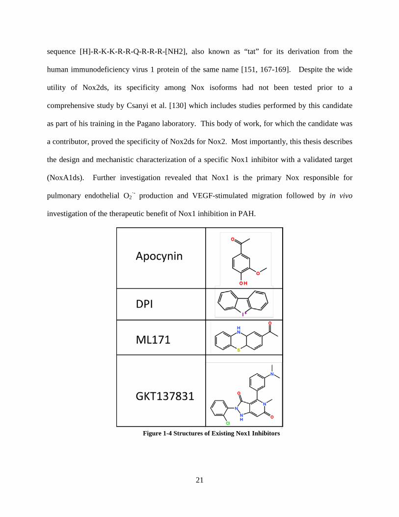
21
sequence [H]-R-K-K-R-R-Q-R-R-R-[NH2], also known as “tat” for its derivation from the
human immunodeficiency virus 1 protein of the same name [151, 167-169]. Despite the wide
utility of Nox2ds, its specificity among Nox isoforms had not been tested prior to a
comprehensive study by Csanyi et al. [130] which includes studies performed by this candidate
as part of his training in the Pagano laboratory. This body of work, for which the candidate was
a contributor, proved the specificity of Nox2ds for Nox2. Most importantly, this thesis describes
the design and mechanistic characterization of a specific Nox1 inhibitor with a validated target
(NoxA1ds). Further investigation revealed that Nox1 is the primary Nox responsible for
pulmonary endothelial O2·- production and VEGF-stimulated migration followed by in vivo
investigation of the therapeutic benefit of Nox1 inhibition in PAH.
Figure 1-4 Structures of Existing Nox1 Inhibitors

22
Figure 1-5: Tissue, cellular, and intracellular distribution of vascular Nox isoforms.
A) Schematic diagram showing cellular localization (endothelial cells, vascular smooth muscle cells,
fibroblasts, macrophages and T cells) of NADPH oxidase isoforms (NOX1 oxidase, NOX2 oxidase,
NOX4 oxidase and NOX5 oxidase) through a cross-section of an artery. B) Schematic diagram of a
hypothetical cell in which all of the vascular NADPH oxidase isoforms (starting with NOX1 oxidase in
the left hand column and finishing with NOX5 oxidase in the right hand column) are expressed in each of
their possible subcellular locations. H2O2, hydrogen peroxide; NOXA1, NADPH oxidase activator 1; O2•−,
superoxide; p22, p22phox; p40, p40phox; p47, p47phox; p67, p67phox; POLDIP2, polymerase δ-interacting
protein 2.
Reprinted by permission from Macmillan Publishers Ltd: [Nature Reviews Drug Discovery] Ref [72].
23
1.3 PULMONARY ARTERIAL HYPERTENSION
1.3.1 Pathophysiology of Pulmonary Arterial Hypertension
Pulmonary arterial hypertension (PAH) is a debilitating disease with high mortality characterized
by a mean pulmonary artery pressure (mPAP) greater than 25 mmHg. Diagnosis of PAH
through right heart catheterization is often performed after by exclusion of other diseases with
similar symptoms, these symptoms including chest pain and a shortness of breath. Idiopathic
PAH comprises nearly half of all cases of PAH, yet its cause is completely unknown and this
progressive disease has no cure [170]. More common is the reversible, altitude-associated PAH.
At high altitudes (>3500 meters), lung vasculature of healthy individuals compensates for
reduced atmospheric oxygen through pulmonary vasoconstriction. This, in turn causes elevation
of pulmonary artery pressures leading to clinical pulmonary hypertension which is ameliorated
upon return to lower altitudes [171]. Epidemiological data from individuals with altitude-
associated PAH and data indicating severe hypoxia in patients with idiopathic PAH are highly
supportive of the paradigm that hypoxia plays a critical role in the pathogenesis of PAH [170,
171].
While hypoxia is a critical factor in the development of PAH, other factors including
reduced NO bioavailability and vascular remodeling/occlusion contribute to the pathogenesis of
PAH. Through abnormal proliferation of the intima, media and adventitia within the pulmonary
vasculature; pathophysiological vessel remodeling occurs and plexiform lesions (PXLs) arise
[170, 172]. The presence of angiogenic markers in PXLs indicates that these lesions may be the
result of angiogenic signaling processes stimulated by hypoxia. Similarly, observational studies
of clinical patients with PAH reveal that remodeling in pulmonary vessels is largely initiated by
24
endothelial cells and that proximal to the PXL, vessels exhibit an increase in intimal thickness
[172-174]. Circumstantial evidence supporting the hypothesis that hypoxic angiogenic signaling
contributes to PXL formation in PAH also is observed in the similarities between PXL histology
and glomeruloid lesions in glioblastoma multiforme, a malignant cancer with particularly strong
angiogenic signaling [172, 175]. The combination of proximal intimal remodeling and the
redirection of blood flow around the PXLs are major contributors to a steady increase in
pulmonary vascular resistance (PVR), RV ESPVR, and attendant pressure overload in the right
ventricle.
1.3.2 Cellular Signaling Pathways Contributing to Pulmonary Arterial Hypertension
As evidenced by multiple in vitro and in vivo studies, hypoxia contributes to pulmonary vascular
ROS production (via Nox and mitochondria), cell proliferation (through HIF1α), and pulmonary
vessel remodeling [82, 94, 176, 177]. Each of these phenomena plays a role in attenuating
pulmonary artery relaxation during hypoxia, either through O2·- scavenging of NO, pulmonary
microvessel remodeling and occlusion, or reduced vascular compliance [178-181]. Through
investigating cellular potentiators of PAH, caveolin-1 dysfunction has emerged as a consistent
theme in the disease, despite continuing disagreement as to the exact mechanism [182, 183].
Though investigating the role of caveolin-1 in PAH, a clear relationship between it and ROS,
eNOS and PAH has been established, as both caveolar dysfunction and ROS production
attenuate NO bioavailability in pulmonary vessels through eNOS uncoupling [3, 183-186].
Beyond reduced activity of sGC and concomitant constriction of vascular smooth muscle cells,
decreased NO bioavailability is also permissive of endothelial proliferation [187, 188]. ROS
directly reduce NO availability by conversion of NO to ONOO- and by inhibiting NO production
through inactivation of BH4, resulting in uncoupled eNOS and in turn propagating systemic and
25
pulmonary endothelial proliferation [6, 7, 186]. Thus, an imbalance in favor of increased ROS
vs. NO presumably would lead to increased endothelial proliferation and PAH. The two
opposing functions suggest that regulation of eNOS and NO availability by Nox-derived O2·-
may be a primary regulator of endothelial physiology and that the coupling or uncoupling of
eNOS results in either endothelial maintenance or growth, respectively.
Beyond regulation of endothelial physiology by Nox-mediated eNOS uncoupling, soluble
growth factors leading (i.e. VEGF, EGF) to proliferation of pulmonary endothelial cells are also
implicated in the development of PAH [174, 189]. Endothelial growth and migration is a major
component of microvessel remodeling and, along with the reduced compliance of pulmonary
arteries deprived of NO, is a primary contributor to increasing pulmonary vascular resistance
(PVR) and clinical PAH. In early PAH, the RV compensates for the persistent increase in PVR
through thickening, proliferation, and hypertrophy of cardiac tissue. Unfortunately, the RV
cannot compensate indefinitely and RV failure normally ensues in PAH and can occur within 5
years of diagnosis if left untreated [170]. Hemodynamically, RV failure in PAH is observed as a
sharp increase in ESPVR and RV pressure, indicative of greater contractility [190]. Inhibitors of
ROS sources are expected to reduce proliferation and/or lower pulmonary vascular remodeling,
leading to lower PVR and improved prognosis for patients with PAH. Despite the great potential
of ROS inhibitors, no effective antioxidant treatment for this disease has yet been identified.
1.3.3 In Vitro Cellular Models Applicable to Pulmonary Arterial Hypertension
In attempting to model vasculature pathology in PAH using isolated cellular models, it is
essential to consider that the primary vascular etiological factors of clinical PAH are hypoxia,
angiogenic growth factors, and vascular remodeling [170]. To best mimic these etiological
factors, hypoxia or angiogenic growth factors can be used to perturb the phenotype of pulmonary
26
vascular cells followed by phenotypic measurements of ROS production, protein expression,
proliferation, or migration, as in vitro indications of PAH physiology. Of particular importance
are in vitro models utilizing hypoxia as this is both a causative agent and an indicator of disease
severity in PAH [170, 171]. Blood oxygen tension in vivo can vary widely from 1.0-10.0%,
depending on the tissue bed. Typically, smaller vessels and those more distal from conduit
arteries have lower oxygen saturation [191, 192]. Emerging from these in vivo measurements
and clinical data from PAH patients is the current practice of using in vitro oxygen tensions from
1-5.0% as an in vitro approximation of severe pulmonary hypertension [189, 193]. In addition to
hypoxia, other factors observed in clinical PAH that are useful for in vitro approximations
include endothelin-1, estrogen metabolites, free heme, and angiogenic growth factors [194-197].
While each of these stimuli bears important implications in the pathogenesis of PAH, angiogenic
growth factors merit further discussion due to their clear role in vessel remodeling and probable
interactions with Nox.
The overexpression of angiogenic molecules in diseased pulmonary vessels supports the
hypothesis that PAH is driven in part by disordered angiogenesis and has led to investigations on
the role of angiogenic factors as contributors to PAH, in particular, HIF1α, EGF, VEGFR2, and
Tie2 [174, 175, 181, 198]. As such, assays monitoring cell proliferation, mitogenic potential,
migratory ability, and/or potential sprouting of pulmonary artery smooth muscle cells,
endothelial cells, and/or fibroblasts are all widely used as in vitro approximations of pulmonary
vasculature remodeling [84, 199, 200]. During the progression of PAH, the endothelium is a key
player in the remodeling of pulmonary vessels, with additional contributions by the medial and
adventitial layers both in composition of remodeled arteries and enhancement of remodeling via
paracrine signaling factors [172, 173, 175]. While smooth muscle and fibroblast components of
27
pulmonary vessels cannot be ignored and remain critically important in the pathogenesis of PAH,
many in vitro assessments pulmonary vessel remodeling utilize endothelial cells for their
sensitivity to hypoxia and close relationship with the regulation of vessel tone.
Hypoxia and angiogenic growth factors are key factors for perturbing vascular
phenotypes in in vitro approximations of PAH with major phenotypic outcomes including
increased ROS production, cellular proliferation and migration which are major contributors to
increased pulmonary vascular tone and remodeling. Pulmonary endothelial cells are a key
component of vascular remodeling and represent a significant portion of PAH pathology.
However, the endothelium is not alone in PAH pathology and it is appropriate to also consider
utilizing smooth muscle cells and fibroblasts. These stimuli, phenotypes, and cell types are
reasonable representations of pulmonary vascular function in vivo and provide a reductionist
perspective of PAH pathology.
1.3.4 In Vivo Models of Pulmonary Arterial Hypertension
Ongoing attempts to model PAH in vivo include the aforementioned major clinical
characteristics of PAH including elevated mPAP, hypoxia, and vascular remodeling. Current in
vivo models of PAH utilize a variety of toxicological agents, surgical modifications, hypoxic
environments, and combinations of these to generate multiple competing models of PAH that all
attempt to model this complex human disease. While no model is clearly superior to all others in
every situation, there remain consistent advantages and disadvantages for each model.
Pulmonary artery banding of mice using a 27-gauge surgical clip is a useful surgical
model of PAH and results in an immediate pressure overload of the right ventricle with
concomitant increases in RV end systolic pressure of ~35mmHg [58, 201]. This increase in
pressure is chronic so long as the surgical clip remains on the pulmonary artery. The

28
simultaneous advantage and disadvantage of this model is the ability to dissect the effect of
pressure-overload on RV function independently of pulmonary vascular effects. Incidentally,
RV dysfunction without accompanying vasculopathies is observed in some cases of PAH.
In addition to pulmonary artery banding, chronic mouse hypoxia at 10% O2 for at least 3
weeks also remains a consistently popular method to model human PAH [202]. Similar to the
human condition, mice in the hypoxic environment experience a sharp rise in mPAP and RV
hypertrophy that are both reversible when oxygen tension is returned to normoxia [177].
Importantly, this model of PAH does not display the extent of vascular remodeling that is
prevalent in humans.
Moving beyond mice in hypoxia, extensive research in PAH physiology has been also
performed in rats where PAH is induced by a single subcutaneous injection of monocrotaline,
which causes functional alterations in pulmonary endothelial cells without apoptosis [203].
Following monocrotaline injection, at least two weeks of rodent maintenance are necessary to
allow for the disease to develop. The monocrotaline model of PAH is characterized by a
sustained increase in mPAP, RV hypertrophy, and significant pulmonary vasculature
remodeling. This model has been reported to be different from human PAH as it is caused
independent of hypoxia [204]. In addition, monocrotaline injections also induce significant lung
fibrosis, thus further complicating the pathophysiology of PAH in this model and hampering the
ability to dissect precise roles of the vasculature [205].
The most recently developed rodent model of PAH utilizes the combination of the
VEGFR2 antagonist SU5416 and chronic hypoxia (SUCH) in rats to induce elevated mPAP as
well as RV hypertrophy and pulmonary arteriolar remodeling [206]. Of the in vivo models of
PAH, SUCH bears the greatest similarities to human PAH with respect to vessel remodeling. A
29
reported disadvantage of the SUCH model is that inhibition of VEGFR2 by SU5416 causes
endothelial damage distinct from potential endothelial insults in human PAH.
The advantages and disadvantages of each model as well as the desired endpoint of
study are key components that the investigator has to consider to determine which animal model
of PAH is the most appropriate for the proposed experiments. In the present study, the SUCH
model of PAH was chosen to test the effect of Nox1 inhibition in PAH as SUCH disease severity
correlates with severe lung vessel remodeling, a process that was postulated to be attenuated via
Nox1 inhibition.
1.3.5 Clinical Treatment Options for Pulmonary Arterial Hypertension
In the past quarter-century, PAH has progressed from a virtually untreatable disease with rapid
mortality to a disease which can be carefully managed through three main classes of
pharmacological agents. These three classes include prostacyclin analogs, agents that increase
nitric oxide (NO) bioavailability, and endothelin receptor antagonists. Historically, the benefits
of prostacyclin in reducing pulmonary vascular resistance were observed as early as the 1970s in
dogs with human studies occurring later in the early 1980s [207]. Later, increasing NO
bioavailability through inhaled NO or nitrates was developed as an additional treatment for PAH
after demonstrations of clinical efficacy in the early 1990s through drugs enhancing NO
signaling cascades (PDE5 inhibitors) [71, 208-210]. A relatively new class of treatments acts by
preventing endothelin-mediated vasoconstriction, with one of the primary endothelin receptor
antagonists (bosentan) gaining U.S. Food and Drug Administration approval for the treatment of
PAH in 2001.
Each of these treatments individually or in combination is eventually overcome by
unsatisfactory clinical responses. Although highly effective at treating PAH, prostacyclin analogs
30
are also dangerously capable of causing pulmonary edema, leading to patient death [211]. In the
case of NO donors and signal enhancers, unsatisfactory clinical responses may be a result of
desensitization to NO or a reduced capacity of cells to endogenously generate NO, yet a more
gradual and continual delivery may be beneficial [210, 212]. Additionally, the efficacy of NO is
greatly reduced in conditions of increased adiposity and hypercholesterolemia [111, 213]. In the
case of bosentan, about 10% of patients experience mild liver reactions with a portion of these
patients experiencing severe hepatotoxicity [214]. Recent studies implicating endothelin in the
preservation of RV contractility and compensation in response to increased afterload may
contraindicate ET receptor antagonists in a subset of patients [190]. Each treatment option
provides a viable means to treat PAH, yet no treatment is without its significant side effects or
patients who are resistant to that particular therapy. Inasmuch as renin, aldosterone and
angiotensin II, per se, are well-established promoters of reactive oxygen species production in
the lung that are likely to impede and/or or exacerbate the effects of NO, prostacyclin, or
endothelin blockade, the AngII type I receptor antagonists, sartans, could become an important
adjuvant therapy in PAH [36, 58]. Moreover, angiotensin II type 1 receptor antagonist could
serve to enhance NO bioactivity in obese and aging populations.
The most recently approved therapy for PAH is riociguat (Adempas®), a novel stimulator
of soluble guanylate cyclase (sGC) which acts through sensitizing the heme core of sGC to NO
[215, 216]. Following riociguat’s 2013 FDA approval, increased energy has been directed
towards the identification of therapies for PAH that act on ROS-dependent mechanisms. Despite
these advances in the treatment of PAH, no treatment has been developed that acts through
inhibition of angiogenic signaling or ROS production. To date, no Nox inhibitors have been
tested as potential therapeutics for PAH, despite their great potential to improve PAH prognosis

31
through increasing NO biovailability, inhibiting angiogenic signaling pathways, and attenuating
pathological pulmonary vascular remodeling.
Species Mouse/Rat Mouse Mouse/Rat Rat Toxicological Stimuli
none none monocrotaline 40mg/kg
SU5416 20mg/kg
Surgical Stimuli Pulmonary artery banding
None None None
Hypoxia Dependent
No Yes, 10% Oxygen
No Yes, 10% Oxygen
Disease Progression
Causes immediate and sustained elevation of mPAP
Phenotype apparent after 3 weeks, reversible in normoxia
Severe disease 2 weeks post injection, not reversible
Severe disease after 6 weeks, not reversible
Pulmonary Artery Pressure
Immediate elevation, ~35mmHg
Gradual elevation, ~35mmHg
Gradual elevation, ~60mmHg
Gradual elevation ~35mmHg
Degree of RV Remodeling
Moderate Moderate Severe Severe
Degree of Pulmonary Vascular Remodeling
none Minimal Moderate Severe
Advantages Independently dissects RV function in pressure overload
Very good model for human altitude induced PAH, ease of use
Ease of use, greatest pulmonary pressures
Severity of lung vessel remodeling
Disadvantages Independent of pulmonary vasculature
Reversible, not a good model for idiopathic PAH
Extensive fibrosis complicates pathology
Confounding factors resulting from VEGFR2 inhibition
Table 1-3: In vivo models of PAH

32
1.4 OVERVIEW AND SPECIFIC AIMS
1.4.1 Overview
NADPH Oxidases (Nox) are a family of enzymes that are professional producers of reactive
oxygen species (ROS) including superoxide (O2·-). Multiple Nox isoforms are expressed in
pulmonary vessels, of which Nox1 is the most poorly studied. Hypoxia and growth factors have
been shown to promote Nox1 activity in systemic vascular cells leading to cellular proliferation
and invasion, however it is unknown whether Nox1 mediates proliferative cell phenotypes in the
pulmonary vasculature. The role of Nox1 in the development of pulmonary disorders remains
unknown. Furthermore, despite its well characterized association with cardiovascular morbidity
and mortality, there previously has been no specific inhibitor of Nox1 with an established
mechanism of action. We postulated that the putative activation domain of NOXA1 plays a key
role in Nox1 activation. We tested whether a peptide sequence targeting this region specifically
inhibits Nox1, and used it as a tool to investigate the relative role of Nox1 in pulmonary vascular
disorders, and its potential to attenuate the development of PAH.
Pulmonary arterial hypertension (PAH) is a debilitating disease characterized by
abnormal proliferation in pulmonary arterioles, tissue hypoxia, and elevated pulmonary artery
pressures leading to right heart failure. Angiogenic markers in diseased pulmonary vessels
exhibiting endothelial and smooth muscle proliferation imply angiogenic signaling, that
contribute to vessel occlusion and progression of PAH. The roles of Nox2-derived ROS have
been demonstrated in systemic as well as pulmonary artery vascular cell proliferation.
Significantly less is known about the role of other Nox isoforms in angiogenic signaling in the
pulmonary vasculature.

33
While both Nox1 and Nox2 are expressed in pulmonary vessels, their relative roles in
pulmonary vascular physiology and pathology are unknown. It is unknown whether hypoxic
induction of Nox-derived ROS plays a role in the development of PAH.
The overarching hypothesis of this dissertation was that Nox1 promotes endothelial
dysfunction and VEGF-stimulated pulmonary artery EC migration, and that these
processes endow Nox1 with a pivotal role as a potentiator of PAH. In order to test the
hypothesis, the work described herein involved the design and characterization of the first
peptidic Nox1 inhibitor (NoxA1ds). After confirming the specificity of NoxAS1ds, this peptide
was utilized to inhibit Nox1 in in vitro approximations and in vivo models of PAH.
1.4.2 Specific Aim 1: Development and Characterization of NoxA1ds
Recent publications have established the primary importance of NOXA1 domain in functional
Nox1 O2·- production. Amino acids in NOXA1 corresponding to the reported p67phox activation
domain (residues 190-210) demonstrate a high level of homology and thus suggested its role as
an activation domain in the Nox1 oxidase. We derived a peptide from this region of NOXA1
which was named “NoxA1ds” on the theory that it was a NOXA1 docking sequence for Nox,
thus NoxA1ds. NoxA1ds’ potency, efficacy and specificity were tested using heterologous cell-
free assays prepared from whole cells transfected with each of the individual vascular Nox
isoforms. NoxA1ds’ activity was tested by measuring O2·- production in HT29 colon carcinoma
cells exclusively expressing Nox1.
34
1.4.3 Specific Aim 2: To determine the relative contribution of Nox1 vs Nox2 to
endothelial O2·- production and cell migration
While ROS have been correlated with endothelial dysfunction and PAH, the contribution of
Nox1 and Nox2 in these pathways is unclear. Pharmacological inhibitors of Nox1 and Nox2 will
be tested for their isoform specificity and utilized to determine the Nox isoform responsible for
hypoxia-induced O2·- production and endothelial dysfunction as well as VEGF-stimulated wound
healing. Crosstalk between HPAEC and HPASMC will be investigated by hypoxic conditioning
of each cell type to stimulate cytokine production followed by transfer of conditioned media to
naïve HPAEC/SMC. The relative role of Nox1/2 in HPAEC/SMC proliferation in this
conditioned media will be evaluated by MTT assay.
1.4.4 Specific Aim 3: To investigate the role of Nox1 in Pulmonary Arterial Hypertension
using NoxA1ds
We will evaluate the role of Nox1 in an in vivo model of PAH by injecting rats subcutaneously
with SU5416 and exposing the rats to 3wks of chronic hypoxia to induce severe PAH.
Throughout this study, the role of Nox1-derived O2- in PXL formation will be tested by
aerosolized delivery of NoxA1ds to rat lungs or Nox1 morpholino injection, i.v., at 0, 1, and 3
wks. Measures of PAH (Fulton Index and RV pressure) will be taken followed by ex vivo
quantification of ROS production (EPR, cytochrome c) in tissue homogenates.
35
2.0 MATERIALS AND METHODS
2.1.1 Reagents
Cytochrome c, superoxide dismutase (SOD), lithium dodecyl sulfate (LiDS), catalase,
diphenyleneiodonium chloride (DPI), horseradish peroxidase (HRP), hypoxanthine, and
rhodamine B were purchased from Sigma-Aldrich (St. Louis, MO, USA). Xanthine Oxidase was
obtained from Calbiochem (Merck, KGaA, Damstadt, Germany). Amplex Red was purchased
from Invitrogen (Eugene, OR, USA). Protease inhibitor cocktail was purchased from Roche
Diagnostics GmbH (Mannheim, Germany). NoxA1ds and scrambled NoxA1ds were synthesized
by the Tufts University Core Facility (Boston, MA, USA). The sequence of NoxA1ds is as
follows: [NH3] E-P-V-D-A-L-G-K-A-K-V [CONH2]. The scrambled NoxA1ds sequence
(SCRMB) is as follows: [NH3] L-V-K-G-P-D-A-E-K-V-A [CONH2]. The sequence of Nox2ds
is [NH3] C-S-T-R-I-R-R-Q-L[CONH2]. The scrambled Nox2ds sequence (scrmb) is [NH3] C-
L-R-I-T-Q-S-R [CONH2]. The sequence of the cell penetrating peptide “tat” is [NH3] R-K-K-
R-R-Q-R-R-R [CONH2] and, if used, was added to the amino end of the peptide. In all cases the
[NH3] group represents the amino end and [CONH2] represents the amide of the carboxy
terminus, a consequence of the synthetic procedure. Each peptide was prepared in several
batches, with no batch having purity less than 90%. FITC-labeled NoxA1ds was also
synthesized by Tufts University and is identical in sequence to NoxA1ds with FITC linked to the
N-terminus of NoxA1ds via an alpha hydroxyl acid. Rhodamine-labeled NoxA1ds was
synthesized by Fisher Scientific GmbH (Schwerte, Germany) and is identical in sequence to
NoxA1ds with Rhodamine B linked to the N-terminus of NoxA1ds via an alpha hydroxyl acid.

36
2.1.2 Cell Lines
COS-22 (COS-7 cells stably expressing human p22phox) and COS-Nox2 (a.k.a. COS-phox) cells
(COS-7 cells stably expressing human p22phox, Nox2, p47phox and p67phox) were kindly provided
by Dr. Mary C. Dinauer (Indiana University, School of Medicine). COS-22 cells were
maintained in Dulbecco's Modified Eagle Medium (Cellgro) with 4.5 g/l glucose, L-glutamine
and sodium pyruvate containing 10% heat-inactivated fetal bovine serum (FBS, Invitrogen), 100
units/ml penicillin and 100 µg/ml streptomycin (Invitrogen) supplemented with 1.8 mg/ml G418
(Calbiochem/EMB Bioscience, Gibbstown, NJ). COS-Nox2 cells were maintained in otherwise-
identical media supplemented with 1 µg/ml puromycin (Sigma, St Louis, MO) and 0.2 mg/ml
hygromycin B (Invitrogen, Carlsbad, CA). HEK-Nox5 (HEK-293 cells stably expressing human
Nox5) were kindly provided by Dr. David Fulton (Georgia Health Sciences University) and were
maintained in DMEM with 4.5 g/l glucose, L-glutamine and sodium pyruvate containing 10%
FBS, 100 units/ml penicillin and 100 µg/ml streptomycin. HT-29 cells were purchased from
ATCC and cultured in McCoy’s 5a Medium Modified (Manassas, VA). HEK-293 cells were
purchased from ATCC and cultured Dulbecco’s Modified Eagle Medium (Manassas, VA).
HPAEC, HPASMC and their growth medium (EBM-2 or SmGM, respectively) were purchased
from Lonza (Basel, Switzerland). HPAF and their growth medium were purchased from
ScienCell Laboratories (Carlsbad, CA). COS, HEK, and HT-29 cells were used within 7-10
passages for all experiments while HPAEC were used within 5-8 passages.
37
2.1.3 Plasmid Preparation, Amplification and Purification
Plasmids encoding full-length human cDNAs for Nox1 (pcDNA3.1-hNox1), NOXO1
(pcDNA3.1-hNOXO1), NOXA1 (pCMVsport 6-hNOXA1), and Nox4 (pcDNA3-hNox4) were
kindly provided by Dr. David Lambeth (Emory University, GA) [108, 217]. Plasmids encoding
full-length human cDNA’s for Nox1-YFP and NoxA1-CFP were custom subclones purchased
from OriGene (Rockville, MD). Nox1-YFP was subcloned into the pCMV6-AC-mYFP plasmid
and NOXA1-CFP was subcloned into the pCMV6-AC-mCFP plasmid. Both plasmids placed the
fluorophore at the C-terminus of the Nox-protein sequence. A membrane-targeted CFP with
extensive N-terminal myristoylation (CFPm) was kindly provided by Dr. Jean-Pierre Vilardaga
(Pittsburgh, PA) [218]. Plasmids encoding Nox1, NOXO1, NOXA1, Nox1-YFP, NOXA1-CFP,
or CFPm were transformed and amplified into Escherichia coli strain TOP10 (Invitrogen,
Carlsbad, CA). Plasmids were purified using a QIAfilter plasmid purification kit (QIAGEN Inc.,
Valencia, CA.).
2.1.4 Detection of Nox1/2/5-derived Superoxide Anion (O2˙-)
Separate populations of COS-22 cells were transfected with pcDNA3.1-hNox1 (COS-Nox1) or a
co-transfection of pcDNA3.1-hNOXO1 and pCMVsport 6-hNOXA1 (COS-NOXO1/A1).
Adherent cells were harvested by incubating with 0.05 % trypsin /EDTA for 5 min at 37○C.
Following addition of DMEM/10%FBS to neutralize the trypsin, the cells were pelleted by
centrifugation at 1000 x g for 5 min at 4○C and subsequently resuspended at 5 x 10⁷ cell/ml in
lysis buffer (8 mM potassium, sodium phosphate buffer pH 7.0, 131 mM NaCl, 340 mM sucrose,

38
2 mM NaN3, 5 mM MgCl2, 1mM EGTA, 1 mM EDTA, 1 mM DTT and protease inhibitor
cocktail) [219]. The cells were lysed by freeze/thaw cycles (5 cycles), and passed through a 30-
gauge needle 5 times to further lyse the cells and then centrifuged at 1000 x g for 10 min at 4○C
to remove unbroken cells. The supernatant was then centrifuged at 160,000 x g for 60 min at 4○C
to yield a membrane-enriched pellet (membrane fraction). The membrane fraction from COS-
Nox1 was resuspended in lysis buffer and retained whereas the COS-NOXO1/A1 cytosolic
fraction was retained. O2˙- generation was measured in oxidase assay buffer (OAB) (65 mM
sodium phosphate buffer (pH 7.0), 1 mM EGTA, 10 μM FAD, 1 mM MgCl2, 2 mM NaN3, and
0.2 mM cytochrome c with added 1000 U/ml catalase to prevent H2O2-mediated oxidation of
cytochrome c [219]). The components of the cell-free system were added in the following order:
oxidase assay buffer, COS-Nox1 cell membrane fraction (5 x 10⁵ cell equivalents/well,
approximately 5 µg protein/well), NoxA1ds/SCRMB peptides followed by 10-min incubation on
ice, after which COS-NOXO1/A1-containing cytosolic fractions were added (5 x 10⁵ cell
equivalents/well). Plates were placed on an orbital shaker for 5 min at 120 movements/min at
room temperature before addition of 180 μM NADPH to initiate O2˙- production. The
production of O2˙- was calculated from the initial linear rate (over 15 min) of SOD-inhibitable
cytochrome c reduction quantified at 550 nm using an extinction coefficient of 21.1 mM-1 cm-1
(Biotek Synergy 4 Hybrid Multi-Mode Microplate Reader). The concentration of NoxA1ds
peptide that caused 50% inhibition of O2˙- production (IC50) in COS-Nox1 cell lysates was
calculated by Prism 5 (GraphPad Software, Inc. La Jolla, CA, USA).
To measure Nox2-derived O2˙- production, COS-Nox2 cells were separated into membrane and
cytosolic fractions as described above. The production of O2˙- was measured using identical
methods with the addition of 130 μM LiDS after incubation with NoxA1ds or SCRMB. HEK-

39
Nox5 O2˙- production was determined using methods as described previously with NoxA1ds and
SCRMB peptides being added to HEK-Nox5 membrane lysates prior to the addition of calcium
(final concentration 20 µM) [54]. Throughout these procedures, extreme care was taken to
maintain the lysate at a temperature close to 0 ○C.
2.1.5 Detection of Nox4-derived hydrogen peroxide (H2O2)
H2O2 production was quantified in COS-Nox4 cell lysates as described previously [220]. It is
important to note that COS-Nox4 cells do not produce measureable amounts of O2.- [130]. COS-
Nox4 and COS-22 cells were suspended to a concentration of 5×10⁷ cells/ml in ice-cold
disruption buffer (PBS containing 0.1 mM EDTA, 10 % glycerol, protease inhibitor cocktail, and
0.1 mM PMSF). The cells were lysed by five freeze/thaw cycles and passed through a 30-gauge
needle five times to further lyse the cells. Incubation of COS-Nox4 cell lysate (10 μg/100 μl)
with NoxA1ds was performed in assay buffer (25 mM Hepes, pH 7.4, containing 120 mM NaCl,
3 mM KCl, 1 mM MgCl2, 25 μM FAD, 0.1 mM Amplex Red, and 0.32 U/ml of HRP) for 15 min
at room temperature on an orbital shaker (120 movements/min), before the addition of 36 μM
NADPH, to initiate H2O2 production. This relatively low concentration of NADPH was used
because it was found that higher concentrations interfered with Amplex Red fluorescence.
Fluorescence measurements were made using a Biotek Synergy 4 Hybrid Multi-Mode
Microplate Reader (excitation wavelength: 560 nm; emission wavelength: 590 nm). A standard
curve of known H2O2 concentrations was developed using the Amplex Red assay (per the
manufacturer’s instructions, manufacturer: Life Technologies, Carlsbad, CA), and was used to
quantify H2O2 production in the COS-Nox4 cell free system. The reaction was monitored at

40
room temperature for 60 min. The emission increase was linear during this interval. The effect of
NoxA1ds on Nox4-derived H2O2 production was expressed as percent inhibition of Nox4, which
was calculated by designating the amount H2O2 production by control mixtures in the absence of
peptide as 100%.
2.1.6 O2˙- generating activity in HEK 293 cells
In order to test the effect of Nox2ds on inducible Nox1 activity we used Nox1/NOXO1/NOXA1-
transfected HEK 293 cells (hereafter referred to as HEK-Nox1) as it was shown previously that
O2˙- production in HEK-Nox1 cells was significantly stimulated by phorbol myristate acetate
(PMA) treatment[117]. The effect of Nox2ds on inducible Nox1 activity was tested on LiDS-
stimulated O2˙- in HEK-Nox1 cell-free system. HEK 293 cells were separately transfected with
either Nox1, NOXO1 or NOXA1. Nox1-containing membranes, and NOXO1 and NOXA1
cytosolic extracts from each preparation were prepared as described above. The Nox1 organizer
subunit NOXO1 was preincubated with 10 μM Nox2ds for 10 min, and then NOX1 and NOXA1
were added consecutively. After the preincubation period, LiDS (130 µM) was added to induce
the assembly of the oxidase by permitting membrane reorganization. LiDS was followed by
initiation of O2˙- production by 180 μM NADPH. Superoxide production was measured using
cytochrome c.

41
2.1.7 Xanthine oxidase-derived O2˙- production
The nitroxide spin probe 1-hydroxy-3-methoxycarbonyl-2,2,5,5-tetramethylpyrrolidine
hydrochloride (CPH; Alexis Corp., San Diego, CA) was used to examine O2˙- production using a
Bruker eScan Table-Top EPR spectrometer (Bruker Biospin, USA). O2˙- production in semi-
purified preparations of xanthine oxidase initiated by the addition of 100µM xanthine was
measured in Krebs HEPES buffer (100 mM NaCl, 5 mM KCl, 2.5 mM CaCl2, 1.2 mM MgSO4,
25 mM NaHCO3, 1.0 mM KH2PO4, 5.6 mM D-Glucose, 20 mM Na-HEPES) supplemented with
50 μM CPH. Purified xanthine oxidase was incubated with NoxA1ds or SCRMB for 5 min at
room temperature. Analyses of the CPH up-field spectra peak amplitude from peak to nadir
were used to quantify the amount of O2˙- produced by the lysates and were compared with
buffer-only control spectra or spectra in the presence of NoxA1ds, SCRMB, or 200 U/ml SOD.
To minimize the deleterious effects of contaminating metals, the buffers were treated with
Chelex resin and contained 25 µM deferoxamine (Noxygen Science Transfer, Germany). The
EPR instrument settings were as follows: field sweep, 50 G; microwave frequency, 9.78 GHz;
microwave power 20 mW; modulation amplitude, 2 G; conversion time, 327 ms; time constant,
655 ms; receiver gain, 1 x 10⁵, DETC (Noxygen Science Transfer, Germany).
2.1.8 Detection of O2˙- Production by Whole Cells Treated with NoxA1ds
HT-29 cells at 80% confluence were serum starved in media containing 0.5% FBS for 12 hrs
and NoxA1ds (final concentrations of 0.1, 0.3, 1, 3, or 5µM ) was added directly to the growth
media for one hour prior to cell lysis and membrane preparation for cytochrome c assay.
42
To measure HPAEC O2˙- production, cells were grown to 80% confluence prior to 12 hr serum
starvation (0.2 % FBS). Cells were placed in normoxic (20% O2) or hypoxic (1% O2) conditions
for 23 hrs and then treated with 10 µM NoxA1ds, SCRMB NoxA1ds, Nox2ds-tat, or SCRMB
Nox2ds-tat for 1 hr. For both HT-29 and HPAECs, cells were lysed in lysis buffer by 5
freeze/thaw cycles and 5 passages through a 30 gauge needle. Membrane fractions were
collected by centrifugation (28,000 xg, 20 min). Membranes were suspended in OAB and
NADPH-dependent O2˙- production was determined using cytochrome c reduction.
2.1.9 Enzyme-Linked Immunosorbent Assay (ELISA)
ELISA was first used to determine whether NoxA1ds binds Nox1. Neutravidin-coated plates
(Thermo Scientific, Rockford, IL, USA) were incubated with 10 µM biotinylated NoxA1ds or 10
µM biotinylated SCRMB (Tufts University Core Facility, Boston, MA, USA) for 2 hr at room
temperature. The plates were washed 3 times with wash buffer (25 mM Tris, 150 mM NaCl, and
0.05% Tween-20, pH 7.2). After 1 hr incubation at room temperature, 50 µg of membrane
fractions prepared from either COS-22 or COS-Nox1 cells were added to plates in phosphate
buffered saline (pH 7.2) at room temperature and allowed to incubate for 1 hr. Rabbit polyclonal
Nox1 antibody (1:500; Santa Cruz Biotechnology, Santa Cruz, CA, USA) was added to detect
Nox1 bound to NoxA1ds or SCRMB, respectively. After 1 hr incubation and extensive washing,
bound primary antibodies were detected by the addition of FITC-labeled goat anti-rabbit IgG
antibody (1:1000; 30 min Sigma-Aldrich, St. Louis, MO, USA). The fluorescence of each well
was measured using a Biotek Synergy 4 Hybrid Multi-Mode Microplate Reader (Excitation:488
nM, Emission:518 nM- BioTek, Winooski, VT, USA).

43
A second set of ELISA experiments were performed to test the mechanism by which
Nox2ds could inhibit O2·- production in COS-Nox2 and not in the COS-Nox1 cell-free system.
Neutravidin-coated plates (Thermo Scientific, Rockford, IL, USA) were incubated with
biotinylated Nox2ds (Biotin-Nox2ds, 6 μM) or biotinylated scrmb Nox2ds (Biotin-scrmb, 6 μM)
(Tufts University Core Facility, Boston, MA, USA) for 2 hr at room temperature. The plates
were washed 3 times using wash buffer (25 mM Tris, 150 mM NaCl, 0.1% BSA, 0.05% Tween-
20, pH 7.2). After 1 hr incubation at room temperature with COS-22, COS-22-p47phox (COS-22
cells transfected with p47phox) or COS 22-NOXO1 (COS-22 cells transfected with NOXO1)
cytosolic fraction, rabbit polyclonal p47phox (1:500; Santa Cruz Biotechnology, Santa Cruz, CA,
USA) or rabbit NOXO1 (1:500; Rockland Immunochemicals Inc., Gilbertsville, PA, USA)
antibodies were used to detect p47phox or NOXO1 bound to Nox2ds or its scrambled control,
respectively. After 1 hr incubation and extensive washing, bound primary antibodies were
detected by the addition of FITC-labeled goat anti-rabbit IgG antibody (1:500; Sigma-Aldrich,
St. Louis, MO, USA). The fluorescence of each well was measured using a Biotek Synergy 4
Hybrid Multi-Mode Microplate Reader and expressed as relative fluorescence units
(Excitation:488 nM, Emission:518 nM- BioTek, Winooski, VT, USA).
2.1.10 Fluorescence Recovery After Photobleaching (FRAP)
FRAP was performed using an Olympus FV1000 confocal microscope with minor modifications
from studies by Wheeler et al[221]. Briefly, circular regions of interest (1.0 μm2) were selected
and bleached with a 400 ms pulse from a 488-nm (YFP) and a 559-nm (Rhodamine) laser line
using the SIM scanner; recovery data were acquired using the instrument's main scanner and the
44
488 and 559-nm lines of an argon gas laser. This short pulse was selected to ensure a Gaussian
bleaching spot. To maximize reproducibility of the experimental conditions, all data were
acquired in the photon-counting mode of the instrument. Thirty-five images were then collected
at intervals of ~7 s. Average fluorescence intensities of the bleached and control regions in other
cells or far-removed regions of the same cell were obtained and used to account for bleaching
resulting from image acquisition. Fluorescence was normalized to time = 0 and monitored for ~
250 s. Mobile fraction was calculated using equation 1:
Mobile Fraction =End Fluorescence Intensity
Fluorescence Intensity Before Photobleaching
2.1.11 Fluorescence Resonance Energy Transfer (FRET)
COS-22 cells seeded at 20% density on glass-bottomed tissue culture plates were co-transfected
with Nox1-YFP and NOXA1-CFP or Nox1-YFP and CFPm. After 24 h, cells were washed with
PBS and fixed in 2 % paraformaldehyde. The interaction between Nox1 or CFPm and NOXA1
was then detected using a combination laser scanning microscope system (Nikon A1 confocal)
and quantified by acceptor photobleaching. To achieve excitation, the 458 nm line of an argon
ion laser was focused through the Nikon ×60 oil differential interference contrast objective.
Emissions of YFP (the FRET acceptor) and CFP were collected through 530–575 nm and 475–
500 nm barrier filters, respectively, over a segment of the cell ~5 microns in diameter.
Photobleaching was performed with 50 iterations and 100% intensity of a 514-nm laser. Using
methods described previously, average fluorescence intensities/pixel was calculated following

45
background subtraction [222]. To determine the effect of NoxA1ds on Nox - NOXA1
association, cells transfected with Nox1-YFP and NOXA1-CFP were incubated with 10 µM
NoxA1ds or SCRMB 24 hrs post transfection for 1 hr before fixing the cells. FRET efficiency
was calculated using Equation 2.
Equation 2:
FRET Efficiency = E = 1 − CFPpost-bleach intensity
CFPpre-bleach intensity∗ 100 = 1 −
CFPpost
CFPpre∗ 100
2.1.12 Cell Migration Assay
Scratch assay was performed as described by Liang et al. with minor modification [223].
Briefly, HPAECs were grown to 90% confluence in 35-mm dishes before growth media was
replaced with starvation media (1:10 dilution of growth media). Before cell seeding, each dish
was marked on its bottom with ink bisecting the dish into two halves. HPAEC were starved for
16 hours before the cell monolayer was disrupted with a P1000 pipet tip in one stroke that passed
through both halves of the dish. The scratch was photographed as positions immediately above
and below the halfway mark, cells were treated with +/- 20 nM VEGF, +/- 10 µM NoxA1ds, +/-
10 µM SCRMB NoxA1ds, +/- 10 µM Nox2ds-tat, +/- 10 µM SCRMB Nox2ds-tat and placed in
either 1.0% O2 or 20% O2 environment at 37° for 24 hours. VEGF was added only at time 0
whereas peptides were added at time 0 and each 4 hrs thereafter. After 24 hours, the cells were
again photographed immediately above and below the halfway mark on the dish. ImageJ
software was used to calculate the change in distance between the cell fronts as compared to the

46
time 0 photograph. Distance between the cell fronts was calculated as the width of the
rectangular are between the cell fronts containing 16 cells.
2.1.13 Quantification of in vitro VEGF Production
Human pulmonary artery endothelial cells (HPAEC), human pulmonary artery smooth muscle
cells (HPASMC), or human pulmonary artery fibroblasts (HPAF) were seeded as 40,000
cells/well in a 96 well plate in 100µl of their respective culture media. After 24hrs, cells were
conditioned at either normoxia (20% O2) or hypoxia (1.0% O2) for 24 hrs. After normoxic or
hypoxic conditioning, media was removed from the cells and VEGF quantified in the supernatant
using an ELISA kit and a SECTOR Imager 2400 from Meso Scale Discovery (Rockville, MD).
Data were expressed as pg/ml VEGF.
2.1.14 MTT (Methylthiazolyldiphenyl-tetrazolium bromide) Assay of Cell Proliferation
HPAF were seeded as 40,000 cells/well in a 96-well plate in 100 µl of their respective culture
media. HPAF were conditioned at either normoxia (20% O2) or hypoxia (1.0% O2) for 24 hrs.
During HPAF hypoxic conditioning, HPAEC were seeded as 10,000 cells/well in a 96 well plate
in 100µl of their respective culture media. After HPAF oxygen conditioning, media was
aspirated from HPAEC and replaced with HPAF-conditioned media. After 24 hrs, 10µl of MTT
reagent (final concentration 0.5mg/ml) was added to each well containing HPAEC cultured in
HPAF-conditioned media. After 2 hrs, media was aspirated from the HPAEC and replaced with
100µl DMSO to solubilize metabolized MTT reagent as per manufacturer’s (Sigma Aldrich)

47
protocol. After 15-minute incubation with mild agitation, absorbance of the DMSO solution was
read at 570nm. Absorbance is an indication of cellular respiration and the data were expressed as
relative absorbance.
2.1.15 Rodent Model of Pulmonary Arterial Hypertension (PAH)
To induce a rodent model of PAH, the protocol described by Abe et al. was followed with minor
modification [206]. Briefly, female Sprague-Dawley rats (Charles River) weighing between
210-225 grams were injected with 100mg/kg SU5416 and housed in either room air (21% O2) or
hypoxic air (10% O2) for three weeks, at which point all animals housed in hypoxic air were
removed from hypoxia and housed in room air for an additional one week. This treatment course
is referred to here as SU5416 with chronic hypoxia (SUCH). Control animals were housed in the
same room as hypoxic animals for the same time, yet in room air. During the entire four-week
course of the experiment, rats were treated with 10mg/kg NoxA1ds or SCRMB via aerosol
inhalation for 30 minutes twice a week. The concentration of NoxA1ds/SCRMB for aerosol
preparation was calculated to deliver 10µM peptide to the lung epithelium. Pressure-volume
loop analysis of the left and right ventricles of the heart was performed through the apex of the
heart in an open chest cavity, as described by Pacher et al. using catheters provided by Transonic
(Ithaca, NY) [64]. After RV and LV hemodynamic phenotyping, animals were sacrificed by
exsanguination immediately followed by tissue collection and Fulton index calculation. All
animal experiments were performed in accordance with the University of Pittsburgh Institutional
Animal Care and Use Committee regulations.

48
2.1.16 Measurement of ROS in Tissue Homogenates
Tissues from female SUCH-treated rats were flash frozen in liquid nitrogen immediately after
sacrifice. Cryopreserved tissues were homogenized in phosphate buffered saline followed by
centrifugation at 5,000xg for 10 minutes to remove debris. Homogenates were passed through a
30ga needle 5 times to completely lyse remaining cells before suspension in OAB. NADPH-
dependent O2˙- production was determined using cytochrome c reduction as described above,
using 5ug protein per sample.
2.1.17 Statistical Analysis
All results are expressed as means ± S.E. A Student's t-test was used for simple comparisons
between two data sets. Two-way ANOVA with replication followed by a Bonferroni post-test
was used to discern point differences in data sets with more than one group and with multiple
treatments in each group, e.g. measurements from 2-3 treatment regimens over various time
points. Statistical analyses and IC50 determinations were performed using GraphPad Prism 5
software. A p value of <0.05 was considered statistically significant.
49
3.0 DEVELOPMENT AND CHARACTERIZATION OF AN ISOFORM-SPECIFIC
NOX1 INHIBITOR
3.1 INTRODUCTION
The production of ROS by Nox enzymes is achieved through a conserved catalytic mechanism
contained within the enzyme’s transmembrane domain that causes the transfer of electrons from
NADPH through FAD and two heme groups to molecular oxygen to form O2˙- and/or H2O2
[224]. While this catalytic core is conserved among Nox isoforms, it alone is insufficient for
activity in a subclass of Noxes including Nox1. Indeed, key interactions between Nox1 and its
co-activating cytosolic subunits play a major role in activation of this enzyme domain by
forming a heterodimeric complex essential for ROS generation [36, 115]. Canonical Nox2
oxidase, the closest homologue of Nox1 and for which much more is known, requires the
GTPase Rac, organizer subunit p47phox and activator subunit p67phox for activation [54, 132, 225]
whereas canonical Nox1 requires interaction with homologous organizer NOXO1 and activator
NOXA1 [226]. Nox1 also requires the small GTPase Rac and the transmembrane protein p22phox
[36]. The assembled complex of subunits (Nox1, NOXO1, NOXA1, Rac, p22phox) comprises the
functional Nox1 oxidase. The biochemical structure and function of NOXA1 and NOXO1 are
largely unknown due to their more recent discovery. That said, p67phox contains an “activation
domain” spanning amino acids 190-210 that participates in the catalytic reduction of FAD [127].
This domain shares 50% and 80% homology with corresponding residues 191-211 and 198-208
of NOXA1, respectively [116]. Targeted and specific inhibition of Nox1 has long been desired
and prior to the design of NoxA1ds, there was an absence of Nox1-specific inhibitors with a
50
validated target and established mechanism. We hypothesized that residues 191-211 of NOXA1
serve a function similar to that of the activation domain of p67phox and, in turn, that a peptide
derived from this domain could serve as a viable inhibitor of NOXA1-Nox1 binding and
activation. With the knowledge that mutagenesis of a phenylalanine for an alanine at residue 199
of NOXA1 reduces Nox1-derived O2˙- production by > 95% [129], we postulated that the same
substitution would render this truncated peptide a highly-effective inhibitor of Nox1-derived O2˙-
production. In addition, by including amino acids that are homologous (residues 200 to 205) and
non-homologous between NOXA1 and p67phox (residues 195-198), we surmised that the peptide
would bind to Nox1, rather than Nox2. The peptide representing amino acids 195-205 of
NOXA1 with a F199A substitution is heretofore referred to as NOXA1 docking sequence
(NoxA1ds) and was tested for its efficacy as a Nox1 inhibitor (Figure 3-1).

51
Figure 3-1: Design of NoxA1ds.
Alignment of p67phox and NOXA1 indicates a highly homologous region between the two sequences
within the activation domain. Phe-199 (F199) in NOXA1 was previously shown to be critical for
enzymatic activity of Nox1. We selected an 11-mer sequence that contained critical portions of the
activation domain (i.e. Phe-199), substituted Phe-199 to Ala, and used the resulting peptide as NoxA1ds.
52
3.2 RESULTS
3.2.1 NoxA1ds is a Potent Inhibitor of Nox1-Derived O2˙- in Cell-Free Preparations
To investigate the ability of NoxA1ds ([NH3]-E-P-V-D-A-L-G-K-A-K-V-[CONH2]) to inhibit
Nox1, O2˙- production was assessed in a reconstituted canonical COS-Nox1 oxidase system
expressing Nox1, p22phox, NOXA1, and NOXO1. Previous reports suggested that a related
previously ascribed “activation domain” in NOXA1 homolog p67phox interacts with FAD sites on
Nox2’s C-terminal tail [128]. To maximize the potential for NoxA1ds and Nox1 to interact in
this manner, membrane-integrated fractions from COS-Nox1 cells containing holoprotein Nox1
with its C-terminal tail, were incubated with cumulative concentrations of NoxA1ds (10-12 – 10-5
M) before adding cytosolic fractions containing NOXA1 and NOXO1. NoxA1ds concentration-
dependently inhibited O2˙- production with an IC50 of 20 nM (Fig. 3-2A). Maximal inhibition of
Nox1 was achieved at 1.0 µM NoxA1ds. In concert with these experiments, we designed a
scrambled control peptide consisting of an identical amino acid composition in randomized order
(SCRMB, [NH3]-L-V-K-G-P-D-A-E-K-V-A-[CONH2]) and verified that minimal to no matches
were found in protein database (i.e. BLAST). Multiple variations of SCRMB with different
amino acid sequences were initially considered as control peptides. This sequence was chosen as
the control peptide as it did not preserve the order of charged residues nor the central key amino
acid sequence of the activation domain (V-D-A-L). SCRMB did not inhibit Nox1 (Fig. 3-2B).
3.2.2 NoxA1ds is an Isoform-Specific Nox1 Inhibitor
Isoform-specificity was explored by testing the ability of NoxA1ds to inhibit Nox2, Nox4, and
Nox5. Cell lysates were prepared from COS-Nox2, COS-Nox4, or HEK-Nox5 cells and O2˙-

53
(Nox2/Nox5) or H2O2 (Nox4) production was measured using the reduction of cytochrome c or
Amplex Red fluorescence, respectively. NoxA1ds was applied to prepared membrane fractions
before addition of fractions containing cytosolic subunits (absent in Nox4 and 5 preparations) to
maximize its ability to inhibit the oxidase. NoxA1ds did not inhibit Nox2-derived O2˙-
production, Nox4-derived H2O2 production, or Nox5-derived O2˙- production (Fig. 3-2C-E).
Additionally, the ability of NoxA1ds to inhibit O2˙- production by xanthine oxidase (XO), a
molybdenum-dependent enzyme that produces O2˙-, was investigated. XO produces O2˙- as a
result of purine catabolism where electrons from the substrate (hypoxanthine) are transferred to
oxygen as the enzyme produces xanthine and uric acid [227, 228]. The hydroxylamine EPR spin
probe CPH was used to detect O2˙- produced by XO in the presence or absence of O2˙- scavenger
SOD or the XO-specific inhibitor allopurinol (measuring CP˙ signal intensity). Concentrations of
NoxA1ds up to 1 µM did not inhibit XO-derived O2˙- (Fig. 3-2F) as compared to SOD and
allopurinol controls which abolished the signal. Each Nox and XO was also tested for to the
effect of SCRMB control peptide; and no effect on ROS production by any of these oxidases was
observed (Figure 3-3). These data indicate that NoxA1ds does not inhibit Nox2-, Nox4-, Nox5-,
or XO-derived ROS. In addition, the data indicate that NoxA1ds does not scavenge either O2˙-
from XO or H2O2 from Nox4.

54
Figure 3-2: NoxA1ds inhibits Nox1-derived O2˙- but not related enzymes.
Production of O2˙- was calculated by monitoring the reduction of cytochrome c for 15 minutes post
NADPH addition and subtracting baseline cytochrome c reduction occurring in the presence of SOD. A)
Production of O2˙- as measured by the reduction of cytochrome c by cell lysates from COS cells
transiently transfected with the Nox1 oxidase. Increasing concentrations (from 0.1 nM to 10,000 nM) of
NoxA1ds caused a dose dependent inhibition of O2˙- production with an IC50 of 20nM. Maximal

55
inhibition of 88% of total O2˙- production was achieved at a dose of 1.0 µM NoxA1ds. B) Increasing
concentrations of SCRMB did not inhibit Nox1-derived O2˙-. C) Production of O2˙- as measured by the
reduction of cytochrome c by cell lysates from COS cells transiently transfected with the Nox2 oxidase.
Lysates were treated with increasing concentrations of NoxA1ds (from 0.1 nM to 10,000 nM) prior to
stimulation of enzyme assembly with 130 μM LiDS and enzyme activation with 180 μM NADPH. D)
Production of H2O2 as measured by Amplex red fluorescence 15 min minutes post NADP addition by
membrane fractions prepared from COS cells transiently transfected with the Nox4 oxidase. Lysates
were treated with increasing concentrations of NoxA1ds (from 100 nM to 10,000 nM). E) Production of
O2˙- as measured by the reduction of cytochrome c by cell lysates from HEK293 cells stably transfected
with the Nox5 oxidase stimulated with Ca2+ as described by Banfi et al (Ref. 13), and treated with
increasing concentrations of NoxA1ds (from 0.1 nM to 10,000 nM). F) Production of O2˙- as measured by
Electron Paramagnetic Resonance from pure xanthine oxidase enzyme preparations, stimulated with
hypoxanthine and pre-treated with increasing concentrations of NoxA1ds (from 0.1 nM to 10,000 nM).
In all panels, enzyme activity was evaluated 15 min post enzyme activation with substrate (20 min post
NoxA1ds treatment) and no inhibition of the signal was observed. n = 9-12, three to four separate
experiments.
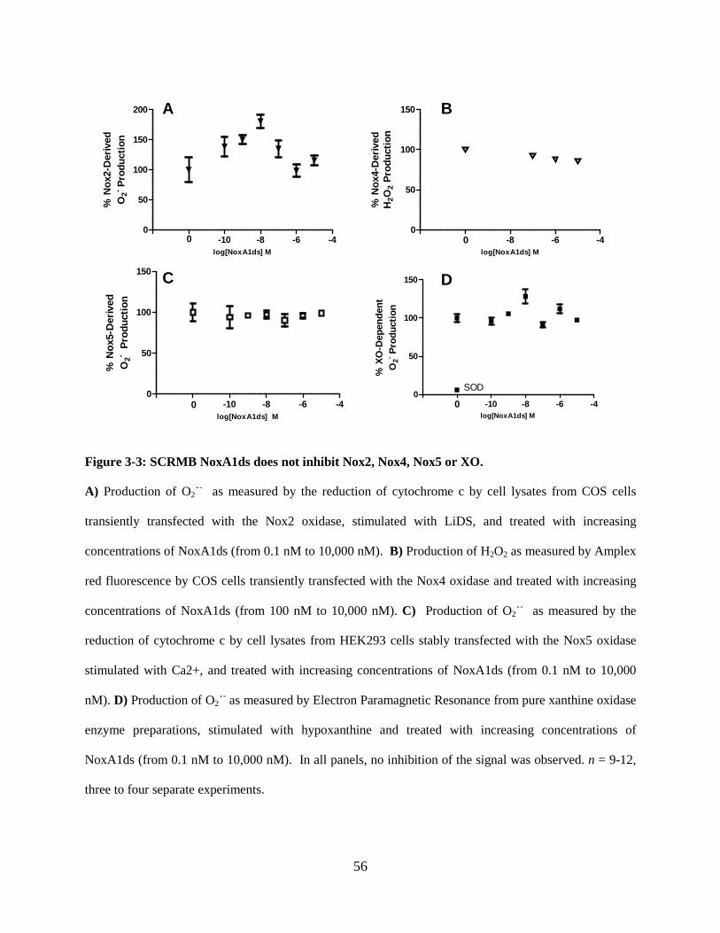
56
Figure 3-3: SCRMB NoxA1ds does not inhibit Nox2, Nox4, Nox5 or XO.
A) Production of O2˙- as measured by the reduction of cytochrome c by cell lysates from COS cells
transiently transfected with the Nox2 oxidase, stimulated with LiDS, and treated with increasing
concentrations of NoxA1ds (from 0.1 nM to 10,000 nM). B) Production of H2O2 as measured by Amplex
red fluorescence by COS cells transiently transfected with the Nox4 oxidase and treated with increasing
concentrations of NoxA1ds (from 100 nM to 10,000 nM). C) Production of O2˙- as measured by the
reduction of cytochrome c by cell lysates from HEK293 cells stably transfected with the Nox5 oxidase
stimulated with Ca2+, and treated with increasing concentrations of NoxA1ds (from 0.1 nM to 10,000
nM). D) Production of O2˙- as measured by Electron Paramagnetic Resonance from pure xanthine oxidase
enzyme preparations, stimulated with hypoxanthine and treated with increasing concentrations of
NoxA1ds (from 0.1 nM to 10,000 nM). In all panels, no inhibition of the signal was observed. n = 9-12,
three to four separate experiments.
-14 -12 -10 -8 -6 -40
50
100
150
200
log[NoxA1ds] M
% N
ox2-
Der
ived
O2- P
rodu
ctio
n
-12 -10 -8 -6 -40
50
100
150
log[NoxA1ds] M
% N
ox4-
Der
ived
H2O
2 Pr
oduc
tion
-14 -12 -10 -8 -6 -40
50
100
150
log[NoxA1ds] M
% N
ox5-
Der
ived
O2- P
rodu
ctio
n
-14 -12 -10 -8 -6 -40
50
100
150
SOD
log[NoxA1ds] M
% X
O-D
epen
dent
O2- P
rodu
ctio
n
A B
C D
0
0 0
0

57
3.2.3 NoxA1ds Inhibits O2˙- in Whole Cells
We used HT-29 cells to explore whether NoxA1ds could be used as a viable pharmacological
inhibitor in intact cells. HT-29 are a unique population of colon carcinoma cells that solely
express Nox1 without expression of any other Nox isoform [229]. To test for permeability, HT-
29 cells were treated with a FITC-labeled NoxA1ds variant for 1 hr before imaging. Confocal
microscopy revealed that NoxA1ds permeated the cell membrane of HT-29 cells and localized to
the cytoplasm (Fig. 3-4A-D). To test whether NoxA1ds inhibits Nox1-derived O2˙- production in
intact cells containing Nox1, HT-29 cells were incubated with increasing NoxA1ds
concentrations and O2˙- was measured in membrane fractions using cytochrome c. Fig. 3-4E
illustrates concentration-dependent inhibition of HT-29 O2˙- that was absent in cells treated with
SCRMB control peptide. In whole HT-29 cells, the IC50 was 1.0 µM with maximal inhibition at
5.0 µM NoxA1ds. As measured in intact HT-29 cells via EPR, Nox1 inhibition in HT-29 cells
by NoxA1ds was matched by Nox1 siRNA control (Fig 3-4F). As an additional control, we
investigated whether NoxA1ds inhibited O2˙- production in cells that do not express Nox1.
Peritoneal macrophages isolated from Nox1-null mice were pretreated with NoxA1ds for 1 hr
before addition of PMA to stimulate O2˙- production. No inhibition of O2˙- production was
observed using L-012 luminescence (Figure 3-4H).
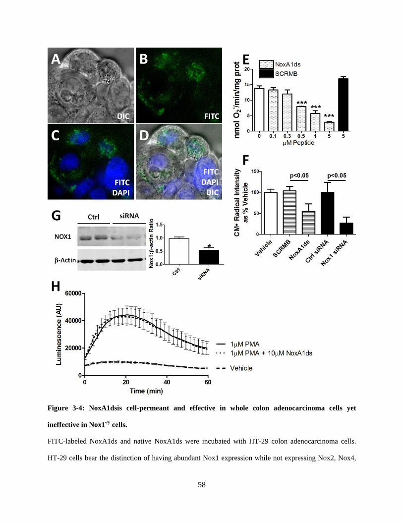
58
Figure 3-4: NoxA1dsis cell-permeant and effective in whole colon adenocarcinoma cells yet
ineffective in Nox1-/y cells.
FITC-labeled NoxA1ds and native NoxA1ds were incubated with HT-29 colon adenocarcinoma cells.
HT-29 cells bear the distinction of having abundant Nox1 expression while not expressing Nox2, Nox4,

59
or Nox5. A-D) FITC-labeled NoxA1ds was incubated with HT-29 colon adenocarcinoma cells for 1hr
prior to imaging. Confocal microscopy of these cells indicates that FITC-NoxA1ds penetrated the
extracellular membrane and that its distribution is cytosolic and perinuclear. E) As measured by SOD
inhibitable reduction of cytochrome c, increasing doses of NoxA1ds caused dose-dependent inhibition of
O2˙- production by HT-29 cells. SCRMB peptide did not inhibit O2˙- production, *** p<0.05 for 0.5, 1,
and 5 µM NoxA1ds vs 0µM NoxA1ds by one-way ANOVA. F) As measured by EPR, 10 μM NoxA1ds
and Nox1 siRNA both significantly inhibited O2˙- production by HT29 cells, p values determined by one-
way ANOVA G) Western blot analysis of Nox1/β-actin protein from Nox1 siRNA or control treated
HT29 cells. H) Peritoneal macrophages were isolated from Nox1 null mice and O2˙- production was
measured by L-012 chemiluminescence after 2 hr incubation with NoxA1ds and subsequent PMA
stimulation. No difference was observed between NoxA1ds treated and untreated samples. All values are
expressed as n = 9, except for H which was quantified via three separate experiments n = 10-12, cells
from four individual mice.
60
3.2.4 NoxA1ds Binds to Nox1
An ELISA-based assay was used to test whether NoxA1ds binds to Nox1. Biotin-tagged
NoxA1ds (B-NoxA1ds) applied to neutravidin-coated 96-well plates was added membrane
fractions from Nox1-transfected or non-transfected COS-22 cells followed by treatment with
fluorescent secondary antibodies. After repeated washing, a 30% increase in fluorescence was
observed in wells treated with membrane fractions of Nox1-transfected cells vs. untransfected
cells. This increase was not observed in wells treated with Nox1-transfected cell membranes
added to wells containing biotin-tagged SCRMB control peptide (Fig. 3-5A). A similar ELISA-
based assay indicated that Nox2 from COS-Nox2 membrane fractions did not bind NoxA1ds
(Fig 3-5B).
To corroborate these results, Fluorescence Recovery After Photobleaching (FRAP) of a
Rhodamine-labeled NoxA1ds (R-NoxA1ds) in untransfected cells and those transfected with
Nox1-YFP was quantified. As seen in Fig. 3-5C, R-NoxA1ds completely recovered after 250 s
in untransfected cells while R-NoxA1ds recovery was significantly slower and markedly
attenuated in cells transfected with Nox1-YFP, i.e. by 250 s, R-NoxA1ds had recovered less than
50% in Nox1-YFP-transfected cells. The significant decrease in the mobile fraction of R-
NoxA1ds and Nox1-YFP-transfected vs. non-transfected cells corroborates the ELISA data,
indicating binding between Nox1 and NoxA1ds (Fig. 3-5)

61
Figure 3-5: NoxA1ds binds to Nox1 but does not detectably bind Nox2.
A) Neutravidin coated plates were incubated with biotin-tagged NoxA1ds (Biotin-NoxA1ds) or biotin-
tagged SCRMB (Biotin-SCRMB) before addition of cell membranes prepared from cells transfected with
Nox1 (Nox1 membrane) or transfected with an empty vector (COS22 membrane). Bound Nox1 was
detected through a FITC conjugated secondary antibody bound to the Nox1 primary antibody. FITC
fluorescence was expressed as binding as % COS22 membranes on each experimental day. There was no
difference in binding between COS22 membranes and Nox1 membranes incubated with Biotin-SCRMB
whereas membranes incubated with Biotin-NoxA1ds showed a significant increase in binding when
transfected with Nox1. n = 10-12, three separate experiments, *p<0.05, two-way ANOVA with
Bonferroni post-hoc t-test. B) Neutravidin coated plates were incubated with biotin-tagged NoxA1ds
(Biotin-NoxA1ds) or biotin-tagged SCRMB (Biotin-SCRMB) before addition of cell membranes
prepared from cells transfected with Nox2 (Nox2 membrane) or transfected with an empty vector (COS22
membrane). Bound Nox2 was detected through an Alexa 488 conjugated secondary antibody bound to
62
the Nox1 primary antibody. Fluorescence was expressed as binding as % COS22 membranes on each
experimental day. There was no difference in binding between COS22 membranes and Nox2 membranes
incubated with Biotin-SCRMB or Biotin-NoxA1ds, C) Fluorescence Recovery After Photobleaching
(FRAP) of COS22 cells treated with 70nM Rhodamine B-labeled NoxA1ds (R-NoxA1ds) in the absence
and presence of Nox1YFP transfection and FRAP of Nox1YFP alone. Panel to the right represents
mobile fraction of these groups after 250 sec, *p<0.05, one-way ANOVA with Bonferroni post-hoc t-test.
63
3.2.5 NoxA1ds Interrupts Nox1 : NOXA1 Association
Upon observation of NoxA1ds-Nox1 binding, we proposed that NoxA1ds would interrupt
association of Nox1 and NOXA1. To test this, we employed a FRET-based assay in which
COS-22 cells were transfected with Nox1-YFP and NOXA1-CFP. Cells were analyzed by fixing
followed by irreversible YFP-photobleaching to determine if FRET was occurring between YFP
and CFP. FRET is observed indicating protein interaction between two partners when the donor
emission (CFP) signal increases after a nearby acceptor fluorophore (YFP) is inactivated by
irreversible photobleaching. In these cells, photobleaching of the YFP label resulted in a
concomitant increase in CFP fluorescence, together with a decrease in YFP signal intensity,
indicating a direct interaction between Nox1-YFP and NOXA1-CFP (Fig. 3-6A). Hence,
photobleaching effectively prevented dipole-dipole coupled acceptor (YFP-Nox1) from
accepting a quantum energy transfer from the donor (CFP), thus enhancing CFP fluorescence.
We used a myristolated CFP (CFPm) which is exclusively targeted to the cell membrane as a
negative control [25]. In cells expressing CFPm and Nox1-YFP, photobleaching of YFP did not
result in an increase in CFP fluorescence, indicating FRET was not a result of coincidental
membrane abundance and co-localization of these proteins (Fig 3-7).
COS-22 cells transfected with Nox1-YFP and NOXA1-CFP were then incubated with 10
µM SCRMB NoxA1ds or NoxA1ds for 1 hr before imaging. The SCRMB control peptide had no
effect on CFP/YFP FRET coupling (Fig. 3-6B, D). On the other hand, NoxA1ds significantly
reduced FRET efficiency (Fig. 3-6C, D). These results indicate that Nox1 and NOXA1 directly
interact and that this interaction is disrupted by NoxA1ds, but not by control peptide.

64
Figure 3-6. NoxA1ds disrupts Nox1-NOXA1 interaction.
FRET between Nox1-YFP and NoxA1-CFP transfected COS22 cells in the presence or absence of 10µM
NoxA1ds or SCRMB was evaluated. Relative fluorescence of CFP is green while YFP is red. Traces
underneath the images indicate fluorescent intensities of CFP and YFP underneath the arrow overlaid on
each cell. A) Transfected COS22 cells were treated with vehicle for one hour prior to imaging cells,
photobleaching of Nox1-YFP was complete and resulted in a concomitant increase in CFP fluorescence.
B) Transfected COS22 cells were treated with 10µM SCRMB peptide for one hour prior to imaging
cells, photobleaching of Nox1-YFP was complete and also resulted in a concomitant increase in CFP
fluorescence. C) Transfected COS22 cells were treated with 10µM NoxA1ds peptide for one hour prior
to imaging cells, photobleaching of Nox1-YFP was complete but did not result in a concomitant increase
in CFP fluorescence. D) Quantification of FRET efficiency from images A-C. Values expressed as n= 8,
three separate experiments ***p<0.001 by one-way ANOVA and Bonferroni post-test

65
Figure 3-7: Nox1:NOXA1 FRET interaction is not a result of CFP and YFP proximity in the
membrane.
Potential FRET between Nox1-YFP and a myristolated-CFP (CFP-Myr) transfected COS22 cells was
evaluated. Relative fluorescence of CFP is green while YFP is red. Traces underneath the images
indicate fluorescent intensities of CFP and YFP underneath the arrow overlaid on each cell.
Photobleaching of Nox1-YFP was complete but did not result in a concomitant increase in CFP
fluorescence.
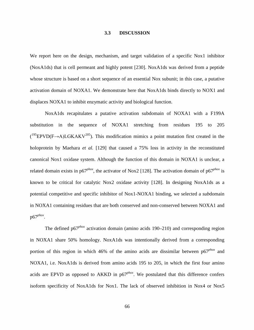
66
3.3 DISCUSSION
We report here on the design, mechanism, and target validation of a specific Nox1 inhibitor
(NoxA1ds) that is cell permeant and highly potent [230]. NoxA1ds was derived from a peptide
whose structure is based on a short sequence of an essential Nox subunit; in this case, a putative
activation domain of NOXA1. We demonstrate here that NoxA1ds binds directly to NOX1 and
displaces NOXA1 to inhibit enzymatic activity and biological function.
NoxA1ds recapitulates a putative activation subdomain of NOXA1 with a F199A
substitution in the sequence of NOXA1 stretching from residues 195 to 205
(195EPVD(F→A)LGKAKV205). This modification mimics a point mutation first created in the
holoprotein by Maehara et al. [129] that caused a 75% loss in activity in the reconstituted
canonical Nox1 oxidase system. Although the function of this domain in NOXA1 is unclear, a
related domain exists in p67phox, the activator of Nox2 [128]. The activation domain of p67phox is
known to be critical for catalytic Nox2 oxidase activity [128]. In designing NoxA1ds as a
potential competitive and specific inhibitor of Nox1-NOXA1 binding, we selected a subdomain
in NOXA1 containing residues that are both conserved and non-conserved between NOXA1 and
p67phox.
The defined p67phox activation domain (amino acids 190–210) and corresponding region
in NOXA1 share 50% homology. NoxA1ds was intentionally derived from a corresponding
portion of this region in which 46% of the amino acids are dissimilar between p67phox and
NOXA1, i.e. NoxA1ds is derived from amino acids 195 to 205, in which the first four amino
acids are EPVD as opposed to AKKD in p67phox. We postulated that this difference confers
isoform specificity of NoxA1ds for Nox1. The lack of observed inhibition in Nox4 or Nox5

67
systems is likely a result of these Nox systems having no cytosolic activator subunit requirement.
Finally, the lack of inhibition of XO by NoxA1ds is further confirmation that its inhibitory effect
is not germane to all oxidases and is not a consequence of ROS scavenging.
NoxA1ds could potentially mimic functional sites in other proteins and thus interfere
with their function. We used BLAST to compare the sequence of NoxA1ds to the National
Institutes of Health translated nucleotide database to determine potential nonspecific protein
interactions with NoxA1ds. We were surprised to observe that fewer than five mammalian
proteins outside of the NADPH oxidase family shared significant homology with NoxA1ds.
These hits were hypothetical proteins based on isolated DNA sequences. As such, we believe
nonspecific actions of NoxA1ds should be negligible.
When NoxA1ds was added to cell-free preparations of the canonical Nox1 oxidase,
composed of catalytic subunit Nox1, activating subunit NOXA1 and organizing subunit NOXO1
along with Rac, NoxA1ds inhibited Nox1-derived O2˙- production with an IC50 of 20 nM
achieving maximum inhibition of 90% at 1.0 μM. In addition, we showed that NoxA1ds does not
scavenge either O2˙- or H2O2 and does not inhibit Nox2, Nox4, Nox5, or XO activity. These
results support NoxA1ds as an isoform-specific inhibitor of Nox1.
We extended our studies to determine the in vitro efficacy of NoxA1ds and observed that
fluorescent analogues of NoxA1ds (Noxa1ds-FITC) are capable of crossing the plasma
membrane and localizing to the cytosol. Many short peptides require the small HIV “tat” peptide
moiety to penetrate cell membranes [112, 231]. In contrast, our findings herein demonstrated that
“tat” is unnecessary for NoxA1ds to cross the plasma membrane, and we attribute this in part to
the positively charged lysines in the C terminus of NoxA1ds and the alternating
hydrophobic/hydrophilic amino acid structure of NoxA1ds. Indeed, other short peptides with
68
similar hydropathy patterns have proven highly cell permeant and thus support our findings
[232]. Upon demonstrating its permeability in whole cells, we tested whether NoxA1ds is an
effective inhibitor of O2˙- production in a primary cancer cell line (HT-29) that exclusively
expresses Nox1 oxidase [229]. We observed that NoxA1ds significantly attenuated O2˙-
production in HT-29.
ELISA and FRAP data revealed that NoxA1ds binds to the catalytic Nox1 subunit, and
FRET demonstrated that NoxA1ds disrupted the critical association of Nox1 with NOXA1 and
that this binding is specific to Nox1 in HPAEC. Our FRET data support the notion that not only
is this domain an activation domain critical for enzyme activity in Nox1 but that this domain is
also a key binding region in Nox1-NOXA1 complexes [129].
Notwithstanding the broadly-demonstrated effectiveness of related peptides by parenteral,
peritoneal, subcutaneous, and direct application to blood vessels using gene therapy [159, 232],
the limitations in the use of peptides as “druggable” therapeutics are obvious. These include a
very limited oral bioavailability due to peptide degradation in the gut. As mentioned earlier,
however, these issues are being circumvented by novel technologies, including the use of
nanotechnologies such as microbubble mediated drug delivery [233, 234]. Beyond targeted
peptide delivery, there are a number of peptide modifications that can support bioavailability by
reducing protease degradation. These include hydrocarbon stapling and partial substitution of d-
amino acids [235, 236].
Maehara et al. [129] first reported that the NOXA1 F199A mutation that we incorporated
into NoxA1ds interferes with FAD reduction in the enzyme complex and thus may prevent
catalytic O2˙- production. While it is plausible that NoxA1ds inhibits Nox1 through a similar
mechanism, our data are more in line with NoxA1ds preventing Nox1-NOXA1 binding and

69
inhibiting O2˙- production. Thus, we propose that this domain is crucial for the binding and
activation of Nox1. Taken together, our data indicate that the peptide NoxA1ds is a specific
inhibitor of Nox1 in cell-free and whole cell preparation and that this inhibition occurs via
binding to the catalytic Nox1 subunit and blockade of Nox1-NOXA1 binding (Figure 3-8). It
stands to reason that this site-specific binding is important for subsequent FAD reduction.
However, whether the domain somehow catalyzes or facilitates FAD reduction either directly or
via an allosteric change in the Nox1 C-terminus is still an open question.
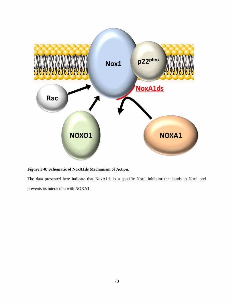
70
Figure 3-8: Schematic of NoxA1ds Mechanism of Action.
The data presented here indicate that NoxA1ds is a specific Nox1 inhibitor that binds to Nox1 and
prevents its interaction with NOXA1.

71
4.0 DETERMINE RELATIVE ROLES OF NOX1 VS NOX2 IN ENDOTHELIAL O2·-
PRODUCTION AND CELL MIGRATION
4.1 INTRODUCTION
The first rationally-designed, peptidic Nox inhibitor, Nox2ds-tat (Nox2 docking sequence-tat;
Nox2ds-tat a.k.a. gp91ds-tat), was identified by the Pagano laboratory in 1999 and published in
2001 as a compound that decreased angiotensin II-induced vascular O2˙- generation and attenuate
systemic blood pressure elevation in hypertensive mice [112]. Nox2ds-tat was designed as a
peptide mimicking the second intracellular loop of Nox2, with the “tat” sequence added to the C-
terminus to facilitate cell permeation [112]. Since its development, the utility of Nox2ds-tat has
been demonstrated as a Nox inhibitor in a wide variety of cell and organ systems representing
different disease phenotypes, including cardiovascular and neurodegenerative manifestations of
disease consistent with the expression of Nox2 [151, 167, 237, 238]. However, despite the
widespread efficacy and utility of Nox2ds-tat, specificity of Nox2ds for the Nox2-oxidase had
not been fully tested. This gap in knowledge was in part a result of the development of Nox2ds-
tat prior to or simultaneous with the discovery of new Nox homologues. Indeed, Nox1 was
discovered in prostate cancer cells in 1999 [100] while Nox4 was discovered in kidney cells in
2001 [131] and Nox5 was discovered in testis in 2001 [53]. At the time of Nox2ds-tat’s
discovery, vascular expression, physiological roles, and genetic data of these Noxes was
completely lacking, thereby precluding investigation of the specificity of Nox2ds-tat against
these novel Nox isoforms [108]. What’s more, heterologous systems (i.e., COS or HEK cell
preparations) were not generally available.

72
The significant homology between Nox1 and Nox2 has raised a possibility that Nox2ds
may also inhibit Nox1 despite its derivation from components of the Nox2 oxidase. Since its
discovery in 2001, Nox2ds-tat has seen continued use as a generic Nox inhibitor and an untested
bias that it would be effective at inhibiting other Noxes. As the major vascular isoforms of Nox
are Nox1, Nox2, and Nox4 (with the specificity of Nox2ds vs Nox4 being tested elsewhere), our
laboratory proceeded to characterize the specificity of Nox2ds-tat for Nox2 vs Nox1 and Nox4
and to determine the relative roles of various isoforms of Nox in endothelial cell biology. The
group long postulated that Nox2ds preferentially inhibits Nox2 rather than Nox1 and 4 by virtue
of its design. Furthermore, elucidation of the primary Nox isoform responsible for ROS
production in HPAEC and subsequent effect on cell physiology was sought in this dissertation.
Here, as part of a comprehensive study performed by Drs. Csanyi and Pagano (and co-workers),
I performed a subset of experiments using cell-free biochemical assays of Nox activity to
determine the pharmacological profile of Nox2ds against Nox1, Nox2 and Nox4. Nox2ds’
specificity was tested without the “tat” conjugation as these assays were performed in cell-free
systems. Following certification of Nox2ds specificity, I utilized NoxA1ds and Nox2ds-tat to
investigate the relative contribution of Nox1 and Nox2 to hypoxia-induced O2˙- production and
VEGF-mediated migration of endothelial cells. The “tat” cell penetrating peptide leader
sequence was added to Nox2ds and used for our endothelial cell migration experiments.

73
4.2 RESULTS
4.2.1 Nox2ds inhibits O2˙- production from Nox2-oxidase
To investigate whether Nox2ds inhibits O2˙- production in the Nox2 oxidase system, SOD-
inhibitable cytochrome c reduction was measured in Nox2ds-pretreated COS-Nox2 cell lysates.
Addition of LiDS to lysates derived from COS-Nox2 cells stimulated O2˙- production in a
reaction that was dependent on the presence of NADPH (1.33±0.1 and 0.40±0.1 nmol O2˙-
/min/107 COS-Nox2 lysate-cell equivalents for LiDS- and vehicle-treated COS-Nox2 lysates,
respectively, p < 0.05). O2˙- production in non-transfected COS-22 cell lysates (control) was
0.16±0.1 nmol O2˙-/min/107 COS-22 lysate-cell equivalents (Fig. 4-1). As demonstrated in Fig.
4-1, preincubation of COS-Nox2 cell lysates with Nox2ds (before LiDS-treatment)
concentration-dependently inhibited O2˙- production, displaying an IC50 of 0.74 μM. In contrast,
preincubation of COS-Nox2 cell lysates with scrmb Nox2ds did not inhibit Nox2-oxidase. Note:
Experiments described in Sections 4.2.1 through 4.2.3 are part of a larger study performed by Dr.
Gabor Csanyi et al. in the Pagano laboratory and are germane to this dissertation. The
candidate’s specific experimental contributions to this body of work are noted below.
4.2.2 Nox2ds does not inhibit O2˙- production from Canonical or Hybrid Nox1-oxidase
The series of experiments described in this subsection were personally carried out by the
candidate and are directly relevant to the validation of Nox2ds for the purposes of this
dissertation. In contrast to the COS-Nox2 cell-free system and in line with the constitutive
activity of COS-Nox1/NOXO1/NOXA1 cells, the cytochrome c assay of COS-
Nox1/NOXO1/NOXA1 cell free system revealed that the canonical COS-Nox1 oxidase does not

74
require LiDS for activation (nmol O2˙-/min/107 cell equivalents were: 1.30±0.3 and 0.16±0.1 for
COS-Nox1 and COS-22 cell lysates, respectively, p < 0.05). Preincubation of COS-Nox1 cell
lysates with different concentrations of Nox2ds did not inhibit O2˙- production (Fig. 4-2).
In separate experiments, we tested whether the lack of an inhibitory effect of Nox2ds on
the Nox1 system is due to the stable interaction of NOXO1 and NOXA1 with Nox1. The Nox1
organizer subunit NOXO1 was preincubated with vehicle (control) or 10 μM Nox2ds for 10 min,
and then NOX1 and NOXA1 were added consecutively. After the preincubation period, LiDS
was added and O2˙- production was measured. Our results demonstrate that preincubation of
NOXO1 with Nox2ds did not inhibit the canonical Nox1 oxidase (Table 4-1). Previous studies
reported that Nox1 can be activated by not only NOXO1 and NOXA1 but also by p47phox and
p67phox [226]. We therefore tested the effect of Nox2ds on O2˙- production on all possible
combinations of Nox1 oxidase system. As shown in Table 4-1, Nox2ds did not inhibit O2˙-
production in either the canonical or the hybrid Nox1 system.

75
Figure 4-1. Nox2ds dose-dependently inhibits O2˙- production from Nox2-oxidase.
COS-Nox2 cell lysate was preincubated with various concentrations of Nox2ds (from 0.1 μM to 10 μM)
and Scrmb Nox2ds (from 0.1 μM to 10 μM) for 5 min at 25○C. After the addition of 130 μM LiDS, O2˙-
production was initiated by the addition of 180 μM NADPH and measured by the initial linear rate of
SOD-inhibitable cytochrome c reduction. O2˙- production is expressed as nmol O2˙-/min/107 cell
equivalents. Data represent the mean ± SEM of 7-16 experiments. For comparison, O2˙- production in
non-transfected COS-22 cell lysate is shown. *p < 0.05 indicates significant differences in O2˙- production
between Nox2ds- and scrmb Nox2ds-treatment. †p < 0.05 indicates significant difference between COS-
Nox2 and COS-22 cell lysate activity.
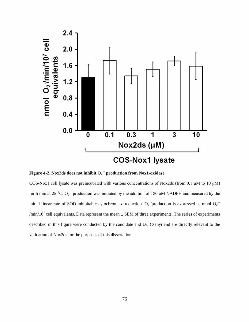
76
Figure 4-2. Nox2ds does not inhibit O2˙- production from Nox1-oxidase.
COS-Nox1 cell lysate was preincubated with various concentrations of Nox2ds (from 0.1 μM to 10 μM)
for 5 min at 25 ○C. O2˙- production was initiated by the addition of 180 μM NADPH and measured by the
initial linear rate of SOD-inhibitable cytochrome c reduction. O2˙-production is expressed as nmol O2˙-
/min/107 cell equivalents. Data represent the mean ± SEM of three experiments. The series of experiments
described in this figure were conducted by the candidate and Dr. Csanyi and are directly relevant to the
validation of Nox2ds for the purposes of this dissertation.
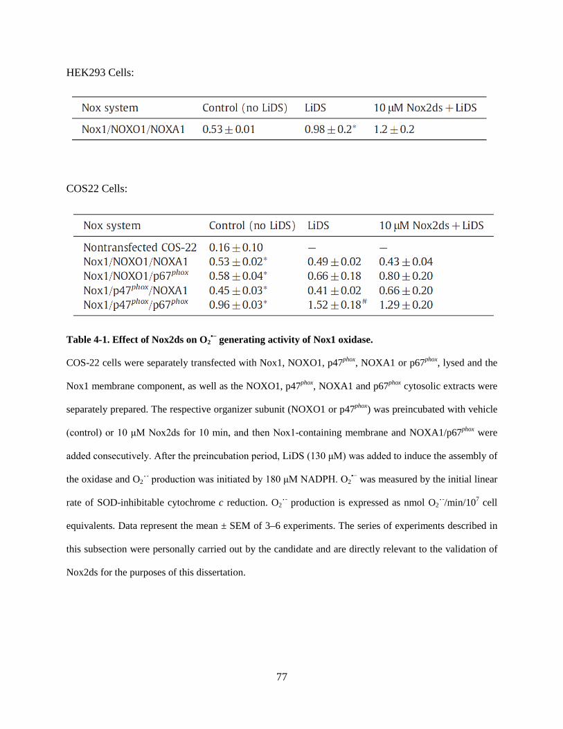
77
HEK293 Cells:
COS22 Cells:
Table 4-1. Effect of Nox2ds on O2•− generating activity of Nox1 oxidase.
COS-22 cells were separately transfected with Nox1, NOXO1, p47phox, NOXA1 or p67phox, lysed and the
Nox1 membrane component, as well as the NOXO1, p47phox, NOXA1 and p67phox cytosolic extracts were
separately prepared. The respective organizer subunit (NOXO1 or p47phox) was preincubated with vehicle
(control) or 10 μM Nox2ds for 10 min, and then Nox1-containing membrane and NOXA1/p67phox were
added consecutively. After the preincubation period, LiDS (130 μM) was added to induce the assembly of
the oxidase and O2˙- production was initiated by 180 μM NADPH. O2•− was measured by the initial linear
rate of SOD-inhibitable cytochrome c reduction. O2˙- production is expressed as nmol O2˙-/min/107 cell
equivalents. Data represent the mean ± SEM of 3–6 experiments. The series of experiments described in
this subsection were personally carried out by the candidate and are directly relevant to the validation of
Nox2ds for the purposes of this dissertation.

78
4.2.3 p47phox, but not NOXO1, binds to Nox2ds
Nox2ds was designed to selectively inhibit the interaction between the cytosolic B-loop of Nox2
and p47phox by binding to p47phox and preventing its translocation to the membrane. To confirm
this binding and to rule out binding of Nox2ds to NOXO1, ELISA experiments were performed
on lysates from p47phox- or NOXO1-transfected COS-22 cells incubated in ELISA plates with
neutravidin-immobilized biotinylated Nox2ds or its scrambled control. As shown in Fig. 4-3A,
using p47phox antibody to detect binding (followed by fluorescently tagged secondary antibody),
fluorescence intensity in biotinylated Nox2ds-bound wells was significantly higher when
cytosolic fractions of COS-22-p47phox vs. COS-22 were added. These data indicate that p47phox
binds to Nox2ds. Binding of p47phox was sequence specific as p47phox binding to the scrambled
sequence produced significantly less fluorescence (Fig. 4-3A). In contrast, there was no
difference in fluorescence intensity when cytosolic fractions of COS-22 NOXO1 vs. COS-22
cytosol were added to plates bound with Nox2ds, followed by incubation with NOXO1 antibody
(Fig. 4-3B).
4.2.4 Nox1, but not Nox2-derived O2˙- Mediates Endothelial Cell Migration
To test the role of this key Nox1-NOXA1 interaction in a pathophysiologically relevant vascular
cell phenotype, we investigated whether Nox1-NoxA1ds binding and inhibition was effective in
HPAEC as these cells express abundant Nox1 and Nox2 with Nox4 being the most highly
expressed (Fig. 4-4B). ELISA data indicated that NoxA1ds bound to Nox1 but not Nox2 or -4 in
HPAEC (Fig. 4-4A). HPAEC were exposed to 1.0% oxygen for 24 h and treated with 10 μm
NoxA1ds or SCRMB for 1 h before measuring O2˙- production. Hypoxia caused a 3-fold
increase in O2˙- production that was completely inhibited by preincubation with NoxA1ds but not
79
with SCRMB or by Nox2ds-tat (Fig 4-4C,F). The effect of NoxA1ds was partially replicated
using Nox1 siRNA, with detectable yet statistically insignificant inhibition of O2˙- production.
The incomplete inhibition of O2˙- production was likely due to incomplete knockdown of Nox1
by the siRNA (Fig. 4-4E). To determine the effect of NoxA1ds on migration, we performed a
wound assay on VEGF-stimulated HPAEC migration under hypoxic conditions (1.0% O2).
NoxA1ds caused a significant reversion in endothelial cell migration to vehicle control levels, an
effect that was not observed in cells treated with SCRMB or Nox2ds-tat (Fig. 4-5). Additionally,
HPAEC transfected with Nox1-YFP and NOXA1-CFP showed an increase in FRET when
treated with 20 nm VEGF that was inhibitable by NoxA1ds, indicating that VEGF promotes
Nox1-NOXA1 association (Fig.4-6).
4.2.5 Fibroblasts are a Potential in vivo Source for VEGF during Hypoxia
To determine potential sources for VEGF and thus Nox1 activation in the vessel, HPAEC,
HPASMC and HPAF were cultured at either 20% O2 or 1.0%O2 for 24 hrs before quantification
of VEGF in the cell culture media. HPAEC consumed nearly all VEGF initially present in the
media while HPASMC produced substantial VEGF, yet in a hypoxia independent manner.
Interestingly, HPAF were the only tested vascular cell type to exhibit enhanced VEGF
production during hypoxia (Figure 4-7). This VEGF-enriched media derived from hypoxia
conditioned HPAF was then used to culture HPAEC and was observed to support HPAEC
mitogenic propensity significantly more than media derived from normoxia conditioned HPAF
as measured by MTT assay. This stimulation of mitogenic propensity was inhibited by
NoxA1ds, confirming a role for Nox1 in VEGF stimulated HPAEC proliferation (Figure 4-7D).

80
Figure 4-3: p47phox, but not NOXO1, binds to Nox2ds.
ELISA experiments were performed to test whether p47phox (A) and NOXO1 (B) bind to Nox2ds and/or
scrmb Nox2ds. Biotinylated Nox2ds and scrambled peptides were first bound to neutravidin-coated plates
and then incubated with COS-22, COS-22-p47phox (COS-22 cells transfected with p47phox) or COS-22-
NOXO1 (COS-22 cells transfected with NOXO1) cytosolic extracts for 1 hr at room temperature. Rabbit
polyclonal p47phox or rabbit NOXO1 antibodies were used to detect Nox2ds-bound p47phox or NOXO1,
respectively. After 1 hr incubation and extensive washing, bound primary antibodies were detected by
FITC-labeled goat anti rabbit IgG antibody. The fluorescence of each well was measured using a Biotek
Synergy 4 Hybrid Multi-Mode Microplate Reader (Excitation:488 nM, Emission:518 nM). Data represent
the mean ± SEM of 3–4 experiments. *p<0.05 between COS-22 and COS-22-p47phox cytosolic extracts in
Nox2ds wells. #p<0.05 between Nox2ds and Scrmb Nox2ds wells incubated with COS-22-p47phox extracts

81
Figure 4-4: NoxA1ds attenuates hypoxia-induced O2˙-production.
A) NoxA1ds binds to Nox1 but not Nox2 or Nox4 in HPAEC. Neutravidin coated plates were incubated
with biotin-tagged NoxA1ds (B-NoxA1ds) or biotin-tagged SCRMB (B-SCRMB) before addition of
HPAEC cell membranes. Captured Noxes were detected through a Alexa-488 conjugated secondary

82
antibody bound to the Nox1,2 or 4 primary antibody. Fluorescence was expressed with background lysis
buffer fluorescence subtracted. When fluorescence was detected via Nox1 primary antibody, there was a
significant increase in binding as compared to B-SCRMB. n = 4, *p<0.05, unpaired t-test. B) Relative
Nox expression in HPAEC quantified by qPCR. C) In hypoxia and normoxia, SCRMB and NoxA1ds
peptides were added to cells at 10uM for 1hr prior to cell lysis and quantification of enzyme activity.
Cells were transfected with Nox1 siRNA 24hrs prior to 24hr normoxic/hypoxic treatment followed by
cell lysis and quantification of enzyme activity. SCRMB, NoxA1ds, and Nox1 siRNA had a negligible
effect on O2˙-production under normoxic conditions. Hypoxia (1.0% O2, 24hrs) treatment resulted in a
three-fold increase in O2˙- production that was unaffected by SCRMB. Upon treatment with NoxA1ds,
O2˙-production by cells subjected to hypoxia returned to the amount observed under normoxia. D)
Representative experimental trace for enzyme activity in “B” shown as the SOD-inhibitable reduction of
cytochrome c over time. n = 9, three separate experiments. E) Western blot analysis of Nox1/β-actin
protein from Nox1 siRNA or control treated human pulmonary artery endothelial cells (HPAEC). Nox1
knockdown was incomplete and approximates the degree of knockdown observed as O2˙- production F)
Hypoxia (1.0% O2, 24hrs) treatment resulted in a two-fold increase in HPAEC (Human Pulmonary Artery
Endothelial Cells) O2˙- production that was unaffected by Nox2ds-tat or its control (SCRMB). n = 9,
three separate experiments. *p<0.05, **p<0.01, one-way ANOVA with Bonferroni post-hoc t-test.

83
Figure 4-5: Nox1, but not Nox2, is responsible for VEGF-stimulated wound healing.
Confluent HPAEC were scratched with P1000 pipet tip, photographed, treated with 20nM VEGF +/-
10µM NoxA1ds, and photographed again after 24 hours. A) Representative images of HPAEC
immediately after scratch wounding and 24hrs post scratch wound. B) Quantification of HPAEC wound
closure VEGF +/- 10µM NoxA1ds C) Quantification of HPAEC wound closure treated with VEGF +/-
10µM Nox2ds-tat. Values represent n=6-8, three to four separate experiments, *p<0.05 one-way
ANOVA with Bonferroni post-test as compared to vehicle treatment.
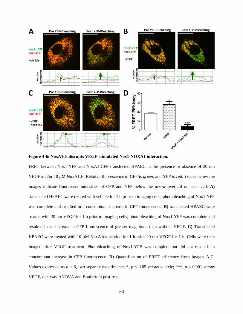
84
Figure 4-6: NoxA1ds disrupts VEGF-stimulated Nox1-NOXA1 interaction.
FRET between Nox1-YFP and NoxA1-CFP transfected HPAEC in the presence or absence of 20 nm
VEGF and/or 10 μM NoxA1ds. Relative fluorescence of CFP is green, and YFP is red. Traces below the
images indicate fluorescent intensities of CFP and YFP below the arrow overlaid on each cell. A)
transfected HPAEC were treated with vehicle for 1 h prior to imaging cells; photobleaching of Nox1-YFP
was complete and resulted in a concomitant increase in CFP fluorescence. B) transfected HPAEC were
treated with 20 nm VEGF for 1 h prior to imaging cells; photobleaching of Nox1-YFP was complete and
resulted in an increase in CFP fluorescence of greater magnitude than without VEGF. C) Transfected
HPAEC were treated with 10 μM NoxA1ds peptide for 1 h prior 20 nm VEGF for 1 h. Cells were then
imaged after VEGF treatment. Photobleaching of Nox1-YFP was complete but did not result in a
concomitant increase in CFP fluorescence. D) Quantification of FRET efficiency from images A-C.
Values expressed as n = 6, two separate experiments; *, p < 0.05 versus vehicle; ***, p < 0.001 versus
VEGF, one-way ANOVA and Bonferroni post-test.

85
Figure 4-7: VEGF Production by HPAF is enhanced by hypoxia.
A) VEGF production by HPAEC measured by MSD ELISA indicated that HPAEC consumed most
VEGF present in the media after 24hrs in either hypoxic (1.0% O2) or normoxic (20% O2) media, n=3. B)
HPASMC produce VEGF with respect to naïve media without cells but in an oxygen-insensitive manner,
n=3. C) VEGF production by HPAF measured by MSD ELISA indicated that HPAF produce VEGF with
respect to naïve media without cells and that this is enhanced by 1.5 fold in hypoxic media versus
normoxic media, D) Normoxic HPAF conditioned media (Nrmx CM) or hypoxic HPAF-conditioned
media (Hypx CM) was used to culture HPAEC for 24 hrs ±NoxA1ds, after which cellular proliferative
propensity was determined by MTT assay. Hypx CM potentiated HPAEC proliferative propensity in a
Nox1 dependent manner. n=3.
2
20% O 2
1.0% O
Growth m
edia
0
20
40
60
80
100pg
/ml V
EGF
2
20% O 2
1.0% O
Growth m
edia
0
200
400
600
800
pg/m
l VEG
F
Nrmx C
M
Hypx C
M
M NoxA
1ds
µ
Hypx C
M +10
0102080
90
100
110p<0.05, ANOVA
Rel
ativ
e M
TT A
bsor
banc
e
2
20% O 2
1.0% O
Growth m
edia
0
5
10
15
20p<0.05
pg/m
l VEG
FA B
C D

86
4.3 DISCUSSION
Numerous studies have established the ability of Nox2ds-tat to inhibit Nox activity in vitro and
in vivo [46, 122, 139, 197]. However, until now the specificity of inhibition of Nox2ds (non-
chimeric B-loop peptide) between Nox1 and Nox2 had not been investigated. To test the
specificity of Nox2ds, we examined its potential inhibitory activity in cell-free assays using
reconstituted systems of Nox1 while others demonstrated that Nox2ds does not inhibit Nox4
[130]. Our data demonstrate that Nox2ds concentration-dependently inhibited O2˙- production in
a COS-Nox2 cell-free system and that Nox2ds is a potent and efficacious inhibitor of Nox2
NADPH oxidase with an IC50 of 0.74 μM. Furthermore, our results demonstrated that Nox2ds
does not inhibit ROS production in the COS-Nox1 system with others determining that Nox2ds
does not inhibit ROS production by COS-Nox4 or XO. The results of the present study
demonstrate selectivity of Nox2ds peptide in differentiating the contribution of Nox2- vs. Nox1-
oxidase to Nox-derived ROS production. Furthermore, it was also determined that Nox2ds does
not inhibit hybrid Nox1 systems where either NOXO1 or NOXA1 is replaced with p47phox or
p67phox, respectively. These findings are of particular importance as we showed that p47phox,
which is present in Nox1 hybrid systems in vascular smooth muscle cells, is the binding target of
Nox2ds. These findings have broad implications for distinguishing the role of Nox2 in a wide
range of disease processes and support its potential use as a Nox2-targeted therapeutic agent.
We next conjugated Nox2ds to the cell penetrating peptide sequence “tat,”[169] thus
enabling us to use Nox2ds-tat and NoxA1ds to comparatively analyze the role of Nox1 vs Nox2
in whole HPAEC O2˙- production in response to hypoxia and VEGF-induced HPAEC migration
in a scratch wound healing assay. In vitro experiments determined that Nox2 plays an

87
insignificant role in hypoxia-induced O2˙- production as well as VEGF-induced HPAEC
migration. These observations are largely in agreement with those made by Garrido-Urbani et.
al. showing that Nox1 rather than Nox2 plays a greater role in angiogenesis [98]. Finally, the
data presented here establish that Nox1 can be directly activated by VEGF and present a new
pathway through which Nox1 can directly influence vascular (patho)physiology.
To investigate potential sources of VEGF in the pulmonary vasculature, HPAEC,
HPASMC, and HPAF were incubated under normoxic and hypoxic conditions for 24 hrs and
VEGF was quantified in the media. Relative to naïve media devoid of cells, HPAEC consumed
nearly all VEGF present, regardless of the oxygen tension. HPASMC produced a substantial
amount of VEGF relative to naïve media, but in an oxygen independent manner. Normoxic
HPAF produced a significant quantity of VEGF versus naïve media, and this was enhanced by
1.5 fold through hypoxic conditioning (10.56 vs 16.81 pg/ml VEGF), with values comparable to
physiological human serum VEGF concentrations (between 10 and 200pg/ml) [239]. When
hypoxic HPAF conditioned media enriched in VEGF was used to culture HPAEC, the mitogenic
propensity of the HPAEC significantly increased in a Nox1-dependent manner, suggesting that
hypoxic HPAF may contribute to HPAEC Nox1 activation in vivo through paracrine VEGF
signaling in addition to myriad other factors present in the extracellular milieu. While HPAF
conditioned media enriched in VEGF was capable of enhancing the mitogenic potential of
HPAEC in a Nox1-dependent manner, the magnitude of this 5% increase is minor when
compared to the 100% increase in Nox1 activity when HPAEC were treated with hypoxia alone.
These data suggest that hypoxia, rather than HPAF-mediated paracrine signaling, is the primary
insult that potentiates Nox1 activation in HPAEC.

88
In collaboration with Drs. Csanyi and Pagano, I observed that Nox2ds is a Nox2 specific
inhibitor that does not inhibit canonical, induced, or hybrid Nox1 systems. By using NoxA1ds
and Nox2ds-tat to specifically inhibit Nox1 and Nox2, respectively, we determined that Nox1,
but not Nox2, is responsible for hypoxic induction of O2˙- production and VEGF-stimulated cell
migration of HPAEC. Furthermore, we identified a novel pathway where VEGF can directly
activate Nox1:NOXA1 assembly. Our data also demonstrated that HPAF are a potential source
for VEGF under hypoxic conditions, although HPAF-sourced VEGF seems to play a minor role
in HPAEC physiology when compared to hypoxia alone. These data indicate that targeting Nox1
with NoxA1ds may provide effective therapy for diseases of the pulmonary vasculature,
including pulmonary arterial hypertension.
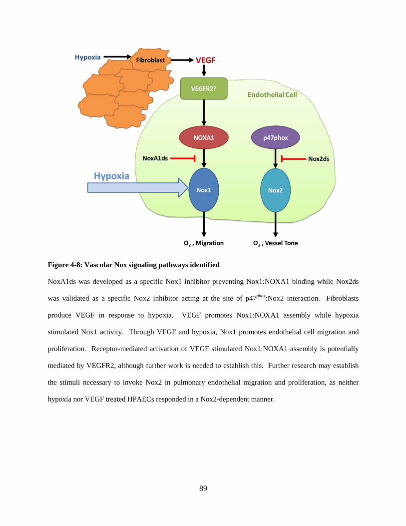
89
Figure 4-8: Vascular Nox signaling pathways identified
NoxA1ds was developed as a specific Nox1 inhibitor preventing Nox1:NOXA1 binding while Nox2ds
was validated as a specific Nox2 inhibitor acting at the site of p47phox:Nox2 interaction. Fibroblasts
produce VEGF in response to hypoxia. VEGF promotes Nox1:NOXA1 assembly while hypoxia
stimulated Nox1 activity. Through VEGF and hypoxia, Nox1 promotes endothelial cell migration and
proliferation. Receptor-mediated activation of VEGF stimulated Nox1:NOXA1 assembly is potentially
mediated by VEGFR2, although further work is needed to establish this. Further research may establish
the stimuli necessary to invoke Nox2 in pulmonary endothelial migration and proliferation, as neither
hypoxia nor VEGF treated HPAECs responded in a Nox2-dependent manner.

90
5.0 INTERROGATE A ROLE FOR NOX1 IN PULMONARY ARTERIAL
HYPERTENSION
5.1 INTRODUCTION
5.1.1 Etiology of Pulmonary Arterial Hypertension
Pulmonary arterial hypertension (PAH) is a debilitating disease with high mortality characterized
by a mean pulmonary artery pressure (mPAP) greater than 25 mmHg, in part a consequence of
abnormal pulmonary vessel intimal remodeling as well as reduced NO bioavailability [170]. In
many patients, vessel remodeling often partially or completely occludes the lumen, increasing
pulmonary vascular resistance and pulmonary pressure eventually leading to right heart failure
[170]. Hypoxia stimulates Nox-derived ROS production, causing reduced NO bioavailability,
vascular cell proliferation and vessel remodeling [94, 179]. In addition to hypoxia, mitogenic
factors promoting vascular cell proliferation, including EGF and VEGF, have been implicated in
the development of PAH [174, 181]. Despite this association with hypoxia and growth factors,
no cause for idiopathic PAH has been identified and current treatment regimens symptomatically
treat the disease [170]. These treatments include prostacyclin, nitrates, beta-blockers, and sartans
[240]. Unfortunately, all of these treatments fail to cure PAH and are eventually overcome by
desensitized responses, leading to disease progression. Inhibitors of ROS sources, including
Nox1, have shown promise in promoting NO bioavailability, attenuating intimal and medial
remodeling, thus lowering PVR and providing an alternative and potentially curative therapy for
PAH [241, 242]. Despite their potential, no specific Nox inhibitor or antioxidant therapy has yet
been identified for PAH.

91
5.1.2 Pulmonary Vascular Hemodynamics in PAH
The defining hemodynamic phenotype in PAH is a mean pulmonary artery pressure (mPAP)
greater than 25 mmHg, causing chronically increased afterload on the right ventricle [170, 243].
This chronic pressure overload results in compensatory right heart hypertrophy associated with
preserved contractility eventually leading to decompensation and right heart failure [170].
Indicative of right heart hypertrophy, the Fulton index is a key indicator of PAH that quantifies
the relative size of the right heart versus the left heart + interventricular septum (Equation 2)
[71]. Beyond simple increases in mPAP and consequent RV hypertrophy, substantial changes in
the hemodynamic profile of the RV follow, with each parameter providing a portion of a more
detailed picture on the pathology of PAH.
Equation 2:
Fulton index =Right Ventricle Mass
Left Ventricle Mass + Septum Mass
Pressure-volume catheterization and the resulting pressure-volume loops (PV loops) is
the primary method of evaluating hemodynamic function within the RV or LV in both clinical
and scientific cardiology settings. The PV loop quantifies simultaneous pressure and volume
changes in the ventricle of the heart though which the catheter is inserted during the cyclical
beating of the heart through both systole and diastole. These loops quantitatively describe the
filling, contraction, ejection, and relaxation of the ventricle into which the catheter is inserted
(Figure 5-1) [65, 67]. Hemodynamic criteria evaluated by PV loop analysis include, but are not
limited to, pressure within each ventricle of the heart, the volume of a full ventricle (end diastolic
volume, EDV), the amount of blood pumped by the ventricle in unit time (cardiac output), and
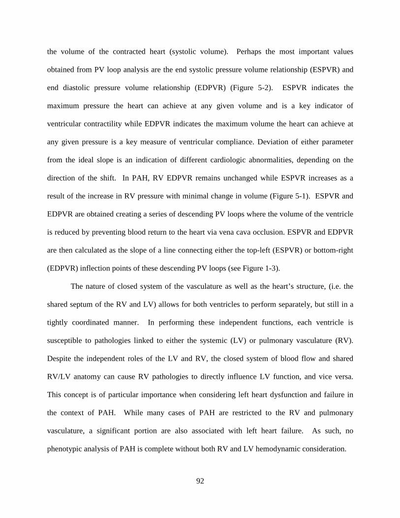
92
the volume of the contracted heart (systolic volume). Perhaps the most important values
obtained from PV loop analysis are the end systolic pressure volume relationship (ESPVR) and
end diastolic pressure volume relationship (EDPVR) (Figure 5-2). ESPVR indicates the
maximum pressure the heart can achieve at any given volume and is a key indicator of
ventricular contractility while EDPVR indicates the maximum volume the heart can achieve at
any given pressure is a key measure of ventricular compliance. Deviation of either parameter
from the ideal slope is an indication of different cardiologic abnormalities, depending on the
direction of the shift. In PAH, RV EDPVR remains unchanged while ESPVR increases as a
result of the increase in RV pressure with minimal change in volume (Figure 5-1). ESPVR and
EDPVR are obtained creating a series of descending PV loops where the volume of the ventricle
is reduced by preventing blood return to the heart via vena cava occlusion. ESPVR and EDPVR
are then calculated as the slope of a line connecting either the top-left (ESPVR) or bottom-right
(EDPVR) inflection points of these descending PV loops (see Figure 1-3).
The nature of closed system of the vasculature as well as the heart’s structure, (i.e. the
shared septum of the RV and LV) allows for both ventricles to perform separately, but still in a
tightly coordinated manner. In performing these independent functions, each ventricle is
susceptible to pathologies linked to either the systemic (LV) or pulmonary vasculature (RV).
Despite the independent roles of the LV and RV, the closed system of blood flow and shared
RV/LV anatomy can cause RV pathologies to directly influence LV function, and vice versa.
This concept is of particular importance when considering left heart dysfunction and failure in
the context of PAH. While many cases of PAH are restricted to the RV and pulmonary
vasculature, a significant portion are also associated with left heart failure. As such, no
phenotypic analysis of PAH is complete without both RV and LV hemodynamic consideration.

93
Figure 5-1: Normal vs PAH RV PV-loops.
Normal RV PV-loop with corresponding EDPVR and ESPVR is shown in black. PAH diseased PV loop
with corresponding EDPVR and ESPVR is shown in red. EDPVR is unchanged between normal and
diseased RV PV-loops while ESPVR increases in PAH.
In this study we quantified RV and LV hemodynamic function in a SU5416 and chronic
hypoxia (SUCH) rat model of PAH. As Nox1 was previously shown to be the major Nox
isoform responsible for HPAEC migration and hypoxia-induced O2˙- production, we tested
whether Nox1 inhibition by aerosolized NoxA1ds can prevent the decline of RV and LV
cardiological function in the severe SUCH model of PAH.

94
5.2 RESULTS
5.2.1 Aerosolized NoxA1ds inhibits SUCH induced RV O2˙- production and
insignificantly reduces RV hypertrophy
Female Sprague-Dawley rats injected with 100mg/kg subcutaneous injection of SU5416
followed by a 3 wk course of hypoxia at 10% O2 (SUCH) to induce severe PAH. After this time,
rats were sacrificed and tissues harvested. Cytochrome c detection of O2˙- in RV homogenates
indicated that SUCH treatment potentiated O2˙- production in the RV and that this increase was
inhibitable by twice weekly treatment with aerosolized NoxA1ds (Figure 5-3). Aerosolization
was used for NoxA1ds delivery to bypass potential degradation of orally administered peptides.
Additionally, SUCH treatment caused a significant increase in the Fulton index (0.22±.02 vs
0.59±0.05, control vs SUCH). Twice a week treatment with NoxA1ds aerosol had a statistically
insignificant effect on the Fulton index (0.59±0.05 vs 0.50±0.03, SUCH vs SUCH+NoxA1ds)
(Figure 4-4). These experiments were conducted as part of a collaboration with Dr. Imad Al
Ghouleh, Dr. Stevan Tofovic and coworkers in the Vascular Medicine Institute.
5.2.2 Nox1 inhibition does not improve RV hemodynamic dysfunction in severe PAH
Female Sprague-Dawley rats at the completion of SUCH treatment were phenotyped by in vivo
PV loop analysis of the right heart. SUCH treated rats exhibited a marked increase in mPAP,
RV max pressure, and ESPVR vs control rats (mPAP: 15.5±1.2 vs 38.0±2.4mmHg,
23±1.8mmHg vs 57.6±3.7mmHg, ESPVR: 0.094±0.02 vs 0.67±.22 all values control vs SUCH)
and a mild but insignificant increase in RV end diastolic volume (186±38µl vs 295±109µl,

95
control vs SUCH). Twice weekly delivery of NoxA1ds aerosol had no significant effect on any
hemodynamic parameters of RV dysfunction (Fig. 5-4 & Fig. 5-5).

96
Control
SUCH
SUCH + NoxA
1ds
0
2
4
6
8 *
nmol
O2- /m
in/m
g pr
ot
Figure 5-2: SUCH treatment enhances Nox1 mediated O2˙- production in the RV.
RV tissue homogenates from SUCH treated rats produced significantly more O2˙- than RV tissue
homogenates from either untreated control animals or SUCH animals treated with aerosolized NoxA1ds,
n= 4 rats per group, *p<0.05 via ANOVA followed by Bonferroni post-test compared to control.
Control
SUCH
SUCH+NoxA
1ds
0.0
0.2
0.4
0.6
0.8
1.0
Fulto
n In
dex
(RV/
[LV+
Sept
um])
p<0.05
Figure 5-3: SUCH treatment causes RV hypertrophy with a marginal, but insignificant, effect of
NoxA1ds.
Fulton index of rat heats indicate a significant increase in RV hypertrophy resulting from SUCH
treatment with an insignificant effect of NoxA1ds on RV hypertrophy in SUCH treatment (n=8).

97
Figure 5-4: Minimal therapeutic benefit of aerosolized NoxA1ds in RV hemodynamic dysfunction.
SUCH treatment significantly increased rat A) mPAP, B) RV Max Pressure and D) ESPVR with mild but
insignificant increases in C) RV EDV and E) EDPVR. In no phenotypic measurement did NoxA1ds
aerosol attenuate the effects of SUCH treatment, in all panels n = 6-7 rats per treatment.
Control
SUCH
SUCH+NoxA
1ds
0.0
0.5
1.0
1.5
2.0
2.5
p<0.05
RV
ESPV
R
Control
SUCH
SUCH+NoxA
1ds
0.00
0.02
0.04
0.06
0.08
0.10
RV
EDPV
R
Control
SUCH
SUCH+NoxA
1ds
0
20
40
60
80
p<0.05m
PAP
(mm
Hg)
Control
SUCH
SUCH+NoxA
1ds
0
20
40
60
80
100p<0.05
RV
Max
Pre
ssur
e (m
mH
g)
Control
SUCH
SUCH+NoxA
1ds
0
100
200
300
400
RV
EDV
( µL)
A B C
D E

98
Figure 5-5: Representative Right Ventricle PV-loops.
5.2.3 Nox1 inhibition prevents LV dilatory cardiomyopathy
Immediately after subjecting rats to RV catheterization and PV loop analysis, the LV was
catheterized and PV loops from the LV were subsequently recorded. SUCH treatment had no
detectable effect on LV pressures or LV ESPVR. Unexpectedly, SUCH caused a significant
increase in EDV and a corresponding increase in LV cardiac output. SUCH rats treated with
twice weekly NoxA1ds via aerosol did not display an increase in either LV EDV or cardiac
output (Fig. 5-7 & Fig. 5-8). Considering both LV pressures and volumes, the LV EDPVR was
decreased by SUCH treatment and this decrease was prevented by NoxA1ds aerosol. Statistical
analysis was not performed on LV EDPVR as there was no detectable difference between the LV
EDPVRs of SUCH treated animals.
-5
5
15
25
35
45
55
65
75
0 50 100 150Pr
essu
re (m
mHg
)Volume (µL)
SUCH
-5
5
15
25
35
45
55
65
75
0 50 100 150
Pres
sure
(mm
Hg)
Volume (µL)
SUCH + NoxA1ds
-5
5
15
25
35
45
55
65
75
0 50 100 150
Pres
sure
(mm
Hg)
Volume (µL)
Control
ESPVR = 0.20
ESPVR = 0.72 ESPVR = 0.53

99
Figure 5-6. SUCH causes LV dysfunction that is prevented by NoxA1ds.
SUCH treatment had no significant effect on A) LV max pressure or D) LV ESPVR but significantly
increased B) LV EDV and C) LV cardiac output with a corresponding decrease in E) LV EDPVR.
NoxA1ds prevented the effect of SUCH treatment on LV EDV, cardiac output, and LV EDPVR.
Statistical analysis was not performed on the data in “E” as all animals with SUCH treatment exhibited an
identical LV EDPVR (0.01), in all panels n=6-7 rats per treatment.
Control
SUCH
SUCH+NoxA
1ds
0
1
2
3
4
LV E
SPVR
Control
SUCH
SUCH+NoxA
1ds
0.00
0.01
0.02
0.03
0.04
0.05
LV E
DPV
R
Control
SUCH
SUCH+NoxA
1ds
0
50
100
150
p<0.05
Car
diac
Out
put (
mL/
min
)
Control
SUCH
SUCH+NoxA
1ds
0
100
200
300
400 p<0.05
LV E
DV
( µL)
Control
SUCH
SUCH+NoxA
1ds
0
50
100
150LV
Max
Pre
ssur
e (m
mH
g)A B C
D E
100
Figure 5-7: Representative Left Ventricle PV-loops.
5.3 DISCUSSION
In this study, we tested the therapeutic potential of Nox1 inhibition in a rodent model of PAH
utilizing the combination of SUCH. Of the current leading rodent models of PAH, SUCH was
utilized as it exhibits the greatest severity of pulmonary vessel remodeling among rodent models
of PAH [206]. Reduced NO bioavailability and endothelial remodeling are both major
contributors to increased pulmonary vascular resistance and clinical PAH. Independently of
clinical data and in vivo models, these factors have also been linked to Nox1 [9, 230]. As Nox1
is more closely linked with endothelial pathophysiology in PAH, we therefore hypothesized that
Nox1 inhibition, reduces PAH progression and leading to corresponding changes in pulmonary
vascular resistance, RV hemodynamics, and RV remodeling. Beyond its effects on endothelial
remodeling, secondary benefits of Nox1 inhibition in PAH likely include potentiation of
vasodilation through reduced NO scavenging by Nox1-derived O2˙-. Together, these provide
ample rationale for investigating the therapeutic potential of Nox1 inhibition in PAH.
-5
15
35
55
75
95
0 100 200 300 400
Pres
sure
(mm
Hg)
Volume (µl)
SUCH
-5
15
35
55
75
95
0 100 200 300 400
Pres
sure
(mm
Hg)
Volume (µl)
SUCH + NoxA1ds
-5
15
35
55
75
95
0 100 200 300 400
Pres
sure
(mm
Hg)
Volume (µl)
Control
EDPVR = 0.02 EDPVR = 0.01 EDPVR = 0.02

101
Through the established SUCH model of PAH [206], female rats were treated with twice
weekly aerosol delivery of either PBS or 10 mg/kg/day NoxA1ds as calculated to deliver 10 µM
NoxA1ds to the lung epithelia. Female rats were utilized to as PAH is predominately observed in
human female patients. Biweekly aerosolized NoxA1ds effectively inhibited O2˙- produced in
RV tissue homogenates, indicating favorable pharmacokinetics and pharmacodynamics of
NoxA1ds. Despite the clear effect of NoxA1ds on RV O2˙- production, PV loop analysis of the
right ventricle revealed that NoxA1ds treatment had no therapeutic benefit on RV hemodynamics
of PAH rats while the Fulton index displayed a marginal, but insignificant, effect of NoxA1ds on
RV hypertrophy.
These data displaying a minimal therapeutic benefit of Nox1 inhibition on RV
hypertrophy/hemodynamics in the SUCH model of PAH indicate that Nox1 may not be the
primary protein responsible for RV dysfunction in PAH. Among the many established
contributors to the progression, other likely contributors to RV dysfunction PAH aside from
Nox1 include Nox2, Nox4, XO, uncoupled eNOS, and estradiol metabolites [6, 196, 244, 245].
Indeed, at the time of writing, a current publication indicates that PAH in SUCH treated rats is
not only Nox4 dependent, but that all of the activating/organizing subunits of Nox1 and Nox2
(p47phox, p67phox, NOXO1, and NOXA1) are significantly downregulated during SUCH, likely
precluding enzyme activity [246]. These data, along with those in the present study, strongly
suggest that the SUCH model of PAH is independent of Nox1 and Nox2 activity. Beyond simple
downregulation of Nox1, SUCH instigated PAH may be independent of Nox1 also through
inhibition of VEGFR2 by SU5416 and subsequent inhibition of direct signaling between VEGF
and Nox1.

102
Simultaneous with the PV loop analysis of RV hemodynamics in SUCH treated rats, PV
loop analysis of the LV of SUCH treated rats was also performed. Unexpectedly, SUCH rats
demonstrated LV dilatory cardiomyopathy as indicated by increased EDV, cardiac output, and
reduced EDPVR. This LV dilated cardiomyopathy was independent of left heart failure, as
evidenced by unchanged LV systolic and diastolic pressures and unchanged ejection fraction and
heart rate (Fig. 5-9). Interestingly, development of LV dysfunction was almost completely
inhibited by NoxA1ds treatment. Nox2 has previously been identified as a contributor to LV
dilatory cardiomyopathy, but prior to this study, it was previously unknown whether Nox1
played a role in this pathology [247]. Clinically, dilatory cardiomyopathy is often seen in
patients displaying symptoms of heart failure with preserved ejection fraction (HFpEF). HFpEF
has been suspected to be correlated with ROS production, and to our knowledge, Nox1 has not
been investigated as a contributor to this disease [248]. While this study was not designed to
directly investigate the role of Nox1 in HFpEF, our data present Nox1 as an interesting potential
therapeutic target for HFpEF, a disease with poor treatment options and outcomes. As in vivo
models for HFpEF are developed we may be able to test this indication for Nox1 inhibition more
conclusively.
Other groups have reported that both SU5416 and hypoxia are independently associated
with LV dysfunction and systemic hypertension in animals and humans [249-252]. Indeed, the
initial clinical trials testing the efficacy of SU5416 against tumor progression noted systemic
hypertension as well as cardiac dysfunction as significant side effects [253-255]. Unfortunately,
no systemic measures of vascular tone were obtained in the present study, and the effect of Nox1
inhibition on LV dysfunction must be evaluated independently of potential systemic effects,
although there is abundant evidence from other studies indicating that SU5416 and hypoxia each

103
contribute to LV dysfunction in part through systemic hypertension. Future studies will evaluate
the effect of Nox1 inhibition in systemic models of hypertension and associated LV dysfunction.
As a whole, our results demonstrate that Nox1 plays a key role in dilatory
cardiomyopathy with preserved ejection fraction and that Nox1’s role in the development of PAH
is likely superseded by other proteins in the SUCH model of PAH. These data indicate that other
models of PAH independent of VEGFR inhibition may be better suited for evaluating the role of
Nox1 in PAH and also strongly suggest further investigation concerning Nox1’s role in the
systemic vasculature, LV dysfunction, and HFpEF.

104
Control
SUCH
SUCH+NoxA
1ds
0
50
100
150
% E
ject
ion
Control
SUCH
SUCH+NoxA
1ds
0
100
200
300
400
Hea
rt R
ate
(bpm
)
Figure 5-8: LV ejection fraction and heart rate during SUCH treatment
There was no detectable difference in LV ejection fraction or heat rate between control, SUCH, and
SUCH + NoxA1ds treated rats.

105
6.0 GENERAL DISCUSSION AND CONCLUSION
6.1 GENERAL DISCUSSION
Nox1-derived O2˙- is associated with morbidity and mortality of multiple diseases including
atherosclerosis, systemic hypertension, inflammatory bowel disease, and cancer [9, 83, 101, 137,
256]. Specific pharmacological inhibitors of Nox1 have long been desired as potential
therapeutics for these diseases and as tools to evaluate the role of Nox1 while avoiding the
confounding factors inexorably tied with genetic manipulation of cellular and animal models. In
a major step towards meeting this great unmet clinical and scientific need, we have developed
the first specific Nox1 inhibitor with a validated target and established mechanism of action and
called this inhibitor NoxA1ds. After verifying Nox2 specificity of the established Nox inhibitor
Nox2ds-tat, NoxA1ds and Nox2ds-tat were then used to evaluate the role of Nox1 vs Nox2 in
hypoxia stimulated O2˙- production and VEGF-stimulated migration by HPAECs in vitro. As
Nox1 was established as the primary contributor to pathways linked to the pathophysiology of
PAH (endothelial O2˙- production and VEGF stimulated migration), NoxA1ds was tested as a
potential therapeutic in an in vivo model of PAH. Although limited therapeutic benefit of
NoxA1ds in the RV was observed in the tested models in vivo, a therapeutic benefit of NoxA1ds
was observed in the LV, bearing implications for left ventricular HFpEF in association with PAH
and systemic cardiovascular dysfunction.
106
6.1.1 Specific Aim 1: Develop and Characterize an Isoform Specific Nox1 Inhibitor
The lack of specific Nox inhibitors has stymied progress into the role of these enzymes in
physiology and disease. To date, major existing Nox inhibitors include diphenyleneiodonium
(DPI), ML171, GKT137831, and Nox2ds-tat among others [112, 157-160]. DPI has long been
considered a potent Nox inhibitor, yet it is burdened by its lack of specificity for any Nox
isoform and its inhibition of other flavoproteins [157, 158]. The compound ML171 was recently
reported as a specific Nox1 inhibitor with no reported inhibition of Nox2 or Nox4 activity [159].
Unfortunately, the mechanism of action and protein target of ML171 remain unanswered
questions. It also remains unknown whether ML171 acts on Nox4, Nox5, or XO-derived ROS
production. The compound GKT137831 is a potent Nox1/4 inhibitor and bears the distinction
of being the first Nox inhibitor to have reached phase I clinical trials [160]. Of the above
compounds, DPI and GKT137831 are plagued by their lack of specificity against a single Nox
isoform while the reports on ML171 and GKT137831 lack mechanistic insight. However, these
compounds remain potentially useful in situations where multiple Noxes contribute to a single
phenotype.
The design and mechanistic clarity of the Nox1 inhibitor NoxA1ds represents a
significant step forward in the investigation of NADPH oxidase roles in physiology and
pathophysiology. Indeed, prior to this study, a specific Nox1 inhibitor with validated target and
established mechanism did not exist. By recapitulating residues 195-205 of NOXA1 reputed to
activate Nox1 and including the F199A first made by Maehara et al, we generated the peptide
NoxA1ds [195-EPVD(FA)LGKAKV-205][129]. NoxA1ds was added in increasing
concentrations to cell-free preparations of vascular Nox isoforms Nox1, Nox2, Nox4, Nox5 and
XO. Resulting ROS production demonstrated that NoxA1ds, but not its scrambled control,
107
inhibited Nox1 derived O2˙- with an IC50 of 20nM. Neither NoxA1ds nor SCRMB had any
inhibitory effect on Nox2, Nox4, Nox5, or XO, demonstrating that NoxA1ds is specific for Nox1
and does not scavenge either O2˙- or H2O2.
All of the initial assays testing the specificity of NoxA1ds were conducted in cell-free
assays to maximize our ability to identify putative Nox1 inhibitors. As such, we proceeded to
verify that NoxA1ds was also effective in whole cells. We first verified the ability of NoxA1ds
to penetrate cells through the observation that FITC-tagged NoxA1ds crossed the plasma
membrane of intact cells within 1 hr. We next tested the effect of NoxA1ds on HT-29 cells that
exclusively express Nox1 and not any other Nox isoform and determined that NoxA1ds inhibited
O2˙- production by HT-29 cells with an IC50 of ~100nM. Using ELISA, we demonstrated that
NoxA1ds bound to Nox1, but not Nox2. Furthermore, using FRAP, we determined that
fluorescently labeled NoxA1ds is less mobile in cells transfected with Nox1, further confirming
that NoxA1ds either directly binds to Nox1 or it binds to an as of yet unidentified closely
associating protein. Finally, a FRET assay utilizing Nox1-YFP as the energy acceptor and
NOXA1-CFP as the energy donor indicated that NoxA1ds disrupted energy transfer between
Nox1-YFP and NOXA1-CFP. These data provide a complete mechanistic paradigm where
NoxA1ds directly binds to Nox1 and prevents Nox1:NOXA1 association and subsequent enzyme
activation, and that inhibition of Nox1 by NoxA1ds is highly specific and efficacious.
Specific Aim 2: Determine Relative Roles of Nox1 vs Nox2 in Endothelial O2·- Production
and Cell Migration
After fully characterizing the mechanism of NoxA1ds, we sought to evaluate the contribution of
Nox1 vs Nox2 derived O2˙- in vitro using specific pharmacological inhibitors. To this end, we
108
first validated that an established Nox inhibitor, Nox2ds, does not inhibit Nox1 in reconstituted
cell-free heterologous Nox systems transiently expressed in either COS22 or HEK293 cells.
Through our characterization of Nox2ds, we discovered that this peptide targets a cytosolic
component of the Nox2 oxidase (p47phox). Initially, this raised serious concerns as the cytosolic
organizing and activating Nox subunits have the potential to activate either Nox1 or Nox2 and
that these hybrid systems would not be specifically inhibited by compounds targeting the
cytosolic subunits. For this reason, we confirmed that p47phox can substitute for NOXO1 and that
p67phox can substitute for NOXA1 in the organization and activation of Nox1 and thus create a
hybrid Nox1 system. As this hybrid Nox1 could potentially be inhibited by Nox2ds, we utilized
heterologous cell-free systems to verify that Nox2ds does not inhibit any permutation of hybrid
Nox1 oxidases with either p47phox substituting for NOXO1 or p67phox substituting for NOXA1.
These data, as part of a larger study conducted by Csanyi et al [130], indicate that Nox2ds is a
specific Nox2 inhibitor.
With newly-validated specific Nox1 and Nox2 inhibitors, we proceeded to utilize these
inhibitors (NoxA1ds and Nox2ds) to determine relative signaling contribution of Nox1 and Nox2
in vitro. HPAEC were identified as an ideal cell type to test the relative role of Nox1 vs Nox2 in
vitro, as both the Nox1 and the Nox2 isoforms are transcribed in equal amounts. ROS
production in HPAEC incubated for 24 hrs in hypoxia (1.0% O2) was significantly increased, as
measured by cytochrome c reduction, and was inhibitable by NoxA1ds and Nox1 siRNA, but not
Nox2ds conjugated to the cell penetrating peptide “tat”. Our data indicate that Nox1 is
responsible for O2˙- produced by HPAEC in response to hypoxia and that Nox1 is also
responsible for the migration of HPAEC in response to VEGF stimulation. Further investigation
revealed that VEGF directly stimulates Nox1:NOXA1 assembly and that pulmonary artery

109
fibroblasts secrete VEGF during hypoxia and are capable of promoting Nox1-dependent
endothelial cell metabolism and its associated proliferation. These data suggested that Nox1 is a
more significant contributor than Nox2 to pulmonary endothelial dysfunction.
6.1.2 Specific Aim 3: Interrogate a Role for Nox in Pulmonary Arterial Hypertension
Pulmonary arterial hypertension is a debilitating disease with few effective treatment options.
Historical treatment options for PAH include endothelin receptor antagonists, agents that
increase NO bioavailability, and prostacyclin [240]. Endothelin receptor antagonists, such as
bosentan, prevent vasoconstriction initiated by the ET1 receptor while inhaled NO, nitrates, or
PDE5 inhibitors (i.e. sildenafil) are used to facilitate vasodilation via enhanced NO
bioavailability. Unfortunately, each of these treatments is eventually overcome by side effects or
acquired resistance to the therapy. Recent research has identified soluble guanylate cyclase
(sGC) activators as effective therapies for PAH that can bypass endogenous NO scavenging,
although it remains to be seen whether these compounds are eventually overcome by acquired
resistance as well [195, 215]. Nevertheless, clinical management of PAH remains a difficult
goal while curative treatments are completely elusive.
In this study, we tested the efficacy of a novel and specific Nox1 inhibitor (NoxA1ds) as
a treatment during the development of PAH in a rodent model. Nox1 was chosen as the target
molecule due to mounting evidence that Nox1, rather than Nox2, plays a greater role in
endothelial phenotypes observed in PAH, including hypoxia induced O2˙- production, VEGF-
stimulated migration, and angiogenic signaling [98, 230]. We report that NoxA1ds significantly
inhibited RV O2˙- production. Despite the observed reduction in RV O2˙-, no corresponding
improvement in RV hemodynamics was observed in SUCH rats treated with NoxA1ds. These

110
results indicate that while NoxA1ds is effective in vivo at preventing enzymatic O2˙- production,
Nox1 is likely not responsible for cardiomyocyte mediated RV dysfunction in SUCH PAH and
that RV O2˙- production is not the primary instigator of RV hypertrophy and dysfunction.
Rather, other groups have proposed that Nox4 is the primary isoform responsible for RV
dysfunction in this context. Beyond Nox4, it remains possible that other pathways, including
growth receptor signaling and fibrosis contribute to the disease in this model. It remains possible
that Nox1 indirectly contributes to RV dysfuction in PAH through modulation of pulmonary
vascular remodeling. Future studies will elucidate Nox1’s direct effect on pulmonary vascular
remodeling in PAH.
As an unexpected side effect of SUCH treatment, the LV of SUCH treated rats exhibited
a dilatory cardiomyopathy, as indicated by the increased LV EDV and cardiac output paired with
a decreased LV EDPVR. Indeed, dilatory cardiomyopathies have previously been associated
with Nox activity and hypoxia, but it was completely unknown whether Nox1 contributes to this
pathology [247, 251]. Most interestingly, the LV dilatory cardiomyopathy in SUCH treated rats
was prevented via NoxA1ds aerosol treatment. These data indicate that Nox1 may play a more
significant pathophysiological in LV vs the RV.
Prior to this study, virtually nothing was known about the role of Nox1 in the LV or RV
in the context of pulmonary hypertension. Through this study where we treated rats in the
SUCH model of pulmonary hypertension with a novel Nox1 inhibitor NoxA1ds, we determined
that while NoxA1ds effectively inhibits RV O2˙- production in PAH, Nox1 inhibition is an
ineffective treatment for the SUCH model of PAH as measured by the Fulton index, ESPVR, and
mPAP. Surprisingly, we identified a novel role for Nox1 in LV dilatory cardiomyopathy,
111
indicating that Nox1 inhibition may be a potential target for the treatment of HFpEF. It remains
to be seen whether Nox1 inhibition is effective in alternative models PAH.
6.2 CONCLUSION
The overarching goal of this thesis was the effort to pharmacologically test the role of Nox in
vitro and in vivo through the design and characterization of a novel Nox1 inhibitor. In this
respect, I was successful in designing NoxA1ds; a peptide that has proven to be a highly
efficacious and specific inhibitor of Nox1that is effective in vitro and in vivo.
Following a thorough literature review and through creative insight, NoxA1ds was
designed to mimic the putative activation domain of NOXA1. Progressing from the initial
design of NoxA1ds we characterized its pharmacological profile and verified its specificity
against Nox1 and its ability to penetrate and attenuate Nox1 activity in whole cells. Further
studies showed that mechanistically, NoxA1ds inhibits Nox1 by binding Nox1 and preventing its
interaction with NOXA1.
After the successful design and characterization of NoxA1ds, I then proceeded to
investigate the role of Nox1 vs Nox2 in HPAECs, a cell line that transcribes Nox1 and Nox2 in
equal amounts. To do this, we first validated that the established Nox inhibitor, Nox2ds, is
specific for Nox2 over Nox1. Then, using NoxA1ds and Nox2ds to specifically inhibit either
Nox1 or Nox2, I determined that Nox1, rather than Nox2, is responsible for O2˙- produced by
HPAEC in response to hypoxia and HPAEC migration in response to VEGF stimulation.

112
As Nox1 rather than Nox2 was responsible for pulmonary endothelial phenotypes
associated with PAH, we proceeded to test the role of Nox1 in the development of PAH, a
disease thought to be, in part, mediated by dysfunctional endothelium. Through an in vivo model
of PAH, we demonstrated that NoxA1ds is effective at inhibiting RV O2˙- production in vivo, but
that this inhibition is insufficient to prevent RV dysfunction in severe PAH. Unexpectedly, this
same in vivo model of PAH revealed that LV dilatory dysfunction in the context of PAH can be
prevented through Nox1 inhibition, bearing implications for the treatment of HFpEF associated
with PAH. Future studies will investigate the effect of Nox1 inhibition in other models of
cardiovascular disease, including systemic hypertension and associated left heart failure.
Concerning public health, this dissertation has contributed to a greater understanding of
Nox1’s role in the endothelium in cardiovascular disease, potentially catalyzing the development
of treatments for cardiovascular diseases driven by Nox1. Additionally, the Nox1 inhibitor
described here, NoxA1ds, may be utilized as a tool to elucidate Nox1’s role in cellular biology
and animal physiology in any number of disease models.
113
APPENDIX A
ELSEVIER LICENSE TERMS AND CONDITIONS
Aug 19, 2014 This is a License Agreement between Daniel J Ranayhossaini ("You") and Elsevier ("Elsevier") provided by Copyright Clearance Center ("CCC"). The license consists of your order details, the terms and conditions provided by Elsevier, and the payment terms and conditions. All payments must be made in full to CCC. For payment instructions, please see information listed at the bottom of this form. Supplier Elsevier Limited
The Boulevard, Langford Lane Kidlington,Oxford,OX5 1GB,UK
Registered Company Number
1982084 Customer name Daniel J Ranayhossaini Customer address E1200 BST
PITTSBURGH, PA 15213 License number 3401530058793 License date Jun 03, 2014 Licensed content publisher Elsevier Licensed content publication Free Radical Biology and Medicine Licensed content title Oxidases and peroxidases in cardiovascular and lung disease: New
concepts in reactive oxygen species signaling Licensed content author Imad Al Ghouleh, Nicholas K.H. Khoo, Ulla G. Knaus, Kathy K.
Griendling, Rhian M. Touyz, Victor J. Thannickal, Aaron Barchowsky, William M. Nauseef, Eric E. Kelley, Phillip M. Bauer, Victor Darley-Usmar, Sruti Shiva, Eugenia Cifuentes-Pagano, Bruce A. Freeman, et al.
Licensed content date 1 October 2011 Licensed content volume number
51 Licensed content issue number
7 Number of pages 18 Start Page 1271 End Page 1288 Type of Use reuse in a thesis/dissertation Intended publisher of new work
other Portion figures/tables/illustrations Number of figures/tables

114
/illustrations 1
Format both print and electronic Are you the author of this Elsevier article?
No Will you be translating? No Title of your thesis/dissertation
RATIONAL DESIGN AND THERAPEUTIC POTENTIAL OF A NOVEL NOX1 INHIBITOR FOR THE TREATMENT OF PULMONARY HYPERTENSION: IN VITRO AND IN VIVO EFFECTS OF NOX1 INHIBITION
Expected completion date Aug 2014 Estimated size (number of pages)
120 Elsevier VAT number GB 494 6272 12 Permissions price 0.00 USD

115
APPENDIX B
NATURE PUBLISHING GROUP LICENSE TERMS AND CONDITIONS
Aug 19, 2014 This is a License Agreement between Daniel J Ranayhossaini ("You") and Nature Publishing Group ("Nature Publishing Group") provided by Copyright Clearance Center ("CCC"). The license consists of your order details, the terms and conditions provided by Nature Publishing Group, and the payment terms and conditions. All payments must be made in full to CCC. For payment instructions, please see information listed at the bottom of this form. License Number 3401521101637 License date Jun 03, 2014 Licensed content publisher Nature Publishing Group Licensed content publication Nature Reviews Drug Discovery Licensed content title Combating oxidative stress in vascular disease: NADPH oxidases as
therapeutic targets Licensed content author Grant R. Drummond, Stavros Selemidis, Kathy K. Griendling and
Christopher G. Sobey Licensed content date Jun 1, 2011 Volume number 10 Issue number 6 Type of Use reuse in a dissertation / thesis Requestor type academic/educational Format print and electronic Portion figures/tables/illustrations Number of figures/tables /illustrations
1 High-res required no Figures Figure 2 Author of this NPG article no Your reference number None Title of your thesis / dissertation
RATIONAL DESIGN AND THERAPEUTIC POTENTIAL OF A NOVEL NOX1 INHIBITOR FOR THE TREATMENT OF PULMONARY HYPERTENSION: IN VITRO AND IN VIVO EFFECTS OF NOX1 INHIBITION
Expected completion date Aug 2014 Estimated size (number of pages)
120 Total 0.00 USD
116
APPENDIX C
ELSEVIER LICENSE TERMS AND CONDITIONS
Aug 19, 2014 This is a License Agreement between Daniel J Ranayhossaini ("You") and Elsevier ("Elsevier") provided by Copyright Clearance Center ("CCC"). The license consists of your order details, the terms and conditions provided by Elsevier, and the payment terms and conditions. All payments must be made in full to CCC. For payment instructions, please see information listed at the bottom of this form. Supplier Elsevier Limited
The Boulevard, Langford Lane Kidlington, Oxford, OX5 1GB,UK
Registered Company Number
1982084 Customer name Daniel J Ranayhossaini Customer address E1200 BST
PITTSBURGH, PA 15213 License number 3406561441195 License date Jun 12, 2014 Licensed content publisher Elsevier Licensed content publication Free Radical Biology and Medicine Licensed content title Nox2 B-loop peptide, Nox2ds, specifically inhibits the NADPH oxidase
Nox2 Licensed content author Gábor Csányi, Eugenia Cifuentes-Pagano, Imad Al Ghouleh, Daniel J.
Ranayhossaini, Loreto Egaña, Lucia R. Lopes, Heather M. Jackson, Eric E. Kelley, Patrick J. Pagano
Licensed content date 15 September 2011 Licensed content volume number
51 Licensed content issue number
6 Number of pages 10 Start Page 1116 End Page 1125 Type of Use reuse in a thesis/dissertation Portion figures/tables/illustrations Number of figures/tables /illustrations
3 Format both print and electronic Are you the author of this Elsevier article?
Yes Will you be translating? No Title of your thesis/dissertation

117
RATIONAL DESIGN AND THERAPEUTIC POTENTIAL OF A NOVEL NOX1 INHIBITOR FOR THE TREATMENT OF PULMONARY HYPERTENSION: IN VITRO AND IN VIVO EFFECTS OF NOX1 INHIBITION
Expected completion date Aug 2014 Estimated size (number of pages)
120 Elsevier VAT number GB 494 6272 12 Permissions price 0.00 USD VAT/Local Sales Tax 0.00 USD / 0.00 GBP Total 0.00 USD
118
BIBLIOGRAPHY
1.Kelley, E.E., et al., Nitro-oleic acid, a novel and irreversible inhibitor of xanthine oxidoreductase. J Biol Chem, 2008. 283(52): p. 36176-84.
2.Rey, F.E., et al., Perivascular superoxide anion contributes to impairment of endothelium-dependent relaxation: role of gp91(phox). Circulation, 2002. 106(19): p. 2497-502.
3.Kuzkaya, N., et al., Interactions of peroxynitrite, tetrahydrobiopterin, ascorbic acid, and thiols: implications for uncoupling endothelial nitric-oxide synthase. J Biol Chem, 2003. 278(25): p. 22546-54.
4.Gray, B. and A.J. Carmichael, Kinetics of superoxide scavenging by dismutase enzymes and manganese mimics determined by electron spin resonance. Biochem J, 1992. 281(Pt 3): p. 795-802.
5.Tarpey, M.M. and I. Fridovich, Methods of detection of vascular reactive species: nitric oxide, superoxide, hydrogen peroxide, and peroxynitrite. Circ Res, 2001. 89(3): p. 224-36.
6.Tabima, D.M., S. Frizzell, and M.T. Gladwin, Reactive oxygen and nitrogen species in pulmonary hypertension. Free Radic Biol Med, 2012. 52(9): p. 1970-86.
7.Noguchi, K., et al., Increasing dihydrobiopterin causes dysfunction of endothelial nitric oxide synthase in rats in vivo. Am J Physiol Heart Circ Physiol, 2011. 301(3): p. H721-9.
8.Vásquez-Vivar, J., et al., Reaction of tetrahydrobiopterin with superoxide: EPR-kinetic analysis and characterization of the pteridine radical. Free Radical Biology and Medicine, 2001. 31(8): p. 975-985.
9.Dikalova, A.E., et al., Upregulation of Nox1 in vascular smooth muscle leads to impaired endothelium-dependent relaxation via eNOS uncoupling. Am J Physiol Heart Circ Physiol, 2010. 299(3): p. H673-9.
10.Winterbourn, C.C. and D. Metodiewa, Reactivity of biologically important thiol compounds with superoxide and hydrogen peroxide. Free Radic Biol Med, 1999. 27(3-4): p. 322-8.
11.Breithaupt, J., Summary Review of Available Literature for Hydrogen Peroxide and Peroxyacetic Acid for New Use to Treat Wastewater, P. Office of Prevention, and Toxic Substances, Editor 2007. p. 35.
12.Neville, R.G., The Oxidation of cysteine by Iron and Hydrogen Peroxide. Journal of the American Chemical Society, 1957. 79(10): p. 2456-2457.
13.Luo, D., S.W. Smith, and B.D. Anderson, Kinetics and mechanism of the reaction of cysteine and hydrogen peroxide in aqueous solution. J Pharm Sci, 2005. 94(2): p. 304-16.
14.Loew, O., A NEW ENZYME OF GENERAL OCCURRENCE IN ORGANISMIS. Science, 1900. 11(279): p. 701-2.
15.Jones, P. and A. Suggett, The catalase–hydrogen peroxide system. Kinetics of catalatic action at high substrate concentrations. Biochem J, 1968. 110(4): p. 617-20.
16.Foster, J., et al., Oxygenases1962, New York: Academic Press. 588. 17.Dunford, H.B., Peroxidases and Catalases2010, Hoboken, NJ: John Wiley & Sons. 459. 18.Furchgott, R.F. and P.M. Vanhoutte, Endothelium-derived relaxing and contracting factors.
FASEB J, 1989. 3(9): p. 2007-18. 19.Griffith, T.M., et al., The nature of endothelium-derived vascular relaxant factor. Nature,
1984. 308(5960): p. 645-7.

119
20.Castiglione, N., et al., Nitrite and nitrite reductases: from molecular mechanisms to significance in human health and disease. Antioxid Redox Signal, 2012. 17(4): p. 684-716.
21.Beckman, J.S. and W.H. Koppenol, Nitric oxide, superoxide, and peroxynitrite: the good, the bad, and ugly. Am J Physiol, 1996. 271(5 Pt 1): p. C1424-37.
22.Herrera, M., N.J. Hong, and J.L. Garvin, Aquaporin-1 transports NO across cell membranes. Hypertension, 2006. 48(1): p. 157-64.
23.Squadrito, G.L. and W.A. Pryor, The formation of peroxynitrite in vivo from nitric oxide and superoxide. Chem Biol Interact, 1995. 96(2): p. 203-6.
24.Huie, R.E. and S. Padmaja, The reaction of no with superoxide. Free Radic Res Commun, 1993. 18(4): p. 195-9.
25.Tsai JH, H.T., Harrison JG, Jablowski M, van der Woerd M, Martin JC, Beckman JS, Role of peroxynitrite conformation with its stability and toxicity. J Am Chem Soc, 1994. 116: p. 4115-4116.
26.Whiteman, M. and B. Halliwell, Protection against peroxynitrite-dependent tyrosine nitration and alpha 1-antiproteinase inactivation by ascorbic acid. A comparison with other biological antioxidants. Free Radic Res, 1996. 25(3): p. 275-83.
27.Beckman, J.S., et al., Apparent hydroxyl radical production by peroxynitrite: implications for endothelial injury from nitric oxide and superoxide. Proc Natl Acad Sci U S A, 1990. 87(4): p. 1620-4.
28.Ischiropoulos, H., et al., Peroxynitrite-mediated tyrosine nitration catalyzed by superoxide dismutase. Arch Biochem Biophys, 1992. 298(2): p. 431-7.
29.Radi, R., et al., Peroxynitrite oxidation of sulfhydryls. The cytotoxic potential of superoxide and nitric oxide. J Biol Chem, 1991. 266(7): p. 4244-50.
30.Pryor, W.A. and G.L. Squadrito, The chemistry of peroxynitrite: a product from the reaction of nitric oxide with superoxide. Am J Physiol, 1995. 268(5 Pt 1): p. L699-722.
31.Halliwell, B., J.M.C. Gutteridge, and O.I. Aruoma, The deoxyribose method: A simple “test-tube” assay for determination of rate constants for reactions of hydroxyl radicals. Analytical Biochemistry, 1987. 165(1): p. 215-219.
32.Martinez, A., et al., Structural and molecular basis of the peroxynitrite-mediated nitration and inactivation of Trypanosoma cruzi iron-superoxide dismutases (Fe-SODs) A and B: disparate susceptibilities due to the repair of Tyr35 radical by Cys83 in Fe-SODB through intramolecular electron transfer. J Biol Chem, 2014. 289(18): p. 12760-78.
33.Lupidi, G., et al., Peroxynitrite-mediated oxidation of fibrinogen inhibits clot formation. FEBS Lett, 1999. 462(3): p. 236-40.
34.Cassina, A.M., et al., Cytochrome c nitration by peroxynitrite. J Biol Chem, 2000. 275(28): p. 21409-15.
35.Franco, M.C., et al., Nitration of Hsp90 induces cell death. Proc Natl Acad Sci U S A, 2013. 110(12): p. E1102-11.
36.Al Ghouleh, I., et al., Oxidases and peroxidases in cardiovascular and lung disease: New concepts in reactive oxygen species signaling. Free Radic Biol Med, 2011. 51(7): p. 1271-88.
37.Sbarra, A.J. and M.L. Karnovsky, The biochemical basis of phagocytosis. 2. Incorporation of C14-labeled building blocks into lipid, protein, and glycogen of leukocytes during phagocytosis. J Biol Chem, 1960. 235: p. 2224-9.
120
38.Sbarra, A.J. and M.L. Karnovsky, The biochemical basis of phagocytosis. I. Metabolic changes during the ingestion of particles by polymorphonuclear leukocytes. J Biol Chem, 1959. 234(6): p. 1355-62.
39.Karnovsky, M.L. and A.J. Sbarra, Metabolic changes accompanying the ingestion of particulate matter by cells. Am J Clin Nutr, 1960. 8: p. 147-55.
40.Griendling, K.K., D. Sorescu, and M. Ushio-Fukai, NAD(P)H oxidase: role in cardiovascular biology and disease. Circ Res, 2000. 86(5): p. 494-501.
41.Cui, X.L., et al., Expression of NADPH oxidase isoform 1 (Nox1) in human placenta: involvement in preeclampsia. Placenta, 2006. 27(4-5): p. 422-31.
42.Banfi, B., et al., A mammalian H+ channel generated through alternative splicing of the NADPH oxidase homolog NOH-1. Science, 2000. 287(5450): p. 138-42.
43.Miller, F.J., Jr., et al., Cytokine activation of nuclear factor kappa B in vascular smooth muscle cells requires signaling endosomes containing Nox1 and ClC-3. Circ Res, 2007. 101(7): p. 663-71.
44.Jones, S.A., et al., Expression of phagocyte NADPH oxidase components in human endothelial cells. Am J Physiol, 1996. 271(4 Pt 2): p. H1626-34.
45.Bedard, K. and K.H. Krause, The NOX family of ROS-generating NADPH oxidases: physiology and pathophysiology. Physiol Rev, 2007. 87(1): p. 245-313.
46.Touyz, R.M., et al., Expression of a functionally active gp91phox-containing neutrophil-type NAD(P)H oxidase in smooth muscle cells from human resistance arteries: regulation by angiotensin II. Circ Res, 2002. 90(11): p. 1205-13.
47.Gorlach, A., et al., A gp91phox containing NADPH oxidase selectively expressed in endothelial cells is a major source of oxygen radical generation in the arterial wall. Circ Res, 2000. 87(1): p. 26-32.
48.Petry, A., et al., NOX2 and NOX4 mediate proliferative response in endothelial cells. Antioxid Redox Signal, 2006. 8(9-10): p. 1473-84.
49.Nakano, Y., et al., Critical roles for p22phox in the structural maturation and subcellular targeting of Nox3. Biochem J, 2007. 403(1): p. 97-108.
50.Banfi, B., et al., NOX3, a superoxide-generating NADPH oxidase of the inner ear. J Biol Chem, 2004. 279(44): p. 46065-72.
51.Hilenski, L.L., et al., Distinct subcellular localizations of Nox1 and Nox4 in vascular smooth muscle cells. Arterioscler Thromb Vasc Biol, 2004. 24(4): p. 677-83.
52.Geiszt, M., et al., Identification of renox, an NAD(P)H oxidase in kidney. Proc Natl Acad Sci U S A, 2000. 97(14): p. 8010-4.
53.Banfi, B., et al., A Ca(2+)-activated NADPH oxidase in testis, spleen, and lymph nodes. J Biol Chem, 2001. 276(40): p. 37594-601.
54.Banfi, B., et al., Mechanism of Ca2+ activation of the NADPH oxidase 5 (NOX5). J Biol Chem, 2004. 279(18): p. 18583-91.
55.De Deken, X., et al., Characterization of ThOX proteins as components of the thyroid H(2)O(2)-generating system. Exp Cell Res, 2002. 273(2): p. 187-96.
56.De Deken, X., et al., Cloning of two human thyroid cDNAs encoding new members of the NADPH oxidase family. J Biol Chem, 2000. 275(30): p. 23227-33.
57.Sedeek, M., et al., Molecular mechanisms of hypertension: role of Nox family NADPH oxidases. Curr Opin Nephrol Hypertens, 2009. 18(2): p. 122-7.

121
58.Frazziano, G., H.C. Champion, and P.J. Pagano, NADPH oxidase-derived ROS and the regulation of pulmonary vessel tone. Am J Physiol Heart Circ Physiol, 2012. 302(11): p. H2166-77.
59.Bishopric, N.H., Evolution of the heart from bacteria to man. Ann N Y Acad Sci, 2005. 1047: p. 13-29.
60.Hall, J.E., Guyton and Hall Textbook of Medical Physiology. 12 ed2011, Philadelphia: Saunders Elsevier.
61.Mitchell, G.A., The innervation of the heart. Br Heart J, 1953. 15(2): p. 159-71. 62.Roy, C.S. and J.G. Adami, Contributions To The Physiology And Pathology Of The
Mammalian Heart. The British Medical Journal, 1892. 1(1626): p. 428-430. 63.Kent, A.F.S., Researches on the Structure and Function of the Mammalian Heart. J Physiol,
1893. 14(4-5): p. i2-254. 64.Pacher, P., et al., Measurement of cardiac function using pressure-volume conductance
catheter technique in mice and rats. Nat Protoc, 2008. 3(9): p. 1422-34. 65.Chesler, N.C., et al., How to measure pulmonary vascular and right ventricular function.
Conf Proc IEEE Eng Med Biol Soc, 2009. 2009: p. 177-80. 66.Mazzei, W.J., Lecture Notes: Cardiac Physiology and Monitoring, 1998, SciSense. p. 7. 67.Suga, H. and K. Sagawa, Instantaneous pressure-volume relationships and their ratio in the
excised, supported canine left ventricle. Circ Res, 1974. 35(1): p. 117-26. 68.Hayward, C.S., W.V. Kalnins, and R.P. Kelly, Gender-related differences in left ventricular
chamber function. Cardiovasc Res, 2001. 49(2): p. 340-50. 69.Nishio, R., S. Sasayama, and A. Matsumori, Left ventricular pressure-volume relationship in
a murine model of congestive heart failure due to acute viral myocarditis. J Am Coll Cardiol, 2002. 40(8): p. 1506-14.
70.Kass, D.A., et al., Comparative influence of load versus inotropic states on indexes of ventricular contractility: experimental and theoretical analysis based on pressure-volume relationships. Circulation, 1987. 76(6): p. 1422-36.
71.Takimoto, E., et al., Chronic inhibition of cyclic GMP phosphodiesterase 5A prevents and reverses cardiac hypertrophy. Nat Med, 2005. 11(2): p. 214-22.
72.Drummond, G.R., et al., Combating oxidative stress in vascular disease: NADPH oxidases as therapeutic targets. Nat Rev Drug Discov, 2011. 10(6): p. 453-71.
73.Orekhov, A.N., Y.V. Bobryshev, and D.A. Chistiakov, The complexity of cell composition of the intima of large arteries: focus on pericyte-like cells. Cardiovasc Res, 2014.
74.Villanueva, A.G., et al., Stimulation of fibroblast collagen and total protein formation by an endothelial cell-derived factor. Circ Res, 1991. 69(1): p. 134-41.
75.Pagano, P.J., et al., Localization of a constitutively active, phagocyte-like NADPH oxidase in rabbit aortic adventitia: enhancement by angiotensin II. Proc Natl Acad Sci U S A, 1997. 94(26): p. 14483-8.
76.Pagano, P.J., et al., An NADPH oxidase superoxide-generating system in the rabbit aorta. Am J Physiol, 1995. 268(6 Pt 2): p. H2274-80.
77.Bohr, D.F., Vascular smooth muscle updated. Circ Res, 1973. 32(6): p. 665-72. 78.Ignarro, L.J., Endothelium-derived nitric oxide: actions and properties. FASEB J, 1989. 3(1):
p. 31-6. 79.Oltman, C.L., et al., Reactive oxygen species mediate arachidonic acid-induced dilation in
porcine coronary microvessels. Am J Physiol Heart Circ Physiol, 2003. 285(6): p. H2309-15.

122
80.Miura, H., et al., Role for hydrogen peroxide in flow-induced dilation of human coronary arterioles. Circ Res, 2003. 92(2): p. e31-40.
81.Hein, T.W., L. Belardinelli, and L. Kuo, Adenosine A(2A) receptors mediate coronary microvascular dilation to adenosine: role of nitric oxide and ATP-sensitive potassium channels. J Pharmacol Exp Ther, 1999. 291(2): p. 655-64.
82.Knighton, D.R., I.A. Silver, and T.K. Hunt, Regulation of wound-healing angiogenesis-effect of oxygen gradients and inspired oxygen concentration. Surgery, 1981. 90(2): p. 262-70.
83.Arbiser, J.L., et al., Reactive oxygen generated by Nox1 triggers the angiogenic switch. Proc Natl Acad Sci U S A, 2002. 99(2): p. 715-20.
84.Leung, D.W., et al., Vascular endothelial growth factor is a secreted angiogenic mitogen. Science, 1989. 246(4935): p. 1306-9.
85.Arany, Z., et al., An essential role for p300/CBP in the cellular response to hypoxia. Proc Natl Acad Sci U S A, 1996. 93(23): p. 12969-73.
86.Keck, P.J., et al., Vascular permeability factor, an endothelial cell mitogen related to PDGF. Science, 1989. 246(4935): p. 1309-12.
87.Connolly, D.T., et al., Tumor vascular permeability factor stimulates endothelial cell growth and angiogenesis. J Clin Invest, 1989. 84(5): p. 1470-8.
88.Kim, K.J., et al., Inhibition of vascular endothelial growth factor-induced angiogenesis suppresses tumour growth in vivo. Nature, 1993. 362(6423): p. 841-4.
89.Senger, D.R., et al., Tumor cells secrete a vascular permeability factor that promotes accumulation of ascites fluid. Science, 1983. 219(4587): p. 983-5.
90.Brekken, R.A., et al., Selective inhibition of vascular endothelial growth factor (VEGF) receptor 2 (KDR/Flk-1) activity by a monoclonal anti-VEGF antibody blocks tumor growth in mice. Cancer Res, 2000. 60(18): p. 5117-24.
91.Tsuzuki, Y., et al., Vascular endothelial growth factor (VEGF) modulation by targeting hypoxia-inducible factor-1alpha--> hypoxia response element--> VEGF cascade differentially regulates vascular response and growth rate in tumors. Cancer Res, 2000. 60(22): p. 6248-52.
92.Acker, T., J. Fandrey, and H. Acker, The good, the bad and the ugly in oxygen-sensing: ROS, cytochromes and prolyl-hydroxylases. Cardiovasc Res, 2006. 71(2): p. 195-207.
93.Hill, P., et al., Inhibition of hypoxia inducible factor hydroxylases protects against renal ischemia-reperfusion injury. J Am Soc Nephrol, 2008. 19(1): p. 39-46.
94.Yuan, G., et al., Hypoxia-inducible factor 1 mediates increased expression of NADPH oxidase-2 in response to intermittent hypoxia. J Cell Physiol, 2011. 226(11): p. 2925-33.
95.Kar, S., et al., Redox-control of matrix metalloproteinase-1: a critical link between free radicals, matrix remodeling and degenerative disease. Respir Physiol Neurobiol, 2010. 174(3): p. 299-306.
96.Martinez-Lemus, L.A., et al., Inward remodeling of resistance arteries requires reactive oxygen species-dependent activation of matrix metalloproteinases. Am J Physiol Heart Circ Physiol, 2011. 300(6): p. H2005-15.
97.Ushio-Fukai, M., et al., Novel role of gp91(phox)-containing NAD(P)H oxidase in vascular endothelial growth factor-induced signaling and angiogenesis. Circ Res, 2002. 91(12): p. 1160-7.
98.Garrido-Urbani, S., et al., Targeting vascular NADPH oxidase 1 blocks tumor angiogenesis through a PPARalpha mediated mechanism. PLoS One, 2011. 6(2): p. e14665.

123
99.Zhang, M., et al., NADPH oxidase-4 mediates protection against chronic load-induced stress in mouse hearts by enhancing angiogenesis. Proc Natl Acad Sci U S A, 2010. 107(42): p. 18121-6.
100.Suh, Y.A., et al., Cell transformation by the superoxide-generating oxidase Mox1. Nature, 1999. 401(6748): p. 79-82.
101.Gray, S.P., et al., NADPH oxidase 1 plays a key role in diabetes mellitus-accelerated atherosclerosis. Circulation, 2013. 127(18): p. 1888-902.
102.Morel, F. and P.V. Vignais, Purification of cytochrome b558 from bovine polymorphonuclear neutrophils. Biochem Biophys Res Commun, 1987. 149(1): p. 46-55.
103.Bellavite, P., The superoxide-forming enzymatic system of phagocytes. Free Radic Biol Med, 1988. 4(4): p. 225-61.
104.Corcionivoschi, N., et al., Mucosal reactive oxygen species decrease virulence by disrupting Campylobacter jejuni phosphotyrosine signaling. Cell Host Microbe, 2012. 12(1): p. 47-59.
105.McPhail, L.C., L.R. DeChatelet, and P.S. Shirley, Further characterization of NADPH oxidase activity of human polymorphonuclear leukocytes. J Clin Invest, 1976. 58(4): p. 774-80.
106.Babior, B.M., The respiratory burst of phagocytes. J Clin Invest, 1984. 73(3): p. 599-601. 107.Wingler, K., et al., Upregulation of the vascular NAD(P)H-oxidase isoforms Nox1 and Nox4
by the renin-angiotensin system in vitro and in vivo. Free Radic Biol Med, 2001. 31(11): p. 1456-64.
108.Cheng, G., et al., Homologs of gp91phox: cloning and tissue expression of Nox3, Nox4, and Nox5. Gene, 2001. 269(1-2): p. 131-40.
109.Bedard, K., V. Jaquet, and K.H. Krause, NOX5: from basic biology to signaling and disease. Free Radic Biol Med, 2012. 52(4): p. 725-34.
110.Sorescu, D., et al., Superoxide production and expression of nox family proteins in human atherosclerosis. Circulation, 2002. 105(12): p. 1429-35.
111.Weisbrod, R.M., et al., Reduced responsiveness of hypercholesterolemic rabbit aortic smooth muscle cells to nitric oxide. Arterioscler Thromb Vasc Biol, 1997. 17(2): p. 394-402.
112.Rey, F.E., et al., Novel competitive inhibitor of NAD(P)H oxidase assembly attenuates vascular O(2)(-) and systolic blood pressure in mice. Circ Res, 2001. 89(5): p. 408-14.
113.Matsushima, S., et al., Increased Oxidative Stress in the Nucleus Caused by Nox4 Mediates Oxidation of HDAC4 and Cardiac Hypertrophy. Circulation Research, 2013. 112(4): p. 651-663.
114.Rivera, J., et al., Nox isoforms in vascular pathophysiology: insights from transgenic and knockout mouse models. Redox Rep, 2010. 15(2): p. 50-63.
115.Ambasta, R.K., et al., Direct interaction of the novel Nox proteins with p22phox is required for the formation of a functionally active NADPH oxidase. J Biol Chem, 2004. 279(44): p. 45935-41.
116.Ambasta, R.K., et al., Noxa1 is a central component of the smooth muscle NADPH oxidase in mice. Free Radic Biol Med, 2006. 41(2): p. 193-201.
117.Takeya, R., et al., Novel human homologues of p47phox and p67phox participate in activation of superoxide-producing NADPH oxidases. J Biol Chem, 2003. 278(27): p. 25234-46.

124
118.Baumer, A.T., et al., Phosphatidylinositol 3-kinase-dependent membrane recruitment of Rac-1 and p47phox is critical for alpha-platelet-derived growth factor receptor-induced production of reactive oxygen species. J Biol Chem, 2008. 283(12): p. 7864-76.
119.Gorzalczany, Y., et al., A prenylated p67phox-Rac1 chimera elicits NADPH-dependent superoxide production by phagocyte membranes in the absence of an activator and of p47phox: conversion of a pagan NADPH oxidase to monotheism. J Biol Chem, 2002. 277(21): p. 18605-10.
120.Nobuhisa, I., et al., Activation of the superoxide-producing phagocyte NADPH oxidase requires co-operation between the tandem SH3 domains of p47phox in recognition of a polyproline type II helix and an adjacent alpha-helix of p22phox. Biochem J, 2006. 396(1): p. 183-92.
121.DeLeo, F.R. and M.T. Quinn, Assembly of the phagocyte NADPH oxidase: molecular interaction of oxidase proteins. J Leukoc Biol, 1996. 60(6): p. 677-91.
122.DeLeo, F.R., et al., Mapping sites of interaction of p47-phox and flavocytochrome b with random-sequence peptide phage display libraries. Proc Natl Acad Sci U S A, 1995. 92(15): p. 7110-4.
123.Bromberg, Y. and E. Pick, Activation of NADPH-dependent superoxide production in a cell-free system by sodium dodecyl sulfate. J Biol Chem, 1985. 260(25): p. 13539-45.
124.Roos, D., et al., Hematologically important mutations: the autosomal recessive forms of chronic granulomatous disease (second update). Blood Cells Mol Dis, 2010. 44(4): p. 291-9.
125.Burnham, D.N., D.J. Uhlinger, and J.D. Lambeth, Diradylglycerol synergizes with an anionic amphiphile to activate superoxide generation and phosphorylation of p47phox in a cell-free system from human neutrophils. J Biol Chem, 1990. 265(29): p. 17550-9.
126.Yuzawa, S., et al., Solution structure of the tandem Src homology 3 domains of p47phox in an autoinhibited form. J Biol Chem, 2004. 279(28): p. 29752-60.
127.Han, C.H., et al., Regulation of the neutrophil respiratory burst oxidase. Identification of an activation domain in p67(phox). J Biol Chem, 1998. 273(27): p. 16663-8.
128.Nisimoto, Y., et al., The p67(phox) activation domain regulates electron flow from NADPH to flavin in flavocytochrome b(558). J Biol Chem, 1999. 274(33): p. 22999-3005.
129.Maehara, Y., et al., A conserved region between the TPR and activation domains of p67phox participates in activation of the phagocyte NADPH oxidase. J Biol Chem, 2010. 285(41): p. 31435-45.
130.Csanyi, G., et al., Nox2 B-loop peptide, Nox2ds, specifically inhibits the NADPH oxidase Nox2. Free Radic Biol Med, 2011. 51(6): p. 1116-25.
131.Shiose, A., et al., A novel superoxide-producing NAD(P)H oxidase in kidney. J Biol Chem, 2001. 276(2): p. 1417-23.
132.Lyle, A.N., et al., Poldip2, a novel regulator of Nox4 and cytoskeletal integrity in vascular smooth muscle cells. Circ Res, 2009. 105(3): p. 249-59.
133.Pollock, D.M., T.L. Keith, and R.F. Highsmith, Endothelin receptors and calcium signaling. FASEB J, 1995. 9(12): p. 1196-204.
134.Liu, J., et al., Gene transfer of NAD(P)H oxidase inhibitor to the vascular adventitia attenuates medial smooth muscle hypertrophy. Circ Res, 2004. 95(6): p. 587-94.
135.Csanyi, G., et al., Thrombospondin-1 Regulates Blood Flow via CD47 Receptor-Mediated Activation of NADPH Oxidase 1. Arterioscler Thromb Vasc Biol, 2012. 32(12): p. 2966-73.
125
136.Dikalova, A., et al., Nox1 overexpression potentiates angiotensin II-induced hypertension and vascular smooth muscle hypertrophy in transgenic mice. Circulation, 2005. 112(17): p. 2668-76.
137.Matsuno, K., et al., Nox1 is involved in angiotensin II-mediated hypertension: a study in Nox1-deficient mice. Circulation, 2005. 112(17): p. 2677-85.
138.Lee, M.Y., et al., Mechanisms of vascular smooth muscle NADPH oxidase 1 (Nox1) contribution to injury-induced neointimal formation. Arterioscler Thromb Vasc Biol, 2009. 29(4): p. 480-7.
139.Jacobson, G.M., et al., Novel NAD(P)H oxidase inhibitor suppresses angioplasty-induced superoxide and neointimal hyperplasia of rat carotid artery. Circ Res, 2003. 92(6): p. 637-43.
140.Sullivan, J.M., R.L. Prewitt, and J.A. Josephs, Attenuation of the microcirculation in young patients with high-output borderline hypertension. Hypertension, 1983. 5(6): p. 844-51.
141.Takeshita, A., et al., Limited maximal vasodilator capacity of forearm resistance vessels in normotensive young men with a familial predisposition to hypertension. Circ Res, 1982. 50(5): p. 671-7.
142.Hahn, N.E., et al., NOX5 expression is increased in intramyocardial blood vessels and cardiomyocytes after acute myocardial infarction in humans. Am J Pathol, 2012. 180(6): p. 2222-9.
143.Holterman, C.E., et al., Nephropathy and elevated BP in mice with podocyte-specific NADPH oxidase 5 expression. J Am Soc Nephrol, 2014. 25(4): p. 784-97.
144.Musset, B., et al., NOX5 in human spermatozoa: expression, function, and regulation. J Biol Chem, 2012. 287(12): p. 9376-88.
145.Wang, X., et al., NADPH oxidase activation is required in reactive oxygen species generation and cell transformation induced by hexavalent chromium. Toxicol Sci, 2011. 123(2): p. 399-410.
146.Li, D. and W. Cao, Role of intracellular calcium and NADPH oxidase NOX5-S in acid-induced DNA damage in Barrett's cells and Barrett's esophageal adenocarcinoma cells. Am J Physiol Gastrointest Liver Physiol, 2014. 306(10): p. G863-72.
147.Cucoranu, I., et al., NAD(P)H oxidase 4 mediates transforming growth factor-beta1-induced differentiation of cardiac fibroblasts into myofibroblasts. Circ Res, 2005. 97(9): p. 900-7.
148.Zhao, Y., et al., Nox2 NADPH oxidase promotes pathologic cardiac remodeling associated with Doxorubicin chemotherapy. Cancer Res, 2010. 70(22): p. 9287-97.
149.Nabeebaccus, A., M. Zhang, and A.M. Shah, NADPH oxidases and cardiac remodelling. Heart Fail Rev, 2011. 16(1): p. 5-12.
150.Judkins, C.P., et al., Direct evidence of a role for Nox2 in superoxide production, reduced nitric oxide bioavailability, and early atherosclerotic plaque formation in ApoE-/- mice. Am J Physiol Heart Circ Physiol, 2010. 298(1): p. H24-32.
151.Al-Shabrawey, M., et al., Inhibition of NAD(P)H oxidase activity blocks vascular endothelial growth factor overexpression and neovascularization during ischemic retinopathy. Am J Pathol, 2005. 167(2): p. 599-607.
152.van Golen, R.F., T.M. van Gulik, and M. Heger, Mechanistic overview of reactive species-induced degradation of the endothelial glycocalyx during hepatic ischemia/reperfusion injury. Free Radic Biol Med, 2012.
153.Al Ghouleh, I., et al., Aquaporin 1, Nox1, and Ask1 mediate oxidant-induced smooth muscle cell hypertrophy. Cardiovasc Res, 2013. 97(1): p. 134-42.

126
154.Oak, J.H. and H. Cai, Attenuation of angiotensin II signaling recouples eNOS and inhibits nonendothelial NOX activity in diabetic mice. Diabetes, 2007. 56(1): p. 118-26.
155.Wind, S., et al., Oxidative stress and endothelial dysfunction in aortas of aged spontaneously hypertensive rats by NOX1/2 is reversed by NADPH oxidase inhibition. Hypertension, 2010. 56(3): p. 490-7.
156.Xu, S., et al., Increased expression of Nox1 in neointimal smooth muscle cells promotes activation of matrix metalloproteinase-9. J Vasc Res, 2012. 49(3): p. 242-8.
157.McGuire, J.J., D.J. Anderson, and B.M. Bennett, Inhibition of the biotransformation and pharmacological actions of glyceryl trinitrate by the flavoprotein inhibitor, diphenyleneiodonium sulfate. J Pharmacol Exp Ther, 1994. 271(2): p. 708-14.
158.Hancock, J.T. and O.T. Jones, The inhibition by diphenyleneiodonium and its analogues of superoxide generation by macrophages. Biochem J, 1987. 242(1): p. 103-7.
159.Gianni, D., et al., A novel and specific NADPH oxidase-1 (Nox1) small-molecule inhibitor blocks the formation of functional invadopodia in human colon cancer cells. ACS Chem Biol, 2010. 5(10): p. 981-93.
160.Gaggini, F., et al., Design, synthesis and biological activity of original pyrazolo-pyrido-diazepine, -pyrazine and -oxazine dione derivatives as novel dual Nox4/Nox1 inhibitors. Bioorg Med Chem, 2011. 19(23): p. 6989-99.
161.Thom, S.R., V.M. Bhopale, and M. Yang, Neutrophils Generate Microparticles during Exposure to Inert Gases Due to Cytoskeletal Oxidative Stress. J Biol Chem, 2014. 289(27): p. 18831-45.
162.Straub, A.C., et al., Arsenic-stimulated liver sinusoidal capillarization in mice requires NADPH oxidase-generated superoxide. J Clin Invest, 2008. 118(12): p. 3980-9.
163.Selemidis, S., et al., NADPH oxidases in the vasculature: molecular features, roles in disease and pharmacological inhibition. Pharmacol Ther, 2008. 120(3): p. 254-91.
164.Aldieri, E., et al., Classical inhibitors of NOX NAD(P)H oxidases are not specific. Curr Drug Metab, 2008. 9(8): p. 686-96.
165.Heumuller, S., et al., Apocynin is not an inhibitor of vascular NADPH oxidases but an antioxidant. Hypertension, 2008. 51(2): p. 211-7.
166.GKT137831. June 11 2014 [cited 2014 July 7]. 167.Girouard, H., et al., Angiotensin II attenuates endothelium-dependent responses in the
cerebral microcirculation through nox-2-derived radicals. Arterioscler Thromb Vasc Biol, 2006. 26(4): p. 826-32.
168.Sun, C., et al., NAD(P)H oxidase inhibition attenuates neuronal chronotropic actions of angiotensin II. Circ Res, 2005. 96(6): p. 659-66.
169.Fawell, S., et al., Tat-mediated delivery of heterologous proteins into cells. Proceedings of the National Academy of Sciences, 1994. 91(2): p. 664-668.
170.McLaughlin, V.V., et al., ACCF/AHA 2009 expert consensus document on pulmonary hypertension a report of the American College of Cardiology Foundation Task Force on Expert Consensus Documents and the American Heart Association developed in collaboration with the American College of Chest Physicians; American Thoracic Society, Inc.; and the Pulmonary Hypertension Association. J Am Coll Cardiol, 2009. 53(17): p. 1573-619.
171.Penaloza, D. and J. Arias-Stella, The heart and pulmonary circulation at high altitudes: healthy highlanders and chronic mountain sickness. Circulation, 2007. 115(9): p. 1132-46.

127
172.Tuder, R.M., et al., Exuberant endothelial cell growth and elements of inflammation are present in plexiform lesions of pulmonary hypertension. Am J Pathol, 1994. 144(2): p. 275-85.
173.Lee, S.D., et al., Monoclonal endothelial cell proliferation is present in primary but not secondary pulmonary hypertension. J Clin Invest, 1998. 101(5): p. 927-34.
174.Tuder, R.M., et al., Expression of angiogenesis-related molecules in plexiform lesions in severe pulmonary hypertension: evidence for a process of disordered angiogenesis. J Pathol, 2001. 195(3): p. 367-74.
175.Jonigk, D., et al., Plexiform lesions in pulmonary arterial hypertension composition, architecture, and microenvironment. Am J Pathol, 2011. 179(1): p. 167-79.
176.Schroder, K., et al., NADPH oxidase Nox2 is required for hypoxia-induced mobilization of endothelial progenitor cells. Circ Res, 2009. 105(6): p. 537-44.
177.Quinlan, T.R., et al., eNOS-deficient mice show reduced pulmonary vascular proliferation and remodeling to chronic hypoxia. Am J Physiol Lung Cell Mol Physiol, 2000. 279(4): p. L641-50.
178.Murata, T., et al., Hypoxia impairs endothelium-dependent relaxation in organ cultured pulmonary artery. Eur J Pharmacol, 2001. 421(1): p. 45-53.
179.Mittal, M., et al., Hypoxia-dependent regulation of nonphagocytic NADPH oxidase subunit NOX4 in the pulmonary vasculature. Circ Res, 2007. 101(3): p. 258-67.
180.Rafikova, O., et al., Bosentan inhibits oxidative and nitrosative stress and rescues occlusive pulmonary hypertension. Free Radic Biol Med, 2012.
181.Norton, C.E., et al., Enhanced depolarization-induced pulmonary vasoconstriction following chronic hypoxia requires EGFR-dependent activation of NAD(P)H oxidase 2. Antioxid Redox Signal, 2013. 18(14): p. 1777-88.
182.Austin, E.D., et al., Whole Exome Sequencing to Identify a Novel Gene (Caveolin-1) Associated with Human Pulmonary Arterial Hypertension. Circ Cardiovasc Genet, 2012.
183.Mathew, R., Cell-specific dual role of caveolin-1 in pulmonary hypertension. Pulm Med, 2011. 2011: p. 573432.
184.Drab, M., et al., Loss of caveolae, vascular dysfunction, and pulmonary defects in caveolin-1 gene-disrupted mice. Science, 2001. 293(5539): p. 2449-52.
185.Garcia-Cardena, G., et al., Endothelial nitric oxide synthase is regulated by tyrosine phosphorylation and interacts with caveolin-1. J Biol Chem, 1996. 271(44): p. 27237-40.
186.Landmesser, U., et al., Oxidation of tetrahydrobiopterin leads to uncoupling of endothelial cell nitric oxide synthase in hypertension. J Clin Invest, 2003. 111(8): p. 1201-9.
187.Ozaki, M., et al., Overexpression of endothelial nitric oxide synthase accelerates atherosclerotic lesion formation in apoE-deficient mice. J Clin Invest, 2002. 110(3): p. 331-40.
188.Babaei, S., et al., Role of nitric oxide in the angiogenic response in vitro to basic fibroblast growth factor. Circ Res, 1998. 82(9): p. 1007-15.
189.Toby, I.T., et al., Hypoxia-induced proliferation of human pulmonary microvascular endothelial cells depends on epidermal growth factor receptor tyrosine kinase activation. Am J Physiol Lung Cell Mol Physiol, 2010. 298(4): p. L600-6.
190.Nagendran, J., et al., Endothelin axis is upregulated in human and rat right ventricular hypertrophy. Circ Res, 2013. 112(2): p. 347-54.
191.Spencer, J.A., et al., Direct measurement of local oxygen concentration in the bone marrow of live animals. Nature, 2014. 508(7495): p. 269-73.

128
192.Sakadzic, S., et al., Two-photon high-resolution measurement of partial pressure of oxygen in cerebral vasculature and tissue. Nat Methods, 2010. 7(9): p. 755-9.
193.Gammella, E., et al., Evidence of synergistic/additive effects of sildenafil and erythropoietin in enhancing survival and migration of hypoxic endothelial cells. Am J Physiol Lung Cell Mol Physiol, 2012.
194.de Jesus Perez, V.A., Making sense of the estrogen paradox in pulmonary arterial hypertension. Am J Respir Crit Care Med, 2011. 184(6): p. 629-30.
195.Raat, N.J., et al., Direct sGC activation bypasses NO scavenging reactions of intravascular free oxy-hemoglobin and limits vasoconstriction. Antioxid Redox Signal, 2013. 19(18): p. 2232-43.
196.Tofovic, S.P. and O. Rafikova, Preventative and therapeutic effects of 2-methoxyestradiol, but not estradiol, in severe occlusive pulmonary arterial hypertension in female rats. Am. J. Respir. Crit. Care Med., 2009. 179.
197.Zeng, Q., et al., Endothelin-1 regulates cardiac L-type calcium channels via NAD(P)H oxidase-derived superoxide. J Pharmacol Exp Ther, 2008. 326(3): p. 732-8.
198.Dahal, B.K., et al., Role of epidermal growth factor inhibition in experimental pulmonary hypertension. Am J Respir Crit Care Med, 2010. 181(2): p. 158-67.
199.Tavolari, S., et al., Selected polychlorobiphenyls congeners bind to estrogen receptor alpha in human umbilical vascular endothelial (HUVE) cells modulating angiogenesis. Toxicology, 2006. 218(1): p. 67-74.
200.Han, Y., et al., Inhibition of ARNT severely compromises endothelial cell viability and function in response to moderate hypoxia. Angiogenesis, 2012.
201.Johnson, J.A., et al., ACE2 improves right ventricular function in a pressure overload model. PLoS One, 2011. 6(6): p. e20828.
202.Sharma, S., et al., ApoA-I Mimetic Peptide 4F Rescues Pulmonary Hypertension by Inducing MicroRNA-193-3p. Circulation, 2014.
203.Wilson, D.W., et al., Mechanisms and pathology of monocrotaline pulmonary toxicity. Crit Rev Toxicol, 1992. 22(5-6): p. 307-25.
204.Huang, J., et al., Pyrrolidine dithiocarbamate restores endothelial cell membrane integrity and attenuates monocrotaline-induced pulmonary artery hypertension. Am J Physiol Lung Cell Mol Physiol, 2008. 294(6): p. L1250-9.
205.Molteni, A., et al., Monocrotaline-induced pulmonary fibrosis in rats: amelioration by captopril and penicillamine. Proc Soc Exp Biol Med, 1985. 180(1): p. 112-20.
206.Abe, K., et al., Formation of plexiform lesions in experimental severe pulmonary arterial hypertension. Circulation, 2010. 121(25): p. 2747-54.
207.Rubin, L.J., et al., Prostacyclin-induced acute pulmonary vasodilation in primary pulmonary hypertension. Circulation, 1982. 66(2): p. 334-8.
208.Hunter, C.J., et al., Inhaled nebulized nitrite is a hypoxia-sensitive NO-dependent selective pulmonary vasodilator. Nat Med, 2004. 10(10): p. 1122-7.
209.Wessel, D.L., et al., Use of inhaled nitric oxide and acetylcholine in the evaluation of pulmonary hypertension and endothelial function after cardiopulmonary bypass. Circulation, 1993. 88(5 Pt 1): p. 2128-38.
210.Zuckerbraun, B.S., P. George, and M.T. Gladwin, Nitrite in pulmonary arterial hypertension: therapeutic avenues in the setting of dysregulated arginine/nitric oxide synthase signalling. Cardiovasc Res, 2011. 89(3): p. 542-52.
129
211.Humbert, M., et al., Pulmonary edema complicating continuous intravenous prostacyclin in pulmonary capillary hemangiomatosis. Am J Respir Crit Care Med, 1998. 157(5 Pt 1): p. 1681-5.
212.Bellamy, T.C., et al., Rapid desensitization of the nitric oxide receptor, soluble guanylyl cyclase, underlies diversity of cellular cGMP responses. Proc Natl Acad Sci U S A, 2000. 97(6): p. 2928-33.
213.Christou, D.D., et al., Vascular Smooth Muscle Responsiveness to Nitric Oxide is Reduced in Healthy Adults with Increased Adiposity. Am J Physiol Heart Circ Physiol, 2012.
214.Eriksson, C., et al., Hepatotoxicity by bosentan in a patient with portopulmonary hypertension: a case-report and review of the literature. J Gastrointestin Liver Dis, 2011. 20(1): p. 77-80.
215.Ghofrani, H.A., et al., Riociguat for the treatment of pulmonary arterial hypertension. N Engl J Med, 2013. 369(4): p. 330-40.
216.Ghofrani, H.A. and F. Grimminger, Soluble guanylate cyclase stimulation: an emerging option in pulmonary hypertension therapy. Eur Respir Rev, 2009. 18(111): p. 35-41.
217.Cheng, G. and J.D. Lambeth, NOXO1, regulation of lipid binding, localization, and activation of Nox1 by the Phox homology (PX) domain. J Biol Chem, 2004. 279(6): p. 4737-42.
218.Vilardaga, J.P., et al., Conformational cross-talk between alpha2A-adrenergic and mu-opioid receptors controls cell signaling. Nat Chem Biol, 2008. 4(2): p. 126-31.
219.Molshanski-Mor, S., et al., Cell-free assays: the reductionist approach to the study of NADPH oxidase assembly, or "all you wanted to know about cell-free assays but did not dare to ask". Methods Mol Biol, 2007. 412: p. 385-428.
220.Nisimoto, Y., et al., Constitutive NADPH-dependent electron transferase activity of the Nox4 dehydrogenase domain. Biochemistry. 49(11): p. 2433-42.
221.Wheeler, D., et al., NHERF-1 and the cytoskeleton regulate the traffic and membrane dynamics of G protein-coupled receptors. J Biol Chem, 2007. 282(34): p. 25076-87.
222.Chen, B.B. and R.K. Mallampalli, Masking of a nuclear signal motif by monoubiquitination leads to mislocalization and degradation of the regulatory enzyme cytidylyltransferase. Mol Cell Biol, 2009. 29(11): p. 3062-75.
223.Liang, C.-C., A.Y. Park, and J.-L. Guan, In vitro scratch assay: a convenient and inexpensive method for analysis of cell migration in vitro. Nat. Protocols, 2007. 2(2): p. 329-333.
224.Lassegue, B. and R.E. Clempus, Vascular NAD(P)H oxidases: specific features, expression, and regulation. Am J Physiol Regul Integr Comp Physiol, 2003. 285(2): p. R277-97.
225.Leto, T.L., et al., Characterization of neutrophil NADPH oxidase factors p47-phox and p67-phox from recombinant baculoviruses. J Biol Chem, 1991. 266(29): p. 19812-8.
226.Banfi, B., et al., Two novel proteins activate superoxide generation by the NADPH oxidase NOX1. J Biol Chem, 2003. 278(6): p. 3510-3.
227.Kellogg, E.W., 3rd and I. Fridovich, Superoxide, hydrogen peroxide, and singlet oxygen in lipid peroxidation by a xanthine oxidase system. J Biol Chem, 1975. 250(22): p. 8812-7.
228.Harrison, R., Structure and function of xanthine oxidoreductase: where are we now? Free Radic Biol Med, 2002. 33(6): p. 774-97.
229.Gianni, D., et al., The involvement of the tyrosine kinase c-Src in the regulation of reactive oxygen species generation mediated by NADPH oxidase-1. Mol Biol Cell, 2008. 19(7): p. 2984-94.
130
230.Ranayhossaini, D.J., et al., Selective recapitulation of conserved and nonconserved regions of putative NOXA1 protein activation domain confers isoform-specific inhibition of Nox1 oxidase and attenuation of endothelial cell migration. J Biol Chem, 2013. 288(51): p. 36437-50.
231.Mishra, A., et al., Translocation of HIV TAT peptide and analogues induced by multiplexed membrane and cytoskeletal interactions. Proc Natl Acad Sci U S A, 2011. 108(41): p. 16883-8.
232.Poillot, C., et al., Small efficient cell-penetrating peptides derived from scorpion toxin maurocalcine. J Biol Chem, 2012. 287(21): p. 17331-42.
233.Kinoshita, M. and K. Hynynen, Intracellular delivery of Bak BH3 peptide by microbubble-enhanced ultrasound. Pharm Res, 2005. 22(5): p. 716-20.
234.Meijering, B.D., et al., Ultrasound and microbubble-targeted delivery of macromolecules is regulated by induction of endocytosis and pore formation. Circ Res, 2009. 104(5): p. 679-87.
235.Walensky, L.D., et al., Activation of apoptosis in vivo by a hydrocarbon-stapled BH3 helix. Science, 2004. 305(5689): p. 1466-70.
236.Tugyi, R., et al., Partial D-amino acid substitution: Improved enzymatic stability and preserved Ab recognition of a MUC2 epitope peptide. Proc Natl Acad Sci U S A, 2005. 102(2): p. 413-8.
237.Zhang, Q.G., et al., Critical role of NADPH oxidase in neuronal oxidative damage and microglia activation following traumatic brain injury. PLoS One, 2012. 7(4): p. e34504.
238.Zhou, X., et al., NAD(P)H oxidase-derived peroxide mediates elevated basal and impaired flow-induced NO production in SHR mesenteric arteries in vivo. Am J Physiol Heart Circ Physiol, 2008. 295(3): p. H1008-H1016.
239.Larsson, A., E. Skoldenberg, and H. Ericson, Serum and plasma levels of FGF-2 and VEGF in healthy blood donors. Angiogenesis, 2002. 5(1-2): p. 107-10.
240.Ranayhossaini, D. and P.J. Pagano, TrACEing angiotensin II type 1 to right ventricular hypertrophy: are the "sartans" a viable course to treating pulmonary arterial hypertension? Am J Respir Crit Care Med, 2012. 186(8): p. 705-7.
241.Veit, F., et al., Function of NADPH oxidase 1 in pulmonary arterial smooth muscle cells after monocrotaline-induced pulmonary vascular remodeling. Antioxid Redox Signal, 2013. 19(18): p. 2213-31.
242.Freund-Michel, V., et al., Reactive oxygen species as therapeutic targets in pulmonary hypertension. Ther Adv Respir Dis, 2013. 7(3): p. 175-200.
243.Hoeper, M.M., et al., Definitions and diagnosis of pulmonary hypertension. J Am Coll Cardiol, 2013. 62(25 Suppl): p. D42-50.
244.Tofovic, S.P., Estrogens and development of pulmonary hypertension: interaction of estradiol metabolism and pulmonary vascular disease. J Cardiovasc Pharmacol, 2010. 56(6): p. 696-708.
245.Zhao, Y.Y., et al., Persistent eNOS activation secondary to caveolin-1 deficiency induces pulmonary hypertension in mice and humans through PKG nitration. J Clin Invest, 2009. 119(7): p. 2009-18.
246.Barman, S.A., et al., NADPH Oxidase 4 Is Expressed in Pulmonary Artery Adventitia and Contributes to Hypertensive Vascular Remodeling. Arterioscler Thromb Vasc Biol, 2014.
247.Parajuli, N., et al., Loss of NOX 2 (gp91phox) prevents oxidative stress and progression to advanced heart failure. Clin Sci (Lond), 2014.
131
248.Paulus, W.J. and C. Tschope, A novel paradigm for heart failure with preserved ejection fraction: comorbidities drive myocardial dysfunction and remodeling through coronary microvascular endothelial inflammation. J Am Coll Cardiol, 2013. 62(4): p. 263-71.
249.Gu, J.W., et al., Vascular endothelial growth factor receptor inhibitor enhances dietary salt-induced hypertension in Sprague-Dawley rats. Am J Physiol Regul Integr Comp Physiol, 2009. 297(1): p. R142-8.
250.Rouwet, E.V., et al., Hypoxia induces aortic hypertrophic growth, left ventricular dysfunction, and sympathetic hyperinnervation of peripheral arteries in the chick embryo. Circulation, 2002. 105(23): p. 2791-6.
251.Larsen, K.O., et al., Alveolar hypoxia induces left ventricular diastolic dysfunction and reduces phosphorylation of phospholamban in mice. Am J Physiol Heart Circ Physiol, 2006. 291(2): p. H507-16.
252.Chen, L., et al., Left ventricular dysfunction and associated cellular injury in rats exposed to chronic intermittent hypoxia. J Appl Physiol, 2008. 104(1): p. 218-23.
253.Zangari, M., et al., Phase II study of SU5416, a small molecule vascular endothelial growth factor tyrosine kinase receptor inhibitor, in patients with refractory multiple myeloma. Clin Cancer Res, 2004. 10(1 Pt 1): p. 88-95.
254.Lockhart, A.C., et al., Phase I/pilot study of SU5416 (semaxinib) in combination with irinotecan/bolus 5-FU/LV (IFL) in patients with metastatic colorectal cancer. Am J Clin Oncol, 2006. 29(2): p. 109-15.
255.Chen, M.H., R. Kerkela, and T. Force, Mechanisms of cardiac dysfunction associated with tyrosine kinase inhibitor cancer therapeutics. Circulation, 2008. 118(1): p. 84-95.
256.Szanto, I., et al., Expression of NOX1, a superoxide-generating NADPH oxidase, in colon cancer and inflammatory bowel disease. J Pathol, 2005. 207(2): p. 164-76.