
Rathayibacter toxicus - Purdue University
16
1 Rathayibacter toxicus Scientific Name Rathayibacter toxicus (Riley and Ophel, 1992; Sasaki et al., 1998) Synonyms Clavibacter toxicus, Corynebacterium rathayi Preferred Common Name Annual Ryegrass Toxicity (ARGT) Other Common Name(s) Rathayibacter Poisoning, Annual Ryegrass Staggers, Parasitized Annual Ryegrass, Ryegrass Toxicity, Toxic Annual Ryegrass, Tunicamycin Poisoning, Wimmera, Ryegrass Toxicity, Black Springs Syndrome, Flood Plain Staggers, Blown Grass/Beard Grass Poisoning, Corynetoxin Poisoning, Corynetoxicosis, Stewarts range syndrome, Tunicaminyluracil Toxicosis, Bacterial Galls, Gumming Disease, Seed Gall Nematode Type of Pest Toxigenic Bacterium Taxonomic Position Class: Actinobacteria Order: Actinomycetales Family: Microbacteriaceae Reason for Inclusion in Manual USDA-PPQ Select Agent CAPS FY 2009 AHP Master Pest List CAPS FY 2013, 2017-18 Additional Pests of Concern Background Information Rathayibacter toxicus is a nematode-vectored Gram-positive bacterium that causes disease in animals that eat infected host plants. The bacterium causes this disease by producing corynetoxins in host plants. Corynetoxins have proven to be lethal to all animal species exposed naturally or in experimental conditions, including: Sheep, cattle, horses, donkeys, pigs, guinea pigs, rats, mice, and chickens. They are cumulative toxins, so the total lethal dose is the same Figure 1. Gumming disease of Ryegrass due to Rathayibacter toxicus. Photo courtesy of J. Allen, Department of Agriculture and Food, Western Australia.
Transcript of Rathayibacter toxicus - Purdue University

1
Rathayibacter toxicus Scientific Name Rathayibacter toxicus (Riley and Ophel, 1992; Sasaki et al., 1998) Synonyms Clavibacter toxicus, Corynebacterium rathayi Preferred Common Name Annual Ryegrass Toxicity (ARGT)
Other Common Name(s) Rathayibacter Poisoning, Annual Ryegrass Staggers, Parasitized Annual Ryegrass, Ryegrass Toxicity, Toxic Annual Ryegrass, Tunicamycin Poisoning, Wimmera, Ryegrass Toxicity, Black Springs Syndrome, Flood Plain Staggers, Blown Grass/Beard Grass Poisoning, Corynetoxin Poisoning, Corynetoxicosis, Stewarts range syndrome, Tunicaminyluracil Toxicosis, Bacterial Galls, Gumming Disease, Seed Gall Nematode Type of Pest Toxigenic Bacterium Taxonomic Position Class: Actinobacteria Order: Actinomycetales Family: Microbacteriaceae Reason for Inclusion in Manual USDA-PPQ Select Agent CAPS FY 2009 AHP Master Pest List CAPS FY 2013, 2017-18 Additional Pests of Concern Background Information Rathayibacter toxicus is a nematode-vectored Gram-positive bacterium that causes disease in animals that eat infected host plants. The bacterium causes this disease by producing corynetoxins in host plants. Corynetoxins have proven to be lethal to all animal species exposed naturally or in experimental conditions, including: Sheep, cattle, horses, donkeys, pigs, guinea pigs, rats, mice, and chickens. They are cumulative toxins, so the total lethal dose is the same
Figure 1. Gumming disease of Ryegrass due to Rathayibacter toxicus. Photo courtesy of J. Allen, Department of Agriculture and Food, Western Australia.

2
whether given as a single dose or as repeated, smaller doses up to two months apart (Allen, 2012). In Australia, R. toxicus is most commonly found in Lolium rigidum (annual ryegrass) with the nematode Anguina funesta (Fig. 2, 3). The bacterium is also found in Polypogon monspeliensis (annual beard grass) and Lachnagrostis filiformis (blown grass), where is it typically associated with the nematode Anguina paludicola (Bertozzi and Davis, 2009). Rathayibacter toxicus can also colonize and produce toxins in other cereals and fodder grasses (Murray et al., 2014), but these are usually incidental hosts near heavily colonized annual ryegrass pastures. The pathogen can go undetected for long periods, making visual detection of a serious challenge (Murray et al., 2014). Pest Description Anguina funesta (as described in Price et al., 1979): Female: L= 2.07 (1.65-2.44) mm; a= 20.1 (16.8-20.1); b= 18.2 (9.3- 34.0); c= 32.2 (18.1-41.2); V= 90.8 (86.9-94.0%); length of post-vulval sac 75 (62-112) µm; length of p-v sac as percentage of vulva to anus distance= 64.1-72%; stylet length= 8.3 (7-10) µm. “Young females were fully motile, but older females, in which gross expansion of the ovary had occurred, were strongly curved towards the ventral side and were capable only of weak movements of head and tail. Many of the obese females appeared to lack osmoregulatory capability also, because when placed in tap water they continued to absorb fluid until they burst”. “When older enlarged females were killed the body curved ventrally to form a complete circle, with head and tail overlapping. Lips were slightly offset and rounded in front, and the head framework lightly cuticularised. Stylet with conical and cylindrical portions of roughly equal length and with well-developed circular knobs. Esophagus 64-178 µm long with wide procorpus opening to a muscular median bulb, ovate to spheroid, 17-25 µm long with three crescentic thickenings sometimes centrally situated, sometimes slightly anterior to center. A small tubular isthmus connected median and basal bulb, with frequently an accessory bulb joining the anterior part of the dorsal esophageal gland. The junction of the basal bulb and intestine obscured by the large
Figure 2: Microphotograph of an Anguina spp. nematode juvenile (J2). a=head, b=tail. Courtesy of Dr. T.O. Powers, University of Nebraska-Lincoln.
a b

3
dorsal gland 45-72 µm in length, ovate to spathulate with a prominent nucleus. The terminal bulb overlaps the intestine slightly. Orifice of dorsal gland duct just posterior to stylet knobs and duct prominent. Sub-ventral glands difficult to detect but their nuclei close to that of the dorsal gland. Hemizonid between base of median bulb and anterior end of dorsal esophageal gland at 80-100 µm from anterior end. Excretory pore located more posteriorly, 105-155 µm from anterior end. Lateral field difficult to see because of expansion of the body. Oocytes arranged in three or four lines about a rachis, except near the base of the ovary where this increased to 5 or 6 lines. Spermatheca ovate or elongated, long and 25-40 µm wide. Crustaformeria long and slender, made up of more than four columns of cells and up to 350 µm long, separated from spermatheca and uterus by short constrictions, 10-15 µm long. The crustaformeria often contained sperm cells and up to eight eggs”. “The thick-walled uterus 70-100 µm long, opened to the exterior via the vagina, 15-25 µm long, and the prominent rounded lips of the vulva. The post-vulval sac was approximately the same length as the uterus, 62-112 µm, and occasionally had a muscular wall. Sometimes there was a very small, finger-like process extending backwards from the posterior wall of the sac. The tail was 48-112 µm long while vulva to anus distance was 86-173 µm. Body width at anus was about half that at vulva. Tail was occasionally bluntly rounded, but was more usually conically pointed. Occasionally a small process extended from the tail tip.” Male: L= 1.17 (0.78-1.52) mm; a= 25.2 (20.3-30.9); b= 8.0 (6.3-9.5); c= 20.3 (16.1-24.9); spicule= 23 (16-28) µm; gubernaculum= 11 (9-14) µm; bursa length= 75 (44-114) µm; stylet= 8.3 (7-10) µm. Holotype male. L= 1.52 mm; a= 30.5; b= 9.2; c= 23.8; spicula= 26 µm; gubernaculum= 14 µm. “Males were shorter and thinner than females and remained motile till death. When killed the body relaxed into a straight or slightly curved position. The head and esophageal regions of the male essentially similar to those of the female, the major differences being the male's larger, almost rectangular, dorsal esophageal gland, 50-78 µm long, and larger accessory bulb. Hemizonid 71-90 µm, and excretory pore, 102-147 µm from anterior end. Lateral field with five or six incisures, broken at intervals and occupying 1/4 to 1/3 of body width. Testis nearly always reflexed once, the flexure occurring some distance posterior to the base of the dorsal esophageal gland, so that a band of dark material (intestinal contents) was exposed between the light-colored testis and the semi- transparent
Figure 3: Anhydrobiotic Anguina funesta juveniles (J2) as seen by scanning electron microscopy. Photo courtesy of Michael McClure, University of Arizona, Bugwood.org

4
gland. Testis with spermatocytes in multiple rows about a rachis. Spicules paired, non-fused and arcuate, each 16-28 µm long and shaped like a curved dagger. They bore characteristic bulges on the manubrium and where the manubrium and shaft joined. Gubernaculum slim and trough-like. Bursa extended almost to tail-tip and was of variable length as it sometimes arose just anterior to the spicules or some distance further forward; its edges were finely crenate. Tail was 43-72 µm long with terminus conically pointed. Body width at cloaca 17-43 µm”. Anguina paludicola is closely related to A. funesta and also a known vector of R. toxicus. For detailed descriptions of A. paludicola, see Bertozzi and Davies (2009). See ‘Known Vectors” for a list of additional nematode vectors. Bacterium description: Rathayibacter toxicus is a toxigenic pleomorphic bacterium (Riley and Ophal, 1992). It requires a nematode vector to initiate
Figure 4. The annual life cycle of the bacterium causing Rathayibacter poisoning. This diagram is for illustrative purposes only and is not drawn to scale. Courtesy of Dr. Anne Vidaver, University of Nebraska-Lincoln.

5
disease in plants, where it causes gumming disease (gummosis) (Fig.1), named for the bacterial exudates sometimes present on infected grasses. The bacterium produces several corynetoxins, which frequently cause lethal neurological and liver damage to animals that ingest contaminated fodder (Murray et al., 2014). Disease in animals, when fed on infected Lolium rigidum, is known as annual ryegrass toxicity (ARGT). When animals eat infected Agrostis avenacea or Polypogon monspeliensis, the malady is called flood plain staggers. Toxin description: Corynetoxins are generally produced as the plants become senescent and the bacterial biomass has peaked. Corynetoxins are heat stable, highly toxic (lethal dose for sheep is 3-6 mg/kg body weight) glycolipids with cumulative effects. As many as 16 toxins may be produced, differing only in their side chains (Murray et al., 2014). Biology and Ecology Several species of Anguina (seed and leaf gall nematodes) carry R. toxicus into the host plant, where it resides in the inflorescence (developing seedhead), and galls are formed (Fig.4). In Australia, nematode and bacteria-infested seed galls oversummer in the ground. The nematodes survive the high temperatures of late summer as stage 2 juveniles (J2s) in a state of anhydrobiosis in the galls (Putnam, 2009) (Fig. 3). When autumn rainfall moistens the galls, the J2s emerge and migrate through the soil to germinating seedlings, carrying the bacteria adhered to their cuticles. Anguina funesta nematodes find their way to the grass meristem and remain there until the inflorescence forms, which they then infect (Murray et al., 2014). The presence of the nematode stimulates gall formation, replacing the intact seed with a nematode-infested gall (Fig. 5). The J2s undergo three molts, become adults, and lay eggs in the galls. The J2s form within the eggs and remain there until the following winter (Putnam, 2009). Galls fall to the ground and under moist soil conditions rot, and nematodes are released to repeat the cycle. The life cycle of A. funesta is independent of R. toxicus. Animals fed nematode galls that are not colonized by bacteria do not become ill (Putnam, 2009). The biology of Anguina paludica is slightly different from A. funesta. Unlike A. funesta, A. paludicola can form galls in vegetative meristems of their hosts in addition to the flowering meristems. This allows A. paludicola to complete multiple lifecycles before flowering meristems are produced. In addition, studies suggest that galls in infected vegetative meristems contain more cornytoxins than galls in infected flowering meristems (Bertozzi, 2017, personal communication). Rathayibacter toxicus grows in the seed heads and galls formed by the nematode after being carried into the host plant from soil. Bacteria re-enter the soil when infected seeds fall (Murray et al., 2014). If environmental conditions are favorable, the bacteria also proliferate and can outgrow the nematodes, killing them and forming a bacterial gall (Fig 5). Bacterial-and nematode-galls drop to the soil, completing the disease cycle (Fig. 4) (Putnam, 2009). Corynetoxins are

6
produced by R. toxicus as the grass matures. When infected grasses are consumed, animals suffer neurological symptoms, toxicosis and death (Allen et al., 2012). When bacteria are present, some toxin is present when seed heads emerge, and the level of toxin increases until the plants dry off. There is strong evidence that toxin production is associated with bacteriophage infection of the bacteria (Ophel et al., 1993; Kowalski et al., 2007). Within bacterial galls, R. toxicus produces corynetoxins that cause ARGT during the very short period from the end of flowering, through seed set, to maturity of the seeds. No further toxin production occurs after the seeds have matured (Allen, 2012). Only mature pasture plants are toxic, and death of stock may start about three weeks before the pasture dries off (Kerr and Gibb, 1997). Symptoms/Signs Inspection of the seed should show whether it is infected by the nematode vector. The seeds at the base of the spikelet are the most likely to be infected. A healthy ryegrass seed is rounded at the top and is green, purple or buff, depending on the stage of maturity. In annual ryegrass, nematode-infected seeds are pointed, shorter than normal, and are black or yellow (Fig. 5). A yellow coloration indicates that bacteria are present in the gall (Fig. 5) (Clarke and Nurse, 1999). In Ploypogon monspeliensis and Lachnagrostis filiformis, infection of seed heads is visible, but infection of vegetative meristems, which account for the majority of actual infestations in these hosts, are not visible (Bertozzi, 2017, personal communication). When introduced to a gall, Rathayibacter toxicus bacteria rapidly multiply soon after the gall is initiated, smother the nematode larvae, and colonize the structure to form a bacterial gall (Fig. 5). If the bacteria are deposited outside the galls but within the developing inflorescence, under suitable environmental conditions they
Figure 5. a) Healthy ryegrass seed. b) A. funesta nematode gall. c) R. toxicus bacterial gall. d) From left to right: L. rigidum dispersal unit (diaspore), L. rigidum healthy seed, A. funesta gall, bacterial gall. Photos (a-c) courtesy of J. Allen. Photo (d) courtesy of Ian Riley.
a) b) c)
d)

7
can multiply to form a yellow slime that will become apparent on the surface of the plant at the time of head emergence (Murray et al., 2014) (Fig. 1, 5). With time, the slime hardens and turns orange and then brown. The slime does not always appear and may also be washed off by rain (Clarke and Nurse, 1999). Clinical signs in livestock appear abruptly, usually following some external stimulation. They include tremors, ataxia, adoption of a wide-based stance, stumbling and falling over, nystagmus, convulsions while recumbent and death (Allen, 2012). Animals may appear to recover between episodes of ataxia and convulsions. Sheep often exhibit a high stepping gait with their forelimbs, while the head is held high. When forced to run, some sheep have a stiff-legged or “rocking horse” gait. Some cattle appear disorientated and wander aimlessly between episodes of convulsions. Signs may appear as soon as four days after introduction to toxic pasture or feed. Animals may continue to exhibit neurological signs and die for up to 10 days after removal from toxic feed. Ewes may abort (Allen, 2012). Rathayibacter toxicus can go undetected for long periods, making visual detection a serious challenge. Most of the infected plants do not show any visible symptoms. Thus, absence of visible slime (gummosis) does not necessarily mean that a pasture is free of R. toxicus. In many cases, bacteria and nematode infections go undetected or the disease is misidentified. The nematode vector and bacterium can survive in the dry state for many years (Murray et al., 2014). Pest Importance In 2008, Rathayibacter toxicus was added to the United States Department of Agriculture (USDA) Plant Protection and Quarantine (PPQ) select agents and toxins list. Pursuant to federal rules 42 USC 262a and 7 USC 8401, select agents and toxins are a subset of biological agents and toxins that the Departments of Health and Human Services (HHS) and Agriculture have determined to have the potential to pose a severe threat to public health and safety, to animal or plant health, or to animal or plant products (USDA-APHIS, 2017). The Western Australia Department of Agriculture and Food estimates the cost of ARGT in this state to be $40M/yr. The major visual impact is livestock deaths, but this only accounts for approximately 5% of the overall cost of the disease. There are subclinical effects on wool and reproduction and suspected effects on meat production and feed conversion efficiency. Daily monitoring of stock is expensive and psychologically draining on producers. Export hay producers can also suffer as hay containing more than 1 bacterial gall/kg is rejected. Quality assurance programs to assure the absence of contamination in grain deliveries are tedious and expensive (Kessell, 2010). Annual and perennial ryegrasses (Lolium multiflorum and L. perenne, respectively) are economically important crops grown in Oregon. In 2010,

8
ryegrass seed production in Oregon accounted for 69% of the total production in the United States (Meng et al., 2012). Annual ryegrass accounted for 55% of all ryegrass production in Oregon in 2010, with a farmgate value of $50.5 million. The majority of annual ryegrass produced in Oregon is exported as seed to other countries (Meng et al., 2012). Animal disease due to R. toxicus could also present a threat to the livestock industry in the United States, which was valued in 2008 at over $95 billion for cattle and $836 million for sheep (Putnam, 2009). At the genus level, Rathayibacter spp. is listed as a harmful organism in Australia and Nauru (USDA-PCIT, 2017). There may be trade implications with these countries if this bacterium becomes established in the United States. Known Hosts Major hosts: Agrostis avenacea (common blown grass), Festuca nigrescens (Festuca nigrescens), Lachnagrostis filiformis (blown grass), Lolium multiflorum (Italian ryegrass), Lolium rigidum (annual ryegrass), Lolium spp. (ryegrass), and Polypogon monspeliensis (annual beard grass) (Putnam, 2009; Murray et al., 2014). Minor hosts: Avena spp. (oat), Phalaris minor (lesser canary grass), Phalaris paradoxa (paradoxa grass), and Vulpia myuros (rat-tail fescue) (Putnam, 2009). Experimental hosts: Austrodanthonia caespitosa (white top or ringed wallaby grass), Ehrharta longiflora (Annual veldtgrass), and Triticum aestivum (wheat) (Putnam, 2009). Known Vectors Rathayibacter toxicus is primarily vectored by Anguina funesta. It is also naturally vectored by A. paludicola (Murray et al., 2017). Other nematodes of the same genus are reported vectors of the bacterium, including: A. agrostis, A. australis, A. paludicola, A. tritici, and an Anguina spp. from Holcus lanatus (velvet grass) (Riley et al., 2001; Bertozzi & Davies, 2009; Putnam, 2009, Murray et al., 2014). Known Distribution Rathayibacter toxicus: Australia (Meng et al., 2012). ARGT has been reported in South Africa in 1981 and 2009 (Murray et al., 2017), and also in Japan in 1997 (Meng et al., 2012). The Japan report was associated with the importation of contaminated ryegrass seed from Australia (Meng et al., 2012). The South Africa reports were associated with toxigenic bacterium in the field (Murray et al., 2017), but Rathayibacter toxicus was not verified to be the cause of these outbreaks.

9
A similar disease involving the association of Agrostis spp. and a grass (Festuca nigrescens, Chewings fescue) occurred in Oregon until about 1960 but has not been seen since (Murray et al., 2017). Recent examination of stored specimens suggests that corynetoxin-like toxins produced by a variant of R. toxicus caused this disease (Allen, 2012). In addition, a disease considered clinically similar to ARGT occurred in cattle grazing annual ryegrass and fescue in the Central Valley of California in 1996 and 1997 (Allen, 2012). However, the cause of this outbreak was never identified. Anguina funesta: Australia (Allen, 2012), United States (Oregon) (Meng et al., 2012). Anguina funesta was identified in Linn County Oregon, in 2010. The nematode was found in 14 different Lolium multiflorum seed lots that were submitted for phytosanitary testing. Each of these seed lots were samples of seed that was destined for export. Further testing for R. toxicus in these samples was negative (Meng et al., 2012).
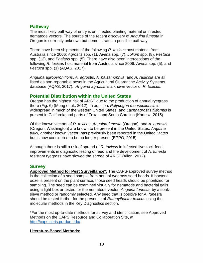
Figure 6: Rathayibacter toxicus combined host density map. Map courtesy of USDA-APHIS-PPQ-CPHST.
10
Pathway The most likely pathway of entry is on infected planting material or infected nematode vectors. The source of the recent discovery of Anguina funesta in Oregon is currently unknown but demonstrates a possible pathway. There have been shipments of the following R. toxicus host material from Australia since 2006: Agrostis spp. (1), Avena spp. (7), Lolium spp. (8), Festuca spp. (12), and Phalaris spp. (5). There have also been interceptions of the following R. toxicus host material from Australia since 2006: Avena spp. (5), and Festuca spp. (1) (AQAS, 2017). Anguina agropyronifloris, A. agrostis, A. balsamophila, and A. radicola are all listed as non-reportable pests in the Agricultural Quarantine Activity Systems database (AQAS, 2017). Anguina agrostis is a known vector of R. toxicus. Potential Distribution within the United States Oregon has the highest risk of ARGT due to the production of annual ryegrass there (Fig. 6) (Meng et al., 2012). In addition, Polypogon monspeliensis is widespread in much of the western United States, and Lachnagrostis filiformis is present in California and parts of Texas and South Carolina (Kartesz, 2015). Of the known vectors of R. toxicus, Anguina funesta (Oregon), and A. agrostis (Oregon, Washington) are known to be present in the United States. Anguina tritici, another known vector, has previously been reported in the United States but is now considered to be no longer present (EPPO, 2015). Although there is still a risk of spread of R. toxicus in infected livestock feed, improvements in diagnostic testing of feed and the development of A. funesta resistant ryegrass have slowed the spread of ARGT (Allen, 2012). Survey Approved Method for Pest Surveillance*: The CAPS-approved survey method is the collection of a seed sample from annual ryegrass seed heads. If bacterial ooze is present on the plant surface, those seed heads should be prioritized for sampling. The seed can be examined visually for nematode and bacterial galls using a light box or tested for the nematode vector, Anguina funesta, by a soak-sieve method or randomly selected. Any seed that is positive for A. funesta should be tested further for the presence of Rathayibacter toxicus using the molecular methods in the Key Diagnostics section. *For the most up-to-date methods for survey and identification, see Approved Methods on the CAPS Resource and Collaboration Site, at http://caps.ceris.purdue.edu/. Literature-Based Methods:

11
“The preferred method of survey is to collect annual ryegrass seed heads for laboratory analysis. For pasture or stubble, a recommended sampling strategy for paddocks up to 40 ha is to walk a “W” across the paddock, stopping to collect 3–5 seed heads at each of at least 100 locations along the “W”. All the collected seed heads can be combined into a single sample for testing. If the ryegrass is unevenly distributed in the paddock, then more collections should be made in the areas of greater ryegrass density. If the paddock is greater than 40 Ha (99 ac) it should be subdivided, and samples submitted for each subdivision. Relative risks of the feed causing ARGT are provided on the basis of the quantities of the bacterium detected, which is reported in terms of bacterial galls/kg” (Allen, 2012). Feed can also be physically examined for the presence of ryegrass seeds, and nematode and bacterial galls can be identified using a light box (Allen, 2012). Seedheads may also be sampled from Polypogon monspeliensis and Lachnagrostis filiformis, but infected seedheads represent a small fraction of actual infestations in these two hosts (Bertozzi, 2017, personal communication). Key Diagnostics Approved Method for Pest Surveillance*: Molecular: Although serological assays have been developed, they are not commercially available in the United States. Currently the screening for this pathogen is laborious, as there is no good selective medium, and the investigator only has colony color and a Gram stain for use in diagnostics. There are real-time PCR assays available to detect Rathayibacter toxicus in plants and for identification of four species of Anguina (funesta, tritici, agrostis, and pacificae). Although work instructions are available from CPHST Beltsville for both assays, they are being evaluated and validated.
1. TaqMan Real-time PCR for Detection of Rathayibacter toxicus in Plants and Nematodes (Postnikova et al., 2017).
o Work Instructions for the above have been developed, but CPHST Beltsville did not previously have access to a sufficiently large collection of R. toxicus isolates to properly evaluate and validate the R. toxicus assays. They are working with colleagues at Kansas State University to ring-test R. toxicus assays using their extensive collection of R. toxicus field isolates from Australia this year.
2. TaqMan Real-time PCR for Identification of Nematodes Anguina funesta,
A. tritici, A. agrostis, and A. pacificae based on Li et al. (2015). o Once CPHST Beltsville receives additional positive controls they
plan to validate and hold hands-on training workshops so that the work instructions can be used for screening.

12
*For the most up-to-date methods for survey and identification, see Approved Methods on the CAPS Resource and Collaboration Site, at http://caps.ceris.purdue.edu/. Literature-Based Methods: The International Plant Protection Convention (IPPC) has released compilation of diagnostic protocols for Anguina species (IPPC, 2017), which includes current morphological and molecular identification method for the vector A. funesta. Li et al. (2015) developed a real-time PCR which can detect the vector Anguina funesta. Atha et al. (2015) have developed a real-time PCR which can detect R. toxicus. There is reportedly real-time PCR under development by the USDA-ARS Foreign Disease-Weed Science Research Unit, Ft. Detrick, for detection of R. toxicus (Murray et al., 2014). This may be the same project as Atha et al., (2015) above. PCR-based assays for identification of Rathayibacter toxicus and the bacteriophage isolate NCPPB 3778 have also been described (Kowalski et al., 2007). Masters et al. (2006) developed an Enzyme Linked Immunosorbent Assay (ELISA) to detect R. toxicus in feeds. This ELISA has since been improved (Masters et al., 2011) and can also be used to detect the presence of the organism in rumen contents and feces. An ELISA which tests for the R. toxicus antigen is under development and shows good correlation with levels of cornytoxin in tested hay (Masters, personal communication, 2017). An ELISA that detects and quantifies the corynetoxins in feed has also been developed, but it is not as quick and is more costly than the ELISA for R. toxicus, and studies have shown a good correlation between the results from both ELISAs. Therefore, the ELISA for the bacterium is still the preferred test (Allen, 2012). Easily Confused Pests ARGT can be confused with perennial ryegrass staggers (caused by a toxin produced by an endophytic fungus in the grass), botulism, and tetanus. However, animals affected by ARGT may have periods of apparent recovery, and this distinguishes ARGT from these other diseases (Kerr and Gibb, 1997). Rathayibacter rathayi, a non-toxigenic species, causes bacterial head blight or Rathay’s disease in orchardgrass (Dactylus glomerata L.) in the United States. Symptoms look superficially similar to infection by R. toxicus, and molecular or serological confirmation of the causal agent is required to differentiate the two bacterial species (Putnam, 2009). Rathayibacter tritici causes spike blight or gumming disease in wheat, but is not known to be present in the United States. Rathayibacter iranicus also causes a gumming disease of wheat but it has been documented only from Iran and Turkey (Putnam, 2009).

13
Anguina funesta was once synonymized with congeners A. agrostis and A. wevelli. There are now at least 11 valid species of Anguina, and Powers et al. (2001) and IPPC (2017) provide methods to differentiate them using molecular analysis. In addition to molecular differences, these nematodes are generally host specific. Anguina paludicola is also a vector of R. toxicus, but it is associated with different hosts than annual ryegrass (Bertozzi and Davies, 2009, see Background Information). References Allen, J. 2012. Annual ryegrass toxicity - an animal disease caused by toxins produced by a bacterial plant pathogen. Microbiology Australia 33(1): 18-21. AQAS. 2017. Agricultural Quarantine Activity Systems. Queried March 21, 2017 from, https://aqas.aphis.usda.gov/aqas/ Atha, B. W., Sechler, A., King, J., Blanc, J., and Schneider, W. 2015. Development of a PCR assay for the tunicamycin gene cluster of Rathayibacter toxicus. (Abstr.) Phytopathology 105(Suppl. 3):S3.1. http://dx.doi.org/10.1094/PHYTO-105-9-S3.1 Bertozzi. 2017. Biology of and importance of Anguina paludicola and grass hosts in R. toxicus distribution. Personal communication to M. Sullivan on May 16, 2017 by T. Bertozzi, South Australia Museum. Bertozzi, T., and K. A. Davies. 2009. Anguina paludicola sp n. (Tylenchida: Anguinidae): The nematode associated with Rathayibacter toxicus infection in Polypogon monspeliensis and Lachnagrostis filiformis in Australia. Zootaxa, 2060: 33-46. Clarke, R., and G. Nurse. 1999. Annual Ryegrass Toxicity (Note Number: AG0129). State Government of Victoria, Department of Primary Industries. Retrieved 1/13/16 from, http://agriculture.vic.gov.au/agriculture/pests-diseases-and-weeds/animal-diseases/beef-and-dairy-cows/annual-ryegrass-toxicity EPPO. 2015. PQR - EPPO database on quarantine pests (available online). http://www.eppo.int IPPC. 2017. Anguina spp. International Plant Protection Convention ISPM 27, DP18, 38pp. Kartesz, J.T. 2015. The Biota of North America Program (BONAP). North American Plant Atlas. (http://bonap.net/napa). Chapel Hill, N.C. [maps generated from Kartesz, J.T. 2015. Floristic Synthesis of North America, Version 1.0. Biota of North America Program (BONAP). (in press)].

14
Kerr, A., and K. Gibb. 1997. Bacterial and Phytoplasma Diseases and Their Control. In J. F. Brown & H. J. Ogle (Eds.), Plant Pathogens and Plant Diseases, 465-487. NSW Australia: Rockvale Publications. Kessell, D. 2010. Annual ryegrass toxicity - current situation. Farmnote, 417, February, 2010. Government of Western Australia, Department of Agriculture and Food. Kowalski, M. C., D. Cahill, T.J. Doran and S. M. Colegate. 2007. Development and application of polymerase chain reaction-based assays for Rathayibacter toxicus and a bacteriophage associated with annual ryegrass (Lolium rigidum) toxicity. Aust. J. Exp. Agric. 47: 177–183. Li, W., Y. Zonghe, M. K. Nakhla, and A. M. Skantar. 2015. Real-time PCR for detection and identification of Anguina funesta, A. agrostis, A. tritici, and A. pacificae. Plant Disease 99: 1584-1589. Masters. A. 2017. ELISA-based screening for cornytoxins in hay. Personal communication to M. Sullivan on May 10, 2017 from A. Masters (Dept. of Agriculture and Food, Western Australia). Masters, A. M., A. R. Gregory, R. J. Evans, J. E. Speijers, and S. S. Sutherland. 2006. An enzyme-linked immunosorbent assay for the detection of Rathayibacter toxicus, the bacterium involved in annual ryegrass toxicity, in hay. Australian Journal of Agricultural Research 57: 731–742. Masters, A. M., B. Samarasinghe, M. J. Kalkhoven, G. L. den Hollander, and D. G. Palmer. 2011. Improvements to the immunoassay for detection of Rathayibacter toxicus in hay. Crop and Pasture Science 62(6): 523-530. Meng, S., S. Alderman, C. Fraley, R. Ludy, F. Sun, and N. Osterbauer. 2012. Identification of Anguina funesta from annual ryegrass seed lots in Oregon. Online. Plant Health Progress doi:10.1094/PHP-2012-1024-01-RS. Murray, T. D., I. Agarkova, S. Alderman, J. Allen, R. Bulluck, J. Chitambar, C. Divan, I. Riley, B. Schroeder, A. Sechler, and S. Subbotin. 2014. Recovery Plan for Rathayibacter Poisoning caused by Rathayibacter toxicus (syn. Clavibacter toxicus) National Plant Disease Recovery System, a cooperative project of The American Phytopathological Society and The United States Department of Agriculture, posted at http://www.ars.usda.gov/research/npdrs. Murray, T. D., B. K. Schroeder, W. Schneider, D. Luster, A. Sechler, E. E. Rogers, and S. Subbotin. 2017. Rathayibacter toxicus, other Rathayibacter species inducing Bacterial Head Blight of Grasses, and the Potential for Livestock Poisonings. Phytopathology: doi: 10.1094/PHYTO-02-17-0047-RVW.

15
Ophel, K.M., A. F. Bird, and A. Kerr. 1993. Association of bacteriophage particles with toxin production by Clavibacter toxicus, the causal agent of annual ryegrass toxicity. Phytopathology 83(6): 676-81. Postnikova, E., I. V. Agarkova, W. L. Schneider, A. J. Sechler, and I. T. Riley. 2017. Detection of Rathayibacter spp. in seeds of cereals and grasses. In: Detection of plant-pathogenic bacteria in seed and other planting material. Eds.: M’B. Fatmi, R.R. Walcott, and N.W. Schaad. Second ed. Chapter 15. APS Pres. P. 95- 101. Powers, T. O., A. L. Szalanski, P. G. Mullin, T. S. Harris, T. Bertozzi, and J. A. Griesbach. 2001. Identification of Seed Gall Nematodes of Agronomic and Regulatory Concern with PCR-RFLP of ITS1. Journal of Nematology, 33(4): 191-194. Price, P. C., J. M. Fisher, and A. Kerr. 1979. On Anguina funesta n. sp. and its association with Corynebacterium sp., in infecting Lolium rigidum. Nematologica 25: 76-85. Putnam, M. 2009. Rathayibacter toxicus, select agent [Poster]. 2nd Annual meeting of the National Plant Diagnostic Network. Miami, FL, (December 6-10, 2009). Retrieved from http://plant-clinic.bpp.oregonstate.edu/files/plant_clinic/R%20toxicus%20poster%20final%20for%20NPDN%20mtg%202009%20%282%29.pdf Riley, I.T., A. Schmitz, and P. de Silva. 2001. Anguina australis, a vector for Rathayibacter toxicus in Ehrharta longiflora. Australasian Plant Pathology 30: 171-175. USDA-APHIS. 2017. Select Agents and Toxins List. Retrieved March 8, 2017 from, http://www.selectagents.gov/SelectAgentsandToxinsList.html USDA-PCIT. 2017. Phytosanitary Certificate Issuance & Tracking System. Queried March 25, 2017 from, https://pcit.aphis.usda.gov/pcit/ This datasheet was developed by USDA-APHIS-PPQ-CPHST staff. Cite this document as: Mackesy, D. Z., and M. Sullivan. 2017. CPHST Pest Datasheet for Rathayibacter toxicus. USDA-APHISPPQ-CPHST. Reviewers:
• Irina Agarkova and Anne Vidaver, Dept. of Plant Pathology, University of Nebraska, Lincoln.
• Terry Bertozzi, Research Scientist, South Australian Museum.

16
• Anne Masters and Dieter Palmer, DAFWA Diagnostic Laboratory Services, Dept. of Agriculture and Food, Western Australia
• Tim Murray, Dept. of Plant Pathology Washington State University • Andrea Skantar, Research Molecular Biologist/Nematologist, USDA-ARS,
Beltsville, MD • Stefano Costanzo, USDA-APHIS-PPQ-CPHST, Beltsville, MD
Update History: March 2017: Draft version written May 2017: Datasheet sent out for subject matter review. May 2017: Datasheet published on CAPS Research and Collaboration website.