RARE/Turbo Spin Echo Imaging with Simultaneous Multislice...
10
RAPID COMMUNICATION RARE/Turbo Spin Echo Imaging with Simultaneous Multislice Wave-CAIPI Borjan A. Gagoski, 1,2y * Berkin Bilgic, 2,3y * Cornelius Eichner, 3,4 Himanshu Bhat, 5 P. Ellen Grant, 1,2 Lawrence L. Wald, 2,3,6 and Kawin Setsompop 2,3 Purpose: To enable highly accelerated RARE/Turbo Spin Echo (TSE) imaging using Simultaneous MultiSlice (SMS) Wave- CAIPI acquisition with reduced g-factor penalty. Methods: SMS Wave-CAIPI incurs slice shifts across simulta- neously excited slices while playing sinusoidal gradient wave- forms during the readout of each encoding line. This results in an efficient k-space coverage that spreads aliasing in all three dimensions to fully harness the encoding power of coil sensi- tivities. The novel MultiPINS radiofrequency (RF) pulses dra- matically reduce the power deposition of multiband (MB) refocusing pulse, thus allowing high MB factors within the Specific Absorption Rate (SAR) limit. Results: Wave-CAIPI acquisition with MultiPINS permits whole brain coverage with 1 mm isotropic resolution in 70 s at effec- tive MB factor 13, with maximum and average g-factor penal- ties of g max ¼ 1.34 and g avg ¼ 1.12, and without R penalty. With blipped-CAIPI, the g-factor performance was degraded to g max ¼ 3.24 and g avg ¼ 1.42; a 2.4-fold increase in g max rela- tive to Wave-CAIPI. At this MB factor, the SAR of the Multi- Band and PINS pulses are 4.2 and 1.9 times that of the MultiPINS pulse, while the peak RF power are 19.4 and 3.9 times higher. Conclusion: Combination of the two technologies, Wave-CAIPI and MultiPINS pulse, enables highly accelerated RARE/TSE imaging with low SNR penalty at reduced SAR. Magn Reson Med 000:000–000, 2015. V C 2015 Wiley Periodicals, Inc. Key words: simultaneous multislice, multiband; Wave-CAIPI; MultiPINS; CAIPIRINHA; RARE/TSE/FSE INTRODUCTION Rapid acquisition with refocusing echoes (RARE) (1), also known as turbo spin echo (TSE) or fast spin echo (FSE), is by far the most commonly used image readout sequence in clinical imaging. This versatile tool can be tailored to provide T1, T2, and proton density weighted acquisitions. It provides rapid imaging compared with conventional single-echo spin-echo by acquiring multi- ple phase encoding lines per repetition time (TR). Fol- lowing a 90 excitation, multiple 180 radiofrequency (RF) pulses are used to continually refocus the transverse magnetization, thereby recording multiple spin echos. While this strategy permits efficient sampling of k-space, the application of large number of refocusing RF pulses results in increased power deposition to the target tissue. It is possible to mitigate this issue by simply reducing the flip angle of the refocusing pulses (2), or by fully refocusing the echo signals acquired at the center of k- space, and using lower flip angles to encode higher spa- tial frequencies (3). RARE acquisition has been sped up though the use of in-plane parallel imaging, which undersamples two- dimensional (2D) k-space to reduce the number of phase encoding lines that needs to be acquired. However, at acceleration factor of 3 or above, the g-factor noise ampli- fication in reconstructing this 2D undersampled acquisi- tion and the R noise penalty becomes too large. The RARE sequence can be further accelerated with the use of simultaneous multislice (SMS) imaging (4–9), wherein multiple slices are excited, refocused and read-out simultaneously. Such acquisition enables acceleration without reducing the number of k-space lines and hence without the R penalty (9). Furthermore, SMS imaging has been shown to be amenable to controlled aliasing in two dimensions (y-z) by means of CAIPIRINHA tech- nique, or CAIPI for short, to increase distance between aliased voxels and significantly reduce g-factor penalty. However, the application of SMS in RARE imaging fur- ther aggravates the specific absorption rate (SAR) issue because conventional multiband (MB) RF pulses are gen- erated by means of superposition of single band sinc RF pulses, which makes the total deposited RF power pro- portional to the MB factor (10). A novel proposal termed power independent of number of slices (PINS) (11) cre- ates a periodic excitation pattern obtained by undersam- pling of single-slice RF pulses. In contrast to conventional MB excitation, total power deposition of PINS is independent of the MB factor, thus making it particularly useful for ultra high field applications, slice accelerated spin echo functional MRI and diffusion 1 Fetal-Neonatal Neuroimaging & Developmental Science Center, Boston Children’s Hospital, Boston, Massachusetts, USA. 2 Department of Radiology, Harvard Medical School, Boston, Massachu- setts, USA. 3 Athinoula A. Martinos Center for Biomedical Imaging, Massachusetts Gen- eral Hospital, Charlestown, Massachusetts, USA. 4 Max Planck Institute for Human Cognitive and Brain Sciences, Leipzig, Germany. 5 Siemens Medical Solutions USA Inc., Charlestown, Massachusetts, USA. 6 Harvard-MIT Health Sciences and Technology, Cambridge, Massachu- setts, USA Grant sponsor: NIH; Grant numbers: R00EB012107; P41RR14075; R01EB017337; Grant sponsor: NIH Blueprint for Neuroscience; Grant num- ber: 1U01MH093765 (Human Connectome Project). *Correspondence to: Borjan A. Gagoski, PhD, 1 Autumn Street, AU457, Boston, MA, 02215. E-mail: [email protected] and Berkin Bilgic, PhD, Building 75, 13th Street, Charlestown, MA, 02129 Additional Supporting Information may be found in the online version of this article. y Drs. Gagoski and Bilgic contributed equally to this work. Received 10 October 2014; revised 21 December 2014; accepted 22 December 2014 DOI 10.1002/mrm.25615 Published online 00 Month 2014 in Wiley Online Library (wileyonlinelibrary. com). Magnetic Resonance in Medicine 00:00–00 (2015) V C 2015 Wiley Periodicals, Inc. 1
Transcript of RARE/Turbo Spin Echo Imaging with Simultaneous Multislice...
RAPIDCOMMUNICATION
RARE/Turbo Spin Echo Imaging with SimultaneousMultislice Wave-CAIPI
Borjan A. Gagoski,1,2y* Berkin Bilgic,2,3y* Cornelius Eichner,3,4 Himanshu Bhat,5
P. Ellen Grant,1,2 Lawrence L. Wald,2,3,6 and Kawin Setsompop2,3
Purpose: To enable highly accelerated RARE/Turbo Spin Echo(TSE) imaging using Simultaneous MultiSlice (SMS) Wave-
CAIPI acquisition with reduced g-factor penalty.Methods: SMS Wave-CAIPI incurs slice shifts across simulta-
neously excited slices while playing sinusoidal gradient wave-forms during the readout of each encoding line. This results inan efficient k-space coverage that spreads aliasing in all three
dimensions to fully harness the encoding power of coil sensi-tivities. The novel MultiPINS radiofrequency (RF) pulses dra-
matically reduce the power deposition of multiband (MB)refocusing pulse, thus allowing high MB factors within theSpecific Absorption Rate (SAR) limit.
Results: Wave-CAIPI acquisition with MultiPINS permits wholebrain coverage with 1 mm isotropic resolution in 70 s at effec-tive MB factor 13, with maximum and average g-factor penal-
ties of gmax¼1.34 and gavg¼1.12, and without �R penalty.With blipped-CAIPI, the g-factor performance was degraded
to gmax¼3.24 and gavg¼1.42; a 2.4-fold increase in gmax rela-tive to Wave-CAIPI. At this MB factor, the SAR of the Multi-Band and PINS pulses are 4.2 and 1.9 times that of the
MultiPINS pulse, while the peak RF power are 19.4 and 3.9times higher.
Conclusion: Combination of the two technologies, Wave-CAIPIand MultiPINS pulse, enables highly accelerated RARE/TSEimaging with low SNR penalty at reduced SAR. Magn ResonMed 000:000–000, 2015. VC 2015 Wiley Periodicals, Inc.
Key words: simultaneous multislice, multiband; Wave-CAIPI;MultiPINS; CAIPIRINHA; RARE/TSE/FSE
INTRODUCTION
Rapid acquisition with refocusing echoes (RARE) (1),also known as turbo spin echo (TSE) or fast spin echo(FSE), is by far the most commonly used image readoutsequence in clinical imaging. This versatile tool can betailored to provide T1, T2, and proton density weightedacquisitions. It provides rapid imaging compared withconventional single-echo spin-echo by acquiring multi-ple phase encoding lines per repetition time (TR). Fol-lowing a 90� excitation, multiple 180� radiofrequency(RF) pulses are used to continually refocus the transversemagnetization, thereby recording multiple spin echos.While this strategy permits efficient sampling of k-space,the application of large number of refocusing RF pulsesresults in increased power deposition to the target tissue.It is possible to mitigate this issue by simply reducingthe flip angle of the refocusing pulses (2), or by fullyrefocusing the echo signals acquired at the center of k-space, and using lower flip angles to encode higher spa-tial frequencies (3).
RARE acquisition has been sped up though the use ofin-plane parallel imaging, which undersamples two-dimensional (2D) k-space to reduce the number of phaseencoding lines that needs to be acquired. However, atacceleration factor of 3 or above, the g-factor noise ampli-fication in reconstructing this 2D undersampled acquisi-tion and the �R noise penalty becomes too large. TheRARE sequence can be further accelerated with the useof simultaneous multislice (SMS) imaging (4–9), whereinmultiple slices are excited, refocused and read-outsimultaneously. Such acquisition enables accelerationwithout reducing the number of k-space lines and hencewithout the �R penalty (9). Furthermore, SMS imaginghas been shown to be amenable to controlled aliasing intwo dimensions (y-z) by means of CAIPIRINHA tech-nique, or CAIPI for short, to increase distance betweenaliased voxels and significantly reduce g-factor penalty.However, the application of SMS in RARE imaging fur-ther aggravates the specific absorption rate (SAR) issuebecause conventional multiband (MB) RF pulses are gen-erated by means of superposition of single band sinc RFpulses, which makes the total deposited RF power pro-portional to the MB factor (10). A novel proposal termedpower independent of number of slices (PINS) (11) cre-ates a periodic excitation pattern obtained by undersam-pling of single-slice RF pulses. In contrast toconventional MB excitation, total power deposition ofPINS is independent of the MB factor, thus making itparticularly useful for ultra high field applications, sliceaccelerated spin echo functional MRI and diffusion
1Fetal-Neonatal Neuroimaging & Developmental Science Center, BostonChildren’s Hospital, Boston, Massachusetts, USA.2Department of Radiology, Harvard Medical School, Boston, Massachu-setts, USA.3Athinoula A. Martinos Center for Biomedical Imaging, Massachusetts Gen-eral Hospital, Charlestown, Massachusetts, USA.4Max Planck Institute for Human Cognitive and Brain Sciences, Leipzig,Germany.5Siemens Medical Solutions USA Inc., Charlestown, Massachusetts, USA.6Harvard-MIT Health Sciences and Technology, Cambridge, Massachu-setts, USA
Grant sponsor: NIH; Grant numbers: R00EB012107; P41RR14075;R01EB017337; Grant sponsor: NIH Blueprint for Neuroscience; Grant num-ber: 1U01MH093765 (Human Connectome Project).
*Correspondence to: Borjan A. Gagoski, PhD, 1 Autumn Street, AU457,Boston, MA, 02215. E-mail: [email protected] andBerkin Bilgic, PhD, Building 75, 13th Street, Charlestown, MA, 02129
Additional Supporting Information may be found in the online version ofthis article.yDrs. Gagoski and Bilgic contributed equally to this work.
Received 10 October 2014; revised 21 December 2014; accepted 22December 2014
DOI 10.1002/mrm.25615Published online 00 Month 2014 in Wiley Online Library (wileyonlinelibrary.com).
Magnetic Resonance in Medicine 00:00–00 (2015)
VC 2015 Wiley Periodicals, Inc. 1

imaging experiments (12,13). PINS pulses were alsorecently applied to the RARE sequence (14) to enableSMS-RARE imaging at 7T. This previous study usedBlipped-CAIPI acquisition that creates interslice shiftsacross collapsed slices for improved parallel imaging(4,7), which yielded high quality images at an MB accel-eration factor of 8 with 2 mm and 3 mm slice thickness.
In this contribution, we push the SMS accelerationeven further to reach an MB factor of 15, thereby ena-bling whole brain RARE acquisition with an echo trainlength (ETL) of 12 and 1 mm isotropic resolution in70 s. With this MB15 implementation, the coronal slicefield of view (FOV) was set to 255 mm and becausetwo of the excited slices in each slice group remainoutside the head for our test subject, the effective MBfactor (MBeff) in this case is 13. The two limiting fac-tors that impede attaining such acceleration factors are(i) the increased power deposition needed for highquality PINS excitation and refocusing of thin sliceimaging at short pulse duration of 5–6 ms required forefficient RARE imaging, and (ii) the substantial g-factorpenalty that would be incurred by existing parallelimaging methods. We address both of these issues byusing the novel MultiPINS (15) RF pulses that enablelow SAR refocusing with Wave-CAIPI acquisition (16)that fully harnesses the spatial variation in coil sensi-tivity profiles to mitigate the g-factor penalty. Thecombination of the two technologies yield maximumand average g-factors of gmax¼ 1.34 and gavg¼ 1.12 witha commercial 32-channel brain array while substan-tially reducing imaging at MBeff�13 under SAR safetyconstraint at 3T.
The specific contributions of this work are: (i) Usingnovel MultiPINS refocusing pulses to dramaticallyreduce the RF power deposition in RARE experiments,thus permitting high MB factors to be achieved in vivowithin the SAR limit. (ii) Deploying Wave-CAIPI acquisi-tion/reconstruction framework in SMS RARE imaging toachieve MBeff factor 13 with reduced g-factor penalty.This enables a whole brain T2-weighted acquisition at1 mm isotropic resolution in 70 s. (iii) Releasing supple-mentary Matlab code that replicates in vivo Wave-CAIPIand blipped-CAIPI reconstructions with MBeff factor 13at martinos.org/�berkin/software.
METHODS
MultiPINS: Low Power RF Pulse for SMS Excitation andRefocusing
Conventional SMS excitation involves MB pulses thatare formed by the addition of multiple single-slice RFwaveforms (17). The drawback of this superposition isthe linear increase in transmitted energy and peak powerdeposition with the MB factor. Peak power of an MBpulse can be reduced through an optimized RF phaseschedule (18) or a pulse time-shifting scheme (19), butthese techniques do not reduce SAR. The VERSE algo-rithm (20) reduces both peak power and SAR, but canresult in unacceptable slice-profile distortion and longpulse duration at high MB factors.
In contrast, PINS pulses (11) create a periodic excita-tion pattern obtained by undersampling of a single-slice
RF pulse, which makes the energy deposition independ-ent of the number of excited slices. As the PINS pulsesare divided into distinct time-bands containing RF sub-pulses to achieve such undersampling, fast traversal ofexcitation k-space becomes difficult. These pulses can belengthy for thin slice imaging, leading to undesirablylong echo-train length in RARE imaging and large off-resonance slice shift. PINS pulses can be shortened byreducing the sub-pulse duration, but this comes at thecost of increased SAR and peak RF power.
In MultiPINS (15), MB and PINS pulses are synergisti-cally combined to reduce energy transmission and peakRF power, which also permits shorter pulses withoutexceeding SAR limits. We have demonstrated the effi-cacy of MultiPINS for high-resolution diffusion imagingat 7 Tesla (T), where SAR was reduced by 51% com-pared with PINS excitation (15). Herein, we demonstrateits efficacy for SMS-RARE imaging with 1mm slice thick-ness. While these MultiPINS are designed to excite 15slices simultaneously within a slice FOV of 255 mm, theeffective MB factor is 13 because two of the excited sli-ces usually remain outside the head due to large FOV.
Because PINS sub-pulses are played only between thegradients blips, MultiPINS uses the time interval duringthe blips to play MB pulses. MultiPINS uses this strategywith an optimal mixing ratio of the two types of pulses,leading to reduced peak RF power and SAR specifica-tion. Figure 1 further demonstrates the application ofMultiPINS to attain high SMS acceleration factor.
Wave-CAIPI for Highly Accelerated SMS Imaging
CAIPIRINHA (abbreviated as CAIPI) controlled aliasingtechnique creates interslice shifts across simultaneouslyexcited slices to improve unaliasing in SMS imaging (4).This is achieved by modulating the phase of the readoutlines to create a phase ramp in k-space, which leads to aslice shift in the image space. Such modulation can begenerated either by performing RF excitation with dis-tinct phase cycling schemes across the different slices(4), or by playing Gz gradient blips that incur a phaseramp in z (slice) direction (7). CAIPI acquisition hasbeen successfully deployed in volumetric (21), steadystate free precession (SSFP) (22), diffusion (23), perfu-sion (24–26), and functional imaging (27). Clinical inves-tigations of this strategy have demonstrated good imagequality up to an acceleration factor of 4 in structuralimaging (28,29). Acceleration factor of up to 8–9 hadbeen shown (14), but such acquisitions suffer from g-factor penalty that can reduce SNR by as much as 30–40%, particularly in the central brain region whereintrinsic SNR of the coil array is low (16).
Wave-CAIPI overcomes this issue by efficient samplingof k-space that spreads the aliasing evenly in all threedimensions to fully use the variation in the coil sensitiv-ity profiles, thereby providing dramatic scan time reduc-tion at negligible SNR and artifact penalties. We havepreviously deployed Wave-CAIPI in 3D-GRE imaging,and demonstrated that maximum g-factor gmax remainsbelow 1.10 for R¼nine-fold acceleration (16). Herein, weextend the Wave-CAIPI concept to multiband imagingusing the SMS 3D k-space formalism (30).
2 Gagoski et al.
It is possible to represent SMS imaging as undersam-pling in 3D k-space (30,31) by relying on the fact thatalternating gradient blips (or phase of RF pulses) conductDiscrete Fourier Transform (DFT) along the slice axis.Recognizing the distance between the simultaneouslyexcited slices Dz (i.e., slice separation) as the inherentslice resolution, the k-space sampling period Dkz needs tobe 1/(N�Dz) to fully sample the N slices. In the presenceof field-of-view (FOV) shifting in the phase encodingdirection by an amount FOV/L, the blipped-CAIPI sam-pling period becomes Dkz
blip¼ 1/(L�Dz), thus effectivelyundersampling the SMS 3D k-space. The staggered k-space sampling pattern that gives rise to slice shifting isdemonstrated in Figure 2a for MB factor 15 (N¼ 15), withFOV/4 shift (L¼4). We note that the CAIPIRINHAmethod, which uses RF phase modulation, also createsthe same k-space shift as the blipped-CAIPI method.
The effect of playing the sinusoidal Wave gradients dur-ing the readout period of each RARE/TSE phase encodingline is to create a helical k-space trajectory that increasesthe coverage in the undersampled SMS 3D k-space. Whenthe centers of each helix is staggered in the blipped-CAIPImanner, the resulting trajectory spreads the undersam-pling to all three k-space axes, rather than fully samplingthe readout dimension and sparsely sampling the ky–kz
plane (Fig. 2b). This allows Wave-CAIPI to use all degreesof freedom in the coil sensitivity maps and improve the g-factor. If we look at this trajectory in the ky–kz plane byfixing a particular kx position, the relative position ofeach sample will be identical to that of blipped-CAIPIsampling, meaning that the helical trajectory of each kx
readout line does not intersect with the other kx lines in
the Wave-CAIPI acquisition. Wave-CAIPI builds upon theBunched Phase Encoding (BPE) (32) and zigzag samplingstrategies (33,34) to improve the k-space coverage alongphase encoded dimensions by oversampling in the read-out direction. A similar strategy has also been applied toGRASE (gradient and spin echo) imaging (35,36), whereinzigzag sampling allowed distribution of T2 modulationsand off-resonance effects to different axes (vGRASE) (37).These methods, however, require either gridding or multi-ple GRAPPA (38) kernels for reconstruction.
As further demonstrated in Figure 2b, the non-Cartesiantrajectory in Wave-CAIPI acquisition can be expressed asconvolution in Cartesian space, thereby obviating the needfor gridding or nonuniform Fourier Transform. As detailedin Bilgic et al (16), each readout line is convolved with adifferent point spread function (PSF) to create differentamount of voxel spreading as a function of space. Notethat the spreading effect is only along the readout (x) direc-tion, with the amount of spreading varies based on the (y,z) coordinate of the voxels. Combined with interslice FOVshifts, the sinusoidal Wave gradients create highly spread-out data in image space. This increases the average dis-tance between collapsed voxels in the SMS acquisitionand substantially improves the parallel imaging capability.
Characterization of Off-Resonance Effects for Wave-CAIPI
Wave-CAIPI provides a rapid acquisition without undesir-able image distortion/blurring from B0 inhomogeneity.This is because Wave-CAIPI traverses k-space in the read-out direction with the same constant rate as conventionalacquisitions, with B0 inhomogeneity-related phase
FIG. 1. Comparison of MultiBand, PINS and MultiPINS refocusing pulses at MB-15 and 1-mm slice thickness. a: Compared with Multi-
PINS, MultiBand and PINS designs have increased SAR and peak power deposition (4.2� and 1.9� SAR, and 19.4� and 3.9� peakpower in this example). b: Gradient waveforms and excitation k-space trajectories for MultiBand and MultiPINS. Despite differences inthe gradient waveforms, the k-space traversals are very similar; minor deviations from linear transversal of MultiPINS are shown in the
zoomed panel. As such, MultiPINS exhibits good off-resonance performance with negligible slice distortion. c: Similar refocusing profilesof MultiBand and MultiPINS, with additional sidelobes outside the volume of interest for MultiPINS from the PINS component of the
pulse. d: Zoomed in comparison of profiles at 0 and 100 Hz. The profile at 100 Hz contains a simple shift essentially identical to Multi-Band profile at the same frequency. No slice profile distortions can be observed.
RARE/TSE with SMS Wave-CAIPI 3
evolving solely as a function of kx. To validate this, awater phantom was scanned at 3T using conventionaland Wave-CAIPI 3D-GRE sequences with 2 mm isotropicresolution, 96 � 96 � 60 matrix size, FOV¼ 192 � 192 �120, TR/TE¼20/10 ms and 100 Hz/pixel bandwidth.Both datasets were fully sampled and acquired in thepresence of large B0 off-resonance (500 Hz) imposed bymanually offsetting the B0 shims. To serve as groundtruth, normal GRE data were also collected on-resonancewith otherwise identical parameters. B0 mapping wasconducted using three sequentially acquired normal GREvolumes with echo times TE1/TE2/TE3¼ 9.5/10/10.5 ms.
Data Acquisition and Reconstruction
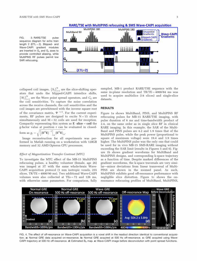
RARE/TSE sequence was modified to include sinusoidalGy and Gz gradient modules as well as gradient blips thatcreate interslice shifts used in Wave-CAIPI trajectories.The pulse sequence diagram including the 90� MB and
180� MultiPINS pulses is depicted in Figure 3, where theecho train length (ETL) is set to 2 for display purposes.
A healthy subject (male, age 33) was imaged on SiemensTim Trio 3T system in accordance with the institutionalreview board protocol to acquire MB-15 RARE/TSE datawith 1 mm isotropic resolution. The effective MB factor,MBeff, was 13 because two slices in each slice groupremained outside the head. For signal reception, 32-channelSiemens head coil was used. The bandwidth was chosen tobe similar to the protocol in Norris et al (14), and was set to130 Hz/pixel and ETL¼ 12. The in-plane matrix size was256 � 192 with 255 coronal slices, and the repetition (TR)and echo times (TE) were TR/TE¼ 4000/90 ms with an echospacing of 18 ms. Using the same parameter setting, Blipped-CAIPI and Wave-CAIPI data were acquired within an acqui-sition time of Tacq¼ 70 s. Because SMS imaging excites mul-tiple slices simultaneously and records them for the sameduration in an unaccelerated 2D scan (9), it does not incurthe intrinsic �R SNR penalty that occurs when the time win-dow of signal accumulation is reduced. The SNR advantageof eliminating the �R penalty is particularly significant athigh acceleration: the �R penalty alone would have led to a�3.9-fold drop in SNR for 15-fold in-plane acceleration. Assuch, the present study used SMS acceleration only.
For comparison, MB-1, fully sampled, product RARE/TSEdata were acquired. Owing to the stepwise increments in theecho time, TE was set to 84 ms, while the echo spacing was16.8 ms. Only 14 slices could be acquired within the sameacquisition time of 70 s. All other parameters were identicalto the Blipped- and Wave-CAIPI acquisitions. The WavePSFs used in slice unaliasing reconstruction were deter-mined using the k-space trajectory estimation scheme in Bil-gic et al (16). For the PSF calibration, data were acquired ona phantom before the in vivo acquisition.
Coil sensitivity profiles were estimated using low-resolution (2 � 2 � 3 mm3) TSE data, and were iterativelyrefined using J-SENSE algorithm (39) with 7th order poly-nomial fitting for smoothing. As presented in the flow-chart in Supporting Figure S1, which is available online,estimated PSFs and coil sensitivities are used in general-ized SENSE reconstruction (40,41) to unalias simultane-ously excited slices in the Wave-CAIPI acquisition.
g-Factor Map Computation
Follows the g-factor formulation for SENSE reconstruc-tion outlined in Pruessmann et al (40), and has beenmodified it to incorporate the Wave point spread func-tions. Specifically, Wave-CAIPI decouples the recon-struction into subproblems that are solvedindependently for each set of collapsed readout rows. AtMB15, we solve for one readout row from 15 slices at atime, and loop over phase encoding positions and slicegroups. This leads to the generalized SENSE model,
W1C11S1 . . . WNC1NSN
� � �
W1CM1S1 � � � WNCMNSN
2664
3775
slice1
�
sliceN
2664
3775 ¼
coil1
�
coilM
2664
3775 [1]
where slicej
� �N
j¼1are the readout rows from each slice
within the collapsed slice group, coilif gMi¼1 are the
FIG. 2. K-space and image space view of controlled aliasing withBlipped- and Wave-CAIPI. a: Blipped-CAIPI SMS imaging can bereformulated as undersampling in 3D k-space with staggered
sampling, which induces slice shifts in the phase encoding direc-tion. b: Wave-CAIPI traverses a helical trajectory that distributes
the undersampling to all k-space axes along with staggered sam-pling of PE. The helical trajectory incurs voxel spreading in imagespace in the readout direction that increases the average distance
across collapsed voxels, thus improving g-factor substantially.Non-Cartesian Wave trajectory can be represented as convolution
with a spatially varying point spread function that permits Carte-sian reconstruction.
4 Gagoski et al.
collapsed coil images, Sj
� �N
i¼1are the slice-shifting oper-
ators that undo the blipped-CAIPI interslice shifts,
Wj
� �N
i¼1are the Wave point spread operators, and Cij are
the coil sensitivities. To capture the noise correlationacross the receive channels, the coil sensitivities and thecoil images are prewhitened with the inverse square root
of the covariance matrix, C�1=2. For the current experi-ments, RF pulses are designed to excite N¼ 15 slicessimultaneously and M¼ 32 coils are used for reception.Compactly representing this system as E � slice ¼ coil theg-factor value at position r can be evaluated in closed-
form as gr ¼ffiffiffiffiffiffiffiffiffiffiffiffiffiffiffiffiffiffiffiffiffiffiffiffiffiffiffiffiffiffiffiffiffiffiffiffiffiffiffiffiffiffiffi
EH E� ��1h i
rrEH E� �
rr
r.
Image reconstruction for all experiments was per-formed in Matlab running on a workstation with 128GBmemory and 32 AMD Opteron CPU processors.
Effect of Magnetization Transfer Contrast (MTC)
To investigate the MTC effect of the MB-15 MultiPINSrefocusing pulses, a healthy volunteer (female, age 26)was imaged at 3T with the same whole-brain Wave-CAIPI acquisition protocol (1 mm isotropic voxels, 255slices, TR/TE¼ 4000/90 ms). Two additional Wave-CAIPIvolumes were also collected at TEs¼ 72 and 126 ms,with otherwise same parameters. For comparison, fully
sampled, MB-1 product RARE/TSE sequence with thesame in-plane resolution and TR/TE¼ 4000/84 ms wasused to acquire multislice (14 slices) and single-slicedatasets.
RESULTS
Figure 1a shows MultiBand, PINS, and MultiPINS RFrefocusing pulses for MB-15 RARE/TSE imaging, withpulse duration of 6 ms and time-bandwidth product of2.4, on the same order as in single slice RF in clinicalRARE imaging. In this example, the SAR of the Multi-Band and PINS pulses are 4.2 and 1.9 times that of theMultiPINS pulse, while the peak power (proportional tosquare of maximum voltage) were 19.4 and 3.9 timeshigher. The MultiPINS pulse was the only one that couldbe used for in vivo MB-15 SMS-RARE imaging withoutexceeding the SAR limit (results in Figures 5 and 6). Fig-ure 1b shows gradient waveforms for MultiBand andMultiPINS designs, and corresponding k-space trajectoryas a function of time. Despite marked differences of thegradient waveforms, the k-space traversals are very simi-lar—minor deviations from linear transversal of Multi-PINS are shown in the zoomed panel. As such,MultiPINS exhibits good off-resonance performance withnegligible slice distortion. Figure 1c shows the on-resonance refocusing profiles of MultiBand, MultiPINS,
FIG. 3. RARE/TSE pulse
sequence diagram for echo trainlength 2 (ETL¼2). Blipped- andWave-CAIPI gradient modules
are inserted in Gy and Gz axes toprovide controlled aliasing, whileMultiPINS RF pulses permit low
SAR refocusing.
FIG. 4. The effect of off-resonance on Wave-CAIPI acquisition is a voxel shift in the readout direction identical to conventional acquisi-tion. a: Normal GRE data acquired on-resonance. b: Normal GRE acquired at 500 Hz off-resonance. c: GRE acquired using Wave-
CAIPI trajectory at 500 Hz off-resonance. d: Estimated B0 map. e: Wave-CAIPI image before deconvolution with point spread functions.
RARE/TSE with SMS Wave-CAIPI 5
and ideal excitation, while Figure 1d presents a zoomedin comparison of profiles at 0 and 100 Hz. The profile at100 Hz contains mostly a simple shift essentially identi-cal to MultiBand’s profile at the same frequency (correla-tion coeff.> 0.99). The slice profile also shows partialrefocusing outside the volume of interest resulting fromthe PINS component of the MultiPINS pulse. This wouldaffect axial imaging, but can be overcome using a MBexcitation before MultiPINS refocusing, similar to whatwe have demonstrated for PINS diffusion imaging (15).
The effect of off-resonance acquisition for Wave-CAIPIis demonstrated in Figure 4, where the Wave-CAIPIreconstruction results in a sharp image with the sameimage shift of five voxels along the readout direction asthe standard acquisition.
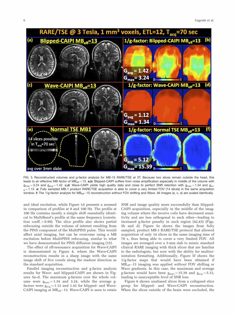
Parallel imaging reconstruction and g-factor analysisresults for Wave- and blipped-CAIPI are shown in Fig-ures 5a–d. The maximum g-factors over the whole vol-ume were gmax¼ 1.34 and 3.24, while the average g-factors were gavg¼ 1.12 and 1.42 for blipped- and Wave-CAIPI imaging at MBeff�13. Wave-CAIPI is seen to retain
SNR and image quality more successfully than blipped-CAIPI acquisition, especially in the middle of the imag-ing volume where the receive coils have decreased sensi-tivity and are less orthogonal to each other—leading toincreased g-factor penalty in such region (42,43) (Figs.5b and d). Figure 5e shows the images from fullysampled, product MB-1 RARE/TSE protocol that allowedacquisition of only 14 slices in the same imaging time of70 s, thus being able to cover a very limited FOV. Allimages are averaged over a 3-mm slab to mimic standardclinical RARE imaging with thick slices that are familiarto the radiologists, but now with the ability for multior-ientation formatting. Additionally, Figure 5f shows the1/g-factor maps that would have been obtained ifMBeff�13 imaging was applied without FOV shifting orWave gradients. In this case, the maximum and averageg-factors would have been gmax¼ 15.39 and gavg¼5.12,leading to unacceptable level of SNR loss.
Figure 6 shows unaliased slices from a collapsed slicegroup for blipped- and Wave-CAIPI reconstruction.When the slices outside of the brain were excluded, the
FIG. 5. Reconstructed volumes and g-factor analysis for MB-15 RARE/TSE at 3T. Because two slices remain outside the head, thisleads to an effective MB factor of MBeff¼13. a,b: Blipped-CAIPI suffers from noise amplification especially in middle of the volume withgmax¼3.24 and gavg¼1.42. c,d: Wave-CAIPI yields high quality data and close to perfect SNR retention with gmax¼1.34 and gav-
g¼1.12. e: Fully sampled MB-1 product RARE/TSE acquisition is able to cover a very limited FOV (14 slices) in the same acquisitionwindow. f: The 1/g-factor analysis for MBeff�15 reconstruction without FOV shifting and Wave. All images (a, c, e) are scaled identically.
6 Gagoski et al.
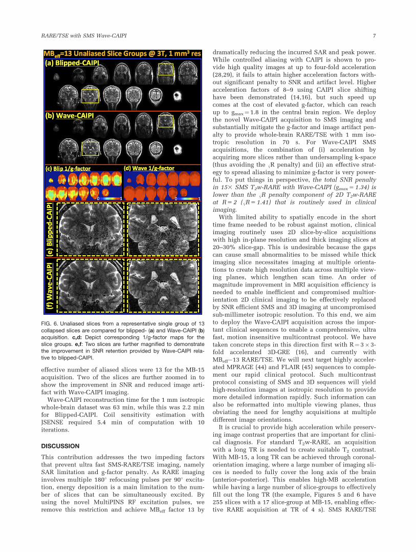
effective number of aliased slices were 13 for the MB-15acquisition. Two of the slices are further zoomed in toshow the improvement in SNR and reduced image arti-fact with Wave-CAIPI imaging.
Wave-CAIPI reconstruction time for the 1 mm isotropicwhole-brain dataset was 63 min, while this was 2.2 minfor Blipped-CAIPI. Coil sensitivity estimation withJSENSE required 5.4 min of computation with 10iterations.
DISCUSSION
This contribution addresses the two impeding factorsthat prevent ultra fast SMS-RARE/TSE imaging, namelySAR limitation and g-factor penalty. As RARE imaginginvolves multiple 180� refocusing pulses per 90� excita-tion, energy deposition is a main limitation to the num-ber of slices that can be simultaneously excited. Byusing the novel MultiPINS RF excitation pulses, weremove this restriction and achieve MBeff factor 13 by
dramatically reducing the incurred SAR and peak power.While controlled aliasing with CAIPI is shown to pro-vide high quality images at up to four-fold acceleration(28,29), it fails to attain higher acceleration factors with-out significant penalty to SNR and artifact level. Higheracceleration factors of 8–9 using CAIPI slice shiftinghave been demonstrated (14,16), but such speed upcomes at the cost of elevated g-factor, which can reachup to gmax¼1.8 in the central brain region. We deploythe novel Wave-CAIPI acquisition to SMS imaging andsubstantially mitigate the g-factor and image artifact pen-alty to provide whole-brain RARE/TSE with 1 mm iso-tropic resolution in 70 s. For Wave-CAIPI SMSacquisitions, the combination of (i) acceleration byacquiring more slices rather than undersampling k-space(thus avoiding the �R penalty) and (ii) an effective strat-egy to spread aliasing to minimize g-factor is very power-ful. To put things in perspective, the total SNR penaltyin 153 SMS T2w-RARE with Wave-CAIPI (gmax 5 1.34) islower than the �R penalty component of 2D T2w-RAREat R 5 2 (�R 5 1.41) that is routinely used in clinicalimaging.
With limited ability to spatially encode in the shorttime frame needed to be robust against motion, clinicalimaging routinely uses 2D slice-by-slice acquisitionswith high in-plane resolution and thick imaging slices at20–30% slice-gap. This is undesirable because the gapscan cause small abnormalities to be missed while thickimaging slice necessitates imaging at multiple orienta-tions to create high resolution data across multiple view-ing planes, which lengthen scan time. An order ofmagnitude improvement in MRI acquisition efficiency isneeded to enable inefficient and compromised multior-ientation 2D clinical imaging to be effectively replacedby SNR efficient SMS and 3D imaging at uncompromisedsub-millimeter isotropic resolution. To this end, we aimto deploy the Wave-CAIPI acquisition across the impor-tant clinical sequences to enable a comprehensive, ultrafast, motion insensitive multicontrast protocol. We havetaken concrete steps in this direction first with R¼ 3� 3-fold accelerated 3D-GRE (16), and currently withMBeff�13 RARE/TSE. We will next target highly acceler-ated MPRAGE (44) and FLAIR (45) sequences to comple-ment our rapid clinical protocol. Such multicontrastprotocol consisting of SMS and 3D sequences will yieldhigh-resolution images at isotropic resolution to providemore detailed information rapidly. Such information canalso be reformatted into multiple viewing planes, thusobviating the need for lengthy acquisitions at multipledifferent image orientations.
It is crucial to provide high acceleration while preserv-ing image contrast properties that are important for clini-cal diagnosis. For standard T2w-RARE, an acquisitionwith a long TR is needed to create suitable T2 contrast.With MB-15, a long TR can be achieved through coronal-orientation imaging, where a large number of imaging sli-ces is needed to fully cover the long axis of the brain(anterior–posterior). This enables high-MB accelerationwhile having a large number of slice-groups to effectivelyfill out the long TR (the example, Figures 5 and 6 have255 slices with a 17 slice-group at MB-15, enabling effec-tive RARE acquisition at TR of 4 s). SMS RARE/TSE
FIG. 6. Unaliased slices from a representative single group of 13
collapsed slices are compared for blipped- (a) and Wave-CAIPI (b)acquisition. c,d: Depict corresponding 1/g-factor maps for the
slice groups. e,f: Two slices are further magnified to demonstratethe improvement in SNR retention provided by Wave-CAIPI rela-tive to blipped-CAIPI.
RARE/TSE with SMS Wave-CAIPI 7

imaging can be further accelerated by using in-planeundersampling. While R-fold in-plane acceleration willincur �R penalty to the SNR of the reconstructed images,such strategy can be beneficial for speed-up beyond MB-15 without reducing the TR necessary for T2-weightedcontrast. In-plane acceleration will decrease the numberof shots necessary to cover the k-space while retainingthe long TR, thus will allow scan time reduction withoutintroducing undesirable T1 contrast. In particular, Wave-CAIPI acquisition can be modified for synergistic combi-nation with recent developments in multichannel com-pressed sensing using pseudorandom sampling (46–49).
For the comparisons in Figure 1, all refocusing pulseswere of the same time-bandwidth product of 2.4 andalso of the same pulse duration. However, as with actualacquisitions in this work, the Bloch simulation uses a 1-mm-thick 90� MB excitation pulse and a 1.3-mm-thick180� refocusing pulse to provide an overall 1 mm profileas shown in Figure 1. As demonstrated by the Bloch sim-ulation, the use of thicker slice profile for the 180� refo-cusing at 1.3� thickness still provides a good sliceprofile at 1 mm, because the overall profile has alreadybeen defined by the 90� excitation. Furthermore, becauseinterleaved acquisition is used and TR is long, thisthicker refocusing pulse will have minimal effect on thesteady state signal level. Alternatively, a 180� refocusingpulse with 1 mm thickness can also be used, resulting ina time-bandwidth product of 1.8, which would still pro-vide a relatively good refocusing profile.
As detailed in Bilgic et al (16), the non-Cartesian tra-jectory traversed by the sinusoidal Wave gradients canbe captured in PSF formalism, which allows reconstruc-tion with simple fast Fourier transforms (FFTs) anddecoupled SENSE model. Additionally non-Cartesian,Wave-CAIPI trajectory does not sustain B0-distortion thatis encountered in, e.g., EPI or spiral acquisitions. Asshown in Figure 4, Wave trajectory incurs the sameamount of voxel shift in the readout direction due to B0inhomogeneity, as would any Cartesian acquisition, withno additional blurring. As demonstrated in Figure 2b,
the radius of the Wave trajectory used in this work iseven larger than the Blipped-CAIPI sampling periodDkz
blip. As the radius grows larger, the voxel spreadingamount in image space also rises, thus further increasingthe average distance between collapsed voxels and the g-factor performance.
Clinical multislice T2w-RARE imaging contains signifi-cant Magnetization Transfer Contrast (MTC) from theslice-selective RF pulses (50,51), which is substantiallyreduced in SMS T2w-RARE imaging with PINS RF pulses(14). The optimal mixing ratio of MultiBand and PINSrefocusing pulses used in the MB-15 MultiPINS designedto minimize SAR was 50%, leading to substantiallyreduced peak RF power and SAR. The flexibility of Mul-tiPINS refocusing to combine MultiBand and PINS pulsesat different ratios can be used to adjust the MTC asrequired for a specific clinical application, albeit at thecost of operating at sub-optimal peak power and SARspecifications. Figure 7 investigates the MTC contrast inMultiPINS refocusing by comparing the MB-15 recon-structions at different echo times with multi- and single-slice product RARE/TSE datasets at MB-1. As expected,MT effect reduces the signal level in the 14-slice MB-1experiment relative to the single-slice acquisition thatdoes not exhibit MTC. Using MB-15 MultiPINS pulses isseen to further amplify the MT effect due to the combina-tion of two factors:
(i) Because the linewidth of the macromolecules thatcontribute to MTC extends over a large frequency range(1 kHz to 100 kHz) (2), even the RF pulses excitingmuch further slices contribute to the magnetization ofthe slice of interest (3). As the MD component of theMB-15 MultiPINS refocusing pulse excites substantiallymore slices compared with the time-matched MB-1 prod-uct sequence (255 versus 14 slices), the MT effect ismore pronounced.
(ii) The sub-pulses used in the PINS component of thecurrent MultiPINS design have a sub-pulse duration of33 ms, leading to a 633 kHz off-resonance bandwidth.This is much shorter than the sub-pulse durations of
FIG. 7. Comparison of Magnetization Transfer Contrast (MTC) in MultiPINS and MultiBand refocusing. a: Wave-CAIPI reconstructions at
MBeff¼13 with 255 slices at three different echo times, TE¼72, 90, and 126 ms. b: Product RARE/TSE at MB-1 with multi- and single-slice acquisitions at TE¼84 ms. The 14-slice MB-1 images demonstrate reduced signal intensity relative to the single-slice acquisition
due to the MT effect. Wave-CAIPI with MultiPINS refocusing has further increased MTC due to (i) increased number of slices (255 versus14) contributing to MTC, and (ii) high bandwidth PINS sub-pulses that produce off-resonance excitation. To obtain contrast similar tothe MB-1 product sequence, MBeff¼13 acquisition at the lower of TE¼72 ms can be used.
8 Gagoski et al.

PINS pulses in Breuer et al (4) which are designed forexcitation and refocusing of thicker (2 and 3 mm) slices.Therefore, the current PINS design possess a much widerbandwidth, within which MT effects become significant.
The combined effect of (i) and (ii) leads to increasedMTC in the Wave-CAIPI experiment, for which reducingthe echo time is seen to provide similar contrast to theMB-1 multislice product acquisition. Specifically, MB-15acquisition with TE¼72 ms yields similar contrast toMB-1 product TSE at the longer echo time of TE¼ 84 ms.
In this work, we have been able to take advantage ofthe relatively low bandwidth of the Gx readout gradientthat is typically used in standard acquisitions to enableadditional encoding through the corkscrew trajectory.For this, we have chosen to use mild Gy, Gz sinusoidalwaveforms, with low demand on maximum gradientamplitude and slew rate at 6 mT/m and 35 mT/m/s, toprovide large additional k-space coverage as shown inFigure 2, which has proven to be adequate for good par-allel imaging performance at MB-15 acceleration. Suchsinusoidal gradients do not push the gradient systemhardware to the limit and high fidelity gradients wave-forms are created.
Limitations and Drawbacks
As outlined in the flowchart in Supporting Figure 1,Wave-CAIPI uses generalized SENSE reconstruction (41)and relies on coil sensitivity and PSF estimates for theforward data acquisition model. Because the PSFsdepend only on the gradient hardware of the system,they need to be estimated once and can be used in suc-cessive acquisitions. As such, the current work usedPSFs estimated on a phantom scanned before the in vivoacquisition, thus incurring no additional scan time.Receiver sensitivities, however, need to be estimated foreach individual subject due to changes in the coil load-ing based on the interaction between the receivers andthe subject. This work used lower resolution (2 � 2 �3 mm3) fully sampled RARE/TSE data to estimate thecoil sensitivities, but a much more efficient 3D-FLASH(52) calibration acquisition can also be used such as out-lined in Norris et al (14). Such acquisition, which is con-siderably faster, will also reduce the mismatch betweenthe coil sensitivities and the SMS RARE/TSE data due topotential subject motion between the scans. Furthermore,automatic coil sensitivity estimation with ESPIRiT (53)might be a viable substitute for JSENSE, as it obviatesthe need for polynomial fitting for smoothing, and thusthe dependency of sensitivities on the polynomial order.Using recent sensitivity estimation techniques such as(54,55) may also provide high-fidelity coil profiles.
As the Wave-CAIPI technique entails a more involvedreconstruction step, it is computationally more expen-sive than blipped-CAIPI. Without the use of parallelcomputing, the Wave-CAIPI reconstruction time for the1 mm isotropic whole-brain dataset was 63 min, whereasthis was 2.2 min for Blipped-CAIPI. At MB factor 15,groups of 15 collapsed readout rows are being recon-structed sequentially in our current implementation. Aseach collapsed group is independent of the others, thereconstruction is highly parallelizable, and is warrantedto yield significant speed-up when parallel processing is
enabled in C/Cþþ environment. Berkeley AdvancedReconstruction Toolbox (BART) (56) is a library for effi-cient image reconstruction, and is likely to provide sig-nificant time savings during computation. As ESPIRiTcoil sensitivity estimation is included in the BART pack-age, migrating Wave-CAIPI to this platform may enablerapid coil profile estimation and parallel imaging recon-struction, thus facilitating near real-time reconstructionfor clinical applications.
Lastly, the 1-mm isotropic data were acquired in 70 susing a prototype SMS-RARE sequence, where echo spac-ing and gradient timing have not been fully optimized.With additional engineering, we expect that this 1-mmisotropic acquisition can be completed in just 1 min.
CONCLUSIONS
In this work we combine the recently developed Multi-PINS refocusing pulses with the novel Wave-CAIPI con-trolled aliasing strategy and SMS acceleration to achievedramatic speed-up of RARE/TSE imaging. While Multi-PINS pulses allow simultaneous refocusing of large num-ber of slices with reduced SAR and peak RF powerdeposition, Wave-CAIPI substantially reduces the g-factor penalty during SMS reconstruction thanks toimproved usage of 3D coil sensitivity information. Theintegration of these techniques permits 1 mm isotropicRARE/TSE imaging with whole brain coverage in 70 s,for conventional contrast and ETL RARE/TSE acqusi-tions, while incurring maximum and average g-factors ofgmax¼ 1.34 and gavg¼ 1.12 at effective MB factor 13. Thiswork has the potential to be highly impactful to clinicalbrain MRI examinations, which heavily relies on RAREbased imaging. Such technology could be use to dramati-cally shorten clinical brain exams, and thus increasepatient throughput, cost efficiency, and compliance ingeneral. It would improve the diagnostic power of MRIwhile expanding its feasibility and availability in time-sensitive situations such as acute stroke.
REFERENCES
1. Hennig J, Nauerth A, Friedburg H. RARE imaging: a fast imaging
method for clinical MR. Magn Reson Med 1986;3:823–833.
2. Hennig J. Multiecho imaging sequences with low refocusing flip
angles. J Magn Reson 1988;78:397–407.
3. Hennig J, Weigel M, Scheffler K. Multiecho sequences with variable
refocusing flip angles: optimization of signal behavior using smooth
transitions between pseudo steady states (TRAPS). Magn Reson Med
2003;49:527–535.
4. Breuer F, Blaimer M, Heidemann RM, Mueller MF, Griswold MA,
Jakob PM. Controlled aliasing in parallel imaging results in higher
acceleration (CAIPIRINHA) for multi-slice imaging. Magn Reson Med
2005;53:684–691.
5. Feinberg D, Moeller S, Smith S, Auerbach E, Ramanna S, Glasser MF,
Miller K, U�gurbil K, Yacoub E. Multiplexed echo planar imaging for
sub-second whole brain FMRI and fast diffusion imaging. PLoS One
2010;5:e15710.
6. Moeller S, Yacoub E, Olman CA, Auerbach E, Strupp J, Harel N,
U�gurbil K. Multiband multislice GE-EPI at 7 tesla, with 16-fold accel-
eration using partial parallel imaging with application to high spatial
and temporal whole-brain fMRI. Magn Reson Med 2010;63:1144–
1153.
7. Setsompop K, Gagoski B, Polimeni JR, Witzel T, Wedeen VJ, Wald
LL. Blipped-controlled aliasing in parallel imaging for simultaneous
multislice echo planar imaging with reduced g-factor penalty. Magn
Reson Med 2012;67:1210–1224.
RARE/TSE with SMS Wave-CAIPI 9

8. Feinberg D, Setsompop K. Ultra-fast MRI of the human brain with
simultaneous multi-slice imaging. J Magn Reson 2013;229:90–100.
9. Larkman D, Hajnal J, Herlihy AH, Coutts GA, Young IR, Ehnholm G.
Use of multicoil arrays for separation of signal from multiple slices
simultaneously excited. J Magn Reson Imaging 2001;13:313–317.
10. M€uller S. Multifrequency selective rf pulses for multislice MR imag-
ing. Magn Reson Med 1988;6:364–371.
11. Norris D, Koopmans PJ, Boyacio�glu R, Barth M. Power independent of
number of slices (PINS) radiofrequency pulses for low-power simulta-
neous multislice excitation. Magn Reson Med 2011;66:1234–1240.
12. Eichner C, Setsompop K, Koopmans P, Lutzkendorf R, Norris D,
Turner R, Wald L, Heidemann RM. Slice accelerated diffusion-
weighted imaging at ultra-high field strength. Magn Reson Med 2014;
71:1518–1525.
13. Koopmans P, Boyacio�glu R, Barth M, Norris D. Whole brain, high
resolution spin-echo resting state fMRI using PINS multiplexing at
7T. Neuroimage 2012;62:1939–1946.
14. Norris D, Boyacio�glu R, Schulz J, Barth M, Koopmans PJ. Application
of PINS radiofrequency pulses to reduce power deposition in RARE/
turbo spin echo imaging of the human head. Magn Reson Med 2014;
71:44–49.
15. Eichner C, Wald LL, Setsompop K. A low power radiofrequency
pulse for simultaneous multislice excitation and refocusing. Magn
Reson Med 2014;72:949–958.
16. Bilgic B, Gagoski B, Cauley S, Fan AP, Polimeni J, Grant P, Wald L,
Setsompop K. Wave-CAIPI for highly accelerated 3D imaging. Magn
Reson Med 2014. doi: 10.1002/mrm.25347.
17. Souza S, Szumowski J. SIMA: simultaneous multislice acquisition of
MR images by Hadamard-encoded excitation. J Comput Assist
Tomogr 1988;12:1026–1030.
18. Wong E. Optimized phase schedules for minimizing peak RF power
in simultaneous multi-slice RF excitation pulses. In Proceedings of
the 20th Annual Meeting of ISMRM, Melbourne, Australia, 2012.
Abstract 2209.
19. Auerbach EJ, Xu J, Yacoub E, Moeller S, U�gurbil K. Multiband accel-
erated spin-echo echo planar imaging with reduced peak RF power
using time-shifted RF pulses. Magn Reson Med 2013;69:1261–1267.
20. Conolly S, Nishimura D, Mackovski A, Glover G. Variable-rate selec-
tive excitation. J Magn Reson 1988;78:440–458.
21. Breuer F, Blaimer M, Mueller MF, Seiberlich N, Heidemann RM,
Griswold MA, Jakob PM. Controlled aliasing in volumetric parallel
imaging (2D CAIPIRINHA). Magn Reson Med 2006;55.3:549–556.
22. St€ab D, Ritter C, Breuer F, Weng AM, Hahn D, K€ostler H. CAIPIRI-
NHA accelerated SSFP imaging. Magn Reson Med 2011;65:157–164.
23. Setsompop K, Cohen-Adad J, Gagoski B, Raij T, Yendiki A, Keil B,
Wedeen VJ, Wald LL. Improving diffusion MRI using simultaneous
multi-slice echo planar imaging. Neuroimage 2012;63:569–580.
24. Feinberg D, Beckett A, Chen L. Arterial spin labeling with simultaneous
multi-slice echo planar imaging. Magn Reson Med 2013;70:1500–1506.
25. Kim T, Shin W, Zhao T. Whole brain perfusion measurements using
arterial spin labeling with multiband acquisition. Magn Reson Med
2013;70:1653–1661.
26. Eichner C, Jafari-Khouzani K, Cauley S, et al. Slice accelerated gradient-
echo spin-echo dynamic susceptibility contrast imaging with blipped
CAIPI for increased slice coverage. Magn Reson Med 2013;72:770–778.
27. U�gurbil K, Xu J, Auerbach E, et al. Pushing spatial and temporal reso-
lution for functional and diffusion MRI in the Human Connectome
Project. Neuroimage 2013;80:80–104.
28. Wright K, Harrell M, Jesberger J, Landeras L, Nakamoto D, Thomas S,
Nickel D, Kroeker R, Griswold M, Gulani V. Clinical evaluation of
CAIPIRINHA: comparison against a GRAPPA standard. J Magn Reson
Imaging 2014;39:189–194.
29. Yu M, Lee J, Yoon J, Keifer B, Han J, Choi B. Clinical application of
controlled aliasing in parallel imaging results in a higher acceleration
(CAIPIRINHA)-volumetric interpolated breathhold (VIBE) sequence
for gadoxetic acid-enhanced liver MR imaging. J Magn Reson Imaging
2013;38:1020–1026.
30. Zahneisen B, Poser BA, Ernst T, Stenger VA. Three-dimensional Fou-
rier encoding of simultaneously excited slices: generalized acquisition
and reconstruction framework. Magn Reson Med 2014;71:2071–2081.
31. Zhu K, Kerr A, Pauly J. Autocalibrating CAIPIRINHA: reformulating
CAIPIRINHA as a 3D problem. In Proceedings of the 20th Annual
Meeting of ISMRM, Melbourne, Australia, 2012. Abstract 518.
32. Moriguchi H, Duerk J. Bunched phase encoding (BPE): a new fast data
acquisition method in MRI. Magn Reson Med 2006;55.3:633–648.
33. Breuer F, Moriguchi H, Seiberlich N, Blaimer M, Jakob PM, Duerk JL,
Griswold MA. Zigzag sampling for improved parallel imaging. Magn
Reson Med 2008;60.2:474–478.
34. Seiberlich N, Breuer F, Ehses P, Moriguchi H, Blaimer M, Peter M.
Jakob A, Griswold MA. Using the GRAPPA operator and the general-
ized sampling theorem to reconstruct undersampled non-Cartesian
data. Magn Reson Med 2009;61.3:705–715.
35. Feinberg D, Oshio K. GRASE (gradient-and spin-echo) MR imaging: a
new fast clinical imaging technique. Radiology 1991;181:597–602.
36. Oshio K, Feinberg D. GRASE (Gradient-and Spin-Echo) imaging: a
novel fast MRI technique. Magn Reson Med 1991;20:344–349.
37. Oshio K. vGRASE: separating phase and T2 modulations in 2D. Magn
Reson Med 2000;44:383–386.
38. Griswold M, Jakob P, Heidemann RM, Nittka M, Jellus V, Wang J,
Kiefer B, Haase A. Generalized autocalibrating partially parallel
acquisitions (GRAPPA). Magn Reson Imaging 2002;47:1202–1210.
39. Ying L, Sheng J. Joint image reconstruction and sensitivity estimation
in SENSE (JSENSE). Magn Reson Med 2007;57:1196–1202.
40. Pruessmann K, Weiger M, Scheidegger MB, Boesiger P. SENSE: sensi-
tivity encoding for fast MRI. Magn Reson Med 1999;42:952–962.
41. Pruessmann K, Weiger M, M., B€ornert P, Boesiger P. Advances in
sensitivity encoding with arbitrary k-space trajectories. Magn Reson
Med 2001;46:638–651.
42. Keil B, Wald L. Massively parallel MRI detector arrays. J Magn Reson
2013;229:75–89.
43. Wiesinger F, Zanche N De, Pruessmann K. Approaching ultimate
SNR with finite coil arrays. In Proceedings of the 13th Annual Meet-
ing of ISMRM, Miami, Florida, USA, 2005. Abstract 672.
44. Mugler J, Brookeman J. Three-dimensional magnetization-prepared
rapid gradient-echo imaging (3D MP RAGE). Magn Reson Med 1990;
15:152–157.
45. De Coene B, Hajnal J, Gatehouse P, Longmore D, White S, Oatridge
A, Pennock J, Young I, Bydder G. MR of the brain using fluid-
attenuated inversion recovery (FLAIR) pulse sequences. AJNR Am J
Neuroradiol 1992;13:1555–1564.
46. Liang D, Liu B, Wang J, Ying L. Accelerating SENSE using com-
pressed sensing. Magn Reson Med 2009;62.6:1574–1584.
47. Lustig M, Pauly J. SPIRiT: iterative self consistent parallel imaging
reconstruction from arbitrary k space. Magn Reson Med 2010;64.2:
457–471.
48. Cauley S, Xi Y, Bilgic B, Xia J, Adalsteinsson E, Balakrishnan V,
Wald LL, Setsompop K. Fast reconstruction for multichannel com-
pressed sensing using a hierarchically semiseparable solver. Magn
Reson Med 2014. doi: 10.1002/mrm.25222.
49. Otazo R, Kim D, Axel J, Sodickson. DK. Combination of compressed
sensing and parallel imaging for highly accelerated first pass cardiac
perfusion MRI. Magn Reson Med 2010;64.3:767–776.
50. Henkelman R, Stanisz G, Graham S. Magnetization transfer in MRI: a
review. NMR Biomed 2001;14:57–64.
51. Constable R, Anderson A, Zhong J, Gore J. Factors influencing contrast
in fast spin-echo MR imaging. Magn Reson Imaging 1992;10:497–511.
52. Haase A, Frahm J, Matthaei D, Hanicke W, Merboldt K. FLASH imag-
ing. Rapid NMR imaging using low flip-angle pulses. J Magn Reson
1986;67:258–266.
53. Uecker M, Lai P, Murphy M, Virtue P, Elad M, Pauly JM, Vasanawala
S, Lustig M. ESPIRiT—an eigenvalue approach to autocalibrating par-
allel MRI: Where SENSE meets GRAPPA. Magn Reson Med 2014;71:
990–1001.
54. Uecker M, Hohage T, Block KT, Frahm J. Image reconstruction by
regularized nonlinear inversion–joint estimation of coil sensitivities
and image content. Magn Reson Med 2008;60:674–682.
55. Peeters JM, Fuderer M. SENSE with improved tolerance to inaccura-
cies in coil sensitivity maps. Magn Reson Med 2013;69:1665–1669.
56. Uecker M, Virtue P, Ong F, Murphy M, Alley M, Vasanawala S,
Lustig M. Software toolbox and programming library for compressed
sensing and parallel imaging. In ISMRM Workshop on Data Sampling
and Image Reconstruction, Sedona, 2013.
SUPPORTING INFORMATION
Additional Supporting Information may be found in the online version ofthis article.SUPPORTING FIG. S1. SMS Wave-CAIPI reconstruction flowchart. Pointspread functions estimated on phantom data are combined with coil sensi-tivity estimates in decoupled SENSE reconstruction for slice unaliasing.
10 Gagoski et al.