Rapid sequential heart-cut multidimensional gas chromatographic analysis
7
Journal of Chromatography A, 1056 (2004) 163–169 Rapid sequential heart-cut multidimensional gas chromatographic analysis Michael Dunn, Robert Shellie, Paul Morrison, Philip Marriott ∗ Australian Centre for Research on Separation Science, School of Applied Sciences (Applied Chemistry), RMIT University, G.P.O. Box 2476V, Melbourne 3001, Australia Available online 20 June 2004 Abstract A method is described which allows the sequential fast analysis of heart-cuts taken during the first column elution of an essential oil sample in a multidimensional gas chromatography experiment. In this investigation, a relatively short, narrow bore capillary column is used in the second dimension, with fast cryogenic modulation permitting rapid delivery of cryofocussed heart-cuts into the second column of the multidimensional arrangement. In this implementation, the total analysis time on the second column is of the order of 30 s, which is less than the duration of the sampled heart-cuts (1 min), with peak theoretical efficiencies of about 7000 m −1 . Thus, the method should allow on-line heart-cutting of as many discrete heart-cuts as one may require from the first dimension chromatogram, and so two-dimensional separation can be achieved for almost the whole sample in one single analysis without the need for repeat injections or cycling of the oven temperature. The method is demonstrated by the transfer and rapid re-injection of 1 min heart-cuts taken from an initial separation stage of peppermint essential oil. The cryofocussing and fast analysis step is shown to increase signal response by up to 40-fold. Total peak capacity in the described system is measured to be of the order of 1800, with capacities of about 20–30 for each 30 s second dimension analysis. By increasing the frequency of sampling to remove excessive (unused) time to develop the 2 D separation, it should be possible to achieve peak capacities of 5000 or more baseline resolved peaks. © 2004 Elsevier B.V. All rights reserved. Keywords: Gas chromatography, comprehensive two-dimensional; Cryogenic trapping; Rapid remobilisation; Peppermint oil 1. Introduction Multidimensional gas chromatography (MDGC) is the act of subjecting discrete fractions or all of a sample to two or more independent gas chromatography (GC) separation steps. In GC, the transfer of effluent to an independent sepa- ration step is known as heart-cutting and is most commonly achieved using a pneumatic pressure controlled switch (Deans switch) or a moving mechanical valve [1,2]. MDGC was first established around the late 1950s and has become a well-established technique with the motivation being the need for increasing separation power in gas chromatogra- phy [3]. In a two-part review [4,5], Bertsch summarized the history and recent developments of multidimensional gas chromatography. A range of operational variations exist for such cou- pled column instruments [6,7]. For instance, the transferred ∗ Corresponding author. Tel.: +61 3 99252632; fax: +61 3 96391321. E-mail address: [email protected] (P. Marriott). heart-cut may be passed unhindered to the second column or could be focused at the start of the second dimension (normally by cryogenic means). One, or multiple, trap(s) can be used for heart-cut collection where they are stored until the secondary column is available for subsequent indi- vidual analysis. Both columns can be contained within the same oven or in separate ovens according to the demands of the method. Whilst it is recognised that the multidimen- sional approach gives improved separation capability, one of the difficulties in implementation is that it is often not possible or it may be tedious to apply the two-dimensional approach to the whole sample. Thus, target analysis of, e.g. specific compounds in a sample might be the more common application of MDGC. In the investigation of a very complex tobacco flu-cured essential oil sample, Gordon et al. identified 306 components [8], 80 of which were reported in tobacco for the first time. The instrument employed two ovens, and a single trap for the collection of a single heart-cut. After the completion of each heart-cut analysis, the second oven was cooled in preparation 0021-9673/$ – see front matter © 2004 Elsevier B.V. All rights reserved. doi:10.1016/j.chroma.2004.05.048
-
Upload
michael-dunn -
Category
Documents
-
view
215 -
download
3
Transcript of Rapid sequential heart-cut multidimensional gas chromatographic analysis

Journal of Chromatography A, 1056 (2004) 163–169
Rapid sequential heart-cut multidimensionalgas chromatographic analysis
Michael Dunn, Robert Shellie, Paul Morrison, Philip Marriott∗
Australian Centre for Research on Separation Science, School of Applied Sciences (Applied Chemistry),RMIT University, G.P.O. Box 2476V, Melbourne 3001, Australia
Available online 20 June 2004
Abstract
A method is described which allows the sequential fast analysis of heart-cuts taken during the first column elution of an essential oilsample in a multidimensional gas chromatography experiment. In this investigation, a relatively short, narrow bore capillary column is usedin the second dimension, with fast cryogenic modulation permitting rapid delivery of cryofocussed heart-cuts into the second column of themultidimensional arrangement. In this implementation, the total analysis time on the second column is of the order of 30 s, which is less thanthe duration of the sampled heart-cuts (1 min), with peak theoretical efficiencies of about 7000 m−1. Thus, the method should allow on-lineheart-cutting of as many discrete heart-cuts as one may require from the first dimension chromatogram, and so two-dimensional separation canbe achieved for almost the whole sample in one single analysis without the need for repeat injections or cycling of the oven temperature. Themethod is demonstrated by the transfer and rapid re-injection of 1 min heart-cuts taken from an initial separation stage of peppermint essentialoil. The cryofocussing and fast analysis step is shown to increase signal response by up to 40-fold. Total peak capacity in the described systemis measured to be of the order of 1800, with capacities of about 20–30 for each 30 s second dimension analysis. By increasing the frequencyof sampling to remove excessive (unused) time to develop the2D separation, it should be possible to achieve peak capacities of 5000 or morebaseline resolved peaks.© 2004 Elsevier B.V. All rights reserved.
Keywords: Gas chromatography, comprehensive two-dimensional; Cryogenic trapping; Rapid remobilisation; Peppermint oil
1. Introduction
Multidimensional gas chromatography (MDGC) is theact of subjecting discrete fractions or all of a sample to twoor more independent gas chromatography (GC) separationsteps. In GC, the transfer of effluent to an independent sepa-ration step is known as heart-cutting and is most commonlyachieved using a pneumatic pressure controlled switch(Deans switch) or a moving mechanical valve[1,2]. MDGCwas first established around the late 1950s and has becomea well-established technique with the motivation being theneed for increasing separation power in gas chromatogra-phy [3]. In a two-part review[4,5], Bertsch summarized thehistory and recent developments of multidimensional gaschromatography.
A range of operational variations exist for such cou-pled column instruments[6,7]. For instance, the transferred
∗ Corresponding author. Tel.:+61 3 99252632; fax:+61 3 96391321.E-mail address: [email protected] (P. Marriott).
heart-cut may be passed unhindered to the second columnor could be focused at the start of the second dimension(normally by cryogenic means). One, or multiple, trap(s)can be used for heart-cut collection where they are storeduntil the secondary column is available for subsequent indi-vidual analysis. Both columns can be contained within thesame oven or in separate ovens according to the demandsof the method. Whilst it is recognised that the multidimen-sional approach gives improved separation capability, oneof the difficulties in implementation is that it is often notpossible or it may be tedious to apply the two-dimensionalapproach to the whole sample. Thus, target analysis of, e.g.specific compounds in a sample might be the more commonapplication of MDGC.
In the investigation of a very complex tobacco flu-curedessential oil sample, Gordon et al. identified 306 components[8], 80 of which were reported in tobacco for the first time.The instrument employed two ovens, and a single trap for thecollection of a single heart-cut. After the completion of eachheart-cut analysis, the second oven was cooled in preparation
0021-9673/$ – see front matter © 2004 Elsevier B.V. All rights reserved.doi:10.1016/j.chroma.2004.05.048

164 M. Dunn et al. / J. Chromatogr. A 1056 (2004) 163–169
for the analysis of the next heart-cut. Thus, it was feasible toonly analyse one heart-cut per injection. Consequently, thetotal of 23 heart-cuts required 48 h of instrument time. Thistempers the impressive separation performance reported.
Wilkins and coworkers discussed the potential time re-duction that could be achieved using multiple parallel cryo-genic trapping, rather than just a single trap as used in Gor-don et al.’s work[9]. For example, a primary separation of30 min duration could have saved at least 6 h if Gordon useda six-trap instrument[10]. Using an instrument comprisingsix parallel cryogenic traps, Wilkins and coworkers analysedan unleaded gasoline sample[11], taking five contiguous72 s heart-cuts from the one injection which were releasedin turn onto the second column for further separation. Thetotal time for complete analysis of the unleaded gasolinewas 2 h, compared with 3–4 h if only one cryogenic trapwere used. Whilst faster analysis may have been possibleby using higher resolution narrow bore capillary columns,this was not demonstrated due to the practical limitations(mainly sampling speed) of the infrared detector employed.
In 1991, Liu and Phillips[12] reported a “comprehen-sive two-dimensional” GC technique (GC× GC) that couldsubject all sample components to the total separation po-tential of two sequential GC columns, with total analysistime determined only by the elution time of the first column.The GC× GC literature describes applications demonstrat-ing the complexity of cigarette smoke using TOFMS de-tection [19], and high resolution essential oil analysis[13]including chiral analysis[14], and may be compared withthe above traditional MDGC studies. These illustrate thepromise and potential of GC× GC, compared with the dis-crete ‘unit-operation’ approach of MDGC. This essentiallyrealised the proposition by Giddings[15] where maximumseparation power can be achieved by comprehensively cou-pling the two separation dimensions. In GC× GC, a modu-lation process effectively collects sequential parts of the firstcolumn effluent and rapidly re-injects each to the second col-umn. The period of collection/re-injection process should befaster than the bandwidth of an individual peak eluted from1D (the first dimension column), with about four samplingsper1D peak considered acceptable[16], and so each compo-nent is now split into a number of separate sub-peaks. Elutionon 2D (second dimension) must therefore be very fast — ofthe order of the modulation period. Since this must be about1–5 s, then2D must likewise provide very fast analysis, sotypically GC × GC uses short (ca. 1 m), narrow bore (ca0.1 mm i.d.), thin film (ca. 0.1�m film thickness) columnsas the second column separation medium. Perhaps, the onlydrawback of GC× GC, a technique that this group[17] andothers have been developing over the past 5 years, is the needto present the data in two-dimensional format, and the con-sequent questions surrounding automated data processing.
The experience acquired over recent years dedicatedto fast modulation processes and with an appreciation ofclassical MDGC goals has suggested that alternative op-portunities exist for advanced separation analysis, and thus
the present work revisits MDGC approaches. By usingrapid microvalve switching, whilst maintaining the veryeffective re-injection capability achieved with the cryo-genic modulation process (referred to as the longitudinallymodulated cryogenic system, LMCS) developed in this lab-oratory [18,19], the role of fast2D separations combinedwith heart-cutting MDGC can be further studied. In a re-cent study[20], the use of the proposed system describedhere using a column geometry very similar to that used incomprehensive two-dimensional gas chromatography wasdescribed. However, the separation capacity of2D was lim-ited. That study suggests that a longer2D column wouldprovide considerably improved separation, and that withfast elution on2D combined with rapid remobilisation, anew way to perform multiple fast MDGC throughout aconsiderable portion of a primary column separation mightbe possible. The recent study of MacNamara[21], of pes-ticides in lemon oil using a conventional MDGC systemwith normal column dimensions, reported target analysis ofa small number of pesticides and produced peaks of normalcapillary GC dimensions. Such an approach will still not beconducive to significant expansion of total peak capacity.The length of the2D is now only limited by the time takenfor the2D analysis to be completed before the next heart-cutis delivered to the second column. This paper describesinitial experiments and observations using this approach.
2. Experimental
2.1. Gas chromatography system
All analyses were performed using an Agilent Tech-nologies 6890 model gas chromatograph equipped twoflame ionisation detectors, 7683 series auto sampler, twoinjection modules, and Chemstation software. The GC wasretrofitted with an Everest model longitudinally modulatedcryogenic system (Chromatography Concepts, Doncaster,Australia), and a 10-port microswitching valve (modelEH6C10WT, VICI Valco Instruments, Houston, TX). TheGC was equipped with a split/splitless injector, operatedat 250◦C; an injection volume of 1.0�L was employed insplit mode (20:1, 20.0 mL/min) unless otherwise stated. Thecarrier gas was hydrogen, and the column head pressurewas 35.19 psi. The schematic diagram of this equipment isbased on that reported earlier[20], and is shown inFig. 1,incorporating the longer2D column. The switching valvemay be replaced with a Deans switch if required, shouldactivity of a valve be of concern. In the present study, overa period of 1 year and routine operation up to 250◦C, therewas no apparent deterioration in valve performance.
2.2. Separation columns
The column set consisted of a1D fused silica capillary col-umn of 95% methyl–5% phenyl polysilphenylene-siloxane
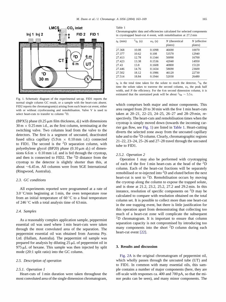
M. Dunn et al. / J. Chromatogr. A 1056 (2004) 163–169 165
Fig. 1. Schematic diagram of the experimental set-up. FID1 reports thenormal single column GC result, or a sample with the heart-cuts absent.FID2 reports the chromatogram(s) arising from each heart-cut event, eitherwith or without cryofocussing and remobilisation. Valve V is used toselect heart-cuts to transfer to column2D.
(BPX5) phase (0.25�m film thickness,df ) with dimensions30 m× 0.25 mm i.d., as the first column, terminating at theswitching valve. Two columns lead from the valve to thedetectors. The first is a segment of uncoated, deactivatedfused silica capillary (5.9 m× 0.10 mm i.d.) connectedto FID1. The second is the2D separation column, withpolyethylene glycol (BP20) phase (0.10�m df ) of dimen-sions 6.6 m× 0.10 mm i.d. and is fed through the cryotrap,and then is connected to FID2. The2D distance from thecryotrap to the detector is slightly shorter than this, atabout∼6.45 m. All columns were from SGE International(Ringwood, Australia).
2.3. GC conditions
All experiments reported were programmed at a rate of3.0◦C/min beginning at 1 min, the oven temperature rosefrom an initial temperature of 60◦C to a final temperatureof 246◦C with a total analysis time of 63 min.
2.4. Samples
As a reasonably complex application sample, peppermintessential oil was used where 1 min heart-cuts were takenthrough the most convoluted area of the separation. Thepeppermint essential oil was obtained from Auroma Pty.Ltd. (Hallam, Australia). The peppermint oil sample wasprepared for analysis by diluting 25�L of peppermint oil in975�L of hexane. This sample was then injected by splitmode (20:1 split ratio) into the GC column.
2.5. Description of operation
2.5.1. Operation 1Heart-cuts of 1 min duration were taken throughout the
most convoluted area of the single dimension chromatogram,
Table 1Chromatographic data and efficiencies calculated for selected componentsin cryotrapped heart-cut 4 event, with remobilisation at 27.2 min
tR (min) 2tR (s) wh (s) N (theoreticalplates)
N (effectiveplates)
27.368 10.08 0.1098 46690 1007027.377 10.62 0.108 53570 1294027.413 12.78 0.1344 50090 1670027.423 13.38 0.1536 42040 1495027.43 13.8 0.1608 40800 1512027.446 14.76 0.1434 58690 2360027.502 18.12 0.1986 46120 2273027.514 18.84 0.1944 52030 26480
tR is the total time taken for the solute to reach the detector,2tR thetime the solute takes to traverse the second column,wh the peak halfwidth, andN the efficiency. For the 6 m second dimension column, it isestimated that the unretained peak will be about2tM ∼ 5.5 s.
which comprises both major and minor components. Thisarea ranged from 20 to 30 min with the five 1 min heart-cutstaken at 20–21, 22–23, 24–25, 26–27 and 28–29 min, re-spectively. The heart-cuts and remobilisation times when thecryotrap is simply moved down (towards the incoming car-rier gas flow, seeFig. 1) are listed inTable 1. Heart-cuttingdiverts the selected zone away from the uncoated capillarytube and to the2D column. Clearly, chromatographic regions21–22, 23–24, 25–26 and 27–28 travel through the uncoatedtube to FID1.
2.5.2. Operation 2Operation 1 may also be performed with cryotrapping
of each of the five 1 min heart-cuts at the head of the2Dcolumn. Each of the heart-cut fractions will be separatelyremobilised or re-injected into2D and eluted before the nextheart-cut is sent to2D. Remobilisation occurs by movingthe cryotrap along the column to expose the trapped solute,and is done at 21.2, 23.2, 25.2, 27.2 and 29.2 min. In thisinstance, resolution of specific components on2D may becalculated to compare with resolution obtained on the totalcolumn set. It is possible to collect more than one heart-cutin the one trapping event, but there is little justification forthis operation apart from demonstrating that collecting toomuch of a heart-cut zone will complicate the subsequent2D chromatogram. It is important to ensure that columnseparation capacity is not compromised by introducing toomany components into the short2D column during eachheart-cut event[22].
3. Results and discussion
Fig. 2A is the original chromatogram of peppermint oil,which wholly passes through the uncoated tube (UT) andto FID1. In common with many essential oils, this sam-ple contains a number of major components (here, they areoff-scale with responses ca. 400 and 700 pA, so that the mi-nor peaks can be seen), and many minor components. The

166 M. Dunn et al. / J. Chromatogr. A 1056 (2004) 163–169
20 22 24 26 28 30 32
100
200
~
54321
FID
Res
pons
e (p
A)
Retention time (min)
~
20 22 24 26 28 300
50
100
150
200FID
Res
pons
e (p
A)
Retention time (min)
20 22 24 26 28 300
50
100
150
200 FID1
FID2
1 432 5
1 432 5
20 22 24 26 28 300
100
200
300
400
500
600
700
800
900
1000
FID
Res
pons
e (p
A)
Retention time (min)
~
1
4
2
35
(A)
(B)
(C)
Fig. 2. (A) Expanded gas chromatogram of peppermint essential oil show-ing the selected heart-cuts and trace components. The two major peakshave response maxima of 400 and 700 pA, respectively, and both exhibitoverloading. (B) FID1: peppermint GC trace on FID1 with heart-cuts (de-noted 1–5) absent (only non-heart-cut sections pass to the FID1). FID2:heart-cut sections 1–5 recorded at FID2, after passage through2D column,but without cryotrapping. (C) The five heart-cuts shown in (B) (FID2)are separately sequential cryotrapped and rapidly remobilised, to producefast GC analysis on2D with good efficiency and response. Heart-cutsand remobilisation times given inSection 2.
five heart-cut events are shown on this chromatogram. Thereis no particular reason for choice of the heart-cut times, butfor convenience are conducted at 1 min intervals, over a to-tal time of 10 min (times are listed inSection 2). The totalnumber of events in this case is controlled by the eventstable in the software, and for the system here is rather lim-ited. A more liberal events table will allow a more extensivenumber of heart-cuts, which in principle can be limitless.Fig. 2Bis a display of the total sample with the heart-cuttingimplemented. Thus,Fig. 2B (FID1) shows that regions 1–5are absent (i.e. the FID1 result) and the lower trace (Fig. 2B,FID2) is the corresponding heart-cuts that travel through thecryotrap (here, without cryogen supplied) to FID2. It shouldbe apparent that the arithmetic summation ofFig. 2B(FID1and FID) should be equivalent toFig. 2A. Since2D is a re-taining column, there is a small finite retention increment(and in this case also a possible small shift in retention) ofthe components, which would have to be taken into accountif a direct comparison of the FID2 result with the selectedheart-cut zones originally seen on FID1 is required.
The effect of cryotrapping on each of the heart-cut zonescan be seen inFig. 2C. The major peak, in heart-cut zone 3,is significantly off scale (response 8000 pA, but is also sig-nificantly overloaded, for the 400 pA peak inFig. 2A). Fora non-overloaded peak comparison, the peak at 28.5 min inFig. 2A has a response of 30 pA, compared with its cor-responding response inFig. 2C of 800 pA (almost 30-foldincrease). The extent of response increase depends upon theextent of zone compression (peak widths) in the first dimen-sion, and the time that a peak elutes on2D — the earlier elut-ing solutes have narrower peaks, and proportionally greaterresponse increase. Note that compared to a normal GC anal-ysis, the fast elution reported here requires a faster data ac-quisition rate, which does increase detector noise marginally.
The narrow peaks inFig. 2Ccannot be readily displayedunless an expanded scale is used.Fig. 3 is an expansion ofeach of the five heart-cuts, where time zero for each plot isthe time that the trap is moved to permit the cryofocussedsolutes to heat up and travel to the2D separation column.Again, to permit the small peaks to be seen, the verticalresponse scale is also expanded. The vertical comparisonalso allows some appreciation of the breadth of the sepa-rated zones or windows on the second column. This shouldcorrespond, to a first approximation, to the polarity rangeof the solutes in each of the heart-cut zones, where lowpolarity solutes are expected to be the least retained on thepolar polyethylene glycol phase. The proper interpretationwould be arrived at if the individual identities of each of thepeaks was known, so that this correlation might be betterrationalised.
Since the oven temperature increments by about 6◦C be-tween successive heart-cuts, it may be asked whether theisolation of the separate heart-cuts serves an improved sep-aration purpose, if the more volatile compounds can be iso-lated from the later peaks if a single narrow bore capillaryanalysis were performed. First,Fig. 3 suggests that there is

M. Dunn et al. / J. Chromatogr. A 1056 (2004) 163–169 167
050
100150200
Retention time (min)
050
100150200
0
100
200
300
FID
Res
pons
e (p
A)
20
40
60
0
50
100
150
H/C 5
H/C 4
H/C 3
H/C 2
0.050.00 0.450.400.350.300.250.200.150.10
H/C 1
Fig. 3. Vertical alignment of each heart-cut (time= 0 corresponds tothe remobilisation time of each heart-cut) to demonstrate the componentelution (separation) window for each heart-cut. Vertical scale expandedto show detail of smaller components.
little spare separation capacity to fit in more components be-fore the separation window in subsequent heart-cuts. But totest this, two neighbouring heart-cut events were collectedtogether, and only after the second, was the cryotrap moved.This result is compared with the selection and trapping ofeach heart-cut in separate analyses, but remobilisation atthe later heart-cut trap movement time.Fig. 4 is the resultof this trial. In Fig. 4A, the dotted trace is the peak-richresult for heart-cut 1, held until the remobilisation step ofheart-cut 2 (i.e. 23.2 min), whilst the solid line is the resultfor heart-cut 2. There is considerable peak overlap, so if toowide a heart-cut is taken, separation performance will dete-riorate.Fig. 4Bshows the result when both heart-cuts 1 and2 are collected together in one single trapping event and re-mobilised at 23.2 min. Clearly, effective separation requiresheart-cuts of limited duration.
23.20 23.25 23.30 23.35 23.40 23.45 23.50 23.55 23.60 23.6510
20
30
40
50
FID
Res
pons
e (p
A)
Retention time (min)
combined heartcuts 1 & 2
23.20 23.25 23.30 23.35 23.40 23.45 23.50 23.55 23.60 23.6510
20
30
40
50 dotted line = H/C 1 (20-21)solid line = H/C 2 (22-23)
(A)
(B)
Fig. 4. Comparison of separate collection of heart-cuts 1 and 2, eachremobilised at 23.2 min (A), with collection of heart-cuts 1 and 2 together,and both remobilised at 23.2 min (B).
As a confirmation of enhanced separation performance ofthe heart-cut/cryotrap process, the result ofFig. 3 (H/C2)can be contrasted with the corresponding normal GC result(Fig. 2A, heart-cut 2) and the heart-cut without cryotrap-ping result (Fig. 2B, heart-cut 2). As stated earlier, thereis a slight difference between the result of the passage ofthe zone through the uncoated tubing, and the same zonepassed through the2D column (see minor differences be-tweenFig. 2A (heart-cut 2) andFig. 2B (heart-cut 2)), butone might think that there are only one, or maybe two peaksin this chromatogram. However, for this equivalent heart-cutregion,Fig. 3 (H/C2) shows many well separated, good re-sponse peaks, that one can only imagine would be unde-tected in normal GC analysis. Note that inFig. 3 (H/C2), astep response is seen above chromatographic detector base-line when no sample is introduced into the2D column. Thiscorresponds to either chemical response (unresolved peaks),and/or column bleed from the long primary column. Thisstep response arises at about 22.1 min, which should approx-imate the unretained peak time (or void time) on2D, i.e.about 6 s. A similar value of 5.5 s is found by calculation.
Since the cryotrap collects all effluent from the end of theprimary column in each of the trapping events, it is impor-tant to conduct blank injections to confirm that the peaks arenot artifacts of, for example, phase bleed. InFig. 5, the dot-ted line is the blank injection result for the region equivalentto heart-cut 4. The solid line indicates that a large numberof peaks arise from the essential oil, and are authentic com-ponents from the sample injection. Many of these peaks willnot be seen in the original GC trace.
Fig. 5 data have been interpreted on the basis of peakwidths and retention times on the second column (time takenfrom the movement of the cryotrap) in order to evaluatecolumn efficiency for selected peaks A–E, and peak capac-ity. Table 1reports the widths and efficiencies (theoreticalplates) of these peaks in this chromatogram. Generally theo-
27.25 27.30 27.35 27.40 27.45 27.50 27.55 27.60 27.650
10
20
30
40
50
60
70
FID
Res
pons
e (p
A)
Retention time (min)
A B
C
D
E
Fig. 5. Heart-cut 4 chromatogram showing overlay of the peppermint in-jection with a separate trace of a blank injection (dotted line). Quantitativedata for identified peaks A–E are given inTable 2.

168 M. Dunn et al. / J. Chromatogr. A 1056 (2004) 163–169
Table 2Quantitative data for selected peaks in heart-cut 4
Peak Replicate 1 Replicate 2 Replicate 3 Overall peak area (%R.S.D.)
Solute tR (min) Peak area (pA s) SolutetR (min) Peak area (pA s) SolutetR (min) Peak area (pA s)
A 27.368 13.37 27.368 13.32 27.369 13.34 0.2B 27.378 6.18 27.377 6.12 27.378 6.19 0.6C 27.431 76.55 27.430 76.43 27.431 75.28 0.9D 27.467 36.1 27.466 36.09 27.467 36.11 0.1E 27.503 2.67 27.502 2.74 27.502 2.81 2.6
Remobilisation time= 27.2 min. Refer toFig. 5 for peak identification.
retical plates of the order of 40,000–50,000 are obtained. Inthe absence of a direct measure of the unretained peak in thisexperiment for each heart-cut, if atM of 5.5 s (giving a car-rier flow velocity of 110 cm/s) is taken for heart-cut 4, theneffective plates of 10,000–20,000 are obtained for selectedpeaks inFig. 5. Thus, theoretical and effective plates ofabout 7000 and 4000 m−1, respectively, are obtained for the100�m i.d. 2D capillary. These data are less than the theo-retical value for a capillary column of these dimensions, andthis may be due to a higher carrier flow than optimum, anda lower coating efficiency for the polar polyethylene glycolphase. The peak capacity of the second dimension can beestimated by taking an average peak width for a second di-mension peak, and calculating the total number of resolvedpeaks that can be placed in the separation window. By usinga peak width definition of 6σ, about 30 equally resolvedpeaks can be separated within about 24 s on the secondcolumn (note that no peaks eluted later than about 30 s onthe second column). In a total analysis time of 60 min, thisequates to a peak capacity of about 1800. Since only 40% ofthe separation space is used in the present case, the samplingfrequency could be increased, or a longer2D column used,to get even greater separation capacity. Taking the valueof 40% as the start point, it can be estimated that the totalanalytical peak capacity might be about 5000 or more. Thisis considerably more than a normal (or even a narrow bore)capillary, and supports the notion that the present methodprovides expanded peak capacity, as well as improvedresponse.
Finally, the analysis of the sample was performed intriplicate, and the same heart-cut compared across thethree chromatograms by both overlaying the expandedheart-cut results, and by calculation of statistical data. Chro-matograms (not shown) indicate that the responses are wellpreserved from run to run in this experiment.Table 2re-ports selected data for five components (A–E) in heart-cutfraction 4, including their total retention times and areas.Note that the recorded retention times differ by no morethan 0.001 min, and that quantitative response data vary byno more than R.S.D. of 1.0% for the replicate injection, ex-cept for the smallest of the selected peaks. Thus, excellentreproducibility is obtained.
There are a few additional points to be recognised in thepresent work. First, it is possible to split peaks between
two different trapping events, and so quantitative analysisin this instance would require addition of each of the peaksof a solute. Second, holding the solutes in the trap requiresthem to be effectively immobilised at the trapping temper-ature. For highly volatile solutes, the trap will require ef-fective supply of cryogen. Note that heart-cut 1 has beenheld in the cryotrap for up to 3 min, without any break-through noted. Being only 3 cm long, the trap must per-form very well in order to hold the more volatile solutes.Note that the solvent tail here is not held for a full 1 mintrapping period, and suffers breakthrough. For less volatilesolutes, the trap performs reproducibly and reliably. Place-ment of the system into a GC–mass spectrometer instrumentwill allow mass spectra for all solutes to be obtained, andthis is the next objective of this work. Note that since thesepeaks are broader than those encountered in comprehensivetwo-dimensional gas chromatography, the quadrupole massspectrometer should be suitable for such work, as opposedto the time-of-flight instrument normally recommended inGC × GC.
4. Conclusions
This work has described a relatively simple processfor performing heart-cutting in an MDGC approach withcryotrapping and rapid re-injection of the heart-cuts into ashort, narrow ID capillary column to allow complete, highresolution separation of each heart-cut within the samplingtime of the primary column effluent. The system is morecomplex than a single column GC set-up, requiring twoinjectors and detectors, a switching valve and the cryotrap-ping unit, however, in respect of multidimensional systems,it is not complex. It should be possible to implement thisapproach for almost a complete sample, with contiguousheart-cuts taken over the whole primary column elutionof the sample, to give a complete multidimensional gaschromatography result for all components of the sample.This method offers considerable time savings, improvedresolution and considerable response increase over conven-tional MDGC. The principles employed here derive fromthe considerations of comprehensive two-dimensional gaschromatography, where high performance modulation andfast second dimension analysis is critical.

M. Dunn et al. / J. Chromatogr. A 1056 (2004) 163–169 169
References
[1] D.R. Deans, Chromatographia 1 (1968) 19.[2] D.J. McEwen, Anal. Chem. 38 (1966) 1047.[3] J.C. Giddings, Anal. Chem. 56 (1984) 1258A.[4] W. Bertsch, J. High Resolut. Chromatogr. 22 (1999) 647.[5] W. Bertsch, J. High Resolut. Chromatogr. 23 (2000) 167.[6] A.C. Lewis, in: L. Mondello, A.C. Lewis, K.D. Bartle (Eds.), Mul-
tidimensional Chromatography, Wiley, Chichester, 2001.[7] B.M. Gordon, C.E. Rix, M.F. Borgerding, J. Chromatogr. Sci. 23
(1985) 1.[8] B.M. Gordon, M.S. Uhrig, M.F. Borgerding, H.L. Chung, W.M.
Coleman, J. Elder Jr. III, F.J.A. Giles, D.S. Moore, C.E. Rix, E.L.White, J. Chromatogr. Sci. 25 (1988) 174.
[9] N. Ragunathan, K.A. Krock, C. Klawun, T.A. Sasaki, C.L. Wilkins,J. Chromatogr. A 703 (1995) 335.
[10] K.A. Krock, C.L. Wilkins, Trends Anal. Chem. 13 (1994) 13.[11] K.A. Krock, N. Ragunathan, C.L. Wilkins, J. Chromatogr. 645 (1993)
153.
[12] Z. Liu, J.B. Phillips, J. Chromatogr. Sci. 29 (1991) 227.[13] P.J. Marriott, R. Shellie, J. Fergeus, R.C.Y. Ong, P.D. Morrison,
Flavour Frag. J. 15 (2000) 225.[14] R. Shellie, P. Marriott, C. Cornwell, P. Morrison, in: Proceedings
of the 23rd International Symposium on Capillary Chromatography,Riva del Garda, Italy, 2000.
[15] J.C. Giddings, J. High Resolut. Chromatogr. 10 (1987) 319.[16] R.E. Murphy, M.R. Schure, J.P. Foley, Anal. Chem. 70 (1998)
1585.[17] P. Marriott, R. Ong, R. Shellie, R. Western, Y. Shao, R. Perera, L.
Xie, A.J. Kueh, P.D. Morrison, Aust. J. Chem. 56 (2003) 187.[18] P.J. Marriott, R.M. Kinghorn, Anal. Chem. 69 (1997) 2582.[19] R.M. Kinghorn, P.J. Marriott, P.A. Dawes, J. High Resolut. Chro-
matogr. 23 (2000) 245.[20] P. Marriott, M. Dunn, R. Shellie, P. Morrison, Anal. Chem. 75 (2003)
5532.[21] K. MacNamara, R. Leardi, A. Hoffmann, LC-GC Eur. 16 (2003)
14.[22] J.M. Davis, J.C. Giddings, Anal. Chem. 55 (1983) 418.


![t e o m ics& Journal of Saha et al., J Proteomics ... · [7]; and multidimensional chromatographic fractionation [3,4,6]. But all these methods are expensive, laborious and time-consuming,](https://static.fdocuments.in/doc/165x107/5f0f60d17e708231d443dcef/t-e-o-m-ics-journal-of-saha-et-al-j-proteomics-7-and-multidimensional.jpg)