Rapid methods in food microbiology Identification of...
118
Sabina Purkrtová Rapid methods in food microbiology Identification of microorganisms
Transcript of Rapid methods in food microbiology Identification of...
Sabina Purkrtovaacute
Rapid methods in food microbiology
Identification of microorganisms
Microbiological analysis procedures
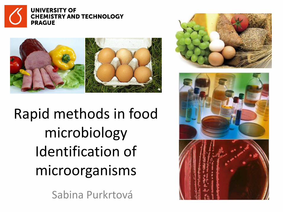
CONVENTIONAL CELL CULTIVATION
bull relatively easy to use but time (requires several days) labour (lots of procedural steps)
and material consuming
bull many of them are recognised as approved for ISO and they are gold standard
procedures
bull Colony count method (CCM)
bull pour plate techniques
bull spread plate techniques
bull Membrane filtration
bull Most Probable Number (MPN)
RAPID METHODS
bull immunonological method (based on antigenantibody-binding)
bull based on molecular biological method (based on PCR)
bull others (ATP Photometry Direct Epifluorescent Filter Techniques (DEFT) Electrical
impedance method Flow cytometry etc)
3
IMMUNOLOGICAL METHODS
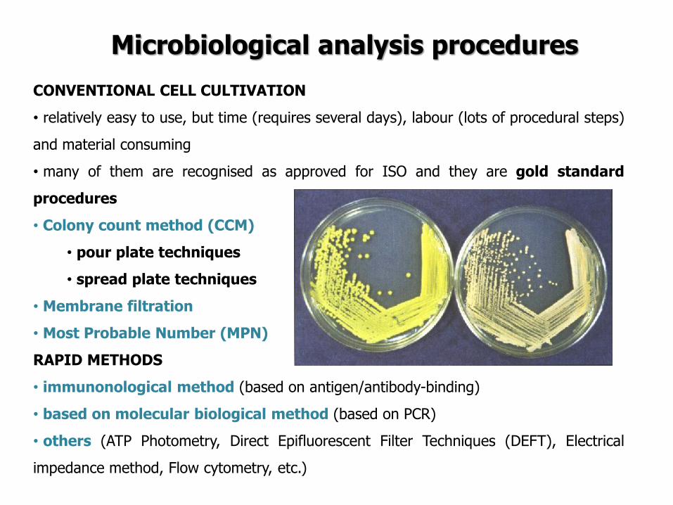
Immunonological method
httpwwwmicrobiologyinfocomwp-contentuploads201505Antigen-Propertiesjpg
A bacterial antigen ndash a molecule on the surface of bacterial cell
httpwwwnanomedicinecomNMIIAFigures158JPG
Immunoglobulins Ig (also antibodies) are glycoprotein molecules produced by B cells plasma cells (white blood cells) according to antigen to mark remaining bacteria for destruction The antibody immune response is highly complex and exceedingly specific
Immunonological method
httpsuploadwikimediaorgwikipediacommonsaa9Antibody_IgG2png
IgG2
httpwwwhighlandseduacademicsdivisionsscipebiologyfacultyharnden2122imagesabstructurejpg
http4bpblogspotcom-Qrm1tS__g6MT56tMi5jmJIAAAAAAAAGasrgLQgP-DfXos320immunoglobulinsjpg
Monomer
Dimer Pentamer
The various immunoglobulin isotypes differ in their biological features structure target specificity and distribution
Immunonological method
httpcontentsedu-iorgCDROMtongko2bio4filesVIEW0031JPG
Polyclonal antibodies The immune response to an antigen generally involves the activation of multiple B-cells all of which target a specific epitope on that antigen As a result a large number of antibodies with different specificities and epitope affinities are produced
httpwwwkyowa-kirincomantibodyabout_antibodyimagesproduction_illustjpg
Monoclonal antibodies are generated by a single B lymphocyte to one specific epitope For production B cell is isolated from from the spleen and lymph nodes of immunised animals and fuse with immortal heteromyleoma The produced hybridoma cells produce only one antibody within the supernatant
7
Immunonological method
bullimmunonological method
(based on antigenantibody-
binding)
Isolation on selective agar plates MacConkey agar with sorbitol (instead of lactose) (SMAC) E coli O157 is sorbitol negative ndash no fermentation ndash colourless colonies Fluorocultreg E coli O157 Agar Chromogenic medium ndash chromogenic substrate for β-D-glucuronidase ndash E coli O157 is negative -colourless colonies 37plusmndegC for 24plusmn 3h
Selective enrichment 1 day 25 g of sample + 225 ml of
Modified Tryptone Soya Broth (mTSB) with novobiocine
homogenization Incubation at 415 degC
Confirmation 4 Day Isolation of a charasteristic
colony on Nutrient Agar and incubation at 37plusmndegC for 24plusmn 3h
5Day testing for positive indole reaction
Immunomagnetic beads separation ISO 166542001 Microbiology of food and animal feeding stuffs - Horizontal method for the detection of Escherichia coli O157
Immunomagnetic isolation
after 8 and 16 hours (2 day)
bull 1 ml of homogenate + 100 microl of paramagnetic beads covered with antibody against E coli O157
bull Present cells E coli O157 are trapped and separated by applying a magnet bull The homogenate with other bacteria is taken away bull The beads with trapped E coli O157 are washed (to add a washing buffer ndash to
release a magnet ndash to mix ndash to apply a magnet) bull Plating out by 50 microl of paramagnetic beads with trapped E coli O157 on 2
selective agar plates
SMAC agar Escherichia coli O157H7 (colourless)
Enzyme-linked immunosorbent assay (ELISA)
httpimageslidesharecdncomelisaonlineversion-110602035913-phpapp0295elisa-test-enzymelinked-immunosorbent-assay-27-728jpgcb=1306989526
How to detect the formation of complex antigen-antibody bull Different systems with
antibodies linked to enzyme (eg horseradish peroxidasealkaline phosphatase) ndash then to add substrate ndash its transformation is measured (eg changes in the absorbance)
Enzyme-linked immunosorbent assay (ELISA)
httpthefutureofthingscomuploadimagearticles2006biopenbiopen-elisa-schematicjpg
bull When the complex antibody-antigen is formed the secondary antibody conjugated with enzyme is to be bound to the antigen and not to be washed off
bull After adding the substrate the substrate is changed and give the signal (eg Measured as absorbance)
Enzyme-linked immunosorbent assay (ELISA)
bull monoclonal or polyclonal antibodies coated microtitre trays to capture target antigen
The captured antigens detected using a second antibody which is conjugated to an
enzyme The addition of enzyme substrate enables the presence of the target antigen to
be visualised
bull considerable specificity
bull can be automated commercially available (Salmonella L monocytogenes
Campylobacter hellipetc)
target antigen
washing
product
second antibody
enzyme substrate
substrate solid phase
with antibody
incubation washing
VIDAS or mini-VIDAS Analyzers
bull the automated multiparametric immunoanalyser
bull based on an enzyme immunoassay which detect target antigens using the
ELFA (Enzyme Linked Fluorescent Assay)
bull 4-methyl-umbelliferone (fluorescent molecule) is released by alkaline phosphatase = fluorescence
VIDAS or mini-VIDAS Analyzers
Each test is composed of two parts
1 The SPRreg acts as a Solid Phase Receptacle for the reaction The SPR is coated
with anti-target antibodies adsorbed on its surface
2 The Strip contains all ready-to-use reagents necessary for the test washing
solution alkaline phosphatase-labeled anti-target antibodies and substrate
Video httpwwwbiomerieux-industrycomfoodvidas-listeria-monocytogenes-detectionVIDAS LMO2 httpswwwyoutubecomwatchv=ZFRuJYynLwk
httpwwwbiomerieuxcom
bioMeacuterieux
VIDAS or mini-VIDAS Analyzers
Validated by ISO 16140 againt ISO 6579 (AFNOR NdegBIO 1216-0905)
Salmonella spp
VIDAS or mini-VIDAS Analyzers
The sample = the sample diluted and enriched in Xpress 2 broth (different from ISO 6579) The used broth must be convenient for the method
Detection of Salmonella spp according to ISO 65792002
01 ml of homogenate to 10 ml RVS incubation at 415degC for 24plusmn3 h
Incubation at 37plusmn1degC for 18plusmn2 h
1 ml of homogenate to 10 ml MKTTn incubation at 37plusmn1degC for 24plusmn3 h
Plating out by 10 micro on two selective media and incubation at 37plusmndegC for 24plusmn 3h
bullXylose Lysine ndash Desoxycholate Agar (XLD)
bullAny other selective medium (BGAHE BS SS DC chromogenic mediahellip)
25 g of sample + 225 ml of buffered peptone water homogenization
Selective enrichment
Pre-enrichment
Isolation on selective agar
plates
Confirmation
Confirmation
Isolation of a charasteristic colony on Nutrient Agar and incubation at 37plusmndegC for 24plusmn 3h
1 day
2 day
3 day
4-6 day
ISO 65792002 Microbiology of food and animal feeding stuffs -- Horizontal method for the detection of Salmonella spp
VIDAS kits
bull E coli O157 - VIDASreg UP E coli O157 (including H7)
bull E coli O157 - VIDASreg ECO
bull E coli O157 (confirmation) - VIDASreg ICE
bull Salmonella spp - VIDASreg SLM
bull VIDASreg ICS
bull Salmonella spp - VIDASreg Easy SLM
bull Listeria spp - VIDASreg LIS
bull Listeria spp - VIDASreg LSX
bull Listeria monocytogenes VIDASreg LMO2
bull Staphylococcal enterotoxins - VIDASreg SET2
bull Campylobacter spp - VIDASreg CAM
GLISA-Rapid Test (Gold Labelled ImmunoSorbent Assay) ndash Lateral Flow Test
Detection limit 105 CFUml in a sample (homogenate of selective enrichment or suspension of colony) (sometimes heat-treatment required - 20 min at 80 degC = cell lysis = better motility of the cell walls with antigens)
httpwwwmeridianbioscienceeumediahome_3columns_images751630jpg
GLISA-Rapid Test
Reaction zone 150 microl of sample
Testing zone Control zone
+ Pos - Neg
Singlepathcopy for detection of Listeria monocytogenes (Merck)
Validation of procedure
Validation of an alternative method bull the validation of an alternative method is the procedure to demonstrate if the alternative method provides equivalent or better results compared to the reference methods
bull Validation is a process within which the method is demonstrated to be suitable
for its purpose It documents methods validity
bull During validation process methods performance characteristics are estimated
The validation of qualitative and quantitative methods comprises two phases
1 a method comparison study of the alternative method against a reference
method (performed by an expert laboratory)
2 a interlaboratory study of the alternative method (organised by an expert
laboratory)
Expert laboratory (organising) laboratory having the qualified staff and skills to
perform the method comparison study and organise the interlaboratory study
Useful documents
ISO 161402003 ndash Microbiology of food and animal feeding stuffs ndash Protocol for the
validation of alternative methods
ISO 170252005 ndash General requirements for the competence of testing and calibration
laboratories
Accreditation bodies that recognize the competence of testing and calibration laboratories
should use this International Standard as the basis for their accreditation
ISOIEC 170112004 Conformity assessment -General requirements for accreditation
bodies accrediting conformity assessment bodies
If a laboratory wishes accreditation for part of all of its testing and calibration activities it
should select an accreditation body that operates in accordance with ISOIEC 17011
It is expected that all laboratories involved in each step of a validation process will have a Quality System or quality assurance (QA) program in place to ensure standardization of laboratory operations as well as adequate quality control (QC) activities
Validation of procedure
Test characteristics The test characteristics for alternative methods are shown
Qualitative methods
1 Selectivity (inclusivity exclusivity)
2 Relative accuracy
3 Detection level
4 Relative sensitivityRelative specificity
5 The agreement between the
methods
Quantitative methods
1 Selectivity (inclusivity exclusivity)
2 Lowest validated level with satisfactory
precision
3 Repeatability
4 Reproducibility
5 Uncertainty of the method
28
PCR METHODS
DNA RNA PROTEINS Deoxyribonucleic acid (DNA)
A linear polymer that consists of four nucleotides
Adenine Cytosine Guanine Thymine Primer binding A ndash T C - G
DNA RNA PROTEINS
DNA
RNA
httpss3amazonawscomclassconnection116flashcards5862116jpgrna-149F82012BD7233971Fjpg httpslh3googleusercontentcomf81cHVr_O-
jXwH1A7g-mfX1LJKZFffUkItKpHPZ3kGF6cGXJaAJ-pqZoNL7T4YNKcP-grQ=s85 httpslh3googleusercontentcomdc9f6E-QDwxJQc-
7MvExwNvrihEjEfgjzwKr2gidCYIt0VmoMb1yEgTDc6mpkFDMUsXsgw=s94
httpsuploadwikimediaorgwikipediacommons00fPeptide_synpng
PCR principle PCR=Polymerase Chain Reaction (1983 by Kary Mullis) in vitro amplification of the part of DNA (usually 100 bp ndash 1500 bp) bordered by two primers (syntetically prepared oligonucleotides)
Typical number of cycles = 30 - 45
httpswwwyoutubecomwatchv=iQsu3Kz9NYo
PCR principle DENATURATION of double-stranded DNA by heat (app 94-95degC initially 5 then in each step 2-3 minutes)
ANNEALING of PRIMERS on the free single strand DNA bull forward primer on the 3acute-
5acute strand bull reverse primer sits on the
5acute-3acute strand Ta = annealing temperature bull 50-65 degC ndash depends on the
primers sequence bull Time 30 s-60 s bull One of the most important
factor for the specifity bull T gtgt Tm= no annealing - no
PCR product bull T ltlt Tm= non-specific
annealing ndash nonspecific products
ELONGATION by DNA polymerase
in vivo starts to synthetize new DNA molecules from 3acute- end of RNA primer
in vitro PCR from 3acute-end the primers
72 degC ndash optimal temperature
45 s-3 minutes (final elongation 5-15 minutes)
httpwwwmuncabiologyscarrPCR_sketch_3gif
PCR principle
httpopenwetwareorgimagesff1CH391L_S12_PCR_exponential_amplificationjpg
Mistake in the beginning ndash the most influence Exponential phase duplication of PCR products amount in each cycle Linear phase The level of amplification is decreased (lower than duplication in each cycle) Plateau Absence or degradation of some component Reasociation can be preferred than primers anealing due to thermodynamical reasons
PCR principle
httpwwwebioecomuploadfile2010031420100314110534183gif
DNA (genomic plasmid DNA)
Primers (forward reverse)
Thermostable
DNA polymerase
Mg2+ (cofactor
for DNA
polymerase)
dNTP mix
(dCTP dATP
dTTP dGTP) ndash
building stones
for newly
synthetized
DNA product
Nuclease free water ndash without Dnase
Rnase presence (enzymes which
degrade DNA RNA)
MASTERMIX
Total volume ndash 25 μl ndash 50 μl
PCR principle
Polymerase Taq DNA polymerase
bull from thermophilic bacteriumThermus aquaticus
(now prepared by recombination)
bull opt temp 75-80degC (150 ntss)
bull active at the room temperature ndashto work on ice
bull inactive at gt90degC but reactivation when cooled
bull only 5acuteexonuklease activity not 3acuteexonuklease
activity (proofreading)
bull 1 mistake at 10-20 000 nts
Hot start polymerases
bull Activity at the room temperature is inhibited - binding on anti-polymerase
antibodies on the wax balls
bull activation after heating at 94 degC (antibodies destroyed)
Other polymerases
bull Proofreading polymerases (with the 3acuteexonuclease activity)
bull Tth polymerase - reverse transcription (Mn2+) or polymerase activity (Mg2+)
1) Releasing DNA from the cell
- lysis of the cell wall by detergents or by heat digesting cell surface proteins by proteinase K
- RNAse treatment for RNA-free genomic DNA
2) Isolation DNA from the released cytoplasmatic contents
- Centrifugation the vaste in pellet (heat isolation)
- Ion-exchange chromatography (commercial kit) ndash binding DNA by ionic interactions to the column (DNA is negative charged) ndash proteins and RNA pass away ndash DNA is later released by using more concentrated salt solution (column is charged positively)
- Phase separation (aqueouschlorophorm phase ndash CTAB method)
DNA isolation
Detection of food-borne pathogens
DNA of microorganism = set of specific genes with specific sequences
Primers are designed to be complementary to sequence of some specific gene
ndash if the complementary sequence is present PCR product will be performed
Main problems about primers designing and applying of PCR
- Is the gene of interest specific enough (can be found only in the detected microorganism)
- Is the primers sequences complementary only to this gene
- Is the primer sequence designed well (requirements about easy and specific annealing ndash depends on the primers sequence)
- Primers can be designed comercially or by using software (httpwwwbiocenterhelsinkifibiProgramsdownloadhtml)
- sequences and genomes needed - wwwpubmedcom
- httpswwwncbinlmnihgovtoolsprimer-blastindexcgiLINK_LOC=BlastHome
PCR products detection The PCR product has got specific length and specific sequence
Visualiton by intercalating agents bind to double-stranded DNA - fluorescence signal bull Ethidium bromide ndash carcinogenteratogen If intercalated the fluorescence of EtBr is significantly higher than in aqueous solution (UV absorbance maxima at 300 and 360nmemitted as orangeyellow light at 590nm) bull Sybr Green DNA-Sybr Green complex absorbs blue light at 488 nm and emits green light at 522 nm Fluorescent tags at 5acute-end of primers Other tag at 5acute -end of primers eg biotin - for immunodetection detected by anti-bodies linked with an enzyme Microarray
Electrophoresis Conventional electrophoresis a single electrical field causes biomolecules to migrate through a matrix according to its mass-to-charge ratio effectively separates DNA fragments up to ~20-30 kb (according to the gel concentration) larger fragments will comigrate (a large band at the top of the gel)
httpwwwrothamstedacuknotebookimagesgelelgif httpstevegallikorgsitesallimagesgelElectrophoresis2jpg
httpabeleewardhawaiieduProtocolsDNA20Gel20Preparation20and20Electrophoresishtm
Electrophoresis Primers (OT1559 a 18ndash1) complementary to DNA coding16S rRNA Campylobacter
jejuni Campylobacter coli a Campylobacter lari
Product is 287 bp
60 V 100 minutes M ndash 100 bp marker (1-5 positive controls) 1- C jejuni CCM 6212 2- C jejuni CCM 6214 3 ndash
C coli CCM 6211 4 ndash C lari CCM 4897 5 ndash C upsaliensis ATCC 43954 6- 15 strains C jejuni Nt- no
template control (negative control)
Genus specific PCR
Electrophoresis DUPLEX PCR =two pairs of primers
C jejuni C1 5 - CAA ATA AAG TTA GAGGTA GAA TGT-3
C4 5 - GGA TAA GCA CTA GCTAGC TGA T-3
Complementary to a part oxidoreductase 159 bp
C coli 18F 5 - GGT ATG ATT TCT ACAAAG CGAG-3
519R 5- ATA AAA GAC TAT CGT CGC GTG-3acute
Complementary to aspartatekinase and flanking ORF 500 bp
Species specific PCR
60 V 100 minutes M ndash 100 bp marker (1-5 positive controls) 1- C jejuni CCM 6212 2- C jejuni CCM
6214 3 ndash C coli CCM 6211 4 ndash C lari CCM 4897 5 ndash C upsaliensis ATCC 43954 6- 15 strains C
jejuni Nt- no template control (negative control)
Melting curve Intercalating dye binds to double-stranded DNA - producing fluorescent signal when excited by light
When samples are heated to melting point double-stranded DNA denatures into single strands freeing dye into solution
ndash Melting temperature (Tm) is the point at which the DNA denatures
ndash Dependent on the length of fragment and G-C content
ndash More base pairs --gt Increase in Tm
ndash Higher G-C content --gt Increase in Tm
bull Unbound dye has reduced fluorescent signal
bull Loss in signal generates melt curve
No Signal Signal Increasing
temperature
Intercalating
dye
Melting curve
Inflex point
bull 1 derivation = 0
bull In this case 2derivation is negative
Real-Time PCR
CT
CT (threshold cycle) ndash the lowest cycle when fluorescence crosses the
determinated value
Video httpswwwyoutubecomwatchv=kvQWKcMdyS4 httpswwwyoutubecomwatchv=pRwoOBuk00c
Real-Time PCR
Food-borne pathogens detection by PCR
Identification of pure cultures - without problems
Detectionenumeration from food matrix directly or after enrichment
bull How to isolate successfully DNA from few cells in 25 g10 g - Efficiency of DNA isolation (isolation by kits heat-lysis CTAB method)
bull How many targets copies are needed for successfully PCR products detection
ndash detection limits
ndash presence of PCR inhibitors (inhibition of polymerase primers annealing)
bull How to distuinguish DNA from dead and live cells - Total DNA is detected (from dead and living cell)
bull For distinguishing of dead cells DNA ndash eg application of EMA (ethidium monoazide) to the sample before DNA isolation ndash EMA irreversibly bound to DNA of dead cells ndash they are bdquoinvisibleldquo for PCR ndash no amplification
bull Direct enumerationdetection only in some specific case ndash simple liquid sample when the required limit is higher than the limit of detection
bull Standardly ndash for PCR is used enriched homogenate (sample diluted in enrichment broth and cultivated) ndash to obtain detectable level of microorganisms
BAXreg System
Salmonella
Listeria monocytogenes
Genus Listeria
Enterobacter sakazakii
E coli O157H7
Staphylococcus aureus Real-time
Campylobacter jejunicolilariReal-time
Vibrio cholerae
parahaemolyticusvulnificus Real-time
Yeast and molds
httpwww2dupontcomDuPon
t_Homeen_USindexhtml
BAX ndash three major steps
On enrichedregrown samples
Prepare samples = perform heat-lysis in two stages
DNA present in the lysate
2 Process samples
3 Review results
Hydrate PCR tablets
Enrichment protocols vary
depending on bacterial target
and type of food yoursquore testing
Salmonella Enterobacter
sakazakii
E coli O157H7 E coli O157H7
MP Campylobacter jejunicoli
Yeast and Mold
37deg +- 2degC for 20 minutes then
95deg +- 3degC for 10 minutes
Listeria and L monocytogenes
55deg +- 2degC for 60 minutes then
95deg +- 3degC for 10 minutes
5 microl for all bacterial targets
20 microl for beef samples enriched
in E coli O157H7 MP media
20 microl for yeast amp mold
PCR tablets bull DNA-polymerase
bull Primers
bull Nucleotids
bull Suplements
bull SybrGreen or fluorescent probe
bull Internal control + primer
bull Mastermix in the tablet in PCR Eppendorf ndash minimal risk of contamination
For solving the problem of distiguishing dead and live cell the process of small volumes
is used ndash only if live cells are present in the sample they able to multiplied to the limit
detection (104 CFUml in the enrichment suspension used for lysis step)
If the result of detection is positive confirmation by plating out is done (live and dead
cells)
+
Target organism DNA BAXreg system
primers
in tablets PCR
+ no detectable
amplification
Background DNA
PCR
BAXreg system
primers
in tablets
Amplification of specific fragments
Detection of specific fragments Fluorescent signal drops with heating to generate a melt curve
Melting curve Raw Data Processed Data
Strong positive
result
Negative
result
Target peak
Positive control peaks
Positive control peak
Melting curves display peaks within a set temperature range for the control and target (if present)
Detection
Melt curve weak INPC in presence of strong Salmonella positive
Real time weak INPC in presence of strong
Campylobacter positives
Direct protocol does not display a positive or
negative result PCR was successful IndeterminateSignal error
Salmonella AFNOR (ISO 16140) protocol Sample 110 BPW
18 h37ordmC
RVS Muumlller-Kauffmann
24 h 415 37 ordmC
5 suspicious colonies on TSA
24 h 37ordm C
Biochemical and serological confirmation
XLD 2 Plate
24 h 37ordmC
BHI
3h 37 ordmC
22 h 37 ordmC
15 d neg and pos result
3 d neg 5 d pos results
Salmonella ( ISO 65792003) versus BAXreg-PCR
International certificates AOAC-RI Performance Tested Method
Salmonella 100201 L monocytogenes
070202 L spp 030502 Saureus 120701
Campylobacter colilarijejuni
AFNOR Certification
Salmonella QUA 183-1102
NordVal Certification
Salmonella 2006-30-5408
Certificates from AFNOR and AOAC are equal
NordVal Certificate
57
OTHER METHODS
ATP bioluminiscence
httpwwwpromegacouk~mediaimagesresourcesfiguresenotesfe0027_fig1jpgla=en
httpwwwbiotekptassetstech_resources143clarity_atp_conc_fig2gif
Principle the detection of the present level of ATP (direct proportion to the presence of microorganisms or their products residues) The enzyme luciferase catalyzes the transformation of d-luciferin to oxyluciferin (indicated by emision of light at 560 nm)
ATP bioluminiscence
httpwwwbioxyscomimages2figure-B11gif
ATP Hygiene Monitoring System - SystemSURE II
httpwwwmicromaticcomdraft-keg-beerline-cleaning-hygiene-pid-ATP-100-tab-reviewshtml
Biosensors
60
httpsuploadwikimediaorgwikipediacommonseecBiosensor_Systemjpg
Biosenzor =the whole cellmicroorganism (CBS ndash cell-based sensors)
Cell-based sensors
Patogenic microorganisms interact during the infection with mammalian
cells ndash detection of cell or toxin by cell culture The damage of cell culture is measured optically or electrically
62
IDENTIFICATION OF MICROORGANISMS
Definition of species in bacteriaarchae
The criterion for a bacterial or archael species a collection of strains that have bull at least one diagnostic phenotypic trait bull ge70 DNAndashDNA hybridization of their purified molecules DNA
bull ~more than 97 16S rRNA sequence identity Under the same conditions some eucaryotic order would be a single bacterial species
The bacterial species definition bull eminent practical significance bull but a very difficult issue
bull very wide and often changes in DNA
bull mutations during replication of DNA
bull horizontal gene transfer through mobile elements plasmids also between species
Criterion for species in prokaryotic cells (bacteria and archea) Collection of strains characterised bull by at least one diagnostic feature bull their purified DNA molecules prove at least 70 hybridisation
which corresponds to at least 97 concordance in the sequence of 16S rRNA gene
Map of genome of B cereus ATCC 14579
httpwwwacgtinccomspecialty_dna_sequencinghtm
Identification based on genotype
64
DNA isolation PCR amplification of 16S rRNA gene (usually)
Sequencing of PCR product
Comparison to sequences in databases (BLAST)
Identification of bacteria PHENOTYPE METHODS bull Morphology of colony and cell (Gram staining shape) bull Requirements for temperature atmosphere nutrition bull Biochemical tests - presence of specific enzymes
bull catalase oxidase fermentation of glucose bull the way how they utilize different compounds
bull Immunochemical tests - specific antigens in the cell wall METHODS BASED ON PROTEIN ANALYSIS
bull MALDI-TOF MS METHODS BASED ON GENOTYPE bull looking for and comparing of specific DNA sequences
bull conserved universal genes (eg 16S rRNA) bull genes specific for genus species
bull done by PCR methods DNA chips sequencing etc
66
BIOCHEMICAL TESTING
Identification based on phenotype I Physiological + biochemical features
bull morfology of colonies and cells (Gram staining shape)
bull requirements for temperature atmosphere nutrient sources
bull basic biochemical tests oxidase catalase fermentationoxidation of glucose haemolysis etc
appearance of colonies on selective or selective-diagnostic agars (for other testing to be reinoculated on non-selective agar)
the classification of an isolate into a certain group
(eg for bacteria catalase positivenegative G+ cocci glucose fermentingnon-fermenting G- rods etc)
biochemical tests specific for certain groups of microorganisms
Identification based on phenotype I
68
Physiological + biochemical features Example Bacillus cereus
httpwwwfoodhaccpcommemberonlynewsletter164html
httpwwwnapavalleyedupeoplesrosePublishingImagesBacillus20cereus202(Gram20stain)jpg
Set of biochemical tests Microgenreg Bacillus ID Panel for the identification of Bacillus spp and related genera (time for performing 48 hours)
MYP agar (Mannitol Egg Yolk Polymyxin Agar) pink colonies with a precipitation zone (no fermentation of manitol + enzyme lecitinase)
catalase positive
httpwwwoxoidcomukblueprod_detailprod_detailasppr=CM0929ampc=ukamplang=ENamporg=9ampimg=CM0929ampsec=
G+ rod endspores forming
β-haemolysis moisty and easy to grow colonies
Principle of biochemical testing
to rank the bacterium to some group having the same basic features bull eg catalase positive gram positive cocci bull Family Enterobacteriaceae
the identification on the genus or species level
bull sets of biochemical tests specific for some group of bacteria
70
Gram staining procedure - principle
https40mediatumblrcom25da2e5b25bbe336819bf1715849d171tumblr_inline_ntrsgn9w1D1sv92o6_540jpg
Gram staining procedure - principle
httpsonlinesciencepsuedusitesdefaultfilesmicrb106CellStructurecow02354_03_15jpg
Gram staining procedure 1-2 drops of sterile water on a slide Spread culture in thin filter in this drop Let dry in airclose to fire Pass slide through flame 2-3x to fix Add crystal violet ndash 15-20 s to act Add Lugol solution (KII2) ndash 15-20 s to act Washing off with a mixture of
ethanolaceton (41) or acetone till the stain removes but max 15 s
Rinse with water decant excess liquid Add safranin 1 minute Rinse with water decant excess liquid Dry and put a drop of imersion oil use an
objective bdquoOILldquo
httpwwwtumblrcomtaggedgram-staining
G- red
G+ violet
Gram staining procedure + Identification
E coli
Family Enterobacteriaceae Gram-negative rods
Micrococcus luteus
Staphylococcus aureus
Gram-positive cocci Bacillus cereus
Gram-positive rods (with endospores)
Biochemical identification of G + bacteria
httpyoungeneticistblogspotcz201106diagnostic-microbiology-labhtml
G+ bacteria bull cocci ndash in clusters in pairs
diplococci bull bacilli
bull endospore bull no endospore
For G+ cocci bull Catalase positive ndash Staphylococcus
sp bull Coagulase + S aureus or some other few species bull Coagulase -
bull Novobiocin S bull S epidermidis etc
bull Novobiocin R bull S saprophyticus etc
bull Catalase negative Streptococcus sp Enterococcus sp
bull kind of haemolysis Other important G + group ndash filamentous bacteria
Catalase test Catalase test Catalase is a common enzyme found in nearly all living organisms that are exposed to oxygen where it catalyzes the decomposition of hydrogen peroxide to water and oxygen
2 H2O + O2 2 H2O2
catalase
bubbling when culture is add in peroxide
httpiws2collinedudcainCCCCD20Microcatalase_testhtm
Catalase testing into a drop of 3 peroxide to transfer a loop of bacterial culture catalase positive ndash releasing of air bubbles
Examples bull G+ cocci catalase positive Staphylococcus sp Micrococcus sp Kocuria sp Dermacoccus sp Macrococcus sp Rothia sp catalase negative Streptococcus sp Enterococcus sp bull G+ rods (bacilli) endospores forming catalase positive Bacillus sp catalase negative Clostridium sp
Resistance to ATBhaemolysis Resistance eg to novobiocin
httpquizletcom20077004mic-lecture-1-t2-flash-cards
Hemolysis on blood agar
Measuring the diameter of inhibition zone (less than 17 mm resistant more or equal to 17 mm sensitive) ndash differing in Staphylococcus sp (Clinical performance)
alpha-hemolysis - oxidizing the iron in the hemoglobin - green discoloration of the medium around beta-hemolysis - rupture of erythrocytes - complete clearing medium around gamma-hemolysis ndash no effect ndash no change
By cultivation on blood agar bacteria can be
differentiated based on their capacity to secrete
hemolysins The hemolysis will cause a clearing zone of
the blood agar around the colonies Bacteria can cause
different types of hemolysis
bullα-hemolysis which means an incomplete clearing
(green haemolysis)
bullβ-hemolysis which means a complete clearing
bullDouble hemolysis of some staphylococci consisting of
an inner β-hemolysis zone and an outer α-hemolysis
zone
bullNo hemolysis which is sometimes referred to as γ-
hemolysis which may seem illogical
Note that the α-hemolysin of staphylococci causes
complete hemolysis whereas their β-hemolysin causes
incomplete hemolysis
The capacity to produce hemolysins may vary between
different strains of a particular bacterial species
httpwwwvetbactorgvetbactdisplayextinfo=67
Identification of bacteria based on haemolysis
Streptococcal hemolysins α (alpha) hemolysis - ldquozone of green discolourationrdquo or ldquogreeningrdquo called partial or incomplete hemolysis produced by alpha-hemolytic streptococci such as Streptococcus suis This type of hemolysis on BA is due to bacterial production of hydrogen peroxide which oxidises hemoglobin to methemoglobin β (beta) hemolysis ndash a clear zone of complete hemolysis produced by beta-hemolytic streptococci such as S equi subsp equi This hemolysis is due to lysis of RBCs in the BA by the cytotoxin Streptolysin S
Source httppeopleupeicajlewishtmlhemolysinshtml
Bacterial hemolysins
Staphylococcal hemolysins Most coagulase-positive staphylococci (CPS) such as Staphylococcus aureus S pseudintermedius ( but not S hycius) typically produce an distinctive pattern of ldquodouble zonerdquo or ldquotargetrdquo hemolysis on blood agar Double-zone hemolysis indicates that the two hemolysins one which produces ldquocompleterdquo and another one that produces ldquoincompleterdquo hemolysis are present and this combined effect is visible due to different diffusion rates of these enzymes through the agar The staphylococcal α (alpha) hemolysin causes a complete type of hemolysis This zone of complete hemolysis is narrow and immediately adjacent to the bacterial colony on blood agar The staphylococcal β (beta) hemolysin causes an incomplete type of hemolysis at 37 degC This broader incomplete zone is outside of the inner complete zone and can be hard to see on early culture plates and also varies with the species of RBCs used to make the BA plate Its action is enhanced by chilling at 4 degC and the hemolysis becomes complete (this effect is referred to as ldquohot-coldrdquohemolysis) There are other staphylococcal cytotoxins that are also hemolysins but their lytic actions are not seen on blood agar
The confusion arises due to the NAMES of the two STAPHYLOCOCCAL HEMOLYSINS and the TYPE of hemolysis they produce These are the REVERSE of the two TYPES of STREPTOCOCCAL HEMOLYSIS Strictly speaking the descriptive terms ldquoalphardquo for ldquoincompleterdquo and ldquobetardquo for ldquocompleterdquo hemolysis apply only to the hemolytic toxins of Streptococcus species However other organisms beside Streptococcus and Staphylococcus species have cytotoxins that cause hemolysis and this hemolysis can be either complete or incomplete It is preferable to use the terms ldquocompleterdquo and ldquoincompleterdquo rather than ldquobetardquo or ldquoalphardquo to describe the hemolysis caused by these organisms but you will notice in bacteriology text books that the terms alpha and beta are commonly used
G ndash bacteria bull Cocci bull Bacilli (Rods) bull Oxidase positivenegative bull Glucose metabolism
bull fermentation bull oxidation bull Inactive
bull Lactose metabolism for coliform
bull Pigment formation for Pseudomonas sp
Biochemical identification of
G - bacteria
Oxidase test
Oxidase positive (OXI +) Aeromonas sp Pseudomonas sp Dark-blue to violet
Oxidase negative (OXI -) Enterobacteriaceae Bacillus sp Colourless
Oxidase bullabbreviated general name for some kind of cytochrome c oxidases (cytochrome oxidase or indophenol oxidase) enzymes involved in aerobic respiratory chain when oxygen is used as the final receptor for hydrogen bullOxidases are usually present only in aerobic organisms bullPresence or absence of oxidase(s) is an important feature of the bacterial metabolism used mainly in the identification process of gram-negative bacteria
Detection Reaction of the culture with redox indicator as eg NN-dimethyl-p-phenylenediamine (DMPD) dark-blue when oxidized and colorless when reduced Different test performances are possible
Fermentation of carbohydrates Type of utilisation of glucose or carbohydrates bull an important feature of the bacterial metabolism bull widely used in the identification process of bacteria bull fermentation strictly means to be under anaerobic conditions (but in bacterial
metabolism which can be but not have to be the same as the anaerobic conditions of cultivation) so very often is used in a general way like bdquoutilisationldquo
bull fermentation (like bdquoutilisationldquo) of carbohydrates to
bull acid and gas (Durham tubes) bull acid without gas bull non-utilised
bull Testing of fermentation or oxidation of carbohydrates
bull eg OF test for glucose bull by oxidation under aerobic conditions bull by fermenation under anaerobic conditions (an oil layer)
Fermentation of sugars to acid
Peptone Water wPhenol Red
(HiMedia M028I)
Ingredients gl
Peptic digest of animal tissue 10000
Sodium chloride 5000
Phenol red 0020
Final pH ( at 25degC) 68plusmn02
httpsuploadwikimediaorgwikipediacommons009Phenol_red_pH_60_-_80jpg
+ after sterilisation
at the room
temperature to add
aseptically sugar
discs and
inoculate
succrose trehalose rhamnose xylose
Incubation 30 degC 24 h
- +
Fermentation of carbohydrates
Casein enzymic hydrolysate and yeast extract provide nitrogenous compounds vitamin B complex and other growth nutrients Dextrose is the fermentable carbohydrate and bromocresol purple acts as the pH indicator with colour change from purple to yellow in acidic conditions Sodium chloride maintains osmotic equilibrium bdquoAnaerobicldquo conditions ndash inoculation by stabbing to used fresh prepared media (oxygen expelled by heating medium in boiling water or running steam for 15 min then to be cooled down quickly)
- +
Enterobacteriaceae ndash glucose fermentation
83
Tryptone Yeast Extract Agar Ingredients Gms Litre Casein enzymic hydrolysate 10000 Yeast extract 1500 Dextrose 10000 Sodium chloride 5000 Bromocresol purple 0015 Agar 15000 Final pH ( at 25degC) 70plusmn02
Test for oxidationfermentation of glucose (OF glucose test) detection of acid production by acidobazic indicator change to yellow (acid present) in lower pH from green (no acid) Anaerobic atmosph ndash covering by sterile mineral oil If fermented anaerobically then also runs aerobically
Fermentation of carbohydrates
Principle of biochemical tests bull The first step in identification is to rank the bacterium to some group of bacteria
having the same basic features ndash eg catalase positive gram positive cocci bull The identification on the genus or species level is finished by other biochemical tests
specific for each group of bacteria bull The tests can be done in tubes or miniaturized plate
ENTEROtest 24N (Erba Lachema) (24 means 24 biochemical tests N= new)
API 20E (bioMeacuterieux)
bull designed for routine identification of clinically important species of family Enterobacteriaceae without the use of reagents
bull A strip test OXItest or OFtest in microtitration wells can be used to confirm that the isolate belongs to Enterobacteriaceae family
bull The results are read visually or automatically using reader MIKROLAreg
bull The identification can be supplemented by diagnostic strip tests INDOLtest COLItest and VPtest
APIreg Gram negative Identification API 20E ndash Speciessubspecies identification of Enterobacteriaceae and groupspecies identification of non-fermenting Gram negative bacteria API Rapid 20E ndash 4-hour identification of Enterobacteriaceae API 20NE ndash 24 to 48-hour identification of Gram negative non-Enterobacteriaceae API Campy ndash 24-hour identification of Campylobacter species
Example Escherichia coli
ENTEROtest 24 N
Biochemical testing - Enterobacteriaceae bull TSI Triple Sugar Iron Agar (use a straight wire to stab the butt and streak the agar surface)
- to determine whether organisms can ferment glucose sucrose andor lactose with or without production of gas and the ability of the organism to produce hydrogen sulphide from thiosulphate in an acid environment is also tested
Formula Liter Enzymatic Digest of Casein 5 g Enzymatic Digest of Animal Tissue 5 g Yeast Enriched Peptone 10 g Dextrose 1 g Lactose 10 g Sucrose 10 g Ferric Ammonium Citrate 02 g Sodium Chloride 5 g Sodium Thiosulfate 03 g Phenol Red 0025 g Agar 135 g Final pH 73 plusmn 02 at 25 degC
inoculation in the butt by stab inoculation on the slant on the surface bull Fermentation of dextrose
(glucose) to acid bull anaerobically - yellow butt bull aerobically leads to products
which are later oxidised by air and pH remains alkaline (red) bull Fermentation of glucose to gas (anaerobically - in the stab) Cracks splits or bubbles in medium indicate gas production bull Production of hydrogen sulfide (H2S) ndash reaction with ferrous sulphate and sodium thiosulphate - a black precipitate in butt bull Lactose andor succrose
utilisation to acid Acid production ndash yellow butt and slant
Results
An alkaline slant-acid butt
(redyellow) - fermentation of
dextrose only
An acid slant-acid butt
(yellowyellow) - fermentation of
dextrose lactose andor sucrose
An alkaline slant-alkaline butt
(redred) - dextrose or lactose not fermented (non-fermenter)
Bacterium fermenting lactose andor succrose are able to ferment glucose
Phenol red ndash alkaline = red acid = yellow
bull TSI Triple Sugar Iron Agar (use a straight wire to stab the butt and streak the agar surface)
- to determine whether organisms can ferment glucose sucrose andor lactose with or without production of gas and the ability of the organism to produce hydrogen sulphide from thiosulphate in an acid environment is also tested
Biochemical testing - Enterobacteriaceae
Urea agar (Christensen) (use a 1 μl loop full)
- to determine whether an organism can split urea into ammonia and
carbon dioxide by the action of the enzyme urease
httpimageslidesharecdncomlab17-141201085153-conversion-gate0195-10-638jpgcb=1417424219
httpimageslidesharecdncomhelicobacterpylori-copy-110516201901-phpapp0195campylobacter-jejunihelicobacter-pylori-39-728jpgcb=1305577391
Phenol red
Medium is acidic - yellow
(pH 68plusmn02)
Alkaline reaction by urea hydrolysis - pink colour
Composition Gms Litre
Peptic digest of animal tissue
1000
Dextrose 1000
Sodium chloride 5000
Disodium phosphate 1200
Monopotassium phosphate 0800
Phenol red 0012
Agar 15000
Final pH ( at 25degC) 68plusmn02
Nutrition
Peptic digest of animal tissues dextrose
Sodium chloride - osmotic equilibrium
Phosphates - buffering of the medium
35-37 degC
Biochemical testing - Enterobacteriaceae
bull L-lysine decarboxylase (use a 1 μl loop full)
- to determine whether an organism can decarboxylate an amino acid (lysine) leading to formation of an
amine After inoculation both tubes are overlayed with sterile paraffin oil
httpfce-studynetdna-sslcomimagesupload-flashcards10295302332621_mjpg
+ CO2 + OH-
Lysine Decarboxylase Broth Typical Composition (glitre) Peptic digest of animal tissue 5000 Yeast extract 3000Dextrose 1000 (glucose prepared from starch chemically equivalent to glucose) L-Lysine hydrochloride 5000 Bromocresol purple 0020 Final pH ( at 25 degC) 68plusmn02 Inoculate below the surface cover by sterile paraffin oil
Two steps detection 1 step Dextrose is fermented ndash H+ is released Acidic conditions bull bromocresol purple changes colour to yellow bull in acidic conditions lysine decarboxylase is
activated 2 step L-lysine is decarboxylated and OH- released Alkaline conditions bull bromocresol purple changes colour back to
purple
Biochemical testing - Enterobacteriaceae
bull β-galactosidase (ONPG) (use a 1 μl loop full)
- to determine whether an organism has the enzyme szlig-galactosidase by using the compound o-
nitrophenyl-beta-D-galactopyranoside (ONPG)
- this test is used to differentiate between lactose-negative and lactose-delayed organisms
- ONPG is hydrolysed by the enzyme to o-nitrophenol which is yellow and galactose
O-Nitrophenyl-β-D-galactopyranoside (ONPG) is structurally similar to lactose (ie ONPG is an analog of lactose) except that orthonitrophenyl has been substituted for glucose ONPG is colorless compound O-nitrophenol is yellow visual evidence of hydrolysis
Lactose fermenting bacteria (ONPG positive) two enzymes Permease ndash transferring of the lactose molecule into the bacterial cell β-galactosidase - cleaving the galactoside bond producing glucose and galactose - inducible enzyme - made ONLY in the presence of the lactose substrate Coliform bacteria as Ecoli Klebsiella spp Enterobacter spp
hydrolysis
Late lactose fermentering bacteria Permease ndash not present delayed in the production of acid from lactose because of sluggish permease activity β-galactosidase ndash present - give a positive ONPG test Citrobacter spp Arizona spp
Non lactose fermenter (ONPG Negative) Permease ndash not present β-galactosidase ndash not present Salmonella spp Shigella spp Proteus spp Providencia spp and Morganella spp
Biochemical testing - Enterobacteriaceae
Procedure Culture Bacteria grown in medium containing lactose (to induce the production of the galactosidase enzyme) such as Kligler iron agar (KIA) or Triple sugar Iron (TSI) agar (Note β-galactosidase enzyme (inducible enzyme) is made ONLY in the presence of the lactose substrate) Inoculation 1A loopful of bacterial growth is emulsified in 005mL of physiologic saline to produce a heavy suspension 2One drop of toluene is added to the suspension and vigorously mixed for a few seconds to release the enzyme for bacterial cells 3An equal quantity of buffered ONPG solution is added to the suspension 4The mixture is placed in a 37 oC water bath 5When Using ONPG Tablets 6A loopful of bacterial suspension is added directly to the ONPG substrate resulting from adding 1mL of distilled water to a tablet in a test tube 7This suspension is also placed in a 37oC water bath
β-galactosidase (ONPG)
Results and Interpretations The rate of hydrolysis of ONPG to o-nitrophenol may be rapid for some organisms producing a visible yellow color reaction within 5 to 10 minutes Most tests are positive within 1 hour however reactions should not be interpreted as negative before 24 hours of incubation The yellow color is usually distinct and indicates that the organism has produced o-nitrophenol from the ONPG substrate through the action of β-galactosidase
Biochemical testing - Enterobacteriaceae
bull VogesndashProskauer (VP) test (use a 1 μl loop full)
- to determine whether an organism can produce acetylmethylcarbinol
(acetoin) from fermentation of glucose If present acetylmethyl carbinol is
converted to diacetyl in the presence of prop- naphthol (to be added first) strong
alkali (40 KOH) and atmospheric oxygen The diacetyl and quanidine-
containing compounds found in the peptones of the broth then condense to
form a pinkish red polymer
MRVP broth (pH 69)
Ingredients per liter of
deionized water
buffered peptone= 70 gm
glucose= 50 gm
dipotassium phosphate= 50 gm Voges-Proskauer Reagent A Barrittrsquos reagent A
Alpha-Naphthol 5 50 gm
Absolute Ethanol 1000
ml
Voges-Proskauer Reagent B Barrittrsquos reagent B
Potassium Hydroxide 400 gm
Deionized Water 1000 ml
Procedure of VogesndashProskauer (VP) Test
1Prior to inoculation allow medium to
equilibrate to room temperature
2Using organisms taken from an 18-24
hour pure culture lightly inoculate the
medium
3Incubate aerobically at 37 degC 24 hours
4Following 24 hours of incubation aliquot
2 ml of the broth to a clean test tube
5Re-incubate the remaining broth for an
additional 24 hours
6Add 6 drops of 5 alpha-naphthol and
mix well to aerate
7Add 2 drops of 40 potassium hydroxide
and mix well to aerate
8Observe for a pink-red color at the surface
within 30 min Shake the tube vigorously
during the 30-min period
Positive Reaction
A pink-red color at the surface
Examples Viridans group streptococci
(except Streptococcus vestibularis) Listeria
Enterobacter Klebsiella Serratia marcescens
Hafnia alvei Vibrio eltor Vibrio alginolyticus etc
Negative Reaction
A lack of a pink-red color
Examples Streptococcus mitis Citrobacter sp
Shigella Yersinia Edwardsiella Salmonella Vibrio
furnissii Vibrio fluvialis Vibrio vulnificus and Vibrio
parahaemolyticus etc
A copper color should be considered negative A
rust color is a weak positive reaction
Quality Control of VogesndashProskauer (VP) Test
VP positive Enterobacter aerogenes (ATCC13048)
VP negative Escherichia coli (ATCC25922)
Biochemical testing - Enterobacteriaceae
94
VogesndashProskauer (VP) test
Rapid test for confirmation of E coli ndash typical features
β-D-glucuronidase (97 strains) production of indole from tryptophan (typical for E coli)
Performance
Previous testing Gram-negative (eg able to grow on VRBD agar) oxidase negative
From pure culture to prepare suspension in saline solution (app 5 ml) at the density 3 McFarland
To transfer 05 ndash 1 ml of this suspension into a new tube ndash to add the strip of COLItest (chemicals ndash fluorogenic substrate for β-D-glucuronidase tryptophane are present in the pad at the end of the strip)
Incubation 37 degC 4 hours
Detecion of β-D-glucuronidase (to be done at first) UV (360 nm) ndash blue fluorescence (GLR + hellipβ-D-glucuronidase positive) ndash GLR test can be read after 1 hour
Detection of indole formation from tryptophane (to be done at second) to add Kovacs reagent (IND + hellipformation of red ring)
95
COLItest - Escherichia coli
Biochemical testing - Enterobacteriaceae
Fluorogenic culture medium
bull One or more fluorogenic substrates
bull For detection of specific enzyme which are able to cleave out the fluorogenic substrate that a designed
fluorophore (responsible for the UV fluorescence) is released
FLUOROGENIC MEDIUM (Colilert) E coli use β-glucuronidase to metabolize MUG and create fluorescence (UV 360 nm) Since most non-coliforms do not have these enzymes they are unable to grow and interfere
httpgawaterusgsgovprojectsbacteriapicturessummaryanalysisecolijpg
E coli (MPN method)
COLItest Biochemical testing - Enterobacteriaceae
COLItest bull Indole test (use a 1 μl loop full)
- to determine whether an organism can split indole from tryptophan
httpwwwmicrobiologyinfocomindole-test-principle-reagents-procedure-result-interpretation-and-limitations
indole production from tryptophan
Production of indole is detected by Kovacacutes reagent
Indole Kovacs Reagent
p-Dimethylaminobenzaldehyde 500 gm
Hydrochloric Acid 37 2500 ml
Amyl Alcohol 7500 ml
Biochemical testing - Enterobacteriaceae
INDOLe test ndash quick variant
Ecoli Citrobacter Enterobacter
Procedure bull on the filtration paper to add a drop of INDOL suspension bull to spread into by a loop some culture bullTo incubate ndash at the room temperature for 5 minutes bullTo read results
Biochemical testing - Enterobacteriaceae
ENTEROtest 24 ndash example of biochemical testing
routine identification of clinically important species of family Enterobacteriaceae the test is focused only on some species often isolated in clinical practis strains from other sources can be slightly different in the dominant biochemical profiles intended exclusively for the identification of family Enterobacteriaceae the preliminary ranking into family Enterobacteriaceae must be done (Gram staining oxidase glucose fermentation) the results of reactions obtained in this modified micromethod may differ from the results obtained using conventional tests and results available in literature sources in case of problem to control positivenegative reactions by control strains or to use for this tests tube variant to check the functionality of the kit not to check the accuracy or success of the identification
Preparation of suspension from over-night culture growing on non-selective agar (for staphylococci Columbia agar blood with 5 of sheep blood is recommended)
Inoculation of microtitre plates anaerobic reaction covered by mineral oil
Incubation
Reading results by both visual and instrumental mode respectively Standard off-line tests INDOLtest or COLItest Other off-line tests OXItest OFtest VPtest Acetoin test
ENTEROtest 24 N ndash example of biochemical testing
bull tube with 5 ml of sterile physiolsol bull suspension (1 McFarland) bull application 100 microl to each hole bull 2 drops of parafin oil into holes D-H of the first line (anaerobic conditions) bull Incubation 37degC 24h bull for check the purity of suspension to streak suspension on TSA (incubation 37 degC ndash 24 h
ENTEROtest 24N
ENTEROtest 24 N - evaluation
Escherichia coli
H 0+0+0=0 G 0+2+0=2 F 1+2+0=3 hellip Identification code 023426
1 1
2
4
ENTEROtest 24 ndash example of biochemical testing
The result = a set of biochemical properties (+-) which is compared to the sets of biochemical properties of different chosen genera and species for them the test is designed
ENTEROtest 24 N
T-index how is the likelihood of the different properties The combination of identication and T-index = the reliability of the result Excelent very good good (this case) acceptable intermediary strain needs some additional tests genus level non-existing taxon
Evaluation by using the identification code - By using a code book - By using software TNW
Identification score the measure of similarity to proposed taxon
The way of identification the obtained profile is compared with profiles in database bull The most similar profiles are found
bull Identification score = of similarity bull But what about the different reactions
bull T-index is calculated bull how is the probability of the different reactions
bull It depends on their importance ndash typical reactions should be the same
bull Reliability of the result bull the combination of identication score and T-index = bull Excelent very good good acceptable intermediary strain needs some
additional tests genus level non-existing taxon
ENTEROtest 24 ndash example of biochemical testing
Biochemical tests - divisions
ENTEROtest 24 species of family Enterobacteriaceae
NEFERMtest 24 Gramnegative non-fermenting bacteria and families Vibrionaceae Aeromonadaceae and Pleiomonas shigelloides present mainly in clinical material
ANAEROtest 23 ANAEROBIC BACTERIA (G + AND G-) routine identification of anaerobic bacteria (G + AND G-) in clinical material and in food
STREPTOtest 24 Streptococcus Enterococcus and related gram positive catalase negative cocci isolated from clinical material
STAPHYtest 24 genus Staphylococcus isolated from clinical material and for their distiguishing from related genera of other gram positive catalase positive cocci
Biochemical tests APIreg Gram positive Identification API Staph ndash Overnight identification of clinical staphylococci and micrococci API 20 Strep ndash 4 or 24-hour identification of streptococci and enterococci API Coryne ndash 24-hour identification of Corynebacteria and coryne-like organisms API Listeria ndash 24-hour identification of all Listeria species APIreg Gram negative Identification API 20E ndash Speciessubspecies identification of Enterobacteriaceae and groupspecies identification of non-fermenting Gram negative bacteria API Rapid 20E ndash 4-hour identification of Enterobacteriaceae API 20NE ndash 24 to 48-hour identification of Gram negative non-Enterobacteriaceae API Campy ndash 24-hour identification of Campylobacter species APIreg Anaerobe Identification API 20A ndash 24-hour identification of anaerobes Rapid ID 32 A ndash 4-hour identification of anaerobes
MicrogenTM GN-ID Convenient 12 substrate identification system for commonly encountered Enterobacteriaceae (GNA) Extended 24 substrate identification system for the complete range of Enterobacteriacea and oxidase positive non-fastidious Gram negative bacilli (GNA+B) MicrogenTM Listeria-ID Enhanced built-in haemolysis test MicrogenTM Bacillus-ID 24 substrate system for the identification of mesophilic Bacillus spp isolated from foods and related samples MicrogenTM Staph-ID 12 substrate system minimizes handling and manipulation Substrates selected specifically for Staphylococcus and related organisms MicrogenTM Strep-ID Substrates selected specifically for Streptococcus Enterococcus and related species Provides excellent differentiation of Enterococci spp especially E casseliflavus E mundtii and E gallinarum
Biochemical tests
Identification of microorganisms by MALDI-TOF MS
Department of Biochemistry and Microbiology University of Chemistry and Technology Prague
Sabina Purkrtovaacute1 Petra Junkovaacute2 1Laboratory of food microbiology 2Laboratory of applied proteomics
SabinaPurkrtovavschtcz PetraJunkovavschtcz
MALDI-TOF MS
1) sample preparation bull microbial culture or its
proteins extract is smearing onto a steel plate and covered over by matrix
bull matrix enables the sample (A) to be desorped and ionised as pseudomolecule ionts [A+H] +
2) MALDI-TOF MS analysis unique mass spectral fingerprint of desorbed microbial cell components (mainly intracellular proteins) different among genera species or also some strains
Matrix-Assisted Laser Desorption Ionization-Time of Flight Mass Spectrometry
httppubsrscorgencontentarticlehtml2014RAC4RA05604C
httpcmrasmorgcontent263547figures-only
3) identification comparison of mass spectrum to those of reference strains in database
MALDI-TOF MS PRINCIPLE
111
2
2
2L
teU
z
m
m mass z charge
L length of drifting zone e
elementary charge U
speeding voltage
Time of flight is a function of the specific ion mass (mz)
httpwwwsigmaaldrichcomtechnical-documentsarticlesbiologycustom-dna-oligos-qc-analysis-by-mass-spectrometryhtml
Soft ionisation method bull low level of sample fragmentation
Animation http cmgmstanfordedupansection_htmlMS
MALDI-TOF MS SAMPLE AND MATRIX
httpcmrasmorgcontent263547figures-only
Matrix bull able to absorb the energy of the laser (usually 337 nm) bull to crystalise with samples - usually acid
character (proton ionisation of sample) dissolved in organic solvent
Sample preparation Direct transfer bull intact cells spread directly onto a steel plate bull lysis of cells occurs during the contact with acid
matrix and by laser desorption bull most bacteria Proteins extraction bull previous extraction of proteins by organic acids
andor alcohol (eg ethanol and 70 formic acid) bull yeasts moulds some species of bacteria (depending
on the cell wall composition)
CHC α-Cyano-4-hydroxycinnamic acid (organic solvent 50 acetonitrile with 25 trifluoracetic acid) SA 35-Dimethoxy-4-hydroxycinnamic acid (sinapic acid)
DHB 25-Dihydroxybenzoic acid
httpwwwsigmaaldrichcomcatalogproductsigmac8982lang=enampregion=CZ
MALDI-TOF MS MICROORGANISMS
113
Mass spectrum protein profile z equals usally to 1+ (so mz usually corresponds to molecula mass) the range usually used for identification 2000 -20 000 mz the intensity of single peaks corresponds to the abundance of the protein Which proteins dominates in the protein profile abundant basic and mediumly hydrophobic mainly ribosomal proteins further cold-shock and heat-shock proteins chaperons etc
conserved house-keeping gene = conserved proteins = the acordance with identification based on DNA
Visualisation of mass spectrum protein profile ndash (software mMass 5 Strohalm et al 2010)
Bacillus cereus
Analysis is recomended (and validated) to be performed from colonies grown onto non-selective agar
MALDI-TOF MS ANALYSIS
114
Comparison of mass spectrum protein profile of unknown sample with these of reference strains present in database by software
Commercial databases from different MALDI-TOF MS producers Bruker Daltonics ndash MALDI BIOTYPER Shimadzu - Shimadzu Launchpad software + SARAMIS database Biomeacuterieux - VITEKreg MS Other databases compatible with different hardware systems (eg Andromas)
BioTyper The statistical analysis for correlation includes peak positions intensities and frequencies across the complete range of microorganisms Score value 0 (none similarity) - 1000 (absolute similarity) But it is expressed in decadic logarithm log(score value) 0-3
Range Description Symbols Color
2300 3000
highly probable species identification
( +++ ) green
2000 2299
secure genus identification probable species identification
( ++ ) green
1700 1999
probable genus identification ( + ) yellow
0000 1699
not reliable identification ( - ) red
Bacillus cereus
115
Range Description Symbols Color
2300 3000
highly probable species identification
( +++ ) green
2000 2299
secure genus identification probable species identification
( ++ ) green
1700 1999
probable genus identification ( + ) yellow
0000 1699
not reliable identification ( - ) red
Rank (Quality)
Matched Pattern Score Value
NCBI Identifier
1 ( +++ )
Bacillus cereus DSM 31T DSM 2554 1396
2 ( ++ )
Bacillus cereus 994000168 LBK 2203 1396
3 ( ++ )
Bacillus weihenstephanensis DSM 11821T DSM
2158 86662
4 ( ++ )
Bacillus mycoides DSM 2048T DSM 2155 1405
5 ( ++ )
Bacillus cereus 4080 LBK 2147 1396
6 ( + )
Bacillus thuringiensis DSM 2046T DSM 1975 1428
7 ( + )
Bacillus pseudomycoides DSM 12442T DSM 1787 64104
8 ( - )
Bacillus bataviensis DSM 15601T DSM 1369 220685
9 ( - )
Brevibacterium linens IMET 11075T HKJ 1347 1703
10 ( - )
Acinetobacter towneri DSM 14962T HAM 1345 202956
Bacillus cereus
Bacillus cereus
MALDI-TOF MS - PROCEDURE Direct method smearing sample in four parallels in lower and higher cells concentrations ndash after drying to cover over by matrix (1-2 microl) and let to crystallise at room temperature
Matrix solution of α-Cyano-4-hydroxycinnamic acid (10 mgml) in 50 acetonitrile with 25 trifluoroacetic acid (prepared with 10 TFA solution)
Protein standard (1 μl) Bruker Bacterial Test Standard (Bruker Daltonics SRN) ndash proteins extracted from z Escherichia coli DH5alpha BRL + some others
Equipment Bruker Autoflex Speed Database MALDI Biotyper 31
116
MALDI-TOF MSPROTEIN STANDARD
117
Analyte name Rank (Quality) Matched Pattern Score Value
F4 1 ( +++ ) Escherichia coli DH5alpha BRL 2439
Thank you for your attention
Questions to be sent to SabinaPurkrtovavschtcz
118
Microbiological analysis procedures
CONVENTIONAL CELL CULTIVATION
bull relatively easy to use but time (requires several days) labour (lots of procedural steps)
and material consuming
bull many of them are recognised as approved for ISO and they are gold standard
procedures
bull Colony count method (CCM)
bull pour plate techniques
bull spread plate techniques
bull Membrane filtration
bull Most Probable Number (MPN)
RAPID METHODS
bull immunonological method (based on antigenantibody-binding)
bull based on molecular biological method (based on PCR)
bull others (ATP Photometry Direct Epifluorescent Filter Techniques (DEFT) Electrical
impedance method Flow cytometry etc)
3
IMMUNOLOGICAL METHODS
Immunonological method
httpwwwmicrobiologyinfocomwp-contentuploads201505Antigen-Propertiesjpg
A bacterial antigen ndash a molecule on the surface of bacterial cell
httpwwwnanomedicinecomNMIIAFigures158JPG
Immunoglobulins Ig (also antibodies) are glycoprotein molecules produced by B cells plasma cells (white blood cells) according to antigen to mark remaining bacteria for destruction The antibody immune response is highly complex and exceedingly specific
Immunonological method
httpsuploadwikimediaorgwikipediacommonsaa9Antibody_IgG2png
IgG2
httpwwwhighlandseduacademicsdivisionsscipebiologyfacultyharnden2122imagesabstructurejpg
http4bpblogspotcom-Qrm1tS__g6MT56tMi5jmJIAAAAAAAAGasrgLQgP-DfXos320immunoglobulinsjpg
Monomer
Dimer Pentamer
The various immunoglobulin isotypes differ in their biological features structure target specificity and distribution
Immunonological method
httpcontentsedu-iorgCDROMtongko2bio4filesVIEW0031JPG
Polyclonal antibodies The immune response to an antigen generally involves the activation of multiple B-cells all of which target a specific epitope on that antigen As a result a large number of antibodies with different specificities and epitope affinities are produced
httpwwwkyowa-kirincomantibodyabout_antibodyimagesproduction_illustjpg
Monoclonal antibodies are generated by a single B lymphocyte to one specific epitope For production B cell is isolated from from the spleen and lymph nodes of immunised animals and fuse with immortal heteromyleoma The produced hybridoma cells produce only one antibody within the supernatant
7
Immunonological method
bullimmunonological method
(based on antigenantibody-
binding)
Isolation on selective agar plates MacConkey agar with sorbitol (instead of lactose) (SMAC) E coli O157 is sorbitol negative ndash no fermentation ndash colourless colonies Fluorocultreg E coli O157 Agar Chromogenic medium ndash chromogenic substrate for β-D-glucuronidase ndash E coli O157 is negative -colourless colonies 37plusmndegC for 24plusmn 3h
Selective enrichment 1 day 25 g of sample + 225 ml of
Modified Tryptone Soya Broth (mTSB) with novobiocine
homogenization Incubation at 415 degC
Confirmation 4 Day Isolation of a charasteristic
colony on Nutrient Agar and incubation at 37plusmndegC for 24plusmn 3h
5Day testing for positive indole reaction
Immunomagnetic beads separation ISO 166542001 Microbiology of food and animal feeding stuffs - Horizontal method for the detection of Escherichia coli O157
Immunomagnetic isolation
after 8 and 16 hours (2 day)
bull 1 ml of homogenate + 100 microl of paramagnetic beads covered with antibody against E coli O157
bull Present cells E coli O157 are trapped and separated by applying a magnet bull The homogenate with other bacteria is taken away bull The beads with trapped E coli O157 are washed (to add a washing buffer ndash to
release a magnet ndash to mix ndash to apply a magnet) bull Plating out by 50 microl of paramagnetic beads with trapped E coli O157 on 2
selective agar plates
SMAC agar Escherichia coli O157H7 (colourless)
Enzyme-linked immunosorbent assay (ELISA)
httpimageslidesharecdncomelisaonlineversion-110602035913-phpapp0295elisa-test-enzymelinked-immunosorbent-assay-27-728jpgcb=1306989526
How to detect the formation of complex antigen-antibody bull Different systems with
antibodies linked to enzyme (eg horseradish peroxidasealkaline phosphatase) ndash then to add substrate ndash its transformation is measured (eg changes in the absorbance)
Enzyme-linked immunosorbent assay (ELISA)
httpthefutureofthingscomuploadimagearticles2006biopenbiopen-elisa-schematicjpg
bull When the complex antibody-antigen is formed the secondary antibody conjugated with enzyme is to be bound to the antigen and not to be washed off
bull After adding the substrate the substrate is changed and give the signal (eg Measured as absorbance)
Enzyme-linked immunosorbent assay (ELISA)
bull monoclonal or polyclonal antibodies coated microtitre trays to capture target antigen
The captured antigens detected using a second antibody which is conjugated to an
enzyme The addition of enzyme substrate enables the presence of the target antigen to
be visualised
bull considerable specificity
bull can be automated commercially available (Salmonella L monocytogenes
Campylobacter hellipetc)
target antigen
washing
product
second antibody
enzyme substrate
substrate solid phase
with antibody
incubation washing
VIDAS or mini-VIDAS Analyzers
bull the automated multiparametric immunoanalyser
bull based on an enzyme immunoassay which detect target antigens using the
ELFA (Enzyme Linked Fluorescent Assay)
bull 4-methyl-umbelliferone (fluorescent molecule) is released by alkaline phosphatase = fluorescence
VIDAS or mini-VIDAS Analyzers
Each test is composed of two parts
1 The SPRreg acts as a Solid Phase Receptacle for the reaction The SPR is coated
with anti-target antibodies adsorbed on its surface
2 The Strip contains all ready-to-use reagents necessary for the test washing
solution alkaline phosphatase-labeled anti-target antibodies and substrate
Video httpwwwbiomerieux-industrycomfoodvidas-listeria-monocytogenes-detectionVIDAS LMO2 httpswwwyoutubecomwatchv=ZFRuJYynLwk
httpwwwbiomerieuxcom
bioMeacuterieux
VIDAS or mini-VIDAS Analyzers
Validated by ISO 16140 againt ISO 6579 (AFNOR NdegBIO 1216-0905)
Salmonella spp
VIDAS or mini-VIDAS Analyzers
The sample = the sample diluted and enriched in Xpress 2 broth (different from ISO 6579) The used broth must be convenient for the method
Detection of Salmonella spp according to ISO 65792002
01 ml of homogenate to 10 ml RVS incubation at 415degC for 24plusmn3 h
Incubation at 37plusmn1degC for 18plusmn2 h
1 ml of homogenate to 10 ml MKTTn incubation at 37plusmn1degC for 24plusmn3 h
Plating out by 10 micro on two selective media and incubation at 37plusmndegC for 24plusmn 3h
bullXylose Lysine ndash Desoxycholate Agar (XLD)
bullAny other selective medium (BGAHE BS SS DC chromogenic mediahellip)
25 g of sample + 225 ml of buffered peptone water homogenization
Selective enrichment
Pre-enrichment
Isolation on selective agar
plates
Confirmation
Confirmation
Isolation of a charasteristic colony on Nutrient Agar and incubation at 37plusmndegC for 24plusmn 3h
1 day
2 day
3 day
4-6 day
ISO 65792002 Microbiology of food and animal feeding stuffs -- Horizontal method for the detection of Salmonella spp
VIDAS kits
bull E coli O157 - VIDASreg UP E coli O157 (including H7)
bull E coli O157 - VIDASreg ECO
bull E coli O157 (confirmation) - VIDASreg ICE
bull Salmonella spp - VIDASreg SLM
bull VIDASreg ICS
bull Salmonella spp - VIDASreg Easy SLM
bull Listeria spp - VIDASreg LIS
bull Listeria spp - VIDASreg LSX
bull Listeria monocytogenes VIDASreg LMO2
bull Staphylococcal enterotoxins - VIDASreg SET2
bull Campylobacter spp - VIDASreg CAM
GLISA-Rapid Test (Gold Labelled ImmunoSorbent Assay) ndash Lateral Flow Test
Detection limit 105 CFUml in a sample (homogenate of selective enrichment or suspension of colony) (sometimes heat-treatment required - 20 min at 80 degC = cell lysis = better motility of the cell walls with antigens)
httpwwwmeridianbioscienceeumediahome_3columns_images751630jpg
GLISA-Rapid Test
Reaction zone 150 microl of sample
Testing zone Control zone
+ Pos - Neg
Singlepathcopy for detection of Listeria monocytogenes (Merck)
Validation of procedure
Validation of an alternative method bull the validation of an alternative method is the procedure to demonstrate if the alternative method provides equivalent or better results compared to the reference methods
bull Validation is a process within which the method is demonstrated to be suitable
for its purpose It documents methods validity
bull During validation process methods performance characteristics are estimated
The validation of qualitative and quantitative methods comprises two phases
1 a method comparison study of the alternative method against a reference
method (performed by an expert laboratory)
2 a interlaboratory study of the alternative method (organised by an expert
laboratory)
Expert laboratory (organising) laboratory having the qualified staff and skills to
perform the method comparison study and organise the interlaboratory study
Useful documents
ISO 161402003 ndash Microbiology of food and animal feeding stuffs ndash Protocol for the
validation of alternative methods
ISO 170252005 ndash General requirements for the competence of testing and calibration
laboratories
Accreditation bodies that recognize the competence of testing and calibration laboratories
should use this International Standard as the basis for their accreditation
ISOIEC 170112004 Conformity assessment -General requirements for accreditation
bodies accrediting conformity assessment bodies
If a laboratory wishes accreditation for part of all of its testing and calibration activities it
should select an accreditation body that operates in accordance with ISOIEC 17011
It is expected that all laboratories involved in each step of a validation process will have a Quality System or quality assurance (QA) program in place to ensure standardization of laboratory operations as well as adequate quality control (QC) activities
Validation of procedure
Test characteristics The test characteristics for alternative methods are shown
Qualitative methods
1 Selectivity (inclusivity exclusivity)
2 Relative accuracy
3 Detection level
4 Relative sensitivityRelative specificity
5 The agreement between the
methods
Quantitative methods
1 Selectivity (inclusivity exclusivity)
2 Lowest validated level with satisfactory
precision
3 Repeatability
4 Reproducibility
5 Uncertainty of the method
28
PCR METHODS
DNA RNA PROTEINS Deoxyribonucleic acid (DNA)
A linear polymer that consists of four nucleotides
Adenine Cytosine Guanine Thymine Primer binding A ndash T C - G
DNA RNA PROTEINS
DNA
RNA
httpss3amazonawscomclassconnection116flashcards5862116jpgrna-149F82012BD7233971Fjpg httpslh3googleusercontentcomf81cHVr_O-
jXwH1A7g-mfX1LJKZFffUkItKpHPZ3kGF6cGXJaAJ-pqZoNL7T4YNKcP-grQ=s85 httpslh3googleusercontentcomdc9f6E-QDwxJQc-
7MvExwNvrihEjEfgjzwKr2gidCYIt0VmoMb1yEgTDc6mpkFDMUsXsgw=s94
httpsuploadwikimediaorgwikipediacommons00fPeptide_synpng
PCR principle PCR=Polymerase Chain Reaction (1983 by Kary Mullis) in vitro amplification of the part of DNA (usually 100 bp ndash 1500 bp) bordered by two primers (syntetically prepared oligonucleotides)
Typical number of cycles = 30 - 45
httpswwwyoutubecomwatchv=iQsu3Kz9NYo
PCR principle DENATURATION of double-stranded DNA by heat (app 94-95degC initially 5 then in each step 2-3 minutes)
ANNEALING of PRIMERS on the free single strand DNA bull forward primer on the 3acute-
5acute strand bull reverse primer sits on the
5acute-3acute strand Ta = annealing temperature bull 50-65 degC ndash depends on the
primers sequence bull Time 30 s-60 s bull One of the most important
factor for the specifity bull T gtgt Tm= no annealing - no
PCR product bull T ltlt Tm= non-specific
annealing ndash nonspecific products
ELONGATION by DNA polymerase
in vivo starts to synthetize new DNA molecules from 3acute- end of RNA primer
in vitro PCR from 3acute-end the primers
72 degC ndash optimal temperature
45 s-3 minutes (final elongation 5-15 minutes)
httpwwwmuncabiologyscarrPCR_sketch_3gif
PCR principle
httpopenwetwareorgimagesff1CH391L_S12_PCR_exponential_amplificationjpg
Mistake in the beginning ndash the most influence Exponential phase duplication of PCR products amount in each cycle Linear phase The level of amplification is decreased (lower than duplication in each cycle) Plateau Absence or degradation of some component Reasociation can be preferred than primers anealing due to thermodynamical reasons
PCR principle
httpwwwebioecomuploadfile2010031420100314110534183gif
DNA (genomic plasmid DNA)
Primers (forward reverse)
Thermostable
DNA polymerase
Mg2+ (cofactor
for DNA
polymerase)
dNTP mix
(dCTP dATP
dTTP dGTP) ndash
building stones
for newly
synthetized
DNA product
Nuclease free water ndash without Dnase
Rnase presence (enzymes which
degrade DNA RNA)
MASTERMIX
Total volume ndash 25 μl ndash 50 μl
PCR principle
Polymerase Taq DNA polymerase
bull from thermophilic bacteriumThermus aquaticus
(now prepared by recombination)
bull opt temp 75-80degC (150 ntss)
bull active at the room temperature ndashto work on ice
bull inactive at gt90degC but reactivation when cooled
bull only 5acuteexonuklease activity not 3acuteexonuklease
activity (proofreading)
bull 1 mistake at 10-20 000 nts
Hot start polymerases
bull Activity at the room temperature is inhibited - binding on anti-polymerase
antibodies on the wax balls
bull activation after heating at 94 degC (antibodies destroyed)
Other polymerases
bull Proofreading polymerases (with the 3acuteexonuclease activity)
bull Tth polymerase - reverse transcription (Mn2+) or polymerase activity (Mg2+)
1) Releasing DNA from the cell
- lysis of the cell wall by detergents or by heat digesting cell surface proteins by proteinase K
- RNAse treatment for RNA-free genomic DNA
2) Isolation DNA from the released cytoplasmatic contents
- Centrifugation the vaste in pellet (heat isolation)
- Ion-exchange chromatography (commercial kit) ndash binding DNA by ionic interactions to the column (DNA is negative charged) ndash proteins and RNA pass away ndash DNA is later released by using more concentrated salt solution (column is charged positively)
- Phase separation (aqueouschlorophorm phase ndash CTAB method)
DNA isolation
Detection of food-borne pathogens
DNA of microorganism = set of specific genes with specific sequences
Primers are designed to be complementary to sequence of some specific gene
ndash if the complementary sequence is present PCR product will be performed
Main problems about primers designing and applying of PCR
- Is the gene of interest specific enough (can be found only in the detected microorganism)
- Is the primers sequences complementary only to this gene
- Is the primer sequence designed well (requirements about easy and specific annealing ndash depends on the primers sequence)
- Primers can be designed comercially or by using software (httpwwwbiocenterhelsinkifibiProgramsdownloadhtml)
- sequences and genomes needed - wwwpubmedcom
- httpswwwncbinlmnihgovtoolsprimer-blastindexcgiLINK_LOC=BlastHome
PCR products detection The PCR product has got specific length and specific sequence
Visualiton by intercalating agents bind to double-stranded DNA - fluorescence signal bull Ethidium bromide ndash carcinogenteratogen If intercalated the fluorescence of EtBr is significantly higher than in aqueous solution (UV absorbance maxima at 300 and 360nmemitted as orangeyellow light at 590nm) bull Sybr Green DNA-Sybr Green complex absorbs blue light at 488 nm and emits green light at 522 nm Fluorescent tags at 5acute-end of primers Other tag at 5acute -end of primers eg biotin - for immunodetection detected by anti-bodies linked with an enzyme Microarray
Electrophoresis Conventional electrophoresis a single electrical field causes biomolecules to migrate through a matrix according to its mass-to-charge ratio effectively separates DNA fragments up to ~20-30 kb (according to the gel concentration) larger fragments will comigrate (a large band at the top of the gel)
httpwwwrothamstedacuknotebookimagesgelelgif httpstevegallikorgsitesallimagesgelElectrophoresis2jpg
httpabeleewardhawaiieduProtocolsDNA20Gel20Preparation20and20Electrophoresishtm
Electrophoresis Primers (OT1559 a 18ndash1) complementary to DNA coding16S rRNA Campylobacter
jejuni Campylobacter coli a Campylobacter lari
Product is 287 bp
60 V 100 minutes M ndash 100 bp marker (1-5 positive controls) 1- C jejuni CCM 6212 2- C jejuni CCM 6214 3 ndash
C coli CCM 6211 4 ndash C lari CCM 4897 5 ndash C upsaliensis ATCC 43954 6- 15 strains C jejuni Nt- no
template control (negative control)
Genus specific PCR
Electrophoresis DUPLEX PCR =two pairs of primers
C jejuni C1 5 - CAA ATA AAG TTA GAGGTA GAA TGT-3
C4 5 - GGA TAA GCA CTA GCTAGC TGA T-3
Complementary to a part oxidoreductase 159 bp
C coli 18F 5 - GGT ATG ATT TCT ACAAAG CGAG-3
519R 5- ATA AAA GAC TAT CGT CGC GTG-3acute
Complementary to aspartatekinase and flanking ORF 500 bp
Species specific PCR
60 V 100 minutes M ndash 100 bp marker (1-5 positive controls) 1- C jejuni CCM 6212 2- C jejuni CCM
6214 3 ndash C coli CCM 6211 4 ndash C lari CCM 4897 5 ndash C upsaliensis ATCC 43954 6- 15 strains C
jejuni Nt- no template control (negative control)
Melting curve Intercalating dye binds to double-stranded DNA - producing fluorescent signal when excited by light
When samples are heated to melting point double-stranded DNA denatures into single strands freeing dye into solution
ndash Melting temperature (Tm) is the point at which the DNA denatures
ndash Dependent on the length of fragment and G-C content
ndash More base pairs --gt Increase in Tm
ndash Higher G-C content --gt Increase in Tm
bull Unbound dye has reduced fluorescent signal
bull Loss in signal generates melt curve
No Signal Signal Increasing
temperature
Intercalating
dye
Melting curve
Inflex point
bull 1 derivation = 0
bull In this case 2derivation is negative
Real-Time PCR
CT
CT (threshold cycle) ndash the lowest cycle when fluorescence crosses the
determinated value
Video httpswwwyoutubecomwatchv=kvQWKcMdyS4 httpswwwyoutubecomwatchv=pRwoOBuk00c
Real-Time PCR
Food-borne pathogens detection by PCR
Identification of pure cultures - without problems
Detectionenumeration from food matrix directly or after enrichment
bull How to isolate successfully DNA from few cells in 25 g10 g - Efficiency of DNA isolation (isolation by kits heat-lysis CTAB method)
bull How many targets copies are needed for successfully PCR products detection
ndash detection limits
ndash presence of PCR inhibitors (inhibition of polymerase primers annealing)
bull How to distuinguish DNA from dead and live cells - Total DNA is detected (from dead and living cell)
bull For distinguishing of dead cells DNA ndash eg application of EMA (ethidium monoazide) to the sample before DNA isolation ndash EMA irreversibly bound to DNA of dead cells ndash they are bdquoinvisibleldquo for PCR ndash no amplification
bull Direct enumerationdetection only in some specific case ndash simple liquid sample when the required limit is higher than the limit of detection
bull Standardly ndash for PCR is used enriched homogenate (sample diluted in enrichment broth and cultivated) ndash to obtain detectable level of microorganisms
BAXreg System
Salmonella
Listeria monocytogenes
Genus Listeria
Enterobacter sakazakii
E coli O157H7
Staphylococcus aureus Real-time
Campylobacter jejunicolilariReal-time
Vibrio cholerae
parahaemolyticusvulnificus Real-time
Yeast and molds
httpwww2dupontcomDuPon
t_Homeen_USindexhtml
BAX ndash three major steps
On enrichedregrown samples
Prepare samples = perform heat-lysis in two stages
DNA present in the lysate
2 Process samples
3 Review results
Hydrate PCR tablets
Enrichment protocols vary
depending on bacterial target
and type of food yoursquore testing
Salmonella Enterobacter
sakazakii
E coli O157H7 E coli O157H7
MP Campylobacter jejunicoli
Yeast and Mold
37deg +- 2degC for 20 minutes then
95deg +- 3degC for 10 minutes
Listeria and L monocytogenes
55deg +- 2degC for 60 minutes then
95deg +- 3degC for 10 minutes
5 microl for all bacterial targets
20 microl for beef samples enriched
in E coli O157H7 MP media
20 microl for yeast amp mold
PCR tablets bull DNA-polymerase
bull Primers
bull Nucleotids
bull Suplements
bull SybrGreen or fluorescent probe
bull Internal control + primer
bull Mastermix in the tablet in PCR Eppendorf ndash minimal risk of contamination
For solving the problem of distiguishing dead and live cell the process of small volumes
is used ndash only if live cells are present in the sample they able to multiplied to the limit
detection (104 CFUml in the enrichment suspension used for lysis step)
If the result of detection is positive confirmation by plating out is done (live and dead
cells)
+
Target organism DNA BAXreg system
primers
in tablets PCR
+ no detectable
amplification
Background DNA
PCR
BAXreg system
primers
in tablets
Amplification of specific fragments
Detection of specific fragments Fluorescent signal drops with heating to generate a melt curve
Melting curve Raw Data Processed Data
Strong positive
result
Negative
result
Target peak
Positive control peaks
Positive control peak
Melting curves display peaks within a set temperature range for the control and target (if present)
Detection
Melt curve weak INPC in presence of strong Salmonella positive
Real time weak INPC in presence of strong
Campylobacter positives
Direct protocol does not display a positive or
negative result PCR was successful IndeterminateSignal error
Salmonella AFNOR (ISO 16140) protocol Sample 110 BPW
18 h37ordmC
RVS Muumlller-Kauffmann
24 h 415 37 ordmC
5 suspicious colonies on TSA
24 h 37ordm C
Biochemical and serological confirmation
XLD 2 Plate
24 h 37ordmC
BHI
3h 37 ordmC
22 h 37 ordmC
15 d neg and pos result
3 d neg 5 d pos results
Salmonella ( ISO 65792003) versus BAXreg-PCR
International certificates AOAC-RI Performance Tested Method
Salmonella 100201 L monocytogenes
070202 L spp 030502 Saureus 120701
Campylobacter colilarijejuni
AFNOR Certification
Salmonella QUA 183-1102
NordVal Certification
Salmonella 2006-30-5408
Certificates from AFNOR and AOAC are equal
NordVal Certificate
57
OTHER METHODS
ATP bioluminiscence
httpwwwpromegacouk~mediaimagesresourcesfiguresenotesfe0027_fig1jpgla=en
httpwwwbiotekptassetstech_resources143clarity_atp_conc_fig2gif
Principle the detection of the present level of ATP (direct proportion to the presence of microorganisms or their products residues) The enzyme luciferase catalyzes the transformation of d-luciferin to oxyluciferin (indicated by emision of light at 560 nm)
ATP bioluminiscence
httpwwwbioxyscomimages2figure-B11gif
ATP Hygiene Monitoring System - SystemSURE II
httpwwwmicromaticcomdraft-keg-beerline-cleaning-hygiene-pid-ATP-100-tab-reviewshtml
Biosensors
60
httpsuploadwikimediaorgwikipediacommonseecBiosensor_Systemjpg
Biosenzor =the whole cellmicroorganism (CBS ndash cell-based sensors)
Cell-based sensors
Patogenic microorganisms interact during the infection with mammalian
cells ndash detection of cell or toxin by cell culture The damage of cell culture is measured optically or electrically
62
IDENTIFICATION OF MICROORGANISMS
Definition of species in bacteriaarchae
The criterion for a bacterial or archael species a collection of strains that have bull at least one diagnostic phenotypic trait bull ge70 DNAndashDNA hybridization of their purified molecules DNA
bull ~more than 97 16S rRNA sequence identity Under the same conditions some eucaryotic order would be a single bacterial species
The bacterial species definition bull eminent practical significance bull but a very difficult issue
bull very wide and often changes in DNA
bull mutations during replication of DNA
bull horizontal gene transfer through mobile elements plasmids also between species
Criterion for species in prokaryotic cells (bacteria and archea) Collection of strains characterised bull by at least one diagnostic feature bull their purified DNA molecules prove at least 70 hybridisation
which corresponds to at least 97 concordance in the sequence of 16S rRNA gene
Map of genome of B cereus ATCC 14579
httpwwwacgtinccomspecialty_dna_sequencinghtm
Identification based on genotype
64
DNA isolation PCR amplification of 16S rRNA gene (usually)
Sequencing of PCR product
Comparison to sequences in databases (BLAST)
Identification of bacteria PHENOTYPE METHODS bull Morphology of colony and cell (Gram staining shape) bull Requirements for temperature atmosphere nutrition bull Biochemical tests - presence of specific enzymes
bull catalase oxidase fermentation of glucose bull the way how they utilize different compounds
bull Immunochemical tests - specific antigens in the cell wall METHODS BASED ON PROTEIN ANALYSIS
bull MALDI-TOF MS METHODS BASED ON GENOTYPE bull looking for and comparing of specific DNA sequences
bull conserved universal genes (eg 16S rRNA) bull genes specific for genus species
bull done by PCR methods DNA chips sequencing etc
66
BIOCHEMICAL TESTING
Identification based on phenotype I Physiological + biochemical features
bull morfology of colonies and cells (Gram staining shape)
bull requirements for temperature atmosphere nutrient sources
bull basic biochemical tests oxidase catalase fermentationoxidation of glucose haemolysis etc
appearance of colonies on selective or selective-diagnostic agars (for other testing to be reinoculated on non-selective agar)
the classification of an isolate into a certain group
(eg for bacteria catalase positivenegative G+ cocci glucose fermentingnon-fermenting G- rods etc)
biochemical tests specific for certain groups of microorganisms
Identification based on phenotype I
68
Physiological + biochemical features Example Bacillus cereus
httpwwwfoodhaccpcommemberonlynewsletter164html
httpwwwnapavalleyedupeoplesrosePublishingImagesBacillus20cereus202(Gram20stain)jpg
Set of biochemical tests Microgenreg Bacillus ID Panel for the identification of Bacillus spp and related genera (time for performing 48 hours)
MYP agar (Mannitol Egg Yolk Polymyxin Agar) pink colonies with a precipitation zone (no fermentation of manitol + enzyme lecitinase)
catalase positive
httpwwwoxoidcomukblueprod_detailprod_detailasppr=CM0929ampc=ukamplang=ENamporg=9ampimg=CM0929ampsec=
G+ rod endspores forming
β-haemolysis moisty and easy to grow colonies
Principle of biochemical testing
to rank the bacterium to some group having the same basic features bull eg catalase positive gram positive cocci bull Family Enterobacteriaceae
the identification on the genus or species level
bull sets of biochemical tests specific for some group of bacteria
70
Gram staining procedure - principle
https40mediatumblrcom25da2e5b25bbe336819bf1715849d171tumblr_inline_ntrsgn9w1D1sv92o6_540jpg
Gram staining procedure - principle
httpsonlinesciencepsuedusitesdefaultfilesmicrb106CellStructurecow02354_03_15jpg
Gram staining procedure 1-2 drops of sterile water on a slide Spread culture in thin filter in this drop Let dry in airclose to fire Pass slide through flame 2-3x to fix Add crystal violet ndash 15-20 s to act Add Lugol solution (KII2) ndash 15-20 s to act Washing off with a mixture of
ethanolaceton (41) or acetone till the stain removes but max 15 s
Rinse with water decant excess liquid Add safranin 1 minute Rinse with water decant excess liquid Dry and put a drop of imersion oil use an
objective bdquoOILldquo
httpwwwtumblrcomtaggedgram-staining
G- red
G+ violet
Gram staining procedure + Identification
E coli
Family Enterobacteriaceae Gram-negative rods
Micrococcus luteus
Staphylococcus aureus
Gram-positive cocci Bacillus cereus
Gram-positive rods (with endospores)
Biochemical identification of G + bacteria
httpyoungeneticistblogspotcz201106diagnostic-microbiology-labhtml
G+ bacteria bull cocci ndash in clusters in pairs
diplococci bull bacilli
bull endospore bull no endospore
For G+ cocci bull Catalase positive ndash Staphylococcus
sp bull Coagulase + S aureus or some other few species bull Coagulase -
bull Novobiocin S bull S epidermidis etc
bull Novobiocin R bull S saprophyticus etc
bull Catalase negative Streptococcus sp Enterococcus sp
bull kind of haemolysis Other important G + group ndash filamentous bacteria
Catalase test Catalase test Catalase is a common enzyme found in nearly all living organisms that are exposed to oxygen where it catalyzes the decomposition of hydrogen peroxide to water and oxygen
2 H2O + O2 2 H2O2
catalase
bubbling when culture is add in peroxide
httpiws2collinedudcainCCCCD20Microcatalase_testhtm
Catalase testing into a drop of 3 peroxide to transfer a loop of bacterial culture catalase positive ndash releasing of air bubbles
Examples bull G+ cocci catalase positive Staphylococcus sp Micrococcus sp Kocuria sp Dermacoccus sp Macrococcus sp Rothia sp catalase negative Streptococcus sp Enterococcus sp bull G+ rods (bacilli) endospores forming catalase positive Bacillus sp catalase negative Clostridium sp
Resistance to ATBhaemolysis Resistance eg to novobiocin
httpquizletcom20077004mic-lecture-1-t2-flash-cards
Hemolysis on blood agar
Measuring the diameter of inhibition zone (less than 17 mm resistant more or equal to 17 mm sensitive) ndash differing in Staphylococcus sp (Clinical performance)
alpha-hemolysis - oxidizing the iron in the hemoglobin - green discoloration of the medium around beta-hemolysis - rupture of erythrocytes - complete clearing medium around gamma-hemolysis ndash no effect ndash no change
By cultivation on blood agar bacteria can be
differentiated based on their capacity to secrete
hemolysins The hemolysis will cause a clearing zone of
the blood agar around the colonies Bacteria can cause
different types of hemolysis
bullα-hemolysis which means an incomplete clearing
(green haemolysis)
bullβ-hemolysis which means a complete clearing
bullDouble hemolysis of some staphylococci consisting of
an inner β-hemolysis zone and an outer α-hemolysis
zone
bullNo hemolysis which is sometimes referred to as γ-
hemolysis which may seem illogical
Note that the α-hemolysin of staphylococci causes
complete hemolysis whereas their β-hemolysin causes
incomplete hemolysis
The capacity to produce hemolysins may vary between
different strains of a particular bacterial species
httpwwwvetbactorgvetbactdisplayextinfo=67
Identification of bacteria based on haemolysis
Streptococcal hemolysins α (alpha) hemolysis - ldquozone of green discolourationrdquo or ldquogreeningrdquo called partial or incomplete hemolysis produced by alpha-hemolytic streptococci such as Streptococcus suis This type of hemolysis on BA is due to bacterial production of hydrogen peroxide which oxidises hemoglobin to methemoglobin β (beta) hemolysis ndash a clear zone of complete hemolysis produced by beta-hemolytic streptococci such as S equi subsp equi This hemolysis is due to lysis of RBCs in the BA by the cytotoxin Streptolysin S
Source httppeopleupeicajlewishtmlhemolysinshtml
Bacterial hemolysins
Staphylococcal hemolysins Most coagulase-positive staphylococci (CPS) such as Staphylococcus aureus S pseudintermedius ( but not S hycius) typically produce an distinctive pattern of ldquodouble zonerdquo or ldquotargetrdquo hemolysis on blood agar Double-zone hemolysis indicates that the two hemolysins one which produces ldquocompleterdquo and another one that produces ldquoincompleterdquo hemolysis are present and this combined effect is visible due to different diffusion rates of these enzymes through the agar The staphylococcal α (alpha) hemolysin causes a complete type of hemolysis This zone of complete hemolysis is narrow and immediately adjacent to the bacterial colony on blood agar The staphylococcal β (beta) hemolysin causes an incomplete type of hemolysis at 37 degC This broader incomplete zone is outside of the inner complete zone and can be hard to see on early culture plates and also varies with the species of RBCs used to make the BA plate Its action is enhanced by chilling at 4 degC and the hemolysis becomes complete (this effect is referred to as ldquohot-coldrdquohemolysis) There are other staphylococcal cytotoxins that are also hemolysins but their lytic actions are not seen on blood agar
The confusion arises due to the NAMES of the two STAPHYLOCOCCAL HEMOLYSINS and the TYPE of hemolysis they produce These are the REVERSE of the two TYPES of STREPTOCOCCAL HEMOLYSIS Strictly speaking the descriptive terms ldquoalphardquo for ldquoincompleterdquo and ldquobetardquo for ldquocompleterdquo hemolysis apply only to the hemolytic toxins of Streptococcus species However other organisms beside Streptococcus and Staphylococcus species have cytotoxins that cause hemolysis and this hemolysis can be either complete or incomplete It is preferable to use the terms ldquocompleterdquo and ldquoincompleterdquo rather than ldquobetardquo or ldquoalphardquo to describe the hemolysis caused by these organisms but you will notice in bacteriology text books that the terms alpha and beta are commonly used
G ndash bacteria bull Cocci bull Bacilli (Rods) bull Oxidase positivenegative bull Glucose metabolism
bull fermentation bull oxidation bull Inactive
bull Lactose metabolism for coliform
bull Pigment formation for Pseudomonas sp
Biochemical identification of
G - bacteria
Oxidase test
Oxidase positive (OXI +) Aeromonas sp Pseudomonas sp Dark-blue to violet
Oxidase negative (OXI -) Enterobacteriaceae Bacillus sp Colourless
Oxidase bullabbreviated general name for some kind of cytochrome c oxidases (cytochrome oxidase or indophenol oxidase) enzymes involved in aerobic respiratory chain when oxygen is used as the final receptor for hydrogen bullOxidases are usually present only in aerobic organisms bullPresence or absence of oxidase(s) is an important feature of the bacterial metabolism used mainly in the identification process of gram-negative bacteria
Detection Reaction of the culture with redox indicator as eg NN-dimethyl-p-phenylenediamine (DMPD) dark-blue when oxidized and colorless when reduced Different test performances are possible
Fermentation of carbohydrates Type of utilisation of glucose or carbohydrates bull an important feature of the bacterial metabolism bull widely used in the identification process of bacteria bull fermentation strictly means to be under anaerobic conditions (but in bacterial
metabolism which can be but not have to be the same as the anaerobic conditions of cultivation) so very often is used in a general way like bdquoutilisationldquo
bull fermentation (like bdquoutilisationldquo) of carbohydrates to
bull acid and gas (Durham tubes) bull acid without gas bull non-utilised
bull Testing of fermentation or oxidation of carbohydrates
bull eg OF test for glucose bull by oxidation under aerobic conditions bull by fermenation under anaerobic conditions (an oil layer)
Fermentation of sugars to acid
Peptone Water wPhenol Red
(HiMedia M028I)
Ingredients gl
Peptic digest of animal tissue 10000
Sodium chloride 5000
Phenol red 0020
Final pH ( at 25degC) 68plusmn02
httpsuploadwikimediaorgwikipediacommons009Phenol_red_pH_60_-_80jpg
+ after sterilisation
at the room
temperature to add
aseptically sugar
discs and
inoculate
succrose trehalose rhamnose xylose
Incubation 30 degC 24 h
- +
Fermentation of carbohydrates
Casein enzymic hydrolysate and yeast extract provide nitrogenous compounds vitamin B complex and other growth nutrients Dextrose is the fermentable carbohydrate and bromocresol purple acts as the pH indicator with colour change from purple to yellow in acidic conditions Sodium chloride maintains osmotic equilibrium bdquoAnaerobicldquo conditions ndash inoculation by stabbing to used fresh prepared media (oxygen expelled by heating medium in boiling water or running steam for 15 min then to be cooled down quickly)
- +
Enterobacteriaceae ndash glucose fermentation
83
Tryptone Yeast Extract Agar Ingredients Gms Litre Casein enzymic hydrolysate 10000 Yeast extract 1500 Dextrose 10000 Sodium chloride 5000 Bromocresol purple 0015 Agar 15000 Final pH ( at 25degC) 70plusmn02
Test for oxidationfermentation of glucose (OF glucose test) detection of acid production by acidobazic indicator change to yellow (acid present) in lower pH from green (no acid) Anaerobic atmosph ndash covering by sterile mineral oil If fermented anaerobically then also runs aerobically
Fermentation of carbohydrates
Principle of biochemical tests bull The first step in identification is to rank the bacterium to some group of bacteria
having the same basic features ndash eg catalase positive gram positive cocci bull The identification on the genus or species level is finished by other biochemical tests
specific for each group of bacteria bull The tests can be done in tubes or miniaturized plate
ENTEROtest 24N (Erba Lachema) (24 means 24 biochemical tests N= new)
API 20E (bioMeacuterieux)
bull designed for routine identification of clinically important species of family Enterobacteriaceae without the use of reagents
bull A strip test OXItest or OFtest in microtitration wells can be used to confirm that the isolate belongs to Enterobacteriaceae family
bull The results are read visually or automatically using reader MIKROLAreg
bull The identification can be supplemented by diagnostic strip tests INDOLtest COLItest and VPtest
APIreg Gram negative Identification API 20E ndash Speciessubspecies identification of Enterobacteriaceae and groupspecies identification of non-fermenting Gram negative bacteria API Rapid 20E ndash 4-hour identification of Enterobacteriaceae API 20NE ndash 24 to 48-hour identification of Gram negative non-Enterobacteriaceae API Campy ndash 24-hour identification of Campylobacter species
Example Escherichia coli
ENTEROtest 24 N
Biochemical testing - Enterobacteriaceae bull TSI Triple Sugar Iron Agar (use a straight wire to stab the butt and streak the agar surface)
- to determine whether organisms can ferment glucose sucrose andor lactose with or without production of gas and the ability of the organism to produce hydrogen sulphide from thiosulphate in an acid environment is also tested
Formula Liter Enzymatic Digest of Casein 5 g Enzymatic Digest of Animal Tissue 5 g Yeast Enriched Peptone 10 g Dextrose 1 g Lactose 10 g Sucrose 10 g Ferric Ammonium Citrate 02 g Sodium Chloride 5 g Sodium Thiosulfate 03 g Phenol Red 0025 g Agar 135 g Final pH 73 plusmn 02 at 25 degC
inoculation in the butt by stab inoculation on the slant on the surface bull Fermentation of dextrose
(glucose) to acid bull anaerobically - yellow butt bull aerobically leads to products
which are later oxidised by air and pH remains alkaline (red) bull Fermentation of glucose to gas (anaerobically - in the stab) Cracks splits or bubbles in medium indicate gas production bull Production of hydrogen sulfide (H2S) ndash reaction with ferrous sulphate and sodium thiosulphate - a black precipitate in butt bull Lactose andor succrose
utilisation to acid Acid production ndash yellow butt and slant
Results
An alkaline slant-acid butt
(redyellow) - fermentation of
dextrose only
An acid slant-acid butt
(yellowyellow) - fermentation of
dextrose lactose andor sucrose
An alkaline slant-alkaline butt
(redred) - dextrose or lactose not fermented (non-fermenter)
Bacterium fermenting lactose andor succrose are able to ferment glucose
Phenol red ndash alkaline = red acid = yellow
bull TSI Triple Sugar Iron Agar (use a straight wire to stab the butt and streak the agar surface)
- to determine whether organisms can ferment glucose sucrose andor lactose with or without production of gas and the ability of the organism to produce hydrogen sulphide from thiosulphate in an acid environment is also tested
Biochemical testing - Enterobacteriaceae
Urea agar (Christensen) (use a 1 μl loop full)
- to determine whether an organism can split urea into ammonia and
carbon dioxide by the action of the enzyme urease
httpimageslidesharecdncomlab17-141201085153-conversion-gate0195-10-638jpgcb=1417424219
httpimageslidesharecdncomhelicobacterpylori-copy-110516201901-phpapp0195campylobacter-jejunihelicobacter-pylori-39-728jpgcb=1305577391
Phenol red
Medium is acidic - yellow
(pH 68plusmn02)
Alkaline reaction by urea hydrolysis - pink colour
Composition Gms Litre
Peptic digest of animal tissue
1000
Dextrose 1000
Sodium chloride 5000
Disodium phosphate 1200
Monopotassium phosphate 0800
Phenol red 0012
Agar 15000
Final pH ( at 25degC) 68plusmn02
Nutrition
Peptic digest of animal tissues dextrose
Sodium chloride - osmotic equilibrium
Phosphates - buffering of the medium
35-37 degC
Biochemical testing - Enterobacteriaceae
bull L-lysine decarboxylase (use a 1 μl loop full)
- to determine whether an organism can decarboxylate an amino acid (lysine) leading to formation of an
amine After inoculation both tubes are overlayed with sterile paraffin oil
httpfce-studynetdna-sslcomimagesupload-flashcards10295302332621_mjpg
+ CO2 + OH-
Lysine Decarboxylase Broth Typical Composition (glitre) Peptic digest of animal tissue 5000 Yeast extract 3000Dextrose 1000 (glucose prepared from starch chemically equivalent to glucose) L-Lysine hydrochloride 5000 Bromocresol purple 0020 Final pH ( at 25 degC) 68plusmn02 Inoculate below the surface cover by sterile paraffin oil
Two steps detection 1 step Dextrose is fermented ndash H+ is released Acidic conditions bull bromocresol purple changes colour to yellow bull in acidic conditions lysine decarboxylase is
activated 2 step L-lysine is decarboxylated and OH- released Alkaline conditions bull bromocresol purple changes colour back to
purple
Biochemical testing - Enterobacteriaceae
bull β-galactosidase (ONPG) (use a 1 μl loop full)
- to determine whether an organism has the enzyme szlig-galactosidase by using the compound o-
nitrophenyl-beta-D-galactopyranoside (ONPG)
- this test is used to differentiate between lactose-negative and lactose-delayed organisms
- ONPG is hydrolysed by the enzyme to o-nitrophenol which is yellow and galactose
O-Nitrophenyl-β-D-galactopyranoside (ONPG) is structurally similar to lactose (ie ONPG is an analog of lactose) except that orthonitrophenyl has been substituted for glucose ONPG is colorless compound O-nitrophenol is yellow visual evidence of hydrolysis
Lactose fermenting bacteria (ONPG positive) two enzymes Permease ndash transferring of the lactose molecule into the bacterial cell β-galactosidase - cleaving the galactoside bond producing glucose and galactose - inducible enzyme - made ONLY in the presence of the lactose substrate Coliform bacteria as Ecoli Klebsiella spp Enterobacter spp
hydrolysis
Late lactose fermentering bacteria Permease ndash not present delayed in the production of acid from lactose because of sluggish permease activity β-galactosidase ndash present - give a positive ONPG test Citrobacter spp Arizona spp
Non lactose fermenter (ONPG Negative) Permease ndash not present β-galactosidase ndash not present Salmonella spp Shigella spp Proteus spp Providencia spp and Morganella spp
Biochemical testing - Enterobacteriaceae
Procedure Culture Bacteria grown in medium containing lactose (to induce the production of the galactosidase enzyme) such as Kligler iron agar (KIA) or Triple sugar Iron (TSI) agar (Note β-galactosidase enzyme (inducible enzyme) is made ONLY in the presence of the lactose substrate) Inoculation 1A loopful of bacterial growth is emulsified in 005mL of physiologic saline to produce a heavy suspension 2One drop of toluene is added to the suspension and vigorously mixed for a few seconds to release the enzyme for bacterial cells 3An equal quantity of buffered ONPG solution is added to the suspension 4The mixture is placed in a 37 oC water bath 5When Using ONPG Tablets 6A loopful of bacterial suspension is added directly to the ONPG substrate resulting from adding 1mL of distilled water to a tablet in a test tube 7This suspension is also placed in a 37oC water bath
β-galactosidase (ONPG)
Results and Interpretations The rate of hydrolysis of ONPG to o-nitrophenol may be rapid for some organisms producing a visible yellow color reaction within 5 to 10 minutes Most tests are positive within 1 hour however reactions should not be interpreted as negative before 24 hours of incubation The yellow color is usually distinct and indicates that the organism has produced o-nitrophenol from the ONPG substrate through the action of β-galactosidase
Biochemical testing - Enterobacteriaceae
bull VogesndashProskauer (VP) test (use a 1 μl loop full)
- to determine whether an organism can produce acetylmethylcarbinol
(acetoin) from fermentation of glucose If present acetylmethyl carbinol is
converted to diacetyl in the presence of prop- naphthol (to be added first) strong
alkali (40 KOH) and atmospheric oxygen The diacetyl and quanidine-
containing compounds found in the peptones of the broth then condense to
form a pinkish red polymer
MRVP broth (pH 69)
Ingredients per liter of
deionized water
buffered peptone= 70 gm
glucose= 50 gm
dipotassium phosphate= 50 gm Voges-Proskauer Reagent A Barrittrsquos reagent A
Alpha-Naphthol 5 50 gm
Absolute Ethanol 1000
ml
Voges-Proskauer Reagent B Barrittrsquos reagent B
Potassium Hydroxide 400 gm
Deionized Water 1000 ml
Procedure of VogesndashProskauer (VP) Test
1Prior to inoculation allow medium to
equilibrate to room temperature
2Using organisms taken from an 18-24
hour pure culture lightly inoculate the
medium
3Incubate aerobically at 37 degC 24 hours
4Following 24 hours of incubation aliquot
2 ml of the broth to a clean test tube
5Re-incubate the remaining broth for an
additional 24 hours
6Add 6 drops of 5 alpha-naphthol and
mix well to aerate
7Add 2 drops of 40 potassium hydroxide
and mix well to aerate
8Observe for a pink-red color at the surface
within 30 min Shake the tube vigorously
during the 30-min period
Positive Reaction
A pink-red color at the surface
Examples Viridans group streptococci
(except Streptococcus vestibularis) Listeria
Enterobacter Klebsiella Serratia marcescens
Hafnia alvei Vibrio eltor Vibrio alginolyticus etc
Negative Reaction
A lack of a pink-red color
Examples Streptococcus mitis Citrobacter sp
Shigella Yersinia Edwardsiella Salmonella Vibrio
furnissii Vibrio fluvialis Vibrio vulnificus and Vibrio
parahaemolyticus etc
A copper color should be considered negative A
rust color is a weak positive reaction
Quality Control of VogesndashProskauer (VP) Test
VP positive Enterobacter aerogenes (ATCC13048)
VP negative Escherichia coli (ATCC25922)
Biochemical testing - Enterobacteriaceae
94
VogesndashProskauer (VP) test
Rapid test for confirmation of E coli ndash typical features
β-D-glucuronidase (97 strains) production of indole from tryptophan (typical for E coli)
Performance
Previous testing Gram-negative (eg able to grow on VRBD agar) oxidase negative
From pure culture to prepare suspension in saline solution (app 5 ml) at the density 3 McFarland
To transfer 05 ndash 1 ml of this suspension into a new tube ndash to add the strip of COLItest (chemicals ndash fluorogenic substrate for β-D-glucuronidase tryptophane are present in the pad at the end of the strip)
Incubation 37 degC 4 hours
Detecion of β-D-glucuronidase (to be done at first) UV (360 nm) ndash blue fluorescence (GLR + hellipβ-D-glucuronidase positive) ndash GLR test can be read after 1 hour
Detection of indole formation from tryptophane (to be done at second) to add Kovacs reagent (IND + hellipformation of red ring)
95
COLItest - Escherichia coli
Biochemical testing - Enterobacteriaceae
Fluorogenic culture medium
bull One or more fluorogenic substrates
bull For detection of specific enzyme which are able to cleave out the fluorogenic substrate that a designed
fluorophore (responsible for the UV fluorescence) is released
FLUOROGENIC MEDIUM (Colilert) E coli use β-glucuronidase to metabolize MUG and create fluorescence (UV 360 nm) Since most non-coliforms do not have these enzymes they are unable to grow and interfere
httpgawaterusgsgovprojectsbacteriapicturessummaryanalysisecolijpg
E coli (MPN method)
COLItest Biochemical testing - Enterobacteriaceae
COLItest bull Indole test (use a 1 μl loop full)
- to determine whether an organism can split indole from tryptophan
httpwwwmicrobiologyinfocomindole-test-principle-reagents-procedure-result-interpretation-and-limitations
indole production from tryptophan
Production of indole is detected by Kovacacutes reagent
Indole Kovacs Reagent
p-Dimethylaminobenzaldehyde 500 gm
Hydrochloric Acid 37 2500 ml
Amyl Alcohol 7500 ml
Biochemical testing - Enterobacteriaceae
INDOLe test ndash quick variant
Ecoli Citrobacter Enterobacter
Procedure bull on the filtration paper to add a drop of INDOL suspension bull to spread into by a loop some culture bullTo incubate ndash at the room temperature for 5 minutes bullTo read results
Biochemical testing - Enterobacteriaceae
ENTEROtest 24 ndash example of biochemical testing
routine identification of clinically important species of family Enterobacteriaceae the test is focused only on some species often isolated in clinical practis strains from other sources can be slightly different in the dominant biochemical profiles intended exclusively for the identification of family Enterobacteriaceae the preliminary ranking into family Enterobacteriaceae must be done (Gram staining oxidase glucose fermentation) the results of reactions obtained in this modified micromethod may differ from the results obtained using conventional tests and results available in literature sources in case of problem to control positivenegative reactions by control strains or to use for this tests tube variant to check the functionality of the kit not to check the accuracy or success of the identification
Preparation of suspension from over-night culture growing on non-selective agar (for staphylococci Columbia agar blood with 5 of sheep blood is recommended)
Inoculation of microtitre plates anaerobic reaction covered by mineral oil
Incubation
Reading results by both visual and instrumental mode respectively Standard off-line tests INDOLtest or COLItest Other off-line tests OXItest OFtest VPtest Acetoin test
ENTEROtest 24 N ndash example of biochemical testing
bull tube with 5 ml of sterile physiolsol bull suspension (1 McFarland) bull application 100 microl to each hole bull 2 drops of parafin oil into holes D-H of the first line (anaerobic conditions) bull Incubation 37degC 24h bull for check the purity of suspension to streak suspension on TSA (incubation 37 degC ndash 24 h
ENTEROtest 24N
ENTEROtest 24 N - evaluation
Escherichia coli
H 0+0+0=0 G 0+2+0=2 F 1+2+0=3 hellip Identification code 023426
1 1
2
4
ENTEROtest 24 ndash example of biochemical testing
The result = a set of biochemical properties (+-) which is compared to the sets of biochemical properties of different chosen genera and species for them the test is designed
ENTEROtest 24 N
T-index how is the likelihood of the different properties The combination of identication and T-index = the reliability of the result Excelent very good good (this case) acceptable intermediary strain needs some additional tests genus level non-existing taxon
Evaluation by using the identification code - By using a code book - By using software TNW
Identification score the measure of similarity to proposed taxon
The way of identification the obtained profile is compared with profiles in database bull The most similar profiles are found
bull Identification score = of similarity bull But what about the different reactions
bull T-index is calculated bull how is the probability of the different reactions
bull It depends on their importance ndash typical reactions should be the same
bull Reliability of the result bull the combination of identication score and T-index = bull Excelent very good good acceptable intermediary strain needs some
additional tests genus level non-existing taxon
ENTEROtest 24 ndash example of biochemical testing
Biochemical tests - divisions
ENTEROtest 24 species of family Enterobacteriaceae
NEFERMtest 24 Gramnegative non-fermenting bacteria and families Vibrionaceae Aeromonadaceae and Pleiomonas shigelloides present mainly in clinical material
ANAEROtest 23 ANAEROBIC BACTERIA (G + AND G-) routine identification of anaerobic bacteria (G + AND G-) in clinical material and in food
STREPTOtest 24 Streptococcus Enterococcus and related gram positive catalase negative cocci isolated from clinical material
STAPHYtest 24 genus Staphylococcus isolated from clinical material and for their distiguishing from related genera of other gram positive catalase positive cocci
Biochemical tests APIreg Gram positive Identification API Staph ndash Overnight identification of clinical staphylococci and micrococci API 20 Strep ndash 4 or 24-hour identification of streptococci and enterococci API Coryne ndash 24-hour identification of Corynebacteria and coryne-like organisms API Listeria ndash 24-hour identification of all Listeria species APIreg Gram negative Identification API 20E ndash Speciessubspecies identification of Enterobacteriaceae and groupspecies identification of non-fermenting Gram negative bacteria API Rapid 20E ndash 4-hour identification of Enterobacteriaceae API 20NE ndash 24 to 48-hour identification of Gram negative non-Enterobacteriaceae API Campy ndash 24-hour identification of Campylobacter species APIreg Anaerobe Identification API 20A ndash 24-hour identification of anaerobes Rapid ID 32 A ndash 4-hour identification of anaerobes
MicrogenTM GN-ID Convenient 12 substrate identification system for commonly encountered Enterobacteriaceae (GNA) Extended 24 substrate identification system for the complete range of Enterobacteriacea and oxidase positive non-fastidious Gram negative bacilli (GNA+B) MicrogenTM Listeria-ID Enhanced built-in haemolysis test MicrogenTM Bacillus-ID 24 substrate system for the identification of mesophilic Bacillus spp isolated from foods and related samples MicrogenTM Staph-ID 12 substrate system minimizes handling and manipulation Substrates selected specifically for Staphylococcus and related organisms MicrogenTM Strep-ID Substrates selected specifically for Streptococcus Enterococcus and related species Provides excellent differentiation of Enterococci spp especially E casseliflavus E mundtii and E gallinarum
Biochemical tests
Identification of microorganisms by MALDI-TOF MS
Department of Biochemistry and Microbiology University of Chemistry and Technology Prague
Sabina Purkrtovaacute1 Petra Junkovaacute2 1Laboratory of food microbiology 2Laboratory of applied proteomics
SabinaPurkrtovavschtcz PetraJunkovavschtcz
MALDI-TOF MS
1) sample preparation bull microbial culture or its
proteins extract is smearing onto a steel plate and covered over by matrix
bull matrix enables the sample (A) to be desorped and ionised as pseudomolecule ionts [A+H] +
2) MALDI-TOF MS analysis unique mass spectral fingerprint of desorbed microbial cell components (mainly intracellular proteins) different among genera species or also some strains
Matrix-Assisted Laser Desorption Ionization-Time of Flight Mass Spectrometry
httppubsrscorgencontentarticlehtml2014RAC4RA05604C
httpcmrasmorgcontent263547figures-only
3) identification comparison of mass spectrum to those of reference strains in database
MALDI-TOF MS PRINCIPLE
111
2
2
2L
teU
z
m
m mass z charge
L length of drifting zone e
elementary charge U
speeding voltage
Time of flight is a function of the specific ion mass (mz)
httpwwwsigmaaldrichcomtechnical-documentsarticlesbiologycustom-dna-oligos-qc-analysis-by-mass-spectrometryhtml
Soft ionisation method bull low level of sample fragmentation
Animation http cmgmstanfordedupansection_htmlMS
MALDI-TOF MS SAMPLE AND MATRIX
httpcmrasmorgcontent263547figures-only
Matrix bull able to absorb the energy of the laser (usually 337 nm) bull to crystalise with samples - usually acid
character (proton ionisation of sample) dissolved in organic solvent
Sample preparation Direct transfer bull intact cells spread directly onto a steel plate bull lysis of cells occurs during the contact with acid
matrix and by laser desorption bull most bacteria Proteins extraction bull previous extraction of proteins by organic acids
andor alcohol (eg ethanol and 70 formic acid) bull yeasts moulds some species of bacteria (depending
on the cell wall composition)
CHC α-Cyano-4-hydroxycinnamic acid (organic solvent 50 acetonitrile with 25 trifluoracetic acid) SA 35-Dimethoxy-4-hydroxycinnamic acid (sinapic acid)
DHB 25-Dihydroxybenzoic acid
httpwwwsigmaaldrichcomcatalogproductsigmac8982lang=enampregion=CZ
MALDI-TOF MS MICROORGANISMS
113
Mass spectrum protein profile z equals usally to 1+ (so mz usually corresponds to molecula mass) the range usually used for identification 2000 -20 000 mz the intensity of single peaks corresponds to the abundance of the protein Which proteins dominates in the protein profile abundant basic and mediumly hydrophobic mainly ribosomal proteins further cold-shock and heat-shock proteins chaperons etc
conserved house-keeping gene = conserved proteins = the acordance with identification based on DNA
Visualisation of mass spectrum protein profile ndash (software mMass 5 Strohalm et al 2010)
Bacillus cereus
Analysis is recomended (and validated) to be performed from colonies grown onto non-selective agar
MALDI-TOF MS ANALYSIS
114
Comparison of mass spectrum protein profile of unknown sample with these of reference strains present in database by software
Commercial databases from different MALDI-TOF MS producers Bruker Daltonics ndash MALDI BIOTYPER Shimadzu - Shimadzu Launchpad software + SARAMIS database Biomeacuterieux - VITEKreg MS Other databases compatible with different hardware systems (eg Andromas)
BioTyper The statistical analysis for correlation includes peak positions intensities and frequencies across the complete range of microorganisms Score value 0 (none similarity) - 1000 (absolute similarity) But it is expressed in decadic logarithm log(score value) 0-3
Range Description Symbols Color
2300 3000
highly probable species identification
( +++ ) green
2000 2299
secure genus identification probable species identification
( ++ ) green
1700 1999
probable genus identification ( + ) yellow
0000 1699
not reliable identification ( - ) red
Bacillus cereus
115
Range Description Symbols Color
2300 3000
highly probable species identification
( +++ ) green
2000 2299
secure genus identification probable species identification
( ++ ) green
1700 1999
probable genus identification ( + ) yellow
0000 1699
not reliable identification ( - ) red
Rank (Quality)
Matched Pattern Score Value
NCBI Identifier
1 ( +++ )
Bacillus cereus DSM 31T DSM 2554 1396
2 ( ++ )
Bacillus cereus 994000168 LBK 2203 1396
3 ( ++ )
Bacillus weihenstephanensis DSM 11821T DSM
2158 86662
4 ( ++ )
Bacillus mycoides DSM 2048T DSM 2155 1405
5 ( ++ )
Bacillus cereus 4080 LBK 2147 1396
6 ( + )
Bacillus thuringiensis DSM 2046T DSM 1975 1428
7 ( + )
Bacillus pseudomycoides DSM 12442T DSM 1787 64104
8 ( - )
Bacillus bataviensis DSM 15601T DSM 1369 220685
9 ( - )
Brevibacterium linens IMET 11075T HKJ 1347 1703
10 ( - )
Acinetobacter towneri DSM 14962T HAM 1345 202956
Bacillus cereus
Bacillus cereus
MALDI-TOF MS - PROCEDURE Direct method smearing sample in four parallels in lower and higher cells concentrations ndash after drying to cover over by matrix (1-2 microl) and let to crystallise at room temperature
Matrix solution of α-Cyano-4-hydroxycinnamic acid (10 mgml) in 50 acetonitrile with 25 trifluoroacetic acid (prepared with 10 TFA solution)
Protein standard (1 μl) Bruker Bacterial Test Standard (Bruker Daltonics SRN) ndash proteins extracted from z Escherichia coli DH5alpha BRL + some others
Equipment Bruker Autoflex Speed Database MALDI Biotyper 31
116
MALDI-TOF MSPROTEIN STANDARD
117
Analyte name Rank (Quality) Matched Pattern Score Value
F4 1 ( +++ ) Escherichia coli DH5alpha BRL 2439
Thank you for your attention
Questions to be sent to SabinaPurkrtovavschtcz
118

Immunonological method
httpsuploadwikimediaorgwikipediacommonsaa9Antibody_IgG2png
IgG2
httpwwwhighlandseduacademicsdivisionsscipebiologyfacultyharnden2122imagesabstructurejpg
http4bpblogspotcom-Qrm1tS__g6MT56tMi5jmJIAAAAAAAAGasrgLQgP-DfXos320immunoglobulinsjpg
Monomer
Dimer Pentamer
The various immunoglobulin isotypes differ in their biological features structure target specificity and distribution
Immunonological method
httpcontentsedu-iorgCDROMtongko2bio4filesVIEW0031JPG
Polyclonal antibodies The immune response to an antigen generally involves the activation of multiple B-cells all of which target a specific epitope on that antigen As a result a large number of antibodies with different specificities and epitope affinities are produced
httpwwwkyowa-kirincomantibodyabout_antibodyimagesproduction_illustjpg
Monoclonal antibodies are generated by a single B lymphocyte to one specific epitope For production B cell is isolated from from the spleen and lymph nodes of immunised animals and fuse with immortal heteromyleoma The produced hybridoma cells produce only one antibody within the supernatant
7
Immunonological method
bullimmunonological method
(based on antigenantibody-
binding)
Isolation on selective agar plates MacConkey agar with sorbitol (instead of lactose) (SMAC) E coli O157 is sorbitol negative ndash no fermentation ndash colourless colonies Fluorocultreg E coli O157 Agar Chromogenic medium ndash chromogenic substrate for β-D-glucuronidase ndash E coli O157 is negative -colourless colonies 37plusmndegC for 24plusmn 3h
Selective enrichment 1 day 25 g of sample + 225 ml of
Modified Tryptone Soya Broth (mTSB) with novobiocine
homogenization Incubation at 415 degC
Confirmation 4 Day Isolation of a charasteristic
colony on Nutrient Agar and incubation at 37plusmndegC for 24plusmn 3h
5Day testing for positive indole reaction
Immunomagnetic beads separation ISO 166542001 Microbiology of food and animal feeding stuffs - Horizontal method for the detection of Escherichia coli O157
Immunomagnetic isolation
after 8 and 16 hours (2 day)
bull 1 ml of homogenate + 100 microl of paramagnetic beads covered with antibody against E coli O157
bull Present cells E coli O157 are trapped and separated by applying a magnet bull The homogenate with other bacteria is taken away bull The beads with trapped E coli O157 are washed (to add a washing buffer ndash to
release a magnet ndash to mix ndash to apply a magnet) bull Plating out by 50 microl of paramagnetic beads with trapped E coli O157 on 2
selective agar plates
SMAC agar Escherichia coli O157H7 (colourless)
Enzyme-linked immunosorbent assay (ELISA)
httpimageslidesharecdncomelisaonlineversion-110602035913-phpapp0295elisa-test-enzymelinked-immunosorbent-assay-27-728jpgcb=1306989526
How to detect the formation of complex antigen-antibody bull Different systems with
antibodies linked to enzyme (eg horseradish peroxidasealkaline phosphatase) ndash then to add substrate ndash its transformation is measured (eg changes in the absorbance)
Enzyme-linked immunosorbent assay (ELISA)
httpthefutureofthingscomuploadimagearticles2006biopenbiopen-elisa-schematicjpg
bull When the complex antibody-antigen is formed the secondary antibody conjugated with enzyme is to be bound to the antigen and not to be washed off
bull After adding the substrate the substrate is changed and give the signal (eg Measured as absorbance)
Enzyme-linked immunosorbent assay (ELISA)
bull monoclonal or polyclonal antibodies coated microtitre trays to capture target antigen
The captured antigens detected using a second antibody which is conjugated to an
enzyme The addition of enzyme substrate enables the presence of the target antigen to
be visualised
bull considerable specificity
bull can be automated commercially available (Salmonella L monocytogenes
Campylobacter hellipetc)
target antigen
washing
product
second antibody
enzyme substrate
substrate solid phase
with antibody
incubation washing
VIDAS or mini-VIDAS Analyzers
bull the automated multiparametric immunoanalyser
bull based on an enzyme immunoassay which detect target antigens using the
ELFA (Enzyme Linked Fluorescent Assay)
bull 4-methyl-umbelliferone (fluorescent molecule) is released by alkaline phosphatase = fluorescence
VIDAS or mini-VIDAS Analyzers
Each test is composed of two parts
1 The SPRreg acts as a Solid Phase Receptacle for the reaction The SPR is coated
with anti-target antibodies adsorbed on its surface
2 The Strip contains all ready-to-use reagents necessary for the test washing
solution alkaline phosphatase-labeled anti-target antibodies and substrate
Video httpwwwbiomerieux-industrycomfoodvidas-listeria-monocytogenes-detectionVIDAS LMO2 httpswwwyoutubecomwatchv=ZFRuJYynLwk
httpwwwbiomerieuxcom
bioMeacuterieux
VIDAS or mini-VIDAS Analyzers
Validated by ISO 16140 againt ISO 6579 (AFNOR NdegBIO 1216-0905)
Salmonella spp
VIDAS or mini-VIDAS Analyzers
The sample = the sample diluted and enriched in Xpress 2 broth (different from ISO 6579) The used broth must be convenient for the method
Detection of Salmonella spp according to ISO 65792002
01 ml of homogenate to 10 ml RVS incubation at 415degC for 24plusmn3 h
Incubation at 37plusmn1degC for 18plusmn2 h
1 ml of homogenate to 10 ml MKTTn incubation at 37plusmn1degC for 24plusmn3 h
Plating out by 10 micro on two selective media and incubation at 37plusmndegC for 24plusmn 3h
bullXylose Lysine ndash Desoxycholate Agar (XLD)
bullAny other selective medium (BGAHE BS SS DC chromogenic mediahellip)
25 g of sample + 225 ml of buffered peptone water homogenization
Selective enrichment
Pre-enrichment
Isolation on selective agar
plates
Confirmation
Confirmation
Isolation of a charasteristic colony on Nutrient Agar and incubation at 37plusmndegC for 24plusmn 3h
1 day
2 day
3 day
4-6 day
ISO 65792002 Microbiology of food and animal feeding stuffs -- Horizontal method for the detection of Salmonella spp
VIDAS kits
bull E coli O157 - VIDASreg UP E coli O157 (including H7)
bull E coli O157 - VIDASreg ECO
bull E coli O157 (confirmation) - VIDASreg ICE
bull Salmonella spp - VIDASreg SLM
bull VIDASreg ICS
bull Salmonella spp - VIDASreg Easy SLM
bull Listeria spp - VIDASreg LIS
bull Listeria spp - VIDASreg LSX
bull Listeria monocytogenes VIDASreg LMO2
bull Staphylococcal enterotoxins - VIDASreg SET2
bull Campylobacter spp - VIDASreg CAM
GLISA-Rapid Test (Gold Labelled ImmunoSorbent Assay) ndash Lateral Flow Test
Detection limit 105 CFUml in a sample (homogenate of selective enrichment or suspension of colony) (sometimes heat-treatment required - 20 min at 80 degC = cell lysis = better motility of the cell walls with antigens)
httpwwwmeridianbioscienceeumediahome_3columns_images751630jpg
GLISA-Rapid Test
Reaction zone 150 microl of sample
Testing zone Control zone
+ Pos - Neg
Singlepathcopy for detection of Listeria monocytogenes (Merck)
Validation of procedure
Validation of an alternative method bull the validation of an alternative method is the procedure to demonstrate if the alternative method provides equivalent or better results compared to the reference methods
bull Validation is a process within which the method is demonstrated to be suitable
for its purpose It documents methods validity
bull During validation process methods performance characteristics are estimated
The validation of qualitative and quantitative methods comprises two phases
1 a method comparison study of the alternative method against a reference
method (performed by an expert laboratory)
2 a interlaboratory study of the alternative method (organised by an expert
laboratory)
Expert laboratory (organising) laboratory having the qualified staff and skills to
perform the method comparison study and organise the interlaboratory study
Useful documents
ISO 161402003 ndash Microbiology of food and animal feeding stuffs ndash Protocol for the
validation of alternative methods
ISO 170252005 ndash General requirements for the competence of testing and calibration
laboratories
Accreditation bodies that recognize the competence of testing and calibration laboratories
should use this International Standard as the basis for their accreditation
ISOIEC 170112004 Conformity assessment -General requirements for accreditation
bodies accrediting conformity assessment bodies
If a laboratory wishes accreditation for part of all of its testing and calibration activities it
should select an accreditation body that operates in accordance with ISOIEC 17011
It is expected that all laboratories involved in each step of a validation process will have a Quality System or quality assurance (QA) program in place to ensure standardization of laboratory operations as well as adequate quality control (QC) activities
Validation of procedure
Test characteristics The test characteristics for alternative methods are shown
Qualitative methods
1 Selectivity (inclusivity exclusivity)
2 Relative accuracy
3 Detection level
4 Relative sensitivityRelative specificity
5 The agreement between the
methods
Quantitative methods
1 Selectivity (inclusivity exclusivity)
2 Lowest validated level with satisfactory
precision
3 Repeatability
4 Reproducibility
5 Uncertainty of the method
28
PCR METHODS
DNA RNA PROTEINS Deoxyribonucleic acid (DNA)
A linear polymer that consists of four nucleotides
Adenine Cytosine Guanine Thymine Primer binding A ndash T C - G
DNA RNA PROTEINS
DNA
RNA
httpss3amazonawscomclassconnection116flashcards5862116jpgrna-149F82012BD7233971Fjpg httpslh3googleusercontentcomf81cHVr_O-
jXwH1A7g-mfX1LJKZFffUkItKpHPZ3kGF6cGXJaAJ-pqZoNL7T4YNKcP-grQ=s85 httpslh3googleusercontentcomdc9f6E-QDwxJQc-
7MvExwNvrihEjEfgjzwKr2gidCYIt0VmoMb1yEgTDc6mpkFDMUsXsgw=s94
httpsuploadwikimediaorgwikipediacommons00fPeptide_synpng
PCR principle PCR=Polymerase Chain Reaction (1983 by Kary Mullis) in vitro amplification of the part of DNA (usually 100 bp ndash 1500 bp) bordered by two primers (syntetically prepared oligonucleotides)
Typical number of cycles = 30 - 45
httpswwwyoutubecomwatchv=iQsu3Kz9NYo
PCR principle DENATURATION of double-stranded DNA by heat (app 94-95degC initially 5 then in each step 2-3 minutes)
ANNEALING of PRIMERS on the free single strand DNA bull forward primer on the 3acute-
5acute strand bull reverse primer sits on the
5acute-3acute strand Ta = annealing temperature bull 50-65 degC ndash depends on the
primers sequence bull Time 30 s-60 s bull One of the most important
factor for the specifity bull T gtgt Tm= no annealing - no
PCR product bull T ltlt Tm= non-specific
annealing ndash nonspecific products
ELONGATION by DNA polymerase
in vivo starts to synthetize new DNA molecules from 3acute- end of RNA primer
in vitro PCR from 3acute-end the primers
72 degC ndash optimal temperature
45 s-3 minutes (final elongation 5-15 minutes)
httpwwwmuncabiologyscarrPCR_sketch_3gif
PCR principle
httpopenwetwareorgimagesff1CH391L_S12_PCR_exponential_amplificationjpg
Mistake in the beginning ndash the most influence Exponential phase duplication of PCR products amount in each cycle Linear phase The level of amplification is decreased (lower than duplication in each cycle) Plateau Absence or degradation of some component Reasociation can be preferred than primers anealing due to thermodynamical reasons
PCR principle
httpwwwebioecomuploadfile2010031420100314110534183gif
DNA (genomic plasmid DNA)
Primers (forward reverse)
Thermostable
DNA polymerase
Mg2+ (cofactor
for DNA
polymerase)
dNTP mix
(dCTP dATP
dTTP dGTP) ndash
building stones
for newly
synthetized
DNA product
Nuclease free water ndash without Dnase
Rnase presence (enzymes which
degrade DNA RNA)
MASTERMIX
Total volume ndash 25 μl ndash 50 μl
PCR principle
Polymerase Taq DNA polymerase
bull from thermophilic bacteriumThermus aquaticus
(now prepared by recombination)
bull opt temp 75-80degC (150 ntss)
bull active at the room temperature ndashto work on ice
bull inactive at gt90degC but reactivation when cooled
bull only 5acuteexonuklease activity not 3acuteexonuklease
activity (proofreading)
bull 1 mistake at 10-20 000 nts
Hot start polymerases
bull Activity at the room temperature is inhibited - binding on anti-polymerase
antibodies on the wax balls
bull activation after heating at 94 degC (antibodies destroyed)
Other polymerases
bull Proofreading polymerases (with the 3acuteexonuclease activity)
bull Tth polymerase - reverse transcription (Mn2+) or polymerase activity (Mg2+)
1) Releasing DNA from the cell
- lysis of the cell wall by detergents or by heat digesting cell surface proteins by proteinase K
- RNAse treatment for RNA-free genomic DNA
2) Isolation DNA from the released cytoplasmatic contents
- Centrifugation the vaste in pellet (heat isolation)
- Ion-exchange chromatography (commercial kit) ndash binding DNA by ionic interactions to the column (DNA is negative charged) ndash proteins and RNA pass away ndash DNA is later released by using more concentrated salt solution (column is charged positively)
- Phase separation (aqueouschlorophorm phase ndash CTAB method)
DNA isolation
Detection of food-borne pathogens
DNA of microorganism = set of specific genes with specific sequences
Primers are designed to be complementary to sequence of some specific gene
ndash if the complementary sequence is present PCR product will be performed
Main problems about primers designing and applying of PCR
- Is the gene of interest specific enough (can be found only in the detected microorganism)
- Is the primers sequences complementary only to this gene
- Is the primer sequence designed well (requirements about easy and specific annealing ndash depends on the primers sequence)
- Primers can be designed comercially or by using software (httpwwwbiocenterhelsinkifibiProgramsdownloadhtml)
- sequences and genomes needed - wwwpubmedcom
- httpswwwncbinlmnihgovtoolsprimer-blastindexcgiLINK_LOC=BlastHome
PCR products detection The PCR product has got specific length and specific sequence
Visualiton by intercalating agents bind to double-stranded DNA - fluorescence signal bull Ethidium bromide ndash carcinogenteratogen If intercalated the fluorescence of EtBr is significantly higher than in aqueous solution (UV absorbance maxima at 300 and 360nmemitted as orangeyellow light at 590nm) bull Sybr Green DNA-Sybr Green complex absorbs blue light at 488 nm and emits green light at 522 nm Fluorescent tags at 5acute-end of primers Other tag at 5acute -end of primers eg biotin - for immunodetection detected by anti-bodies linked with an enzyme Microarray
Electrophoresis Conventional electrophoresis a single electrical field causes biomolecules to migrate through a matrix according to its mass-to-charge ratio effectively separates DNA fragments up to ~20-30 kb (according to the gel concentration) larger fragments will comigrate (a large band at the top of the gel)
httpwwwrothamstedacuknotebookimagesgelelgif httpstevegallikorgsitesallimagesgelElectrophoresis2jpg
httpabeleewardhawaiieduProtocolsDNA20Gel20Preparation20and20Electrophoresishtm
Electrophoresis Primers (OT1559 a 18ndash1) complementary to DNA coding16S rRNA Campylobacter
jejuni Campylobacter coli a Campylobacter lari
Product is 287 bp
60 V 100 minutes M ndash 100 bp marker (1-5 positive controls) 1- C jejuni CCM 6212 2- C jejuni CCM 6214 3 ndash
C coli CCM 6211 4 ndash C lari CCM 4897 5 ndash C upsaliensis ATCC 43954 6- 15 strains C jejuni Nt- no
template control (negative control)
Genus specific PCR
Electrophoresis DUPLEX PCR =two pairs of primers
C jejuni C1 5 - CAA ATA AAG TTA GAGGTA GAA TGT-3
C4 5 - GGA TAA GCA CTA GCTAGC TGA T-3
Complementary to a part oxidoreductase 159 bp
C coli 18F 5 - GGT ATG ATT TCT ACAAAG CGAG-3
519R 5- ATA AAA GAC TAT CGT CGC GTG-3acute
Complementary to aspartatekinase and flanking ORF 500 bp
Species specific PCR
60 V 100 minutes M ndash 100 bp marker (1-5 positive controls) 1- C jejuni CCM 6212 2- C jejuni CCM
6214 3 ndash C coli CCM 6211 4 ndash C lari CCM 4897 5 ndash C upsaliensis ATCC 43954 6- 15 strains C
jejuni Nt- no template control (negative control)
Melting curve Intercalating dye binds to double-stranded DNA - producing fluorescent signal when excited by light
When samples are heated to melting point double-stranded DNA denatures into single strands freeing dye into solution
ndash Melting temperature (Tm) is the point at which the DNA denatures
ndash Dependent on the length of fragment and G-C content
ndash More base pairs --gt Increase in Tm
ndash Higher G-C content --gt Increase in Tm
bull Unbound dye has reduced fluorescent signal
bull Loss in signal generates melt curve
No Signal Signal Increasing
temperature
Intercalating
dye
Melting curve
Inflex point
bull 1 derivation = 0
bull In this case 2derivation is negative
Real-Time PCR
CT
CT (threshold cycle) ndash the lowest cycle when fluorescence crosses the
determinated value
Video httpswwwyoutubecomwatchv=kvQWKcMdyS4 httpswwwyoutubecomwatchv=pRwoOBuk00c
Real-Time PCR
Food-borne pathogens detection by PCR
Identification of pure cultures - without problems
Detectionenumeration from food matrix directly or after enrichment
bull How to isolate successfully DNA from few cells in 25 g10 g - Efficiency of DNA isolation (isolation by kits heat-lysis CTAB method)
bull How many targets copies are needed for successfully PCR products detection
ndash detection limits
ndash presence of PCR inhibitors (inhibition of polymerase primers annealing)
bull How to distuinguish DNA from dead and live cells - Total DNA is detected (from dead and living cell)
bull For distinguishing of dead cells DNA ndash eg application of EMA (ethidium monoazide) to the sample before DNA isolation ndash EMA irreversibly bound to DNA of dead cells ndash they are bdquoinvisibleldquo for PCR ndash no amplification
bull Direct enumerationdetection only in some specific case ndash simple liquid sample when the required limit is higher than the limit of detection
bull Standardly ndash for PCR is used enriched homogenate (sample diluted in enrichment broth and cultivated) ndash to obtain detectable level of microorganisms
BAXreg System
Salmonella
Listeria monocytogenes
Genus Listeria
Enterobacter sakazakii
E coli O157H7
Staphylococcus aureus Real-time
Campylobacter jejunicolilariReal-time
Vibrio cholerae
parahaemolyticusvulnificus Real-time
Yeast and molds
httpwww2dupontcomDuPon
t_Homeen_USindexhtml
BAX ndash three major steps
On enrichedregrown samples
Prepare samples = perform heat-lysis in two stages
DNA present in the lysate
2 Process samples
3 Review results
Hydrate PCR tablets
Enrichment protocols vary
depending on bacterial target
and type of food yoursquore testing
Salmonella Enterobacter
sakazakii
E coli O157H7 E coli O157H7
MP Campylobacter jejunicoli
Yeast and Mold
37deg +- 2degC for 20 minutes then
95deg +- 3degC for 10 minutes
Listeria and L monocytogenes
55deg +- 2degC for 60 minutes then
95deg +- 3degC for 10 minutes
5 microl for all bacterial targets
20 microl for beef samples enriched
in E coli O157H7 MP media
20 microl for yeast amp mold
PCR tablets bull DNA-polymerase
bull Primers
bull Nucleotids
bull Suplements
bull SybrGreen or fluorescent probe
bull Internal control + primer
bull Mastermix in the tablet in PCR Eppendorf ndash minimal risk of contamination
For solving the problem of distiguishing dead and live cell the process of small volumes
is used ndash only if live cells are present in the sample they able to multiplied to the limit
detection (104 CFUml in the enrichment suspension used for lysis step)
If the result of detection is positive confirmation by plating out is done (live and dead
cells)
+
Target organism DNA BAXreg system
primers
in tablets PCR
+ no detectable
amplification
Background DNA
PCR
BAXreg system
primers
in tablets
Amplification of specific fragments
Detection of specific fragments Fluorescent signal drops with heating to generate a melt curve
Melting curve Raw Data Processed Data
Strong positive
result
Negative
result
Target peak
Positive control peaks
Positive control peak
Melting curves display peaks within a set temperature range for the control and target (if present)
Detection
Melt curve weak INPC in presence of strong Salmonella positive
Real time weak INPC in presence of strong
Campylobacter positives
Direct protocol does not display a positive or
negative result PCR was successful IndeterminateSignal error
Salmonella AFNOR (ISO 16140) protocol Sample 110 BPW
18 h37ordmC
RVS Muumlller-Kauffmann
24 h 415 37 ordmC
5 suspicious colonies on TSA
24 h 37ordm C
Biochemical and serological confirmation
XLD 2 Plate
24 h 37ordmC
BHI
3h 37 ordmC
22 h 37 ordmC
15 d neg and pos result
3 d neg 5 d pos results
Salmonella ( ISO 65792003) versus BAXreg-PCR
International certificates AOAC-RI Performance Tested Method
Salmonella 100201 L monocytogenes
070202 L spp 030502 Saureus 120701
Campylobacter colilarijejuni
AFNOR Certification
Salmonella QUA 183-1102
NordVal Certification
Salmonella 2006-30-5408
Certificates from AFNOR and AOAC are equal
NordVal Certificate
57
OTHER METHODS
ATP bioluminiscence
httpwwwpromegacouk~mediaimagesresourcesfiguresenotesfe0027_fig1jpgla=en
httpwwwbiotekptassetstech_resources143clarity_atp_conc_fig2gif
Principle the detection of the present level of ATP (direct proportion to the presence of microorganisms or their products residues) The enzyme luciferase catalyzes the transformation of d-luciferin to oxyluciferin (indicated by emision of light at 560 nm)
ATP bioluminiscence
httpwwwbioxyscomimages2figure-B11gif
ATP Hygiene Monitoring System - SystemSURE II
httpwwwmicromaticcomdraft-keg-beerline-cleaning-hygiene-pid-ATP-100-tab-reviewshtml
Biosensors
60
httpsuploadwikimediaorgwikipediacommonseecBiosensor_Systemjpg
Biosenzor =the whole cellmicroorganism (CBS ndash cell-based sensors)
Cell-based sensors
Patogenic microorganisms interact during the infection with mammalian
cells ndash detection of cell or toxin by cell culture The damage of cell culture is measured optically or electrically
62
IDENTIFICATION OF MICROORGANISMS
Definition of species in bacteriaarchae
The criterion for a bacterial or archael species a collection of strains that have bull at least one diagnostic phenotypic trait bull ge70 DNAndashDNA hybridization of their purified molecules DNA
bull ~more than 97 16S rRNA sequence identity Under the same conditions some eucaryotic order would be a single bacterial species
The bacterial species definition bull eminent practical significance bull but a very difficult issue
bull very wide and often changes in DNA
bull mutations during replication of DNA
bull horizontal gene transfer through mobile elements plasmids also between species
Criterion for species in prokaryotic cells (bacteria and archea) Collection of strains characterised bull by at least one diagnostic feature bull their purified DNA molecules prove at least 70 hybridisation
which corresponds to at least 97 concordance in the sequence of 16S rRNA gene
Map of genome of B cereus ATCC 14579
httpwwwacgtinccomspecialty_dna_sequencinghtm
Identification based on genotype
64
DNA isolation PCR amplification of 16S rRNA gene (usually)
Sequencing of PCR product
Comparison to sequences in databases (BLAST)
Identification of bacteria PHENOTYPE METHODS bull Morphology of colony and cell (Gram staining shape) bull Requirements for temperature atmosphere nutrition bull Biochemical tests - presence of specific enzymes
bull catalase oxidase fermentation of glucose bull the way how they utilize different compounds
bull Immunochemical tests - specific antigens in the cell wall METHODS BASED ON PROTEIN ANALYSIS
bull MALDI-TOF MS METHODS BASED ON GENOTYPE bull looking for and comparing of specific DNA sequences
bull conserved universal genes (eg 16S rRNA) bull genes specific for genus species
bull done by PCR methods DNA chips sequencing etc
66
BIOCHEMICAL TESTING
Identification based on phenotype I Physiological + biochemical features
bull morfology of colonies and cells (Gram staining shape)
bull requirements for temperature atmosphere nutrient sources
bull basic biochemical tests oxidase catalase fermentationoxidation of glucose haemolysis etc
appearance of colonies on selective or selective-diagnostic agars (for other testing to be reinoculated on non-selective agar)
the classification of an isolate into a certain group
(eg for bacteria catalase positivenegative G+ cocci glucose fermentingnon-fermenting G- rods etc)
biochemical tests specific for certain groups of microorganisms
Identification based on phenotype I
68
Physiological + biochemical features Example Bacillus cereus
httpwwwfoodhaccpcommemberonlynewsletter164html
httpwwwnapavalleyedupeoplesrosePublishingImagesBacillus20cereus202(Gram20stain)jpg
Set of biochemical tests Microgenreg Bacillus ID Panel for the identification of Bacillus spp and related genera (time for performing 48 hours)
MYP agar (Mannitol Egg Yolk Polymyxin Agar) pink colonies with a precipitation zone (no fermentation of manitol + enzyme lecitinase)
catalase positive
httpwwwoxoidcomukblueprod_detailprod_detailasppr=CM0929ampc=ukamplang=ENamporg=9ampimg=CM0929ampsec=
G+ rod endspores forming
β-haemolysis moisty and easy to grow colonies
Principle of biochemical testing
to rank the bacterium to some group having the same basic features bull eg catalase positive gram positive cocci bull Family Enterobacteriaceae
the identification on the genus or species level
bull sets of biochemical tests specific for some group of bacteria
70
Gram staining procedure - principle
https40mediatumblrcom25da2e5b25bbe336819bf1715849d171tumblr_inline_ntrsgn9w1D1sv92o6_540jpg
Gram staining procedure - principle
httpsonlinesciencepsuedusitesdefaultfilesmicrb106CellStructurecow02354_03_15jpg
Gram staining procedure 1-2 drops of sterile water on a slide Spread culture in thin filter in this drop Let dry in airclose to fire Pass slide through flame 2-3x to fix Add crystal violet ndash 15-20 s to act Add Lugol solution (KII2) ndash 15-20 s to act Washing off with a mixture of
ethanolaceton (41) or acetone till the stain removes but max 15 s
Rinse with water decant excess liquid Add safranin 1 minute Rinse with water decant excess liquid Dry and put a drop of imersion oil use an
objective bdquoOILldquo
httpwwwtumblrcomtaggedgram-staining
G- red
G+ violet
Gram staining procedure + Identification
E coli
Family Enterobacteriaceae Gram-negative rods
Micrococcus luteus
Staphylococcus aureus
Gram-positive cocci Bacillus cereus
Gram-positive rods (with endospores)
Biochemical identification of G + bacteria
httpyoungeneticistblogspotcz201106diagnostic-microbiology-labhtml
G+ bacteria bull cocci ndash in clusters in pairs
diplococci bull bacilli
bull endospore bull no endospore
For G+ cocci bull Catalase positive ndash Staphylococcus
sp bull Coagulase + S aureus or some other few species bull Coagulase -
bull Novobiocin S bull S epidermidis etc
bull Novobiocin R bull S saprophyticus etc
bull Catalase negative Streptococcus sp Enterococcus sp
bull kind of haemolysis Other important G + group ndash filamentous bacteria
Catalase test Catalase test Catalase is a common enzyme found in nearly all living organisms that are exposed to oxygen where it catalyzes the decomposition of hydrogen peroxide to water and oxygen
2 H2O + O2 2 H2O2
catalase
bubbling when culture is add in peroxide
httpiws2collinedudcainCCCCD20Microcatalase_testhtm
Catalase testing into a drop of 3 peroxide to transfer a loop of bacterial culture catalase positive ndash releasing of air bubbles
Examples bull G+ cocci catalase positive Staphylococcus sp Micrococcus sp Kocuria sp Dermacoccus sp Macrococcus sp Rothia sp catalase negative Streptococcus sp Enterococcus sp bull G+ rods (bacilli) endospores forming catalase positive Bacillus sp catalase negative Clostridium sp
Resistance to ATBhaemolysis Resistance eg to novobiocin
httpquizletcom20077004mic-lecture-1-t2-flash-cards
Hemolysis on blood agar
Measuring the diameter of inhibition zone (less than 17 mm resistant more or equal to 17 mm sensitive) ndash differing in Staphylococcus sp (Clinical performance)
alpha-hemolysis - oxidizing the iron in the hemoglobin - green discoloration of the medium around beta-hemolysis - rupture of erythrocytes - complete clearing medium around gamma-hemolysis ndash no effect ndash no change
By cultivation on blood agar bacteria can be
differentiated based on their capacity to secrete
hemolysins The hemolysis will cause a clearing zone of
the blood agar around the colonies Bacteria can cause
different types of hemolysis
bullα-hemolysis which means an incomplete clearing
(green haemolysis)
bullβ-hemolysis which means a complete clearing
bullDouble hemolysis of some staphylococci consisting of
an inner β-hemolysis zone and an outer α-hemolysis
zone
bullNo hemolysis which is sometimes referred to as γ-
hemolysis which may seem illogical
Note that the α-hemolysin of staphylococci causes
complete hemolysis whereas their β-hemolysin causes
incomplete hemolysis
The capacity to produce hemolysins may vary between
different strains of a particular bacterial species
httpwwwvetbactorgvetbactdisplayextinfo=67
Identification of bacteria based on haemolysis
Streptococcal hemolysins α (alpha) hemolysis - ldquozone of green discolourationrdquo or ldquogreeningrdquo called partial or incomplete hemolysis produced by alpha-hemolytic streptococci such as Streptococcus suis This type of hemolysis on BA is due to bacterial production of hydrogen peroxide which oxidises hemoglobin to methemoglobin β (beta) hemolysis ndash a clear zone of complete hemolysis produced by beta-hemolytic streptococci such as S equi subsp equi This hemolysis is due to lysis of RBCs in the BA by the cytotoxin Streptolysin S
Source httppeopleupeicajlewishtmlhemolysinshtml
Bacterial hemolysins
Staphylococcal hemolysins Most coagulase-positive staphylococci (CPS) such as Staphylococcus aureus S pseudintermedius ( but not S hycius) typically produce an distinctive pattern of ldquodouble zonerdquo or ldquotargetrdquo hemolysis on blood agar Double-zone hemolysis indicates that the two hemolysins one which produces ldquocompleterdquo and another one that produces ldquoincompleterdquo hemolysis are present and this combined effect is visible due to different diffusion rates of these enzymes through the agar The staphylococcal α (alpha) hemolysin causes a complete type of hemolysis This zone of complete hemolysis is narrow and immediately adjacent to the bacterial colony on blood agar The staphylococcal β (beta) hemolysin causes an incomplete type of hemolysis at 37 degC This broader incomplete zone is outside of the inner complete zone and can be hard to see on early culture plates and also varies with the species of RBCs used to make the BA plate Its action is enhanced by chilling at 4 degC and the hemolysis becomes complete (this effect is referred to as ldquohot-coldrdquohemolysis) There are other staphylococcal cytotoxins that are also hemolysins but their lytic actions are not seen on blood agar
The confusion arises due to the NAMES of the two STAPHYLOCOCCAL HEMOLYSINS and the TYPE of hemolysis they produce These are the REVERSE of the two TYPES of STREPTOCOCCAL HEMOLYSIS Strictly speaking the descriptive terms ldquoalphardquo for ldquoincompleterdquo and ldquobetardquo for ldquocompleterdquo hemolysis apply only to the hemolytic toxins of Streptococcus species However other organisms beside Streptococcus and Staphylococcus species have cytotoxins that cause hemolysis and this hemolysis can be either complete or incomplete It is preferable to use the terms ldquocompleterdquo and ldquoincompleterdquo rather than ldquobetardquo or ldquoalphardquo to describe the hemolysis caused by these organisms but you will notice in bacteriology text books that the terms alpha and beta are commonly used
G ndash bacteria bull Cocci bull Bacilli (Rods) bull Oxidase positivenegative bull Glucose metabolism
bull fermentation bull oxidation bull Inactive
bull Lactose metabolism for coliform
bull Pigment formation for Pseudomonas sp
Biochemical identification of
G - bacteria
Oxidase test
Oxidase positive (OXI +) Aeromonas sp Pseudomonas sp Dark-blue to violet
Oxidase negative (OXI -) Enterobacteriaceae Bacillus sp Colourless
Oxidase bullabbreviated general name for some kind of cytochrome c oxidases (cytochrome oxidase or indophenol oxidase) enzymes involved in aerobic respiratory chain when oxygen is used as the final receptor for hydrogen bullOxidases are usually present only in aerobic organisms bullPresence or absence of oxidase(s) is an important feature of the bacterial metabolism used mainly in the identification process of gram-negative bacteria
Detection Reaction of the culture with redox indicator as eg NN-dimethyl-p-phenylenediamine (DMPD) dark-blue when oxidized and colorless when reduced Different test performances are possible
Fermentation of carbohydrates Type of utilisation of glucose or carbohydrates bull an important feature of the bacterial metabolism bull widely used in the identification process of bacteria bull fermentation strictly means to be under anaerobic conditions (but in bacterial
metabolism which can be but not have to be the same as the anaerobic conditions of cultivation) so very often is used in a general way like bdquoutilisationldquo
bull fermentation (like bdquoutilisationldquo) of carbohydrates to
bull acid and gas (Durham tubes) bull acid without gas bull non-utilised
bull Testing of fermentation or oxidation of carbohydrates
bull eg OF test for glucose bull by oxidation under aerobic conditions bull by fermenation under anaerobic conditions (an oil layer)
Fermentation of sugars to acid
Peptone Water wPhenol Red
(HiMedia M028I)
Ingredients gl
Peptic digest of animal tissue 10000
Sodium chloride 5000
Phenol red 0020
Final pH ( at 25degC) 68plusmn02
httpsuploadwikimediaorgwikipediacommons009Phenol_red_pH_60_-_80jpg
+ after sterilisation
at the room
temperature to add
aseptically sugar
discs and
inoculate
succrose trehalose rhamnose xylose
Incubation 30 degC 24 h
- +
Fermentation of carbohydrates
Casein enzymic hydrolysate and yeast extract provide nitrogenous compounds vitamin B complex and other growth nutrients Dextrose is the fermentable carbohydrate and bromocresol purple acts as the pH indicator with colour change from purple to yellow in acidic conditions Sodium chloride maintains osmotic equilibrium bdquoAnaerobicldquo conditions ndash inoculation by stabbing to used fresh prepared media (oxygen expelled by heating medium in boiling water or running steam for 15 min then to be cooled down quickly)
- +
Enterobacteriaceae ndash glucose fermentation
83
Tryptone Yeast Extract Agar Ingredients Gms Litre Casein enzymic hydrolysate 10000 Yeast extract 1500 Dextrose 10000 Sodium chloride 5000 Bromocresol purple 0015 Agar 15000 Final pH ( at 25degC) 70plusmn02
Test for oxidationfermentation of glucose (OF glucose test) detection of acid production by acidobazic indicator change to yellow (acid present) in lower pH from green (no acid) Anaerobic atmosph ndash covering by sterile mineral oil If fermented anaerobically then also runs aerobically
Fermentation of carbohydrates
Principle of biochemical tests bull The first step in identification is to rank the bacterium to some group of bacteria
having the same basic features ndash eg catalase positive gram positive cocci bull The identification on the genus or species level is finished by other biochemical tests
specific for each group of bacteria bull The tests can be done in tubes or miniaturized plate
ENTEROtest 24N (Erba Lachema) (24 means 24 biochemical tests N= new)
API 20E (bioMeacuterieux)
bull designed for routine identification of clinically important species of family Enterobacteriaceae without the use of reagents
bull A strip test OXItest or OFtest in microtitration wells can be used to confirm that the isolate belongs to Enterobacteriaceae family
bull The results are read visually or automatically using reader MIKROLAreg
bull The identification can be supplemented by diagnostic strip tests INDOLtest COLItest and VPtest
APIreg Gram negative Identification API 20E ndash Speciessubspecies identification of Enterobacteriaceae and groupspecies identification of non-fermenting Gram negative bacteria API Rapid 20E ndash 4-hour identification of Enterobacteriaceae API 20NE ndash 24 to 48-hour identification of Gram negative non-Enterobacteriaceae API Campy ndash 24-hour identification of Campylobacter species
Example Escherichia coli
ENTEROtest 24 N
Biochemical testing - Enterobacteriaceae bull TSI Triple Sugar Iron Agar (use a straight wire to stab the butt and streak the agar surface)
- to determine whether organisms can ferment glucose sucrose andor lactose with or without production of gas and the ability of the organism to produce hydrogen sulphide from thiosulphate in an acid environment is also tested
Formula Liter Enzymatic Digest of Casein 5 g Enzymatic Digest of Animal Tissue 5 g Yeast Enriched Peptone 10 g Dextrose 1 g Lactose 10 g Sucrose 10 g Ferric Ammonium Citrate 02 g Sodium Chloride 5 g Sodium Thiosulfate 03 g Phenol Red 0025 g Agar 135 g Final pH 73 plusmn 02 at 25 degC
inoculation in the butt by stab inoculation on the slant on the surface bull Fermentation of dextrose
(glucose) to acid bull anaerobically - yellow butt bull aerobically leads to products
which are later oxidised by air and pH remains alkaline (red) bull Fermentation of glucose to gas (anaerobically - in the stab) Cracks splits or bubbles in medium indicate gas production bull Production of hydrogen sulfide (H2S) ndash reaction with ferrous sulphate and sodium thiosulphate - a black precipitate in butt bull Lactose andor succrose
utilisation to acid Acid production ndash yellow butt and slant
Results
An alkaline slant-acid butt
(redyellow) - fermentation of
dextrose only
An acid slant-acid butt
(yellowyellow) - fermentation of
dextrose lactose andor sucrose
An alkaline slant-alkaline butt
(redred) - dextrose or lactose not fermented (non-fermenter)
Bacterium fermenting lactose andor succrose are able to ferment glucose
Phenol red ndash alkaline = red acid = yellow
bull TSI Triple Sugar Iron Agar (use a straight wire to stab the butt and streak the agar surface)
- to determine whether organisms can ferment glucose sucrose andor lactose with or without production of gas and the ability of the organism to produce hydrogen sulphide from thiosulphate in an acid environment is also tested
Biochemical testing - Enterobacteriaceae
Urea agar (Christensen) (use a 1 μl loop full)
- to determine whether an organism can split urea into ammonia and
carbon dioxide by the action of the enzyme urease
httpimageslidesharecdncomlab17-141201085153-conversion-gate0195-10-638jpgcb=1417424219
httpimageslidesharecdncomhelicobacterpylori-copy-110516201901-phpapp0195campylobacter-jejunihelicobacter-pylori-39-728jpgcb=1305577391
Phenol red
Medium is acidic - yellow
(pH 68plusmn02)
Alkaline reaction by urea hydrolysis - pink colour
Composition Gms Litre
Peptic digest of animal tissue
1000
Dextrose 1000
Sodium chloride 5000
Disodium phosphate 1200
Monopotassium phosphate 0800
Phenol red 0012
Agar 15000
Final pH ( at 25degC) 68plusmn02
Nutrition
Peptic digest of animal tissues dextrose
Sodium chloride - osmotic equilibrium
Phosphates - buffering of the medium
35-37 degC
Biochemical testing - Enterobacteriaceae
bull L-lysine decarboxylase (use a 1 μl loop full)
- to determine whether an organism can decarboxylate an amino acid (lysine) leading to formation of an
amine After inoculation both tubes are overlayed with sterile paraffin oil
httpfce-studynetdna-sslcomimagesupload-flashcards10295302332621_mjpg
+ CO2 + OH-
Lysine Decarboxylase Broth Typical Composition (glitre) Peptic digest of animal tissue 5000 Yeast extract 3000Dextrose 1000 (glucose prepared from starch chemically equivalent to glucose) L-Lysine hydrochloride 5000 Bromocresol purple 0020 Final pH ( at 25 degC) 68plusmn02 Inoculate below the surface cover by sterile paraffin oil
Two steps detection 1 step Dextrose is fermented ndash H+ is released Acidic conditions bull bromocresol purple changes colour to yellow bull in acidic conditions lysine decarboxylase is
activated 2 step L-lysine is decarboxylated and OH- released Alkaline conditions bull bromocresol purple changes colour back to
purple
Biochemical testing - Enterobacteriaceae
bull β-galactosidase (ONPG) (use a 1 μl loop full)
- to determine whether an organism has the enzyme szlig-galactosidase by using the compound o-
nitrophenyl-beta-D-galactopyranoside (ONPG)
- this test is used to differentiate between lactose-negative and lactose-delayed organisms
- ONPG is hydrolysed by the enzyme to o-nitrophenol which is yellow and galactose
O-Nitrophenyl-β-D-galactopyranoside (ONPG) is structurally similar to lactose (ie ONPG is an analog of lactose) except that orthonitrophenyl has been substituted for glucose ONPG is colorless compound O-nitrophenol is yellow visual evidence of hydrolysis
Lactose fermenting bacteria (ONPG positive) two enzymes Permease ndash transferring of the lactose molecule into the bacterial cell β-galactosidase - cleaving the galactoside bond producing glucose and galactose - inducible enzyme - made ONLY in the presence of the lactose substrate Coliform bacteria as Ecoli Klebsiella spp Enterobacter spp
hydrolysis
Late lactose fermentering bacteria Permease ndash not present delayed in the production of acid from lactose because of sluggish permease activity β-galactosidase ndash present - give a positive ONPG test Citrobacter spp Arizona spp
Non lactose fermenter (ONPG Negative) Permease ndash not present β-galactosidase ndash not present Salmonella spp Shigella spp Proteus spp Providencia spp and Morganella spp
Biochemical testing - Enterobacteriaceae
Procedure Culture Bacteria grown in medium containing lactose (to induce the production of the galactosidase enzyme) such as Kligler iron agar (KIA) or Triple sugar Iron (TSI) agar (Note β-galactosidase enzyme (inducible enzyme) is made ONLY in the presence of the lactose substrate) Inoculation 1A loopful of bacterial growth is emulsified in 005mL of physiologic saline to produce a heavy suspension 2One drop of toluene is added to the suspension and vigorously mixed for a few seconds to release the enzyme for bacterial cells 3An equal quantity of buffered ONPG solution is added to the suspension 4The mixture is placed in a 37 oC water bath 5When Using ONPG Tablets 6A loopful of bacterial suspension is added directly to the ONPG substrate resulting from adding 1mL of distilled water to a tablet in a test tube 7This suspension is also placed in a 37oC water bath
β-galactosidase (ONPG)
Results and Interpretations The rate of hydrolysis of ONPG to o-nitrophenol may be rapid for some organisms producing a visible yellow color reaction within 5 to 10 minutes Most tests are positive within 1 hour however reactions should not be interpreted as negative before 24 hours of incubation The yellow color is usually distinct and indicates that the organism has produced o-nitrophenol from the ONPG substrate through the action of β-galactosidase
Biochemical testing - Enterobacteriaceae
bull VogesndashProskauer (VP) test (use a 1 μl loop full)
- to determine whether an organism can produce acetylmethylcarbinol
(acetoin) from fermentation of glucose If present acetylmethyl carbinol is
converted to diacetyl in the presence of prop- naphthol (to be added first) strong
alkali (40 KOH) and atmospheric oxygen The diacetyl and quanidine-
containing compounds found in the peptones of the broth then condense to
form a pinkish red polymer
MRVP broth (pH 69)
Ingredients per liter of
deionized water
buffered peptone= 70 gm
glucose= 50 gm
dipotassium phosphate= 50 gm Voges-Proskauer Reagent A Barrittrsquos reagent A
Alpha-Naphthol 5 50 gm
Absolute Ethanol 1000
ml
Voges-Proskauer Reagent B Barrittrsquos reagent B
Potassium Hydroxide 400 gm
Deionized Water 1000 ml
Procedure of VogesndashProskauer (VP) Test
1Prior to inoculation allow medium to
equilibrate to room temperature
2Using organisms taken from an 18-24
hour pure culture lightly inoculate the
medium
3Incubate aerobically at 37 degC 24 hours
4Following 24 hours of incubation aliquot
2 ml of the broth to a clean test tube
5Re-incubate the remaining broth for an
additional 24 hours
6Add 6 drops of 5 alpha-naphthol and
mix well to aerate
7Add 2 drops of 40 potassium hydroxide
and mix well to aerate
8Observe for a pink-red color at the surface
within 30 min Shake the tube vigorously
during the 30-min period
Positive Reaction
A pink-red color at the surface
Examples Viridans group streptococci
(except Streptococcus vestibularis) Listeria
Enterobacter Klebsiella Serratia marcescens
Hafnia alvei Vibrio eltor Vibrio alginolyticus etc
Negative Reaction
A lack of a pink-red color
Examples Streptococcus mitis Citrobacter sp
Shigella Yersinia Edwardsiella Salmonella Vibrio
furnissii Vibrio fluvialis Vibrio vulnificus and Vibrio
parahaemolyticus etc
A copper color should be considered negative A
rust color is a weak positive reaction
Quality Control of VogesndashProskauer (VP) Test
VP positive Enterobacter aerogenes (ATCC13048)
VP negative Escherichia coli (ATCC25922)
Biochemical testing - Enterobacteriaceae
94
VogesndashProskauer (VP) test
Rapid test for confirmation of E coli ndash typical features
β-D-glucuronidase (97 strains) production of indole from tryptophan (typical for E coli)
Performance
Previous testing Gram-negative (eg able to grow on VRBD agar) oxidase negative
From pure culture to prepare suspension in saline solution (app 5 ml) at the density 3 McFarland
To transfer 05 ndash 1 ml of this suspension into a new tube ndash to add the strip of COLItest (chemicals ndash fluorogenic substrate for β-D-glucuronidase tryptophane are present in the pad at the end of the strip)
Incubation 37 degC 4 hours
Detecion of β-D-glucuronidase (to be done at first) UV (360 nm) ndash blue fluorescence (GLR + hellipβ-D-glucuronidase positive) ndash GLR test can be read after 1 hour
Detection of indole formation from tryptophane (to be done at second) to add Kovacs reagent (IND + hellipformation of red ring)
95
COLItest - Escherichia coli
Biochemical testing - Enterobacteriaceae
Fluorogenic culture medium
bull One or more fluorogenic substrates
bull For detection of specific enzyme which are able to cleave out the fluorogenic substrate that a designed
fluorophore (responsible for the UV fluorescence) is released
FLUOROGENIC MEDIUM (Colilert) E coli use β-glucuronidase to metabolize MUG and create fluorescence (UV 360 nm) Since most non-coliforms do not have these enzymes they are unable to grow and interfere
httpgawaterusgsgovprojectsbacteriapicturessummaryanalysisecolijpg
E coli (MPN method)
COLItest Biochemical testing - Enterobacteriaceae
COLItest bull Indole test (use a 1 μl loop full)
- to determine whether an organism can split indole from tryptophan
httpwwwmicrobiologyinfocomindole-test-principle-reagents-procedure-result-interpretation-and-limitations
indole production from tryptophan
Production of indole is detected by Kovacacutes reagent
Indole Kovacs Reagent
p-Dimethylaminobenzaldehyde 500 gm
Hydrochloric Acid 37 2500 ml
Amyl Alcohol 7500 ml
Biochemical testing - Enterobacteriaceae
INDOLe test ndash quick variant
Ecoli Citrobacter Enterobacter
Procedure bull on the filtration paper to add a drop of INDOL suspension bull to spread into by a loop some culture bullTo incubate ndash at the room temperature for 5 minutes bullTo read results
Biochemical testing - Enterobacteriaceae
ENTEROtest 24 ndash example of biochemical testing
routine identification of clinically important species of family Enterobacteriaceae the test is focused only on some species often isolated in clinical practis strains from other sources can be slightly different in the dominant biochemical profiles intended exclusively for the identification of family Enterobacteriaceae the preliminary ranking into family Enterobacteriaceae must be done (Gram staining oxidase glucose fermentation) the results of reactions obtained in this modified micromethod may differ from the results obtained using conventional tests and results available in literature sources in case of problem to control positivenegative reactions by control strains or to use for this tests tube variant to check the functionality of the kit not to check the accuracy or success of the identification
Preparation of suspension from over-night culture growing on non-selective agar (for staphylococci Columbia agar blood with 5 of sheep blood is recommended)
Inoculation of microtitre plates anaerobic reaction covered by mineral oil
Incubation
Reading results by both visual and instrumental mode respectively Standard off-line tests INDOLtest or COLItest Other off-line tests OXItest OFtest VPtest Acetoin test
ENTEROtest 24 N ndash example of biochemical testing
bull tube with 5 ml of sterile physiolsol bull suspension (1 McFarland) bull application 100 microl to each hole bull 2 drops of parafin oil into holes D-H of the first line (anaerobic conditions) bull Incubation 37degC 24h bull for check the purity of suspension to streak suspension on TSA (incubation 37 degC ndash 24 h
ENTEROtest 24N
ENTEROtest 24 N - evaluation
Escherichia coli
H 0+0+0=0 G 0+2+0=2 F 1+2+0=3 hellip Identification code 023426
1 1
2
4
ENTEROtest 24 ndash example of biochemical testing
The result = a set of biochemical properties (+-) which is compared to the sets of biochemical properties of different chosen genera and species for them the test is designed
ENTEROtest 24 N
T-index how is the likelihood of the different properties The combination of identication and T-index = the reliability of the result Excelent very good good (this case) acceptable intermediary strain needs some additional tests genus level non-existing taxon
Evaluation by using the identification code - By using a code book - By using software TNW
Identification score the measure of similarity to proposed taxon
The way of identification the obtained profile is compared with profiles in database bull The most similar profiles are found
bull Identification score = of similarity bull But what about the different reactions
bull T-index is calculated bull how is the probability of the different reactions
bull It depends on their importance ndash typical reactions should be the same
bull Reliability of the result bull the combination of identication score and T-index = bull Excelent very good good acceptable intermediary strain needs some
additional tests genus level non-existing taxon
ENTEROtest 24 ndash example of biochemical testing
Biochemical tests - divisions
ENTEROtest 24 species of family Enterobacteriaceae
NEFERMtest 24 Gramnegative non-fermenting bacteria and families Vibrionaceae Aeromonadaceae and Pleiomonas shigelloides present mainly in clinical material
ANAEROtest 23 ANAEROBIC BACTERIA (G + AND G-) routine identification of anaerobic bacteria (G + AND G-) in clinical material and in food
STREPTOtest 24 Streptococcus Enterococcus and related gram positive catalase negative cocci isolated from clinical material
STAPHYtest 24 genus Staphylococcus isolated from clinical material and for their distiguishing from related genera of other gram positive catalase positive cocci
Biochemical tests APIreg Gram positive Identification API Staph ndash Overnight identification of clinical staphylococci and micrococci API 20 Strep ndash 4 or 24-hour identification of streptococci and enterococci API Coryne ndash 24-hour identification of Corynebacteria and coryne-like organisms API Listeria ndash 24-hour identification of all Listeria species APIreg Gram negative Identification API 20E ndash Speciessubspecies identification of Enterobacteriaceae and groupspecies identification of non-fermenting Gram negative bacteria API Rapid 20E ndash 4-hour identification of Enterobacteriaceae API 20NE ndash 24 to 48-hour identification of Gram negative non-Enterobacteriaceae API Campy ndash 24-hour identification of Campylobacter species APIreg Anaerobe Identification API 20A ndash 24-hour identification of anaerobes Rapid ID 32 A ndash 4-hour identification of anaerobes
MicrogenTM GN-ID Convenient 12 substrate identification system for commonly encountered Enterobacteriaceae (GNA) Extended 24 substrate identification system for the complete range of Enterobacteriacea and oxidase positive non-fastidious Gram negative bacilli (GNA+B) MicrogenTM Listeria-ID Enhanced built-in haemolysis test MicrogenTM Bacillus-ID 24 substrate system for the identification of mesophilic Bacillus spp isolated from foods and related samples MicrogenTM Staph-ID 12 substrate system minimizes handling and manipulation Substrates selected specifically for Staphylococcus and related organisms MicrogenTM Strep-ID Substrates selected specifically for Streptococcus Enterococcus and related species Provides excellent differentiation of Enterococci spp especially E casseliflavus E mundtii and E gallinarum
Biochemical tests
Identification of microorganisms by MALDI-TOF MS
Department of Biochemistry and Microbiology University of Chemistry and Technology Prague
Sabina Purkrtovaacute1 Petra Junkovaacute2 1Laboratory of food microbiology 2Laboratory of applied proteomics
SabinaPurkrtovavschtcz PetraJunkovavschtcz
MALDI-TOF MS
1) sample preparation bull microbial culture or its
proteins extract is smearing onto a steel plate and covered over by matrix
bull matrix enables the sample (A) to be desorped and ionised as pseudomolecule ionts [A+H] +
2) MALDI-TOF MS analysis unique mass spectral fingerprint of desorbed microbial cell components (mainly intracellular proteins) different among genera species or also some strains
Matrix-Assisted Laser Desorption Ionization-Time of Flight Mass Spectrometry
httppubsrscorgencontentarticlehtml2014RAC4RA05604C
httpcmrasmorgcontent263547figures-only
3) identification comparison of mass spectrum to those of reference strains in database
MALDI-TOF MS PRINCIPLE
111
2
2
2L
teU
z
m
m mass z charge
L length of drifting zone e
elementary charge U
speeding voltage
Time of flight is a function of the specific ion mass (mz)
httpwwwsigmaaldrichcomtechnical-documentsarticlesbiologycustom-dna-oligos-qc-analysis-by-mass-spectrometryhtml
Soft ionisation method bull low level of sample fragmentation
Animation http cmgmstanfordedupansection_htmlMS
MALDI-TOF MS SAMPLE AND MATRIX
httpcmrasmorgcontent263547figures-only
Matrix bull able to absorb the energy of the laser (usually 337 nm) bull to crystalise with samples - usually acid
character (proton ionisation of sample) dissolved in organic solvent
Sample preparation Direct transfer bull intact cells spread directly onto a steel plate bull lysis of cells occurs during the contact with acid
matrix and by laser desorption bull most bacteria Proteins extraction bull previous extraction of proteins by organic acids
andor alcohol (eg ethanol and 70 formic acid) bull yeasts moulds some species of bacteria (depending
on the cell wall composition)
CHC α-Cyano-4-hydroxycinnamic acid (organic solvent 50 acetonitrile with 25 trifluoracetic acid) SA 35-Dimethoxy-4-hydroxycinnamic acid (sinapic acid)
DHB 25-Dihydroxybenzoic acid
httpwwwsigmaaldrichcomcatalogproductsigmac8982lang=enampregion=CZ
MALDI-TOF MS MICROORGANISMS
113
Mass spectrum protein profile z equals usally to 1+ (so mz usually corresponds to molecula mass) the range usually used for identification 2000 -20 000 mz the intensity of single peaks corresponds to the abundance of the protein Which proteins dominates in the protein profile abundant basic and mediumly hydrophobic mainly ribosomal proteins further cold-shock and heat-shock proteins chaperons etc
conserved house-keeping gene = conserved proteins = the acordance with identification based on DNA
Visualisation of mass spectrum protein profile ndash (software mMass 5 Strohalm et al 2010)
Bacillus cereus
Analysis is recomended (and validated) to be performed from colonies grown onto non-selective agar
MALDI-TOF MS ANALYSIS
114
Comparison of mass spectrum protein profile of unknown sample with these of reference strains present in database by software
Commercial databases from different MALDI-TOF MS producers Bruker Daltonics ndash MALDI BIOTYPER Shimadzu - Shimadzu Launchpad software + SARAMIS database Biomeacuterieux - VITEKreg MS Other databases compatible with different hardware systems (eg Andromas)
BioTyper The statistical analysis for correlation includes peak positions intensities and frequencies across the complete range of microorganisms Score value 0 (none similarity) - 1000 (absolute similarity) But it is expressed in decadic logarithm log(score value) 0-3
Range Description Symbols Color
2300 3000
highly probable species identification
( +++ ) green
2000 2299
secure genus identification probable species identification
( ++ ) green
1700 1999
probable genus identification ( + ) yellow
0000 1699
not reliable identification ( - ) red
Bacillus cereus
115
Range Description Symbols Color
2300 3000
highly probable species identification
( +++ ) green
2000 2299
secure genus identification probable species identification
( ++ ) green
1700 1999
probable genus identification ( + ) yellow
0000 1699
not reliable identification ( - ) red
Rank (Quality)
Matched Pattern Score Value
NCBI Identifier
1 ( +++ )
Bacillus cereus DSM 31T DSM 2554 1396
2 ( ++ )
Bacillus cereus 994000168 LBK 2203 1396
3 ( ++ )
Bacillus weihenstephanensis DSM 11821T DSM
2158 86662
4 ( ++ )
Bacillus mycoides DSM 2048T DSM 2155 1405
5 ( ++ )
Bacillus cereus 4080 LBK 2147 1396
6 ( + )
Bacillus thuringiensis DSM 2046T DSM 1975 1428
7 ( + )
Bacillus pseudomycoides DSM 12442T DSM 1787 64104
8 ( - )
Bacillus bataviensis DSM 15601T DSM 1369 220685
9 ( - )
Brevibacterium linens IMET 11075T HKJ 1347 1703
10 ( - )
Acinetobacter towneri DSM 14962T HAM 1345 202956
Bacillus cereus
Bacillus cereus
MALDI-TOF MS - PROCEDURE Direct method smearing sample in four parallels in lower and higher cells concentrations ndash after drying to cover over by matrix (1-2 microl) and let to crystallise at room temperature
Matrix solution of α-Cyano-4-hydroxycinnamic acid (10 mgml) in 50 acetonitrile with 25 trifluoroacetic acid (prepared with 10 TFA solution)
Protein standard (1 μl) Bruker Bacterial Test Standard (Bruker Daltonics SRN) ndash proteins extracted from z Escherichia coli DH5alpha BRL + some others
Equipment Bruker Autoflex Speed Database MALDI Biotyper 31
116
MALDI-TOF MSPROTEIN STANDARD
117
Analyte name Rank (Quality) Matched Pattern Score Value
F4 1 ( +++ ) Escherichia coli DH5alpha BRL 2439
Thank you for your attention
Questions to be sent to SabinaPurkrtovavschtcz
118
Immunonological method
httpcontentsedu-iorgCDROMtongko2bio4filesVIEW0031JPG
Polyclonal antibodies The immune response to an antigen generally involves the activation of multiple B-cells all of which target a specific epitope on that antigen As a result a large number of antibodies with different specificities and epitope affinities are produced
httpwwwkyowa-kirincomantibodyabout_antibodyimagesproduction_illustjpg
Monoclonal antibodies are generated by a single B lymphocyte to one specific epitope For production B cell is isolated from from the spleen and lymph nodes of immunised animals and fuse with immortal heteromyleoma The produced hybridoma cells produce only one antibody within the supernatant
7
Immunonological method
bullimmunonological method
(based on antigenantibody-
binding)
Isolation on selective agar plates MacConkey agar with sorbitol (instead of lactose) (SMAC) E coli O157 is sorbitol negative ndash no fermentation ndash colourless colonies Fluorocultreg E coli O157 Agar Chromogenic medium ndash chromogenic substrate for β-D-glucuronidase ndash E coli O157 is negative -colourless colonies 37plusmndegC for 24plusmn 3h
Selective enrichment 1 day 25 g of sample + 225 ml of
Modified Tryptone Soya Broth (mTSB) with novobiocine
homogenization Incubation at 415 degC
Confirmation 4 Day Isolation of a charasteristic
colony on Nutrient Agar and incubation at 37plusmndegC for 24plusmn 3h
5Day testing for positive indole reaction
Immunomagnetic beads separation ISO 166542001 Microbiology of food and animal feeding stuffs - Horizontal method for the detection of Escherichia coli O157
Immunomagnetic isolation
after 8 and 16 hours (2 day)
bull 1 ml of homogenate + 100 microl of paramagnetic beads covered with antibody against E coli O157
bull Present cells E coli O157 are trapped and separated by applying a magnet bull The homogenate with other bacteria is taken away bull The beads with trapped E coli O157 are washed (to add a washing buffer ndash to
release a magnet ndash to mix ndash to apply a magnet) bull Plating out by 50 microl of paramagnetic beads with trapped E coli O157 on 2
selective agar plates
SMAC agar Escherichia coli O157H7 (colourless)
Enzyme-linked immunosorbent assay (ELISA)
httpimageslidesharecdncomelisaonlineversion-110602035913-phpapp0295elisa-test-enzymelinked-immunosorbent-assay-27-728jpgcb=1306989526
How to detect the formation of complex antigen-antibody bull Different systems with
antibodies linked to enzyme (eg horseradish peroxidasealkaline phosphatase) ndash then to add substrate ndash its transformation is measured (eg changes in the absorbance)
Enzyme-linked immunosorbent assay (ELISA)
httpthefutureofthingscomuploadimagearticles2006biopenbiopen-elisa-schematicjpg
bull When the complex antibody-antigen is formed the secondary antibody conjugated with enzyme is to be bound to the antigen and not to be washed off
bull After adding the substrate the substrate is changed and give the signal (eg Measured as absorbance)
Enzyme-linked immunosorbent assay (ELISA)
bull monoclonal or polyclonal antibodies coated microtitre trays to capture target antigen
The captured antigens detected using a second antibody which is conjugated to an
enzyme The addition of enzyme substrate enables the presence of the target antigen to
be visualised
bull considerable specificity
bull can be automated commercially available (Salmonella L monocytogenes
Campylobacter hellipetc)
target antigen
washing
product
second antibody
enzyme substrate
substrate solid phase
with antibody
incubation washing
VIDAS or mini-VIDAS Analyzers
bull the automated multiparametric immunoanalyser
bull based on an enzyme immunoassay which detect target antigens using the
ELFA (Enzyme Linked Fluorescent Assay)
bull 4-methyl-umbelliferone (fluorescent molecule) is released by alkaline phosphatase = fluorescence
VIDAS or mini-VIDAS Analyzers
Each test is composed of two parts
1 The SPRreg acts as a Solid Phase Receptacle for the reaction The SPR is coated
with anti-target antibodies adsorbed on its surface
2 The Strip contains all ready-to-use reagents necessary for the test washing
solution alkaline phosphatase-labeled anti-target antibodies and substrate
Video httpwwwbiomerieux-industrycomfoodvidas-listeria-monocytogenes-detectionVIDAS LMO2 httpswwwyoutubecomwatchv=ZFRuJYynLwk
httpwwwbiomerieuxcom
bioMeacuterieux
VIDAS or mini-VIDAS Analyzers
Validated by ISO 16140 againt ISO 6579 (AFNOR NdegBIO 1216-0905)
Salmonella spp
VIDAS or mini-VIDAS Analyzers
The sample = the sample diluted and enriched in Xpress 2 broth (different from ISO 6579) The used broth must be convenient for the method
Detection of Salmonella spp according to ISO 65792002
01 ml of homogenate to 10 ml RVS incubation at 415degC for 24plusmn3 h
Incubation at 37plusmn1degC for 18plusmn2 h
1 ml of homogenate to 10 ml MKTTn incubation at 37plusmn1degC for 24plusmn3 h
Plating out by 10 micro on two selective media and incubation at 37plusmndegC for 24plusmn 3h
bullXylose Lysine ndash Desoxycholate Agar (XLD)
bullAny other selective medium (BGAHE BS SS DC chromogenic mediahellip)
25 g of sample + 225 ml of buffered peptone water homogenization
Selective enrichment
Pre-enrichment
Isolation on selective agar
plates
Confirmation
Confirmation
Isolation of a charasteristic colony on Nutrient Agar and incubation at 37plusmndegC for 24plusmn 3h
1 day
2 day
3 day
4-6 day
ISO 65792002 Microbiology of food and animal feeding stuffs -- Horizontal method for the detection of Salmonella spp
VIDAS kits
bull E coli O157 - VIDASreg UP E coli O157 (including H7)
bull E coli O157 - VIDASreg ECO
bull E coli O157 (confirmation) - VIDASreg ICE
bull Salmonella spp - VIDASreg SLM
bull VIDASreg ICS
bull Salmonella spp - VIDASreg Easy SLM
bull Listeria spp - VIDASreg LIS
bull Listeria spp - VIDASreg LSX
bull Listeria monocytogenes VIDASreg LMO2
bull Staphylococcal enterotoxins - VIDASreg SET2
bull Campylobacter spp - VIDASreg CAM
GLISA-Rapid Test (Gold Labelled ImmunoSorbent Assay) ndash Lateral Flow Test
Detection limit 105 CFUml in a sample (homogenate of selective enrichment or suspension of colony) (sometimes heat-treatment required - 20 min at 80 degC = cell lysis = better motility of the cell walls with antigens)
httpwwwmeridianbioscienceeumediahome_3columns_images751630jpg
GLISA-Rapid Test
Reaction zone 150 microl of sample
Testing zone Control zone
+ Pos - Neg
Singlepathcopy for detection of Listeria monocytogenes (Merck)
Validation of procedure
Validation of an alternative method bull the validation of an alternative method is the procedure to demonstrate if the alternative method provides equivalent or better results compared to the reference methods
bull Validation is a process within which the method is demonstrated to be suitable
for its purpose It documents methods validity
bull During validation process methods performance characteristics are estimated
The validation of qualitative and quantitative methods comprises two phases
1 a method comparison study of the alternative method against a reference
method (performed by an expert laboratory)
2 a interlaboratory study of the alternative method (organised by an expert
laboratory)
Expert laboratory (organising) laboratory having the qualified staff and skills to
perform the method comparison study and organise the interlaboratory study
Useful documents
ISO 161402003 ndash Microbiology of food and animal feeding stuffs ndash Protocol for the
validation of alternative methods
ISO 170252005 ndash General requirements for the competence of testing and calibration
laboratories
Accreditation bodies that recognize the competence of testing and calibration laboratories
should use this International Standard as the basis for their accreditation
ISOIEC 170112004 Conformity assessment -General requirements for accreditation
bodies accrediting conformity assessment bodies
If a laboratory wishes accreditation for part of all of its testing and calibration activities it
should select an accreditation body that operates in accordance with ISOIEC 17011
It is expected that all laboratories involved in each step of a validation process will have a Quality System or quality assurance (QA) program in place to ensure standardization of laboratory operations as well as adequate quality control (QC) activities
Validation of procedure
Test characteristics The test characteristics for alternative methods are shown
Qualitative methods
1 Selectivity (inclusivity exclusivity)
2 Relative accuracy
3 Detection level
4 Relative sensitivityRelative specificity
5 The agreement between the
methods
Quantitative methods
1 Selectivity (inclusivity exclusivity)
2 Lowest validated level with satisfactory
precision
3 Repeatability
4 Reproducibility
5 Uncertainty of the method
28
PCR METHODS
DNA RNA PROTEINS Deoxyribonucleic acid (DNA)
A linear polymer that consists of four nucleotides
Adenine Cytosine Guanine Thymine Primer binding A ndash T C - G
DNA RNA PROTEINS
DNA
RNA
httpss3amazonawscomclassconnection116flashcards5862116jpgrna-149F82012BD7233971Fjpg httpslh3googleusercontentcomf81cHVr_O-
jXwH1A7g-mfX1LJKZFffUkItKpHPZ3kGF6cGXJaAJ-pqZoNL7T4YNKcP-grQ=s85 httpslh3googleusercontentcomdc9f6E-QDwxJQc-
7MvExwNvrihEjEfgjzwKr2gidCYIt0VmoMb1yEgTDc6mpkFDMUsXsgw=s94
httpsuploadwikimediaorgwikipediacommons00fPeptide_synpng
PCR principle PCR=Polymerase Chain Reaction (1983 by Kary Mullis) in vitro amplification of the part of DNA (usually 100 bp ndash 1500 bp) bordered by two primers (syntetically prepared oligonucleotides)
Typical number of cycles = 30 - 45
httpswwwyoutubecomwatchv=iQsu3Kz9NYo
PCR principle DENATURATION of double-stranded DNA by heat (app 94-95degC initially 5 then in each step 2-3 minutes)
ANNEALING of PRIMERS on the free single strand DNA bull forward primer on the 3acute-
5acute strand bull reverse primer sits on the
5acute-3acute strand Ta = annealing temperature bull 50-65 degC ndash depends on the
primers sequence bull Time 30 s-60 s bull One of the most important
factor for the specifity bull T gtgt Tm= no annealing - no
PCR product bull T ltlt Tm= non-specific
annealing ndash nonspecific products
ELONGATION by DNA polymerase
in vivo starts to synthetize new DNA molecules from 3acute- end of RNA primer
in vitro PCR from 3acute-end the primers
72 degC ndash optimal temperature
45 s-3 minutes (final elongation 5-15 minutes)
httpwwwmuncabiologyscarrPCR_sketch_3gif
PCR principle
httpopenwetwareorgimagesff1CH391L_S12_PCR_exponential_amplificationjpg
Mistake in the beginning ndash the most influence Exponential phase duplication of PCR products amount in each cycle Linear phase The level of amplification is decreased (lower than duplication in each cycle) Plateau Absence or degradation of some component Reasociation can be preferred than primers anealing due to thermodynamical reasons
PCR principle
httpwwwebioecomuploadfile2010031420100314110534183gif
DNA (genomic plasmid DNA)
Primers (forward reverse)
Thermostable
DNA polymerase
Mg2+ (cofactor
for DNA
polymerase)
dNTP mix
(dCTP dATP
dTTP dGTP) ndash
building stones
for newly
synthetized
DNA product
Nuclease free water ndash without Dnase
Rnase presence (enzymes which
degrade DNA RNA)
MASTERMIX
Total volume ndash 25 μl ndash 50 μl
PCR principle
Polymerase Taq DNA polymerase
bull from thermophilic bacteriumThermus aquaticus
(now prepared by recombination)
bull opt temp 75-80degC (150 ntss)
bull active at the room temperature ndashto work on ice
bull inactive at gt90degC but reactivation when cooled
bull only 5acuteexonuklease activity not 3acuteexonuklease
activity (proofreading)
bull 1 mistake at 10-20 000 nts
Hot start polymerases
bull Activity at the room temperature is inhibited - binding on anti-polymerase
antibodies on the wax balls
bull activation after heating at 94 degC (antibodies destroyed)
Other polymerases
bull Proofreading polymerases (with the 3acuteexonuclease activity)
bull Tth polymerase - reverse transcription (Mn2+) or polymerase activity (Mg2+)
1) Releasing DNA from the cell
- lysis of the cell wall by detergents or by heat digesting cell surface proteins by proteinase K
- RNAse treatment for RNA-free genomic DNA
2) Isolation DNA from the released cytoplasmatic contents
- Centrifugation the vaste in pellet (heat isolation)
- Ion-exchange chromatography (commercial kit) ndash binding DNA by ionic interactions to the column (DNA is negative charged) ndash proteins and RNA pass away ndash DNA is later released by using more concentrated salt solution (column is charged positively)
- Phase separation (aqueouschlorophorm phase ndash CTAB method)
DNA isolation
Detection of food-borne pathogens
DNA of microorganism = set of specific genes with specific sequences
Primers are designed to be complementary to sequence of some specific gene
ndash if the complementary sequence is present PCR product will be performed
Main problems about primers designing and applying of PCR
- Is the gene of interest specific enough (can be found only in the detected microorganism)
- Is the primers sequences complementary only to this gene
- Is the primer sequence designed well (requirements about easy and specific annealing ndash depends on the primers sequence)
- Primers can be designed comercially or by using software (httpwwwbiocenterhelsinkifibiProgramsdownloadhtml)
- sequences and genomes needed - wwwpubmedcom
- httpswwwncbinlmnihgovtoolsprimer-blastindexcgiLINK_LOC=BlastHome
PCR products detection The PCR product has got specific length and specific sequence
Visualiton by intercalating agents bind to double-stranded DNA - fluorescence signal bull Ethidium bromide ndash carcinogenteratogen If intercalated the fluorescence of EtBr is significantly higher than in aqueous solution (UV absorbance maxima at 300 and 360nmemitted as orangeyellow light at 590nm) bull Sybr Green DNA-Sybr Green complex absorbs blue light at 488 nm and emits green light at 522 nm Fluorescent tags at 5acute-end of primers Other tag at 5acute -end of primers eg biotin - for immunodetection detected by anti-bodies linked with an enzyme Microarray
Electrophoresis Conventional electrophoresis a single electrical field causes biomolecules to migrate through a matrix according to its mass-to-charge ratio effectively separates DNA fragments up to ~20-30 kb (according to the gel concentration) larger fragments will comigrate (a large band at the top of the gel)
httpwwwrothamstedacuknotebookimagesgelelgif httpstevegallikorgsitesallimagesgelElectrophoresis2jpg
httpabeleewardhawaiieduProtocolsDNA20Gel20Preparation20and20Electrophoresishtm
Electrophoresis Primers (OT1559 a 18ndash1) complementary to DNA coding16S rRNA Campylobacter
jejuni Campylobacter coli a Campylobacter lari
Product is 287 bp
60 V 100 minutes M ndash 100 bp marker (1-5 positive controls) 1- C jejuni CCM 6212 2- C jejuni CCM 6214 3 ndash
C coli CCM 6211 4 ndash C lari CCM 4897 5 ndash C upsaliensis ATCC 43954 6- 15 strains C jejuni Nt- no
template control (negative control)
Genus specific PCR
Electrophoresis DUPLEX PCR =two pairs of primers
C jejuni C1 5 - CAA ATA AAG TTA GAGGTA GAA TGT-3
C4 5 - GGA TAA GCA CTA GCTAGC TGA T-3
Complementary to a part oxidoreductase 159 bp
C coli 18F 5 - GGT ATG ATT TCT ACAAAG CGAG-3
519R 5- ATA AAA GAC TAT CGT CGC GTG-3acute
Complementary to aspartatekinase and flanking ORF 500 bp
Species specific PCR
60 V 100 minutes M ndash 100 bp marker (1-5 positive controls) 1- C jejuni CCM 6212 2- C jejuni CCM
6214 3 ndash C coli CCM 6211 4 ndash C lari CCM 4897 5 ndash C upsaliensis ATCC 43954 6- 15 strains C
jejuni Nt- no template control (negative control)
Melting curve Intercalating dye binds to double-stranded DNA - producing fluorescent signal when excited by light
When samples are heated to melting point double-stranded DNA denatures into single strands freeing dye into solution
ndash Melting temperature (Tm) is the point at which the DNA denatures
ndash Dependent on the length of fragment and G-C content
ndash More base pairs --gt Increase in Tm
ndash Higher G-C content --gt Increase in Tm
bull Unbound dye has reduced fluorescent signal
bull Loss in signal generates melt curve
No Signal Signal Increasing
temperature
Intercalating
dye
Melting curve
Inflex point
bull 1 derivation = 0
bull In this case 2derivation is negative
Real-Time PCR
CT
CT (threshold cycle) ndash the lowest cycle when fluorescence crosses the
determinated value
Video httpswwwyoutubecomwatchv=kvQWKcMdyS4 httpswwwyoutubecomwatchv=pRwoOBuk00c
Real-Time PCR
Food-borne pathogens detection by PCR
Identification of pure cultures - without problems
Detectionenumeration from food matrix directly or after enrichment
bull How to isolate successfully DNA from few cells in 25 g10 g - Efficiency of DNA isolation (isolation by kits heat-lysis CTAB method)
bull How many targets copies are needed for successfully PCR products detection
ndash detection limits
ndash presence of PCR inhibitors (inhibition of polymerase primers annealing)
bull How to distuinguish DNA from dead and live cells - Total DNA is detected (from dead and living cell)
bull For distinguishing of dead cells DNA ndash eg application of EMA (ethidium monoazide) to the sample before DNA isolation ndash EMA irreversibly bound to DNA of dead cells ndash they are bdquoinvisibleldquo for PCR ndash no amplification
bull Direct enumerationdetection only in some specific case ndash simple liquid sample when the required limit is higher than the limit of detection
bull Standardly ndash for PCR is used enriched homogenate (sample diluted in enrichment broth and cultivated) ndash to obtain detectable level of microorganisms
BAXreg System
Salmonella
Listeria monocytogenes
Genus Listeria
Enterobacter sakazakii
E coli O157H7
Staphylococcus aureus Real-time
Campylobacter jejunicolilariReal-time
Vibrio cholerae
parahaemolyticusvulnificus Real-time
Yeast and molds
httpwww2dupontcomDuPon
t_Homeen_USindexhtml
BAX ndash three major steps
On enrichedregrown samples
Prepare samples = perform heat-lysis in two stages
DNA present in the lysate
2 Process samples
3 Review results
Hydrate PCR tablets
Enrichment protocols vary
depending on bacterial target
and type of food yoursquore testing
Salmonella Enterobacter
sakazakii
E coli O157H7 E coli O157H7
MP Campylobacter jejunicoli
Yeast and Mold
37deg +- 2degC for 20 minutes then
95deg +- 3degC for 10 minutes
Listeria and L monocytogenes
55deg +- 2degC for 60 minutes then
95deg +- 3degC for 10 minutes
5 microl for all bacterial targets
20 microl for beef samples enriched
in E coli O157H7 MP media
20 microl for yeast amp mold
PCR tablets bull DNA-polymerase
bull Primers
bull Nucleotids
bull Suplements
bull SybrGreen or fluorescent probe
bull Internal control + primer
bull Mastermix in the tablet in PCR Eppendorf ndash minimal risk of contamination
For solving the problem of distiguishing dead and live cell the process of small volumes
is used ndash only if live cells are present in the sample they able to multiplied to the limit
detection (104 CFUml in the enrichment suspension used for lysis step)
If the result of detection is positive confirmation by plating out is done (live and dead
cells)
+
Target organism DNA BAXreg system
primers
in tablets PCR
+ no detectable
amplification
Background DNA
PCR
BAXreg system
primers
in tablets
Amplification of specific fragments
Detection of specific fragments Fluorescent signal drops with heating to generate a melt curve
Melting curve Raw Data Processed Data
Strong positive
result
Negative
result
Target peak
Positive control peaks
Positive control peak
Melting curves display peaks within a set temperature range for the control and target (if present)
Detection
Melt curve weak INPC in presence of strong Salmonella positive
Real time weak INPC in presence of strong
Campylobacter positives
Direct protocol does not display a positive or
negative result PCR was successful IndeterminateSignal error
Salmonella AFNOR (ISO 16140) protocol Sample 110 BPW
18 h37ordmC
RVS Muumlller-Kauffmann
24 h 415 37 ordmC
5 suspicious colonies on TSA
24 h 37ordm C
Biochemical and serological confirmation
XLD 2 Plate
24 h 37ordmC
BHI
3h 37 ordmC
22 h 37 ordmC
15 d neg and pos result
3 d neg 5 d pos results
Salmonella ( ISO 65792003) versus BAXreg-PCR
International certificates AOAC-RI Performance Tested Method
Salmonella 100201 L monocytogenes
070202 L spp 030502 Saureus 120701
Campylobacter colilarijejuni
AFNOR Certification
Salmonella QUA 183-1102
NordVal Certification
Salmonella 2006-30-5408
Certificates from AFNOR and AOAC are equal
NordVal Certificate
57
OTHER METHODS
ATP bioluminiscence
httpwwwpromegacouk~mediaimagesresourcesfiguresenotesfe0027_fig1jpgla=en
httpwwwbiotekptassetstech_resources143clarity_atp_conc_fig2gif
Principle the detection of the present level of ATP (direct proportion to the presence of microorganisms or their products residues) The enzyme luciferase catalyzes the transformation of d-luciferin to oxyluciferin (indicated by emision of light at 560 nm)
ATP bioluminiscence
httpwwwbioxyscomimages2figure-B11gif
ATP Hygiene Monitoring System - SystemSURE II
httpwwwmicromaticcomdraft-keg-beerline-cleaning-hygiene-pid-ATP-100-tab-reviewshtml
Biosensors
60
httpsuploadwikimediaorgwikipediacommonseecBiosensor_Systemjpg
Biosenzor =the whole cellmicroorganism (CBS ndash cell-based sensors)
Cell-based sensors
Patogenic microorganisms interact during the infection with mammalian
cells ndash detection of cell or toxin by cell culture The damage of cell culture is measured optically or electrically
62
IDENTIFICATION OF MICROORGANISMS
Definition of species in bacteriaarchae
The criterion for a bacterial or archael species a collection of strains that have bull at least one diagnostic phenotypic trait bull ge70 DNAndashDNA hybridization of their purified molecules DNA
bull ~more than 97 16S rRNA sequence identity Under the same conditions some eucaryotic order would be a single bacterial species
The bacterial species definition bull eminent practical significance bull but a very difficult issue
bull very wide and often changes in DNA
bull mutations during replication of DNA
bull horizontal gene transfer through mobile elements plasmids also between species
Criterion for species in prokaryotic cells (bacteria and archea) Collection of strains characterised bull by at least one diagnostic feature bull their purified DNA molecules prove at least 70 hybridisation
which corresponds to at least 97 concordance in the sequence of 16S rRNA gene
Map of genome of B cereus ATCC 14579
httpwwwacgtinccomspecialty_dna_sequencinghtm
Identification based on genotype
64
DNA isolation PCR amplification of 16S rRNA gene (usually)
Sequencing of PCR product
Comparison to sequences in databases (BLAST)
Identification of bacteria PHENOTYPE METHODS bull Morphology of colony and cell (Gram staining shape) bull Requirements for temperature atmosphere nutrition bull Biochemical tests - presence of specific enzymes
bull catalase oxidase fermentation of glucose bull the way how they utilize different compounds
bull Immunochemical tests - specific antigens in the cell wall METHODS BASED ON PROTEIN ANALYSIS
bull MALDI-TOF MS METHODS BASED ON GENOTYPE bull looking for and comparing of specific DNA sequences
bull conserved universal genes (eg 16S rRNA) bull genes specific for genus species
bull done by PCR methods DNA chips sequencing etc
66
BIOCHEMICAL TESTING
Identification based on phenotype I Physiological + biochemical features
bull morfology of colonies and cells (Gram staining shape)
bull requirements for temperature atmosphere nutrient sources
bull basic biochemical tests oxidase catalase fermentationoxidation of glucose haemolysis etc
appearance of colonies on selective or selective-diagnostic agars (for other testing to be reinoculated on non-selective agar)
the classification of an isolate into a certain group
(eg for bacteria catalase positivenegative G+ cocci glucose fermentingnon-fermenting G- rods etc)
biochemical tests specific for certain groups of microorganisms
Identification based on phenotype I
68
Physiological + biochemical features Example Bacillus cereus
httpwwwfoodhaccpcommemberonlynewsletter164html
httpwwwnapavalleyedupeoplesrosePublishingImagesBacillus20cereus202(Gram20stain)jpg
Set of biochemical tests Microgenreg Bacillus ID Panel for the identification of Bacillus spp and related genera (time for performing 48 hours)
MYP agar (Mannitol Egg Yolk Polymyxin Agar) pink colonies with a precipitation zone (no fermentation of manitol + enzyme lecitinase)
catalase positive
httpwwwoxoidcomukblueprod_detailprod_detailasppr=CM0929ampc=ukamplang=ENamporg=9ampimg=CM0929ampsec=
G+ rod endspores forming
β-haemolysis moisty and easy to grow colonies
Principle of biochemical testing
to rank the bacterium to some group having the same basic features bull eg catalase positive gram positive cocci bull Family Enterobacteriaceae
the identification on the genus or species level
bull sets of biochemical tests specific for some group of bacteria
70
Gram staining procedure - principle
https40mediatumblrcom25da2e5b25bbe336819bf1715849d171tumblr_inline_ntrsgn9w1D1sv92o6_540jpg
Gram staining procedure - principle
httpsonlinesciencepsuedusitesdefaultfilesmicrb106CellStructurecow02354_03_15jpg
Gram staining procedure 1-2 drops of sterile water on a slide Spread culture in thin filter in this drop Let dry in airclose to fire Pass slide through flame 2-3x to fix Add crystal violet ndash 15-20 s to act Add Lugol solution (KII2) ndash 15-20 s to act Washing off with a mixture of
ethanolaceton (41) or acetone till the stain removes but max 15 s
Rinse with water decant excess liquid Add safranin 1 minute Rinse with water decant excess liquid Dry and put a drop of imersion oil use an
objective bdquoOILldquo
httpwwwtumblrcomtaggedgram-staining
G- red
G+ violet
Gram staining procedure + Identification
E coli
Family Enterobacteriaceae Gram-negative rods
Micrococcus luteus
Staphylococcus aureus
Gram-positive cocci Bacillus cereus
Gram-positive rods (with endospores)
Biochemical identification of G + bacteria
httpyoungeneticistblogspotcz201106diagnostic-microbiology-labhtml
G+ bacteria bull cocci ndash in clusters in pairs
diplococci bull bacilli
bull endospore bull no endospore
For G+ cocci bull Catalase positive ndash Staphylococcus
sp bull Coagulase + S aureus or some other few species bull Coagulase -
bull Novobiocin S bull S epidermidis etc
bull Novobiocin R bull S saprophyticus etc
bull Catalase negative Streptococcus sp Enterococcus sp
bull kind of haemolysis Other important G + group ndash filamentous bacteria
Catalase test Catalase test Catalase is a common enzyme found in nearly all living organisms that are exposed to oxygen where it catalyzes the decomposition of hydrogen peroxide to water and oxygen
2 H2O + O2 2 H2O2
catalase
bubbling when culture is add in peroxide
httpiws2collinedudcainCCCCD20Microcatalase_testhtm
Catalase testing into a drop of 3 peroxide to transfer a loop of bacterial culture catalase positive ndash releasing of air bubbles
Examples bull G+ cocci catalase positive Staphylococcus sp Micrococcus sp Kocuria sp Dermacoccus sp Macrococcus sp Rothia sp catalase negative Streptococcus sp Enterococcus sp bull G+ rods (bacilli) endospores forming catalase positive Bacillus sp catalase negative Clostridium sp
Resistance to ATBhaemolysis Resistance eg to novobiocin
httpquizletcom20077004mic-lecture-1-t2-flash-cards
Hemolysis on blood agar
Measuring the diameter of inhibition zone (less than 17 mm resistant more or equal to 17 mm sensitive) ndash differing in Staphylococcus sp (Clinical performance)
alpha-hemolysis - oxidizing the iron in the hemoglobin - green discoloration of the medium around beta-hemolysis - rupture of erythrocytes - complete clearing medium around gamma-hemolysis ndash no effect ndash no change
By cultivation on blood agar bacteria can be
differentiated based on their capacity to secrete
hemolysins The hemolysis will cause a clearing zone of
the blood agar around the colonies Bacteria can cause
different types of hemolysis
bullα-hemolysis which means an incomplete clearing
(green haemolysis)
bullβ-hemolysis which means a complete clearing
bullDouble hemolysis of some staphylococci consisting of
an inner β-hemolysis zone and an outer α-hemolysis
zone
bullNo hemolysis which is sometimes referred to as γ-
hemolysis which may seem illogical
Note that the α-hemolysin of staphylococci causes
complete hemolysis whereas their β-hemolysin causes
incomplete hemolysis
The capacity to produce hemolysins may vary between
different strains of a particular bacterial species
httpwwwvetbactorgvetbactdisplayextinfo=67
Identification of bacteria based on haemolysis
Streptococcal hemolysins α (alpha) hemolysis - ldquozone of green discolourationrdquo or ldquogreeningrdquo called partial or incomplete hemolysis produced by alpha-hemolytic streptococci such as Streptococcus suis This type of hemolysis on BA is due to bacterial production of hydrogen peroxide which oxidises hemoglobin to methemoglobin β (beta) hemolysis ndash a clear zone of complete hemolysis produced by beta-hemolytic streptococci such as S equi subsp equi This hemolysis is due to lysis of RBCs in the BA by the cytotoxin Streptolysin S
Source httppeopleupeicajlewishtmlhemolysinshtml
Bacterial hemolysins
Staphylococcal hemolysins Most coagulase-positive staphylococci (CPS) such as Staphylococcus aureus S pseudintermedius ( but not S hycius) typically produce an distinctive pattern of ldquodouble zonerdquo or ldquotargetrdquo hemolysis on blood agar Double-zone hemolysis indicates that the two hemolysins one which produces ldquocompleterdquo and another one that produces ldquoincompleterdquo hemolysis are present and this combined effect is visible due to different diffusion rates of these enzymes through the agar The staphylococcal α (alpha) hemolysin causes a complete type of hemolysis This zone of complete hemolysis is narrow and immediately adjacent to the bacterial colony on blood agar The staphylococcal β (beta) hemolysin causes an incomplete type of hemolysis at 37 degC This broader incomplete zone is outside of the inner complete zone and can be hard to see on early culture plates and also varies with the species of RBCs used to make the BA plate Its action is enhanced by chilling at 4 degC and the hemolysis becomes complete (this effect is referred to as ldquohot-coldrdquohemolysis) There are other staphylococcal cytotoxins that are also hemolysins but their lytic actions are not seen on blood agar
The confusion arises due to the NAMES of the two STAPHYLOCOCCAL HEMOLYSINS and the TYPE of hemolysis they produce These are the REVERSE of the two TYPES of STREPTOCOCCAL HEMOLYSIS Strictly speaking the descriptive terms ldquoalphardquo for ldquoincompleterdquo and ldquobetardquo for ldquocompleterdquo hemolysis apply only to the hemolytic toxins of Streptococcus species However other organisms beside Streptococcus and Staphylococcus species have cytotoxins that cause hemolysis and this hemolysis can be either complete or incomplete It is preferable to use the terms ldquocompleterdquo and ldquoincompleterdquo rather than ldquobetardquo or ldquoalphardquo to describe the hemolysis caused by these organisms but you will notice in bacteriology text books that the terms alpha and beta are commonly used
G ndash bacteria bull Cocci bull Bacilli (Rods) bull Oxidase positivenegative bull Glucose metabolism
bull fermentation bull oxidation bull Inactive
bull Lactose metabolism for coliform
bull Pigment formation for Pseudomonas sp
Biochemical identification of
G - bacteria
Oxidase test
Oxidase positive (OXI +) Aeromonas sp Pseudomonas sp Dark-blue to violet
Oxidase negative (OXI -) Enterobacteriaceae Bacillus sp Colourless
Oxidase bullabbreviated general name for some kind of cytochrome c oxidases (cytochrome oxidase or indophenol oxidase) enzymes involved in aerobic respiratory chain when oxygen is used as the final receptor for hydrogen bullOxidases are usually present only in aerobic organisms bullPresence or absence of oxidase(s) is an important feature of the bacterial metabolism used mainly in the identification process of gram-negative bacteria
Detection Reaction of the culture with redox indicator as eg NN-dimethyl-p-phenylenediamine (DMPD) dark-blue when oxidized and colorless when reduced Different test performances are possible
Fermentation of carbohydrates Type of utilisation of glucose or carbohydrates bull an important feature of the bacterial metabolism bull widely used in the identification process of bacteria bull fermentation strictly means to be under anaerobic conditions (but in bacterial
metabolism which can be but not have to be the same as the anaerobic conditions of cultivation) so very often is used in a general way like bdquoutilisationldquo
bull fermentation (like bdquoutilisationldquo) of carbohydrates to
bull acid and gas (Durham tubes) bull acid without gas bull non-utilised
bull Testing of fermentation or oxidation of carbohydrates
bull eg OF test for glucose bull by oxidation under aerobic conditions bull by fermenation under anaerobic conditions (an oil layer)
Fermentation of sugars to acid
Peptone Water wPhenol Red
(HiMedia M028I)
Ingredients gl
Peptic digest of animal tissue 10000
Sodium chloride 5000
Phenol red 0020
Final pH ( at 25degC) 68plusmn02
httpsuploadwikimediaorgwikipediacommons009Phenol_red_pH_60_-_80jpg
+ after sterilisation
at the room
temperature to add
aseptically sugar
discs and
inoculate
succrose trehalose rhamnose xylose
Incubation 30 degC 24 h
- +
Fermentation of carbohydrates
Casein enzymic hydrolysate and yeast extract provide nitrogenous compounds vitamin B complex and other growth nutrients Dextrose is the fermentable carbohydrate and bromocresol purple acts as the pH indicator with colour change from purple to yellow in acidic conditions Sodium chloride maintains osmotic equilibrium bdquoAnaerobicldquo conditions ndash inoculation by stabbing to used fresh prepared media (oxygen expelled by heating medium in boiling water or running steam for 15 min then to be cooled down quickly)
- +
Enterobacteriaceae ndash glucose fermentation
83
Tryptone Yeast Extract Agar Ingredients Gms Litre Casein enzymic hydrolysate 10000 Yeast extract 1500 Dextrose 10000 Sodium chloride 5000 Bromocresol purple 0015 Agar 15000 Final pH ( at 25degC) 70plusmn02
Test for oxidationfermentation of glucose (OF glucose test) detection of acid production by acidobazic indicator change to yellow (acid present) in lower pH from green (no acid) Anaerobic atmosph ndash covering by sterile mineral oil If fermented anaerobically then also runs aerobically
Fermentation of carbohydrates
Principle of biochemical tests bull The first step in identification is to rank the bacterium to some group of bacteria
having the same basic features ndash eg catalase positive gram positive cocci bull The identification on the genus or species level is finished by other biochemical tests
specific for each group of bacteria bull The tests can be done in tubes or miniaturized plate
ENTEROtest 24N (Erba Lachema) (24 means 24 biochemical tests N= new)
API 20E (bioMeacuterieux)
bull designed for routine identification of clinically important species of family Enterobacteriaceae without the use of reagents
bull A strip test OXItest or OFtest in microtitration wells can be used to confirm that the isolate belongs to Enterobacteriaceae family
bull The results are read visually or automatically using reader MIKROLAreg
bull The identification can be supplemented by diagnostic strip tests INDOLtest COLItest and VPtest
APIreg Gram negative Identification API 20E ndash Speciessubspecies identification of Enterobacteriaceae and groupspecies identification of non-fermenting Gram negative bacteria API Rapid 20E ndash 4-hour identification of Enterobacteriaceae API 20NE ndash 24 to 48-hour identification of Gram negative non-Enterobacteriaceae API Campy ndash 24-hour identification of Campylobacter species
Example Escherichia coli
ENTEROtest 24 N
Biochemical testing - Enterobacteriaceae bull TSI Triple Sugar Iron Agar (use a straight wire to stab the butt and streak the agar surface)
- to determine whether organisms can ferment glucose sucrose andor lactose with or without production of gas and the ability of the organism to produce hydrogen sulphide from thiosulphate in an acid environment is also tested
Formula Liter Enzymatic Digest of Casein 5 g Enzymatic Digest of Animal Tissue 5 g Yeast Enriched Peptone 10 g Dextrose 1 g Lactose 10 g Sucrose 10 g Ferric Ammonium Citrate 02 g Sodium Chloride 5 g Sodium Thiosulfate 03 g Phenol Red 0025 g Agar 135 g Final pH 73 plusmn 02 at 25 degC
inoculation in the butt by stab inoculation on the slant on the surface bull Fermentation of dextrose
(glucose) to acid bull anaerobically - yellow butt bull aerobically leads to products
which are later oxidised by air and pH remains alkaline (red) bull Fermentation of glucose to gas (anaerobically - in the stab) Cracks splits or bubbles in medium indicate gas production bull Production of hydrogen sulfide (H2S) ndash reaction with ferrous sulphate and sodium thiosulphate - a black precipitate in butt bull Lactose andor succrose
utilisation to acid Acid production ndash yellow butt and slant
Results
An alkaline slant-acid butt
(redyellow) - fermentation of
dextrose only
An acid slant-acid butt
(yellowyellow) - fermentation of
dextrose lactose andor sucrose
An alkaline slant-alkaline butt
(redred) - dextrose or lactose not fermented (non-fermenter)
Bacterium fermenting lactose andor succrose are able to ferment glucose
Phenol red ndash alkaline = red acid = yellow
bull TSI Triple Sugar Iron Agar (use a straight wire to stab the butt and streak the agar surface)
- to determine whether organisms can ferment glucose sucrose andor lactose with or without production of gas and the ability of the organism to produce hydrogen sulphide from thiosulphate in an acid environment is also tested
Biochemical testing - Enterobacteriaceae
Urea agar (Christensen) (use a 1 μl loop full)
- to determine whether an organism can split urea into ammonia and
carbon dioxide by the action of the enzyme urease
httpimageslidesharecdncomlab17-141201085153-conversion-gate0195-10-638jpgcb=1417424219
httpimageslidesharecdncomhelicobacterpylori-copy-110516201901-phpapp0195campylobacter-jejunihelicobacter-pylori-39-728jpgcb=1305577391
Phenol red
Medium is acidic - yellow
(pH 68plusmn02)
Alkaline reaction by urea hydrolysis - pink colour
Composition Gms Litre
Peptic digest of animal tissue
1000
Dextrose 1000
Sodium chloride 5000
Disodium phosphate 1200
Monopotassium phosphate 0800
Phenol red 0012
Agar 15000
Final pH ( at 25degC) 68plusmn02
Nutrition
Peptic digest of animal tissues dextrose
Sodium chloride - osmotic equilibrium
Phosphates - buffering of the medium
35-37 degC
Biochemical testing - Enterobacteriaceae
bull L-lysine decarboxylase (use a 1 μl loop full)
- to determine whether an organism can decarboxylate an amino acid (lysine) leading to formation of an
amine After inoculation both tubes are overlayed with sterile paraffin oil
httpfce-studynetdna-sslcomimagesupload-flashcards10295302332621_mjpg
+ CO2 + OH-
Lysine Decarboxylase Broth Typical Composition (glitre) Peptic digest of animal tissue 5000 Yeast extract 3000Dextrose 1000 (glucose prepared from starch chemically equivalent to glucose) L-Lysine hydrochloride 5000 Bromocresol purple 0020 Final pH ( at 25 degC) 68plusmn02 Inoculate below the surface cover by sterile paraffin oil
Two steps detection 1 step Dextrose is fermented ndash H+ is released Acidic conditions bull bromocresol purple changes colour to yellow bull in acidic conditions lysine decarboxylase is
activated 2 step L-lysine is decarboxylated and OH- released Alkaline conditions bull bromocresol purple changes colour back to
purple
Biochemical testing - Enterobacteriaceae
bull β-galactosidase (ONPG) (use a 1 μl loop full)
- to determine whether an organism has the enzyme szlig-galactosidase by using the compound o-
nitrophenyl-beta-D-galactopyranoside (ONPG)
- this test is used to differentiate between lactose-negative and lactose-delayed organisms
- ONPG is hydrolysed by the enzyme to o-nitrophenol which is yellow and galactose
O-Nitrophenyl-β-D-galactopyranoside (ONPG) is structurally similar to lactose (ie ONPG is an analog of lactose) except that orthonitrophenyl has been substituted for glucose ONPG is colorless compound O-nitrophenol is yellow visual evidence of hydrolysis
Lactose fermenting bacteria (ONPG positive) two enzymes Permease ndash transferring of the lactose molecule into the bacterial cell β-galactosidase - cleaving the galactoside bond producing glucose and galactose - inducible enzyme - made ONLY in the presence of the lactose substrate Coliform bacteria as Ecoli Klebsiella spp Enterobacter spp
hydrolysis
Late lactose fermentering bacteria Permease ndash not present delayed in the production of acid from lactose because of sluggish permease activity β-galactosidase ndash present - give a positive ONPG test Citrobacter spp Arizona spp
Non lactose fermenter (ONPG Negative) Permease ndash not present β-galactosidase ndash not present Salmonella spp Shigella spp Proteus spp Providencia spp and Morganella spp
Biochemical testing - Enterobacteriaceae
Procedure Culture Bacteria grown in medium containing lactose (to induce the production of the galactosidase enzyme) such as Kligler iron agar (KIA) or Triple sugar Iron (TSI) agar (Note β-galactosidase enzyme (inducible enzyme) is made ONLY in the presence of the lactose substrate) Inoculation 1A loopful of bacterial growth is emulsified in 005mL of physiologic saline to produce a heavy suspension 2One drop of toluene is added to the suspension and vigorously mixed for a few seconds to release the enzyme for bacterial cells 3An equal quantity of buffered ONPG solution is added to the suspension 4The mixture is placed in a 37 oC water bath 5When Using ONPG Tablets 6A loopful of bacterial suspension is added directly to the ONPG substrate resulting from adding 1mL of distilled water to a tablet in a test tube 7This suspension is also placed in a 37oC water bath
β-galactosidase (ONPG)
Results and Interpretations The rate of hydrolysis of ONPG to o-nitrophenol may be rapid for some organisms producing a visible yellow color reaction within 5 to 10 minutes Most tests are positive within 1 hour however reactions should not be interpreted as negative before 24 hours of incubation The yellow color is usually distinct and indicates that the organism has produced o-nitrophenol from the ONPG substrate through the action of β-galactosidase
Biochemical testing - Enterobacteriaceae
bull VogesndashProskauer (VP) test (use a 1 μl loop full)
- to determine whether an organism can produce acetylmethylcarbinol
(acetoin) from fermentation of glucose If present acetylmethyl carbinol is
converted to diacetyl in the presence of prop- naphthol (to be added first) strong
alkali (40 KOH) and atmospheric oxygen The diacetyl and quanidine-
containing compounds found in the peptones of the broth then condense to
form a pinkish red polymer
MRVP broth (pH 69)
Ingredients per liter of
deionized water
buffered peptone= 70 gm
glucose= 50 gm
dipotassium phosphate= 50 gm Voges-Proskauer Reagent A Barrittrsquos reagent A
Alpha-Naphthol 5 50 gm
Absolute Ethanol 1000
ml
Voges-Proskauer Reagent B Barrittrsquos reagent B
Potassium Hydroxide 400 gm
Deionized Water 1000 ml
Procedure of VogesndashProskauer (VP) Test
1Prior to inoculation allow medium to
equilibrate to room temperature
2Using organisms taken from an 18-24
hour pure culture lightly inoculate the
medium
3Incubate aerobically at 37 degC 24 hours
4Following 24 hours of incubation aliquot
2 ml of the broth to a clean test tube
5Re-incubate the remaining broth for an
additional 24 hours
6Add 6 drops of 5 alpha-naphthol and
mix well to aerate
7Add 2 drops of 40 potassium hydroxide
and mix well to aerate
8Observe for a pink-red color at the surface
within 30 min Shake the tube vigorously
during the 30-min period
Positive Reaction
A pink-red color at the surface
Examples Viridans group streptococci
(except Streptococcus vestibularis) Listeria
Enterobacter Klebsiella Serratia marcescens
Hafnia alvei Vibrio eltor Vibrio alginolyticus etc
Negative Reaction
A lack of a pink-red color
Examples Streptococcus mitis Citrobacter sp
Shigella Yersinia Edwardsiella Salmonella Vibrio
furnissii Vibrio fluvialis Vibrio vulnificus and Vibrio
parahaemolyticus etc
A copper color should be considered negative A
rust color is a weak positive reaction
Quality Control of VogesndashProskauer (VP) Test
VP positive Enterobacter aerogenes (ATCC13048)
VP negative Escherichia coli (ATCC25922)
Biochemical testing - Enterobacteriaceae
94
VogesndashProskauer (VP) test
Rapid test for confirmation of E coli ndash typical features
β-D-glucuronidase (97 strains) production of indole from tryptophan (typical for E coli)
Performance
Previous testing Gram-negative (eg able to grow on VRBD agar) oxidase negative
From pure culture to prepare suspension in saline solution (app 5 ml) at the density 3 McFarland
To transfer 05 ndash 1 ml of this suspension into a new tube ndash to add the strip of COLItest (chemicals ndash fluorogenic substrate for β-D-glucuronidase tryptophane are present in the pad at the end of the strip)
Incubation 37 degC 4 hours
Detecion of β-D-glucuronidase (to be done at first) UV (360 nm) ndash blue fluorescence (GLR + hellipβ-D-glucuronidase positive) ndash GLR test can be read after 1 hour
Detection of indole formation from tryptophane (to be done at second) to add Kovacs reagent (IND + hellipformation of red ring)
95
COLItest - Escherichia coli
Biochemical testing - Enterobacteriaceae
Fluorogenic culture medium
bull One or more fluorogenic substrates
bull For detection of specific enzyme which are able to cleave out the fluorogenic substrate that a designed
fluorophore (responsible for the UV fluorescence) is released
FLUOROGENIC MEDIUM (Colilert) E coli use β-glucuronidase to metabolize MUG and create fluorescence (UV 360 nm) Since most non-coliforms do not have these enzymes they are unable to grow and interfere
httpgawaterusgsgovprojectsbacteriapicturessummaryanalysisecolijpg
E coli (MPN method)
COLItest Biochemical testing - Enterobacteriaceae
COLItest bull Indole test (use a 1 μl loop full)
- to determine whether an organism can split indole from tryptophan
httpwwwmicrobiologyinfocomindole-test-principle-reagents-procedure-result-interpretation-and-limitations
indole production from tryptophan
Production of indole is detected by Kovacacutes reagent
Indole Kovacs Reagent
p-Dimethylaminobenzaldehyde 500 gm
Hydrochloric Acid 37 2500 ml
Amyl Alcohol 7500 ml
Biochemical testing - Enterobacteriaceae
INDOLe test ndash quick variant
Ecoli Citrobacter Enterobacter
Procedure bull on the filtration paper to add a drop of INDOL suspension bull to spread into by a loop some culture bullTo incubate ndash at the room temperature for 5 minutes bullTo read results
Biochemical testing - Enterobacteriaceae
ENTEROtest 24 ndash example of biochemical testing
routine identification of clinically important species of family Enterobacteriaceae the test is focused only on some species often isolated in clinical practis strains from other sources can be slightly different in the dominant biochemical profiles intended exclusively for the identification of family Enterobacteriaceae the preliminary ranking into family Enterobacteriaceae must be done (Gram staining oxidase glucose fermentation) the results of reactions obtained in this modified micromethod may differ from the results obtained using conventional tests and results available in literature sources in case of problem to control positivenegative reactions by control strains or to use for this tests tube variant to check the functionality of the kit not to check the accuracy or success of the identification
Preparation of suspension from over-night culture growing on non-selective agar (for staphylococci Columbia agar blood with 5 of sheep blood is recommended)
Inoculation of microtitre plates anaerobic reaction covered by mineral oil
Incubation
Reading results by both visual and instrumental mode respectively Standard off-line tests INDOLtest or COLItest Other off-line tests OXItest OFtest VPtest Acetoin test
ENTEROtest 24 N ndash example of biochemical testing
bull tube with 5 ml of sterile physiolsol bull suspension (1 McFarland) bull application 100 microl to each hole bull 2 drops of parafin oil into holes D-H of the first line (anaerobic conditions) bull Incubation 37degC 24h bull for check the purity of suspension to streak suspension on TSA (incubation 37 degC ndash 24 h
ENTEROtest 24N
ENTEROtest 24 N - evaluation
Escherichia coli
H 0+0+0=0 G 0+2+0=2 F 1+2+0=3 hellip Identification code 023426
1 1
2
4
ENTEROtest 24 ndash example of biochemical testing
The result = a set of biochemical properties (+-) which is compared to the sets of biochemical properties of different chosen genera and species for them the test is designed
ENTEROtest 24 N
T-index how is the likelihood of the different properties The combination of identication and T-index = the reliability of the result Excelent very good good (this case) acceptable intermediary strain needs some additional tests genus level non-existing taxon
Evaluation by using the identification code - By using a code book - By using software TNW
Identification score the measure of similarity to proposed taxon
The way of identification the obtained profile is compared with profiles in database bull The most similar profiles are found
bull Identification score = of similarity bull But what about the different reactions
bull T-index is calculated bull how is the probability of the different reactions
bull It depends on their importance ndash typical reactions should be the same
bull Reliability of the result bull the combination of identication score and T-index = bull Excelent very good good acceptable intermediary strain needs some
additional tests genus level non-existing taxon
ENTEROtest 24 ndash example of biochemical testing
Biochemical tests - divisions
ENTEROtest 24 species of family Enterobacteriaceae
NEFERMtest 24 Gramnegative non-fermenting bacteria and families Vibrionaceae Aeromonadaceae and Pleiomonas shigelloides present mainly in clinical material
ANAEROtest 23 ANAEROBIC BACTERIA (G + AND G-) routine identification of anaerobic bacteria (G + AND G-) in clinical material and in food
STREPTOtest 24 Streptococcus Enterococcus and related gram positive catalase negative cocci isolated from clinical material
STAPHYtest 24 genus Staphylococcus isolated from clinical material and for their distiguishing from related genera of other gram positive catalase positive cocci
Biochemical tests APIreg Gram positive Identification API Staph ndash Overnight identification of clinical staphylococci and micrococci API 20 Strep ndash 4 or 24-hour identification of streptococci and enterococci API Coryne ndash 24-hour identification of Corynebacteria and coryne-like organisms API Listeria ndash 24-hour identification of all Listeria species APIreg Gram negative Identification API 20E ndash Speciessubspecies identification of Enterobacteriaceae and groupspecies identification of non-fermenting Gram negative bacteria API Rapid 20E ndash 4-hour identification of Enterobacteriaceae API 20NE ndash 24 to 48-hour identification of Gram negative non-Enterobacteriaceae API Campy ndash 24-hour identification of Campylobacter species APIreg Anaerobe Identification API 20A ndash 24-hour identification of anaerobes Rapid ID 32 A ndash 4-hour identification of anaerobes
MicrogenTM GN-ID Convenient 12 substrate identification system for commonly encountered Enterobacteriaceae (GNA) Extended 24 substrate identification system for the complete range of Enterobacteriacea and oxidase positive non-fastidious Gram negative bacilli (GNA+B) MicrogenTM Listeria-ID Enhanced built-in haemolysis test MicrogenTM Bacillus-ID 24 substrate system for the identification of mesophilic Bacillus spp isolated from foods and related samples MicrogenTM Staph-ID 12 substrate system minimizes handling and manipulation Substrates selected specifically for Staphylococcus and related organisms MicrogenTM Strep-ID Substrates selected specifically for Streptococcus Enterococcus and related species Provides excellent differentiation of Enterococci spp especially E casseliflavus E mundtii and E gallinarum
Biochemical tests
Identification of microorganisms by MALDI-TOF MS
Department of Biochemistry and Microbiology University of Chemistry and Technology Prague
Sabina Purkrtovaacute1 Petra Junkovaacute2 1Laboratory of food microbiology 2Laboratory of applied proteomics
SabinaPurkrtovavschtcz PetraJunkovavschtcz
MALDI-TOF MS
1) sample preparation bull microbial culture or its
proteins extract is smearing onto a steel plate and covered over by matrix
bull matrix enables the sample (A) to be desorped and ionised as pseudomolecule ionts [A+H] +
2) MALDI-TOF MS analysis unique mass spectral fingerprint of desorbed microbial cell components (mainly intracellular proteins) different among genera species or also some strains
Matrix-Assisted Laser Desorption Ionization-Time of Flight Mass Spectrometry
httppubsrscorgencontentarticlehtml2014RAC4RA05604C
httpcmrasmorgcontent263547figures-only
3) identification comparison of mass spectrum to those of reference strains in database
MALDI-TOF MS PRINCIPLE
111
2
2
2L
teU
z
m
m mass z charge
L length of drifting zone e
elementary charge U
speeding voltage
Time of flight is a function of the specific ion mass (mz)
httpwwwsigmaaldrichcomtechnical-documentsarticlesbiologycustom-dna-oligos-qc-analysis-by-mass-spectrometryhtml
Soft ionisation method bull low level of sample fragmentation
Animation http cmgmstanfordedupansection_htmlMS
MALDI-TOF MS SAMPLE AND MATRIX
httpcmrasmorgcontent263547figures-only
Matrix bull able to absorb the energy of the laser (usually 337 nm) bull to crystalise with samples - usually acid
character (proton ionisation of sample) dissolved in organic solvent
Sample preparation Direct transfer bull intact cells spread directly onto a steel plate bull lysis of cells occurs during the contact with acid
matrix and by laser desorption bull most bacteria Proteins extraction bull previous extraction of proteins by organic acids
andor alcohol (eg ethanol and 70 formic acid) bull yeasts moulds some species of bacteria (depending
on the cell wall composition)
CHC α-Cyano-4-hydroxycinnamic acid (organic solvent 50 acetonitrile with 25 trifluoracetic acid) SA 35-Dimethoxy-4-hydroxycinnamic acid (sinapic acid)
DHB 25-Dihydroxybenzoic acid
httpwwwsigmaaldrichcomcatalogproductsigmac8982lang=enampregion=CZ
MALDI-TOF MS MICROORGANISMS
113
Mass spectrum protein profile z equals usally to 1+ (so mz usually corresponds to molecula mass) the range usually used for identification 2000 -20 000 mz the intensity of single peaks corresponds to the abundance of the protein Which proteins dominates in the protein profile abundant basic and mediumly hydrophobic mainly ribosomal proteins further cold-shock and heat-shock proteins chaperons etc
conserved house-keeping gene = conserved proteins = the acordance with identification based on DNA
Visualisation of mass spectrum protein profile ndash (software mMass 5 Strohalm et al 2010)
Bacillus cereus
Analysis is recomended (and validated) to be performed from colonies grown onto non-selective agar
MALDI-TOF MS ANALYSIS
114
Comparison of mass spectrum protein profile of unknown sample with these of reference strains present in database by software
Commercial databases from different MALDI-TOF MS producers Bruker Daltonics ndash MALDI BIOTYPER Shimadzu - Shimadzu Launchpad software + SARAMIS database Biomeacuterieux - VITEKreg MS Other databases compatible with different hardware systems (eg Andromas)
BioTyper The statistical analysis for correlation includes peak positions intensities and frequencies across the complete range of microorganisms Score value 0 (none similarity) - 1000 (absolute similarity) But it is expressed in decadic logarithm log(score value) 0-3
Range Description Symbols Color
2300 3000
highly probable species identification
( +++ ) green
2000 2299
secure genus identification probable species identification
( ++ ) green
1700 1999
probable genus identification ( + ) yellow
0000 1699
not reliable identification ( - ) red
Bacillus cereus
115
Range Description Symbols Color
2300 3000
highly probable species identification
( +++ ) green
2000 2299
secure genus identification probable species identification
( ++ ) green
1700 1999
probable genus identification ( + ) yellow
0000 1699
not reliable identification ( - ) red
Rank (Quality)
Matched Pattern Score Value
NCBI Identifier
1 ( +++ )
Bacillus cereus DSM 31T DSM 2554 1396
2 ( ++ )
Bacillus cereus 994000168 LBK 2203 1396
3 ( ++ )
Bacillus weihenstephanensis DSM 11821T DSM
2158 86662
4 ( ++ )
Bacillus mycoides DSM 2048T DSM 2155 1405
5 ( ++ )
Bacillus cereus 4080 LBK 2147 1396
6 ( + )
Bacillus thuringiensis DSM 2046T DSM 1975 1428
7 ( + )
Bacillus pseudomycoides DSM 12442T DSM 1787 64104
8 ( - )
Bacillus bataviensis DSM 15601T DSM 1369 220685
9 ( - )
Brevibacterium linens IMET 11075T HKJ 1347 1703
10 ( - )
Acinetobacter towneri DSM 14962T HAM 1345 202956
Bacillus cereus
Bacillus cereus
MALDI-TOF MS - PROCEDURE Direct method smearing sample in four parallels in lower and higher cells concentrations ndash after drying to cover over by matrix (1-2 microl) and let to crystallise at room temperature
Matrix solution of α-Cyano-4-hydroxycinnamic acid (10 mgml) in 50 acetonitrile with 25 trifluoroacetic acid (prepared with 10 TFA solution)
Protein standard (1 μl) Bruker Bacterial Test Standard (Bruker Daltonics SRN) ndash proteins extracted from z Escherichia coli DH5alpha BRL + some others
Equipment Bruker Autoflex Speed Database MALDI Biotyper 31
116
MALDI-TOF MSPROTEIN STANDARD
117
Analyte name Rank (Quality) Matched Pattern Score Value
F4 1 ( +++ ) Escherichia coli DH5alpha BRL 2439
Thank you for your attention
Questions to be sent to SabinaPurkrtovavschtcz
118
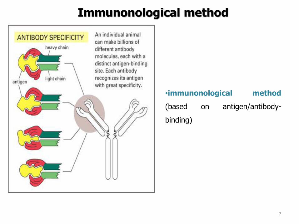

Enzyme-linked immunosorbent assay (ELISA)
httpimageslidesharecdncomelisaonlineversion-110602035913-phpapp0295elisa-test-enzymelinked-immunosorbent-assay-27-728jpgcb=1306989526
How to detect the formation of complex antigen-antibody bull Different systems with
antibodies linked to enzyme (eg horseradish peroxidasealkaline phosphatase) ndash then to add substrate ndash its transformation is measured (eg changes in the absorbance)
Enzyme-linked immunosorbent assay (ELISA)
httpthefutureofthingscomuploadimagearticles2006biopenbiopen-elisa-schematicjpg
bull When the complex antibody-antigen is formed the secondary antibody conjugated with enzyme is to be bound to the antigen and not to be washed off
bull After adding the substrate the substrate is changed and give the signal (eg Measured as absorbance)
Enzyme-linked immunosorbent assay (ELISA)
bull monoclonal or polyclonal antibodies coated microtitre trays to capture target antigen
The captured antigens detected using a second antibody which is conjugated to an
enzyme The addition of enzyme substrate enables the presence of the target antigen to
be visualised
bull considerable specificity
bull can be automated commercially available (Salmonella L monocytogenes
Campylobacter hellipetc)
target antigen
washing
product
second antibody
enzyme substrate
substrate solid phase
with antibody
incubation washing
VIDAS or mini-VIDAS Analyzers
bull the automated multiparametric immunoanalyser
bull based on an enzyme immunoassay which detect target antigens using the
ELFA (Enzyme Linked Fluorescent Assay)
bull 4-methyl-umbelliferone (fluorescent molecule) is released by alkaline phosphatase = fluorescence
VIDAS or mini-VIDAS Analyzers
Each test is composed of two parts
1 The SPRreg acts as a Solid Phase Receptacle for the reaction The SPR is coated
with anti-target antibodies adsorbed on its surface
2 The Strip contains all ready-to-use reagents necessary for the test washing
solution alkaline phosphatase-labeled anti-target antibodies and substrate
Video httpwwwbiomerieux-industrycomfoodvidas-listeria-monocytogenes-detectionVIDAS LMO2 httpswwwyoutubecomwatchv=ZFRuJYynLwk
httpwwwbiomerieuxcom
bioMeacuterieux
VIDAS or mini-VIDAS Analyzers
Validated by ISO 16140 againt ISO 6579 (AFNOR NdegBIO 1216-0905)
Salmonella spp
VIDAS or mini-VIDAS Analyzers
The sample = the sample diluted and enriched in Xpress 2 broth (different from ISO 6579) The used broth must be convenient for the method
Detection of Salmonella spp according to ISO 65792002
01 ml of homogenate to 10 ml RVS incubation at 415degC for 24plusmn3 h
Incubation at 37plusmn1degC for 18plusmn2 h
1 ml of homogenate to 10 ml MKTTn incubation at 37plusmn1degC for 24plusmn3 h
Plating out by 10 micro on two selective media and incubation at 37plusmndegC for 24plusmn 3h
bullXylose Lysine ndash Desoxycholate Agar (XLD)
bullAny other selective medium (BGAHE BS SS DC chromogenic mediahellip)
25 g of sample + 225 ml of buffered peptone water homogenization
Selective enrichment
Pre-enrichment
Isolation on selective agar
plates
Confirmation
Confirmation
Isolation of a charasteristic colony on Nutrient Agar and incubation at 37plusmndegC for 24plusmn 3h
1 day
2 day
3 day
4-6 day
ISO 65792002 Microbiology of food and animal feeding stuffs -- Horizontal method for the detection of Salmonella spp
VIDAS kits
bull E coli O157 - VIDASreg UP E coli O157 (including H7)
bull E coli O157 - VIDASreg ECO
bull E coli O157 (confirmation) - VIDASreg ICE
bull Salmonella spp - VIDASreg SLM
bull VIDASreg ICS
bull Salmonella spp - VIDASreg Easy SLM
bull Listeria spp - VIDASreg LIS
bull Listeria spp - VIDASreg LSX
bull Listeria monocytogenes VIDASreg LMO2
bull Staphylococcal enterotoxins - VIDASreg SET2
bull Campylobacter spp - VIDASreg CAM
GLISA-Rapid Test (Gold Labelled ImmunoSorbent Assay) ndash Lateral Flow Test
Detection limit 105 CFUml in a sample (homogenate of selective enrichment or suspension of colony) (sometimes heat-treatment required - 20 min at 80 degC = cell lysis = better motility of the cell walls with antigens)
httpwwwmeridianbioscienceeumediahome_3columns_images751630jpg
GLISA-Rapid Test
Reaction zone 150 microl of sample
Testing zone Control zone
+ Pos - Neg
Singlepathcopy for detection of Listeria monocytogenes (Merck)
Validation of procedure
Validation of an alternative method bull the validation of an alternative method is the procedure to demonstrate if the alternative method provides equivalent or better results compared to the reference methods
bull Validation is a process within which the method is demonstrated to be suitable
for its purpose It documents methods validity
bull During validation process methods performance characteristics are estimated
The validation of qualitative and quantitative methods comprises two phases
1 a method comparison study of the alternative method against a reference
method (performed by an expert laboratory)
2 a interlaboratory study of the alternative method (organised by an expert
laboratory)
Expert laboratory (organising) laboratory having the qualified staff and skills to
perform the method comparison study and organise the interlaboratory study
Useful documents
ISO 161402003 ndash Microbiology of food and animal feeding stuffs ndash Protocol for the
validation of alternative methods
ISO 170252005 ndash General requirements for the competence of testing and calibration
laboratories
Accreditation bodies that recognize the competence of testing and calibration laboratories
should use this International Standard as the basis for their accreditation
ISOIEC 170112004 Conformity assessment -General requirements for accreditation
bodies accrediting conformity assessment bodies
If a laboratory wishes accreditation for part of all of its testing and calibration activities it
should select an accreditation body that operates in accordance with ISOIEC 17011
It is expected that all laboratories involved in each step of a validation process will have a Quality System or quality assurance (QA) program in place to ensure standardization of laboratory operations as well as adequate quality control (QC) activities
Validation of procedure
Test characteristics The test characteristics for alternative methods are shown
Qualitative methods
1 Selectivity (inclusivity exclusivity)
2 Relative accuracy
3 Detection level
4 Relative sensitivityRelative specificity
5 The agreement between the
methods
Quantitative methods
1 Selectivity (inclusivity exclusivity)
2 Lowest validated level with satisfactory
precision
3 Repeatability
4 Reproducibility
5 Uncertainty of the method
28
PCR METHODS
DNA RNA PROTEINS Deoxyribonucleic acid (DNA)
A linear polymer that consists of four nucleotides
Adenine Cytosine Guanine Thymine Primer binding A ndash T C - G
DNA RNA PROTEINS
DNA
RNA
httpss3amazonawscomclassconnection116flashcards5862116jpgrna-149F82012BD7233971Fjpg httpslh3googleusercontentcomf81cHVr_O-
jXwH1A7g-mfX1LJKZFffUkItKpHPZ3kGF6cGXJaAJ-pqZoNL7T4YNKcP-grQ=s85 httpslh3googleusercontentcomdc9f6E-QDwxJQc-
7MvExwNvrihEjEfgjzwKr2gidCYIt0VmoMb1yEgTDc6mpkFDMUsXsgw=s94
httpsuploadwikimediaorgwikipediacommons00fPeptide_synpng
PCR principle PCR=Polymerase Chain Reaction (1983 by Kary Mullis) in vitro amplification of the part of DNA (usually 100 bp ndash 1500 bp) bordered by two primers (syntetically prepared oligonucleotides)
Typical number of cycles = 30 - 45
httpswwwyoutubecomwatchv=iQsu3Kz9NYo
PCR principle DENATURATION of double-stranded DNA by heat (app 94-95degC initially 5 then in each step 2-3 minutes)
ANNEALING of PRIMERS on the free single strand DNA bull forward primer on the 3acute-
5acute strand bull reverse primer sits on the
5acute-3acute strand Ta = annealing temperature bull 50-65 degC ndash depends on the
primers sequence bull Time 30 s-60 s bull One of the most important
factor for the specifity bull T gtgt Tm= no annealing - no
PCR product bull T ltlt Tm= non-specific
annealing ndash nonspecific products
ELONGATION by DNA polymerase
in vivo starts to synthetize new DNA molecules from 3acute- end of RNA primer
in vitro PCR from 3acute-end the primers
72 degC ndash optimal temperature
45 s-3 minutes (final elongation 5-15 minutes)
httpwwwmuncabiologyscarrPCR_sketch_3gif
PCR principle
httpopenwetwareorgimagesff1CH391L_S12_PCR_exponential_amplificationjpg
Mistake in the beginning ndash the most influence Exponential phase duplication of PCR products amount in each cycle Linear phase The level of amplification is decreased (lower than duplication in each cycle) Plateau Absence or degradation of some component Reasociation can be preferred than primers anealing due to thermodynamical reasons
PCR principle
httpwwwebioecomuploadfile2010031420100314110534183gif
DNA (genomic plasmid DNA)
Primers (forward reverse)
Thermostable
DNA polymerase
Mg2+ (cofactor
for DNA
polymerase)
dNTP mix
(dCTP dATP
dTTP dGTP) ndash
building stones
for newly
synthetized
DNA product
Nuclease free water ndash without Dnase
Rnase presence (enzymes which
degrade DNA RNA)
MASTERMIX
Total volume ndash 25 μl ndash 50 μl
PCR principle
Polymerase Taq DNA polymerase
bull from thermophilic bacteriumThermus aquaticus
(now prepared by recombination)
bull opt temp 75-80degC (150 ntss)
bull active at the room temperature ndashto work on ice
bull inactive at gt90degC but reactivation when cooled
bull only 5acuteexonuklease activity not 3acuteexonuklease
activity (proofreading)
bull 1 mistake at 10-20 000 nts
Hot start polymerases
bull Activity at the room temperature is inhibited - binding on anti-polymerase
antibodies on the wax balls
bull activation after heating at 94 degC (antibodies destroyed)
Other polymerases
bull Proofreading polymerases (with the 3acuteexonuclease activity)
bull Tth polymerase - reverse transcription (Mn2+) or polymerase activity (Mg2+)
1) Releasing DNA from the cell
- lysis of the cell wall by detergents or by heat digesting cell surface proteins by proteinase K
- RNAse treatment for RNA-free genomic DNA
2) Isolation DNA from the released cytoplasmatic contents
- Centrifugation the vaste in pellet (heat isolation)
- Ion-exchange chromatography (commercial kit) ndash binding DNA by ionic interactions to the column (DNA is negative charged) ndash proteins and RNA pass away ndash DNA is later released by using more concentrated salt solution (column is charged positively)
- Phase separation (aqueouschlorophorm phase ndash CTAB method)
DNA isolation
Detection of food-borne pathogens
DNA of microorganism = set of specific genes with specific sequences
Primers are designed to be complementary to sequence of some specific gene
ndash if the complementary sequence is present PCR product will be performed
Main problems about primers designing and applying of PCR
- Is the gene of interest specific enough (can be found only in the detected microorganism)
- Is the primers sequences complementary only to this gene
- Is the primer sequence designed well (requirements about easy and specific annealing ndash depends on the primers sequence)
- Primers can be designed comercially or by using software (httpwwwbiocenterhelsinkifibiProgramsdownloadhtml)
- sequences and genomes needed - wwwpubmedcom
- httpswwwncbinlmnihgovtoolsprimer-blastindexcgiLINK_LOC=BlastHome
PCR products detection The PCR product has got specific length and specific sequence
Visualiton by intercalating agents bind to double-stranded DNA - fluorescence signal bull Ethidium bromide ndash carcinogenteratogen If intercalated the fluorescence of EtBr is significantly higher than in aqueous solution (UV absorbance maxima at 300 and 360nmemitted as orangeyellow light at 590nm) bull Sybr Green DNA-Sybr Green complex absorbs blue light at 488 nm and emits green light at 522 nm Fluorescent tags at 5acute-end of primers Other tag at 5acute -end of primers eg biotin - for immunodetection detected by anti-bodies linked with an enzyme Microarray
Electrophoresis Conventional electrophoresis a single electrical field causes biomolecules to migrate through a matrix according to its mass-to-charge ratio effectively separates DNA fragments up to ~20-30 kb (according to the gel concentration) larger fragments will comigrate (a large band at the top of the gel)
httpwwwrothamstedacuknotebookimagesgelelgif httpstevegallikorgsitesallimagesgelElectrophoresis2jpg
httpabeleewardhawaiieduProtocolsDNA20Gel20Preparation20and20Electrophoresishtm
Electrophoresis Primers (OT1559 a 18ndash1) complementary to DNA coding16S rRNA Campylobacter
jejuni Campylobacter coli a Campylobacter lari
Product is 287 bp
60 V 100 minutes M ndash 100 bp marker (1-5 positive controls) 1- C jejuni CCM 6212 2- C jejuni CCM 6214 3 ndash
C coli CCM 6211 4 ndash C lari CCM 4897 5 ndash C upsaliensis ATCC 43954 6- 15 strains C jejuni Nt- no
template control (negative control)
Genus specific PCR
Electrophoresis DUPLEX PCR =two pairs of primers
C jejuni C1 5 - CAA ATA AAG TTA GAGGTA GAA TGT-3
C4 5 - GGA TAA GCA CTA GCTAGC TGA T-3
Complementary to a part oxidoreductase 159 bp
C coli 18F 5 - GGT ATG ATT TCT ACAAAG CGAG-3
519R 5- ATA AAA GAC TAT CGT CGC GTG-3acute
Complementary to aspartatekinase and flanking ORF 500 bp
Species specific PCR
60 V 100 minutes M ndash 100 bp marker (1-5 positive controls) 1- C jejuni CCM 6212 2- C jejuni CCM
6214 3 ndash C coli CCM 6211 4 ndash C lari CCM 4897 5 ndash C upsaliensis ATCC 43954 6- 15 strains C
jejuni Nt- no template control (negative control)
Melting curve Intercalating dye binds to double-stranded DNA - producing fluorescent signal when excited by light
When samples are heated to melting point double-stranded DNA denatures into single strands freeing dye into solution
ndash Melting temperature (Tm) is the point at which the DNA denatures
ndash Dependent on the length of fragment and G-C content
ndash More base pairs --gt Increase in Tm
ndash Higher G-C content --gt Increase in Tm
bull Unbound dye has reduced fluorescent signal
bull Loss in signal generates melt curve
No Signal Signal Increasing
temperature
Intercalating
dye
Melting curve
Inflex point
bull 1 derivation = 0
bull In this case 2derivation is negative
Real-Time PCR
CT
CT (threshold cycle) ndash the lowest cycle when fluorescence crosses the
determinated value
Video httpswwwyoutubecomwatchv=kvQWKcMdyS4 httpswwwyoutubecomwatchv=pRwoOBuk00c
Real-Time PCR
Food-borne pathogens detection by PCR
Identification of pure cultures - without problems
Detectionenumeration from food matrix directly or after enrichment
bull How to isolate successfully DNA from few cells in 25 g10 g - Efficiency of DNA isolation (isolation by kits heat-lysis CTAB method)
bull How many targets copies are needed for successfully PCR products detection
ndash detection limits
ndash presence of PCR inhibitors (inhibition of polymerase primers annealing)
bull How to distuinguish DNA from dead and live cells - Total DNA is detected (from dead and living cell)
bull For distinguishing of dead cells DNA ndash eg application of EMA (ethidium monoazide) to the sample before DNA isolation ndash EMA irreversibly bound to DNA of dead cells ndash they are bdquoinvisibleldquo for PCR ndash no amplification
bull Direct enumerationdetection only in some specific case ndash simple liquid sample when the required limit is higher than the limit of detection
bull Standardly ndash for PCR is used enriched homogenate (sample diluted in enrichment broth and cultivated) ndash to obtain detectable level of microorganisms
BAXreg System
Salmonella
Listeria monocytogenes
Genus Listeria
Enterobacter sakazakii
E coli O157H7
Staphylococcus aureus Real-time
Campylobacter jejunicolilariReal-time
Vibrio cholerae
parahaemolyticusvulnificus Real-time
Yeast and molds
httpwww2dupontcomDuPon
t_Homeen_USindexhtml
BAX ndash three major steps
On enrichedregrown samples
Prepare samples = perform heat-lysis in two stages
DNA present in the lysate
2 Process samples
3 Review results
Hydrate PCR tablets
Enrichment protocols vary
depending on bacterial target
and type of food yoursquore testing
Salmonella Enterobacter
sakazakii
E coli O157H7 E coli O157H7
MP Campylobacter jejunicoli
Yeast and Mold
37deg +- 2degC for 20 minutes then
95deg +- 3degC for 10 minutes
Listeria and L monocytogenes
55deg +- 2degC for 60 minutes then
95deg +- 3degC for 10 minutes
5 microl for all bacterial targets
20 microl for beef samples enriched
in E coli O157H7 MP media
20 microl for yeast amp mold
PCR tablets bull DNA-polymerase
bull Primers
bull Nucleotids
bull Suplements
bull SybrGreen or fluorescent probe
bull Internal control + primer
bull Mastermix in the tablet in PCR Eppendorf ndash minimal risk of contamination
For solving the problem of distiguishing dead and live cell the process of small volumes
is used ndash only if live cells are present in the sample they able to multiplied to the limit
detection (104 CFUml in the enrichment suspension used for lysis step)
If the result of detection is positive confirmation by plating out is done (live and dead
cells)
+
Target organism DNA BAXreg system
primers
in tablets PCR
+ no detectable
amplification
Background DNA
PCR
BAXreg system
primers
in tablets
Amplification of specific fragments
Detection of specific fragments Fluorescent signal drops with heating to generate a melt curve
Melting curve Raw Data Processed Data
Strong positive
result
Negative
result
Target peak
Positive control peaks
Positive control peak
Melting curves display peaks within a set temperature range for the control and target (if present)
Detection
Melt curve weak INPC in presence of strong Salmonella positive
Real time weak INPC in presence of strong
Campylobacter positives
Direct protocol does not display a positive or
negative result PCR was successful IndeterminateSignal error
Salmonella AFNOR (ISO 16140) protocol Sample 110 BPW
18 h37ordmC
RVS Muumlller-Kauffmann
24 h 415 37 ordmC
5 suspicious colonies on TSA
24 h 37ordm C
Biochemical and serological confirmation
XLD 2 Plate
24 h 37ordmC
BHI
3h 37 ordmC
22 h 37 ordmC
15 d neg and pos result
3 d neg 5 d pos results
Salmonella ( ISO 65792003) versus BAXreg-PCR
International certificates AOAC-RI Performance Tested Method
Salmonella 100201 L monocytogenes
070202 L spp 030502 Saureus 120701
Campylobacter colilarijejuni
AFNOR Certification
Salmonella QUA 183-1102
NordVal Certification
Salmonella 2006-30-5408
Certificates from AFNOR and AOAC are equal
NordVal Certificate
57
OTHER METHODS
ATP bioluminiscence
httpwwwpromegacouk~mediaimagesresourcesfiguresenotesfe0027_fig1jpgla=en
httpwwwbiotekptassetstech_resources143clarity_atp_conc_fig2gif
Principle the detection of the present level of ATP (direct proportion to the presence of microorganisms or their products residues) The enzyme luciferase catalyzes the transformation of d-luciferin to oxyluciferin (indicated by emision of light at 560 nm)
ATP bioluminiscence
httpwwwbioxyscomimages2figure-B11gif
ATP Hygiene Monitoring System - SystemSURE II
httpwwwmicromaticcomdraft-keg-beerline-cleaning-hygiene-pid-ATP-100-tab-reviewshtml
Biosensors
60
httpsuploadwikimediaorgwikipediacommonseecBiosensor_Systemjpg
Biosenzor =the whole cellmicroorganism (CBS ndash cell-based sensors)
Cell-based sensors
Patogenic microorganisms interact during the infection with mammalian
cells ndash detection of cell or toxin by cell culture The damage of cell culture is measured optically or electrically
62
IDENTIFICATION OF MICROORGANISMS
Definition of species in bacteriaarchae
The criterion for a bacterial or archael species a collection of strains that have bull at least one diagnostic phenotypic trait bull ge70 DNAndashDNA hybridization of their purified molecules DNA
bull ~more than 97 16S rRNA sequence identity Under the same conditions some eucaryotic order would be a single bacterial species
The bacterial species definition bull eminent practical significance bull but a very difficult issue
bull very wide and often changes in DNA
bull mutations during replication of DNA
bull horizontal gene transfer through mobile elements plasmids also between species
Criterion for species in prokaryotic cells (bacteria and archea) Collection of strains characterised bull by at least one diagnostic feature bull their purified DNA molecules prove at least 70 hybridisation
which corresponds to at least 97 concordance in the sequence of 16S rRNA gene
Map of genome of B cereus ATCC 14579
httpwwwacgtinccomspecialty_dna_sequencinghtm
Identification based on genotype
64
DNA isolation PCR amplification of 16S rRNA gene (usually)
Sequencing of PCR product
Comparison to sequences in databases (BLAST)
Identification of bacteria PHENOTYPE METHODS bull Morphology of colony and cell (Gram staining shape) bull Requirements for temperature atmosphere nutrition bull Biochemical tests - presence of specific enzymes
bull catalase oxidase fermentation of glucose bull the way how they utilize different compounds
bull Immunochemical tests - specific antigens in the cell wall METHODS BASED ON PROTEIN ANALYSIS
bull MALDI-TOF MS METHODS BASED ON GENOTYPE bull looking for and comparing of specific DNA sequences
bull conserved universal genes (eg 16S rRNA) bull genes specific for genus species
bull done by PCR methods DNA chips sequencing etc
66
BIOCHEMICAL TESTING
Identification based on phenotype I Physiological + biochemical features
bull morfology of colonies and cells (Gram staining shape)
bull requirements for temperature atmosphere nutrient sources
bull basic biochemical tests oxidase catalase fermentationoxidation of glucose haemolysis etc
appearance of colonies on selective or selective-diagnostic agars (for other testing to be reinoculated on non-selective agar)
the classification of an isolate into a certain group
(eg for bacteria catalase positivenegative G+ cocci glucose fermentingnon-fermenting G- rods etc)
biochemical tests specific for certain groups of microorganisms
Identification based on phenotype I
68
Physiological + biochemical features Example Bacillus cereus
httpwwwfoodhaccpcommemberonlynewsletter164html
httpwwwnapavalleyedupeoplesrosePublishingImagesBacillus20cereus202(Gram20stain)jpg
Set of biochemical tests Microgenreg Bacillus ID Panel for the identification of Bacillus spp and related genera (time for performing 48 hours)
MYP agar (Mannitol Egg Yolk Polymyxin Agar) pink colonies with a precipitation zone (no fermentation of manitol + enzyme lecitinase)
catalase positive
httpwwwoxoidcomukblueprod_detailprod_detailasppr=CM0929ampc=ukamplang=ENamporg=9ampimg=CM0929ampsec=
G+ rod endspores forming
β-haemolysis moisty and easy to grow colonies
Principle of biochemical testing
to rank the bacterium to some group having the same basic features bull eg catalase positive gram positive cocci bull Family Enterobacteriaceae
the identification on the genus or species level
bull sets of biochemical tests specific for some group of bacteria
70
Gram staining procedure - principle
https40mediatumblrcom25da2e5b25bbe336819bf1715849d171tumblr_inline_ntrsgn9w1D1sv92o6_540jpg
Gram staining procedure - principle
httpsonlinesciencepsuedusitesdefaultfilesmicrb106CellStructurecow02354_03_15jpg
Gram staining procedure 1-2 drops of sterile water on a slide Spread culture in thin filter in this drop Let dry in airclose to fire Pass slide through flame 2-3x to fix Add crystal violet ndash 15-20 s to act Add Lugol solution (KII2) ndash 15-20 s to act Washing off with a mixture of
ethanolaceton (41) or acetone till the stain removes but max 15 s
Rinse with water decant excess liquid Add safranin 1 minute Rinse with water decant excess liquid Dry and put a drop of imersion oil use an
objective bdquoOILldquo
httpwwwtumblrcomtaggedgram-staining
G- red
G+ violet
Gram staining procedure + Identification
E coli
Family Enterobacteriaceae Gram-negative rods
Micrococcus luteus
Staphylococcus aureus
Gram-positive cocci Bacillus cereus
Gram-positive rods (with endospores)
Biochemical identification of G + bacteria
httpyoungeneticistblogspotcz201106diagnostic-microbiology-labhtml
G+ bacteria bull cocci ndash in clusters in pairs
diplococci bull bacilli
bull endospore bull no endospore
For G+ cocci bull Catalase positive ndash Staphylococcus
sp bull Coagulase + S aureus or some other few species bull Coagulase -
bull Novobiocin S bull S epidermidis etc
bull Novobiocin R bull S saprophyticus etc
bull Catalase negative Streptococcus sp Enterococcus sp
bull kind of haemolysis Other important G + group ndash filamentous bacteria
Catalase test Catalase test Catalase is a common enzyme found in nearly all living organisms that are exposed to oxygen where it catalyzes the decomposition of hydrogen peroxide to water and oxygen
2 H2O + O2 2 H2O2
catalase
bubbling when culture is add in peroxide
httpiws2collinedudcainCCCCD20Microcatalase_testhtm
Catalase testing into a drop of 3 peroxide to transfer a loop of bacterial culture catalase positive ndash releasing of air bubbles
Examples bull G+ cocci catalase positive Staphylococcus sp Micrococcus sp Kocuria sp Dermacoccus sp Macrococcus sp Rothia sp catalase negative Streptococcus sp Enterococcus sp bull G+ rods (bacilli) endospores forming catalase positive Bacillus sp catalase negative Clostridium sp
Resistance to ATBhaemolysis Resistance eg to novobiocin
httpquizletcom20077004mic-lecture-1-t2-flash-cards
Hemolysis on blood agar
Measuring the diameter of inhibition zone (less than 17 mm resistant more or equal to 17 mm sensitive) ndash differing in Staphylococcus sp (Clinical performance)
alpha-hemolysis - oxidizing the iron in the hemoglobin - green discoloration of the medium around beta-hemolysis - rupture of erythrocytes - complete clearing medium around gamma-hemolysis ndash no effect ndash no change
By cultivation on blood agar bacteria can be
differentiated based on their capacity to secrete
hemolysins The hemolysis will cause a clearing zone of
the blood agar around the colonies Bacteria can cause
different types of hemolysis
bullα-hemolysis which means an incomplete clearing
(green haemolysis)
bullβ-hemolysis which means a complete clearing
bullDouble hemolysis of some staphylococci consisting of
an inner β-hemolysis zone and an outer α-hemolysis
zone
bullNo hemolysis which is sometimes referred to as γ-
hemolysis which may seem illogical
Note that the α-hemolysin of staphylococci causes
complete hemolysis whereas their β-hemolysin causes
incomplete hemolysis
The capacity to produce hemolysins may vary between
different strains of a particular bacterial species
httpwwwvetbactorgvetbactdisplayextinfo=67
Identification of bacteria based on haemolysis
Streptococcal hemolysins α (alpha) hemolysis - ldquozone of green discolourationrdquo or ldquogreeningrdquo called partial or incomplete hemolysis produced by alpha-hemolytic streptococci such as Streptococcus suis This type of hemolysis on BA is due to bacterial production of hydrogen peroxide which oxidises hemoglobin to methemoglobin β (beta) hemolysis ndash a clear zone of complete hemolysis produced by beta-hemolytic streptococci such as S equi subsp equi This hemolysis is due to lysis of RBCs in the BA by the cytotoxin Streptolysin S
Source httppeopleupeicajlewishtmlhemolysinshtml
Bacterial hemolysins
Staphylococcal hemolysins Most coagulase-positive staphylococci (CPS) such as Staphylococcus aureus S pseudintermedius ( but not S hycius) typically produce an distinctive pattern of ldquodouble zonerdquo or ldquotargetrdquo hemolysis on blood agar Double-zone hemolysis indicates that the two hemolysins one which produces ldquocompleterdquo and another one that produces ldquoincompleterdquo hemolysis are present and this combined effect is visible due to different diffusion rates of these enzymes through the agar The staphylococcal α (alpha) hemolysin causes a complete type of hemolysis This zone of complete hemolysis is narrow and immediately adjacent to the bacterial colony on blood agar The staphylococcal β (beta) hemolysin causes an incomplete type of hemolysis at 37 degC This broader incomplete zone is outside of the inner complete zone and can be hard to see on early culture plates and also varies with the species of RBCs used to make the BA plate Its action is enhanced by chilling at 4 degC and the hemolysis becomes complete (this effect is referred to as ldquohot-coldrdquohemolysis) There are other staphylococcal cytotoxins that are also hemolysins but their lytic actions are not seen on blood agar
The confusion arises due to the NAMES of the two STAPHYLOCOCCAL HEMOLYSINS and the TYPE of hemolysis they produce These are the REVERSE of the two TYPES of STREPTOCOCCAL HEMOLYSIS Strictly speaking the descriptive terms ldquoalphardquo for ldquoincompleterdquo and ldquobetardquo for ldquocompleterdquo hemolysis apply only to the hemolytic toxins of Streptococcus species However other organisms beside Streptococcus and Staphylococcus species have cytotoxins that cause hemolysis and this hemolysis can be either complete or incomplete It is preferable to use the terms ldquocompleterdquo and ldquoincompleterdquo rather than ldquobetardquo or ldquoalphardquo to describe the hemolysis caused by these organisms but you will notice in bacteriology text books that the terms alpha and beta are commonly used
G ndash bacteria bull Cocci bull Bacilli (Rods) bull Oxidase positivenegative bull Glucose metabolism
bull fermentation bull oxidation bull Inactive
bull Lactose metabolism for coliform
bull Pigment formation for Pseudomonas sp
Biochemical identification of
G - bacteria
Oxidase test
Oxidase positive (OXI +) Aeromonas sp Pseudomonas sp Dark-blue to violet
Oxidase negative (OXI -) Enterobacteriaceae Bacillus sp Colourless
Oxidase bullabbreviated general name for some kind of cytochrome c oxidases (cytochrome oxidase or indophenol oxidase) enzymes involved in aerobic respiratory chain when oxygen is used as the final receptor for hydrogen bullOxidases are usually present only in aerobic organisms bullPresence or absence of oxidase(s) is an important feature of the bacterial metabolism used mainly in the identification process of gram-negative bacteria
Detection Reaction of the culture with redox indicator as eg NN-dimethyl-p-phenylenediamine (DMPD) dark-blue when oxidized and colorless when reduced Different test performances are possible
Fermentation of carbohydrates Type of utilisation of glucose or carbohydrates bull an important feature of the bacterial metabolism bull widely used in the identification process of bacteria bull fermentation strictly means to be under anaerobic conditions (but in bacterial
metabolism which can be but not have to be the same as the anaerobic conditions of cultivation) so very often is used in a general way like bdquoutilisationldquo
bull fermentation (like bdquoutilisationldquo) of carbohydrates to
bull acid and gas (Durham tubes) bull acid without gas bull non-utilised
bull Testing of fermentation or oxidation of carbohydrates
bull eg OF test for glucose bull by oxidation under aerobic conditions bull by fermenation under anaerobic conditions (an oil layer)
Fermentation of sugars to acid
Peptone Water wPhenol Red
(HiMedia M028I)
Ingredients gl
Peptic digest of animal tissue 10000
Sodium chloride 5000
Phenol red 0020
Final pH ( at 25degC) 68plusmn02
httpsuploadwikimediaorgwikipediacommons009Phenol_red_pH_60_-_80jpg
+ after sterilisation
at the room
temperature to add
aseptically sugar
discs and
inoculate
succrose trehalose rhamnose xylose
Incubation 30 degC 24 h
- +
Fermentation of carbohydrates
Casein enzymic hydrolysate and yeast extract provide nitrogenous compounds vitamin B complex and other growth nutrients Dextrose is the fermentable carbohydrate and bromocresol purple acts as the pH indicator with colour change from purple to yellow in acidic conditions Sodium chloride maintains osmotic equilibrium bdquoAnaerobicldquo conditions ndash inoculation by stabbing to used fresh prepared media (oxygen expelled by heating medium in boiling water or running steam for 15 min then to be cooled down quickly)
- +
Enterobacteriaceae ndash glucose fermentation
83
Tryptone Yeast Extract Agar Ingredients Gms Litre Casein enzymic hydrolysate 10000 Yeast extract 1500 Dextrose 10000 Sodium chloride 5000 Bromocresol purple 0015 Agar 15000 Final pH ( at 25degC) 70plusmn02
Test for oxidationfermentation of glucose (OF glucose test) detection of acid production by acidobazic indicator change to yellow (acid present) in lower pH from green (no acid) Anaerobic atmosph ndash covering by sterile mineral oil If fermented anaerobically then also runs aerobically
Fermentation of carbohydrates
Principle of biochemical tests bull The first step in identification is to rank the bacterium to some group of bacteria
having the same basic features ndash eg catalase positive gram positive cocci bull The identification on the genus or species level is finished by other biochemical tests
specific for each group of bacteria bull The tests can be done in tubes or miniaturized plate
ENTEROtest 24N (Erba Lachema) (24 means 24 biochemical tests N= new)
API 20E (bioMeacuterieux)
bull designed for routine identification of clinically important species of family Enterobacteriaceae without the use of reagents
bull A strip test OXItest or OFtest in microtitration wells can be used to confirm that the isolate belongs to Enterobacteriaceae family
bull The results are read visually or automatically using reader MIKROLAreg
bull The identification can be supplemented by diagnostic strip tests INDOLtest COLItest and VPtest
APIreg Gram negative Identification API 20E ndash Speciessubspecies identification of Enterobacteriaceae and groupspecies identification of non-fermenting Gram negative bacteria API Rapid 20E ndash 4-hour identification of Enterobacteriaceae API 20NE ndash 24 to 48-hour identification of Gram negative non-Enterobacteriaceae API Campy ndash 24-hour identification of Campylobacter species
Example Escherichia coli
ENTEROtest 24 N
Biochemical testing - Enterobacteriaceae bull TSI Triple Sugar Iron Agar (use a straight wire to stab the butt and streak the agar surface)
- to determine whether organisms can ferment glucose sucrose andor lactose with or without production of gas and the ability of the organism to produce hydrogen sulphide from thiosulphate in an acid environment is also tested
Formula Liter Enzymatic Digest of Casein 5 g Enzymatic Digest of Animal Tissue 5 g Yeast Enriched Peptone 10 g Dextrose 1 g Lactose 10 g Sucrose 10 g Ferric Ammonium Citrate 02 g Sodium Chloride 5 g Sodium Thiosulfate 03 g Phenol Red 0025 g Agar 135 g Final pH 73 plusmn 02 at 25 degC
inoculation in the butt by stab inoculation on the slant on the surface bull Fermentation of dextrose
(glucose) to acid bull anaerobically - yellow butt bull aerobically leads to products
which are later oxidised by air and pH remains alkaline (red) bull Fermentation of glucose to gas (anaerobically - in the stab) Cracks splits or bubbles in medium indicate gas production bull Production of hydrogen sulfide (H2S) ndash reaction with ferrous sulphate and sodium thiosulphate - a black precipitate in butt bull Lactose andor succrose
utilisation to acid Acid production ndash yellow butt and slant
Results
An alkaline slant-acid butt
(redyellow) - fermentation of
dextrose only
An acid slant-acid butt
(yellowyellow) - fermentation of
dextrose lactose andor sucrose
An alkaline slant-alkaline butt
(redred) - dextrose or lactose not fermented (non-fermenter)
Bacterium fermenting lactose andor succrose are able to ferment glucose
Phenol red ndash alkaline = red acid = yellow
bull TSI Triple Sugar Iron Agar (use a straight wire to stab the butt and streak the agar surface)
- to determine whether organisms can ferment glucose sucrose andor lactose with or without production of gas and the ability of the organism to produce hydrogen sulphide from thiosulphate in an acid environment is also tested
Biochemical testing - Enterobacteriaceae
Urea agar (Christensen) (use a 1 μl loop full)
- to determine whether an organism can split urea into ammonia and
carbon dioxide by the action of the enzyme urease
httpimageslidesharecdncomlab17-141201085153-conversion-gate0195-10-638jpgcb=1417424219
httpimageslidesharecdncomhelicobacterpylori-copy-110516201901-phpapp0195campylobacter-jejunihelicobacter-pylori-39-728jpgcb=1305577391
Phenol red
Medium is acidic - yellow
(pH 68plusmn02)
Alkaline reaction by urea hydrolysis - pink colour
Composition Gms Litre
Peptic digest of animal tissue
1000
Dextrose 1000
Sodium chloride 5000
Disodium phosphate 1200
Monopotassium phosphate 0800
Phenol red 0012
Agar 15000
Final pH ( at 25degC) 68plusmn02
Nutrition
Peptic digest of animal tissues dextrose
Sodium chloride - osmotic equilibrium
Phosphates - buffering of the medium
35-37 degC
Biochemical testing - Enterobacteriaceae
bull L-lysine decarboxylase (use a 1 μl loop full)
- to determine whether an organism can decarboxylate an amino acid (lysine) leading to formation of an
amine After inoculation both tubes are overlayed with sterile paraffin oil
httpfce-studynetdna-sslcomimagesupload-flashcards10295302332621_mjpg
+ CO2 + OH-
Lysine Decarboxylase Broth Typical Composition (glitre) Peptic digest of animal tissue 5000 Yeast extract 3000Dextrose 1000 (glucose prepared from starch chemically equivalent to glucose) L-Lysine hydrochloride 5000 Bromocresol purple 0020 Final pH ( at 25 degC) 68plusmn02 Inoculate below the surface cover by sterile paraffin oil
Two steps detection 1 step Dextrose is fermented ndash H+ is released Acidic conditions bull bromocresol purple changes colour to yellow bull in acidic conditions lysine decarboxylase is
activated 2 step L-lysine is decarboxylated and OH- released Alkaline conditions bull bromocresol purple changes colour back to
purple
Biochemical testing - Enterobacteriaceae
bull β-galactosidase (ONPG) (use a 1 μl loop full)
- to determine whether an organism has the enzyme szlig-galactosidase by using the compound o-
nitrophenyl-beta-D-galactopyranoside (ONPG)
- this test is used to differentiate between lactose-negative and lactose-delayed organisms
- ONPG is hydrolysed by the enzyme to o-nitrophenol which is yellow and galactose
O-Nitrophenyl-β-D-galactopyranoside (ONPG) is structurally similar to lactose (ie ONPG is an analog of lactose) except that orthonitrophenyl has been substituted for glucose ONPG is colorless compound O-nitrophenol is yellow visual evidence of hydrolysis
Lactose fermenting bacteria (ONPG positive) two enzymes Permease ndash transferring of the lactose molecule into the bacterial cell β-galactosidase - cleaving the galactoside bond producing glucose and galactose - inducible enzyme - made ONLY in the presence of the lactose substrate Coliform bacteria as Ecoli Klebsiella spp Enterobacter spp
hydrolysis
Late lactose fermentering bacteria Permease ndash not present delayed in the production of acid from lactose because of sluggish permease activity β-galactosidase ndash present - give a positive ONPG test Citrobacter spp Arizona spp
Non lactose fermenter (ONPG Negative) Permease ndash not present β-galactosidase ndash not present Salmonella spp Shigella spp Proteus spp Providencia spp and Morganella spp
Biochemical testing - Enterobacteriaceae
Procedure Culture Bacteria grown in medium containing lactose (to induce the production of the galactosidase enzyme) such as Kligler iron agar (KIA) or Triple sugar Iron (TSI) agar (Note β-galactosidase enzyme (inducible enzyme) is made ONLY in the presence of the lactose substrate) Inoculation 1A loopful of bacterial growth is emulsified in 005mL of physiologic saline to produce a heavy suspension 2One drop of toluene is added to the suspension and vigorously mixed for a few seconds to release the enzyme for bacterial cells 3An equal quantity of buffered ONPG solution is added to the suspension 4The mixture is placed in a 37 oC water bath 5When Using ONPG Tablets 6A loopful of bacterial suspension is added directly to the ONPG substrate resulting from adding 1mL of distilled water to a tablet in a test tube 7This suspension is also placed in a 37oC water bath
β-galactosidase (ONPG)
Results and Interpretations The rate of hydrolysis of ONPG to o-nitrophenol may be rapid for some organisms producing a visible yellow color reaction within 5 to 10 minutes Most tests are positive within 1 hour however reactions should not be interpreted as negative before 24 hours of incubation The yellow color is usually distinct and indicates that the organism has produced o-nitrophenol from the ONPG substrate through the action of β-galactosidase
Biochemical testing - Enterobacteriaceae
bull VogesndashProskauer (VP) test (use a 1 μl loop full)
- to determine whether an organism can produce acetylmethylcarbinol
(acetoin) from fermentation of glucose If present acetylmethyl carbinol is
converted to diacetyl in the presence of prop- naphthol (to be added first) strong
alkali (40 KOH) and atmospheric oxygen The diacetyl and quanidine-
containing compounds found in the peptones of the broth then condense to
form a pinkish red polymer
MRVP broth (pH 69)
Ingredients per liter of
deionized water
buffered peptone= 70 gm
glucose= 50 gm
dipotassium phosphate= 50 gm Voges-Proskauer Reagent A Barrittrsquos reagent A
Alpha-Naphthol 5 50 gm
Absolute Ethanol 1000
ml
Voges-Proskauer Reagent B Barrittrsquos reagent B
Potassium Hydroxide 400 gm
Deionized Water 1000 ml
Procedure of VogesndashProskauer (VP) Test
1Prior to inoculation allow medium to
equilibrate to room temperature
2Using organisms taken from an 18-24
hour pure culture lightly inoculate the
medium
3Incubate aerobically at 37 degC 24 hours
4Following 24 hours of incubation aliquot
2 ml of the broth to a clean test tube
5Re-incubate the remaining broth for an
additional 24 hours
6Add 6 drops of 5 alpha-naphthol and
mix well to aerate
7Add 2 drops of 40 potassium hydroxide
and mix well to aerate
8Observe for a pink-red color at the surface
within 30 min Shake the tube vigorously
during the 30-min period
Positive Reaction
A pink-red color at the surface
Examples Viridans group streptococci
(except Streptococcus vestibularis) Listeria
Enterobacter Klebsiella Serratia marcescens
Hafnia alvei Vibrio eltor Vibrio alginolyticus etc
Negative Reaction
A lack of a pink-red color
Examples Streptococcus mitis Citrobacter sp
Shigella Yersinia Edwardsiella Salmonella Vibrio
furnissii Vibrio fluvialis Vibrio vulnificus and Vibrio
parahaemolyticus etc
A copper color should be considered negative A
rust color is a weak positive reaction
Quality Control of VogesndashProskauer (VP) Test
VP positive Enterobacter aerogenes (ATCC13048)
VP negative Escherichia coli (ATCC25922)
Biochemical testing - Enterobacteriaceae
94
VogesndashProskauer (VP) test
Rapid test for confirmation of E coli ndash typical features
β-D-glucuronidase (97 strains) production of indole from tryptophan (typical for E coli)
Performance
Previous testing Gram-negative (eg able to grow on VRBD agar) oxidase negative
From pure culture to prepare suspension in saline solution (app 5 ml) at the density 3 McFarland
To transfer 05 ndash 1 ml of this suspension into a new tube ndash to add the strip of COLItest (chemicals ndash fluorogenic substrate for β-D-glucuronidase tryptophane are present in the pad at the end of the strip)
Incubation 37 degC 4 hours
Detecion of β-D-glucuronidase (to be done at first) UV (360 nm) ndash blue fluorescence (GLR + hellipβ-D-glucuronidase positive) ndash GLR test can be read after 1 hour
Detection of indole formation from tryptophane (to be done at second) to add Kovacs reagent (IND + hellipformation of red ring)
95
COLItest - Escherichia coli
Biochemical testing - Enterobacteriaceae
Fluorogenic culture medium
bull One or more fluorogenic substrates
bull For detection of specific enzyme which are able to cleave out the fluorogenic substrate that a designed
fluorophore (responsible for the UV fluorescence) is released
FLUOROGENIC MEDIUM (Colilert) E coli use β-glucuronidase to metabolize MUG and create fluorescence (UV 360 nm) Since most non-coliforms do not have these enzymes they are unable to grow and interfere
httpgawaterusgsgovprojectsbacteriapicturessummaryanalysisecolijpg
E coli (MPN method)
COLItest Biochemical testing - Enterobacteriaceae
COLItest bull Indole test (use a 1 μl loop full)
- to determine whether an organism can split indole from tryptophan
httpwwwmicrobiologyinfocomindole-test-principle-reagents-procedure-result-interpretation-and-limitations
indole production from tryptophan
Production of indole is detected by Kovacacutes reagent
Indole Kovacs Reagent
p-Dimethylaminobenzaldehyde 500 gm
Hydrochloric Acid 37 2500 ml
Amyl Alcohol 7500 ml
Biochemical testing - Enterobacteriaceae
INDOLe test ndash quick variant
Ecoli Citrobacter Enterobacter
Procedure bull on the filtration paper to add a drop of INDOL suspension bull to spread into by a loop some culture bullTo incubate ndash at the room temperature for 5 minutes bullTo read results
Biochemical testing - Enterobacteriaceae
ENTEROtest 24 ndash example of biochemical testing
routine identification of clinically important species of family Enterobacteriaceae the test is focused only on some species often isolated in clinical practis strains from other sources can be slightly different in the dominant biochemical profiles intended exclusively for the identification of family Enterobacteriaceae the preliminary ranking into family Enterobacteriaceae must be done (Gram staining oxidase glucose fermentation) the results of reactions obtained in this modified micromethod may differ from the results obtained using conventional tests and results available in literature sources in case of problem to control positivenegative reactions by control strains or to use for this tests tube variant to check the functionality of the kit not to check the accuracy or success of the identification
Preparation of suspension from over-night culture growing on non-selective agar (for staphylococci Columbia agar blood with 5 of sheep blood is recommended)
Inoculation of microtitre plates anaerobic reaction covered by mineral oil
Incubation
Reading results by both visual and instrumental mode respectively Standard off-line tests INDOLtest or COLItest Other off-line tests OXItest OFtest VPtest Acetoin test
ENTEROtest 24 N ndash example of biochemical testing
bull tube with 5 ml of sterile physiolsol bull suspension (1 McFarland) bull application 100 microl to each hole bull 2 drops of parafin oil into holes D-H of the first line (anaerobic conditions) bull Incubation 37degC 24h bull for check the purity of suspension to streak suspension on TSA (incubation 37 degC ndash 24 h
ENTEROtest 24N
ENTEROtest 24 N - evaluation
Escherichia coli
H 0+0+0=0 G 0+2+0=2 F 1+2+0=3 hellip Identification code 023426
1 1
2
4
ENTEROtest 24 ndash example of biochemical testing
The result = a set of biochemical properties (+-) which is compared to the sets of biochemical properties of different chosen genera and species for them the test is designed
ENTEROtest 24 N
T-index how is the likelihood of the different properties The combination of identication and T-index = the reliability of the result Excelent very good good (this case) acceptable intermediary strain needs some additional tests genus level non-existing taxon
Evaluation by using the identification code - By using a code book - By using software TNW
Identification score the measure of similarity to proposed taxon
The way of identification the obtained profile is compared with profiles in database bull The most similar profiles are found
bull Identification score = of similarity bull But what about the different reactions
bull T-index is calculated bull how is the probability of the different reactions
bull It depends on their importance ndash typical reactions should be the same
bull Reliability of the result bull the combination of identication score and T-index = bull Excelent very good good acceptable intermediary strain needs some
additional tests genus level non-existing taxon
ENTEROtest 24 ndash example of biochemical testing
Biochemical tests - divisions
ENTEROtest 24 species of family Enterobacteriaceae
NEFERMtest 24 Gramnegative non-fermenting bacteria and families Vibrionaceae Aeromonadaceae and Pleiomonas shigelloides present mainly in clinical material
ANAEROtest 23 ANAEROBIC BACTERIA (G + AND G-) routine identification of anaerobic bacteria (G + AND G-) in clinical material and in food
STREPTOtest 24 Streptococcus Enterococcus and related gram positive catalase negative cocci isolated from clinical material
STAPHYtest 24 genus Staphylococcus isolated from clinical material and for their distiguishing from related genera of other gram positive catalase positive cocci
Biochemical tests APIreg Gram positive Identification API Staph ndash Overnight identification of clinical staphylococci and micrococci API 20 Strep ndash 4 or 24-hour identification of streptococci and enterococci API Coryne ndash 24-hour identification of Corynebacteria and coryne-like organisms API Listeria ndash 24-hour identification of all Listeria species APIreg Gram negative Identification API 20E ndash Speciessubspecies identification of Enterobacteriaceae and groupspecies identification of non-fermenting Gram negative bacteria API Rapid 20E ndash 4-hour identification of Enterobacteriaceae API 20NE ndash 24 to 48-hour identification of Gram negative non-Enterobacteriaceae API Campy ndash 24-hour identification of Campylobacter species APIreg Anaerobe Identification API 20A ndash 24-hour identification of anaerobes Rapid ID 32 A ndash 4-hour identification of anaerobes
MicrogenTM GN-ID Convenient 12 substrate identification system for commonly encountered Enterobacteriaceae (GNA) Extended 24 substrate identification system for the complete range of Enterobacteriacea and oxidase positive non-fastidious Gram negative bacilli (GNA+B) MicrogenTM Listeria-ID Enhanced built-in haemolysis test MicrogenTM Bacillus-ID 24 substrate system for the identification of mesophilic Bacillus spp isolated from foods and related samples MicrogenTM Staph-ID 12 substrate system minimizes handling and manipulation Substrates selected specifically for Staphylococcus and related organisms MicrogenTM Strep-ID Substrates selected specifically for Streptococcus Enterococcus and related species Provides excellent differentiation of Enterococci spp especially E casseliflavus E mundtii and E gallinarum
Biochemical tests
Identification of microorganisms by MALDI-TOF MS
Department of Biochemistry and Microbiology University of Chemistry and Technology Prague
Sabina Purkrtovaacute1 Petra Junkovaacute2 1Laboratory of food microbiology 2Laboratory of applied proteomics
SabinaPurkrtovavschtcz PetraJunkovavschtcz
MALDI-TOF MS
1) sample preparation bull microbial culture or its
proteins extract is smearing onto a steel plate and covered over by matrix
bull matrix enables the sample (A) to be desorped and ionised as pseudomolecule ionts [A+H] +
2) MALDI-TOF MS analysis unique mass spectral fingerprint of desorbed microbial cell components (mainly intracellular proteins) different among genera species or also some strains
Matrix-Assisted Laser Desorption Ionization-Time of Flight Mass Spectrometry
httppubsrscorgencontentarticlehtml2014RAC4RA05604C
httpcmrasmorgcontent263547figures-only
3) identification comparison of mass spectrum to those of reference strains in database
MALDI-TOF MS PRINCIPLE
111
2
2
2L
teU
z
m
m mass z charge
L length of drifting zone e
elementary charge U
speeding voltage
Time of flight is a function of the specific ion mass (mz)
httpwwwsigmaaldrichcomtechnical-documentsarticlesbiologycustom-dna-oligos-qc-analysis-by-mass-spectrometryhtml
Soft ionisation method bull low level of sample fragmentation
Animation http cmgmstanfordedupansection_htmlMS
MALDI-TOF MS SAMPLE AND MATRIX
httpcmrasmorgcontent263547figures-only
Matrix bull able to absorb the energy of the laser (usually 337 nm) bull to crystalise with samples - usually acid
character (proton ionisation of sample) dissolved in organic solvent
Sample preparation Direct transfer bull intact cells spread directly onto a steel plate bull lysis of cells occurs during the contact with acid
matrix and by laser desorption bull most bacteria Proteins extraction bull previous extraction of proteins by organic acids
andor alcohol (eg ethanol and 70 formic acid) bull yeasts moulds some species of bacteria (depending
on the cell wall composition)
CHC α-Cyano-4-hydroxycinnamic acid (organic solvent 50 acetonitrile with 25 trifluoracetic acid) SA 35-Dimethoxy-4-hydroxycinnamic acid (sinapic acid)
DHB 25-Dihydroxybenzoic acid
httpwwwsigmaaldrichcomcatalogproductsigmac8982lang=enampregion=CZ
MALDI-TOF MS MICROORGANISMS
113
Mass spectrum protein profile z equals usally to 1+ (so mz usually corresponds to molecula mass) the range usually used for identification 2000 -20 000 mz the intensity of single peaks corresponds to the abundance of the protein Which proteins dominates in the protein profile abundant basic and mediumly hydrophobic mainly ribosomal proteins further cold-shock and heat-shock proteins chaperons etc
conserved house-keeping gene = conserved proteins = the acordance with identification based on DNA
Visualisation of mass spectrum protein profile ndash (software mMass 5 Strohalm et al 2010)
Bacillus cereus
Analysis is recomended (and validated) to be performed from colonies grown onto non-selective agar
MALDI-TOF MS ANALYSIS
114
Comparison of mass spectrum protein profile of unknown sample with these of reference strains present in database by software
Commercial databases from different MALDI-TOF MS producers Bruker Daltonics ndash MALDI BIOTYPER Shimadzu - Shimadzu Launchpad software + SARAMIS database Biomeacuterieux - VITEKreg MS Other databases compatible with different hardware systems (eg Andromas)
BioTyper The statistical analysis for correlation includes peak positions intensities and frequencies across the complete range of microorganisms Score value 0 (none similarity) - 1000 (absolute similarity) But it is expressed in decadic logarithm log(score value) 0-3
Range Description Symbols Color
2300 3000
highly probable species identification
( +++ ) green
2000 2299
secure genus identification probable species identification
( ++ ) green
1700 1999
probable genus identification ( + ) yellow
0000 1699
not reliable identification ( - ) red
Bacillus cereus
115
Range Description Symbols Color
2300 3000
highly probable species identification
( +++ ) green
2000 2299
secure genus identification probable species identification
( ++ ) green
1700 1999
probable genus identification ( + ) yellow
0000 1699
not reliable identification ( - ) red
Rank (Quality)
Matched Pattern Score Value
NCBI Identifier
1 ( +++ )
Bacillus cereus DSM 31T DSM 2554 1396
2 ( ++ )
Bacillus cereus 994000168 LBK 2203 1396
3 ( ++ )
Bacillus weihenstephanensis DSM 11821T DSM
2158 86662
4 ( ++ )
Bacillus mycoides DSM 2048T DSM 2155 1405
5 ( ++ )
Bacillus cereus 4080 LBK 2147 1396
6 ( + )
Bacillus thuringiensis DSM 2046T DSM 1975 1428
7 ( + )
Bacillus pseudomycoides DSM 12442T DSM 1787 64104
8 ( - )
Bacillus bataviensis DSM 15601T DSM 1369 220685
9 ( - )
Brevibacterium linens IMET 11075T HKJ 1347 1703
10 ( - )
Acinetobacter towneri DSM 14962T HAM 1345 202956
Bacillus cereus
Bacillus cereus
MALDI-TOF MS - PROCEDURE Direct method smearing sample in four parallels in lower and higher cells concentrations ndash after drying to cover over by matrix (1-2 microl) and let to crystallise at room temperature
Matrix solution of α-Cyano-4-hydroxycinnamic acid (10 mgml) in 50 acetonitrile with 25 trifluoroacetic acid (prepared with 10 TFA solution)
Protein standard (1 μl) Bruker Bacterial Test Standard (Bruker Daltonics SRN) ndash proteins extracted from z Escherichia coli DH5alpha BRL + some others
Equipment Bruker Autoflex Speed Database MALDI Biotyper 31
116
MALDI-TOF MSPROTEIN STANDARD
117
Analyte name Rank (Quality) Matched Pattern Score Value
F4 1 ( +++ ) Escherichia coli DH5alpha BRL 2439
Thank you for your attention
Questions to be sent to SabinaPurkrtovavschtcz
118
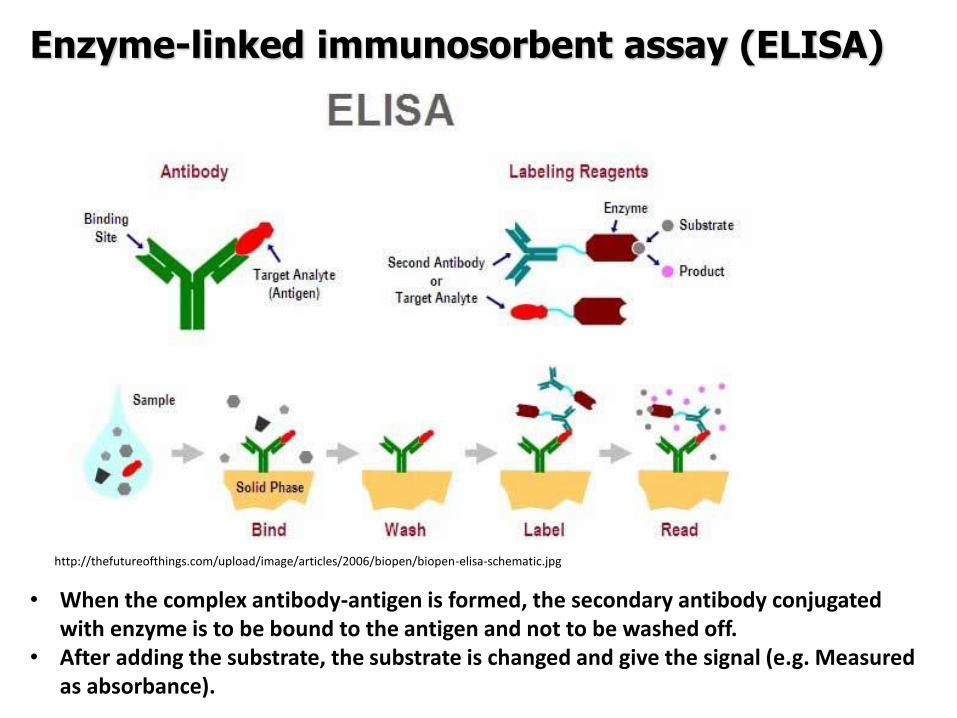
Enzyme-linked immunosorbent assay (ELISA)
httpthefutureofthingscomuploadimagearticles2006biopenbiopen-elisa-schematicjpg
bull When the complex antibody-antigen is formed the secondary antibody conjugated with enzyme is to be bound to the antigen and not to be washed off
bull After adding the substrate the substrate is changed and give the signal (eg Measured as absorbance)
Enzyme-linked immunosorbent assay (ELISA)
bull monoclonal or polyclonal antibodies coated microtitre trays to capture target antigen
The captured antigens detected using a second antibody which is conjugated to an
enzyme The addition of enzyme substrate enables the presence of the target antigen to
be visualised
bull considerable specificity
bull can be automated commercially available (Salmonella L monocytogenes
Campylobacter hellipetc)
target antigen
washing
product
second antibody
enzyme substrate
substrate solid phase
with antibody
incubation washing
VIDAS or mini-VIDAS Analyzers
bull the automated multiparametric immunoanalyser
bull based on an enzyme immunoassay which detect target antigens using the
ELFA (Enzyme Linked Fluorescent Assay)
bull 4-methyl-umbelliferone (fluorescent molecule) is released by alkaline phosphatase = fluorescence
VIDAS or mini-VIDAS Analyzers
Each test is composed of two parts
1 The SPRreg acts as a Solid Phase Receptacle for the reaction The SPR is coated
with anti-target antibodies adsorbed on its surface
2 The Strip contains all ready-to-use reagents necessary for the test washing
solution alkaline phosphatase-labeled anti-target antibodies and substrate
Video httpwwwbiomerieux-industrycomfoodvidas-listeria-monocytogenes-detectionVIDAS LMO2 httpswwwyoutubecomwatchv=ZFRuJYynLwk
httpwwwbiomerieuxcom
bioMeacuterieux
VIDAS or mini-VIDAS Analyzers
Validated by ISO 16140 againt ISO 6579 (AFNOR NdegBIO 1216-0905)
Salmonella spp
VIDAS or mini-VIDAS Analyzers
The sample = the sample diluted and enriched in Xpress 2 broth (different from ISO 6579) The used broth must be convenient for the method
Detection of Salmonella spp according to ISO 65792002
01 ml of homogenate to 10 ml RVS incubation at 415degC for 24plusmn3 h
Incubation at 37plusmn1degC for 18plusmn2 h
1 ml of homogenate to 10 ml MKTTn incubation at 37plusmn1degC for 24plusmn3 h
Plating out by 10 micro on two selective media and incubation at 37plusmndegC for 24plusmn 3h
bullXylose Lysine ndash Desoxycholate Agar (XLD)
bullAny other selective medium (BGAHE BS SS DC chromogenic mediahellip)
25 g of sample + 225 ml of buffered peptone water homogenization
Selective enrichment
Pre-enrichment
Isolation on selective agar
plates
Confirmation
Confirmation
Isolation of a charasteristic colony on Nutrient Agar and incubation at 37plusmndegC for 24plusmn 3h
1 day
2 day
3 day
4-6 day
ISO 65792002 Microbiology of food and animal feeding stuffs -- Horizontal method for the detection of Salmonella spp
VIDAS kits
bull E coli O157 - VIDASreg UP E coli O157 (including H7)
bull E coli O157 - VIDASreg ECO
bull E coli O157 (confirmation) - VIDASreg ICE
bull Salmonella spp - VIDASreg SLM
bull VIDASreg ICS
bull Salmonella spp - VIDASreg Easy SLM
bull Listeria spp - VIDASreg LIS
bull Listeria spp - VIDASreg LSX
bull Listeria monocytogenes VIDASreg LMO2
bull Staphylococcal enterotoxins - VIDASreg SET2
bull Campylobacter spp - VIDASreg CAM
GLISA-Rapid Test (Gold Labelled ImmunoSorbent Assay) ndash Lateral Flow Test
Detection limit 105 CFUml in a sample (homogenate of selective enrichment or suspension of colony) (sometimes heat-treatment required - 20 min at 80 degC = cell lysis = better motility of the cell walls with antigens)
httpwwwmeridianbioscienceeumediahome_3columns_images751630jpg
GLISA-Rapid Test
Reaction zone 150 microl of sample
Testing zone Control zone
+ Pos - Neg
Singlepathcopy for detection of Listeria monocytogenes (Merck)
Validation of procedure
Validation of an alternative method bull the validation of an alternative method is the procedure to demonstrate if the alternative method provides equivalent or better results compared to the reference methods
bull Validation is a process within which the method is demonstrated to be suitable
for its purpose It documents methods validity
bull During validation process methods performance characteristics are estimated
The validation of qualitative and quantitative methods comprises two phases
1 a method comparison study of the alternative method against a reference
method (performed by an expert laboratory)
2 a interlaboratory study of the alternative method (organised by an expert
laboratory)
Expert laboratory (organising) laboratory having the qualified staff and skills to
perform the method comparison study and organise the interlaboratory study
Useful documents
ISO 161402003 ndash Microbiology of food and animal feeding stuffs ndash Protocol for the
validation of alternative methods
ISO 170252005 ndash General requirements for the competence of testing and calibration
laboratories
Accreditation bodies that recognize the competence of testing and calibration laboratories
should use this International Standard as the basis for their accreditation
ISOIEC 170112004 Conformity assessment -General requirements for accreditation
bodies accrediting conformity assessment bodies
If a laboratory wishes accreditation for part of all of its testing and calibration activities it
should select an accreditation body that operates in accordance with ISOIEC 17011
It is expected that all laboratories involved in each step of a validation process will have a Quality System or quality assurance (QA) program in place to ensure standardization of laboratory operations as well as adequate quality control (QC) activities
Validation of procedure
Test characteristics The test characteristics for alternative methods are shown
Qualitative methods
1 Selectivity (inclusivity exclusivity)
2 Relative accuracy
3 Detection level
4 Relative sensitivityRelative specificity
5 The agreement between the
methods
Quantitative methods
1 Selectivity (inclusivity exclusivity)
2 Lowest validated level with satisfactory
precision
3 Repeatability
4 Reproducibility
5 Uncertainty of the method
28
PCR METHODS
DNA RNA PROTEINS Deoxyribonucleic acid (DNA)
A linear polymer that consists of four nucleotides
Adenine Cytosine Guanine Thymine Primer binding A ndash T C - G
DNA RNA PROTEINS
DNA
RNA
httpss3amazonawscomclassconnection116flashcards5862116jpgrna-149F82012BD7233971Fjpg httpslh3googleusercontentcomf81cHVr_O-
jXwH1A7g-mfX1LJKZFffUkItKpHPZ3kGF6cGXJaAJ-pqZoNL7T4YNKcP-grQ=s85 httpslh3googleusercontentcomdc9f6E-QDwxJQc-
7MvExwNvrihEjEfgjzwKr2gidCYIt0VmoMb1yEgTDc6mpkFDMUsXsgw=s94
httpsuploadwikimediaorgwikipediacommons00fPeptide_synpng
PCR principle PCR=Polymerase Chain Reaction (1983 by Kary Mullis) in vitro amplification of the part of DNA (usually 100 bp ndash 1500 bp) bordered by two primers (syntetically prepared oligonucleotides)
Typical number of cycles = 30 - 45
httpswwwyoutubecomwatchv=iQsu3Kz9NYo
PCR principle DENATURATION of double-stranded DNA by heat (app 94-95degC initially 5 then in each step 2-3 minutes)
ANNEALING of PRIMERS on the free single strand DNA bull forward primer on the 3acute-
5acute strand bull reverse primer sits on the
5acute-3acute strand Ta = annealing temperature bull 50-65 degC ndash depends on the
primers sequence bull Time 30 s-60 s bull One of the most important
factor for the specifity bull T gtgt Tm= no annealing - no
PCR product bull T ltlt Tm= non-specific
annealing ndash nonspecific products
ELONGATION by DNA polymerase
in vivo starts to synthetize new DNA molecules from 3acute- end of RNA primer
in vitro PCR from 3acute-end the primers
72 degC ndash optimal temperature
45 s-3 minutes (final elongation 5-15 minutes)
httpwwwmuncabiologyscarrPCR_sketch_3gif
PCR principle
httpopenwetwareorgimagesff1CH391L_S12_PCR_exponential_amplificationjpg
Mistake in the beginning ndash the most influence Exponential phase duplication of PCR products amount in each cycle Linear phase The level of amplification is decreased (lower than duplication in each cycle) Plateau Absence or degradation of some component Reasociation can be preferred than primers anealing due to thermodynamical reasons
PCR principle
httpwwwebioecomuploadfile2010031420100314110534183gif
DNA (genomic plasmid DNA)
Primers (forward reverse)
Thermostable
DNA polymerase
Mg2+ (cofactor
for DNA
polymerase)
dNTP mix
(dCTP dATP
dTTP dGTP) ndash
building stones
for newly
synthetized
DNA product
Nuclease free water ndash without Dnase
Rnase presence (enzymes which
degrade DNA RNA)
MASTERMIX
Total volume ndash 25 μl ndash 50 μl
PCR principle
Polymerase Taq DNA polymerase
bull from thermophilic bacteriumThermus aquaticus
(now prepared by recombination)
bull opt temp 75-80degC (150 ntss)
bull active at the room temperature ndashto work on ice
bull inactive at gt90degC but reactivation when cooled
bull only 5acuteexonuklease activity not 3acuteexonuklease
activity (proofreading)
bull 1 mistake at 10-20 000 nts
Hot start polymerases
bull Activity at the room temperature is inhibited - binding on anti-polymerase
antibodies on the wax balls
bull activation after heating at 94 degC (antibodies destroyed)
Other polymerases
bull Proofreading polymerases (with the 3acuteexonuclease activity)
bull Tth polymerase - reverse transcription (Mn2+) or polymerase activity (Mg2+)
1) Releasing DNA from the cell
- lysis of the cell wall by detergents or by heat digesting cell surface proteins by proteinase K
- RNAse treatment for RNA-free genomic DNA
2) Isolation DNA from the released cytoplasmatic contents
- Centrifugation the vaste in pellet (heat isolation)
- Ion-exchange chromatography (commercial kit) ndash binding DNA by ionic interactions to the column (DNA is negative charged) ndash proteins and RNA pass away ndash DNA is later released by using more concentrated salt solution (column is charged positively)
- Phase separation (aqueouschlorophorm phase ndash CTAB method)
DNA isolation
Detection of food-borne pathogens
DNA of microorganism = set of specific genes with specific sequences
Primers are designed to be complementary to sequence of some specific gene
ndash if the complementary sequence is present PCR product will be performed
Main problems about primers designing and applying of PCR
- Is the gene of interest specific enough (can be found only in the detected microorganism)
- Is the primers sequences complementary only to this gene
- Is the primer sequence designed well (requirements about easy and specific annealing ndash depends on the primers sequence)
- Primers can be designed comercially or by using software (httpwwwbiocenterhelsinkifibiProgramsdownloadhtml)
- sequences and genomes needed - wwwpubmedcom
- httpswwwncbinlmnihgovtoolsprimer-blastindexcgiLINK_LOC=BlastHome
PCR products detection The PCR product has got specific length and specific sequence
Visualiton by intercalating agents bind to double-stranded DNA - fluorescence signal bull Ethidium bromide ndash carcinogenteratogen If intercalated the fluorescence of EtBr is significantly higher than in aqueous solution (UV absorbance maxima at 300 and 360nmemitted as orangeyellow light at 590nm) bull Sybr Green DNA-Sybr Green complex absorbs blue light at 488 nm and emits green light at 522 nm Fluorescent tags at 5acute-end of primers Other tag at 5acute -end of primers eg biotin - for immunodetection detected by anti-bodies linked with an enzyme Microarray
Electrophoresis Conventional electrophoresis a single electrical field causes biomolecules to migrate through a matrix according to its mass-to-charge ratio effectively separates DNA fragments up to ~20-30 kb (according to the gel concentration) larger fragments will comigrate (a large band at the top of the gel)
httpwwwrothamstedacuknotebookimagesgelelgif httpstevegallikorgsitesallimagesgelElectrophoresis2jpg
httpabeleewardhawaiieduProtocolsDNA20Gel20Preparation20and20Electrophoresishtm
Electrophoresis Primers (OT1559 a 18ndash1) complementary to DNA coding16S rRNA Campylobacter
jejuni Campylobacter coli a Campylobacter lari
Product is 287 bp
60 V 100 minutes M ndash 100 bp marker (1-5 positive controls) 1- C jejuni CCM 6212 2- C jejuni CCM 6214 3 ndash
C coli CCM 6211 4 ndash C lari CCM 4897 5 ndash C upsaliensis ATCC 43954 6- 15 strains C jejuni Nt- no
template control (negative control)
Genus specific PCR
Electrophoresis DUPLEX PCR =two pairs of primers
C jejuni C1 5 - CAA ATA AAG TTA GAGGTA GAA TGT-3
C4 5 - GGA TAA GCA CTA GCTAGC TGA T-3
Complementary to a part oxidoreductase 159 bp
C coli 18F 5 - GGT ATG ATT TCT ACAAAG CGAG-3
519R 5- ATA AAA GAC TAT CGT CGC GTG-3acute
Complementary to aspartatekinase and flanking ORF 500 bp
Species specific PCR
60 V 100 minutes M ndash 100 bp marker (1-5 positive controls) 1- C jejuni CCM 6212 2- C jejuni CCM
6214 3 ndash C coli CCM 6211 4 ndash C lari CCM 4897 5 ndash C upsaliensis ATCC 43954 6- 15 strains C
jejuni Nt- no template control (negative control)
Melting curve Intercalating dye binds to double-stranded DNA - producing fluorescent signal when excited by light
When samples are heated to melting point double-stranded DNA denatures into single strands freeing dye into solution
ndash Melting temperature (Tm) is the point at which the DNA denatures
ndash Dependent on the length of fragment and G-C content
ndash More base pairs --gt Increase in Tm
ndash Higher G-C content --gt Increase in Tm
bull Unbound dye has reduced fluorescent signal
bull Loss in signal generates melt curve
No Signal Signal Increasing
temperature
Intercalating
dye
Melting curve
Inflex point
bull 1 derivation = 0
bull In this case 2derivation is negative
Real-Time PCR
CT
CT (threshold cycle) ndash the lowest cycle when fluorescence crosses the
determinated value
Video httpswwwyoutubecomwatchv=kvQWKcMdyS4 httpswwwyoutubecomwatchv=pRwoOBuk00c
Real-Time PCR
Food-borne pathogens detection by PCR
Identification of pure cultures - without problems
Detectionenumeration from food matrix directly or after enrichment
bull How to isolate successfully DNA from few cells in 25 g10 g - Efficiency of DNA isolation (isolation by kits heat-lysis CTAB method)
bull How many targets copies are needed for successfully PCR products detection
ndash detection limits
ndash presence of PCR inhibitors (inhibition of polymerase primers annealing)
bull How to distuinguish DNA from dead and live cells - Total DNA is detected (from dead and living cell)
bull For distinguishing of dead cells DNA ndash eg application of EMA (ethidium monoazide) to the sample before DNA isolation ndash EMA irreversibly bound to DNA of dead cells ndash they are bdquoinvisibleldquo for PCR ndash no amplification
bull Direct enumerationdetection only in some specific case ndash simple liquid sample when the required limit is higher than the limit of detection
bull Standardly ndash for PCR is used enriched homogenate (sample diluted in enrichment broth and cultivated) ndash to obtain detectable level of microorganisms
BAXreg System
Salmonella
Listeria monocytogenes
Genus Listeria
Enterobacter sakazakii
E coli O157H7
Staphylococcus aureus Real-time
Campylobacter jejunicolilariReal-time
Vibrio cholerae
parahaemolyticusvulnificus Real-time
Yeast and molds
httpwww2dupontcomDuPon
t_Homeen_USindexhtml
BAX ndash three major steps
On enrichedregrown samples
Prepare samples = perform heat-lysis in two stages
DNA present in the lysate
2 Process samples
3 Review results
Hydrate PCR tablets
Enrichment protocols vary
depending on bacterial target
and type of food yoursquore testing
Salmonella Enterobacter
sakazakii
E coli O157H7 E coli O157H7
MP Campylobacter jejunicoli
Yeast and Mold
37deg +- 2degC for 20 minutes then
95deg +- 3degC for 10 minutes
Listeria and L monocytogenes
55deg +- 2degC for 60 minutes then
95deg +- 3degC for 10 minutes
5 microl for all bacterial targets
20 microl for beef samples enriched
in E coli O157H7 MP media
20 microl for yeast amp mold
PCR tablets bull DNA-polymerase
bull Primers
bull Nucleotids
bull Suplements
bull SybrGreen or fluorescent probe
bull Internal control + primer
bull Mastermix in the tablet in PCR Eppendorf ndash minimal risk of contamination
For solving the problem of distiguishing dead and live cell the process of small volumes
is used ndash only if live cells are present in the sample they able to multiplied to the limit
detection (104 CFUml in the enrichment suspension used for lysis step)
If the result of detection is positive confirmation by plating out is done (live and dead
cells)
+
Target organism DNA BAXreg system
primers
in tablets PCR
+ no detectable
amplification
Background DNA
PCR
BAXreg system
primers
in tablets
Amplification of specific fragments
Detection of specific fragments Fluorescent signal drops with heating to generate a melt curve
Melting curve Raw Data Processed Data
Strong positive
result
Negative
result
Target peak
Positive control peaks
Positive control peak
Melting curves display peaks within a set temperature range for the control and target (if present)
Detection
Melt curve weak INPC in presence of strong Salmonella positive
Real time weak INPC in presence of strong
Campylobacter positives
Direct protocol does not display a positive or
negative result PCR was successful IndeterminateSignal error
Salmonella AFNOR (ISO 16140) protocol Sample 110 BPW
18 h37ordmC
RVS Muumlller-Kauffmann
24 h 415 37 ordmC
5 suspicious colonies on TSA
24 h 37ordm C
Biochemical and serological confirmation
XLD 2 Plate
24 h 37ordmC
BHI
3h 37 ordmC
22 h 37 ordmC
15 d neg and pos result
3 d neg 5 d pos results
Salmonella ( ISO 65792003) versus BAXreg-PCR
International certificates AOAC-RI Performance Tested Method
Salmonella 100201 L monocytogenes
070202 L spp 030502 Saureus 120701
Campylobacter colilarijejuni
AFNOR Certification
Salmonella QUA 183-1102
NordVal Certification
Salmonella 2006-30-5408
Certificates from AFNOR and AOAC are equal
NordVal Certificate
57
OTHER METHODS
ATP bioluminiscence
httpwwwpromegacouk~mediaimagesresourcesfiguresenotesfe0027_fig1jpgla=en
httpwwwbiotekptassetstech_resources143clarity_atp_conc_fig2gif
Principle the detection of the present level of ATP (direct proportion to the presence of microorganisms or their products residues) The enzyme luciferase catalyzes the transformation of d-luciferin to oxyluciferin (indicated by emision of light at 560 nm)
ATP bioluminiscence
httpwwwbioxyscomimages2figure-B11gif
ATP Hygiene Monitoring System - SystemSURE II
httpwwwmicromaticcomdraft-keg-beerline-cleaning-hygiene-pid-ATP-100-tab-reviewshtml
Biosensors
60
httpsuploadwikimediaorgwikipediacommonseecBiosensor_Systemjpg
Biosenzor =the whole cellmicroorganism (CBS ndash cell-based sensors)
Cell-based sensors
Patogenic microorganisms interact during the infection with mammalian
cells ndash detection of cell or toxin by cell culture The damage of cell culture is measured optically or electrically
62
IDENTIFICATION OF MICROORGANISMS
Definition of species in bacteriaarchae
The criterion for a bacterial or archael species a collection of strains that have bull at least one diagnostic phenotypic trait bull ge70 DNAndashDNA hybridization of their purified molecules DNA
bull ~more than 97 16S rRNA sequence identity Under the same conditions some eucaryotic order would be a single bacterial species
The bacterial species definition bull eminent practical significance bull but a very difficult issue
bull very wide and often changes in DNA
bull mutations during replication of DNA
bull horizontal gene transfer through mobile elements plasmids also between species
Criterion for species in prokaryotic cells (bacteria and archea) Collection of strains characterised bull by at least one diagnostic feature bull their purified DNA molecules prove at least 70 hybridisation
which corresponds to at least 97 concordance in the sequence of 16S rRNA gene
Map of genome of B cereus ATCC 14579
httpwwwacgtinccomspecialty_dna_sequencinghtm
Identification based on genotype
64
DNA isolation PCR amplification of 16S rRNA gene (usually)
Sequencing of PCR product
Comparison to sequences in databases (BLAST)
Identification of bacteria PHENOTYPE METHODS bull Morphology of colony and cell (Gram staining shape) bull Requirements for temperature atmosphere nutrition bull Biochemical tests - presence of specific enzymes
bull catalase oxidase fermentation of glucose bull the way how they utilize different compounds
bull Immunochemical tests - specific antigens in the cell wall METHODS BASED ON PROTEIN ANALYSIS
bull MALDI-TOF MS METHODS BASED ON GENOTYPE bull looking for and comparing of specific DNA sequences
bull conserved universal genes (eg 16S rRNA) bull genes specific for genus species
bull done by PCR methods DNA chips sequencing etc
66
BIOCHEMICAL TESTING
Identification based on phenotype I Physiological + biochemical features
bull morfology of colonies and cells (Gram staining shape)
bull requirements for temperature atmosphere nutrient sources
bull basic biochemical tests oxidase catalase fermentationoxidation of glucose haemolysis etc
appearance of colonies on selective or selective-diagnostic agars (for other testing to be reinoculated on non-selective agar)
the classification of an isolate into a certain group
(eg for bacteria catalase positivenegative G+ cocci glucose fermentingnon-fermenting G- rods etc)
biochemical tests specific for certain groups of microorganisms
Identification based on phenotype I
68
Physiological + biochemical features Example Bacillus cereus
httpwwwfoodhaccpcommemberonlynewsletter164html
httpwwwnapavalleyedupeoplesrosePublishingImagesBacillus20cereus202(Gram20stain)jpg
Set of biochemical tests Microgenreg Bacillus ID Panel for the identification of Bacillus spp and related genera (time for performing 48 hours)
MYP agar (Mannitol Egg Yolk Polymyxin Agar) pink colonies with a precipitation zone (no fermentation of manitol + enzyme lecitinase)
catalase positive
httpwwwoxoidcomukblueprod_detailprod_detailasppr=CM0929ampc=ukamplang=ENamporg=9ampimg=CM0929ampsec=
G+ rod endspores forming
β-haemolysis moisty and easy to grow colonies
Principle of biochemical testing
to rank the bacterium to some group having the same basic features bull eg catalase positive gram positive cocci bull Family Enterobacteriaceae
the identification on the genus or species level
bull sets of biochemical tests specific for some group of bacteria
70
Gram staining procedure - principle
https40mediatumblrcom25da2e5b25bbe336819bf1715849d171tumblr_inline_ntrsgn9w1D1sv92o6_540jpg
Gram staining procedure - principle
httpsonlinesciencepsuedusitesdefaultfilesmicrb106CellStructurecow02354_03_15jpg
Gram staining procedure 1-2 drops of sterile water on a slide Spread culture in thin filter in this drop Let dry in airclose to fire Pass slide through flame 2-3x to fix Add crystal violet ndash 15-20 s to act Add Lugol solution (KII2) ndash 15-20 s to act Washing off with a mixture of
ethanolaceton (41) or acetone till the stain removes but max 15 s
Rinse with water decant excess liquid Add safranin 1 minute Rinse with water decant excess liquid Dry and put a drop of imersion oil use an
objective bdquoOILldquo
httpwwwtumblrcomtaggedgram-staining
G- red
G+ violet
Gram staining procedure + Identification
E coli
Family Enterobacteriaceae Gram-negative rods
Micrococcus luteus
Staphylococcus aureus
Gram-positive cocci Bacillus cereus
Gram-positive rods (with endospores)
Biochemical identification of G + bacteria
httpyoungeneticistblogspotcz201106diagnostic-microbiology-labhtml
G+ bacteria bull cocci ndash in clusters in pairs
diplococci bull bacilli
bull endospore bull no endospore
For G+ cocci bull Catalase positive ndash Staphylococcus
sp bull Coagulase + S aureus or some other few species bull Coagulase -
bull Novobiocin S bull S epidermidis etc
bull Novobiocin R bull S saprophyticus etc
bull Catalase negative Streptococcus sp Enterococcus sp
bull kind of haemolysis Other important G + group ndash filamentous bacteria
Catalase test Catalase test Catalase is a common enzyme found in nearly all living organisms that are exposed to oxygen where it catalyzes the decomposition of hydrogen peroxide to water and oxygen
2 H2O + O2 2 H2O2
catalase
bubbling when culture is add in peroxide
httpiws2collinedudcainCCCCD20Microcatalase_testhtm
Catalase testing into a drop of 3 peroxide to transfer a loop of bacterial culture catalase positive ndash releasing of air bubbles
Examples bull G+ cocci catalase positive Staphylococcus sp Micrococcus sp Kocuria sp Dermacoccus sp Macrococcus sp Rothia sp catalase negative Streptococcus sp Enterococcus sp bull G+ rods (bacilli) endospores forming catalase positive Bacillus sp catalase negative Clostridium sp
Resistance to ATBhaemolysis Resistance eg to novobiocin
httpquizletcom20077004mic-lecture-1-t2-flash-cards
Hemolysis on blood agar
Measuring the diameter of inhibition zone (less than 17 mm resistant more or equal to 17 mm sensitive) ndash differing in Staphylococcus sp (Clinical performance)
alpha-hemolysis - oxidizing the iron in the hemoglobin - green discoloration of the medium around beta-hemolysis - rupture of erythrocytes - complete clearing medium around gamma-hemolysis ndash no effect ndash no change
By cultivation on blood agar bacteria can be
differentiated based on their capacity to secrete
hemolysins The hemolysis will cause a clearing zone of
the blood agar around the colonies Bacteria can cause
different types of hemolysis
bullα-hemolysis which means an incomplete clearing
(green haemolysis)
bullβ-hemolysis which means a complete clearing
bullDouble hemolysis of some staphylococci consisting of
an inner β-hemolysis zone and an outer α-hemolysis
zone
bullNo hemolysis which is sometimes referred to as γ-
hemolysis which may seem illogical
Note that the α-hemolysin of staphylococci causes
complete hemolysis whereas their β-hemolysin causes
incomplete hemolysis
The capacity to produce hemolysins may vary between
different strains of a particular bacterial species
httpwwwvetbactorgvetbactdisplayextinfo=67
Identification of bacteria based on haemolysis
Streptococcal hemolysins α (alpha) hemolysis - ldquozone of green discolourationrdquo or ldquogreeningrdquo called partial or incomplete hemolysis produced by alpha-hemolytic streptococci such as Streptococcus suis This type of hemolysis on BA is due to bacterial production of hydrogen peroxide which oxidises hemoglobin to methemoglobin β (beta) hemolysis ndash a clear zone of complete hemolysis produced by beta-hemolytic streptococci such as S equi subsp equi This hemolysis is due to lysis of RBCs in the BA by the cytotoxin Streptolysin S
Source httppeopleupeicajlewishtmlhemolysinshtml
Bacterial hemolysins
Staphylococcal hemolysins Most coagulase-positive staphylococci (CPS) such as Staphylococcus aureus S pseudintermedius ( but not S hycius) typically produce an distinctive pattern of ldquodouble zonerdquo or ldquotargetrdquo hemolysis on blood agar Double-zone hemolysis indicates that the two hemolysins one which produces ldquocompleterdquo and another one that produces ldquoincompleterdquo hemolysis are present and this combined effect is visible due to different diffusion rates of these enzymes through the agar The staphylococcal α (alpha) hemolysin causes a complete type of hemolysis This zone of complete hemolysis is narrow and immediately adjacent to the bacterial colony on blood agar The staphylococcal β (beta) hemolysin causes an incomplete type of hemolysis at 37 degC This broader incomplete zone is outside of the inner complete zone and can be hard to see on early culture plates and also varies with the species of RBCs used to make the BA plate Its action is enhanced by chilling at 4 degC and the hemolysis becomes complete (this effect is referred to as ldquohot-coldrdquohemolysis) There are other staphylococcal cytotoxins that are also hemolysins but their lytic actions are not seen on blood agar
The confusion arises due to the NAMES of the two STAPHYLOCOCCAL HEMOLYSINS and the TYPE of hemolysis they produce These are the REVERSE of the two TYPES of STREPTOCOCCAL HEMOLYSIS Strictly speaking the descriptive terms ldquoalphardquo for ldquoincompleterdquo and ldquobetardquo for ldquocompleterdquo hemolysis apply only to the hemolytic toxins of Streptococcus species However other organisms beside Streptococcus and Staphylococcus species have cytotoxins that cause hemolysis and this hemolysis can be either complete or incomplete It is preferable to use the terms ldquocompleterdquo and ldquoincompleterdquo rather than ldquobetardquo or ldquoalphardquo to describe the hemolysis caused by these organisms but you will notice in bacteriology text books that the terms alpha and beta are commonly used
G ndash bacteria bull Cocci bull Bacilli (Rods) bull Oxidase positivenegative bull Glucose metabolism
bull fermentation bull oxidation bull Inactive
bull Lactose metabolism for coliform
bull Pigment formation for Pseudomonas sp
Biochemical identification of
G - bacteria
Oxidase test
Oxidase positive (OXI +) Aeromonas sp Pseudomonas sp Dark-blue to violet
Oxidase negative (OXI -) Enterobacteriaceae Bacillus sp Colourless
Oxidase bullabbreviated general name for some kind of cytochrome c oxidases (cytochrome oxidase or indophenol oxidase) enzymes involved in aerobic respiratory chain when oxygen is used as the final receptor for hydrogen bullOxidases are usually present only in aerobic organisms bullPresence or absence of oxidase(s) is an important feature of the bacterial metabolism used mainly in the identification process of gram-negative bacteria
Detection Reaction of the culture with redox indicator as eg NN-dimethyl-p-phenylenediamine (DMPD) dark-blue when oxidized and colorless when reduced Different test performances are possible
Fermentation of carbohydrates Type of utilisation of glucose or carbohydrates bull an important feature of the bacterial metabolism bull widely used in the identification process of bacteria bull fermentation strictly means to be under anaerobic conditions (but in bacterial
metabolism which can be but not have to be the same as the anaerobic conditions of cultivation) so very often is used in a general way like bdquoutilisationldquo
bull fermentation (like bdquoutilisationldquo) of carbohydrates to
bull acid and gas (Durham tubes) bull acid without gas bull non-utilised
bull Testing of fermentation or oxidation of carbohydrates
bull eg OF test for glucose bull by oxidation under aerobic conditions bull by fermenation under anaerobic conditions (an oil layer)
Fermentation of sugars to acid
Peptone Water wPhenol Red
(HiMedia M028I)
Ingredients gl
Peptic digest of animal tissue 10000
Sodium chloride 5000
Phenol red 0020
Final pH ( at 25degC) 68plusmn02
httpsuploadwikimediaorgwikipediacommons009Phenol_red_pH_60_-_80jpg
+ after sterilisation
at the room
temperature to add
aseptically sugar
discs and
inoculate
succrose trehalose rhamnose xylose
Incubation 30 degC 24 h
- +
Fermentation of carbohydrates
Casein enzymic hydrolysate and yeast extract provide nitrogenous compounds vitamin B complex and other growth nutrients Dextrose is the fermentable carbohydrate and bromocresol purple acts as the pH indicator with colour change from purple to yellow in acidic conditions Sodium chloride maintains osmotic equilibrium bdquoAnaerobicldquo conditions ndash inoculation by stabbing to used fresh prepared media (oxygen expelled by heating medium in boiling water or running steam for 15 min then to be cooled down quickly)
- +
Enterobacteriaceae ndash glucose fermentation
83
Tryptone Yeast Extract Agar Ingredients Gms Litre Casein enzymic hydrolysate 10000 Yeast extract 1500 Dextrose 10000 Sodium chloride 5000 Bromocresol purple 0015 Agar 15000 Final pH ( at 25degC) 70plusmn02
Test for oxidationfermentation of glucose (OF glucose test) detection of acid production by acidobazic indicator change to yellow (acid present) in lower pH from green (no acid) Anaerobic atmosph ndash covering by sterile mineral oil If fermented anaerobically then also runs aerobically
Fermentation of carbohydrates
Principle of biochemical tests bull The first step in identification is to rank the bacterium to some group of bacteria
having the same basic features ndash eg catalase positive gram positive cocci bull The identification on the genus or species level is finished by other biochemical tests
specific for each group of bacteria bull The tests can be done in tubes or miniaturized plate
ENTEROtest 24N (Erba Lachema) (24 means 24 biochemical tests N= new)
API 20E (bioMeacuterieux)
bull designed for routine identification of clinically important species of family Enterobacteriaceae without the use of reagents
bull A strip test OXItest or OFtest in microtitration wells can be used to confirm that the isolate belongs to Enterobacteriaceae family
bull The results are read visually or automatically using reader MIKROLAreg
bull The identification can be supplemented by diagnostic strip tests INDOLtest COLItest and VPtest
APIreg Gram negative Identification API 20E ndash Speciessubspecies identification of Enterobacteriaceae and groupspecies identification of non-fermenting Gram negative bacteria API Rapid 20E ndash 4-hour identification of Enterobacteriaceae API 20NE ndash 24 to 48-hour identification of Gram negative non-Enterobacteriaceae API Campy ndash 24-hour identification of Campylobacter species
Example Escherichia coli
ENTEROtest 24 N
Biochemical testing - Enterobacteriaceae bull TSI Triple Sugar Iron Agar (use a straight wire to stab the butt and streak the agar surface)
- to determine whether organisms can ferment glucose sucrose andor lactose with or without production of gas and the ability of the organism to produce hydrogen sulphide from thiosulphate in an acid environment is also tested
Formula Liter Enzymatic Digest of Casein 5 g Enzymatic Digest of Animal Tissue 5 g Yeast Enriched Peptone 10 g Dextrose 1 g Lactose 10 g Sucrose 10 g Ferric Ammonium Citrate 02 g Sodium Chloride 5 g Sodium Thiosulfate 03 g Phenol Red 0025 g Agar 135 g Final pH 73 plusmn 02 at 25 degC
inoculation in the butt by stab inoculation on the slant on the surface bull Fermentation of dextrose
(glucose) to acid bull anaerobically - yellow butt bull aerobically leads to products
which are later oxidised by air and pH remains alkaline (red) bull Fermentation of glucose to gas (anaerobically - in the stab) Cracks splits or bubbles in medium indicate gas production bull Production of hydrogen sulfide (H2S) ndash reaction with ferrous sulphate and sodium thiosulphate - a black precipitate in butt bull Lactose andor succrose
utilisation to acid Acid production ndash yellow butt and slant
Results
An alkaline slant-acid butt
(redyellow) - fermentation of
dextrose only
An acid slant-acid butt
(yellowyellow) - fermentation of
dextrose lactose andor sucrose
An alkaline slant-alkaline butt
(redred) - dextrose or lactose not fermented (non-fermenter)
Bacterium fermenting lactose andor succrose are able to ferment glucose
Phenol red ndash alkaline = red acid = yellow
bull TSI Triple Sugar Iron Agar (use a straight wire to stab the butt and streak the agar surface)
- to determine whether organisms can ferment glucose sucrose andor lactose with or without production of gas and the ability of the organism to produce hydrogen sulphide from thiosulphate in an acid environment is also tested
Biochemical testing - Enterobacteriaceae
Urea agar (Christensen) (use a 1 μl loop full)
- to determine whether an organism can split urea into ammonia and
carbon dioxide by the action of the enzyme urease
httpimageslidesharecdncomlab17-141201085153-conversion-gate0195-10-638jpgcb=1417424219
httpimageslidesharecdncomhelicobacterpylori-copy-110516201901-phpapp0195campylobacter-jejunihelicobacter-pylori-39-728jpgcb=1305577391
Phenol red
Medium is acidic - yellow
(pH 68plusmn02)
Alkaline reaction by urea hydrolysis - pink colour
Composition Gms Litre
Peptic digest of animal tissue
1000
Dextrose 1000
Sodium chloride 5000
Disodium phosphate 1200
Monopotassium phosphate 0800
Phenol red 0012
Agar 15000
Final pH ( at 25degC) 68plusmn02
Nutrition
Peptic digest of animal tissues dextrose
Sodium chloride - osmotic equilibrium
Phosphates - buffering of the medium
35-37 degC
Biochemical testing - Enterobacteriaceae
bull L-lysine decarboxylase (use a 1 μl loop full)
- to determine whether an organism can decarboxylate an amino acid (lysine) leading to formation of an
amine After inoculation both tubes are overlayed with sterile paraffin oil
httpfce-studynetdna-sslcomimagesupload-flashcards10295302332621_mjpg
+ CO2 + OH-
Lysine Decarboxylase Broth Typical Composition (glitre) Peptic digest of animal tissue 5000 Yeast extract 3000Dextrose 1000 (glucose prepared from starch chemically equivalent to glucose) L-Lysine hydrochloride 5000 Bromocresol purple 0020 Final pH ( at 25 degC) 68plusmn02 Inoculate below the surface cover by sterile paraffin oil
Two steps detection 1 step Dextrose is fermented ndash H+ is released Acidic conditions bull bromocresol purple changes colour to yellow bull in acidic conditions lysine decarboxylase is
activated 2 step L-lysine is decarboxylated and OH- released Alkaline conditions bull bromocresol purple changes colour back to
purple
Biochemical testing - Enterobacteriaceae
bull β-galactosidase (ONPG) (use a 1 μl loop full)
- to determine whether an organism has the enzyme szlig-galactosidase by using the compound o-
nitrophenyl-beta-D-galactopyranoside (ONPG)
- this test is used to differentiate between lactose-negative and lactose-delayed organisms
- ONPG is hydrolysed by the enzyme to o-nitrophenol which is yellow and galactose
O-Nitrophenyl-β-D-galactopyranoside (ONPG) is structurally similar to lactose (ie ONPG is an analog of lactose) except that orthonitrophenyl has been substituted for glucose ONPG is colorless compound O-nitrophenol is yellow visual evidence of hydrolysis
Lactose fermenting bacteria (ONPG positive) two enzymes Permease ndash transferring of the lactose molecule into the bacterial cell β-galactosidase - cleaving the galactoside bond producing glucose and galactose - inducible enzyme - made ONLY in the presence of the lactose substrate Coliform bacteria as Ecoli Klebsiella spp Enterobacter spp
hydrolysis
Late lactose fermentering bacteria Permease ndash not present delayed in the production of acid from lactose because of sluggish permease activity β-galactosidase ndash present - give a positive ONPG test Citrobacter spp Arizona spp
Non lactose fermenter (ONPG Negative) Permease ndash not present β-galactosidase ndash not present Salmonella spp Shigella spp Proteus spp Providencia spp and Morganella spp
Biochemical testing - Enterobacteriaceae
Procedure Culture Bacteria grown in medium containing lactose (to induce the production of the galactosidase enzyme) such as Kligler iron agar (KIA) or Triple sugar Iron (TSI) agar (Note β-galactosidase enzyme (inducible enzyme) is made ONLY in the presence of the lactose substrate) Inoculation 1A loopful of bacterial growth is emulsified in 005mL of physiologic saline to produce a heavy suspension 2One drop of toluene is added to the suspension and vigorously mixed for a few seconds to release the enzyme for bacterial cells 3An equal quantity of buffered ONPG solution is added to the suspension 4The mixture is placed in a 37 oC water bath 5When Using ONPG Tablets 6A loopful of bacterial suspension is added directly to the ONPG substrate resulting from adding 1mL of distilled water to a tablet in a test tube 7This suspension is also placed in a 37oC water bath
β-galactosidase (ONPG)
Results and Interpretations The rate of hydrolysis of ONPG to o-nitrophenol may be rapid for some organisms producing a visible yellow color reaction within 5 to 10 minutes Most tests are positive within 1 hour however reactions should not be interpreted as negative before 24 hours of incubation The yellow color is usually distinct and indicates that the organism has produced o-nitrophenol from the ONPG substrate through the action of β-galactosidase
Biochemical testing - Enterobacteriaceae
bull VogesndashProskauer (VP) test (use a 1 μl loop full)
- to determine whether an organism can produce acetylmethylcarbinol
(acetoin) from fermentation of glucose If present acetylmethyl carbinol is
converted to diacetyl in the presence of prop- naphthol (to be added first) strong
alkali (40 KOH) and atmospheric oxygen The diacetyl and quanidine-
containing compounds found in the peptones of the broth then condense to
form a pinkish red polymer
MRVP broth (pH 69)
Ingredients per liter of
deionized water
buffered peptone= 70 gm
glucose= 50 gm
dipotassium phosphate= 50 gm Voges-Proskauer Reagent A Barrittrsquos reagent A
Alpha-Naphthol 5 50 gm
Absolute Ethanol 1000
ml
Voges-Proskauer Reagent B Barrittrsquos reagent B
Potassium Hydroxide 400 gm
Deionized Water 1000 ml
Procedure of VogesndashProskauer (VP) Test
1Prior to inoculation allow medium to
equilibrate to room temperature
2Using organisms taken from an 18-24
hour pure culture lightly inoculate the
medium
3Incubate aerobically at 37 degC 24 hours
4Following 24 hours of incubation aliquot
2 ml of the broth to a clean test tube
5Re-incubate the remaining broth for an
additional 24 hours
6Add 6 drops of 5 alpha-naphthol and
mix well to aerate
7Add 2 drops of 40 potassium hydroxide
and mix well to aerate
8Observe for a pink-red color at the surface
within 30 min Shake the tube vigorously
during the 30-min period
Positive Reaction
A pink-red color at the surface
Examples Viridans group streptococci
(except Streptococcus vestibularis) Listeria
Enterobacter Klebsiella Serratia marcescens
Hafnia alvei Vibrio eltor Vibrio alginolyticus etc
Negative Reaction
A lack of a pink-red color
Examples Streptococcus mitis Citrobacter sp
Shigella Yersinia Edwardsiella Salmonella Vibrio
furnissii Vibrio fluvialis Vibrio vulnificus and Vibrio
parahaemolyticus etc
A copper color should be considered negative A
rust color is a weak positive reaction
Quality Control of VogesndashProskauer (VP) Test
VP positive Enterobacter aerogenes (ATCC13048)
VP negative Escherichia coli (ATCC25922)
Biochemical testing - Enterobacteriaceae
94
VogesndashProskauer (VP) test
Rapid test for confirmation of E coli ndash typical features
β-D-glucuronidase (97 strains) production of indole from tryptophan (typical for E coli)
Performance
Previous testing Gram-negative (eg able to grow on VRBD agar) oxidase negative
From pure culture to prepare suspension in saline solution (app 5 ml) at the density 3 McFarland
To transfer 05 ndash 1 ml of this suspension into a new tube ndash to add the strip of COLItest (chemicals ndash fluorogenic substrate for β-D-glucuronidase tryptophane are present in the pad at the end of the strip)
Incubation 37 degC 4 hours
Detecion of β-D-glucuronidase (to be done at first) UV (360 nm) ndash blue fluorescence (GLR + hellipβ-D-glucuronidase positive) ndash GLR test can be read after 1 hour
Detection of indole formation from tryptophane (to be done at second) to add Kovacs reagent (IND + hellipformation of red ring)
95
COLItest - Escherichia coli
Biochemical testing - Enterobacteriaceae
Fluorogenic culture medium
bull One or more fluorogenic substrates
bull For detection of specific enzyme which are able to cleave out the fluorogenic substrate that a designed
fluorophore (responsible for the UV fluorescence) is released
FLUOROGENIC MEDIUM (Colilert) E coli use β-glucuronidase to metabolize MUG and create fluorescence (UV 360 nm) Since most non-coliforms do not have these enzymes they are unable to grow and interfere
httpgawaterusgsgovprojectsbacteriapicturessummaryanalysisecolijpg
E coli (MPN method)
COLItest Biochemical testing - Enterobacteriaceae
COLItest bull Indole test (use a 1 μl loop full)
- to determine whether an organism can split indole from tryptophan
httpwwwmicrobiologyinfocomindole-test-principle-reagents-procedure-result-interpretation-and-limitations
indole production from tryptophan
Production of indole is detected by Kovacacutes reagent
Indole Kovacs Reagent
p-Dimethylaminobenzaldehyde 500 gm
Hydrochloric Acid 37 2500 ml
Amyl Alcohol 7500 ml
Biochemical testing - Enterobacteriaceae
INDOLe test ndash quick variant
Ecoli Citrobacter Enterobacter
Procedure bull on the filtration paper to add a drop of INDOL suspension bull to spread into by a loop some culture bullTo incubate ndash at the room temperature for 5 minutes bullTo read results
Biochemical testing - Enterobacteriaceae
ENTEROtest 24 ndash example of biochemical testing
routine identification of clinically important species of family Enterobacteriaceae the test is focused only on some species often isolated in clinical practis strains from other sources can be slightly different in the dominant biochemical profiles intended exclusively for the identification of family Enterobacteriaceae the preliminary ranking into family Enterobacteriaceae must be done (Gram staining oxidase glucose fermentation) the results of reactions obtained in this modified micromethod may differ from the results obtained using conventional tests and results available in literature sources in case of problem to control positivenegative reactions by control strains or to use for this tests tube variant to check the functionality of the kit not to check the accuracy or success of the identification
Preparation of suspension from over-night culture growing on non-selective agar (for staphylococci Columbia agar blood with 5 of sheep blood is recommended)
Inoculation of microtitre plates anaerobic reaction covered by mineral oil
Incubation
Reading results by both visual and instrumental mode respectively Standard off-line tests INDOLtest or COLItest Other off-line tests OXItest OFtest VPtest Acetoin test
ENTEROtest 24 N ndash example of biochemical testing
bull tube with 5 ml of sterile physiolsol bull suspension (1 McFarland) bull application 100 microl to each hole bull 2 drops of parafin oil into holes D-H of the first line (anaerobic conditions) bull Incubation 37degC 24h bull for check the purity of suspension to streak suspension on TSA (incubation 37 degC ndash 24 h
ENTEROtest 24N
ENTEROtest 24 N - evaluation
Escherichia coli
H 0+0+0=0 G 0+2+0=2 F 1+2+0=3 hellip Identification code 023426
1 1
2
4
ENTEROtest 24 ndash example of biochemical testing
The result = a set of biochemical properties (+-) which is compared to the sets of biochemical properties of different chosen genera and species for them the test is designed
ENTEROtest 24 N
T-index how is the likelihood of the different properties The combination of identication and T-index = the reliability of the result Excelent very good good (this case) acceptable intermediary strain needs some additional tests genus level non-existing taxon
Evaluation by using the identification code - By using a code book - By using software TNW
Identification score the measure of similarity to proposed taxon
The way of identification the obtained profile is compared with profiles in database bull The most similar profiles are found
bull Identification score = of similarity bull But what about the different reactions
bull T-index is calculated bull how is the probability of the different reactions
bull It depends on their importance ndash typical reactions should be the same
bull Reliability of the result bull the combination of identication score and T-index = bull Excelent very good good acceptable intermediary strain needs some
additional tests genus level non-existing taxon
ENTEROtest 24 ndash example of biochemical testing
Biochemical tests - divisions
ENTEROtest 24 species of family Enterobacteriaceae
NEFERMtest 24 Gramnegative non-fermenting bacteria and families Vibrionaceae Aeromonadaceae and Pleiomonas shigelloides present mainly in clinical material
ANAEROtest 23 ANAEROBIC BACTERIA (G + AND G-) routine identification of anaerobic bacteria (G + AND G-) in clinical material and in food
STREPTOtest 24 Streptococcus Enterococcus and related gram positive catalase negative cocci isolated from clinical material
STAPHYtest 24 genus Staphylococcus isolated from clinical material and for their distiguishing from related genera of other gram positive catalase positive cocci
Biochemical tests APIreg Gram positive Identification API Staph ndash Overnight identification of clinical staphylococci and micrococci API 20 Strep ndash 4 or 24-hour identification of streptococci and enterococci API Coryne ndash 24-hour identification of Corynebacteria and coryne-like organisms API Listeria ndash 24-hour identification of all Listeria species APIreg Gram negative Identification API 20E ndash Speciessubspecies identification of Enterobacteriaceae and groupspecies identification of non-fermenting Gram negative bacteria API Rapid 20E ndash 4-hour identification of Enterobacteriaceae API 20NE ndash 24 to 48-hour identification of Gram negative non-Enterobacteriaceae API Campy ndash 24-hour identification of Campylobacter species APIreg Anaerobe Identification API 20A ndash 24-hour identification of anaerobes Rapid ID 32 A ndash 4-hour identification of anaerobes
MicrogenTM GN-ID Convenient 12 substrate identification system for commonly encountered Enterobacteriaceae (GNA) Extended 24 substrate identification system for the complete range of Enterobacteriacea and oxidase positive non-fastidious Gram negative bacilli (GNA+B) MicrogenTM Listeria-ID Enhanced built-in haemolysis test MicrogenTM Bacillus-ID 24 substrate system for the identification of mesophilic Bacillus spp isolated from foods and related samples MicrogenTM Staph-ID 12 substrate system minimizes handling and manipulation Substrates selected specifically for Staphylococcus and related organisms MicrogenTM Strep-ID Substrates selected specifically for Streptococcus Enterococcus and related species Provides excellent differentiation of Enterococci spp especially E casseliflavus E mundtii and E gallinarum
Biochemical tests
Identification of microorganisms by MALDI-TOF MS
Department of Biochemistry and Microbiology University of Chemistry and Technology Prague
Sabina Purkrtovaacute1 Petra Junkovaacute2 1Laboratory of food microbiology 2Laboratory of applied proteomics
SabinaPurkrtovavschtcz PetraJunkovavschtcz
MALDI-TOF MS
1) sample preparation bull microbial culture or its
proteins extract is smearing onto a steel plate and covered over by matrix
bull matrix enables the sample (A) to be desorped and ionised as pseudomolecule ionts [A+H] +
2) MALDI-TOF MS analysis unique mass spectral fingerprint of desorbed microbial cell components (mainly intracellular proteins) different among genera species or also some strains
Matrix-Assisted Laser Desorption Ionization-Time of Flight Mass Spectrometry
httppubsrscorgencontentarticlehtml2014RAC4RA05604C
httpcmrasmorgcontent263547figures-only
3) identification comparison of mass spectrum to those of reference strains in database
MALDI-TOF MS PRINCIPLE
111
2
2
2L
teU
z
m
m mass z charge
L length of drifting zone e
elementary charge U
speeding voltage
Time of flight is a function of the specific ion mass (mz)
httpwwwsigmaaldrichcomtechnical-documentsarticlesbiologycustom-dna-oligos-qc-analysis-by-mass-spectrometryhtml
Soft ionisation method bull low level of sample fragmentation
Animation http cmgmstanfordedupansection_htmlMS
MALDI-TOF MS SAMPLE AND MATRIX
httpcmrasmorgcontent263547figures-only
Matrix bull able to absorb the energy of the laser (usually 337 nm) bull to crystalise with samples - usually acid
character (proton ionisation of sample) dissolved in organic solvent
Sample preparation Direct transfer bull intact cells spread directly onto a steel plate bull lysis of cells occurs during the contact with acid
matrix and by laser desorption bull most bacteria Proteins extraction bull previous extraction of proteins by organic acids
andor alcohol (eg ethanol and 70 formic acid) bull yeasts moulds some species of bacteria (depending
on the cell wall composition)
CHC α-Cyano-4-hydroxycinnamic acid (organic solvent 50 acetonitrile with 25 trifluoracetic acid) SA 35-Dimethoxy-4-hydroxycinnamic acid (sinapic acid)
DHB 25-Dihydroxybenzoic acid
httpwwwsigmaaldrichcomcatalogproductsigmac8982lang=enampregion=CZ
MALDI-TOF MS MICROORGANISMS
113
Mass spectrum protein profile z equals usally to 1+ (so mz usually corresponds to molecula mass) the range usually used for identification 2000 -20 000 mz the intensity of single peaks corresponds to the abundance of the protein Which proteins dominates in the protein profile abundant basic and mediumly hydrophobic mainly ribosomal proteins further cold-shock and heat-shock proteins chaperons etc
conserved house-keeping gene = conserved proteins = the acordance with identification based on DNA
Visualisation of mass spectrum protein profile ndash (software mMass 5 Strohalm et al 2010)
Bacillus cereus
Analysis is recomended (and validated) to be performed from colonies grown onto non-selective agar
MALDI-TOF MS ANALYSIS
114
Comparison of mass spectrum protein profile of unknown sample with these of reference strains present in database by software
Commercial databases from different MALDI-TOF MS producers Bruker Daltonics ndash MALDI BIOTYPER Shimadzu - Shimadzu Launchpad software + SARAMIS database Biomeacuterieux - VITEKreg MS Other databases compatible with different hardware systems (eg Andromas)
BioTyper The statistical analysis for correlation includes peak positions intensities and frequencies across the complete range of microorganisms Score value 0 (none similarity) - 1000 (absolute similarity) But it is expressed in decadic logarithm log(score value) 0-3
Range Description Symbols Color
2300 3000
highly probable species identification
( +++ ) green
2000 2299
secure genus identification probable species identification
( ++ ) green
1700 1999
probable genus identification ( + ) yellow
0000 1699
not reliable identification ( - ) red
Bacillus cereus
115
Range Description Symbols Color
2300 3000
highly probable species identification
( +++ ) green
2000 2299
secure genus identification probable species identification
( ++ ) green
1700 1999
probable genus identification ( + ) yellow
0000 1699
not reliable identification ( - ) red
Rank (Quality)
Matched Pattern Score Value
NCBI Identifier
1 ( +++ )
Bacillus cereus DSM 31T DSM 2554 1396
2 ( ++ )
Bacillus cereus 994000168 LBK 2203 1396
3 ( ++ )
Bacillus weihenstephanensis DSM 11821T DSM
2158 86662
4 ( ++ )
Bacillus mycoides DSM 2048T DSM 2155 1405
5 ( ++ )
Bacillus cereus 4080 LBK 2147 1396
6 ( + )
Bacillus thuringiensis DSM 2046T DSM 1975 1428
7 ( + )
Bacillus pseudomycoides DSM 12442T DSM 1787 64104
8 ( - )
Bacillus bataviensis DSM 15601T DSM 1369 220685
9 ( - )
Brevibacterium linens IMET 11075T HKJ 1347 1703
10 ( - )
Acinetobacter towneri DSM 14962T HAM 1345 202956
Bacillus cereus
Bacillus cereus
MALDI-TOF MS - PROCEDURE Direct method smearing sample in four parallels in lower and higher cells concentrations ndash after drying to cover over by matrix (1-2 microl) and let to crystallise at room temperature
Matrix solution of α-Cyano-4-hydroxycinnamic acid (10 mgml) in 50 acetonitrile with 25 trifluoroacetic acid (prepared with 10 TFA solution)
Protein standard (1 μl) Bruker Bacterial Test Standard (Bruker Daltonics SRN) ndash proteins extracted from z Escherichia coli DH5alpha BRL + some others
Equipment Bruker Autoflex Speed Database MALDI Biotyper 31
116
MALDI-TOF MSPROTEIN STANDARD
117
Analyte name Rank (Quality) Matched Pattern Score Value
F4 1 ( +++ ) Escherichia coli DH5alpha BRL 2439
Thank you for your attention
Questions to be sent to SabinaPurkrtovavschtcz
118
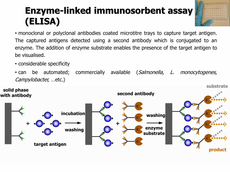
Enzyme-linked immunosorbent assay (ELISA)
bull monoclonal or polyclonal antibodies coated microtitre trays to capture target antigen
The captured antigens detected using a second antibody which is conjugated to an
enzyme The addition of enzyme substrate enables the presence of the target antigen to
be visualised
bull considerable specificity
bull can be automated commercially available (Salmonella L monocytogenes
Campylobacter hellipetc)
target antigen
washing
product
second antibody
enzyme substrate
substrate solid phase
with antibody
incubation washing
VIDAS or mini-VIDAS Analyzers
bull the automated multiparametric immunoanalyser
bull based on an enzyme immunoassay which detect target antigens using the
ELFA (Enzyme Linked Fluorescent Assay)
bull 4-methyl-umbelliferone (fluorescent molecule) is released by alkaline phosphatase = fluorescence
VIDAS or mini-VIDAS Analyzers
Each test is composed of two parts
1 The SPRreg acts as a Solid Phase Receptacle for the reaction The SPR is coated
with anti-target antibodies adsorbed on its surface
2 The Strip contains all ready-to-use reagents necessary for the test washing
solution alkaline phosphatase-labeled anti-target antibodies and substrate
Video httpwwwbiomerieux-industrycomfoodvidas-listeria-monocytogenes-detectionVIDAS LMO2 httpswwwyoutubecomwatchv=ZFRuJYynLwk
httpwwwbiomerieuxcom
bioMeacuterieux
VIDAS or mini-VIDAS Analyzers
Validated by ISO 16140 againt ISO 6579 (AFNOR NdegBIO 1216-0905)
Salmonella spp
VIDAS or mini-VIDAS Analyzers
The sample = the sample diluted and enriched in Xpress 2 broth (different from ISO 6579) The used broth must be convenient for the method
Detection of Salmonella spp according to ISO 65792002
01 ml of homogenate to 10 ml RVS incubation at 415degC for 24plusmn3 h
Incubation at 37plusmn1degC for 18plusmn2 h
1 ml of homogenate to 10 ml MKTTn incubation at 37plusmn1degC for 24plusmn3 h
Plating out by 10 micro on two selective media and incubation at 37plusmndegC for 24plusmn 3h
bullXylose Lysine ndash Desoxycholate Agar (XLD)
bullAny other selective medium (BGAHE BS SS DC chromogenic mediahellip)
25 g of sample + 225 ml of buffered peptone water homogenization
Selective enrichment
Pre-enrichment
Isolation on selective agar
plates
Confirmation
Confirmation
Isolation of a charasteristic colony on Nutrient Agar and incubation at 37plusmndegC for 24plusmn 3h
1 day
2 day
3 day
4-6 day
ISO 65792002 Microbiology of food and animal feeding stuffs -- Horizontal method for the detection of Salmonella spp
VIDAS kits
bull E coli O157 - VIDASreg UP E coli O157 (including H7)
bull E coli O157 - VIDASreg ECO
bull E coli O157 (confirmation) - VIDASreg ICE
bull Salmonella spp - VIDASreg SLM
bull VIDASreg ICS
bull Salmonella spp - VIDASreg Easy SLM
bull Listeria spp - VIDASreg LIS
bull Listeria spp - VIDASreg LSX
bull Listeria monocytogenes VIDASreg LMO2
bull Staphylococcal enterotoxins - VIDASreg SET2
bull Campylobacter spp - VIDASreg CAM
GLISA-Rapid Test (Gold Labelled ImmunoSorbent Assay) ndash Lateral Flow Test
Detection limit 105 CFUml in a sample (homogenate of selective enrichment or suspension of colony) (sometimes heat-treatment required - 20 min at 80 degC = cell lysis = better motility of the cell walls with antigens)
httpwwwmeridianbioscienceeumediahome_3columns_images751630jpg
GLISA-Rapid Test
Reaction zone 150 microl of sample
Testing zone Control zone
+ Pos - Neg
Singlepathcopy for detection of Listeria monocytogenes (Merck)
Validation of procedure
Validation of an alternative method bull the validation of an alternative method is the procedure to demonstrate if the alternative method provides equivalent or better results compared to the reference methods
bull Validation is a process within which the method is demonstrated to be suitable
for its purpose It documents methods validity
bull During validation process methods performance characteristics are estimated
The validation of qualitative and quantitative methods comprises two phases
1 a method comparison study of the alternative method against a reference
method (performed by an expert laboratory)
2 a interlaboratory study of the alternative method (organised by an expert
laboratory)
Expert laboratory (organising) laboratory having the qualified staff and skills to
perform the method comparison study and organise the interlaboratory study
Useful documents
ISO 161402003 ndash Microbiology of food and animal feeding stuffs ndash Protocol for the
validation of alternative methods
ISO 170252005 ndash General requirements for the competence of testing and calibration
laboratories
Accreditation bodies that recognize the competence of testing and calibration laboratories
should use this International Standard as the basis for their accreditation
ISOIEC 170112004 Conformity assessment -General requirements for accreditation
bodies accrediting conformity assessment bodies
If a laboratory wishes accreditation for part of all of its testing and calibration activities it
should select an accreditation body that operates in accordance with ISOIEC 17011
It is expected that all laboratories involved in each step of a validation process will have a Quality System or quality assurance (QA) program in place to ensure standardization of laboratory operations as well as adequate quality control (QC) activities
Validation of procedure
Test characteristics The test characteristics for alternative methods are shown
Qualitative methods
1 Selectivity (inclusivity exclusivity)
2 Relative accuracy
3 Detection level
4 Relative sensitivityRelative specificity
5 The agreement between the
methods
Quantitative methods
1 Selectivity (inclusivity exclusivity)
2 Lowest validated level with satisfactory
precision
3 Repeatability
4 Reproducibility
5 Uncertainty of the method
28
PCR METHODS
DNA RNA PROTEINS Deoxyribonucleic acid (DNA)
A linear polymer that consists of four nucleotides
Adenine Cytosine Guanine Thymine Primer binding A ndash T C - G
DNA RNA PROTEINS
DNA
RNA
httpss3amazonawscomclassconnection116flashcards5862116jpgrna-149F82012BD7233971Fjpg httpslh3googleusercontentcomf81cHVr_O-
jXwH1A7g-mfX1LJKZFffUkItKpHPZ3kGF6cGXJaAJ-pqZoNL7T4YNKcP-grQ=s85 httpslh3googleusercontentcomdc9f6E-QDwxJQc-
7MvExwNvrihEjEfgjzwKr2gidCYIt0VmoMb1yEgTDc6mpkFDMUsXsgw=s94
httpsuploadwikimediaorgwikipediacommons00fPeptide_synpng
PCR principle PCR=Polymerase Chain Reaction (1983 by Kary Mullis) in vitro amplification of the part of DNA (usually 100 bp ndash 1500 bp) bordered by two primers (syntetically prepared oligonucleotides)
Typical number of cycles = 30 - 45
httpswwwyoutubecomwatchv=iQsu3Kz9NYo
PCR principle DENATURATION of double-stranded DNA by heat (app 94-95degC initially 5 then in each step 2-3 minutes)
ANNEALING of PRIMERS on the free single strand DNA bull forward primer on the 3acute-
5acute strand bull reverse primer sits on the
5acute-3acute strand Ta = annealing temperature bull 50-65 degC ndash depends on the
primers sequence bull Time 30 s-60 s bull One of the most important
factor for the specifity bull T gtgt Tm= no annealing - no
PCR product bull T ltlt Tm= non-specific
annealing ndash nonspecific products
ELONGATION by DNA polymerase
in vivo starts to synthetize new DNA molecules from 3acute- end of RNA primer
in vitro PCR from 3acute-end the primers
72 degC ndash optimal temperature
45 s-3 minutes (final elongation 5-15 minutes)
httpwwwmuncabiologyscarrPCR_sketch_3gif
PCR principle
httpopenwetwareorgimagesff1CH391L_S12_PCR_exponential_amplificationjpg
Mistake in the beginning ndash the most influence Exponential phase duplication of PCR products amount in each cycle Linear phase The level of amplification is decreased (lower than duplication in each cycle) Plateau Absence or degradation of some component Reasociation can be preferred than primers anealing due to thermodynamical reasons
PCR principle
httpwwwebioecomuploadfile2010031420100314110534183gif
DNA (genomic plasmid DNA)
Primers (forward reverse)
Thermostable
DNA polymerase
Mg2+ (cofactor
for DNA
polymerase)
dNTP mix
(dCTP dATP
dTTP dGTP) ndash
building stones
for newly
synthetized
DNA product
Nuclease free water ndash without Dnase
Rnase presence (enzymes which
degrade DNA RNA)
MASTERMIX
Total volume ndash 25 μl ndash 50 μl
PCR principle
Polymerase Taq DNA polymerase
bull from thermophilic bacteriumThermus aquaticus
(now prepared by recombination)
bull opt temp 75-80degC (150 ntss)
bull active at the room temperature ndashto work on ice
bull inactive at gt90degC but reactivation when cooled
bull only 5acuteexonuklease activity not 3acuteexonuklease
activity (proofreading)
bull 1 mistake at 10-20 000 nts
Hot start polymerases
bull Activity at the room temperature is inhibited - binding on anti-polymerase
antibodies on the wax balls
bull activation after heating at 94 degC (antibodies destroyed)
Other polymerases
bull Proofreading polymerases (with the 3acuteexonuclease activity)
bull Tth polymerase - reverse transcription (Mn2+) or polymerase activity (Mg2+)
1) Releasing DNA from the cell
- lysis of the cell wall by detergents or by heat digesting cell surface proteins by proteinase K
- RNAse treatment for RNA-free genomic DNA
2) Isolation DNA from the released cytoplasmatic contents
- Centrifugation the vaste in pellet (heat isolation)
- Ion-exchange chromatography (commercial kit) ndash binding DNA by ionic interactions to the column (DNA is negative charged) ndash proteins and RNA pass away ndash DNA is later released by using more concentrated salt solution (column is charged positively)
- Phase separation (aqueouschlorophorm phase ndash CTAB method)
DNA isolation
Detection of food-borne pathogens
DNA of microorganism = set of specific genes with specific sequences
Primers are designed to be complementary to sequence of some specific gene
ndash if the complementary sequence is present PCR product will be performed
Main problems about primers designing and applying of PCR
- Is the gene of interest specific enough (can be found only in the detected microorganism)
- Is the primers sequences complementary only to this gene
- Is the primer sequence designed well (requirements about easy and specific annealing ndash depends on the primers sequence)
- Primers can be designed comercially or by using software (httpwwwbiocenterhelsinkifibiProgramsdownloadhtml)
- sequences and genomes needed - wwwpubmedcom
- httpswwwncbinlmnihgovtoolsprimer-blastindexcgiLINK_LOC=BlastHome
PCR products detection The PCR product has got specific length and specific sequence
Visualiton by intercalating agents bind to double-stranded DNA - fluorescence signal bull Ethidium bromide ndash carcinogenteratogen If intercalated the fluorescence of EtBr is significantly higher than in aqueous solution (UV absorbance maxima at 300 and 360nmemitted as orangeyellow light at 590nm) bull Sybr Green DNA-Sybr Green complex absorbs blue light at 488 nm and emits green light at 522 nm Fluorescent tags at 5acute-end of primers Other tag at 5acute -end of primers eg biotin - for immunodetection detected by anti-bodies linked with an enzyme Microarray
Electrophoresis Conventional electrophoresis a single electrical field causes biomolecules to migrate through a matrix according to its mass-to-charge ratio effectively separates DNA fragments up to ~20-30 kb (according to the gel concentration) larger fragments will comigrate (a large band at the top of the gel)
httpwwwrothamstedacuknotebookimagesgelelgif httpstevegallikorgsitesallimagesgelElectrophoresis2jpg
httpabeleewardhawaiieduProtocolsDNA20Gel20Preparation20and20Electrophoresishtm
Electrophoresis Primers (OT1559 a 18ndash1) complementary to DNA coding16S rRNA Campylobacter
jejuni Campylobacter coli a Campylobacter lari
Product is 287 bp
60 V 100 minutes M ndash 100 bp marker (1-5 positive controls) 1- C jejuni CCM 6212 2- C jejuni CCM 6214 3 ndash
C coli CCM 6211 4 ndash C lari CCM 4897 5 ndash C upsaliensis ATCC 43954 6- 15 strains C jejuni Nt- no
template control (negative control)
Genus specific PCR
Electrophoresis DUPLEX PCR =two pairs of primers
C jejuni C1 5 - CAA ATA AAG TTA GAGGTA GAA TGT-3
C4 5 - GGA TAA GCA CTA GCTAGC TGA T-3
Complementary to a part oxidoreductase 159 bp
C coli 18F 5 - GGT ATG ATT TCT ACAAAG CGAG-3
519R 5- ATA AAA GAC TAT CGT CGC GTG-3acute
Complementary to aspartatekinase and flanking ORF 500 bp
Species specific PCR
60 V 100 minutes M ndash 100 bp marker (1-5 positive controls) 1- C jejuni CCM 6212 2- C jejuni CCM
6214 3 ndash C coli CCM 6211 4 ndash C lari CCM 4897 5 ndash C upsaliensis ATCC 43954 6- 15 strains C
jejuni Nt- no template control (negative control)
Melting curve Intercalating dye binds to double-stranded DNA - producing fluorescent signal when excited by light
When samples are heated to melting point double-stranded DNA denatures into single strands freeing dye into solution
ndash Melting temperature (Tm) is the point at which the DNA denatures
ndash Dependent on the length of fragment and G-C content
ndash More base pairs --gt Increase in Tm
ndash Higher G-C content --gt Increase in Tm
bull Unbound dye has reduced fluorescent signal
bull Loss in signal generates melt curve
No Signal Signal Increasing
temperature
Intercalating
dye
Melting curve
Inflex point
bull 1 derivation = 0
bull In this case 2derivation is negative
Real-Time PCR
CT
CT (threshold cycle) ndash the lowest cycle when fluorescence crosses the
determinated value
Video httpswwwyoutubecomwatchv=kvQWKcMdyS4 httpswwwyoutubecomwatchv=pRwoOBuk00c
Real-Time PCR
Food-borne pathogens detection by PCR
Identification of pure cultures - without problems
Detectionenumeration from food matrix directly or after enrichment
bull How to isolate successfully DNA from few cells in 25 g10 g - Efficiency of DNA isolation (isolation by kits heat-lysis CTAB method)
bull How many targets copies are needed for successfully PCR products detection
ndash detection limits
ndash presence of PCR inhibitors (inhibition of polymerase primers annealing)
bull How to distuinguish DNA from dead and live cells - Total DNA is detected (from dead and living cell)
bull For distinguishing of dead cells DNA ndash eg application of EMA (ethidium monoazide) to the sample before DNA isolation ndash EMA irreversibly bound to DNA of dead cells ndash they are bdquoinvisibleldquo for PCR ndash no amplification
bull Direct enumerationdetection only in some specific case ndash simple liquid sample when the required limit is higher than the limit of detection
bull Standardly ndash for PCR is used enriched homogenate (sample diluted in enrichment broth and cultivated) ndash to obtain detectable level of microorganisms
BAXreg System
Salmonella
Listeria monocytogenes
Genus Listeria
Enterobacter sakazakii
E coli O157H7
Staphylococcus aureus Real-time
Campylobacter jejunicolilariReal-time
Vibrio cholerae
parahaemolyticusvulnificus Real-time
Yeast and molds
httpwww2dupontcomDuPon
t_Homeen_USindexhtml
BAX ndash three major steps
On enrichedregrown samples
Prepare samples = perform heat-lysis in two stages
DNA present in the lysate
2 Process samples
3 Review results
Hydrate PCR tablets
Enrichment protocols vary
depending on bacterial target
and type of food yoursquore testing
Salmonella Enterobacter
sakazakii
E coli O157H7 E coli O157H7
MP Campylobacter jejunicoli
Yeast and Mold
37deg +- 2degC for 20 minutes then
95deg +- 3degC for 10 minutes
Listeria and L monocytogenes
55deg +- 2degC for 60 minutes then
95deg +- 3degC for 10 minutes
5 microl for all bacterial targets
20 microl for beef samples enriched
in E coli O157H7 MP media
20 microl for yeast amp mold
PCR tablets bull DNA-polymerase
bull Primers
bull Nucleotids
bull Suplements
bull SybrGreen or fluorescent probe
bull Internal control + primer
bull Mastermix in the tablet in PCR Eppendorf ndash minimal risk of contamination
For solving the problem of distiguishing dead and live cell the process of small volumes
is used ndash only if live cells are present in the sample they able to multiplied to the limit
detection (104 CFUml in the enrichment suspension used for lysis step)
If the result of detection is positive confirmation by plating out is done (live and dead
cells)
+
Target organism DNA BAXreg system
primers
in tablets PCR
+ no detectable
amplification
Background DNA
PCR
BAXreg system
primers
in tablets
Amplification of specific fragments
Detection of specific fragments Fluorescent signal drops with heating to generate a melt curve
Melting curve Raw Data Processed Data
Strong positive
result
Negative
result
Target peak
Positive control peaks
Positive control peak
Melting curves display peaks within a set temperature range for the control and target (if present)
Detection
Melt curve weak INPC in presence of strong Salmonella positive
Real time weak INPC in presence of strong
Campylobacter positives
Direct protocol does not display a positive or
negative result PCR was successful IndeterminateSignal error
Salmonella AFNOR (ISO 16140) protocol Sample 110 BPW
18 h37ordmC
RVS Muumlller-Kauffmann
24 h 415 37 ordmC
5 suspicious colonies on TSA
24 h 37ordm C
Biochemical and serological confirmation
XLD 2 Plate
24 h 37ordmC
BHI
3h 37 ordmC
22 h 37 ordmC
15 d neg and pos result
3 d neg 5 d pos results
Salmonella ( ISO 65792003) versus BAXreg-PCR
International certificates AOAC-RI Performance Tested Method
Salmonella 100201 L monocytogenes
070202 L spp 030502 Saureus 120701
Campylobacter colilarijejuni
AFNOR Certification
Salmonella QUA 183-1102
NordVal Certification
Salmonella 2006-30-5408
Certificates from AFNOR and AOAC are equal
NordVal Certificate
57
OTHER METHODS
ATP bioluminiscence
httpwwwpromegacouk~mediaimagesresourcesfiguresenotesfe0027_fig1jpgla=en
httpwwwbiotekptassetstech_resources143clarity_atp_conc_fig2gif
Principle the detection of the present level of ATP (direct proportion to the presence of microorganisms or their products residues) The enzyme luciferase catalyzes the transformation of d-luciferin to oxyluciferin (indicated by emision of light at 560 nm)
ATP bioluminiscence
httpwwwbioxyscomimages2figure-B11gif
ATP Hygiene Monitoring System - SystemSURE II
httpwwwmicromaticcomdraft-keg-beerline-cleaning-hygiene-pid-ATP-100-tab-reviewshtml
Biosensors
60
httpsuploadwikimediaorgwikipediacommonseecBiosensor_Systemjpg
Biosenzor =the whole cellmicroorganism (CBS ndash cell-based sensors)
Cell-based sensors
Patogenic microorganisms interact during the infection with mammalian
cells ndash detection of cell or toxin by cell culture The damage of cell culture is measured optically or electrically
62
IDENTIFICATION OF MICROORGANISMS
Definition of species in bacteriaarchae
The criterion for a bacterial or archael species a collection of strains that have bull at least one diagnostic phenotypic trait bull ge70 DNAndashDNA hybridization of their purified molecules DNA
bull ~more than 97 16S rRNA sequence identity Under the same conditions some eucaryotic order would be a single bacterial species
The bacterial species definition bull eminent practical significance bull but a very difficult issue
bull very wide and often changes in DNA
bull mutations during replication of DNA
bull horizontal gene transfer through mobile elements plasmids also between species
Criterion for species in prokaryotic cells (bacteria and archea) Collection of strains characterised bull by at least one diagnostic feature bull their purified DNA molecules prove at least 70 hybridisation
which corresponds to at least 97 concordance in the sequence of 16S rRNA gene
Map of genome of B cereus ATCC 14579
httpwwwacgtinccomspecialty_dna_sequencinghtm
Identification based on genotype
64
DNA isolation PCR amplification of 16S rRNA gene (usually)
Sequencing of PCR product
Comparison to sequences in databases (BLAST)
Identification of bacteria PHENOTYPE METHODS bull Morphology of colony and cell (Gram staining shape) bull Requirements for temperature atmosphere nutrition bull Biochemical tests - presence of specific enzymes
bull catalase oxidase fermentation of glucose bull the way how they utilize different compounds
bull Immunochemical tests - specific antigens in the cell wall METHODS BASED ON PROTEIN ANALYSIS
bull MALDI-TOF MS METHODS BASED ON GENOTYPE bull looking for and comparing of specific DNA sequences
bull conserved universal genes (eg 16S rRNA) bull genes specific for genus species
bull done by PCR methods DNA chips sequencing etc
66
BIOCHEMICAL TESTING
Identification based on phenotype I Physiological + biochemical features
bull morfology of colonies and cells (Gram staining shape)
bull requirements for temperature atmosphere nutrient sources
bull basic biochemical tests oxidase catalase fermentationoxidation of glucose haemolysis etc
appearance of colonies on selective or selective-diagnostic agars (for other testing to be reinoculated on non-selective agar)
the classification of an isolate into a certain group
(eg for bacteria catalase positivenegative G+ cocci glucose fermentingnon-fermenting G- rods etc)
biochemical tests specific for certain groups of microorganisms
Identification based on phenotype I
68
Physiological + biochemical features Example Bacillus cereus
httpwwwfoodhaccpcommemberonlynewsletter164html
httpwwwnapavalleyedupeoplesrosePublishingImagesBacillus20cereus202(Gram20stain)jpg
Set of biochemical tests Microgenreg Bacillus ID Panel for the identification of Bacillus spp and related genera (time for performing 48 hours)
MYP agar (Mannitol Egg Yolk Polymyxin Agar) pink colonies with a precipitation zone (no fermentation of manitol + enzyme lecitinase)
catalase positive
httpwwwoxoidcomukblueprod_detailprod_detailasppr=CM0929ampc=ukamplang=ENamporg=9ampimg=CM0929ampsec=
G+ rod endspores forming
β-haemolysis moisty and easy to grow colonies
Principle of biochemical testing
to rank the bacterium to some group having the same basic features bull eg catalase positive gram positive cocci bull Family Enterobacteriaceae
the identification on the genus or species level
bull sets of biochemical tests specific for some group of bacteria
70
Gram staining procedure - principle
https40mediatumblrcom25da2e5b25bbe336819bf1715849d171tumblr_inline_ntrsgn9w1D1sv92o6_540jpg
Gram staining procedure - principle
httpsonlinesciencepsuedusitesdefaultfilesmicrb106CellStructurecow02354_03_15jpg
Gram staining procedure 1-2 drops of sterile water on a slide Spread culture in thin filter in this drop Let dry in airclose to fire Pass slide through flame 2-3x to fix Add crystal violet ndash 15-20 s to act Add Lugol solution (KII2) ndash 15-20 s to act Washing off with a mixture of
ethanolaceton (41) or acetone till the stain removes but max 15 s
Rinse with water decant excess liquid Add safranin 1 minute Rinse with water decant excess liquid Dry and put a drop of imersion oil use an
objective bdquoOILldquo
httpwwwtumblrcomtaggedgram-staining
G- red
G+ violet
Gram staining procedure + Identification
E coli
Family Enterobacteriaceae Gram-negative rods
Micrococcus luteus
Staphylococcus aureus
Gram-positive cocci Bacillus cereus
Gram-positive rods (with endospores)
Biochemical identification of G + bacteria
httpyoungeneticistblogspotcz201106diagnostic-microbiology-labhtml
G+ bacteria bull cocci ndash in clusters in pairs
diplococci bull bacilli
bull endospore bull no endospore
For G+ cocci bull Catalase positive ndash Staphylococcus
sp bull Coagulase + S aureus or some other few species bull Coagulase -
bull Novobiocin S bull S epidermidis etc
bull Novobiocin R bull S saprophyticus etc
bull Catalase negative Streptococcus sp Enterococcus sp
bull kind of haemolysis Other important G + group ndash filamentous bacteria
Catalase test Catalase test Catalase is a common enzyme found in nearly all living organisms that are exposed to oxygen where it catalyzes the decomposition of hydrogen peroxide to water and oxygen
2 H2O + O2 2 H2O2
catalase
bubbling when culture is add in peroxide
httpiws2collinedudcainCCCCD20Microcatalase_testhtm
Catalase testing into a drop of 3 peroxide to transfer a loop of bacterial culture catalase positive ndash releasing of air bubbles
Examples bull G+ cocci catalase positive Staphylococcus sp Micrococcus sp Kocuria sp Dermacoccus sp Macrococcus sp Rothia sp catalase negative Streptococcus sp Enterococcus sp bull G+ rods (bacilli) endospores forming catalase positive Bacillus sp catalase negative Clostridium sp
Resistance to ATBhaemolysis Resistance eg to novobiocin
httpquizletcom20077004mic-lecture-1-t2-flash-cards
Hemolysis on blood agar
Measuring the diameter of inhibition zone (less than 17 mm resistant more or equal to 17 mm sensitive) ndash differing in Staphylococcus sp (Clinical performance)
alpha-hemolysis - oxidizing the iron in the hemoglobin - green discoloration of the medium around beta-hemolysis - rupture of erythrocytes - complete clearing medium around gamma-hemolysis ndash no effect ndash no change
By cultivation on blood agar bacteria can be
differentiated based on their capacity to secrete
hemolysins The hemolysis will cause a clearing zone of
the blood agar around the colonies Bacteria can cause
different types of hemolysis
bullα-hemolysis which means an incomplete clearing
(green haemolysis)
bullβ-hemolysis which means a complete clearing
bullDouble hemolysis of some staphylococci consisting of
an inner β-hemolysis zone and an outer α-hemolysis
zone
bullNo hemolysis which is sometimes referred to as γ-
hemolysis which may seem illogical
Note that the α-hemolysin of staphylococci causes
complete hemolysis whereas their β-hemolysin causes
incomplete hemolysis
The capacity to produce hemolysins may vary between
different strains of a particular bacterial species
httpwwwvetbactorgvetbactdisplayextinfo=67
Identification of bacteria based on haemolysis
Streptococcal hemolysins α (alpha) hemolysis - ldquozone of green discolourationrdquo or ldquogreeningrdquo called partial or incomplete hemolysis produced by alpha-hemolytic streptococci such as Streptococcus suis This type of hemolysis on BA is due to bacterial production of hydrogen peroxide which oxidises hemoglobin to methemoglobin β (beta) hemolysis ndash a clear zone of complete hemolysis produced by beta-hemolytic streptococci such as S equi subsp equi This hemolysis is due to lysis of RBCs in the BA by the cytotoxin Streptolysin S
Source httppeopleupeicajlewishtmlhemolysinshtml
Bacterial hemolysins
Staphylococcal hemolysins Most coagulase-positive staphylococci (CPS) such as Staphylococcus aureus S pseudintermedius ( but not S hycius) typically produce an distinctive pattern of ldquodouble zonerdquo or ldquotargetrdquo hemolysis on blood agar Double-zone hemolysis indicates that the two hemolysins one which produces ldquocompleterdquo and another one that produces ldquoincompleterdquo hemolysis are present and this combined effect is visible due to different diffusion rates of these enzymes through the agar The staphylococcal α (alpha) hemolysin causes a complete type of hemolysis This zone of complete hemolysis is narrow and immediately adjacent to the bacterial colony on blood agar The staphylococcal β (beta) hemolysin causes an incomplete type of hemolysis at 37 degC This broader incomplete zone is outside of the inner complete zone and can be hard to see on early culture plates and also varies with the species of RBCs used to make the BA plate Its action is enhanced by chilling at 4 degC and the hemolysis becomes complete (this effect is referred to as ldquohot-coldrdquohemolysis) There are other staphylococcal cytotoxins that are also hemolysins but their lytic actions are not seen on blood agar
The confusion arises due to the NAMES of the two STAPHYLOCOCCAL HEMOLYSINS and the TYPE of hemolysis they produce These are the REVERSE of the two TYPES of STREPTOCOCCAL HEMOLYSIS Strictly speaking the descriptive terms ldquoalphardquo for ldquoincompleterdquo and ldquobetardquo for ldquocompleterdquo hemolysis apply only to the hemolytic toxins of Streptococcus species However other organisms beside Streptococcus and Staphylococcus species have cytotoxins that cause hemolysis and this hemolysis can be either complete or incomplete It is preferable to use the terms ldquocompleterdquo and ldquoincompleterdquo rather than ldquobetardquo or ldquoalphardquo to describe the hemolysis caused by these organisms but you will notice in bacteriology text books that the terms alpha and beta are commonly used
G ndash bacteria bull Cocci bull Bacilli (Rods) bull Oxidase positivenegative bull Glucose metabolism
bull fermentation bull oxidation bull Inactive
bull Lactose metabolism for coliform
bull Pigment formation for Pseudomonas sp
Biochemical identification of
G - bacteria
Oxidase test
Oxidase positive (OXI +) Aeromonas sp Pseudomonas sp Dark-blue to violet
Oxidase negative (OXI -) Enterobacteriaceae Bacillus sp Colourless
Oxidase bullabbreviated general name for some kind of cytochrome c oxidases (cytochrome oxidase or indophenol oxidase) enzymes involved in aerobic respiratory chain when oxygen is used as the final receptor for hydrogen bullOxidases are usually present only in aerobic organisms bullPresence or absence of oxidase(s) is an important feature of the bacterial metabolism used mainly in the identification process of gram-negative bacteria
Detection Reaction of the culture with redox indicator as eg NN-dimethyl-p-phenylenediamine (DMPD) dark-blue when oxidized and colorless when reduced Different test performances are possible
Fermentation of carbohydrates Type of utilisation of glucose or carbohydrates bull an important feature of the bacterial metabolism bull widely used in the identification process of bacteria bull fermentation strictly means to be under anaerobic conditions (but in bacterial
metabolism which can be but not have to be the same as the anaerobic conditions of cultivation) so very often is used in a general way like bdquoutilisationldquo
bull fermentation (like bdquoutilisationldquo) of carbohydrates to
bull acid and gas (Durham tubes) bull acid without gas bull non-utilised
bull Testing of fermentation or oxidation of carbohydrates
bull eg OF test for glucose bull by oxidation under aerobic conditions bull by fermenation under anaerobic conditions (an oil layer)
Fermentation of sugars to acid
Peptone Water wPhenol Red
(HiMedia M028I)
Ingredients gl
Peptic digest of animal tissue 10000
Sodium chloride 5000
Phenol red 0020
Final pH ( at 25degC) 68plusmn02
httpsuploadwikimediaorgwikipediacommons009Phenol_red_pH_60_-_80jpg
+ after sterilisation
at the room
temperature to add
aseptically sugar
discs and
inoculate
succrose trehalose rhamnose xylose
Incubation 30 degC 24 h
- +
Fermentation of carbohydrates
Casein enzymic hydrolysate and yeast extract provide nitrogenous compounds vitamin B complex and other growth nutrients Dextrose is the fermentable carbohydrate and bromocresol purple acts as the pH indicator with colour change from purple to yellow in acidic conditions Sodium chloride maintains osmotic equilibrium bdquoAnaerobicldquo conditions ndash inoculation by stabbing to used fresh prepared media (oxygen expelled by heating medium in boiling water or running steam for 15 min then to be cooled down quickly)
- +
Enterobacteriaceae ndash glucose fermentation
83
Tryptone Yeast Extract Agar Ingredients Gms Litre Casein enzymic hydrolysate 10000 Yeast extract 1500 Dextrose 10000 Sodium chloride 5000 Bromocresol purple 0015 Agar 15000 Final pH ( at 25degC) 70plusmn02
Test for oxidationfermentation of glucose (OF glucose test) detection of acid production by acidobazic indicator change to yellow (acid present) in lower pH from green (no acid) Anaerobic atmosph ndash covering by sterile mineral oil If fermented anaerobically then also runs aerobically
Fermentation of carbohydrates
Principle of biochemical tests bull The first step in identification is to rank the bacterium to some group of bacteria
having the same basic features ndash eg catalase positive gram positive cocci bull The identification on the genus or species level is finished by other biochemical tests
specific for each group of bacteria bull The tests can be done in tubes or miniaturized plate
ENTEROtest 24N (Erba Lachema) (24 means 24 biochemical tests N= new)
API 20E (bioMeacuterieux)
bull designed for routine identification of clinically important species of family Enterobacteriaceae without the use of reagents
bull A strip test OXItest or OFtest in microtitration wells can be used to confirm that the isolate belongs to Enterobacteriaceae family
bull The results are read visually or automatically using reader MIKROLAreg
bull The identification can be supplemented by diagnostic strip tests INDOLtest COLItest and VPtest
APIreg Gram negative Identification API 20E ndash Speciessubspecies identification of Enterobacteriaceae and groupspecies identification of non-fermenting Gram negative bacteria API Rapid 20E ndash 4-hour identification of Enterobacteriaceae API 20NE ndash 24 to 48-hour identification of Gram negative non-Enterobacteriaceae API Campy ndash 24-hour identification of Campylobacter species
Example Escherichia coli
ENTEROtest 24 N
Biochemical testing - Enterobacteriaceae bull TSI Triple Sugar Iron Agar (use a straight wire to stab the butt and streak the agar surface)
- to determine whether organisms can ferment glucose sucrose andor lactose with or without production of gas and the ability of the organism to produce hydrogen sulphide from thiosulphate in an acid environment is also tested
Formula Liter Enzymatic Digest of Casein 5 g Enzymatic Digest of Animal Tissue 5 g Yeast Enriched Peptone 10 g Dextrose 1 g Lactose 10 g Sucrose 10 g Ferric Ammonium Citrate 02 g Sodium Chloride 5 g Sodium Thiosulfate 03 g Phenol Red 0025 g Agar 135 g Final pH 73 plusmn 02 at 25 degC
inoculation in the butt by stab inoculation on the slant on the surface bull Fermentation of dextrose
(glucose) to acid bull anaerobically - yellow butt bull aerobically leads to products
which are later oxidised by air and pH remains alkaline (red) bull Fermentation of glucose to gas (anaerobically - in the stab) Cracks splits or bubbles in medium indicate gas production bull Production of hydrogen sulfide (H2S) ndash reaction with ferrous sulphate and sodium thiosulphate - a black precipitate in butt bull Lactose andor succrose
utilisation to acid Acid production ndash yellow butt and slant
Results
An alkaline slant-acid butt
(redyellow) - fermentation of
dextrose only
An acid slant-acid butt
(yellowyellow) - fermentation of
dextrose lactose andor sucrose
An alkaline slant-alkaline butt
(redred) - dextrose or lactose not fermented (non-fermenter)
Bacterium fermenting lactose andor succrose are able to ferment glucose
Phenol red ndash alkaline = red acid = yellow
bull TSI Triple Sugar Iron Agar (use a straight wire to stab the butt and streak the agar surface)
- to determine whether organisms can ferment glucose sucrose andor lactose with or without production of gas and the ability of the organism to produce hydrogen sulphide from thiosulphate in an acid environment is also tested
Biochemical testing - Enterobacteriaceae
Urea agar (Christensen) (use a 1 μl loop full)
- to determine whether an organism can split urea into ammonia and
carbon dioxide by the action of the enzyme urease
httpimageslidesharecdncomlab17-141201085153-conversion-gate0195-10-638jpgcb=1417424219
httpimageslidesharecdncomhelicobacterpylori-copy-110516201901-phpapp0195campylobacter-jejunihelicobacter-pylori-39-728jpgcb=1305577391
Phenol red
Medium is acidic - yellow
(pH 68plusmn02)
Alkaline reaction by urea hydrolysis - pink colour
Composition Gms Litre
Peptic digest of animal tissue
1000
Dextrose 1000
Sodium chloride 5000
Disodium phosphate 1200
Monopotassium phosphate 0800
Phenol red 0012
Agar 15000
Final pH ( at 25degC) 68plusmn02
Nutrition
Peptic digest of animal tissues dextrose
Sodium chloride - osmotic equilibrium
Phosphates - buffering of the medium
35-37 degC
Biochemical testing - Enterobacteriaceae
bull L-lysine decarboxylase (use a 1 μl loop full)
- to determine whether an organism can decarboxylate an amino acid (lysine) leading to formation of an
amine After inoculation both tubes are overlayed with sterile paraffin oil
httpfce-studynetdna-sslcomimagesupload-flashcards10295302332621_mjpg
+ CO2 + OH-
Lysine Decarboxylase Broth Typical Composition (glitre) Peptic digest of animal tissue 5000 Yeast extract 3000Dextrose 1000 (glucose prepared from starch chemically equivalent to glucose) L-Lysine hydrochloride 5000 Bromocresol purple 0020 Final pH ( at 25 degC) 68plusmn02 Inoculate below the surface cover by sterile paraffin oil
Two steps detection 1 step Dextrose is fermented ndash H+ is released Acidic conditions bull bromocresol purple changes colour to yellow bull in acidic conditions lysine decarboxylase is
activated 2 step L-lysine is decarboxylated and OH- released Alkaline conditions bull bromocresol purple changes colour back to
purple
Biochemical testing - Enterobacteriaceae
bull β-galactosidase (ONPG) (use a 1 μl loop full)
- to determine whether an organism has the enzyme szlig-galactosidase by using the compound o-
nitrophenyl-beta-D-galactopyranoside (ONPG)
- this test is used to differentiate between lactose-negative and lactose-delayed organisms
- ONPG is hydrolysed by the enzyme to o-nitrophenol which is yellow and galactose
O-Nitrophenyl-β-D-galactopyranoside (ONPG) is structurally similar to lactose (ie ONPG is an analog of lactose) except that orthonitrophenyl has been substituted for glucose ONPG is colorless compound O-nitrophenol is yellow visual evidence of hydrolysis
Lactose fermenting bacteria (ONPG positive) two enzymes Permease ndash transferring of the lactose molecule into the bacterial cell β-galactosidase - cleaving the galactoside bond producing glucose and galactose - inducible enzyme - made ONLY in the presence of the lactose substrate Coliform bacteria as Ecoli Klebsiella spp Enterobacter spp
hydrolysis
Late lactose fermentering bacteria Permease ndash not present delayed in the production of acid from lactose because of sluggish permease activity β-galactosidase ndash present - give a positive ONPG test Citrobacter spp Arizona spp
Non lactose fermenter (ONPG Negative) Permease ndash not present β-galactosidase ndash not present Salmonella spp Shigella spp Proteus spp Providencia spp and Morganella spp
Biochemical testing - Enterobacteriaceae
Procedure Culture Bacteria grown in medium containing lactose (to induce the production of the galactosidase enzyme) such as Kligler iron agar (KIA) or Triple sugar Iron (TSI) agar (Note β-galactosidase enzyme (inducible enzyme) is made ONLY in the presence of the lactose substrate) Inoculation 1A loopful of bacterial growth is emulsified in 005mL of physiologic saline to produce a heavy suspension 2One drop of toluene is added to the suspension and vigorously mixed for a few seconds to release the enzyme for bacterial cells 3An equal quantity of buffered ONPG solution is added to the suspension 4The mixture is placed in a 37 oC water bath 5When Using ONPG Tablets 6A loopful of bacterial suspension is added directly to the ONPG substrate resulting from adding 1mL of distilled water to a tablet in a test tube 7This suspension is also placed in a 37oC water bath
β-galactosidase (ONPG)
Results and Interpretations The rate of hydrolysis of ONPG to o-nitrophenol may be rapid for some organisms producing a visible yellow color reaction within 5 to 10 minutes Most tests are positive within 1 hour however reactions should not be interpreted as negative before 24 hours of incubation The yellow color is usually distinct and indicates that the organism has produced o-nitrophenol from the ONPG substrate through the action of β-galactosidase
Biochemical testing - Enterobacteriaceae
bull VogesndashProskauer (VP) test (use a 1 μl loop full)
- to determine whether an organism can produce acetylmethylcarbinol
(acetoin) from fermentation of glucose If present acetylmethyl carbinol is
converted to diacetyl in the presence of prop- naphthol (to be added first) strong
alkali (40 KOH) and atmospheric oxygen The diacetyl and quanidine-
containing compounds found in the peptones of the broth then condense to
form a pinkish red polymer
MRVP broth (pH 69)
Ingredients per liter of
deionized water
buffered peptone= 70 gm
glucose= 50 gm
dipotassium phosphate= 50 gm Voges-Proskauer Reagent A Barrittrsquos reagent A
Alpha-Naphthol 5 50 gm
Absolute Ethanol 1000
ml
Voges-Proskauer Reagent B Barrittrsquos reagent B
Potassium Hydroxide 400 gm
Deionized Water 1000 ml
Procedure of VogesndashProskauer (VP) Test
1Prior to inoculation allow medium to
equilibrate to room temperature
2Using organisms taken from an 18-24
hour pure culture lightly inoculate the
medium
3Incubate aerobically at 37 degC 24 hours
4Following 24 hours of incubation aliquot
2 ml of the broth to a clean test tube
5Re-incubate the remaining broth for an
additional 24 hours
6Add 6 drops of 5 alpha-naphthol and
mix well to aerate
7Add 2 drops of 40 potassium hydroxide
and mix well to aerate
8Observe for a pink-red color at the surface
within 30 min Shake the tube vigorously
during the 30-min period
Positive Reaction
A pink-red color at the surface
Examples Viridans group streptococci
(except Streptococcus vestibularis) Listeria
Enterobacter Klebsiella Serratia marcescens
Hafnia alvei Vibrio eltor Vibrio alginolyticus etc
Negative Reaction
A lack of a pink-red color
Examples Streptococcus mitis Citrobacter sp
Shigella Yersinia Edwardsiella Salmonella Vibrio
furnissii Vibrio fluvialis Vibrio vulnificus and Vibrio
parahaemolyticus etc
A copper color should be considered negative A
rust color is a weak positive reaction
Quality Control of VogesndashProskauer (VP) Test
VP positive Enterobacter aerogenes (ATCC13048)
VP negative Escherichia coli (ATCC25922)
Biochemical testing - Enterobacteriaceae
94
VogesndashProskauer (VP) test
Rapid test for confirmation of E coli ndash typical features
β-D-glucuronidase (97 strains) production of indole from tryptophan (typical for E coli)
Performance
Previous testing Gram-negative (eg able to grow on VRBD agar) oxidase negative
From pure culture to prepare suspension in saline solution (app 5 ml) at the density 3 McFarland
To transfer 05 ndash 1 ml of this suspension into a new tube ndash to add the strip of COLItest (chemicals ndash fluorogenic substrate for β-D-glucuronidase tryptophane are present in the pad at the end of the strip)
Incubation 37 degC 4 hours
Detecion of β-D-glucuronidase (to be done at first) UV (360 nm) ndash blue fluorescence (GLR + hellipβ-D-glucuronidase positive) ndash GLR test can be read after 1 hour
Detection of indole formation from tryptophane (to be done at second) to add Kovacs reagent (IND + hellipformation of red ring)
95
COLItest - Escherichia coli
Biochemical testing - Enterobacteriaceae
Fluorogenic culture medium
bull One or more fluorogenic substrates
bull For detection of specific enzyme which are able to cleave out the fluorogenic substrate that a designed
fluorophore (responsible for the UV fluorescence) is released
FLUOROGENIC MEDIUM (Colilert) E coli use β-glucuronidase to metabolize MUG and create fluorescence (UV 360 nm) Since most non-coliforms do not have these enzymes they are unable to grow and interfere
httpgawaterusgsgovprojectsbacteriapicturessummaryanalysisecolijpg
E coli (MPN method)
COLItest Biochemical testing - Enterobacteriaceae
COLItest bull Indole test (use a 1 μl loop full)
- to determine whether an organism can split indole from tryptophan
httpwwwmicrobiologyinfocomindole-test-principle-reagents-procedure-result-interpretation-and-limitations
indole production from tryptophan
Production of indole is detected by Kovacacutes reagent
Indole Kovacs Reagent
p-Dimethylaminobenzaldehyde 500 gm
Hydrochloric Acid 37 2500 ml
Amyl Alcohol 7500 ml
Biochemical testing - Enterobacteriaceae
INDOLe test ndash quick variant
Ecoli Citrobacter Enterobacter
Procedure bull on the filtration paper to add a drop of INDOL suspension bull to spread into by a loop some culture bullTo incubate ndash at the room temperature for 5 minutes bullTo read results
Biochemical testing - Enterobacteriaceae
ENTEROtest 24 ndash example of biochemical testing
routine identification of clinically important species of family Enterobacteriaceae the test is focused only on some species often isolated in clinical practis strains from other sources can be slightly different in the dominant biochemical profiles intended exclusively for the identification of family Enterobacteriaceae the preliminary ranking into family Enterobacteriaceae must be done (Gram staining oxidase glucose fermentation) the results of reactions obtained in this modified micromethod may differ from the results obtained using conventional tests and results available in literature sources in case of problem to control positivenegative reactions by control strains or to use for this tests tube variant to check the functionality of the kit not to check the accuracy or success of the identification
Preparation of suspension from over-night culture growing on non-selective agar (for staphylococci Columbia agar blood with 5 of sheep blood is recommended)
Inoculation of microtitre plates anaerobic reaction covered by mineral oil
Incubation
Reading results by both visual and instrumental mode respectively Standard off-line tests INDOLtest or COLItest Other off-line tests OXItest OFtest VPtest Acetoin test
ENTEROtest 24 N ndash example of biochemical testing
bull tube with 5 ml of sterile physiolsol bull suspension (1 McFarland) bull application 100 microl to each hole bull 2 drops of parafin oil into holes D-H of the first line (anaerobic conditions) bull Incubation 37degC 24h bull for check the purity of suspension to streak suspension on TSA (incubation 37 degC ndash 24 h
ENTEROtest 24N
ENTEROtest 24 N - evaluation
Escherichia coli
H 0+0+0=0 G 0+2+0=2 F 1+2+0=3 hellip Identification code 023426
1 1
2
4
ENTEROtest 24 ndash example of biochemical testing
The result = a set of biochemical properties (+-) which is compared to the sets of biochemical properties of different chosen genera and species for them the test is designed
ENTEROtest 24 N
T-index how is the likelihood of the different properties The combination of identication and T-index = the reliability of the result Excelent very good good (this case) acceptable intermediary strain needs some additional tests genus level non-existing taxon
Evaluation by using the identification code - By using a code book - By using software TNW
Identification score the measure of similarity to proposed taxon
The way of identification the obtained profile is compared with profiles in database bull The most similar profiles are found
bull Identification score = of similarity bull But what about the different reactions
bull T-index is calculated bull how is the probability of the different reactions
bull It depends on their importance ndash typical reactions should be the same
bull Reliability of the result bull the combination of identication score and T-index = bull Excelent very good good acceptable intermediary strain needs some
additional tests genus level non-existing taxon
ENTEROtest 24 ndash example of biochemical testing
Biochemical tests - divisions
ENTEROtest 24 species of family Enterobacteriaceae
NEFERMtest 24 Gramnegative non-fermenting bacteria and families Vibrionaceae Aeromonadaceae and Pleiomonas shigelloides present mainly in clinical material
ANAEROtest 23 ANAEROBIC BACTERIA (G + AND G-) routine identification of anaerobic bacteria (G + AND G-) in clinical material and in food
STREPTOtest 24 Streptococcus Enterococcus and related gram positive catalase negative cocci isolated from clinical material
STAPHYtest 24 genus Staphylococcus isolated from clinical material and for their distiguishing from related genera of other gram positive catalase positive cocci
Biochemical tests APIreg Gram positive Identification API Staph ndash Overnight identification of clinical staphylococci and micrococci API 20 Strep ndash 4 or 24-hour identification of streptococci and enterococci API Coryne ndash 24-hour identification of Corynebacteria and coryne-like organisms API Listeria ndash 24-hour identification of all Listeria species APIreg Gram negative Identification API 20E ndash Speciessubspecies identification of Enterobacteriaceae and groupspecies identification of non-fermenting Gram negative bacteria API Rapid 20E ndash 4-hour identification of Enterobacteriaceae API 20NE ndash 24 to 48-hour identification of Gram negative non-Enterobacteriaceae API Campy ndash 24-hour identification of Campylobacter species APIreg Anaerobe Identification API 20A ndash 24-hour identification of anaerobes Rapid ID 32 A ndash 4-hour identification of anaerobes
MicrogenTM GN-ID Convenient 12 substrate identification system for commonly encountered Enterobacteriaceae (GNA) Extended 24 substrate identification system for the complete range of Enterobacteriacea and oxidase positive non-fastidious Gram negative bacilli (GNA+B) MicrogenTM Listeria-ID Enhanced built-in haemolysis test MicrogenTM Bacillus-ID 24 substrate system for the identification of mesophilic Bacillus spp isolated from foods and related samples MicrogenTM Staph-ID 12 substrate system minimizes handling and manipulation Substrates selected specifically for Staphylococcus and related organisms MicrogenTM Strep-ID Substrates selected specifically for Streptococcus Enterococcus and related species Provides excellent differentiation of Enterococci spp especially E casseliflavus E mundtii and E gallinarum
Biochemical tests
Identification of microorganisms by MALDI-TOF MS
Department of Biochemistry and Microbiology University of Chemistry and Technology Prague
Sabina Purkrtovaacute1 Petra Junkovaacute2 1Laboratory of food microbiology 2Laboratory of applied proteomics
SabinaPurkrtovavschtcz PetraJunkovavschtcz
MALDI-TOF MS
1) sample preparation bull microbial culture or its
proteins extract is smearing onto a steel plate and covered over by matrix
bull matrix enables the sample (A) to be desorped and ionised as pseudomolecule ionts [A+H] +
2) MALDI-TOF MS analysis unique mass spectral fingerprint of desorbed microbial cell components (mainly intracellular proteins) different among genera species or also some strains
Matrix-Assisted Laser Desorption Ionization-Time of Flight Mass Spectrometry
httppubsrscorgencontentarticlehtml2014RAC4RA05604C
httpcmrasmorgcontent263547figures-only
3) identification comparison of mass spectrum to those of reference strains in database
MALDI-TOF MS PRINCIPLE
111
2
2
2L
teU
z
m
m mass z charge
L length of drifting zone e
elementary charge U
speeding voltage
Time of flight is a function of the specific ion mass (mz)
httpwwwsigmaaldrichcomtechnical-documentsarticlesbiologycustom-dna-oligos-qc-analysis-by-mass-spectrometryhtml
Soft ionisation method bull low level of sample fragmentation
Animation http cmgmstanfordedupansection_htmlMS
MALDI-TOF MS SAMPLE AND MATRIX
httpcmrasmorgcontent263547figures-only
Matrix bull able to absorb the energy of the laser (usually 337 nm) bull to crystalise with samples - usually acid
character (proton ionisation of sample) dissolved in organic solvent
Sample preparation Direct transfer bull intact cells spread directly onto a steel plate bull lysis of cells occurs during the contact with acid
matrix and by laser desorption bull most bacteria Proteins extraction bull previous extraction of proteins by organic acids
andor alcohol (eg ethanol and 70 formic acid) bull yeasts moulds some species of bacteria (depending
on the cell wall composition)
CHC α-Cyano-4-hydroxycinnamic acid (organic solvent 50 acetonitrile with 25 trifluoracetic acid) SA 35-Dimethoxy-4-hydroxycinnamic acid (sinapic acid)
DHB 25-Dihydroxybenzoic acid
httpwwwsigmaaldrichcomcatalogproductsigmac8982lang=enampregion=CZ
MALDI-TOF MS MICROORGANISMS
113
Mass spectrum protein profile z equals usally to 1+ (so mz usually corresponds to molecula mass) the range usually used for identification 2000 -20 000 mz the intensity of single peaks corresponds to the abundance of the protein Which proteins dominates in the protein profile abundant basic and mediumly hydrophobic mainly ribosomal proteins further cold-shock and heat-shock proteins chaperons etc
conserved house-keeping gene = conserved proteins = the acordance with identification based on DNA
Visualisation of mass spectrum protein profile ndash (software mMass 5 Strohalm et al 2010)
Bacillus cereus
Analysis is recomended (and validated) to be performed from colonies grown onto non-selective agar
MALDI-TOF MS ANALYSIS
114
Comparison of mass spectrum protein profile of unknown sample with these of reference strains present in database by software
Commercial databases from different MALDI-TOF MS producers Bruker Daltonics ndash MALDI BIOTYPER Shimadzu - Shimadzu Launchpad software + SARAMIS database Biomeacuterieux - VITEKreg MS Other databases compatible with different hardware systems (eg Andromas)
BioTyper The statistical analysis for correlation includes peak positions intensities and frequencies across the complete range of microorganisms Score value 0 (none similarity) - 1000 (absolute similarity) But it is expressed in decadic logarithm log(score value) 0-3
Range Description Symbols Color
2300 3000
highly probable species identification
( +++ ) green
2000 2299
secure genus identification probable species identification
( ++ ) green
1700 1999
probable genus identification ( + ) yellow
0000 1699
not reliable identification ( - ) red
Bacillus cereus
115
Range Description Symbols Color
2300 3000
highly probable species identification
( +++ ) green
2000 2299
secure genus identification probable species identification
( ++ ) green
1700 1999
probable genus identification ( + ) yellow
0000 1699
not reliable identification ( - ) red
Rank (Quality)
Matched Pattern Score Value
NCBI Identifier
1 ( +++ )
Bacillus cereus DSM 31T DSM 2554 1396
2 ( ++ )
Bacillus cereus 994000168 LBK 2203 1396
3 ( ++ )
Bacillus weihenstephanensis DSM 11821T DSM
2158 86662
4 ( ++ )
Bacillus mycoides DSM 2048T DSM 2155 1405
5 ( ++ )
Bacillus cereus 4080 LBK 2147 1396
6 ( + )
Bacillus thuringiensis DSM 2046T DSM 1975 1428
7 ( + )
Bacillus pseudomycoides DSM 12442T DSM 1787 64104
8 ( - )
Bacillus bataviensis DSM 15601T DSM 1369 220685
9 ( - )
Brevibacterium linens IMET 11075T HKJ 1347 1703
10 ( - )
Acinetobacter towneri DSM 14962T HAM 1345 202956
Bacillus cereus
Bacillus cereus
MALDI-TOF MS - PROCEDURE Direct method smearing sample in four parallels in lower and higher cells concentrations ndash after drying to cover over by matrix (1-2 microl) and let to crystallise at room temperature
Matrix solution of α-Cyano-4-hydroxycinnamic acid (10 mgml) in 50 acetonitrile with 25 trifluoroacetic acid (prepared with 10 TFA solution)
Protein standard (1 μl) Bruker Bacterial Test Standard (Bruker Daltonics SRN) ndash proteins extracted from z Escherichia coli DH5alpha BRL + some others
Equipment Bruker Autoflex Speed Database MALDI Biotyper 31
116
MALDI-TOF MSPROTEIN STANDARD
117
Analyte name Rank (Quality) Matched Pattern Score Value
F4 1 ( +++ ) Escherichia coli DH5alpha BRL 2439
Thank you for your attention
Questions to be sent to SabinaPurkrtovavschtcz
118

VIDAS or mini-VIDAS Analyzers
Each test is composed of two parts
1 The SPRreg acts as a Solid Phase Receptacle for the reaction The SPR is coated
with anti-target antibodies adsorbed on its surface
2 The Strip contains all ready-to-use reagents necessary for the test washing
solution alkaline phosphatase-labeled anti-target antibodies and substrate
Video httpwwwbiomerieux-industrycomfoodvidas-listeria-monocytogenes-detectionVIDAS LMO2 httpswwwyoutubecomwatchv=ZFRuJYynLwk
httpwwwbiomerieuxcom
bioMeacuterieux
VIDAS or mini-VIDAS Analyzers
Validated by ISO 16140 againt ISO 6579 (AFNOR NdegBIO 1216-0905)
Salmonella spp
VIDAS or mini-VIDAS Analyzers
The sample = the sample diluted and enriched in Xpress 2 broth (different from ISO 6579) The used broth must be convenient for the method
Detection of Salmonella spp according to ISO 65792002
01 ml of homogenate to 10 ml RVS incubation at 415degC for 24plusmn3 h
Incubation at 37plusmn1degC for 18plusmn2 h
1 ml of homogenate to 10 ml MKTTn incubation at 37plusmn1degC for 24plusmn3 h
Plating out by 10 micro on two selective media and incubation at 37plusmndegC for 24plusmn 3h
bullXylose Lysine ndash Desoxycholate Agar (XLD)
bullAny other selective medium (BGAHE BS SS DC chromogenic mediahellip)
25 g of sample + 225 ml of buffered peptone water homogenization
Selective enrichment
Pre-enrichment
Isolation on selective agar
plates
Confirmation
Confirmation
Isolation of a charasteristic colony on Nutrient Agar and incubation at 37plusmndegC for 24plusmn 3h
1 day
2 day
3 day
4-6 day
ISO 65792002 Microbiology of food and animal feeding stuffs -- Horizontal method for the detection of Salmonella spp
VIDAS kits
bull E coli O157 - VIDASreg UP E coli O157 (including H7)
bull E coli O157 - VIDASreg ECO
bull E coli O157 (confirmation) - VIDASreg ICE
bull Salmonella spp - VIDASreg SLM
bull VIDASreg ICS
bull Salmonella spp - VIDASreg Easy SLM
bull Listeria spp - VIDASreg LIS
bull Listeria spp - VIDASreg LSX
bull Listeria monocytogenes VIDASreg LMO2
bull Staphylococcal enterotoxins - VIDASreg SET2
bull Campylobacter spp - VIDASreg CAM
GLISA-Rapid Test (Gold Labelled ImmunoSorbent Assay) ndash Lateral Flow Test
Detection limit 105 CFUml in a sample (homogenate of selective enrichment or suspension of colony) (sometimes heat-treatment required - 20 min at 80 degC = cell lysis = better motility of the cell walls with antigens)
httpwwwmeridianbioscienceeumediahome_3columns_images751630jpg
GLISA-Rapid Test
Reaction zone 150 microl of sample
Testing zone Control zone
+ Pos - Neg
Singlepathcopy for detection of Listeria monocytogenes (Merck)
Validation of procedure
Validation of an alternative method bull the validation of an alternative method is the procedure to demonstrate if the alternative method provides equivalent or better results compared to the reference methods
bull Validation is a process within which the method is demonstrated to be suitable
for its purpose It documents methods validity
bull During validation process methods performance characteristics are estimated
The validation of qualitative and quantitative methods comprises two phases
1 a method comparison study of the alternative method against a reference
method (performed by an expert laboratory)
2 a interlaboratory study of the alternative method (organised by an expert
laboratory)
Expert laboratory (organising) laboratory having the qualified staff and skills to
perform the method comparison study and organise the interlaboratory study
Useful documents
ISO 161402003 ndash Microbiology of food and animal feeding stuffs ndash Protocol for the
validation of alternative methods
ISO 170252005 ndash General requirements for the competence of testing and calibration
laboratories
Accreditation bodies that recognize the competence of testing and calibration laboratories
should use this International Standard as the basis for their accreditation
ISOIEC 170112004 Conformity assessment -General requirements for accreditation
bodies accrediting conformity assessment bodies
If a laboratory wishes accreditation for part of all of its testing and calibration activities it
should select an accreditation body that operates in accordance with ISOIEC 17011
It is expected that all laboratories involved in each step of a validation process will have a Quality System or quality assurance (QA) program in place to ensure standardization of laboratory operations as well as adequate quality control (QC) activities
Validation of procedure
Test characteristics The test characteristics for alternative methods are shown
Qualitative methods
1 Selectivity (inclusivity exclusivity)
2 Relative accuracy
3 Detection level
4 Relative sensitivityRelative specificity
5 The agreement between the
methods
Quantitative methods
1 Selectivity (inclusivity exclusivity)
2 Lowest validated level with satisfactory
precision
3 Repeatability
4 Reproducibility
5 Uncertainty of the method
28
PCR METHODS
DNA RNA PROTEINS Deoxyribonucleic acid (DNA)
A linear polymer that consists of four nucleotides
Adenine Cytosine Guanine Thymine Primer binding A ndash T C - G
DNA RNA PROTEINS
DNA
RNA
httpss3amazonawscomclassconnection116flashcards5862116jpgrna-149F82012BD7233971Fjpg httpslh3googleusercontentcomf81cHVr_O-
jXwH1A7g-mfX1LJKZFffUkItKpHPZ3kGF6cGXJaAJ-pqZoNL7T4YNKcP-grQ=s85 httpslh3googleusercontentcomdc9f6E-QDwxJQc-
7MvExwNvrihEjEfgjzwKr2gidCYIt0VmoMb1yEgTDc6mpkFDMUsXsgw=s94
httpsuploadwikimediaorgwikipediacommons00fPeptide_synpng
PCR principle PCR=Polymerase Chain Reaction (1983 by Kary Mullis) in vitro amplification of the part of DNA (usually 100 bp ndash 1500 bp) bordered by two primers (syntetically prepared oligonucleotides)
Typical number of cycles = 30 - 45
httpswwwyoutubecomwatchv=iQsu3Kz9NYo
PCR principle DENATURATION of double-stranded DNA by heat (app 94-95degC initially 5 then in each step 2-3 minutes)
ANNEALING of PRIMERS on the free single strand DNA bull forward primer on the 3acute-
5acute strand bull reverse primer sits on the
5acute-3acute strand Ta = annealing temperature bull 50-65 degC ndash depends on the
primers sequence bull Time 30 s-60 s bull One of the most important
factor for the specifity bull T gtgt Tm= no annealing - no
PCR product bull T ltlt Tm= non-specific
annealing ndash nonspecific products
ELONGATION by DNA polymerase
in vivo starts to synthetize new DNA molecules from 3acute- end of RNA primer
in vitro PCR from 3acute-end the primers
72 degC ndash optimal temperature
45 s-3 minutes (final elongation 5-15 minutes)
httpwwwmuncabiologyscarrPCR_sketch_3gif
PCR principle
httpopenwetwareorgimagesff1CH391L_S12_PCR_exponential_amplificationjpg
Mistake in the beginning ndash the most influence Exponential phase duplication of PCR products amount in each cycle Linear phase The level of amplification is decreased (lower than duplication in each cycle) Plateau Absence or degradation of some component Reasociation can be preferred than primers anealing due to thermodynamical reasons
PCR principle
httpwwwebioecomuploadfile2010031420100314110534183gif
DNA (genomic plasmid DNA)
Primers (forward reverse)
Thermostable
DNA polymerase
Mg2+ (cofactor
for DNA
polymerase)
dNTP mix
(dCTP dATP
dTTP dGTP) ndash
building stones
for newly
synthetized
DNA product
Nuclease free water ndash without Dnase
Rnase presence (enzymes which
degrade DNA RNA)
MASTERMIX
Total volume ndash 25 μl ndash 50 μl
PCR principle
Polymerase Taq DNA polymerase
bull from thermophilic bacteriumThermus aquaticus
(now prepared by recombination)
bull opt temp 75-80degC (150 ntss)
bull active at the room temperature ndashto work on ice
bull inactive at gt90degC but reactivation when cooled
bull only 5acuteexonuklease activity not 3acuteexonuklease
activity (proofreading)
bull 1 mistake at 10-20 000 nts
Hot start polymerases
bull Activity at the room temperature is inhibited - binding on anti-polymerase
antibodies on the wax balls
bull activation after heating at 94 degC (antibodies destroyed)
Other polymerases
bull Proofreading polymerases (with the 3acuteexonuclease activity)
bull Tth polymerase - reverse transcription (Mn2+) or polymerase activity (Mg2+)
1) Releasing DNA from the cell
- lysis of the cell wall by detergents or by heat digesting cell surface proteins by proteinase K
- RNAse treatment for RNA-free genomic DNA
2) Isolation DNA from the released cytoplasmatic contents
- Centrifugation the vaste in pellet (heat isolation)
- Ion-exchange chromatography (commercial kit) ndash binding DNA by ionic interactions to the column (DNA is negative charged) ndash proteins and RNA pass away ndash DNA is later released by using more concentrated salt solution (column is charged positively)
- Phase separation (aqueouschlorophorm phase ndash CTAB method)
DNA isolation
Detection of food-borne pathogens
DNA of microorganism = set of specific genes with specific sequences
Primers are designed to be complementary to sequence of some specific gene
ndash if the complementary sequence is present PCR product will be performed
Main problems about primers designing and applying of PCR
- Is the gene of interest specific enough (can be found only in the detected microorganism)
- Is the primers sequences complementary only to this gene
- Is the primer sequence designed well (requirements about easy and specific annealing ndash depends on the primers sequence)
- Primers can be designed comercially or by using software (httpwwwbiocenterhelsinkifibiProgramsdownloadhtml)
- sequences and genomes needed - wwwpubmedcom
- httpswwwncbinlmnihgovtoolsprimer-blastindexcgiLINK_LOC=BlastHome
PCR products detection The PCR product has got specific length and specific sequence
Visualiton by intercalating agents bind to double-stranded DNA - fluorescence signal bull Ethidium bromide ndash carcinogenteratogen If intercalated the fluorescence of EtBr is significantly higher than in aqueous solution (UV absorbance maxima at 300 and 360nmemitted as orangeyellow light at 590nm) bull Sybr Green DNA-Sybr Green complex absorbs blue light at 488 nm and emits green light at 522 nm Fluorescent tags at 5acute-end of primers Other tag at 5acute -end of primers eg biotin - for immunodetection detected by anti-bodies linked with an enzyme Microarray
Electrophoresis Conventional electrophoresis a single electrical field causes biomolecules to migrate through a matrix according to its mass-to-charge ratio effectively separates DNA fragments up to ~20-30 kb (according to the gel concentration) larger fragments will comigrate (a large band at the top of the gel)
httpwwwrothamstedacuknotebookimagesgelelgif httpstevegallikorgsitesallimagesgelElectrophoresis2jpg
httpabeleewardhawaiieduProtocolsDNA20Gel20Preparation20and20Electrophoresishtm
Electrophoresis Primers (OT1559 a 18ndash1) complementary to DNA coding16S rRNA Campylobacter
jejuni Campylobacter coli a Campylobacter lari
Product is 287 bp
60 V 100 minutes M ndash 100 bp marker (1-5 positive controls) 1- C jejuni CCM 6212 2- C jejuni CCM 6214 3 ndash
C coli CCM 6211 4 ndash C lari CCM 4897 5 ndash C upsaliensis ATCC 43954 6- 15 strains C jejuni Nt- no
template control (negative control)
Genus specific PCR
Electrophoresis DUPLEX PCR =two pairs of primers
C jejuni C1 5 - CAA ATA AAG TTA GAGGTA GAA TGT-3
C4 5 - GGA TAA GCA CTA GCTAGC TGA T-3
Complementary to a part oxidoreductase 159 bp
C coli 18F 5 - GGT ATG ATT TCT ACAAAG CGAG-3
519R 5- ATA AAA GAC TAT CGT CGC GTG-3acute
Complementary to aspartatekinase and flanking ORF 500 bp
Species specific PCR
60 V 100 minutes M ndash 100 bp marker (1-5 positive controls) 1- C jejuni CCM 6212 2- C jejuni CCM
6214 3 ndash C coli CCM 6211 4 ndash C lari CCM 4897 5 ndash C upsaliensis ATCC 43954 6- 15 strains C
jejuni Nt- no template control (negative control)
Melting curve Intercalating dye binds to double-stranded DNA - producing fluorescent signal when excited by light
When samples are heated to melting point double-stranded DNA denatures into single strands freeing dye into solution
ndash Melting temperature (Tm) is the point at which the DNA denatures
ndash Dependent on the length of fragment and G-C content
ndash More base pairs --gt Increase in Tm
ndash Higher G-C content --gt Increase in Tm
bull Unbound dye has reduced fluorescent signal
bull Loss in signal generates melt curve
No Signal Signal Increasing
temperature
Intercalating
dye
Melting curve
Inflex point
bull 1 derivation = 0
bull In this case 2derivation is negative
Real-Time PCR
CT
CT (threshold cycle) ndash the lowest cycle when fluorescence crosses the
determinated value
Video httpswwwyoutubecomwatchv=kvQWKcMdyS4 httpswwwyoutubecomwatchv=pRwoOBuk00c
Real-Time PCR
Food-borne pathogens detection by PCR
Identification of pure cultures - without problems
Detectionenumeration from food matrix directly or after enrichment
bull How to isolate successfully DNA from few cells in 25 g10 g - Efficiency of DNA isolation (isolation by kits heat-lysis CTAB method)
bull How many targets copies are needed for successfully PCR products detection
ndash detection limits
ndash presence of PCR inhibitors (inhibition of polymerase primers annealing)
bull How to distuinguish DNA from dead and live cells - Total DNA is detected (from dead and living cell)
bull For distinguishing of dead cells DNA ndash eg application of EMA (ethidium monoazide) to the sample before DNA isolation ndash EMA irreversibly bound to DNA of dead cells ndash they are bdquoinvisibleldquo for PCR ndash no amplification
bull Direct enumerationdetection only in some specific case ndash simple liquid sample when the required limit is higher than the limit of detection
bull Standardly ndash for PCR is used enriched homogenate (sample diluted in enrichment broth and cultivated) ndash to obtain detectable level of microorganisms
BAXreg System
Salmonella
Listeria monocytogenes
Genus Listeria
Enterobacter sakazakii
E coli O157H7
Staphylococcus aureus Real-time
Campylobacter jejunicolilariReal-time
Vibrio cholerae
parahaemolyticusvulnificus Real-time
Yeast and molds
httpwww2dupontcomDuPon
t_Homeen_USindexhtml
BAX ndash three major steps
On enrichedregrown samples
Prepare samples = perform heat-lysis in two stages
DNA present in the lysate
2 Process samples
3 Review results
Hydrate PCR tablets
Enrichment protocols vary
depending on bacterial target
and type of food yoursquore testing
Salmonella Enterobacter
sakazakii
E coli O157H7 E coli O157H7
MP Campylobacter jejunicoli
Yeast and Mold
37deg +- 2degC for 20 minutes then
95deg +- 3degC for 10 minutes
Listeria and L monocytogenes
55deg +- 2degC for 60 minutes then
95deg +- 3degC for 10 minutes
5 microl for all bacterial targets
20 microl for beef samples enriched
in E coli O157H7 MP media
20 microl for yeast amp mold
PCR tablets bull DNA-polymerase
bull Primers
bull Nucleotids
bull Suplements
bull SybrGreen or fluorescent probe
bull Internal control + primer
bull Mastermix in the tablet in PCR Eppendorf ndash minimal risk of contamination
For solving the problem of distiguishing dead and live cell the process of small volumes
is used ndash only if live cells are present in the sample they able to multiplied to the limit
detection (104 CFUml in the enrichment suspension used for lysis step)
If the result of detection is positive confirmation by plating out is done (live and dead
cells)
+
Target organism DNA BAXreg system
primers
in tablets PCR
+ no detectable
amplification
Background DNA
PCR
BAXreg system
primers
in tablets
Amplification of specific fragments
Detection of specific fragments Fluorescent signal drops with heating to generate a melt curve
Melting curve Raw Data Processed Data
Strong positive
result
Negative
result
Target peak
Positive control peaks
Positive control peak
Melting curves display peaks within a set temperature range for the control and target (if present)
Detection
Melt curve weak INPC in presence of strong Salmonella positive
Real time weak INPC in presence of strong
Campylobacter positives
Direct protocol does not display a positive or
negative result PCR was successful IndeterminateSignal error
Salmonella AFNOR (ISO 16140) protocol Sample 110 BPW
18 h37ordmC
RVS Muumlller-Kauffmann
24 h 415 37 ordmC
5 suspicious colonies on TSA
24 h 37ordm C
Biochemical and serological confirmation
XLD 2 Plate
24 h 37ordmC
BHI
3h 37 ordmC
22 h 37 ordmC
15 d neg and pos result
3 d neg 5 d pos results
Salmonella ( ISO 65792003) versus BAXreg-PCR
International certificates AOAC-RI Performance Tested Method
Salmonella 100201 L monocytogenes
070202 L spp 030502 Saureus 120701
Campylobacter colilarijejuni
AFNOR Certification
Salmonella QUA 183-1102
NordVal Certification
Salmonella 2006-30-5408
Certificates from AFNOR and AOAC are equal
NordVal Certificate
57
OTHER METHODS
ATP bioluminiscence
httpwwwpromegacouk~mediaimagesresourcesfiguresenotesfe0027_fig1jpgla=en
httpwwwbiotekptassetstech_resources143clarity_atp_conc_fig2gif
Principle the detection of the present level of ATP (direct proportion to the presence of microorganisms or their products residues) The enzyme luciferase catalyzes the transformation of d-luciferin to oxyluciferin (indicated by emision of light at 560 nm)
ATP bioluminiscence
httpwwwbioxyscomimages2figure-B11gif
ATP Hygiene Monitoring System - SystemSURE II
httpwwwmicromaticcomdraft-keg-beerline-cleaning-hygiene-pid-ATP-100-tab-reviewshtml
Biosensors
60
httpsuploadwikimediaorgwikipediacommonseecBiosensor_Systemjpg
Biosenzor =the whole cellmicroorganism (CBS ndash cell-based sensors)
Cell-based sensors
Patogenic microorganisms interact during the infection with mammalian
cells ndash detection of cell or toxin by cell culture The damage of cell culture is measured optically or electrically
62
IDENTIFICATION OF MICROORGANISMS
Definition of species in bacteriaarchae
The criterion for a bacterial or archael species a collection of strains that have bull at least one diagnostic phenotypic trait bull ge70 DNAndashDNA hybridization of their purified molecules DNA
bull ~more than 97 16S rRNA sequence identity Under the same conditions some eucaryotic order would be a single bacterial species
The bacterial species definition bull eminent practical significance bull but a very difficult issue
bull very wide and often changes in DNA
bull mutations during replication of DNA
bull horizontal gene transfer through mobile elements plasmids also between species
Criterion for species in prokaryotic cells (bacteria and archea) Collection of strains characterised bull by at least one diagnostic feature bull their purified DNA molecules prove at least 70 hybridisation
which corresponds to at least 97 concordance in the sequence of 16S rRNA gene
Map of genome of B cereus ATCC 14579
httpwwwacgtinccomspecialty_dna_sequencinghtm
Identification based on genotype
64
DNA isolation PCR amplification of 16S rRNA gene (usually)
Sequencing of PCR product
Comparison to sequences in databases (BLAST)
Identification of bacteria PHENOTYPE METHODS bull Morphology of colony and cell (Gram staining shape) bull Requirements for temperature atmosphere nutrition bull Biochemical tests - presence of specific enzymes
bull catalase oxidase fermentation of glucose bull the way how they utilize different compounds
bull Immunochemical tests - specific antigens in the cell wall METHODS BASED ON PROTEIN ANALYSIS
bull MALDI-TOF MS METHODS BASED ON GENOTYPE bull looking for and comparing of specific DNA sequences
bull conserved universal genes (eg 16S rRNA) bull genes specific for genus species
bull done by PCR methods DNA chips sequencing etc
66
BIOCHEMICAL TESTING
Identification based on phenotype I Physiological + biochemical features
bull morfology of colonies and cells (Gram staining shape)
bull requirements for temperature atmosphere nutrient sources
bull basic biochemical tests oxidase catalase fermentationoxidation of glucose haemolysis etc
appearance of colonies on selective or selective-diagnostic agars (for other testing to be reinoculated on non-selective agar)
the classification of an isolate into a certain group
(eg for bacteria catalase positivenegative G+ cocci glucose fermentingnon-fermenting G- rods etc)
biochemical tests specific for certain groups of microorganisms
Identification based on phenotype I
68
Physiological + biochemical features Example Bacillus cereus
httpwwwfoodhaccpcommemberonlynewsletter164html
httpwwwnapavalleyedupeoplesrosePublishingImagesBacillus20cereus202(Gram20stain)jpg
Set of biochemical tests Microgenreg Bacillus ID Panel for the identification of Bacillus spp and related genera (time for performing 48 hours)
MYP agar (Mannitol Egg Yolk Polymyxin Agar) pink colonies with a precipitation zone (no fermentation of manitol + enzyme lecitinase)
catalase positive
httpwwwoxoidcomukblueprod_detailprod_detailasppr=CM0929ampc=ukamplang=ENamporg=9ampimg=CM0929ampsec=
G+ rod endspores forming
β-haemolysis moisty and easy to grow colonies
Principle of biochemical testing
to rank the bacterium to some group having the same basic features bull eg catalase positive gram positive cocci bull Family Enterobacteriaceae
the identification on the genus or species level
bull sets of biochemical tests specific for some group of bacteria
70
Gram staining procedure - principle
https40mediatumblrcom25da2e5b25bbe336819bf1715849d171tumblr_inline_ntrsgn9w1D1sv92o6_540jpg
Gram staining procedure - principle
httpsonlinesciencepsuedusitesdefaultfilesmicrb106CellStructurecow02354_03_15jpg
Gram staining procedure 1-2 drops of sterile water on a slide Spread culture in thin filter in this drop Let dry in airclose to fire Pass slide through flame 2-3x to fix Add crystal violet ndash 15-20 s to act Add Lugol solution (KII2) ndash 15-20 s to act Washing off with a mixture of
ethanolaceton (41) or acetone till the stain removes but max 15 s
Rinse with water decant excess liquid Add safranin 1 minute Rinse with water decant excess liquid Dry and put a drop of imersion oil use an
objective bdquoOILldquo
httpwwwtumblrcomtaggedgram-staining
G- red
G+ violet
Gram staining procedure + Identification
E coli
Family Enterobacteriaceae Gram-negative rods
Micrococcus luteus
Staphylococcus aureus
Gram-positive cocci Bacillus cereus
Gram-positive rods (with endospores)
Biochemical identification of G + bacteria
httpyoungeneticistblogspotcz201106diagnostic-microbiology-labhtml
G+ bacteria bull cocci ndash in clusters in pairs
diplococci bull bacilli
bull endospore bull no endospore
For G+ cocci bull Catalase positive ndash Staphylococcus
sp bull Coagulase + S aureus or some other few species bull Coagulase -
bull Novobiocin S bull S epidermidis etc
bull Novobiocin R bull S saprophyticus etc
bull Catalase negative Streptococcus sp Enterococcus sp
bull kind of haemolysis Other important G + group ndash filamentous bacteria
Catalase test Catalase test Catalase is a common enzyme found in nearly all living organisms that are exposed to oxygen where it catalyzes the decomposition of hydrogen peroxide to water and oxygen
2 H2O + O2 2 H2O2
catalase
bubbling when culture is add in peroxide
httpiws2collinedudcainCCCCD20Microcatalase_testhtm
Catalase testing into a drop of 3 peroxide to transfer a loop of bacterial culture catalase positive ndash releasing of air bubbles
Examples bull G+ cocci catalase positive Staphylococcus sp Micrococcus sp Kocuria sp Dermacoccus sp Macrococcus sp Rothia sp catalase negative Streptococcus sp Enterococcus sp bull G+ rods (bacilli) endospores forming catalase positive Bacillus sp catalase negative Clostridium sp
Resistance to ATBhaemolysis Resistance eg to novobiocin
httpquizletcom20077004mic-lecture-1-t2-flash-cards
Hemolysis on blood agar
Measuring the diameter of inhibition zone (less than 17 mm resistant more or equal to 17 mm sensitive) ndash differing in Staphylococcus sp (Clinical performance)
alpha-hemolysis - oxidizing the iron in the hemoglobin - green discoloration of the medium around beta-hemolysis - rupture of erythrocytes - complete clearing medium around gamma-hemolysis ndash no effect ndash no change
By cultivation on blood agar bacteria can be
differentiated based on their capacity to secrete
hemolysins The hemolysis will cause a clearing zone of
the blood agar around the colonies Bacteria can cause
different types of hemolysis
bullα-hemolysis which means an incomplete clearing
(green haemolysis)
bullβ-hemolysis which means a complete clearing
bullDouble hemolysis of some staphylococci consisting of
an inner β-hemolysis zone and an outer α-hemolysis
zone
bullNo hemolysis which is sometimes referred to as γ-
hemolysis which may seem illogical
Note that the α-hemolysin of staphylococci causes
complete hemolysis whereas their β-hemolysin causes
incomplete hemolysis
The capacity to produce hemolysins may vary between
different strains of a particular bacterial species
httpwwwvetbactorgvetbactdisplayextinfo=67
Identification of bacteria based on haemolysis
Streptococcal hemolysins α (alpha) hemolysis - ldquozone of green discolourationrdquo or ldquogreeningrdquo called partial or incomplete hemolysis produced by alpha-hemolytic streptococci such as Streptococcus suis This type of hemolysis on BA is due to bacterial production of hydrogen peroxide which oxidises hemoglobin to methemoglobin β (beta) hemolysis ndash a clear zone of complete hemolysis produced by beta-hemolytic streptococci such as S equi subsp equi This hemolysis is due to lysis of RBCs in the BA by the cytotoxin Streptolysin S
Source httppeopleupeicajlewishtmlhemolysinshtml
Bacterial hemolysins
Staphylococcal hemolysins Most coagulase-positive staphylococci (CPS) such as Staphylococcus aureus S pseudintermedius ( but not S hycius) typically produce an distinctive pattern of ldquodouble zonerdquo or ldquotargetrdquo hemolysis on blood agar Double-zone hemolysis indicates that the two hemolysins one which produces ldquocompleterdquo and another one that produces ldquoincompleterdquo hemolysis are present and this combined effect is visible due to different diffusion rates of these enzymes through the agar The staphylococcal α (alpha) hemolysin causes a complete type of hemolysis This zone of complete hemolysis is narrow and immediately adjacent to the bacterial colony on blood agar The staphylococcal β (beta) hemolysin causes an incomplete type of hemolysis at 37 degC This broader incomplete zone is outside of the inner complete zone and can be hard to see on early culture plates and also varies with the species of RBCs used to make the BA plate Its action is enhanced by chilling at 4 degC and the hemolysis becomes complete (this effect is referred to as ldquohot-coldrdquohemolysis) There are other staphylococcal cytotoxins that are also hemolysins but their lytic actions are not seen on blood agar
The confusion arises due to the NAMES of the two STAPHYLOCOCCAL HEMOLYSINS and the TYPE of hemolysis they produce These are the REVERSE of the two TYPES of STREPTOCOCCAL HEMOLYSIS Strictly speaking the descriptive terms ldquoalphardquo for ldquoincompleterdquo and ldquobetardquo for ldquocompleterdquo hemolysis apply only to the hemolytic toxins of Streptococcus species However other organisms beside Streptococcus and Staphylococcus species have cytotoxins that cause hemolysis and this hemolysis can be either complete or incomplete It is preferable to use the terms ldquocompleterdquo and ldquoincompleterdquo rather than ldquobetardquo or ldquoalphardquo to describe the hemolysis caused by these organisms but you will notice in bacteriology text books that the terms alpha and beta are commonly used
G ndash bacteria bull Cocci bull Bacilli (Rods) bull Oxidase positivenegative bull Glucose metabolism
bull fermentation bull oxidation bull Inactive
bull Lactose metabolism for coliform
bull Pigment formation for Pseudomonas sp
Biochemical identification of
G - bacteria
Oxidase test
Oxidase positive (OXI +) Aeromonas sp Pseudomonas sp Dark-blue to violet
Oxidase negative (OXI -) Enterobacteriaceae Bacillus sp Colourless
Oxidase bullabbreviated general name for some kind of cytochrome c oxidases (cytochrome oxidase or indophenol oxidase) enzymes involved in aerobic respiratory chain when oxygen is used as the final receptor for hydrogen bullOxidases are usually present only in aerobic organisms bullPresence or absence of oxidase(s) is an important feature of the bacterial metabolism used mainly in the identification process of gram-negative bacteria
Detection Reaction of the culture with redox indicator as eg NN-dimethyl-p-phenylenediamine (DMPD) dark-blue when oxidized and colorless when reduced Different test performances are possible
Fermentation of carbohydrates Type of utilisation of glucose or carbohydrates bull an important feature of the bacterial metabolism bull widely used in the identification process of bacteria bull fermentation strictly means to be under anaerobic conditions (but in bacterial
metabolism which can be but not have to be the same as the anaerobic conditions of cultivation) so very often is used in a general way like bdquoutilisationldquo
bull fermentation (like bdquoutilisationldquo) of carbohydrates to
bull acid and gas (Durham tubes) bull acid without gas bull non-utilised
bull Testing of fermentation or oxidation of carbohydrates
bull eg OF test for glucose bull by oxidation under aerobic conditions bull by fermenation under anaerobic conditions (an oil layer)
Fermentation of sugars to acid
Peptone Water wPhenol Red
(HiMedia M028I)
Ingredients gl
Peptic digest of animal tissue 10000
Sodium chloride 5000
Phenol red 0020
Final pH ( at 25degC) 68plusmn02
httpsuploadwikimediaorgwikipediacommons009Phenol_red_pH_60_-_80jpg
+ after sterilisation
at the room
temperature to add
aseptically sugar
discs and
inoculate
succrose trehalose rhamnose xylose
Incubation 30 degC 24 h
- +
Fermentation of carbohydrates
Casein enzymic hydrolysate and yeast extract provide nitrogenous compounds vitamin B complex and other growth nutrients Dextrose is the fermentable carbohydrate and bromocresol purple acts as the pH indicator with colour change from purple to yellow in acidic conditions Sodium chloride maintains osmotic equilibrium bdquoAnaerobicldquo conditions ndash inoculation by stabbing to used fresh prepared media (oxygen expelled by heating medium in boiling water or running steam for 15 min then to be cooled down quickly)
- +
Enterobacteriaceae ndash glucose fermentation
83
Tryptone Yeast Extract Agar Ingredients Gms Litre Casein enzymic hydrolysate 10000 Yeast extract 1500 Dextrose 10000 Sodium chloride 5000 Bromocresol purple 0015 Agar 15000 Final pH ( at 25degC) 70plusmn02
Test for oxidationfermentation of glucose (OF glucose test) detection of acid production by acidobazic indicator change to yellow (acid present) in lower pH from green (no acid) Anaerobic atmosph ndash covering by sterile mineral oil If fermented anaerobically then also runs aerobically
Fermentation of carbohydrates
Principle of biochemical tests bull The first step in identification is to rank the bacterium to some group of bacteria
having the same basic features ndash eg catalase positive gram positive cocci bull The identification on the genus or species level is finished by other biochemical tests
specific for each group of bacteria bull The tests can be done in tubes or miniaturized plate
ENTEROtest 24N (Erba Lachema) (24 means 24 biochemical tests N= new)
API 20E (bioMeacuterieux)
bull designed for routine identification of clinically important species of family Enterobacteriaceae without the use of reagents
bull A strip test OXItest or OFtest in microtitration wells can be used to confirm that the isolate belongs to Enterobacteriaceae family
bull The results are read visually or automatically using reader MIKROLAreg
bull The identification can be supplemented by diagnostic strip tests INDOLtest COLItest and VPtest
APIreg Gram negative Identification API 20E ndash Speciessubspecies identification of Enterobacteriaceae and groupspecies identification of non-fermenting Gram negative bacteria API Rapid 20E ndash 4-hour identification of Enterobacteriaceae API 20NE ndash 24 to 48-hour identification of Gram negative non-Enterobacteriaceae API Campy ndash 24-hour identification of Campylobacter species
Example Escherichia coli
ENTEROtest 24 N
Biochemical testing - Enterobacteriaceae bull TSI Triple Sugar Iron Agar (use a straight wire to stab the butt and streak the agar surface)
- to determine whether organisms can ferment glucose sucrose andor lactose with or without production of gas and the ability of the organism to produce hydrogen sulphide from thiosulphate in an acid environment is also tested
Formula Liter Enzymatic Digest of Casein 5 g Enzymatic Digest of Animal Tissue 5 g Yeast Enriched Peptone 10 g Dextrose 1 g Lactose 10 g Sucrose 10 g Ferric Ammonium Citrate 02 g Sodium Chloride 5 g Sodium Thiosulfate 03 g Phenol Red 0025 g Agar 135 g Final pH 73 plusmn 02 at 25 degC
inoculation in the butt by stab inoculation on the slant on the surface bull Fermentation of dextrose
(glucose) to acid bull anaerobically - yellow butt bull aerobically leads to products
which are later oxidised by air and pH remains alkaline (red) bull Fermentation of glucose to gas (anaerobically - in the stab) Cracks splits or bubbles in medium indicate gas production bull Production of hydrogen sulfide (H2S) ndash reaction with ferrous sulphate and sodium thiosulphate - a black precipitate in butt bull Lactose andor succrose
utilisation to acid Acid production ndash yellow butt and slant
Results
An alkaline slant-acid butt
(redyellow) - fermentation of
dextrose only
An acid slant-acid butt
(yellowyellow) - fermentation of
dextrose lactose andor sucrose
An alkaline slant-alkaline butt
(redred) - dextrose or lactose not fermented (non-fermenter)
Bacterium fermenting lactose andor succrose are able to ferment glucose
Phenol red ndash alkaline = red acid = yellow
bull TSI Triple Sugar Iron Agar (use a straight wire to stab the butt and streak the agar surface)
- to determine whether organisms can ferment glucose sucrose andor lactose with or without production of gas and the ability of the organism to produce hydrogen sulphide from thiosulphate in an acid environment is also tested
Biochemical testing - Enterobacteriaceae
Urea agar (Christensen) (use a 1 μl loop full)
- to determine whether an organism can split urea into ammonia and
carbon dioxide by the action of the enzyme urease
httpimageslidesharecdncomlab17-141201085153-conversion-gate0195-10-638jpgcb=1417424219
httpimageslidesharecdncomhelicobacterpylori-copy-110516201901-phpapp0195campylobacter-jejunihelicobacter-pylori-39-728jpgcb=1305577391
Phenol red
Medium is acidic - yellow
(pH 68plusmn02)
Alkaline reaction by urea hydrolysis - pink colour
Composition Gms Litre
Peptic digest of animal tissue
1000
Dextrose 1000
Sodium chloride 5000
Disodium phosphate 1200
Monopotassium phosphate 0800
Phenol red 0012
Agar 15000
Final pH ( at 25degC) 68plusmn02
Nutrition
Peptic digest of animal tissues dextrose
Sodium chloride - osmotic equilibrium
Phosphates - buffering of the medium
35-37 degC
Biochemical testing - Enterobacteriaceae
bull L-lysine decarboxylase (use a 1 μl loop full)
- to determine whether an organism can decarboxylate an amino acid (lysine) leading to formation of an
amine After inoculation both tubes are overlayed with sterile paraffin oil
httpfce-studynetdna-sslcomimagesupload-flashcards10295302332621_mjpg
+ CO2 + OH-
Lysine Decarboxylase Broth Typical Composition (glitre) Peptic digest of animal tissue 5000 Yeast extract 3000Dextrose 1000 (glucose prepared from starch chemically equivalent to glucose) L-Lysine hydrochloride 5000 Bromocresol purple 0020 Final pH ( at 25 degC) 68plusmn02 Inoculate below the surface cover by sterile paraffin oil
Two steps detection 1 step Dextrose is fermented ndash H+ is released Acidic conditions bull bromocresol purple changes colour to yellow bull in acidic conditions lysine decarboxylase is
activated 2 step L-lysine is decarboxylated and OH- released Alkaline conditions bull bromocresol purple changes colour back to
purple
Biochemical testing - Enterobacteriaceae
bull β-galactosidase (ONPG) (use a 1 μl loop full)
- to determine whether an organism has the enzyme szlig-galactosidase by using the compound o-
nitrophenyl-beta-D-galactopyranoside (ONPG)
- this test is used to differentiate between lactose-negative and lactose-delayed organisms
- ONPG is hydrolysed by the enzyme to o-nitrophenol which is yellow and galactose
O-Nitrophenyl-β-D-galactopyranoside (ONPG) is structurally similar to lactose (ie ONPG is an analog of lactose) except that orthonitrophenyl has been substituted for glucose ONPG is colorless compound O-nitrophenol is yellow visual evidence of hydrolysis
Lactose fermenting bacteria (ONPG positive) two enzymes Permease ndash transferring of the lactose molecule into the bacterial cell β-galactosidase - cleaving the galactoside bond producing glucose and galactose - inducible enzyme - made ONLY in the presence of the lactose substrate Coliform bacteria as Ecoli Klebsiella spp Enterobacter spp
hydrolysis
Late lactose fermentering bacteria Permease ndash not present delayed in the production of acid from lactose because of sluggish permease activity β-galactosidase ndash present - give a positive ONPG test Citrobacter spp Arizona spp
Non lactose fermenter (ONPG Negative) Permease ndash not present β-galactosidase ndash not present Salmonella spp Shigella spp Proteus spp Providencia spp and Morganella spp
Biochemical testing - Enterobacteriaceae
Procedure Culture Bacteria grown in medium containing lactose (to induce the production of the galactosidase enzyme) such as Kligler iron agar (KIA) or Triple sugar Iron (TSI) agar (Note β-galactosidase enzyme (inducible enzyme) is made ONLY in the presence of the lactose substrate) Inoculation 1A loopful of bacterial growth is emulsified in 005mL of physiologic saline to produce a heavy suspension 2One drop of toluene is added to the suspension and vigorously mixed for a few seconds to release the enzyme for bacterial cells 3An equal quantity of buffered ONPG solution is added to the suspension 4The mixture is placed in a 37 oC water bath 5When Using ONPG Tablets 6A loopful of bacterial suspension is added directly to the ONPG substrate resulting from adding 1mL of distilled water to a tablet in a test tube 7This suspension is also placed in a 37oC water bath
β-galactosidase (ONPG)
Results and Interpretations The rate of hydrolysis of ONPG to o-nitrophenol may be rapid for some organisms producing a visible yellow color reaction within 5 to 10 minutes Most tests are positive within 1 hour however reactions should not be interpreted as negative before 24 hours of incubation The yellow color is usually distinct and indicates that the organism has produced o-nitrophenol from the ONPG substrate through the action of β-galactosidase
Biochemical testing - Enterobacteriaceae
bull VogesndashProskauer (VP) test (use a 1 μl loop full)
- to determine whether an organism can produce acetylmethylcarbinol
(acetoin) from fermentation of glucose If present acetylmethyl carbinol is
converted to diacetyl in the presence of prop- naphthol (to be added first) strong
alkali (40 KOH) and atmospheric oxygen The diacetyl and quanidine-
containing compounds found in the peptones of the broth then condense to
form a pinkish red polymer
MRVP broth (pH 69)
Ingredients per liter of
deionized water
buffered peptone= 70 gm
glucose= 50 gm
dipotassium phosphate= 50 gm Voges-Proskauer Reagent A Barrittrsquos reagent A
Alpha-Naphthol 5 50 gm
Absolute Ethanol 1000
ml
Voges-Proskauer Reagent B Barrittrsquos reagent B
Potassium Hydroxide 400 gm
Deionized Water 1000 ml
Procedure of VogesndashProskauer (VP) Test
1Prior to inoculation allow medium to
equilibrate to room temperature
2Using organisms taken from an 18-24
hour pure culture lightly inoculate the
medium
3Incubate aerobically at 37 degC 24 hours
4Following 24 hours of incubation aliquot
2 ml of the broth to a clean test tube
5Re-incubate the remaining broth for an
additional 24 hours
6Add 6 drops of 5 alpha-naphthol and
mix well to aerate
7Add 2 drops of 40 potassium hydroxide
and mix well to aerate
8Observe for a pink-red color at the surface
within 30 min Shake the tube vigorously
during the 30-min period
Positive Reaction
A pink-red color at the surface
Examples Viridans group streptococci
(except Streptococcus vestibularis) Listeria
Enterobacter Klebsiella Serratia marcescens
Hafnia alvei Vibrio eltor Vibrio alginolyticus etc
Negative Reaction
A lack of a pink-red color
Examples Streptococcus mitis Citrobacter sp
Shigella Yersinia Edwardsiella Salmonella Vibrio
furnissii Vibrio fluvialis Vibrio vulnificus and Vibrio
parahaemolyticus etc
A copper color should be considered negative A
rust color is a weak positive reaction
Quality Control of VogesndashProskauer (VP) Test
VP positive Enterobacter aerogenes (ATCC13048)
VP negative Escherichia coli (ATCC25922)
Biochemical testing - Enterobacteriaceae
94
VogesndashProskauer (VP) test
Rapid test for confirmation of E coli ndash typical features
β-D-glucuronidase (97 strains) production of indole from tryptophan (typical for E coli)
Performance
Previous testing Gram-negative (eg able to grow on VRBD agar) oxidase negative
From pure culture to prepare suspension in saline solution (app 5 ml) at the density 3 McFarland
To transfer 05 ndash 1 ml of this suspension into a new tube ndash to add the strip of COLItest (chemicals ndash fluorogenic substrate for β-D-glucuronidase tryptophane are present in the pad at the end of the strip)
Incubation 37 degC 4 hours
Detecion of β-D-glucuronidase (to be done at first) UV (360 nm) ndash blue fluorescence (GLR + hellipβ-D-glucuronidase positive) ndash GLR test can be read after 1 hour
Detection of indole formation from tryptophane (to be done at second) to add Kovacs reagent (IND + hellipformation of red ring)
95
COLItest - Escherichia coli
Biochemical testing - Enterobacteriaceae
Fluorogenic culture medium
bull One or more fluorogenic substrates
bull For detection of specific enzyme which are able to cleave out the fluorogenic substrate that a designed
fluorophore (responsible for the UV fluorescence) is released
FLUOROGENIC MEDIUM (Colilert) E coli use β-glucuronidase to metabolize MUG and create fluorescence (UV 360 nm) Since most non-coliforms do not have these enzymes they are unable to grow and interfere
httpgawaterusgsgovprojectsbacteriapicturessummaryanalysisecolijpg
E coli (MPN method)
COLItest Biochemical testing - Enterobacteriaceae
COLItest bull Indole test (use a 1 μl loop full)
- to determine whether an organism can split indole from tryptophan
httpwwwmicrobiologyinfocomindole-test-principle-reagents-procedure-result-interpretation-and-limitations
indole production from tryptophan
Production of indole is detected by Kovacacutes reagent
Indole Kovacs Reagent
p-Dimethylaminobenzaldehyde 500 gm
Hydrochloric Acid 37 2500 ml
Amyl Alcohol 7500 ml
Biochemical testing - Enterobacteriaceae
INDOLe test ndash quick variant
Ecoli Citrobacter Enterobacter
Procedure bull on the filtration paper to add a drop of INDOL suspension bull to spread into by a loop some culture bullTo incubate ndash at the room temperature for 5 minutes bullTo read results
Biochemical testing - Enterobacteriaceae
ENTEROtest 24 ndash example of biochemical testing
routine identification of clinically important species of family Enterobacteriaceae the test is focused only on some species often isolated in clinical practis strains from other sources can be slightly different in the dominant biochemical profiles intended exclusively for the identification of family Enterobacteriaceae the preliminary ranking into family Enterobacteriaceae must be done (Gram staining oxidase glucose fermentation) the results of reactions obtained in this modified micromethod may differ from the results obtained using conventional tests and results available in literature sources in case of problem to control positivenegative reactions by control strains or to use for this tests tube variant to check the functionality of the kit not to check the accuracy or success of the identification
Preparation of suspension from over-night culture growing on non-selective agar (for staphylococci Columbia agar blood with 5 of sheep blood is recommended)
Inoculation of microtitre plates anaerobic reaction covered by mineral oil
Incubation
Reading results by both visual and instrumental mode respectively Standard off-line tests INDOLtest or COLItest Other off-line tests OXItest OFtest VPtest Acetoin test
ENTEROtest 24 N ndash example of biochemical testing
bull tube with 5 ml of sterile physiolsol bull suspension (1 McFarland) bull application 100 microl to each hole bull 2 drops of parafin oil into holes D-H of the first line (anaerobic conditions) bull Incubation 37degC 24h bull for check the purity of suspension to streak suspension on TSA (incubation 37 degC ndash 24 h
ENTEROtest 24N
ENTEROtest 24 N - evaluation
Escherichia coli
H 0+0+0=0 G 0+2+0=2 F 1+2+0=3 hellip Identification code 023426
1 1
2
4
ENTEROtest 24 ndash example of biochemical testing
The result = a set of biochemical properties (+-) which is compared to the sets of biochemical properties of different chosen genera and species for them the test is designed
ENTEROtest 24 N
T-index how is the likelihood of the different properties The combination of identication and T-index = the reliability of the result Excelent very good good (this case) acceptable intermediary strain needs some additional tests genus level non-existing taxon
Evaluation by using the identification code - By using a code book - By using software TNW
Identification score the measure of similarity to proposed taxon
The way of identification the obtained profile is compared with profiles in database bull The most similar profiles are found
bull Identification score = of similarity bull But what about the different reactions
bull T-index is calculated bull how is the probability of the different reactions
bull It depends on their importance ndash typical reactions should be the same
bull Reliability of the result bull the combination of identication score and T-index = bull Excelent very good good acceptable intermediary strain needs some
additional tests genus level non-existing taxon
ENTEROtest 24 ndash example of biochemical testing
Biochemical tests - divisions
ENTEROtest 24 species of family Enterobacteriaceae
NEFERMtest 24 Gramnegative non-fermenting bacteria and families Vibrionaceae Aeromonadaceae and Pleiomonas shigelloides present mainly in clinical material
ANAEROtest 23 ANAEROBIC BACTERIA (G + AND G-) routine identification of anaerobic bacteria (G + AND G-) in clinical material and in food
STREPTOtest 24 Streptococcus Enterococcus and related gram positive catalase negative cocci isolated from clinical material
STAPHYtest 24 genus Staphylococcus isolated from clinical material and for their distiguishing from related genera of other gram positive catalase positive cocci
Biochemical tests APIreg Gram positive Identification API Staph ndash Overnight identification of clinical staphylococci and micrococci API 20 Strep ndash 4 or 24-hour identification of streptococci and enterococci API Coryne ndash 24-hour identification of Corynebacteria and coryne-like organisms API Listeria ndash 24-hour identification of all Listeria species APIreg Gram negative Identification API 20E ndash Speciessubspecies identification of Enterobacteriaceae and groupspecies identification of non-fermenting Gram negative bacteria API Rapid 20E ndash 4-hour identification of Enterobacteriaceae API 20NE ndash 24 to 48-hour identification of Gram negative non-Enterobacteriaceae API Campy ndash 24-hour identification of Campylobacter species APIreg Anaerobe Identification API 20A ndash 24-hour identification of anaerobes Rapid ID 32 A ndash 4-hour identification of anaerobes
MicrogenTM GN-ID Convenient 12 substrate identification system for commonly encountered Enterobacteriaceae (GNA) Extended 24 substrate identification system for the complete range of Enterobacteriacea and oxidase positive non-fastidious Gram negative bacilli (GNA+B) MicrogenTM Listeria-ID Enhanced built-in haemolysis test MicrogenTM Bacillus-ID 24 substrate system for the identification of mesophilic Bacillus spp isolated from foods and related samples MicrogenTM Staph-ID 12 substrate system minimizes handling and manipulation Substrates selected specifically for Staphylococcus and related organisms MicrogenTM Strep-ID Substrates selected specifically for Streptococcus Enterococcus and related species Provides excellent differentiation of Enterococci spp especially E casseliflavus E mundtii and E gallinarum
Biochemical tests
Identification of microorganisms by MALDI-TOF MS
Department of Biochemistry and Microbiology University of Chemistry and Technology Prague
Sabina Purkrtovaacute1 Petra Junkovaacute2 1Laboratory of food microbiology 2Laboratory of applied proteomics
SabinaPurkrtovavschtcz PetraJunkovavschtcz
MALDI-TOF MS
1) sample preparation bull microbial culture or its
proteins extract is smearing onto a steel plate and covered over by matrix
bull matrix enables the sample (A) to be desorped and ionised as pseudomolecule ionts [A+H] +
2) MALDI-TOF MS analysis unique mass spectral fingerprint of desorbed microbial cell components (mainly intracellular proteins) different among genera species or also some strains
Matrix-Assisted Laser Desorption Ionization-Time of Flight Mass Spectrometry
httppubsrscorgencontentarticlehtml2014RAC4RA05604C
httpcmrasmorgcontent263547figures-only
3) identification comparison of mass spectrum to those of reference strains in database
MALDI-TOF MS PRINCIPLE
111
2
2
2L
teU
z
m
m mass z charge
L length of drifting zone e
elementary charge U
speeding voltage
Time of flight is a function of the specific ion mass (mz)
httpwwwsigmaaldrichcomtechnical-documentsarticlesbiologycustom-dna-oligos-qc-analysis-by-mass-spectrometryhtml
Soft ionisation method bull low level of sample fragmentation
Animation http cmgmstanfordedupansection_htmlMS
MALDI-TOF MS SAMPLE AND MATRIX
httpcmrasmorgcontent263547figures-only
Matrix bull able to absorb the energy of the laser (usually 337 nm) bull to crystalise with samples - usually acid
character (proton ionisation of sample) dissolved in organic solvent
Sample preparation Direct transfer bull intact cells spread directly onto a steel plate bull lysis of cells occurs during the contact with acid
matrix and by laser desorption bull most bacteria Proteins extraction bull previous extraction of proteins by organic acids
andor alcohol (eg ethanol and 70 formic acid) bull yeasts moulds some species of bacteria (depending
on the cell wall composition)
CHC α-Cyano-4-hydroxycinnamic acid (organic solvent 50 acetonitrile with 25 trifluoracetic acid) SA 35-Dimethoxy-4-hydroxycinnamic acid (sinapic acid)
DHB 25-Dihydroxybenzoic acid
httpwwwsigmaaldrichcomcatalogproductsigmac8982lang=enampregion=CZ
MALDI-TOF MS MICROORGANISMS
113
Mass spectrum protein profile z equals usally to 1+ (so mz usually corresponds to molecula mass) the range usually used for identification 2000 -20 000 mz the intensity of single peaks corresponds to the abundance of the protein Which proteins dominates in the protein profile abundant basic and mediumly hydrophobic mainly ribosomal proteins further cold-shock and heat-shock proteins chaperons etc
conserved house-keeping gene = conserved proteins = the acordance with identification based on DNA
Visualisation of mass spectrum protein profile ndash (software mMass 5 Strohalm et al 2010)
Bacillus cereus
Analysis is recomended (and validated) to be performed from colonies grown onto non-selective agar
MALDI-TOF MS ANALYSIS
114
Comparison of mass spectrum protein profile of unknown sample with these of reference strains present in database by software
Commercial databases from different MALDI-TOF MS producers Bruker Daltonics ndash MALDI BIOTYPER Shimadzu - Shimadzu Launchpad software + SARAMIS database Biomeacuterieux - VITEKreg MS Other databases compatible with different hardware systems (eg Andromas)
BioTyper The statistical analysis for correlation includes peak positions intensities and frequencies across the complete range of microorganisms Score value 0 (none similarity) - 1000 (absolute similarity) But it is expressed in decadic logarithm log(score value) 0-3
Range Description Symbols Color
2300 3000
highly probable species identification
( +++ ) green
2000 2299
secure genus identification probable species identification
( ++ ) green
1700 1999
probable genus identification ( + ) yellow
0000 1699
not reliable identification ( - ) red
Bacillus cereus
115
Range Description Symbols Color
2300 3000
highly probable species identification
( +++ ) green
2000 2299
secure genus identification probable species identification
( ++ ) green
1700 1999
probable genus identification ( + ) yellow
0000 1699
not reliable identification ( - ) red
Rank (Quality)
Matched Pattern Score Value
NCBI Identifier
1 ( +++ )
Bacillus cereus DSM 31T DSM 2554 1396
2 ( ++ )
Bacillus cereus 994000168 LBK 2203 1396
3 ( ++ )
Bacillus weihenstephanensis DSM 11821T DSM
2158 86662
4 ( ++ )
Bacillus mycoides DSM 2048T DSM 2155 1405
5 ( ++ )
Bacillus cereus 4080 LBK 2147 1396
6 ( + )
Bacillus thuringiensis DSM 2046T DSM 1975 1428
7 ( + )
Bacillus pseudomycoides DSM 12442T DSM 1787 64104
8 ( - )
Bacillus bataviensis DSM 15601T DSM 1369 220685
9 ( - )
Brevibacterium linens IMET 11075T HKJ 1347 1703
10 ( - )
Acinetobacter towneri DSM 14962T HAM 1345 202956
Bacillus cereus
Bacillus cereus
MALDI-TOF MS - PROCEDURE Direct method smearing sample in four parallels in lower and higher cells concentrations ndash after drying to cover over by matrix (1-2 microl) and let to crystallise at room temperature
Matrix solution of α-Cyano-4-hydroxycinnamic acid (10 mgml) in 50 acetonitrile with 25 trifluoroacetic acid (prepared with 10 TFA solution)
Protein standard (1 μl) Bruker Bacterial Test Standard (Bruker Daltonics SRN) ndash proteins extracted from z Escherichia coli DH5alpha BRL + some others
Equipment Bruker Autoflex Speed Database MALDI Biotyper 31
116
MALDI-TOF MSPROTEIN STANDARD
117
Analyte name Rank (Quality) Matched Pattern Score Value
F4 1 ( +++ ) Escherichia coli DH5alpha BRL 2439
Thank you for your attention
Questions to be sent to SabinaPurkrtovavschtcz
118
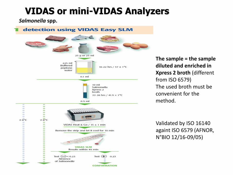
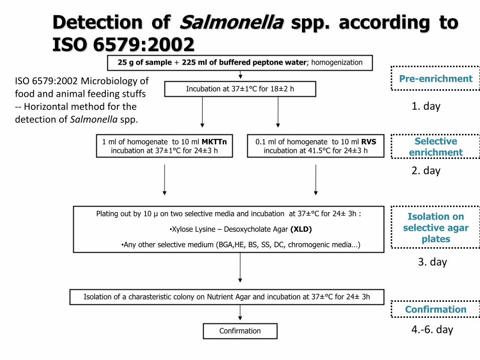
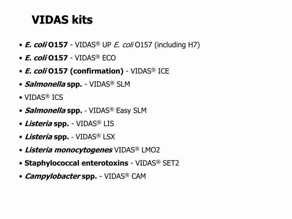
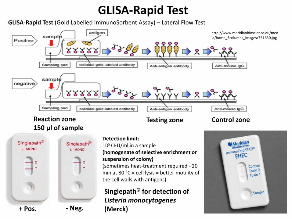

Validation of procedure
Validation of an alternative method bull the validation of an alternative method is the procedure to demonstrate if the alternative method provides equivalent or better results compared to the reference methods
bull Validation is a process within which the method is demonstrated to be suitable
for its purpose It documents methods validity
bull During validation process methods performance characteristics are estimated
The validation of qualitative and quantitative methods comprises two phases
1 a method comparison study of the alternative method against a reference
method (performed by an expert laboratory)
2 a interlaboratory study of the alternative method (organised by an expert
laboratory)
Expert laboratory (organising) laboratory having the qualified staff and skills to
perform the method comparison study and organise the interlaboratory study
Useful documents
ISO 161402003 ndash Microbiology of food and animal feeding stuffs ndash Protocol for the
validation of alternative methods
ISO 170252005 ndash General requirements for the competence of testing and calibration
laboratories
Accreditation bodies that recognize the competence of testing and calibration laboratories
should use this International Standard as the basis for their accreditation
ISOIEC 170112004 Conformity assessment -General requirements for accreditation
bodies accrediting conformity assessment bodies
If a laboratory wishes accreditation for part of all of its testing and calibration activities it
should select an accreditation body that operates in accordance with ISOIEC 17011
It is expected that all laboratories involved in each step of a validation process will have a Quality System or quality assurance (QA) program in place to ensure standardization of laboratory operations as well as adequate quality control (QC) activities
Validation of procedure
Test characteristics The test characteristics for alternative methods are shown
Qualitative methods
1 Selectivity (inclusivity exclusivity)
2 Relative accuracy
3 Detection level
4 Relative sensitivityRelative specificity
5 The agreement between the
methods
Quantitative methods
1 Selectivity (inclusivity exclusivity)
2 Lowest validated level with satisfactory
precision
3 Repeatability
4 Reproducibility
5 Uncertainty of the method
28
PCR METHODS
DNA RNA PROTEINS Deoxyribonucleic acid (DNA)
A linear polymer that consists of four nucleotides
Adenine Cytosine Guanine Thymine Primer binding A ndash T C - G
DNA RNA PROTEINS
DNA
RNA
httpss3amazonawscomclassconnection116flashcards5862116jpgrna-149F82012BD7233971Fjpg httpslh3googleusercontentcomf81cHVr_O-
jXwH1A7g-mfX1LJKZFffUkItKpHPZ3kGF6cGXJaAJ-pqZoNL7T4YNKcP-grQ=s85 httpslh3googleusercontentcomdc9f6E-QDwxJQc-
7MvExwNvrihEjEfgjzwKr2gidCYIt0VmoMb1yEgTDc6mpkFDMUsXsgw=s94
httpsuploadwikimediaorgwikipediacommons00fPeptide_synpng
PCR principle PCR=Polymerase Chain Reaction (1983 by Kary Mullis) in vitro amplification of the part of DNA (usually 100 bp ndash 1500 bp) bordered by two primers (syntetically prepared oligonucleotides)
Typical number of cycles = 30 - 45
httpswwwyoutubecomwatchv=iQsu3Kz9NYo
PCR principle DENATURATION of double-stranded DNA by heat (app 94-95degC initially 5 then in each step 2-3 minutes)
ANNEALING of PRIMERS on the free single strand DNA bull forward primer on the 3acute-
5acute strand bull reverse primer sits on the
5acute-3acute strand Ta = annealing temperature bull 50-65 degC ndash depends on the
primers sequence bull Time 30 s-60 s bull One of the most important
factor for the specifity bull T gtgt Tm= no annealing - no
PCR product bull T ltlt Tm= non-specific
annealing ndash nonspecific products
ELONGATION by DNA polymerase
in vivo starts to synthetize new DNA molecules from 3acute- end of RNA primer
in vitro PCR from 3acute-end the primers
72 degC ndash optimal temperature
45 s-3 minutes (final elongation 5-15 minutes)
httpwwwmuncabiologyscarrPCR_sketch_3gif
PCR principle
httpopenwetwareorgimagesff1CH391L_S12_PCR_exponential_amplificationjpg
Mistake in the beginning ndash the most influence Exponential phase duplication of PCR products amount in each cycle Linear phase The level of amplification is decreased (lower than duplication in each cycle) Plateau Absence or degradation of some component Reasociation can be preferred than primers anealing due to thermodynamical reasons
PCR principle
httpwwwebioecomuploadfile2010031420100314110534183gif
DNA (genomic plasmid DNA)
Primers (forward reverse)
Thermostable
DNA polymerase
Mg2+ (cofactor
for DNA
polymerase)
dNTP mix
(dCTP dATP
dTTP dGTP) ndash
building stones
for newly
synthetized
DNA product
Nuclease free water ndash without Dnase
Rnase presence (enzymes which
degrade DNA RNA)
MASTERMIX
Total volume ndash 25 μl ndash 50 μl
PCR principle
Polymerase Taq DNA polymerase
bull from thermophilic bacteriumThermus aquaticus
(now prepared by recombination)
bull opt temp 75-80degC (150 ntss)
bull active at the room temperature ndashto work on ice
bull inactive at gt90degC but reactivation when cooled
bull only 5acuteexonuklease activity not 3acuteexonuklease
activity (proofreading)
bull 1 mistake at 10-20 000 nts
Hot start polymerases
bull Activity at the room temperature is inhibited - binding on anti-polymerase
antibodies on the wax balls
bull activation after heating at 94 degC (antibodies destroyed)
Other polymerases
bull Proofreading polymerases (with the 3acuteexonuclease activity)
bull Tth polymerase - reverse transcription (Mn2+) or polymerase activity (Mg2+)
1) Releasing DNA from the cell
- lysis of the cell wall by detergents or by heat digesting cell surface proteins by proteinase K
- RNAse treatment for RNA-free genomic DNA
2) Isolation DNA from the released cytoplasmatic contents
- Centrifugation the vaste in pellet (heat isolation)
- Ion-exchange chromatography (commercial kit) ndash binding DNA by ionic interactions to the column (DNA is negative charged) ndash proteins and RNA pass away ndash DNA is later released by using more concentrated salt solution (column is charged positively)
- Phase separation (aqueouschlorophorm phase ndash CTAB method)
DNA isolation
Detection of food-borne pathogens
DNA of microorganism = set of specific genes with specific sequences
Primers are designed to be complementary to sequence of some specific gene
ndash if the complementary sequence is present PCR product will be performed
Main problems about primers designing and applying of PCR
- Is the gene of interest specific enough (can be found only in the detected microorganism)
- Is the primers sequences complementary only to this gene
- Is the primer sequence designed well (requirements about easy and specific annealing ndash depends on the primers sequence)
- Primers can be designed comercially or by using software (httpwwwbiocenterhelsinkifibiProgramsdownloadhtml)
- sequences and genomes needed - wwwpubmedcom
- httpswwwncbinlmnihgovtoolsprimer-blastindexcgiLINK_LOC=BlastHome
PCR products detection The PCR product has got specific length and specific sequence
Visualiton by intercalating agents bind to double-stranded DNA - fluorescence signal bull Ethidium bromide ndash carcinogenteratogen If intercalated the fluorescence of EtBr is significantly higher than in aqueous solution (UV absorbance maxima at 300 and 360nmemitted as orangeyellow light at 590nm) bull Sybr Green DNA-Sybr Green complex absorbs blue light at 488 nm and emits green light at 522 nm Fluorescent tags at 5acute-end of primers Other tag at 5acute -end of primers eg biotin - for immunodetection detected by anti-bodies linked with an enzyme Microarray
Electrophoresis Conventional electrophoresis a single electrical field causes biomolecules to migrate through a matrix according to its mass-to-charge ratio effectively separates DNA fragments up to ~20-30 kb (according to the gel concentration) larger fragments will comigrate (a large band at the top of the gel)
httpwwwrothamstedacuknotebookimagesgelelgif httpstevegallikorgsitesallimagesgelElectrophoresis2jpg
httpabeleewardhawaiieduProtocolsDNA20Gel20Preparation20and20Electrophoresishtm
Electrophoresis Primers (OT1559 a 18ndash1) complementary to DNA coding16S rRNA Campylobacter
jejuni Campylobacter coli a Campylobacter lari
Product is 287 bp
60 V 100 minutes M ndash 100 bp marker (1-5 positive controls) 1- C jejuni CCM 6212 2- C jejuni CCM 6214 3 ndash
C coli CCM 6211 4 ndash C lari CCM 4897 5 ndash C upsaliensis ATCC 43954 6- 15 strains C jejuni Nt- no
template control (negative control)
Genus specific PCR
Electrophoresis DUPLEX PCR =two pairs of primers
C jejuni C1 5 - CAA ATA AAG TTA GAGGTA GAA TGT-3
C4 5 - GGA TAA GCA CTA GCTAGC TGA T-3
Complementary to a part oxidoreductase 159 bp
C coli 18F 5 - GGT ATG ATT TCT ACAAAG CGAG-3
519R 5- ATA AAA GAC TAT CGT CGC GTG-3acute
Complementary to aspartatekinase and flanking ORF 500 bp
Species specific PCR
60 V 100 minutes M ndash 100 bp marker (1-5 positive controls) 1- C jejuni CCM 6212 2- C jejuni CCM
6214 3 ndash C coli CCM 6211 4 ndash C lari CCM 4897 5 ndash C upsaliensis ATCC 43954 6- 15 strains C
jejuni Nt- no template control (negative control)
Melting curve Intercalating dye binds to double-stranded DNA - producing fluorescent signal when excited by light
When samples are heated to melting point double-stranded DNA denatures into single strands freeing dye into solution
ndash Melting temperature (Tm) is the point at which the DNA denatures
ndash Dependent on the length of fragment and G-C content
ndash More base pairs --gt Increase in Tm
ndash Higher G-C content --gt Increase in Tm
bull Unbound dye has reduced fluorescent signal
bull Loss in signal generates melt curve
No Signal Signal Increasing
temperature
Intercalating
dye
Melting curve
Inflex point
bull 1 derivation = 0
bull In this case 2derivation is negative
Real-Time PCR
CT
CT (threshold cycle) ndash the lowest cycle when fluorescence crosses the
determinated value
Video httpswwwyoutubecomwatchv=kvQWKcMdyS4 httpswwwyoutubecomwatchv=pRwoOBuk00c
Real-Time PCR
Food-borne pathogens detection by PCR
Identification of pure cultures - without problems
Detectionenumeration from food matrix directly or after enrichment
bull How to isolate successfully DNA from few cells in 25 g10 g - Efficiency of DNA isolation (isolation by kits heat-lysis CTAB method)
bull How many targets copies are needed for successfully PCR products detection
ndash detection limits
ndash presence of PCR inhibitors (inhibition of polymerase primers annealing)
bull How to distuinguish DNA from dead and live cells - Total DNA is detected (from dead and living cell)
bull For distinguishing of dead cells DNA ndash eg application of EMA (ethidium monoazide) to the sample before DNA isolation ndash EMA irreversibly bound to DNA of dead cells ndash they are bdquoinvisibleldquo for PCR ndash no amplification
bull Direct enumerationdetection only in some specific case ndash simple liquid sample when the required limit is higher than the limit of detection
bull Standardly ndash for PCR is used enriched homogenate (sample diluted in enrichment broth and cultivated) ndash to obtain detectable level of microorganisms
BAXreg System
Salmonella
Listeria monocytogenes
Genus Listeria
Enterobacter sakazakii
E coli O157H7
Staphylococcus aureus Real-time
Campylobacter jejunicolilariReal-time
Vibrio cholerae
parahaemolyticusvulnificus Real-time
Yeast and molds
httpwww2dupontcomDuPon
t_Homeen_USindexhtml
BAX ndash three major steps
On enrichedregrown samples
Prepare samples = perform heat-lysis in two stages
DNA present in the lysate
2 Process samples
3 Review results
Hydrate PCR tablets
Enrichment protocols vary
depending on bacterial target
and type of food yoursquore testing
Salmonella Enterobacter
sakazakii
E coli O157H7 E coli O157H7
MP Campylobacter jejunicoli
Yeast and Mold
37deg +- 2degC for 20 minutes then
95deg +- 3degC for 10 minutes
Listeria and L monocytogenes
55deg +- 2degC for 60 minutes then
95deg +- 3degC for 10 minutes
5 microl for all bacterial targets
20 microl for beef samples enriched
in E coli O157H7 MP media
20 microl for yeast amp mold
PCR tablets bull DNA-polymerase
bull Primers
bull Nucleotids
bull Suplements
bull SybrGreen or fluorescent probe
bull Internal control + primer
bull Mastermix in the tablet in PCR Eppendorf ndash minimal risk of contamination
For solving the problem of distiguishing dead and live cell the process of small volumes
is used ndash only if live cells are present in the sample they able to multiplied to the limit
detection (104 CFUml in the enrichment suspension used for lysis step)
If the result of detection is positive confirmation by plating out is done (live and dead
cells)
+
Target organism DNA BAXreg system
primers
in tablets PCR
+ no detectable
amplification
Background DNA
PCR
BAXreg system
primers
in tablets
Amplification of specific fragments
Detection of specific fragments Fluorescent signal drops with heating to generate a melt curve
Melting curve Raw Data Processed Data
Strong positive
result
Negative
result
Target peak
Positive control peaks
Positive control peak
Melting curves display peaks within a set temperature range for the control and target (if present)
Detection
Melt curve weak INPC in presence of strong Salmonella positive
Real time weak INPC in presence of strong
Campylobacter positives
Direct protocol does not display a positive or
negative result PCR was successful IndeterminateSignal error
Salmonella AFNOR (ISO 16140) protocol Sample 110 BPW
18 h37ordmC
RVS Muumlller-Kauffmann
24 h 415 37 ordmC
5 suspicious colonies on TSA
24 h 37ordm C
Biochemical and serological confirmation
XLD 2 Plate
24 h 37ordmC
BHI
3h 37 ordmC
22 h 37 ordmC
15 d neg and pos result
3 d neg 5 d pos results
Salmonella ( ISO 65792003) versus BAXreg-PCR
International certificates AOAC-RI Performance Tested Method
Salmonella 100201 L monocytogenes
070202 L spp 030502 Saureus 120701
Campylobacter colilarijejuni
AFNOR Certification
Salmonella QUA 183-1102
NordVal Certification
Salmonella 2006-30-5408
Certificates from AFNOR and AOAC are equal
NordVal Certificate
57
OTHER METHODS
ATP bioluminiscence
httpwwwpromegacouk~mediaimagesresourcesfiguresenotesfe0027_fig1jpgla=en
httpwwwbiotekptassetstech_resources143clarity_atp_conc_fig2gif
Principle the detection of the present level of ATP (direct proportion to the presence of microorganisms or their products residues) The enzyme luciferase catalyzes the transformation of d-luciferin to oxyluciferin (indicated by emision of light at 560 nm)
ATP bioluminiscence
httpwwwbioxyscomimages2figure-B11gif
ATP Hygiene Monitoring System - SystemSURE II
httpwwwmicromaticcomdraft-keg-beerline-cleaning-hygiene-pid-ATP-100-tab-reviewshtml
Biosensors
60
httpsuploadwikimediaorgwikipediacommonseecBiosensor_Systemjpg
Biosenzor =the whole cellmicroorganism (CBS ndash cell-based sensors)
Cell-based sensors
Patogenic microorganisms interact during the infection with mammalian
cells ndash detection of cell or toxin by cell culture The damage of cell culture is measured optically or electrically
62
IDENTIFICATION OF MICROORGANISMS
Definition of species in bacteriaarchae
The criterion for a bacterial or archael species a collection of strains that have bull at least one diagnostic phenotypic trait bull ge70 DNAndashDNA hybridization of their purified molecules DNA
bull ~more than 97 16S rRNA sequence identity Under the same conditions some eucaryotic order would be a single bacterial species
The bacterial species definition bull eminent practical significance bull but a very difficult issue
bull very wide and often changes in DNA
bull mutations during replication of DNA
bull horizontal gene transfer through mobile elements plasmids also between species
Criterion for species in prokaryotic cells (bacteria and archea) Collection of strains characterised bull by at least one diagnostic feature bull their purified DNA molecules prove at least 70 hybridisation
which corresponds to at least 97 concordance in the sequence of 16S rRNA gene
Map of genome of B cereus ATCC 14579
httpwwwacgtinccomspecialty_dna_sequencinghtm
Identification based on genotype
64
DNA isolation PCR amplification of 16S rRNA gene (usually)
Sequencing of PCR product
Comparison to sequences in databases (BLAST)
Identification of bacteria PHENOTYPE METHODS bull Morphology of colony and cell (Gram staining shape) bull Requirements for temperature atmosphere nutrition bull Biochemical tests - presence of specific enzymes
bull catalase oxidase fermentation of glucose bull the way how they utilize different compounds
bull Immunochemical tests - specific antigens in the cell wall METHODS BASED ON PROTEIN ANALYSIS
bull MALDI-TOF MS METHODS BASED ON GENOTYPE bull looking for and comparing of specific DNA sequences
bull conserved universal genes (eg 16S rRNA) bull genes specific for genus species
bull done by PCR methods DNA chips sequencing etc
66
BIOCHEMICAL TESTING
Identification based on phenotype I Physiological + biochemical features
bull morfology of colonies and cells (Gram staining shape)
bull requirements for temperature atmosphere nutrient sources
bull basic biochemical tests oxidase catalase fermentationoxidation of glucose haemolysis etc
appearance of colonies on selective or selective-diagnostic agars (for other testing to be reinoculated on non-selective agar)
the classification of an isolate into a certain group
(eg for bacteria catalase positivenegative G+ cocci glucose fermentingnon-fermenting G- rods etc)
biochemical tests specific for certain groups of microorganisms
Identification based on phenotype I
68
Physiological + biochemical features Example Bacillus cereus
httpwwwfoodhaccpcommemberonlynewsletter164html
httpwwwnapavalleyedupeoplesrosePublishingImagesBacillus20cereus202(Gram20stain)jpg
Set of biochemical tests Microgenreg Bacillus ID Panel for the identification of Bacillus spp and related genera (time for performing 48 hours)
MYP agar (Mannitol Egg Yolk Polymyxin Agar) pink colonies with a precipitation zone (no fermentation of manitol + enzyme lecitinase)
catalase positive
httpwwwoxoidcomukblueprod_detailprod_detailasppr=CM0929ampc=ukamplang=ENamporg=9ampimg=CM0929ampsec=
G+ rod endspores forming
β-haemolysis moisty and easy to grow colonies
Principle of biochemical testing
to rank the bacterium to some group having the same basic features bull eg catalase positive gram positive cocci bull Family Enterobacteriaceae
the identification on the genus or species level
bull sets of biochemical tests specific for some group of bacteria
70
Gram staining procedure - principle
https40mediatumblrcom25da2e5b25bbe336819bf1715849d171tumblr_inline_ntrsgn9w1D1sv92o6_540jpg
Gram staining procedure - principle
httpsonlinesciencepsuedusitesdefaultfilesmicrb106CellStructurecow02354_03_15jpg
Gram staining procedure 1-2 drops of sterile water on a slide Spread culture in thin filter in this drop Let dry in airclose to fire Pass slide through flame 2-3x to fix Add crystal violet ndash 15-20 s to act Add Lugol solution (KII2) ndash 15-20 s to act Washing off with a mixture of
ethanolaceton (41) or acetone till the stain removes but max 15 s
Rinse with water decant excess liquid Add safranin 1 minute Rinse with water decant excess liquid Dry and put a drop of imersion oil use an
objective bdquoOILldquo
httpwwwtumblrcomtaggedgram-staining
G- red
G+ violet
Gram staining procedure + Identification
E coli
Family Enterobacteriaceae Gram-negative rods
Micrococcus luteus
Staphylococcus aureus
Gram-positive cocci Bacillus cereus
Gram-positive rods (with endospores)
Biochemical identification of G + bacteria
httpyoungeneticistblogspotcz201106diagnostic-microbiology-labhtml
G+ bacteria bull cocci ndash in clusters in pairs
diplococci bull bacilli
bull endospore bull no endospore
For G+ cocci bull Catalase positive ndash Staphylococcus
sp bull Coagulase + S aureus or some other few species bull Coagulase -
bull Novobiocin S bull S epidermidis etc
bull Novobiocin R bull S saprophyticus etc
bull Catalase negative Streptococcus sp Enterococcus sp
bull kind of haemolysis Other important G + group ndash filamentous bacteria
Catalase test Catalase test Catalase is a common enzyme found in nearly all living organisms that are exposed to oxygen where it catalyzes the decomposition of hydrogen peroxide to water and oxygen
2 H2O + O2 2 H2O2
catalase
bubbling when culture is add in peroxide
httpiws2collinedudcainCCCCD20Microcatalase_testhtm
Catalase testing into a drop of 3 peroxide to transfer a loop of bacterial culture catalase positive ndash releasing of air bubbles
Examples bull G+ cocci catalase positive Staphylococcus sp Micrococcus sp Kocuria sp Dermacoccus sp Macrococcus sp Rothia sp catalase negative Streptococcus sp Enterococcus sp bull G+ rods (bacilli) endospores forming catalase positive Bacillus sp catalase negative Clostridium sp
Resistance to ATBhaemolysis Resistance eg to novobiocin
httpquizletcom20077004mic-lecture-1-t2-flash-cards
Hemolysis on blood agar
Measuring the diameter of inhibition zone (less than 17 mm resistant more or equal to 17 mm sensitive) ndash differing in Staphylococcus sp (Clinical performance)
alpha-hemolysis - oxidizing the iron in the hemoglobin - green discoloration of the medium around beta-hemolysis - rupture of erythrocytes - complete clearing medium around gamma-hemolysis ndash no effect ndash no change
By cultivation on blood agar bacteria can be
differentiated based on their capacity to secrete
hemolysins The hemolysis will cause a clearing zone of
the blood agar around the colonies Bacteria can cause
different types of hemolysis
bullα-hemolysis which means an incomplete clearing
(green haemolysis)
bullβ-hemolysis which means a complete clearing
bullDouble hemolysis of some staphylococci consisting of
an inner β-hemolysis zone and an outer α-hemolysis
zone
bullNo hemolysis which is sometimes referred to as γ-
hemolysis which may seem illogical
Note that the α-hemolysin of staphylococci causes
complete hemolysis whereas their β-hemolysin causes
incomplete hemolysis
The capacity to produce hemolysins may vary between
different strains of a particular bacterial species
httpwwwvetbactorgvetbactdisplayextinfo=67
Identification of bacteria based on haemolysis
Streptococcal hemolysins α (alpha) hemolysis - ldquozone of green discolourationrdquo or ldquogreeningrdquo called partial or incomplete hemolysis produced by alpha-hemolytic streptococci such as Streptococcus suis This type of hemolysis on BA is due to bacterial production of hydrogen peroxide which oxidises hemoglobin to methemoglobin β (beta) hemolysis ndash a clear zone of complete hemolysis produced by beta-hemolytic streptococci such as S equi subsp equi This hemolysis is due to lysis of RBCs in the BA by the cytotoxin Streptolysin S
Source httppeopleupeicajlewishtmlhemolysinshtml
Bacterial hemolysins
Staphylococcal hemolysins Most coagulase-positive staphylococci (CPS) such as Staphylococcus aureus S pseudintermedius ( but not S hycius) typically produce an distinctive pattern of ldquodouble zonerdquo or ldquotargetrdquo hemolysis on blood agar Double-zone hemolysis indicates that the two hemolysins one which produces ldquocompleterdquo and another one that produces ldquoincompleterdquo hemolysis are present and this combined effect is visible due to different diffusion rates of these enzymes through the agar The staphylococcal α (alpha) hemolysin causes a complete type of hemolysis This zone of complete hemolysis is narrow and immediately adjacent to the bacterial colony on blood agar The staphylococcal β (beta) hemolysin causes an incomplete type of hemolysis at 37 degC This broader incomplete zone is outside of the inner complete zone and can be hard to see on early culture plates and also varies with the species of RBCs used to make the BA plate Its action is enhanced by chilling at 4 degC and the hemolysis becomes complete (this effect is referred to as ldquohot-coldrdquohemolysis) There are other staphylococcal cytotoxins that are also hemolysins but their lytic actions are not seen on blood agar
The confusion arises due to the NAMES of the two STAPHYLOCOCCAL HEMOLYSINS and the TYPE of hemolysis they produce These are the REVERSE of the two TYPES of STREPTOCOCCAL HEMOLYSIS Strictly speaking the descriptive terms ldquoalphardquo for ldquoincompleterdquo and ldquobetardquo for ldquocompleterdquo hemolysis apply only to the hemolytic toxins of Streptococcus species However other organisms beside Streptococcus and Staphylococcus species have cytotoxins that cause hemolysis and this hemolysis can be either complete or incomplete It is preferable to use the terms ldquocompleterdquo and ldquoincompleterdquo rather than ldquobetardquo or ldquoalphardquo to describe the hemolysis caused by these organisms but you will notice in bacteriology text books that the terms alpha and beta are commonly used
G ndash bacteria bull Cocci bull Bacilli (Rods) bull Oxidase positivenegative bull Glucose metabolism
bull fermentation bull oxidation bull Inactive
bull Lactose metabolism for coliform
bull Pigment formation for Pseudomonas sp
Biochemical identification of
G - bacteria
Oxidase test
Oxidase positive (OXI +) Aeromonas sp Pseudomonas sp Dark-blue to violet
Oxidase negative (OXI -) Enterobacteriaceae Bacillus sp Colourless
Oxidase bullabbreviated general name for some kind of cytochrome c oxidases (cytochrome oxidase or indophenol oxidase) enzymes involved in aerobic respiratory chain when oxygen is used as the final receptor for hydrogen bullOxidases are usually present only in aerobic organisms bullPresence or absence of oxidase(s) is an important feature of the bacterial metabolism used mainly in the identification process of gram-negative bacteria
Detection Reaction of the culture with redox indicator as eg NN-dimethyl-p-phenylenediamine (DMPD) dark-blue when oxidized and colorless when reduced Different test performances are possible
Fermentation of carbohydrates Type of utilisation of glucose or carbohydrates bull an important feature of the bacterial metabolism bull widely used in the identification process of bacteria bull fermentation strictly means to be under anaerobic conditions (but in bacterial
metabolism which can be but not have to be the same as the anaerobic conditions of cultivation) so very often is used in a general way like bdquoutilisationldquo
bull fermentation (like bdquoutilisationldquo) of carbohydrates to
bull acid and gas (Durham tubes) bull acid without gas bull non-utilised
bull Testing of fermentation or oxidation of carbohydrates
bull eg OF test for glucose bull by oxidation under aerobic conditions bull by fermenation under anaerobic conditions (an oil layer)
Fermentation of sugars to acid
Peptone Water wPhenol Red
(HiMedia M028I)
Ingredients gl
Peptic digest of animal tissue 10000
Sodium chloride 5000
Phenol red 0020
Final pH ( at 25degC) 68plusmn02
httpsuploadwikimediaorgwikipediacommons009Phenol_red_pH_60_-_80jpg
+ after sterilisation
at the room
temperature to add
aseptically sugar
discs and
inoculate
succrose trehalose rhamnose xylose
Incubation 30 degC 24 h
- +
Fermentation of carbohydrates
Casein enzymic hydrolysate and yeast extract provide nitrogenous compounds vitamin B complex and other growth nutrients Dextrose is the fermentable carbohydrate and bromocresol purple acts as the pH indicator with colour change from purple to yellow in acidic conditions Sodium chloride maintains osmotic equilibrium bdquoAnaerobicldquo conditions ndash inoculation by stabbing to used fresh prepared media (oxygen expelled by heating medium in boiling water or running steam for 15 min then to be cooled down quickly)
- +
Enterobacteriaceae ndash glucose fermentation
83
Tryptone Yeast Extract Agar Ingredients Gms Litre Casein enzymic hydrolysate 10000 Yeast extract 1500 Dextrose 10000 Sodium chloride 5000 Bromocresol purple 0015 Agar 15000 Final pH ( at 25degC) 70plusmn02
Test for oxidationfermentation of glucose (OF glucose test) detection of acid production by acidobazic indicator change to yellow (acid present) in lower pH from green (no acid) Anaerobic atmosph ndash covering by sterile mineral oil If fermented anaerobically then also runs aerobically
Fermentation of carbohydrates
Principle of biochemical tests bull The first step in identification is to rank the bacterium to some group of bacteria
having the same basic features ndash eg catalase positive gram positive cocci bull The identification on the genus or species level is finished by other biochemical tests
specific for each group of bacteria bull The tests can be done in tubes or miniaturized plate
ENTEROtest 24N (Erba Lachema) (24 means 24 biochemical tests N= new)
API 20E (bioMeacuterieux)
bull designed for routine identification of clinically important species of family Enterobacteriaceae without the use of reagents
bull A strip test OXItest or OFtest in microtitration wells can be used to confirm that the isolate belongs to Enterobacteriaceae family
bull The results are read visually or automatically using reader MIKROLAreg
bull The identification can be supplemented by diagnostic strip tests INDOLtest COLItest and VPtest
APIreg Gram negative Identification API 20E ndash Speciessubspecies identification of Enterobacteriaceae and groupspecies identification of non-fermenting Gram negative bacteria API Rapid 20E ndash 4-hour identification of Enterobacteriaceae API 20NE ndash 24 to 48-hour identification of Gram negative non-Enterobacteriaceae API Campy ndash 24-hour identification of Campylobacter species
Example Escherichia coli
ENTEROtest 24 N
Biochemical testing - Enterobacteriaceae bull TSI Triple Sugar Iron Agar (use a straight wire to stab the butt and streak the agar surface)
- to determine whether organisms can ferment glucose sucrose andor lactose with or without production of gas and the ability of the organism to produce hydrogen sulphide from thiosulphate in an acid environment is also tested
Formula Liter Enzymatic Digest of Casein 5 g Enzymatic Digest of Animal Tissue 5 g Yeast Enriched Peptone 10 g Dextrose 1 g Lactose 10 g Sucrose 10 g Ferric Ammonium Citrate 02 g Sodium Chloride 5 g Sodium Thiosulfate 03 g Phenol Red 0025 g Agar 135 g Final pH 73 plusmn 02 at 25 degC
inoculation in the butt by stab inoculation on the slant on the surface bull Fermentation of dextrose
(glucose) to acid bull anaerobically - yellow butt bull aerobically leads to products
which are later oxidised by air and pH remains alkaline (red) bull Fermentation of glucose to gas (anaerobically - in the stab) Cracks splits or bubbles in medium indicate gas production bull Production of hydrogen sulfide (H2S) ndash reaction with ferrous sulphate and sodium thiosulphate - a black precipitate in butt bull Lactose andor succrose
utilisation to acid Acid production ndash yellow butt and slant
Results
An alkaline slant-acid butt
(redyellow) - fermentation of
dextrose only
An acid slant-acid butt
(yellowyellow) - fermentation of
dextrose lactose andor sucrose
An alkaline slant-alkaline butt
(redred) - dextrose or lactose not fermented (non-fermenter)
Bacterium fermenting lactose andor succrose are able to ferment glucose
Phenol red ndash alkaline = red acid = yellow
bull TSI Triple Sugar Iron Agar (use a straight wire to stab the butt and streak the agar surface)
- to determine whether organisms can ferment glucose sucrose andor lactose with or without production of gas and the ability of the organism to produce hydrogen sulphide from thiosulphate in an acid environment is also tested
Biochemical testing - Enterobacteriaceae
Urea agar (Christensen) (use a 1 μl loop full)
- to determine whether an organism can split urea into ammonia and
carbon dioxide by the action of the enzyme urease
httpimageslidesharecdncomlab17-141201085153-conversion-gate0195-10-638jpgcb=1417424219
httpimageslidesharecdncomhelicobacterpylori-copy-110516201901-phpapp0195campylobacter-jejunihelicobacter-pylori-39-728jpgcb=1305577391
Phenol red
Medium is acidic - yellow
(pH 68plusmn02)
Alkaline reaction by urea hydrolysis - pink colour
Composition Gms Litre
Peptic digest of animal tissue
1000
Dextrose 1000
Sodium chloride 5000
Disodium phosphate 1200
Monopotassium phosphate 0800
Phenol red 0012
Agar 15000
Final pH ( at 25degC) 68plusmn02
Nutrition
Peptic digest of animal tissues dextrose
Sodium chloride - osmotic equilibrium
Phosphates - buffering of the medium
35-37 degC
Biochemical testing - Enterobacteriaceae
bull L-lysine decarboxylase (use a 1 μl loop full)
- to determine whether an organism can decarboxylate an amino acid (lysine) leading to formation of an
amine After inoculation both tubes are overlayed with sterile paraffin oil
httpfce-studynetdna-sslcomimagesupload-flashcards10295302332621_mjpg
+ CO2 + OH-
Lysine Decarboxylase Broth Typical Composition (glitre) Peptic digest of animal tissue 5000 Yeast extract 3000Dextrose 1000 (glucose prepared from starch chemically equivalent to glucose) L-Lysine hydrochloride 5000 Bromocresol purple 0020 Final pH ( at 25 degC) 68plusmn02 Inoculate below the surface cover by sterile paraffin oil
Two steps detection 1 step Dextrose is fermented ndash H+ is released Acidic conditions bull bromocresol purple changes colour to yellow bull in acidic conditions lysine decarboxylase is
activated 2 step L-lysine is decarboxylated and OH- released Alkaline conditions bull bromocresol purple changes colour back to
purple
Biochemical testing - Enterobacteriaceae
bull β-galactosidase (ONPG) (use a 1 μl loop full)
- to determine whether an organism has the enzyme szlig-galactosidase by using the compound o-
nitrophenyl-beta-D-galactopyranoside (ONPG)
- this test is used to differentiate between lactose-negative and lactose-delayed organisms
- ONPG is hydrolysed by the enzyme to o-nitrophenol which is yellow and galactose
O-Nitrophenyl-β-D-galactopyranoside (ONPG) is structurally similar to lactose (ie ONPG is an analog of lactose) except that orthonitrophenyl has been substituted for glucose ONPG is colorless compound O-nitrophenol is yellow visual evidence of hydrolysis
Lactose fermenting bacteria (ONPG positive) two enzymes Permease ndash transferring of the lactose molecule into the bacterial cell β-galactosidase - cleaving the galactoside bond producing glucose and galactose - inducible enzyme - made ONLY in the presence of the lactose substrate Coliform bacteria as Ecoli Klebsiella spp Enterobacter spp
hydrolysis
Late lactose fermentering bacteria Permease ndash not present delayed in the production of acid from lactose because of sluggish permease activity β-galactosidase ndash present - give a positive ONPG test Citrobacter spp Arizona spp
Non lactose fermenter (ONPG Negative) Permease ndash not present β-galactosidase ndash not present Salmonella spp Shigella spp Proteus spp Providencia spp and Morganella spp
Biochemical testing - Enterobacteriaceae
Procedure Culture Bacteria grown in medium containing lactose (to induce the production of the galactosidase enzyme) such as Kligler iron agar (KIA) or Triple sugar Iron (TSI) agar (Note β-galactosidase enzyme (inducible enzyme) is made ONLY in the presence of the lactose substrate) Inoculation 1A loopful of bacterial growth is emulsified in 005mL of physiologic saline to produce a heavy suspension 2One drop of toluene is added to the suspension and vigorously mixed for a few seconds to release the enzyme for bacterial cells 3An equal quantity of buffered ONPG solution is added to the suspension 4The mixture is placed in a 37 oC water bath 5When Using ONPG Tablets 6A loopful of bacterial suspension is added directly to the ONPG substrate resulting from adding 1mL of distilled water to a tablet in a test tube 7This suspension is also placed in a 37oC water bath
β-galactosidase (ONPG)
Results and Interpretations The rate of hydrolysis of ONPG to o-nitrophenol may be rapid for some organisms producing a visible yellow color reaction within 5 to 10 minutes Most tests are positive within 1 hour however reactions should not be interpreted as negative before 24 hours of incubation The yellow color is usually distinct and indicates that the organism has produced o-nitrophenol from the ONPG substrate through the action of β-galactosidase
Biochemical testing - Enterobacteriaceae
bull VogesndashProskauer (VP) test (use a 1 μl loop full)
- to determine whether an organism can produce acetylmethylcarbinol
(acetoin) from fermentation of glucose If present acetylmethyl carbinol is
converted to diacetyl in the presence of prop- naphthol (to be added first) strong
alkali (40 KOH) and atmospheric oxygen The diacetyl and quanidine-
containing compounds found in the peptones of the broth then condense to
form a pinkish red polymer
MRVP broth (pH 69)
Ingredients per liter of
deionized water
buffered peptone= 70 gm
glucose= 50 gm
dipotassium phosphate= 50 gm Voges-Proskauer Reagent A Barrittrsquos reagent A
Alpha-Naphthol 5 50 gm
Absolute Ethanol 1000
ml
Voges-Proskauer Reagent B Barrittrsquos reagent B
Potassium Hydroxide 400 gm
Deionized Water 1000 ml
Procedure of VogesndashProskauer (VP) Test
1Prior to inoculation allow medium to
equilibrate to room temperature
2Using organisms taken from an 18-24
hour pure culture lightly inoculate the
medium
3Incubate aerobically at 37 degC 24 hours
4Following 24 hours of incubation aliquot
2 ml of the broth to a clean test tube
5Re-incubate the remaining broth for an
additional 24 hours
6Add 6 drops of 5 alpha-naphthol and
mix well to aerate
7Add 2 drops of 40 potassium hydroxide
and mix well to aerate
8Observe for a pink-red color at the surface
within 30 min Shake the tube vigorously
during the 30-min period
Positive Reaction
A pink-red color at the surface
Examples Viridans group streptococci
(except Streptococcus vestibularis) Listeria
Enterobacter Klebsiella Serratia marcescens
Hafnia alvei Vibrio eltor Vibrio alginolyticus etc
Negative Reaction
A lack of a pink-red color
Examples Streptococcus mitis Citrobacter sp
Shigella Yersinia Edwardsiella Salmonella Vibrio
furnissii Vibrio fluvialis Vibrio vulnificus and Vibrio
parahaemolyticus etc
A copper color should be considered negative A
rust color is a weak positive reaction
Quality Control of VogesndashProskauer (VP) Test
VP positive Enterobacter aerogenes (ATCC13048)
VP negative Escherichia coli (ATCC25922)
Biochemical testing - Enterobacteriaceae
94
VogesndashProskauer (VP) test
Rapid test for confirmation of E coli ndash typical features
β-D-glucuronidase (97 strains) production of indole from tryptophan (typical for E coli)
Performance
Previous testing Gram-negative (eg able to grow on VRBD agar) oxidase negative
From pure culture to prepare suspension in saline solution (app 5 ml) at the density 3 McFarland
To transfer 05 ndash 1 ml of this suspension into a new tube ndash to add the strip of COLItest (chemicals ndash fluorogenic substrate for β-D-glucuronidase tryptophane are present in the pad at the end of the strip)
Incubation 37 degC 4 hours
Detecion of β-D-glucuronidase (to be done at first) UV (360 nm) ndash blue fluorescence (GLR + hellipβ-D-glucuronidase positive) ndash GLR test can be read after 1 hour
Detection of indole formation from tryptophane (to be done at second) to add Kovacs reagent (IND + hellipformation of red ring)
95
COLItest - Escherichia coli
Biochemical testing - Enterobacteriaceae
Fluorogenic culture medium
bull One or more fluorogenic substrates
bull For detection of specific enzyme which are able to cleave out the fluorogenic substrate that a designed
fluorophore (responsible for the UV fluorescence) is released
FLUOROGENIC MEDIUM (Colilert) E coli use β-glucuronidase to metabolize MUG and create fluorescence (UV 360 nm) Since most non-coliforms do not have these enzymes they are unable to grow and interfere
httpgawaterusgsgovprojectsbacteriapicturessummaryanalysisecolijpg
E coli (MPN method)
COLItest Biochemical testing - Enterobacteriaceae
COLItest bull Indole test (use a 1 μl loop full)
- to determine whether an organism can split indole from tryptophan
httpwwwmicrobiologyinfocomindole-test-principle-reagents-procedure-result-interpretation-and-limitations
indole production from tryptophan
Production of indole is detected by Kovacacutes reagent
Indole Kovacs Reagent
p-Dimethylaminobenzaldehyde 500 gm
Hydrochloric Acid 37 2500 ml
Amyl Alcohol 7500 ml
Biochemical testing - Enterobacteriaceae
INDOLe test ndash quick variant
Ecoli Citrobacter Enterobacter
Procedure bull on the filtration paper to add a drop of INDOL suspension bull to spread into by a loop some culture bullTo incubate ndash at the room temperature for 5 minutes bullTo read results
Biochemical testing - Enterobacteriaceae
ENTEROtest 24 ndash example of biochemical testing
routine identification of clinically important species of family Enterobacteriaceae the test is focused only on some species often isolated in clinical practis strains from other sources can be slightly different in the dominant biochemical profiles intended exclusively for the identification of family Enterobacteriaceae the preliminary ranking into family Enterobacteriaceae must be done (Gram staining oxidase glucose fermentation) the results of reactions obtained in this modified micromethod may differ from the results obtained using conventional tests and results available in literature sources in case of problem to control positivenegative reactions by control strains or to use for this tests tube variant to check the functionality of the kit not to check the accuracy or success of the identification
Preparation of suspension from over-night culture growing on non-selective agar (for staphylococci Columbia agar blood with 5 of sheep blood is recommended)
Inoculation of microtitre plates anaerobic reaction covered by mineral oil
Incubation
Reading results by both visual and instrumental mode respectively Standard off-line tests INDOLtest or COLItest Other off-line tests OXItest OFtest VPtest Acetoin test
ENTEROtest 24 N ndash example of biochemical testing
bull tube with 5 ml of sterile physiolsol bull suspension (1 McFarland) bull application 100 microl to each hole bull 2 drops of parafin oil into holes D-H of the first line (anaerobic conditions) bull Incubation 37degC 24h bull for check the purity of suspension to streak suspension on TSA (incubation 37 degC ndash 24 h
ENTEROtest 24N
ENTEROtest 24 N - evaluation
Escherichia coli
H 0+0+0=0 G 0+2+0=2 F 1+2+0=3 hellip Identification code 023426
1 1
2
4
ENTEROtest 24 ndash example of biochemical testing
The result = a set of biochemical properties (+-) which is compared to the sets of biochemical properties of different chosen genera and species for them the test is designed
ENTEROtest 24 N
T-index how is the likelihood of the different properties The combination of identication and T-index = the reliability of the result Excelent very good good (this case) acceptable intermediary strain needs some additional tests genus level non-existing taxon
Evaluation by using the identification code - By using a code book - By using software TNW
Identification score the measure of similarity to proposed taxon
The way of identification the obtained profile is compared with profiles in database bull The most similar profiles are found
bull Identification score = of similarity bull But what about the different reactions
bull T-index is calculated bull how is the probability of the different reactions
bull It depends on their importance ndash typical reactions should be the same
bull Reliability of the result bull the combination of identication score and T-index = bull Excelent very good good acceptable intermediary strain needs some
additional tests genus level non-existing taxon
ENTEROtest 24 ndash example of biochemical testing
Biochemical tests - divisions
ENTEROtest 24 species of family Enterobacteriaceae
NEFERMtest 24 Gramnegative non-fermenting bacteria and families Vibrionaceae Aeromonadaceae and Pleiomonas shigelloides present mainly in clinical material
ANAEROtest 23 ANAEROBIC BACTERIA (G + AND G-) routine identification of anaerobic bacteria (G + AND G-) in clinical material and in food
STREPTOtest 24 Streptococcus Enterococcus and related gram positive catalase negative cocci isolated from clinical material
STAPHYtest 24 genus Staphylococcus isolated from clinical material and for their distiguishing from related genera of other gram positive catalase positive cocci
Biochemical tests APIreg Gram positive Identification API Staph ndash Overnight identification of clinical staphylococci and micrococci API 20 Strep ndash 4 or 24-hour identification of streptococci and enterococci API Coryne ndash 24-hour identification of Corynebacteria and coryne-like organisms API Listeria ndash 24-hour identification of all Listeria species APIreg Gram negative Identification API 20E ndash Speciessubspecies identification of Enterobacteriaceae and groupspecies identification of non-fermenting Gram negative bacteria API Rapid 20E ndash 4-hour identification of Enterobacteriaceae API 20NE ndash 24 to 48-hour identification of Gram negative non-Enterobacteriaceae API Campy ndash 24-hour identification of Campylobacter species APIreg Anaerobe Identification API 20A ndash 24-hour identification of anaerobes Rapid ID 32 A ndash 4-hour identification of anaerobes
MicrogenTM GN-ID Convenient 12 substrate identification system for commonly encountered Enterobacteriaceae (GNA) Extended 24 substrate identification system for the complete range of Enterobacteriacea and oxidase positive non-fastidious Gram negative bacilli (GNA+B) MicrogenTM Listeria-ID Enhanced built-in haemolysis test MicrogenTM Bacillus-ID 24 substrate system for the identification of mesophilic Bacillus spp isolated from foods and related samples MicrogenTM Staph-ID 12 substrate system minimizes handling and manipulation Substrates selected specifically for Staphylococcus and related organisms MicrogenTM Strep-ID Substrates selected specifically for Streptococcus Enterococcus and related species Provides excellent differentiation of Enterococci spp especially E casseliflavus E mundtii and E gallinarum
Biochemical tests
Identification of microorganisms by MALDI-TOF MS
Department of Biochemistry and Microbiology University of Chemistry and Technology Prague
Sabina Purkrtovaacute1 Petra Junkovaacute2 1Laboratory of food microbiology 2Laboratory of applied proteomics
SabinaPurkrtovavschtcz PetraJunkovavschtcz
MALDI-TOF MS
1) sample preparation bull microbial culture or its
proteins extract is smearing onto a steel plate and covered over by matrix
bull matrix enables the sample (A) to be desorped and ionised as pseudomolecule ionts [A+H] +
2) MALDI-TOF MS analysis unique mass spectral fingerprint of desorbed microbial cell components (mainly intracellular proteins) different among genera species or also some strains
Matrix-Assisted Laser Desorption Ionization-Time of Flight Mass Spectrometry
httppubsrscorgencontentarticlehtml2014RAC4RA05604C
httpcmrasmorgcontent263547figures-only
3) identification comparison of mass spectrum to those of reference strains in database
MALDI-TOF MS PRINCIPLE
111
2
2
2L
teU
z
m
m mass z charge
L length of drifting zone e
elementary charge U
speeding voltage
Time of flight is a function of the specific ion mass (mz)
httpwwwsigmaaldrichcomtechnical-documentsarticlesbiologycustom-dna-oligos-qc-analysis-by-mass-spectrometryhtml
Soft ionisation method bull low level of sample fragmentation
Animation http cmgmstanfordedupansection_htmlMS
MALDI-TOF MS SAMPLE AND MATRIX
httpcmrasmorgcontent263547figures-only
Matrix bull able to absorb the energy of the laser (usually 337 nm) bull to crystalise with samples - usually acid
character (proton ionisation of sample) dissolved in organic solvent
Sample preparation Direct transfer bull intact cells spread directly onto a steel plate bull lysis of cells occurs during the contact with acid
matrix and by laser desorption bull most bacteria Proteins extraction bull previous extraction of proteins by organic acids
andor alcohol (eg ethanol and 70 formic acid) bull yeasts moulds some species of bacteria (depending
on the cell wall composition)
CHC α-Cyano-4-hydroxycinnamic acid (organic solvent 50 acetonitrile with 25 trifluoracetic acid) SA 35-Dimethoxy-4-hydroxycinnamic acid (sinapic acid)
DHB 25-Dihydroxybenzoic acid
httpwwwsigmaaldrichcomcatalogproductsigmac8982lang=enampregion=CZ
MALDI-TOF MS MICROORGANISMS
113
Mass spectrum protein profile z equals usally to 1+ (so mz usually corresponds to molecula mass) the range usually used for identification 2000 -20 000 mz the intensity of single peaks corresponds to the abundance of the protein Which proteins dominates in the protein profile abundant basic and mediumly hydrophobic mainly ribosomal proteins further cold-shock and heat-shock proteins chaperons etc
conserved house-keeping gene = conserved proteins = the acordance with identification based on DNA
Visualisation of mass spectrum protein profile ndash (software mMass 5 Strohalm et al 2010)
Bacillus cereus
Analysis is recomended (and validated) to be performed from colonies grown onto non-selective agar
MALDI-TOF MS ANALYSIS
114
Comparison of mass spectrum protein profile of unknown sample with these of reference strains present in database by software
Commercial databases from different MALDI-TOF MS producers Bruker Daltonics ndash MALDI BIOTYPER Shimadzu - Shimadzu Launchpad software + SARAMIS database Biomeacuterieux - VITEKreg MS Other databases compatible with different hardware systems (eg Andromas)
BioTyper The statistical analysis for correlation includes peak positions intensities and frequencies across the complete range of microorganisms Score value 0 (none similarity) - 1000 (absolute similarity) But it is expressed in decadic logarithm log(score value) 0-3
Range Description Symbols Color
2300 3000
highly probable species identification
( +++ ) green
2000 2299
secure genus identification probable species identification
( ++ ) green
1700 1999
probable genus identification ( + ) yellow
0000 1699
not reliable identification ( - ) red
Bacillus cereus
115
Range Description Symbols Color
2300 3000
highly probable species identification
( +++ ) green
2000 2299
secure genus identification probable species identification
( ++ ) green
1700 1999
probable genus identification ( + ) yellow
0000 1699
not reliable identification ( - ) red
Rank (Quality)
Matched Pattern Score Value
NCBI Identifier
1 ( +++ )
Bacillus cereus DSM 31T DSM 2554 1396
2 ( ++ )
Bacillus cereus 994000168 LBK 2203 1396
3 ( ++ )
Bacillus weihenstephanensis DSM 11821T DSM
2158 86662
4 ( ++ )
Bacillus mycoides DSM 2048T DSM 2155 1405
5 ( ++ )
Bacillus cereus 4080 LBK 2147 1396
6 ( + )
Bacillus thuringiensis DSM 2046T DSM 1975 1428
7 ( + )
Bacillus pseudomycoides DSM 12442T DSM 1787 64104
8 ( - )
Bacillus bataviensis DSM 15601T DSM 1369 220685
9 ( - )
Brevibacterium linens IMET 11075T HKJ 1347 1703
10 ( - )
Acinetobacter towneri DSM 14962T HAM 1345 202956
Bacillus cereus
Bacillus cereus
MALDI-TOF MS - PROCEDURE Direct method smearing sample in four parallels in lower and higher cells concentrations ndash after drying to cover over by matrix (1-2 microl) and let to crystallise at room temperature
Matrix solution of α-Cyano-4-hydroxycinnamic acid (10 mgml) in 50 acetonitrile with 25 trifluoroacetic acid (prepared with 10 TFA solution)
Protein standard (1 μl) Bruker Bacterial Test Standard (Bruker Daltonics SRN) ndash proteins extracted from z Escherichia coli DH5alpha BRL + some others
Equipment Bruker Autoflex Speed Database MALDI Biotyper 31
116
MALDI-TOF MSPROTEIN STANDARD
117
Analyte name Rank (Quality) Matched Pattern Score Value
F4 1 ( +++ ) Escherichia coli DH5alpha BRL 2439
Thank you for your attention
Questions to be sent to SabinaPurkrtovavschtcz
118
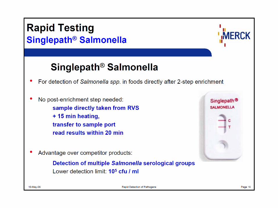
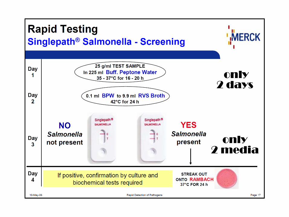
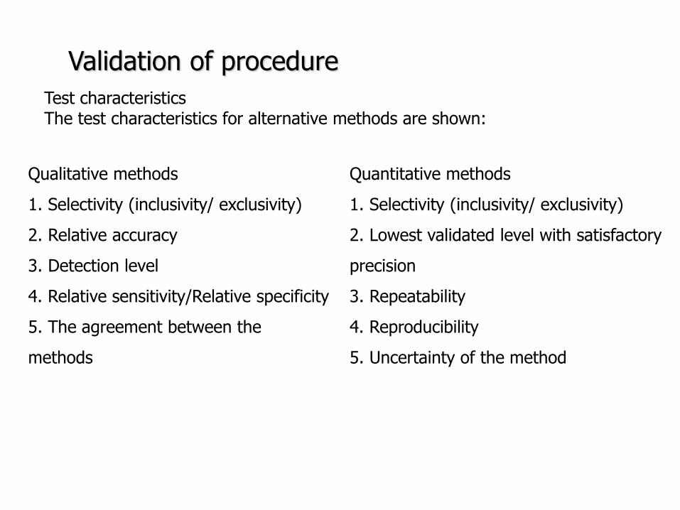
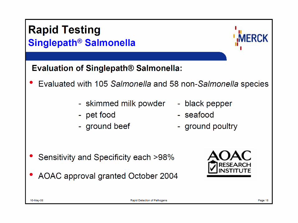
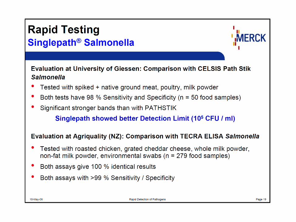
PCR principle
httpopenwetwareorgimagesff1CH391L_S12_PCR_exponential_amplificationjpg
Mistake in the beginning ndash the most influence Exponential phase duplication of PCR products amount in each cycle Linear phase The level of amplification is decreased (lower than duplication in each cycle) Plateau Absence or degradation of some component Reasociation can be preferred than primers anealing due to thermodynamical reasons
PCR principle
httpwwwebioecomuploadfile2010031420100314110534183gif
DNA (genomic plasmid DNA)
Primers (forward reverse)
Thermostable
DNA polymerase
Mg2+ (cofactor
for DNA
polymerase)
dNTP mix
(dCTP dATP
dTTP dGTP) ndash
building stones
for newly
synthetized
DNA product
Nuclease free water ndash without Dnase
Rnase presence (enzymes which
degrade DNA RNA)
MASTERMIX
Total volume ndash 25 μl ndash 50 μl
PCR principle
Polymerase Taq DNA polymerase
bull from thermophilic bacteriumThermus aquaticus
(now prepared by recombination)
bull opt temp 75-80degC (150 ntss)
bull active at the room temperature ndashto work on ice
bull inactive at gt90degC but reactivation when cooled
bull only 5acuteexonuklease activity not 3acuteexonuklease
activity (proofreading)
bull 1 mistake at 10-20 000 nts
Hot start polymerases
bull Activity at the room temperature is inhibited - binding on anti-polymerase
antibodies on the wax balls
bull activation after heating at 94 degC (antibodies destroyed)
Other polymerases
bull Proofreading polymerases (with the 3acuteexonuclease activity)
bull Tth polymerase - reverse transcription (Mn2+) or polymerase activity (Mg2+)
1) Releasing DNA from the cell
- lysis of the cell wall by detergents or by heat digesting cell surface proteins by proteinase K
- RNAse treatment for RNA-free genomic DNA
2) Isolation DNA from the released cytoplasmatic contents
- Centrifugation the vaste in pellet (heat isolation)
- Ion-exchange chromatography (commercial kit) ndash binding DNA by ionic interactions to the column (DNA is negative charged) ndash proteins and RNA pass away ndash DNA is later released by using more concentrated salt solution (column is charged positively)
- Phase separation (aqueouschlorophorm phase ndash CTAB method)
DNA isolation
Detection of food-borne pathogens
DNA of microorganism = set of specific genes with specific sequences
Primers are designed to be complementary to sequence of some specific gene
ndash if the complementary sequence is present PCR product will be performed
Main problems about primers designing and applying of PCR
- Is the gene of interest specific enough (can be found only in the detected microorganism)
- Is the primers sequences complementary only to this gene
- Is the primer sequence designed well (requirements about easy and specific annealing ndash depends on the primers sequence)
- Primers can be designed comercially or by using software (httpwwwbiocenterhelsinkifibiProgramsdownloadhtml)
- sequences and genomes needed - wwwpubmedcom
- httpswwwncbinlmnihgovtoolsprimer-blastindexcgiLINK_LOC=BlastHome
PCR products detection The PCR product has got specific length and specific sequence
Visualiton by intercalating agents bind to double-stranded DNA - fluorescence signal bull Ethidium bromide ndash carcinogenteratogen If intercalated the fluorescence of EtBr is significantly higher than in aqueous solution (UV absorbance maxima at 300 and 360nmemitted as orangeyellow light at 590nm) bull Sybr Green DNA-Sybr Green complex absorbs blue light at 488 nm and emits green light at 522 nm Fluorescent tags at 5acute-end of primers Other tag at 5acute -end of primers eg biotin - for immunodetection detected by anti-bodies linked with an enzyme Microarray
Electrophoresis Conventional electrophoresis a single electrical field causes biomolecules to migrate through a matrix according to its mass-to-charge ratio effectively separates DNA fragments up to ~20-30 kb (according to the gel concentration) larger fragments will comigrate (a large band at the top of the gel)
httpwwwrothamstedacuknotebookimagesgelelgif httpstevegallikorgsitesallimagesgelElectrophoresis2jpg
httpabeleewardhawaiieduProtocolsDNA20Gel20Preparation20and20Electrophoresishtm
Electrophoresis Primers (OT1559 a 18ndash1) complementary to DNA coding16S rRNA Campylobacter
jejuni Campylobacter coli a Campylobacter lari
Product is 287 bp
60 V 100 minutes M ndash 100 bp marker (1-5 positive controls) 1- C jejuni CCM 6212 2- C jejuni CCM 6214 3 ndash
C coli CCM 6211 4 ndash C lari CCM 4897 5 ndash C upsaliensis ATCC 43954 6- 15 strains C jejuni Nt- no
template control (negative control)
Genus specific PCR
Electrophoresis DUPLEX PCR =two pairs of primers
C jejuni C1 5 - CAA ATA AAG TTA GAGGTA GAA TGT-3
C4 5 - GGA TAA GCA CTA GCTAGC TGA T-3
Complementary to a part oxidoreductase 159 bp
C coli 18F 5 - GGT ATG ATT TCT ACAAAG CGAG-3
519R 5- ATA AAA GAC TAT CGT CGC GTG-3acute
Complementary to aspartatekinase and flanking ORF 500 bp
Species specific PCR
60 V 100 minutes M ndash 100 bp marker (1-5 positive controls) 1- C jejuni CCM 6212 2- C jejuni CCM
6214 3 ndash C coli CCM 6211 4 ndash C lari CCM 4897 5 ndash C upsaliensis ATCC 43954 6- 15 strains C
jejuni Nt- no template control (negative control)
Melting curve Intercalating dye binds to double-stranded DNA - producing fluorescent signal when excited by light
When samples are heated to melting point double-stranded DNA denatures into single strands freeing dye into solution
ndash Melting temperature (Tm) is the point at which the DNA denatures
ndash Dependent on the length of fragment and G-C content
ndash More base pairs --gt Increase in Tm
ndash Higher G-C content --gt Increase in Tm
bull Unbound dye has reduced fluorescent signal
bull Loss in signal generates melt curve
No Signal Signal Increasing
temperature
Intercalating
dye
Melting curve
Inflex point
bull 1 derivation = 0
bull In this case 2derivation is negative
Real-Time PCR
CT
CT (threshold cycle) ndash the lowest cycle when fluorescence crosses the
determinated value
Video httpswwwyoutubecomwatchv=kvQWKcMdyS4 httpswwwyoutubecomwatchv=pRwoOBuk00c
Real-Time PCR
Food-borne pathogens detection by PCR
Identification of pure cultures - without problems
Detectionenumeration from food matrix directly or after enrichment
bull How to isolate successfully DNA from few cells in 25 g10 g - Efficiency of DNA isolation (isolation by kits heat-lysis CTAB method)
bull How many targets copies are needed for successfully PCR products detection
ndash detection limits
ndash presence of PCR inhibitors (inhibition of polymerase primers annealing)
bull How to distuinguish DNA from dead and live cells - Total DNA is detected (from dead and living cell)
bull For distinguishing of dead cells DNA ndash eg application of EMA (ethidium monoazide) to the sample before DNA isolation ndash EMA irreversibly bound to DNA of dead cells ndash they are bdquoinvisibleldquo for PCR ndash no amplification
bull Direct enumerationdetection only in some specific case ndash simple liquid sample when the required limit is higher than the limit of detection
bull Standardly ndash for PCR is used enriched homogenate (sample diluted in enrichment broth and cultivated) ndash to obtain detectable level of microorganisms
BAXreg System
Salmonella
Listeria monocytogenes
Genus Listeria
Enterobacter sakazakii
E coli O157H7
Staphylococcus aureus Real-time
Campylobacter jejunicolilariReal-time
Vibrio cholerae
parahaemolyticusvulnificus Real-time
Yeast and molds
httpwww2dupontcomDuPon
t_Homeen_USindexhtml
BAX ndash three major steps
On enrichedregrown samples
Prepare samples = perform heat-lysis in two stages
DNA present in the lysate
2 Process samples
3 Review results
Hydrate PCR tablets
Enrichment protocols vary
depending on bacterial target
and type of food yoursquore testing
Salmonella Enterobacter
sakazakii
E coli O157H7 E coli O157H7
MP Campylobacter jejunicoli
Yeast and Mold
37deg +- 2degC for 20 minutes then
95deg +- 3degC for 10 minutes
Listeria and L monocytogenes
55deg +- 2degC for 60 minutes then
95deg +- 3degC for 10 minutes
5 microl for all bacterial targets
20 microl for beef samples enriched
in E coli O157H7 MP media
20 microl for yeast amp mold
PCR tablets bull DNA-polymerase
bull Primers
bull Nucleotids
bull Suplements
bull SybrGreen or fluorescent probe
bull Internal control + primer
bull Mastermix in the tablet in PCR Eppendorf ndash minimal risk of contamination
For solving the problem of distiguishing dead and live cell the process of small volumes
is used ndash only if live cells are present in the sample they able to multiplied to the limit
detection (104 CFUml in the enrichment suspension used for lysis step)
If the result of detection is positive confirmation by plating out is done (live and dead
cells)
+
Target organism DNA BAXreg system
primers
in tablets PCR
+ no detectable
amplification
Background DNA
PCR
BAXreg system
primers
in tablets
Amplification of specific fragments
Detection of specific fragments Fluorescent signal drops with heating to generate a melt curve
Melting curve Raw Data Processed Data
Strong positive
result
Negative
result
Target peak
Positive control peaks
Positive control peak
Melting curves display peaks within a set temperature range for the control and target (if present)
Detection
Melt curve weak INPC in presence of strong Salmonella positive
Real time weak INPC in presence of strong
Campylobacter positives
Direct protocol does not display a positive or
negative result PCR was successful IndeterminateSignal error
Salmonella AFNOR (ISO 16140) protocol Sample 110 BPW
18 h37ordmC
RVS Muumlller-Kauffmann
24 h 415 37 ordmC
5 suspicious colonies on TSA
24 h 37ordm C
Biochemical and serological confirmation
XLD 2 Plate
24 h 37ordmC
BHI
3h 37 ordmC
22 h 37 ordmC
15 d neg and pos result
3 d neg 5 d pos results
Salmonella ( ISO 65792003) versus BAXreg-PCR
International certificates AOAC-RI Performance Tested Method
Salmonella 100201 L monocytogenes
070202 L spp 030502 Saureus 120701
Campylobacter colilarijejuni
AFNOR Certification
Salmonella QUA 183-1102
NordVal Certification
Salmonella 2006-30-5408
Certificates from AFNOR and AOAC are equal
NordVal Certificate
57
OTHER METHODS
ATP bioluminiscence
httpwwwpromegacouk~mediaimagesresourcesfiguresenotesfe0027_fig1jpgla=en
httpwwwbiotekptassetstech_resources143clarity_atp_conc_fig2gif
Principle the detection of the present level of ATP (direct proportion to the presence of microorganisms or their products residues) The enzyme luciferase catalyzes the transformation of d-luciferin to oxyluciferin (indicated by emision of light at 560 nm)
ATP bioluminiscence
httpwwwbioxyscomimages2figure-B11gif
ATP Hygiene Monitoring System - SystemSURE II
httpwwwmicromaticcomdraft-keg-beerline-cleaning-hygiene-pid-ATP-100-tab-reviewshtml
Biosensors
60
httpsuploadwikimediaorgwikipediacommonseecBiosensor_Systemjpg
Biosenzor =the whole cellmicroorganism (CBS ndash cell-based sensors)
Cell-based sensors
Patogenic microorganisms interact during the infection with mammalian
cells ndash detection of cell or toxin by cell culture The damage of cell culture is measured optically or electrically
62
IDENTIFICATION OF MICROORGANISMS
Definition of species in bacteriaarchae
The criterion for a bacterial or archael species a collection of strains that have bull at least one diagnostic phenotypic trait bull ge70 DNAndashDNA hybridization of their purified molecules DNA
bull ~more than 97 16S rRNA sequence identity Under the same conditions some eucaryotic order would be a single bacterial species
The bacterial species definition bull eminent practical significance bull but a very difficult issue
bull very wide and often changes in DNA
bull mutations during replication of DNA
bull horizontal gene transfer through mobile elements plasmids also between species
Criterion for species in prokaryotic cells (bacteria and archea) Collection of strains characterised bull by at least one diagnostic feature bull their purified DNA molecules prove at least 70 hybridisation
which corresponds to at least 97 concordance in the sequence of 16S rRNA gene
Map of genome of B cereus ATCC 14579
httpwwwacgtinccomspecialty_dna_sequencinghtm
Identification based on genotype
64
DNA isolation PCR amplification of 16S rRNA gene (usually)
Sequencing of PCR product
Comparison to sequences in databases (BLAST)
Identification of bacteria PHENOTYPE METHODS bull Morphology of colony and cell (Gram staining shape) bull Requirements for temperature atmosphere nutrition bull Biochemical tests - presence of specific enzymes
bull catalase oxidase fermentation of glucose bull the way how they utilize different compounds
bull Immunochemical tests - specific antigens in the cell wall METHODS BASED ON PROTEIN ANALYSIS
bull MALDI-TOF MS METHODS BASED ON GENOTYPE bull looking for and comparing of specific DNA sequences
bull conserved universal genes (eg 16S rRNA) bull genes specific for genus species
bull done by PCR methods DNA chips sequencing etc
66
BIOCHEMICAL TESTING
Identification based on phenotype I Physiological + biochemical features
bull morfology of colonies and cells (Gram staining shape)
bull requirements for temperature atmosphere nutrient sources
bull basic biochemical tests oxidase catalase fermentationoxidation of glucose haemolysis etc
appearance of colonies on selective or selective-diagnostic agars (for other testing to be reinoculated on non-selective agar)
the classification of an isolate into a certain group
(eg for bacteria catalase positivenegative G+ cocci glucose fermentingnon-fermenting G- rods etc)
biochemical tests specific for certain groups of microorganisms
Identification based on phenotype I
68
Physiological + biochemical features Example Bacillus cereus
httpwwwfoodhaccpcommemberonlynewsletter164html
httpwwwnapavalleyedupeoplesrosePublishingImagesBacillus20cereus202(Gram20stain)jpg
Set of biochemical tests Microgenreg Bacillus ID Panel for the identification of Bacillus spp and related genera (time for performing 48 hours)
MYP agar (Mannitol Egg Yolk Polymyxin Agar) pink colonies with a precipitation zone (no fermentation of manitol + enzyme lecitinase)
catalase positive
httpwwwoxoidcomukblueprod_detailprod_detailasppr=CM0929ampc=ukamplang=ENamporg=9ampimg=CM0929ampsec=
G+ rod endspores forming
β-haemolysis moisty and easy to grow colonies
Principle of biochemical testing
to rank the bacterium to some group having the same basic features bull eg catalase positive gram positive cocci bull Family Enterobacteriaceae
the identification on the genus or species level
bull sets of biochemical tests specific for some group of bacteria
70
Gram staining procedure - principle
https40mediatumblrcom25da2e5b25bbe336819bf1715849d171tumblr_inline_ntrsgn9w1D1sv92o6_540jpg
Gram staining procedure - principle
httpsonlinesciencepsuedusitesdefaultfilesmicrb106CellStructurecow02354_03_15jpg
Gram staining procedure 1-2 drops of sterile water on a slide Spread culture in thin filter in this drop Let dry in airclose to fire Pass slide through flame 2-3x to fix Add crystal violet ndash 15-20 s to act Add Lugol solution (KII2) ndash 15-20 s to act Washing off with a mixture of
ethanolaceton (41) or acetone till the stain removes but max 15 s
Rinse with water decant excess liquid Add safranin 1 minute Rinse with water decant excess liquid Dry and put a drop of imersion oil use an
objective bdquoOILldquo
httpwwwtumblrcomtaggedgram-staining
G- red
G+ violet
Gram staining procedure + Identification
E coli
Family Enterobacteriaceae Gram-negative rods
Micrococcus luteus
Staphylococcus aureus
Gram-positive cocci Bacillus cereus
Gram-positive rods (with endospores)
Biochemical identification of G + bacteria
httpyoungeneticistblogspotcz201106diagnostic-microbiology-labhtml
G+ bacteria bull cocci ndash in clusters in pairs
diplococci bull bacilli
bull endospore bull no endospore
For G+ cocci bull Catalase positive ndash Staphylococcus
sp bull Coagulase + S aureus or some other few species bull Coagulase -
bull Novobiocin S bull S epidermidis etc
bull Novobiocin R bull S saprophyticus etc
bull Catalase negative Streptococcus sp Enterococcus sp
bull kind of haemolysis Other important G + group ndash filamentous bacteria
Catalase test Catalase test Catalase is a common enzyme found in nearly all living organisms that are exposed to oxygen where it catalyzes the decomposition of hydrogen peroxide to water and oxygen
2 H2O + O2 2 H2O2
catalase
bubbling when culture is add in peroxide
httpiws2collinedudcainCCCCD20Microcatalase_testhtm
Catalase testing into a drop of 3 peroxide to transfer a loop of bacterial culture catalase positive ndash releasing of air bubbles
Examples bull G+ cocci catalase positive Staphylococcus sp Micrococcus sp Kocuria sp Dermacoccus sp Macrococcus sp Rothia sp catalase negative Streptococcus sp Enterococcus sp bull G+ rods (bacilli) endospores forming catalase positive Bacillus sp catalase negative Clostridium sp
Resistance to ATBhaemolysis Resistance eg to novobiocin
httpquizletcom20077004mic-lecture-1-t2-flash-cards
Hemolysis on blood agar
Measuring the diameter of inhibition zone (less than 17 mm resistant more or equal to 17 mm sensitive) ndash differing in Staphylococcus sp (Clinical performance)
alpha-hemolysis - oxidizing the iron in the hemoglobin - green discoloration of the medium around beta-hemolysis - rupture of erythrocytes - complete clearing medium around gamma-hemolysis ndash no effect ndash no change
By cultivation on blood agar bacteria can be
differentiated based on their capacity to secrete
hemolysins The hemolysis will cause a clearing zone of
the blood agar around the colonies Bacteria can cause
different types of hemolysis
bullα-hemolysis which means an incomplete clearing
(green haemolysis)
bullβ-hemolysis which means a complete clearing
bullDouble hemolysis of some staphylococci consisting of
an inner β-hemolysis zone and an outer α-hemolysis
zone
bullNo hemolysis which is sometimes referred to as γ-
hemolysis which may seem illogical
Note that the α-hemolysin of staphylococci causes
complete hemolysis whereas their β-hemolysin causes
incomplete hemolysis
The capacity to produce hemolysins may vary between
different strains of a particular bacterial species
httpwwwvetbactorgvetbactdisplayextinfo=67
Identification of bacteria based on haemolysis
Streptococcal hemolysins α (alpha) hemolysis - ldquozone of green discolourationrdquo or ldquogreeningrdquo called partial or incomplete hemolysis produced by alpha-hemolytic streptococci such as Streptococcus suis This type of hemolysis on BA is due to bacterial production of hydrogen peroxide which oxidises hemoglobin to methemoglobin β (beta) hemolysis ndash a clear zone of complete hemolysis produced by beta-hemolytic streptococci such as S equi subsp equi This hemolysis is due to lysis of RBCs in the BA by the cytotoxin Streptolysin S
Source httppeopleupeicajlewishtmlhemolysinshtml
Bacterial hemolysins
Staphylococcal hemolysins Most coagulase-positive staphylococci (CPS) such as Staphylococcus aureus S pseudintermedius ( but not S hycius) typically produce an distinctive pattern of ldquodouble zonerdquo or ldquotargetrdquo hemolysis on blood agar Double-zone hemolysis indicates that the two hemolysins one which produces ldquocompleterdquo and another one that produces ldquoincompleterdquo hemolysis are present and this combined effect is visible due to different diffusion rates of these enzymes through the agar The staphylococcal α (alpha) hemolysin causes a complete type of hemolysis This zone of complete hemolysis is narrow and immediately adjacent to the bacterial colony on blood agar The staphylococcal β (beta) hemolysin causes an incomplete type of hemolysis at 37 degC This broader incomplete zone is outside of the inner complete zone and can be hard to see on early culture plates and also varies with the species of RBCs used to make the BA plate Its action is enhanced by chilling at 4 degC and the hemolysis becomes complete (this effect is referred to as ldquohot-coldrdquohemolysis) There are other staphylococcal cytotoxins that are also hemolysins but their lytic actions are not seen on blood agar
The confusion arises due to the NAMES of the two STAPHYLOCOCCAL HEMOLYSINS and the TYPE of hemolysis they produce These are the REVERSE of the two TYPES of STREPTOCOCCAL HEMOLYSIS Strictly speaking the descriptive terms ldquoalphardquo for ldquoincompleterdquo and ldquobetardquo for ldquocompleterdquo hemolysis apply only to the hemolytic toxins of Streptococcus species However other organisms beside Streptococcus and Staphylococcus species have cytotoxins that cause hemolysis and this hemolysis can be either complete or incomplete It is preferable to use the terms ldquocompleterdquo and ldquoincompleterdquo rather than ldquobetardquo or ldquoalphardquo to describe the hemolysis caused by these organisms but you will notice in bacteriology text books that the terms alpha and beta are commonly used
G ndash bacteria bull Cocci bull Bacilli (Rods) bull Oxidase positivenegative bull Glucose metabolism
bull fermentation bull oxidation bull Inactive
bull Lactose metabolism for coliform
bull Pigment formation for Pseudomonas sp
Biochemical identification of
G - bacteria
Oxidase test
Oxidase positive (OXI +) Aeromonas sp Pseudomonas sp Dark-blue to violet
Oxidase negative (OXI -) Enterobacteriaceae Bacillus sp Colourless
Oxidase bullabbreviated general name for some kind of cytochrome c oxidases (cytochrome oxidase or indophenol oxidase) enzymes involved in aerobic respiratory chain when oxygen is used as the final receptor for hydrogen bullOxidases are usually present only in aerobic organisms bullPresence or absence of oxidase(s) is an important feature of the bacterial metabolism used mainly in the identification process of gram-negative bacteria
Detection Reaction of the culture with redox indicator as eg NN-dimethyl-p-phenylenediamine (DMPD) dark-blue when oxidized and colorless when reduced Different test performances are possible
Fermentation of carbohydrates Type of utilisation of glucose or carbohydrates bull an important feature of the bacterial metabolism bull widely used in the identification process of bacteria bull fermentation strictly means to be under anaerobic conditions (but in bacterial
metabolism which can be but not have to be the same as the anaerobic conditions of cultivation) so very often is used in a general way like bdquoutilisationldquo
bull fermentation (like bdquoutilisationldquo) of carbohydrates to
bull acid and gas (Durham tubes) bull acid without gas bull non-utilised
bull Testing of fermentation or oxidation of carbohydrates
bull eg OF test for glucose bull by oxidation under aerobic conditions bull by fermenation under anaerobic conditions (an oil layer)
Fermentation of sugars to acid
Peptone Water wPhenol Red
(HiMedia M028I)
Ingredients gl
Peptic digest of animal tissue 10000
Sodium chloride 5000
Phenol red 0020
Final pH ( at 25degC) 68plusmn02
httpsuploadwikimediaorgwikipediacommons009Phenol_red_pH_60_-_80jpg
+ after sterilisation
at the room
temperature to add
aseptically sugar
discs and
inoculate
succrose trehalose rhamnose xylose
Incubation 30 degC 24 h
- +
Fermentation of carbohydrates
Casein enzymic hydrolysate and yeast extract provide nitrogenous compounds vitamin B complex and other growth nutrients Dextrose is the fermentable carbohydrate and bromocresol purple acts as the pH indicator with colour change from purple to yellow in acidic conditions Sodium chloride maintains osmotic equilibrium bdquoAnaerobicldquo conditions ndash inoculation by stabbing to used fresh prepared media (oxygen expelled by heating medium in boiling water or running steam for 15 min then to be cooled down quickly)
- +
Enterobacteriaceae ndash glucose fermentation
83
Tryptone Yeast Extract Agar Ingredients Gms Litre Casein enzymic hydrolysate 10000 Yeast extract 1500 Dextrose 10000 Sodium chloride 5000 Bromocresol purple 0015 Agar 15000 Final pH ( at 25degC) 70plusmn02
Test for oxidationfermentation of glucose (OF glucose test) detection of acid production by acidobazic indicator change to yellow (acid present) in lower pH from green (no acid) Anaerobic atmosph ndash covering by sterile mineral oil If fermented anaerobically then also runs aerobically
Fermentation of carbohydrates
Principle of biochemical tests bull The first step in identification is to rank the bacterium to some group of bacteria
having the same basic features ndash eg catalase positive gram positive cocci bull The identification on the genus or species level is finished by other biochemical tests
specific for each group of bacteria bull The tests can be done in tubes or miniaturized plate
ENTEROtest 24N (Erba Lachema) (24 means 24 biochemical tests N= new)
API 20E (bioMeacuterieux)
bull designed for routine identification of clinically important species of family Enterobacteriaceae without the use of reagents
bull A strip test OXItest or OFtest in microtitration wells can be used to confirm that the isolate belongs to Enterobacteriaceae family
bull The results are read visually or automatically using reader MIKROLAreg
bull The identification can be supplemented by diagnostic strip tests INDOLtest COLItest and VPtest
APIreg Gram negative Identification API 20E ndash Speciessubspecies identification of Enterobacteriaceae and groupspecies identification of non-fermenting Gram negative bacteria API Rapid 20E ndash 4-hour identification of Enterobacteriaceae API 20NE ndash 24 to 48-hour identification of Gram negative non-Enterobacteriaceae API Campy ndash 24-hour identification of Campylobacter species
Example Escherichia coli
ENTEROtest 24 N
Biochemical testing - Enterobacteriaceae bull TSI Triple Sugar Iron Agar (use a straight wire to stab the butt and streak the agar surface)
- to determine whether organisms can ferment glucose sucrose andor lactose with or without production of gas and the ability of the organism to produce hydrogen sulphide from thiosulphate in an acid environment is also tested
Formula Liter Enzymatic Digest of Casein 5 g Enzymatic Digest of Animal Tissue 5 g Yeast Enriched Peptone 10 g Dextrose 1 g Lactose 10 g Sucrose 10 g Ferric Ammonium Citrate 02 g Sodium Chloride 5 g Sodium Thiosulfate 03 g Phenol Red 0025 g Agar 135 g Final pH 73 plusmn 02 at 25 degC
inoculation in the butt by stab inoculation on the slant on the surface bull Fermentation of dextrose
(glucose) to acid bull anaerobically - yellow butt bull aerobically leads to products
which are later oxidised by air and pH remains alkaline (red) bull Fermentation of glucose to gas (anaerobically - in the stab) Cracks splits or bubbles in medium indicate gas production bull Production of hydrogen sulfide (H2S) ndash reaction with ferrous sulphate and sodium thiosulphate - a black precipitate in butt bull Lactose andor succrose
utilisation to acid Acid production ndash yellow butt and slant
Results
An alkaline slant-acid butt
(redyellow) - fermentation of
dextrose only
An acid slant-acid butt
(yellowyellow) - fermentation of
dextrose lactose andor sucrose
An alkaline slant-alkaline butt
(redred) - dextrose or lactose not fermented (non-fermenter)
Bacterium fermenting lactose andor succrose are able to ferment glucose
Phenol red ndash alkaline = red acid = yellow
bull TSI Triple Sugar Iron Agar (use a straight wire to stab the butt and streak the agar surface)
- to determine whether organisms can ferment glucose sucrose andor lactose with or without production of gas and the ability of the organism to produce hydrogen sulphide from thiosulphate in an acid environment is also tested
Biochemical testing - Enterobacteriaceae
Urea agar (Christensen) (use a 1 μl loop full)
- to determine whether an organism can split urea into ammonia and
carbon dioxide by the action of the enzyme urease
httpimageslidesharecdncomlab17-141201085153-conversion-gate0195-10-638jpgcb=1417424219
httpimageslidesharecdncomhelicobacterpylori-copy-110516201901-phpapp0195campylobacter-jejunihelicobacter-pylori-39-728jpgcb=1305577391
Phenol red
Medium is acidic - yellow
(pH 68plusmn02)
Alkaline reaction by urea hydrolysis - pink colour
Composition Gms Litre
Peptic digest of animal tissue
1000
Dextrose 1000
Sodium chloride 5000
Disodium phosphate 1200
Monopotassium phosphate 0800
Phenol red 0012
Agar 15000
Final pH ( at 25degC) 68plusmn02
Nutrition
Peptic digest of animal tissues dextrose
Sodium chloride - osmotic equilibrium
Phosphates - buffering of the medium
35-37 degC
Biochemical testing - Enterobacteriaceae
bull L-lysine decarboxylase (use a 1 μl loop full)
- to determine whether an organism can decarboxylate an amino acid (lysine) leading to formation of an
amine After inoculation both tubes are overlayed with sterile paraffin oil
httpfce-studynetdna-sslcomimagesupload-flashcards10295302332621_mjpg
+ CO2 + OH-
Lysine Decarboxylase Broth Typical Composition (glitre) Peptic digest of animal tissue 5000 Yeast extract 3000Dextrose 1000 (glucose prepared from starch chemically equivalent to glucose) L-Lysine hydrochloride 5000 Bromocresol purple 0020 Final pH ( at 25 degC) 68plusmn02 Inoculate below the surface cover by sterile paraffin oil
Two steps detection 1 step Dextrose is fermented ndash H+ is released Acidic conditions bull bromocresol purple changes colour to yellow bull in acidic conditions lysine decarboxylase is
activated 2 step L-lysine is decarboxylated and OH- released Alkaline conditions bull bromocresol purple changes colour back to
purple
Biochemical testing - Enterobacteriaceae
bull β-galactosidase (ONPG) (use a 1 μl loop full)
- to determine whether an organism has the enzyme szlig-galactosidase by using the compound o-
nitrophenyl-beta-D-galactopyranoside (ONPG)
- this test is used to differentiate between lactose-negative and lactose-delayed organisms
- ONPG is hydrolysed by the enzyme to o-nitrophenol which is yellow and galactose
O-Nitrophenyl-β-D-galactopyranoside (ONPG) is structurally similar to lactose (ie ONPG is an analog of lactose) except that orthonitrophenyl has been substituted for glucose ONPG is colorless compound O-nitrophenol is yellow visual evidence of hydrolysis
Lactose fermenting bacteria (ONPG positive) two enzymes Permease ndash transferring of the lactose molecule into the bacterial cell β-galactosidase - cleaving the galactoside bond producing glucose and galactose - inducible enzyme - made ONLY in the presence of the lactose substrate Coliform bacteria as Ecoli Klebsiella spp Enterobacter spp
hydrolysis
Late lactose fermentering bacteria Permease ndash not present delayed in the production of acid from lactose because of sluggish permease activity β-galactosidase ndash present - give a positive ONPG test Citrobacter spp Arizona spp
Non lactose fermenter (ONPG Negative) Permease ndash not present β-galactosidase ndash not present Salmonella spp Shigella spp Proteus spp Providencia spp and Morganella spp
Biochemical testing - Enterobacteriaceae
Procedure Culture Bacteria grown in medium containing lactose (to induce the production of the galactosidase enzyme) such as Kligler iron agar (KIA) or Triple sugar Iron (TSI) agar (Note β-galactosidase enzyme (inducible enzyme) is made ONLY in the presence of the lactose substrate) Inoculation 1A loopful of bacterial growth is emulsified in 005mL of physiologic saline to produce a heavy suspension 2One drop of toluene is added to the suspension and vigorously mixed for a few seconds to release the enzyme for bacterial cells 3An equal quantity of buffered ONPG solution is added to the suspension 4The mixture is placed in a 37 oC water bath 5When Using ONPG Tablets 6A loopful of bacterial suspension is added directly to the ONPG substrate resulting from adding 1mL of distilled water to a tablet in a test tube 7This suspension is also placed in a 37oC water bath
β-galactosidase (ONPG)
Results and Interpretations The rate of hydrolysis of ONPG to o-nitrophenol may be rapid for some organisms producing a visible yellow color reaction within 5 to 10 minutes Most tests are positive within 1 hour however reactions should not be interpreted as negative before 24 hours of incubation The yellow color is usually distinct and indicates that the organism has produced o-nitrophenol from the ONPG substrate through the action of β-galactosidase
Biochemical testing - Enterobacteriaceae
bull VogesndashProskauer (VP) test (use a 1 μl loop full)
- to determine whether an organism can produce acetylmethylcarbinol
(acetoin) from fermentation of glucose If present acetylmethyl carbinol is
converted to diacetyl in the presence of prop- naphthol (to be added first) strong
alkali (40 KOH) and atmospheric oxygen The diacetyl and quanidine-
containing compounds found in the peptones of the broth then condense to
form a pinkish red polymer
MRVP broth (pH 69)
Ingredients per liter of
deionized water
buffered peptone= 70 gm
glucose= 50 gm
dipotassium phosphate= 50 gm Voges-Proskauer Reagent A Barrittrsquos reagent A
Alpha-Naphthol 5 50 gm
Absolute Ethanol 1000
ml
Voges-Proskauer Reagent B Barrittrsquos reagent B
Potassium Hydroxide 400 gm
Deionized Water 1000 ml
Procedure of VogesndashProskauer (VP) Test
1Prior to inoculation allow medium to
equilibrate to room temperature
2Using organisms taken from an 18-24
hour pure culture lightly inoculate the
medium
3Incubate aerobically at 37 degC 24 hours
4Following 24 hours of incubation aliquot
2 ml of the broth to a clean test tube
5Re-incubate the remaining broth for an
additional 24 hours
6Add 6 drops of 5 alpha-naphthol and
mix well to aerate
7Add 2 drops of 40 potassium hydroxide
and mix well to aerate
8Observe for a pink-red color at the surface
within 30 min Shake the tube vigorously
during the 30-min period
Positive Reaction
A pink-red color at the surface
Examples Viridans group streptococci
(except Streptococcus vestibularis) Listeria
Enterobacter Klebsiella Serratia marcescens
Hafnia alvei Vibrio eltor Vibrio alginolyticus etc
Negative Reaction
A lack of a pink-red color
Examples Streptococcus mitis Citrobacter sp
Shigella Yersinia Edwardsiella Salmonella Vibrio
furnissii Vibrio fluvialis Vibrio vulnificus and Vibrio
parahaemolyticus etc
A copper color should be considered negative A
rust color is a weak positive reaction
Quality Control of VogesndashProskauer (VP) Test
VP positive Enterobacter aerogenes (ATCC13048)
VP negative Escherichia coli (ATCC25922)
Biochemical testing - Enterobacteriaceae
94
VogesndashProskauer (VP) test
Rapid test for confirmation of E coli ndash typical features
β-D-glucuronidase (97 strains) production of indole from tryptophan (typical for E coli)
Performance
Previous testing Gram-negative (eg able to grow on VRBD agar) oxidase negative
From pure culture to prepare suspension in saline solution (app 5 ml) at the density 3 McFarland
To transfer 05 ndash 1 ml of this suspension into a new tube ndash to add the strip of COLItest (chemicals ndash fluorogenic substrate for β-D-glucuronidase tryptophane are present in the pad at the end of the strip)
Incubation 37 degC 4 hours
Detecion of β-D-glucuronidase (to be done at first) UV (360 nm) ndash blue fluorescence (GLR + hellipβ-D-glucuronidase positive) ndash GLR test can be read after 1 hour
Detection of indole formation from tryptophane (to be done at second) to add Kovacs reagent (IND + hellipformation of red ring)
95
COLItest - Escherichia coli
Biochemical testing - Enterobacteriaceae
Fluorogenic culture medium
bull One or more fluorogenic substrates
bull For detection of specific enzyme which are able to cleave out the fluorogenic substrate that a designed
fluorophore (responsible for the UV fluorescence) is released
FLUOROGENIC MEDIUM (Colilert) E coli use β-glucuronidase to metabolize MUG and create fluorescence (UV 360 nm) Since most non-coliforms do not have these enzymes they are unable to grow and interfere
httpgawaterusgsgovprojectsbacteriapicturessummaryanalysisecolijpg
E coli (MPN method)
COLItest Biochemical testing - Enterobacteriaceae
COLItest bull Indole test (use a 1 μl loop full)
- to determine whether an organism can split indole from tryptophan
httpwwwmicrobiologyinfocomindole-test-principle-reagents-procedure-result-interpretation-and-limitations
indole production from tryptophan
Production of indole is detected by Kovacacutes reagent
Indole Kovacs Reagent
p-Dimethylaminobenzaldehyde 500 gm
Hydrochloric Acid 37 2500 ml
Amyl Alcohol 7500 ml
Biochemical testing - Enterobacteriaceae
INDOLe test ndash quick variant
Ecoli Citrobacter Enterobacter
Procedure bull on the filtration paper to add a drop of INDOL suspension bull to spread into by a loop some culture bullTo incubate ndash at the room temperature for 5 minutes bullTo read results
Biochemical testing - Enterobacteriaceae
ENTEROtest 24 ndash example of biochemical testing
routine identification of clinically important species of family Enterobacteriaceae the test is focused only on some species often isolated in clinical practis strains from other sources can be slightly different in the dominant biochemical profiles intended exclusively for the identification of family Enterobacteriaceae the preliminary ranking into family Enterobacteriaceae must be done (Gram staining oxidase glucose fermentation) the results of reactions obtained in this modified micromethod may differ from the results obtained using conventional tests and results available in literature sources in case of problem to control positivenegative reactions by control strains or to use for this tests tube variant to check the functionality of the kit not to check the accuracy or success of the identification
Preparation of suspension from over-night culture growing on non-selective agar (for staphylococci Columbia agar blood with 5 of sheep blood is recommended)
Inoculation of microtitre plates anaerobic reaction covered by mineral oil
Incubation
Reading results by both visual and instrumental mode respectively Standard off-line tests INDOLtest or COLItest Other off-line tests OXItest OFtest VPtest Acetoin test
ENTEROtest 24 N ndash example of biochemical testing
bull tube with 5 ml of sterile physiolsol bull suspension (1 McFarland) bull application 100 microl to each hole bull 2 drops of parafin oil into holes D-H of the first line (anaerobic conditions) bull Incubation 37degC 24h bull for check the purity of suspension to streak suspension on TSA (incubation 37 degC ndash 24 h
ENTEROtest 24N
ENTEROtest 24 N - evaluation
Escherichia coli
H 0+0+0=0 G 0+2+0=2 F 1+2+0=3 hellip Identification code 023426
1 1
2
4
ENTEROtest 24 ndash example of biochemical testing
The result = a set of biochemical properties (+-) which is compared to the sets of biochemical properties of different chosen genera and species for them the test is designed
ENTEROtest 24 N
T-index how is the likelihood of the different properties The combination of identication and T-index = the reliability of the result Excelent very good good (this case) acceptable intermediary strain needs some additional tests genus level non-existing taxon
Evaluation by using the identification code - By using a code book - By using software TNW
Identification score the measure of similarity to proposed taxon
The way of identification the obtained profile is compared with profiles in database bull The most similar profiles are found
bull Identification score = of similarity bull But what about the different reactions
bull T-index is calculated bull how is the probability of the different reactions
bull It depends on their importance ndash typical reactions should be the same
bull Reliability of the result bull the combination of identication score and T-index = bull Excelent very good good acceptable intermediary strain needs some
additional tests genus level non-existing taxon
ENTEROtest 24 ndash example of biochemical testing
Biochemical tests - divisions
ENTEROtest 24 species of family Enterobacteriaceae
NEFERMtest 24 Gramnegative non-fermenting bacteria and families Vibrionaceae Aeromonadaceae and Pleiomonas shigelloides present mainly in clinical material
ANAEROtest 23 ANAEROBIC BACTERIA (G + AND G-) routine identification of anaerobic bacteria (G + AND G-) in clinical material and in food
STREPTOtest 24 Streptococcus Enterococcus and related gram positive catalase negative cocci isolated from clinical material
STAPHYtest 24 genus Staphylococcus isolated from clinical material and for their distiguishing from related genera of other gram positive catalase positive cocci
Biochemical tests APIreg Gram positive Identification API Staph ndash Overnight identification of clinical staphylococci and micrococci API 20 Strep ndash 4 or 24-hour identification of streptococci and enterococci API Coryne ndash 24-hour identification of Corynebacteria and coryne-like organisms API Listeria ndash 24-hour identification of all Listeria species APIreg Gram negative Identification API 20E ndash Speciessubspecies identification of Enterobacteriaceae and groupspecies identification of non-fermenting Gram negative bacteria API Rapid 20E ndash 4-hour identification of Enterobacteriaceae API 20NE ndash 24 to 48-hour identification of Gram negative non-Enterobacteriaceae API Campy ndash 24-hour identification of Campylobacter species APIreg Anaerobe Identification API 20A ndash 24-hour identification of anaerobes Rapid ID 32 A ndash 4-hour identification of anaerobes
MicrogenTM GN-ID Convenient 12 substrate identification system for commonly encountered Enterobacteriaceae (GNA) Extended 24 substrate identification system for the complete range of Enterobacteriacea and oxidase positive non-fastidious Gram negative bacilli (GNA+B) MicrogenTM Listeria-ID Enhanced built-in haemolysis test MicrogenTM Bacillus-ID 24 substrate system for the identification of mesophilic Bacillus spp isolated from foods and related samples MicrogenTM Staph-ID 12 substrate system minimizes handling and manipulation Substrates selected specifically for Staphylococcus and related organisms MicrogenTM Strep-ID Substrates selected specifically for Streptococcus Enterococcus and related species Provides excellent differentiation of Enterococci spp especially E casseliflavus E mundtii and E gallinarum
Biochemical tests
Identification of microorganisms by MALDI-TOF MS
Department of Biochemistry and Microbiology University of Chemistry and Technology Prague
Sabina Purkrtovaacute1 Petra Junkovaacute2 1Laboratory of food microbiology 2Laboratory of applied proteomics
SabinaPurkrtovavschtcz PetraJunkovavschtcz
MALDI-TOF MS
1) sample preparation bull microbial culture or its
proteins extract is smearing onto a steel plate and covered over by matrix
bull matrix enables the sample (A) to be desorped and ionised as pseudomolecule ionts [A+H] +
2) MALDI-TOF MS analysis unique mass spectral fingerprint of desorbed microbial cell components (mainly intracellular proteins) different among genera species or also some strains
Matrix-Assisted Laser Desorption Ionization-Time of Flight Mass Spectrometry
httppubsrscorgencontentarticlehtml2014RAC4RA05604C
httpcmrasmorgcontent263547figures-only
3) identification comparison of mass spectrum to those of reference strains in database
MALDI-TOF MS PRINCIPLE
111
2
2
2L
teU
z
m
m mass z charge
L length of drifting zone e
elementary charge U
speeding voltage
Time of flight is a function of the specific ion mass (mz)
httpwwwsigmaaldrichcomtechnical-documentsarticlesbiologycustom-dna-oligos-qc-analysis-by-mass-spectrometryhtml
Soft ionisation method bull low level of sample fragmentation
Animation http cmgmstanfordedupansection_htmlMS
MALDI-TOF MS SAMPLE AND MATRIX
httpcmrasmorgcontent263547figures-only
Matrix bull able to absorb the energy of the laser (usually 337 nm) bull to crystalise with samples - usually acid
character (proton ionisation of sample) dissolved in organic solvent
Sample preparation Direct transfer bull intact cells spread directly onto a steel plate bull lysis of cells occurs during the contact with acid
matrix and by laser desorption bull most bacteria Proteins extraction bull previous extraction of proteins by organic acids
andor alcohol (eg ethanol and 70 formic acid) bull yeasts moulds some species of bacteria (depending
on the cell wall composition)
CHC α-Cyano-4-hydroxycinnamic acid (organic solvent 50 acetonitrile with 25 trifluoracetic acid) SA 35-Dimethoxy-4-hydroxycinnamic acid (sinapic acid)
DHB 25-Dihydroxybenzoic acid
httpwwwsigmaaldrichcomcatalogproductsigmac8982lang=enampregion=CZ
MALDI-TOF MS MICROORGANISMS
113
Mass spectrum protein profile z equals usally to 1+ (so mz usually corresponds to molecula mass) the range usually used for identification 2000 -20 000 mz the intensity of single peaks corresponds to the abundance of the protein Which proteins dominates in the protein profile abundant basic and mediumly hydrophobic mainly ribosomal proteins further cold-shock and heat-shock proteins chaperons etc
conserved house-keeping gene = conserved proteins = the acordance with identification based on DNA
Visualisation of mass spectrum protein profile ndash (software mMass 5 Strohalm et al 2010)
Bacillus cereus
Analysis is recomended (and validated) to be performed from colonies grown onto non-selective agar
MALDI-TOF MS ANALYSIS
114
Comparison of mass spectrum protein profile of unknown sample with these of reference strains present in database by software
Commercial databases from different MALDI-TOF MS producers Bruker Daltonics ndash MALDI BIOTYPER Shimadzu - Shimadzu Launchpad software + SARAMIS database Biomeacuterieux - VITEKreg MS Other databases compatible with different hardware systems (eg Andromas)
BioTyper The statistical analysis for correlation includes peak positions intensities and frequencies across the complete range of microorganisms Score value 0 (none similarity) - 1000 (absolute similarity) But it is expressed in decadic logarithm log(score value) 0-3
Range Description Symbols Color
2300 3000
highly probable species identification
( +++ ) green
2000 2299
secure genus identification probable species identification
( ++ ) green
1700 1999
probable genus identification ( + ) yellow
0000 1699
not reliable identification ( - ) red
Bacillus cereus
115
Range Description Symbols Color
2300 3000
highly probable species identification
( +++ ) green
2000 2299
secure genus identification probable species identification
( ++ ) green
1700 1999
probable genus identification ( + ) yellow
0000 1699
not reliable identification ( - ) red
Rank (Quality)
Matched Pattern Score Value
NCBI Identifier
1 ( +++ )
Bacillus cereus DSM 31T DSM 2554 1396
2 ( ++ )
Bacillus cereus 994000168 LBK 2203 1396
3 ( ++ )
Bacillus weihenstephanensis DSM 11821T DSM
2158 86662
4 ( ++ )
Bacillus mycoides DSM 2048T DSM 2155 1405
5 ( ++ )
Bacillus cereus 4080 LBK 2147 1396
6 ( + )
Bacillus thuringiensis DSM 2046T DSM 1975 1428
7 ( + )
Bacillus pseudomycoides DSM 12442T DSM 1787 64104
8 ( - )
Bacillus bataviensis DSM 15601T DSM 1369 220685
9 ( - )
Brevibacterium linens IMET 11075T HKJ 1347 1703
10 ( - )
Acinetobacter towneri DSM 14962T HAM 1345 202956
Bacillus cereus
Bacillus cereus
MALDI-TOF MS - PROCEDURE Direct method smearing sample in four parallels in lower and higher cells concentrations ndash after drying to cover over by matrix (1-2 microl) and let to crystallise at room temperature
Matrix solution of α-Cyano-4-hydroxycinnamic acid (10 mgml) in 50 acetonitrile with 25 trifluoroacetic acid (prepared with 10 TFA solution)
Protein standard (1 μl) Bruker Bacterial Test Standard (Bruker Daltonics SRN) ndash proteins extracted from z Escherichia coli DH5alpha BRL + some others
Equipment Bruker Autoflex Speed Database MALDI Biotyper 31
116
MALDI-TOF MSPROTEIN STANDARD
117
Analyte name Rank (Quality) Matched Pattern Score Value
F4 1 ( +++ ) Escherichia coli DH5alpha BRL 2439
Thank you for your attention
Questions to be sent to SabinaPurkrtovavschtcz
118
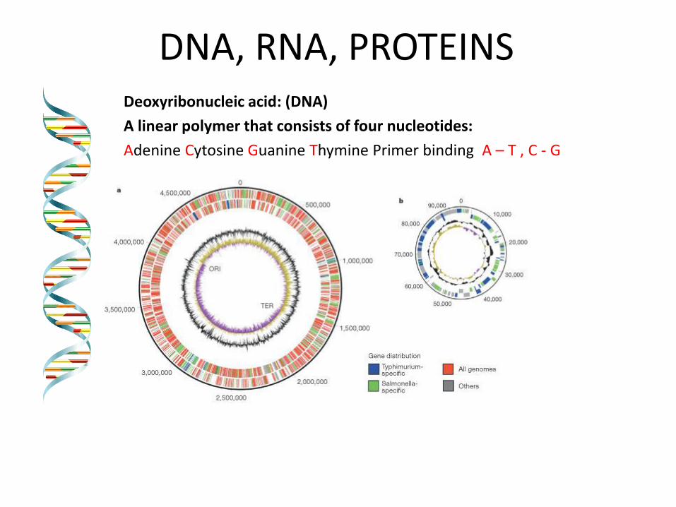
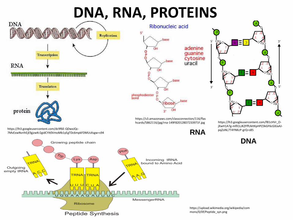
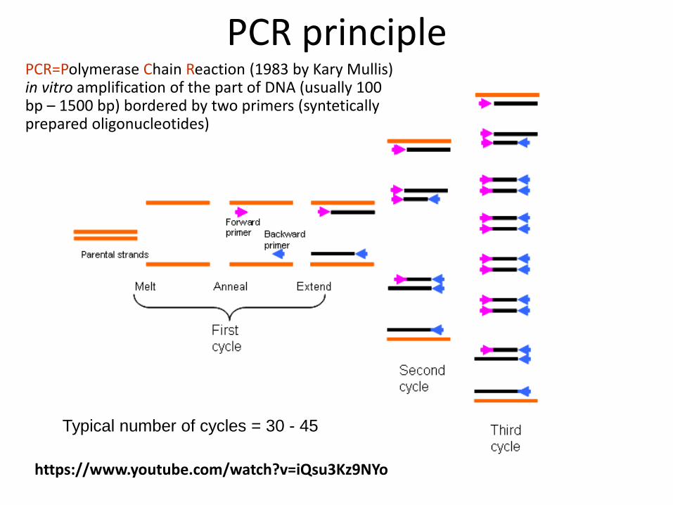
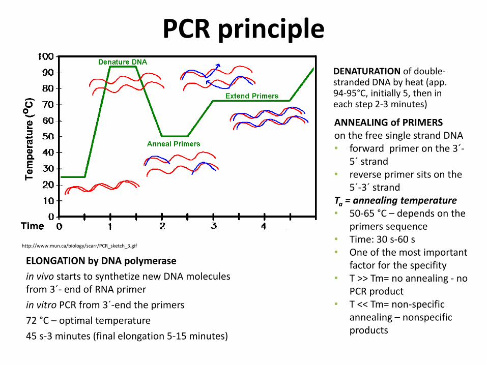
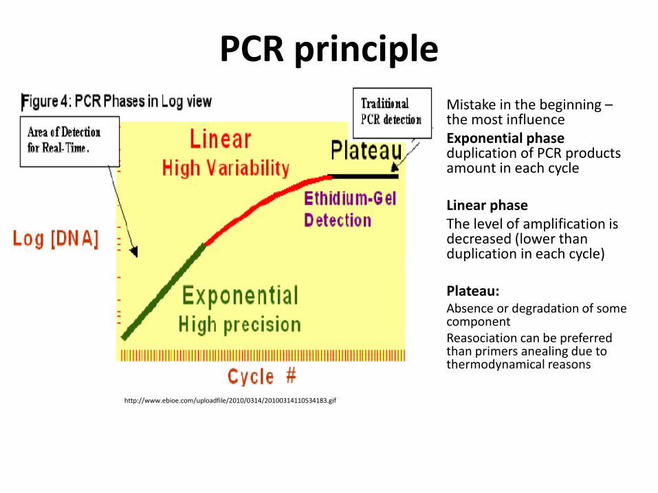
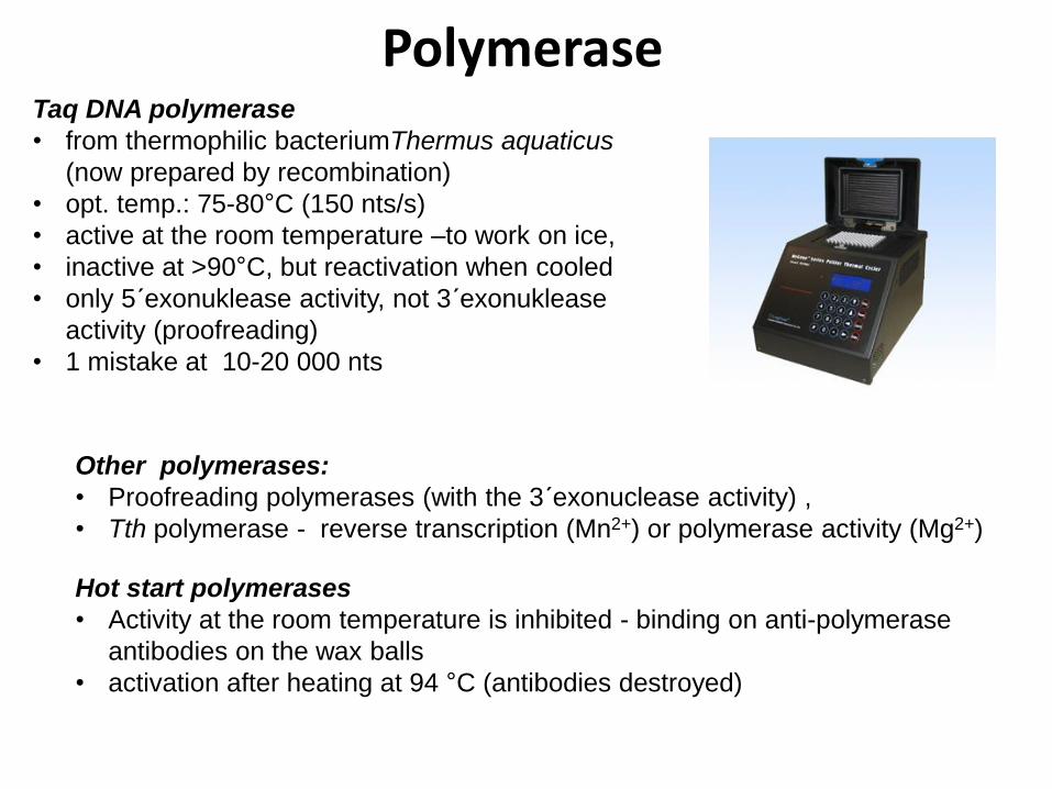
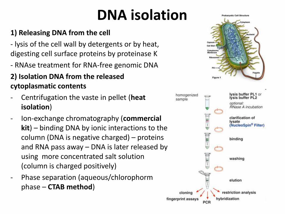
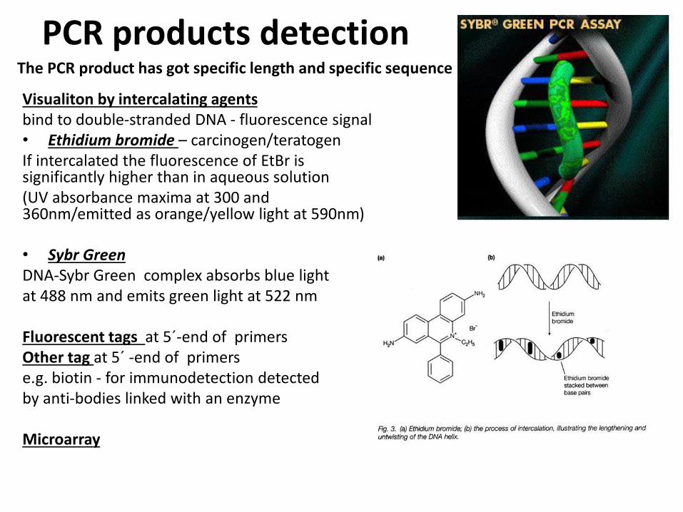
Real-Time PCR
Food-borne pathogens detection by PCR
Identification of pure cultures - without problems
Detectionenumeration from food matrix directly or after enrichment
bull How to isolate successfully DNA from few cells in 25 g10 g - Efficiency of DNA isolation (isolation by kits heat-lysis CTAB method)
bull How many targets copies are needed for successfully PCR products detection
ndash detection limits
ndash presence of PCR inhibitors (inhibition of polymerase primers annealing)
bull How to distuinguish DNA from dead and live cells - Total DNA is detected (from dead and living cell)
bull For distinguishing of dead cells DNA ndash eg application of EMA (ethidium monoazide) to the sample before DNA isolation ndash EMA irreversibly bound to DNA of dead cells ndash they are bdquoinvisibleldquo for PCR ndash no amplification
bull Direct enumerationdetection only in some specific case ndash simple liquid sample when the required limit is higher than the limit of detection
bull Standardly ndash for PCR is used enriched homogenate (sample diluted in enrichment broth and cultivated) ndash to obtain detectable level of microorganisms
BAXreg System
Salmonella
Listeria monocytogenes
Genus Listeria
Enterobacter sakazakii
E coli O157H7
Staphylococcus aureus Real-time
Campylobacter jejunicolilariReal-time
Vibrio cholerae
parahaemolyticusvulnificus Real-time
Yeast and molds
httpwww2dupontcomDuPon
t_Homeen_USindexhtml
BAX ndash three major steps
On enrichedregrown samples
Prepare samples = perform heat-lysis in two stages
DNA present in the lysate
2 Process samples
3 Review results
Hydrate PCR tablets
Enrichment protocols vary
depending on bacterial target
and type of food yoursquore testing
Salmonella Enterobacter
sakazakii
E coli O157H7 E coli O157H7
MP Campylobacter jejunicoli
Yeast and Mold
37deg +- 2degC for 20 minutes then
95deg +- 3degC for 10 minutes
Listeria and L monocytogenes
55deg +- 2degC for 60 minutes then
95deg +- 3degC for 10 minutes
5 microl for all bacterial targets
20 microl for beef samples enriched
in E coli O157H7 MP media
20 microl for yeast amp mold
PCR tablets bull DNA-polymerase
bull Primers
bull Nucleotids
bull Suplements
bull SybrGreen or fluorescent probe
bull Internal control + primer
bull Mastermix in the tablet in PCR Eppendorf ndash minimal risk of contamination
For solving the problem of distiguishing dead and live cell the process of small volumes
is used ndash only if live cells are present in the sample they able to multiplied to the limit
detection (104 CFUml in the enrichment suspension used for lysis step)
If the result of detection is positive confirmation by plating out is done (live and dead
cells)
+
Target organism DNA BAXreg system
primers
in tablets PCR
+ no detectable
amplification
Background DNA
PCR
BAXreg system
primers
in tablets
Amplification of specific fragments
Detection of specific fragments Fluorescent signal drops with heating to generate a melt curve
Melting curve Raw Data Processed Data
Strong positive
result
Negative
result
Target peak
Positive control peaks
Positive control peak
Melting curves display peaks within a set temperature range for the control and target (if present)
Detection
Melt curve weak INPC in presence of strong Salmonella positive
Real time weak INPC in presence of strong
Campylobacter positives
Direct protocol does not display a positive or
negative result PCR was successful IndeterminateSignal error
Salmonella AFNOR (ISO 16140) protocol Sample 110 BPW
18 h37ordmC
RVS Muumlller-Kauffmann
24 h 415 37 ordmC
5 suspicious colonies on TSA
24 h 37ordm C
Biochemical and serological confirmation
XLD 2 Plate
24 h 37ordmC
BHI
3h 37 ordmC
22 h 37 ordmC
15 d neg and pos result
3 d neg 5 d pos results
Salmonella ( ISO 65792003) versus BAXreg-PCR
International certificates AOAC-RI Performance Tested Method
Salmonella 100201 L monocytogenes
070202 L spp 030502 Saureus 120701
Campylobacter colilarijejuni
AFNOR Certification
Salmonella QUA 183-1102
NordVal Certification
Salmonella 2006-30-5408
Certificates from AFNOR and AOAC are equal
NordVal Certificate
57
OTHER METHODS
ATP bioluminiscence
httpwwwpromegacouk~mediaimagesresourcesfiguresenotesfe0027_fig1jpgla=en
httpwwwbiotekptassetstech_resources143clarity_atp_conc_fig2gif
Principle the detection of the present level of ATP (direct proportion to the presence of microorganisms or their products residues) The enzyme luciferase catalyzes the transformation of d-luciferin to oxyluciferin (indicated by emision of light at 560 nm)
ATP bioluminiscence
httpwwwbioxyscomimages2figure-B11gif
ATP Hygiene Monitoring System - SystemSURE II
httpwwwmicromaticcomdraft-keg-beerline-cleaning-hygiene-pid-ATP-100-tab-reviewshtml
Biosensors
60
httpsuploadwikimediaorgwikipediacommonseecBiosensor_Systemjpg
Biosenzor =the whole cellmicroorganism (CBS ndash cell-based sensors)
Cell-based sensors
Patogenic microorganisms interact during the infection with mammalian
cells ndash detection of cell or toxin by cell culture The damage of cell culture is measured optically or electrically
62
IDENTIFICATION OF MICROORGANISMS
Definition of species in bacteriaarchae
The criterion for a bacterial or archael species a collection of strains that have bull at least one diagnostic phenotypic trait bull ge70 DNAndashDNA hybridization of their purified molecules DNA
bull ~more than 97 16S rRNA sequence identity Under the same conditions some eucaryotic order would be a single bacterial species
The bacterial species definition bull eminent practical significance bull but a very difficult issue
bull very wide and often changes in DNA
bull mutations during replication of DNA
bull horizontal gene transfer through mobile elements plasmids also between species
Criterion for species in prokaryotic cells (bacteria and archea) Collection of strains characterised bull by at least one diagnostic feature bull their purified DNA molecules prove at least 70 hybridisation
which corresponds to at least 97 concordance in the sequence of 16S rRNA gene
Map of genome of B cereus ATCC 14579
httpwwwacgtinccomspecialty_dna_sequencinghtm
Identification based on genotype
64
DNA isolation PCR amplification of 16S rRNA gene (usually)
Sequencing of PCR product
Comparison to sequences in databases (BLAST)
Identification of bacteria PHENOTYPE METHODS bull Morphology of colony and cell (Gram staining shape) bull Requirements for temperature atmosphere nutrition bull Biochemical tests - presence of specific enzymes
bull catalase oxidase fermentation of glucose bull the way how they utilize different compounds
bull Immunochemical tests - specific antigens in the cell wall METHODS BASED ON PROTEIN ANALYSIS
bull MALDI-TOF MS METHODS BASED ON GENOTYPE bull looking for and comparing of specific DNA sequences
bull conserved universal genes (eg 16S rRNA) bull genes specific for genus species
bull done by PCR methods DNA chips sequencing etc
66
BIOCHEMICAL TESTING
Identification based on phenotype I Physiological + biochemical features
bull morfology of colonies and cells (Gram staining shape)
bull requirements for temperature atmosphere nutrient sources
bull basic biochemical tests oxidase catalase fermentationoxidation of glucose haemolysis etc
appearance of colonies on selective or selective-diagnostic agars (for other testing to be reinoculated on non-selective agar)
the classification of an isolate into a certain group
(eg for bacteria catalase positivenegative G+ cocci glucose fermentingnon-fermenting G- rods etc)
biochemical tests specific for certain groups of microorganisms
Identification based on phenotype I
68
Physiological + biochemical features Example Bacillus cereus
httpwwwfoodhaccpcommemberonlynewsletter164html
httpwwwnapavalleyedupeoplesrosePublishingImagesBacillus20cereus202(Gram20stain)jpg
Set of biochemical tests Microgenreg Bacillus ID Panel for the identification of Bacillus spp and related genera (time for performing 48 hours)
MYP agar (Mannitol Egg Yolk Polymyxin Agar) pink colonies with a precipitation zone (no fermentation of manitol + enzyme lecitinase)
catalase positive
httpwwwoxoidcomukblueprod_detailprod_detailasppr=CM0929ampc=ukamplang=ENamporg=9ampimg=CM0929ampsec=
G+ rod endspores forming
β-haemolysis moisty and easy to grow colonies
Principle of biochemical testing
to rank the bacterium to some group having the same basic features bull eg catalase positive gram positive cocci bull Family Enterobacteriaceae
the identification on the genus or species level
bull sets of biochemical tests specific for some group of bacteria
70
Gram staining procedure - principle
https40mediatumblrcom25da2e5b25bbe336819bf1715849d171tumblr_inline_ntrsgn9w1D1sv92o6_540jpg
Gram staining procedure - principle
httpsonlinesciencepsuedusitesdefaultfilesmicrb106CellStructurecow02354_03_15jpg
Gram staining procedure 1-2 drops of sterile water on a slide Spread culture in thin filter in this drop Let dry in airclose to fire Pass slide through flame 2-3x to fix Add crystal violet ndash 15-20 s to act Add Lugol solution (KII2) ndash 15-20 s to act Washing off with a mixture of
ethanolaceton (41) or acetone till the stain removes but max 15 s
Rinse with water decant excess liquid Add safranin 1 minute Rinse with water decant excess liquid Dry and put a drop of imersion oil use an
objective bdquoOILldquo
httpwwwtumblrcomtaggedgram-staining
G- red
G+ violet
Gram staining procedure + Identification
E coli
Family Enterobacteriaceae Gram-negative rods
Micrococcus luteus
Staphylococcus aureus
Gram-positive cocci Bacillus cereus
Gram-positive rods (with endospores)
Biochemical identification of G + bacteria
httpyoungeneticistblogspotcz201106diagnostic-microbiology-labhtml
G+ bacteria bull cocci ndash in clusters in pairs
diplococci bull bacilli
bull endospore bull no endospore
For G+ cocci bull Catalase positive ndash Staphylococcus
sp bull Coagulase + S aureus or some other few species bull Coagulase -
bull Novobiocin S bull S epidermidis etc
bull Novobiocin R bull S saprophyticus etc
bull Catalase negative Streptococcus sp Enterococcus sp
bull kind of haemolysis Other important G + group ndash filamentous bacteria
Catalase test Catalase test Catalase is a common enzyme found in nearly all living organisms that are exposed to oxygen where it catalyzes the decomposition of hydrogen peroxide to water and oxygen
2 H2O + O2 2 H2O2
catalase
bubbling when culture is add in peroxide
httpiws2collinedudcainCCCCD20Microcatalase_testhtm
Catalase testing into a drop of 3 peroxide to transfer a loop of bacterial culture catalase positive ndash releasing of air bubbles
Examples bull G+ cocci catalase positive Staphylococcus sp Micrococcus sp Kocuria sp Dermacoccus sp Macrococcus sp Rothia sp catalase negative Streptococcus sp Enterococcus sp bull G+ rods (bacilli) endospores forming catalase positive Bacillus sp catalase negative Clostridium sp
Resistance to ATBhaemolysis Resistance eg to novobiocin
httpquizletcom20077004mic-lecture-1-t2-flash-cards
Hemolysis on blood agar
Measuring the diameter of inhibition zone (less than 17 mm resistant more or equal to 17 mm sensitive) ndash differing in Staphylococcus sp (Clinical performance)
alpha-hemolysis - oxidizing the iron in the hemoglobin - green discoloration of the medium around beta-hemolysis - rupture of erythrocytes - complete clearing medium around gamma-hemolysis ndash no effect ndash no change
By cultivation on blood agar bacteria can be
differentiated based on their capacity to secrete
hemolysins The hemolysis will cause a clearing zone of
the blood agar around the colonies Bacteria can cause
different types of hemolysis
bullα-hemolysis which means an incomplete clearing
(green haemolysis)
bullβ-hemolysis which means a complete clearing
bullDouble hemolysis of some staphylococci consisting of
an inner β-hemolysis zone and an outer α-hemolysis
zone
bullNo hemolysis which is sometimes referred to as γ-
hemolysis which may seem illogical
Note that the α-hemolysin of staphylococci causes
complete hemolysis whereas their β-hemolysin causes
incomplete hemolysis
The capacity to produce hemolysins may vary between
different strains of a particular bacterial species
httpwwwvetbactorgvetbactdisplayextinfo=67
Identification of bacteria based on haemolysis
Streptococcal hemolysins α (alpha) hemolysis - ldquozone of green discolourationrdquo or ldquogreeningrdquo called partial or incomplete hemolysis produced by alpha-hemolytic streptococci such as Streptococcus suis This type of hemolysis on BA is due to bacterial production of hydrogen peroxide which oxidises hemoglobin to methemoglobin β (beta) hemolysis ndash a clear zone of complete hemolysis produced by beta-hemolytic streptococci such as S equi subsp equi This hemolysis is due to lysis of RBCs in the BA by the cytotoxin Streptolysin S
Source httppeopleupeicajlewishtmlhemolysinshtml
Bacterial hemolysins
Staphylococcal hemolysins Most coagulase-positive staphylococci (CPS) such as Staphylococcus aureus S pseudintermedius ( but not S hycius) typically produce an distinctive pattern of ldquodouble zonerdquo or ldquotargetrdquo hemolysis on blood agar Double-zone hemolysis indicates that the two hemolysins one which produces ldquocompleterdquo and another one that produces ldquoincompleterdquo hemolysis are present and this combined effect is visible due to different diffusion rates of these enzymes through the agar The staphylococcal α (alpha) hemolysin causes a complete type of hemolysis This zone of complete hemolysis is narrow and immediately adjacent to the bacterial colony on blood agar The staphylococcal β (beta) hemolysin causes an incomplete type of hemolysis at 37 degC This broader incomplete zone is outside of the inner complete zone and can be hard to see on early culture plates and also varies with the species of RBCs used to make the BA plate Its action is enhanced by chilling at 4 degC and the hemolysis becomes complete (this effect is referred to as ldquohot-coldrdquohemolysis) There are other staphylococcal cytotoxins that are also hemolysins but their lytic actions are not seen on blood agar
The confusion arises due to the NAMES of the two STAPHYLOCOCCAL HEMOLYSINS and the TYPE of hemolysis they produce These are the REVERSE of the two TYPES of STREPTOCOCCAL HEMOLYSIS Strictly speaking the descriptive terms ldquoalphardquo for ldquoincompleterdquo and ldquobetardquo for ldquocompleterdquo hemolysis apply only to the hemolytic toxins of Streptococcus species However other organisms beside Streptococcus and Staphylococcus species have cytotoxins that cause hemolysis and this hemolysis can be either complete or incomplete It is preferable to use the terms ldquocompleterdquo and ldquoincompleterdquo rather than ldquobetardquo or ldquoalphardquo to describe the hemolysis caused by these organisms but you will notice in bacteriology text books that the terms alpha and beta are commonly used
G ndash bacteria bull Cocci bull Bacilli (Rods) bull Oxidase positivenegative bull Glucose metabolism
bull fermentation bull oxidation bull Inactive
bull Lactose metabolism for coliform
bull Pigment formation for Pseudomonas sp
Biochemical identification of
G - bacteria
Oxidase test
Oxidase positive (OXI +) Aeromonas sp Pseudomonas sp Dark-blue to violet
Oxidase negative (OXI -) Enterobacteriaceae Bacillus sp Colourless
Oxidase bullabbreviated general name for some kind of cytochrome c oxidases (cytochrome oxidase or indophenol oxidase) enzymes involved in aerobic respiratory chain when oxygen is used as the final receptor for hydrogen bullOxidases are usually present only in aerobic organisms bullPresence or absence of oxidase(s) is an important feature of the bacterial metabolism used mainly in the identification process of gram-negative bacteria
Detection Reaction of the culture with redox indicator as eg NN-dimethyl-p-phenylenediamine (DMPD) dark-blue when oxidized and colorless when reduced Different test performances are possible
Fermentation of carbohydrates Type of utilisation of glucose or carbohydrates bull an important feature of the bacterial metabolism bull widely used in the identification process of bacteria bull fermentation strictly means to be under anaerobic conditions (but in bacterial
metabolism which can be but not have to be the same as the anaerobic conditions of cultivation) so very often is used in a general way like bdquoutilisationldquo
bull fermentation (like bdquoutilisationldquo) of carbohydrates to
bull acid and gas (Durham tubes) bull acid without gas bull non-utilised
bull Testing of fermentation or oxidation of carbohydrates
bull eg OF test for glucose bull by oxidation under aerobic conditions bull by fermenation under anaerobic conditions (an oil layer)
Fermentation of sugars to acid
Peptone Water wPhenol Red
(HiMedia M028I)
Ingredients gl
Peptic digest of animal tissue 10000
Sodium chloride 5000
Phenol red 0020
Final pH ( at 25degC) 68plusmn02
httpsuploadwikimediaorgwikipediacommons009Phenol_red_pH_60_-_80jpg
+ after sterilisation
at the room
temperature to add
aseptically sugar
discs and
inoculate
succrose trehalose rhamnose xylose
Incubation 30 degC 24 h
- +
Fermentation of carbohydrates
Casein enzymic hydrolysate and yeast extract provide nitrogenous compounds vitamin B complex and other growth nutrients Dextrose is the fermentable carbohydrate and bromocresol purple acts as the pH indicator with colour change from purple to yellow in acidic conditions Sodium chloride maintains osmotic equilibrium bdquoAnaerobicldquo conditions ndash inoculation by stabbing to used fresh prepared media (oxygen expelled by heating medium in boiling water or running steam for 15 min then to be cooled down quickly)
- +
Enterobacteriaceae ndash glucose fermentation
83
Tryptone Yeast Extract Agar Ingredients Gms Litre Casein enzymic hydrolysate 10000 Yeast extract 1500 Dextrose 10000 Sodium chloride 5000 Bromocresol purple 0015 Agar 15000 Final pH ( at 25degC) 70plusmn02
Test for oxidationfermentation of glucose (OF glucose test) detection of acid production by acidobazic indicator change to yellow (acid present) in lower pH from green (no acid) Anaerobic atmosph ndash covering by sterile mineral oil If fermented anaerobically then also runs aerobically
Fermentation of carbohydrates
Principle of biochemical tests bull The first step in identification is to rank the bacterium to some group of bacteria
having the same basic features ndash eg catalase positive gram positive cocci bull The identification on the genus or species level is finished by other biochemical tests
specific for each group of bacteria bull The tests can be done in tubes or miniaturized plate
ENTEROtest 24N (Erba Lachema) (24 means 24 biochemical tests N= new)
API 20E (bioMeacuterieux)
bull designed for routine identification of clinically important species of family Enterobacteriaceae without the use of reagents
bull A strip test OXItest or OFtest in microtitration wells can be used to confirm that the isolate belongs to Enterobacteriaceae family
bull The results are read visually or automatically using reader MIKROLAreg
bull The identification can be supplemented by diagnostic strip tests INDOLtest COLItest and VPtest
APIreg Gram negative Identification API 20E ndash Speciessubspecies identification of Enterobacteriaceae and groupspecies identification of non-fermenting Gram negative bacteria API Rapid 20E ndash 4-hour identification of Enterobacteriaceae API 20NE ndash 24 to 48-hour identification of Gram negative non-Enterobacteriaceae API Campy ndash 24-hour identification of Campylobacter species
Example Escherichia coli
ENTEROtest 24 N
Biochemical testing - Enterobacteriaceae bull TSI Triple Sugar Iron Agar (use a straight wire to stab the butt and streak the agar surface)
- to determine whether organisms can ferment glucose sucrose andor lactose with or without production of gas and the ability of the organism to produce hydrogen sulphide from thiosulphate in an acid environment is also tested
Formula Liter Enzymatic Digest of Casein 5 g Enzymatic Digest of Animal Tissue 5 g Yeast Enriched Peptone 10 g Dextrose 1 g Lactose 10 g Sucrose 10 g Ferric Ammonium Citrate 02 g Sodium Chloride 5 g Sodium Thiosulfate 03 g Phenol Red 0025 g Agar 135 g Final pH 73 plusmn 02 at 25 degC
inoculation in the butt by stab inoculation on the slant on the surface bull Fermentation of dextrose
(glucose) to acid bull anaerobically - yellow butt bull aerobically leads to products
which are later oxidised by air and pH remains alkaline (red) bull Fermentation of glucose to gas (anaerobically - in the stab) Cracks splits or bubbles in medium indicate gas production bull Production of hydrogen sulfide (H2S) ndash reaction with ferrous sulphate and sodium thiosulphate - a black precipitate in butt bull Lactose andor succrose
utilisation to acid Acid production ndash yellow butt and slant
Results
An alkaline slant-acid butt
(redyellow) - fermentation of
dextrose only
An acid slant-acid butt
(yellowyellow) - fermentation of
dextrose lactose andor sucrose
An alkaline slant-alkaline butt
(redred) - dextrose or lactose not fermented (non-fermenter)
Bacterium fermenting lactose andor succrose are able to ferment glucose
Phenol red ndash alkaline = red acid = yellow
bull TSI Triple Sugar Iron Agar (use a straight wire to stab the butt and streak the agar surface)
- to determine whether organisms can ferment glucose sucrose andor lactose with or without production of gas and the ability of the organism to produce hydrogen sulphide from thiosulphate in an acid environment is also tested
Biochemical testing - Enterobacteriaceae
Urea agar (Christensen) (use a 1 μl loop full)
- to determine whether an organism can split urea into ammonia and
carbon dioxide by the action of the enzyme urease
httpimageslidesharecdncomlab17-141201085153-conversion-gate0195-10-638jpgcb=1417424219
httpimageslidesharecdncomhelicobacterpylori-copy-110516201901-phpapp0195campylobacter-jejunihelicobacter-pylori-39-728jpgcb=1305577391
Phenol red
Medium is acidic - yellow
(pH 68plusmn02)
Alkaline reaction by urea hydrolysis - pink colour
Composition Gms Litre
Peptic digest of animal tissue
1000
Dextrose 1000
Sodium chloride 5000
Disodium phosphate 1200
Monopotassium phosphate 0800
Phenol red 0012
Agar 15000
Final pH ( at 25degC) 68plusmn02
Nutrition
Peptic digest of animal tissues dextrose
Sodium chloride - osmotic equilibrium
Phosphates - buffering of the medium
35-37 degC
Biochemical testing - Enterobacteriaceae
bull L-lysine decarboxylase (use a 1 μl loop full)
- to determine whether an organism can decarboxylate an amino acid (lysine) leading to formation of an
amine After inoculation both tubes are overlayed with sterile paraffin oil
httpfce-studynetdna-sslcomimagesupload-flashcards10295302332621_mjpg
+ CO2 + OH-
Lysine Decarboxylase Broth Typical Composition (glitre) Peptic digest of animal tissue 5000 Yeast extract 3000Dextrose 1000 (glucose prepared from starch chemically equivalent to glucose) L-Lysine hydrochloride 5000 Bromocresol purple 0020 Final pH ( at 25 degC) 68plusmn02 Inoculate below the surface cover by sterile paraffin oil
Two steps detection 1 step Dextrose is fermented ndash H+ is released Acidic conditions bull bromocresol purple changes colour to yellow bull in acidic conditions lysine decarboxylase is
activated 2 step L-lysine is decarboxylated and OH- released Alkaline conditions bull bromocresol purple changes colour back to
purple
Biochemical testing - Enterobacteriaceae
bull β-galactosidase (ONPG) (use a 1 μl loop full)
- to determine whether an organism has the enzyme szlig-galactosidase by using the compound o-
nitrophenyl-beta-D-galactopyranoside (ONPG)
- this test is used to differentiate between lactose-negative and lactose-delayed organisms
- ONPG is hydrolysed by the enzyme to o-nitrophenol which is yellow and galactose
O-Nitrophenyl-β-D-galactopyranoside (ONPG) is structurally similar to lactose (ie ONPG is an analog of lactose) except that orthonitrophenyl has been substituted for glucose ONPG is colorless compound O-nitrophenol is yellow visual evidence of hydrolysis
Lactose fermenting bacteria (ONPG positive) two enzymes Permease ndash transferring of the lactose molecule into the bacterial cell β-galactosidase - cleaving the galactoside bond producing glucose and galactose - inducible enzyme - made ONLY in the presence of the lactose substrate Coliform bacteria as Ecoli Klebsiella spp Enterobacter spp
hydrolysis
Late lactose fermentering bacteria Permease ndash not present delayed in the production of acid from lactose because of sluggish permease activity β-galactosidase ndash present - give a positive ONPG test Citrobacter spp Arizona spp
Non lactose fermenter (ONPG Negative) Permease ndash not present β-galactosidase ndash not present Salmonella spp Shigella spp Proteus spp Providencia spp and Morganella spp
Biochemical testing - Enterobacteriaceae
Procedure Culture Bacteria grown in medium containing lactose (to induce the production of the galactosidase enzyme) such as Kligler iron agar (KIA) or Triple sugar Iron (TSI) agar (Note β-galactosidase enzyme (inducible enzyme) is made ONLY in the presence of the lactose substrate) Inoculation 1A loopful of bacterial growth is emulsified in 005mL of physiologic saline to produce a heavy suspension 2One drop of toluene is added to the suspension and vigorously mixed for a few seconds to release the enzyme for bacterial cells 3An equal quantity of buffered ONPG solution is added to the suspension 4The mixture is placed in a 37 oC water bath 5When Using ONPG Tablets 6A loopful of bacterial suspension is added directly to the ONPG substrate resulting from adding 1mL of distilled water to a tablet in a test tube 7This suspension is also placed in a 37oC water bath
β-galactosidase (ONPG)
Results and Interpretations The rate of hydrolysis of ONPG to o-nitrophenol may be rapid for some organisms producing a visible yellow color reaction within 5 to 10 minutes Most tests are positive within 1 hour however reactions should not be interpreted as negative before 24 hours of incubation The yellow color is usually distinct and indicates that the organism has produced o-nitrophenol from the ONPG substrate through the action of β-galactosidase
Biochemical testing - Enterobacteriaceae
bull VogesndashProskauer (VP) test (use a 1 μl loop full)
- to determine whether an organism can produce acetylmethylcarbinol
(acetoin) from fermentation of glucose If present acetylmethyl carbinol is
converted to diacetyl in the presence of prop- naphthol (to be added first) strong
alkali (40 KOH) and atmospheric oxygen The diacetyl and quanidine-
containing compounds found in the peptones of the broth then condense to
form a pinkish red polymer
MRVP broth (pH 69)
Ingredients per liter of
deionized water
buffered peptone= 70 gm
glucose= 50 gm
dipotassium phosphate= 50 gm Voges-Proskauer Reagent A Barrittrsquos reagent A
Alpha-Naphthol 5 50 gm
Absolute Ethanol 1000
ml
Voges-Proskauer Reagent B Barrittrsquos reagent B
Potassium Hydroxide 400 gm
Deionized Water 1000 ml
Procedure of VogesndashProskauer (VP) Test
1Prior to inoculation allow medium to
equilibrate to room temperature
2Using organisms taken from an 18-24
hour pure culture lightly inoculate the
medium
3Incubate aerobically at 37 degC 24 hours
4Following 24 hours of incubation aliquot
2 ml of the broth to a clean test tube
5Re-incubate the remaining broth for an
additional 24 hours
6Add 6 drops of 5 alpha-naphthol and
mix well to aerate
7Add 2 drops of 40 potassium hydroxide
and mix well to aerate
8Observe for a pink-red color at the surface
within 30 min Shake the tube vigorously
during the 30-min period
Positive Reaction
A pink-red color at the surface
Examples Viridans group streptococci
(except Streptococcus vestibularis) Listeria
Enterobacter Klebsiella Serratia marcescens
Hafnia alvei Vibrio eltor Vibrio alginolyticus etc
Negative Reaction
A lack of a pink-red color
Examples Streptococcus mitis Citrobacter sp
Shigella Yersinia Edwardsiella Salmonella Vibrio
furnissii Vibrio fluvialis Vibrio vulnificus and Vibrio
parahaemolyticus etc
A copper color should be considered negative A
rust color is a weak positive reaction
Quality Control of VogesndashProskauer (VP) Test
VP positive Enterobacter aerogenes (ATCC13048)
VP negative Escherichia coli (ATCC25922)
Biochemical testing - Enterobacteriaceae
94
VogesndashProskauer (VP) test
Rapid test for confirmation of E coli ndash typical features
β-D-glucuronidase (97 strains) production of indole from tryptophan (typical for E coli)
Performance
Previous testing Gram-negative (eg able to grow on VRBD agar) oxidase negative
From pure culture to prepare suspension in saline solution (app 5 ml) at the density 3 McFarland
To transfer 05 ndash 1 ml of this suspension into a new tube ndash to add the strip of COLItest (chemicals ndash fluorogenic substrate for β-D-glucuronidase tryptophane are present in the pad at the end of the strip)
Incubation 37 degC 4 hours
Detecion of β-D-glucuronidase (to be done at first) UV (360 nm) ndash blue fluorescence (GLR + hellipβ-D-glucuronidase positive) ndash GLR test can be read after 1 hour
Detection of indole formation from tryptophane (to be done at second) to add Kovacs reagent (IND + hellipformation of red ring)
95
COLItest - Escherichia coli
Biochemical testing - Enterobacteriaceae
Fluorogenic culture medium
bull One or more fluorogenic substrates
bull For detection of specific enzyme which are able to cleave out the fluorogenic substrate that a designed
fluorophore (responsible for the UV fluorescence) is released
FLUOROGENIC MEDIUM (Colilert) E coli use β-glucuronidase to metabolize MUG and create fluorescence (UV 360 nm) Since most non-coliforms do not have these enzymes they are unable to grow and interfere
httpgawaterusgsgovprojectsbacteriapicturessummaryanalysisecolijpg
E coli (MPN method)
COLItest Biochemical testing - Enterobacteriaceae
COLItest bull Indole test (use a 1 μl loop full)
- to determine whether an organism can split indole from tryptophan
httpwwwmicrobiologyinfocomindole-test-principle-reagents-procedure-result-interpretation-and-limitations
indole production from tryptophan
Production of indole is detected by Kovacacutes reagent
Indole Kovacs Reagent
p-Dimethylaminobenzaldehyde 500 gm
Hydrochloric Acid 37 2500 ml
Amyl Alcohol 7500 ml
Biochemical testing - Enterobacteriaceae
INDOLe test ndash quick variant
Ecoli Citrobacter Enterobacter
Procedure bull on the filtration paper to add a drop of INDOL suspension bull to spread into by a loop some culture bullTo incubate ndash at the room temperature for 5 minutes bullTo read results
Biochemical testing - Enterobacteriaceae
ENTEROtest 24 ndash example of biochemical testing
routine identification of clinically important species of family Enterobacteriaceae the test is focused only on some species often isolated in clinical practis strains from other sources can be slightly different in the dominant biochemical profiles intended exclusively for the identification of family Enterobacteriaceae the preliminary ranking into family Enterobacteriaceae must be done (Gram staining oxidase glucose fermentation) the results of reactions obtained in this modified micromethod may differ from the results obtained using conventional tests and results available in literature sources in case of problem to control positivenegative reactions by control strains or to use for this tests tube variant to check the functionality of the kit not to check the accuracy or success of the identification
Preparation of suspension from over-night culture growing on non-selective agar (for staphylococci Columbia agar blood with 5 of sheep blood is recommended)
Inoculation of microtitre plates anaerobic reaction covered by mineral oil
Incubation
Reading results by both visual and instrumental mode respectively Standard off-line tests INDOLtest or COLItest Other off-line tests OXItest OFtest VPtest Acetoin test
ENTEROtest 24 N ndash example of biochemical testing
bull tube with 5 ml of sterile physiolsol bull suspension (1 McFarland) bull application 100 microl to each hole bull 2 drops of parafin oil into holes D-H of the first line (anaerobic conditions) bull Incubation 37degC 24h bull for check the purity of suspension to streak suspension on TSA (incubation 37 degC ndash 24 h
ENTEROtest 24N
ENTEROtest 24 N - evaluation
Escherichia coli
H 0+0+0=0 G 0+2+0=2 F 1+2+0=3 hellip Identification code 023426
1 1
2
4
ENTEROtest 24 ndash example of biochemical testing
The result = a set of biochemical properties (+-) which is compared to the sets of biochemical properties of different chosen genera and species for them the test is designed
ENTEROtest 24 N
T-index how is the likelihood of the different properties The combination of identication and T-index = the reliability of the result Excelent very good good (this case) acceptable intermediary strain needs some additional tests genus level non-existing taxon
Evaluation by using the identification code - By using a code book - By using software TNW
Identification score the measure of similarity to proposed taxon
The way of identification the obtained profile is compared with profiles in database bull The most similar profiles are found
bull Identification score = of similarity bull But what about the different reactions
bull T-index is calculated bull how is the probability of the different reactions
bull It depends on their importance ndash typical reactions should be the same
bull Reliability of the result bull the combination of identication score and T-index = bull Excelent very good good acceptable intermediary strain needs some
additional tests genus level non-existing taxon
ENTEROtest 24 ndash example of biochemical testing
Biochemical tests - divisions
ENTEROtest 24 species of family Enterobacteriaceae
NEFERMtest 24 Gramnegative non-fermenting bacteria and families Vibrionaceae Aeromonadaceae and Pleiomonas shigelloides present mainly in clinical material
ANAEROtest 23 ANAEROBIC BACTERIA (G + AND G-) routine identification of anaerobic bacteria (G + AND G-) in clinical material and in food
STREPTOtest 24 Streptococcus Enterococcus and related gram positive catalase negative cocci isolated from clinical material
STAPHYtest 24 genus Staphylococcus isolated from clinical material and for their distiguishing from related genera of other gram positive catalase positive cocci
Biochemical tests APIreg Gram positive Identification API Staph ndash Overnight identification of clinical staphylococci and micrococci API 20 Strep ndash 4 or 24-hour identification of streptococci and enterococci API Coryne ndash 24-hour identification of Corynebacteria and coryne-like organisms API Listeria ndash 24-hour identification of all Listeria species APIreg Gram negative Identification API 20E ndash Speciessubspecies identification of Enterobacteriaceae and groupspecies identification of non-fermenting Gram negative bacteria API Rapid 20E ndash 4-hour identification of Enterobacteriaceae API 20NE ndash 24 to 48-hour identification of Gram negative non-Enterobacteriaceae API Campy ndash 24-hour identification of Campylobacter species APIreg Anaerobe Identification API 20A ndash 24-hour identification of anaerobes Rapid ID 32 A ndash 4-hour identification of anaerobes
MicrogenTM GN-ID Convenient 12 substrate identification system for commonly encountered Enterobacteriaceae (GNA) Extended 24 substrate identification system for the complete range of Enterobacteriacea and oxidase positive non-fastidious Gram negative bacilli (GNA+B) MicrogenTM Listeria-ID Enhanced built-in haemolysis test MicrogenTM Bacillus-ID 24 substrate system for the identification of mesophilic Bacillus spp isolated from foods and related samples MicrogenTM Staph-ID 12 substrate system minimizes handling and manipulation Substrates selected specifically for Staphylococcus and related organisms MicrogenTM Strep-ID Substrates selected specifically for Streptococcus Enterococcus and related species Provides excellent differentiation of Enterococci spp especially E casseliflavus E mundtii and E gallinarum
Biochemical tests
Identification of microorganisms by MALDI-TOF MS
Department of Biochemistry and Microbiology University of Chemistry and Technology Prague
Sabina Purkrtovaacute1 Petra Junkovaacute2 1Laboratory of food microbiology 2Laboratory of applied proteomics
SabinaPurkrtovavschtcz PetraJunkovavschtcz
MALDI-TOF MS
1) sample preparation bull microbial culture or its
proteins extract is smearing onto a steel plate and covered over by matrix
bull matrix enables the sample (A) to be desorped and ionised as pseudomolecule ionts [A+H] +
2) MALDI-TOF MS analysis unique mass spectral fingerprint of desorbed microbial cell components (mainly intracellular proteins) different among genera species or also some strains
Matrix-Assisted Laser Desorption Ionization-Time of Flight Mass Spectrometry
httppubsrscorgencontentarticlehtml2014RAC4RA05604C
httpcmrasmorgcontent263547figures-only
3) identification comparison of mass spectrum to those of reference strains in database
MALDI-TOF MS PRINCIPLE
111
2
2
2L
teU
z
m
m mass z charge
L length of drifting zone e
elementary charge U
speeding voltage
Time of flight is a function of the specific ion mass (mz)
httpwwwsigmaaldrichcomtechnical-documentsarticlesbiologycustom-dna-oligos-qc-analysis-by-mass-spectrometryhtml
Soft ionisation method bull low level of sample fragmentation
Animation http cmgmstanfordedupansection_htmlMS
MALDI-TOF MS SAMPLE AND MATRIX
httpcmrasmorgcontent263547figures-only
Matrix bull able to absorb the energy of the laser (usually 337 nm) bull to crystalise with samples - usually acid
character (proton ionisation of sample) dissolved in organic solvent
Sample preparation Direct transfer bull intact cells spread directly onto a steel plate bull lysis of cells occurs during the contact with acid
matrix and by laser desorption bull most bacteria Proteins extraction bull previous extraction of proteins by organic acids
andor alcohol (eg ethanol and 70 formic acid) bull yeasts moulds some species of bacteria (depending
on the cell wall composition)
CHC α-Cyano-4-hydroxycinnamic acid (organic solvent 50 acetonitrile with 25 trifluoracetic acid) SA 35-Dimethoxy-4-hydroxycinnamic acid (sinapic acid)
DHB 25-Dihydroxybenzoic acid
httpwwwsigmaaldrichcomcatalogproductsigmac8982lang=enampregion=CZ
MALDI-TOF MS MICROORGANISMS
113
Mass spectrum protein profile z equals usally to 1+ (so mz usually corresponds to molecula mass) the range usually used for identification 2000 -20 000 mz the intensity of single peaks corresponds to the abundance of the protein Which proteins dominates in the protein profile abundant basic and mediumly hydrophobic mainly ribosomal proteins further cold-shock and heat-shock proteins chaperons etc
conserved house-keeping gene = conserved proteins = the acordance with identification based on DNA
Visualisation of mass spectrum protein profile ndash (software mMass 5 Strohalm et al 2010)
Bacillus cereus
Analysis is recomended (and validated) to be performed from colonies grown onto non-selective agar
MALDI-TOF MS ANALYSIS
114
Comparison of mass spectrum protein profile of unknown sample with these of reference strains present in database by software
Commercial databases from different MALDI-TOF MS producers Bruker Daltonics ndash MALDI BIOTYPER Shimadzu - Shimadzu Launchpad software + SARAMIS database Biomeacuterieux - VITEKreg MS Other databases compatible with different hardware systems (eg Andromas)
BioTyper The statistical analysis for correlation includes peak positions intensities and frequencies across the complete range of microorganisms Score value 0 (none similarity) - 1000 (absolute similarity) But it is expressed in decadic logarithm log(score value) 0-3
Range Description Symbols Color
2300 3000
highly probable species identification
( +++ ) green
2000 2299
secure genus identification probable species identification
( ++ ) green
1700 1999
probable genus identification ( + ) yellow
0000 1699
not reliable identification ( - ) red
Bacillus cereus
115
Range Description Symbols Color
2300 3000
highly probable species identification
( +++ ) green
2000 2299
secure genus identification probable species identification
( ++ ) green
1700 1999
probable genus identification ( + ) yellow
0000 1699
not reliable identification ( - ) red
Rank (Quality)
Matched Pattern Score Value
NCBI Identifier
1 ( +++ )
Bacillus cereus DSM 31T DSM 2554 1396
2 ( ++ )
Bacillus cereus 994000168 LBK 2203 1396
3 ( ++ )
Bacillus weihenstephanensis DSM 11821T DSM
2158 86662
4 ( ++ )
Bacillus mycoides DSM 2048T DSM 2155 1405
5 ( ++ )
Bacillus cereus 4080 LBK 2147 1396
6 ( + )
Bacillus thuringiensis DSM 2046T DSM 1975 1428
7 ( + )
Bacillus pseudomycoides DSM 12442T DSM 1787 64104
8 ( - )
Bacillus bataviensis DSM 15601T DSM 1369 220685
9 ( - )
Brevibacterium linens IMET 11075T HKJ 1347 1703
10 ( - )
Acinetobacter towneri DSM 14962T HAM 1345 202956
Bacillus cereus
Bacillus cereus
MALDI-TOF MS - PROCEDURE Direct method smearing sample in four parallels in lower and higher cells concentrations ndash after drying to cover over by matrix (1-2 microl) and let to crystallise at room temperature
Matrix solution of α-Cyano-4-hydroxycinnamic acid (10 mgml) in 50 acetonitrile with 25 trifluoroacetic acid (prepared with 10 TFA solution)
Protein standard (1 μl) Bruker Bacterial Test Standard (Bruker Daltonics SRN) ndash proteins extracted from z Escherichia coli DH5alpha BRL + some others
Equipment Bruker Autoflex Speed Database MALDI Biotyper 31
116
MALDI-TOF MSPROTEIN STANDARD
117
Analyte name Rank (Quality) Matched Pattern Score Value
F4 1 ( +++ ) Escherichia coli DH5alpha BRL 2439
Thank you for your attention
Questions to be sent to SabinaPurkrtovavschtcz
118
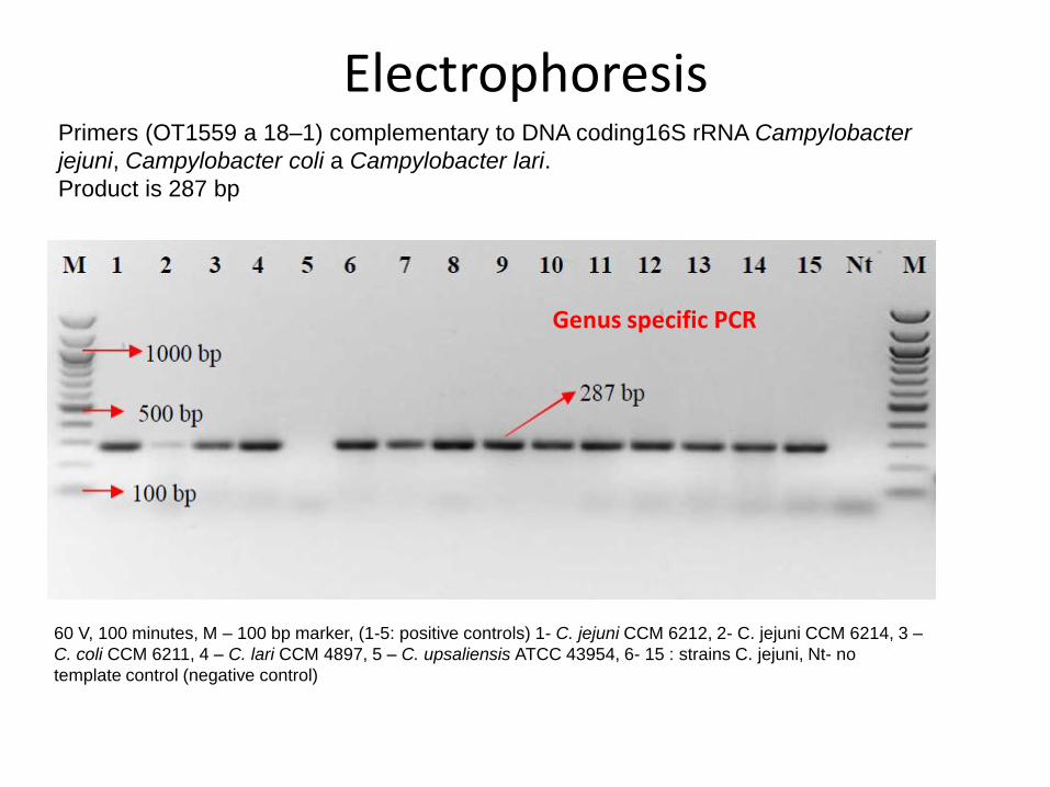
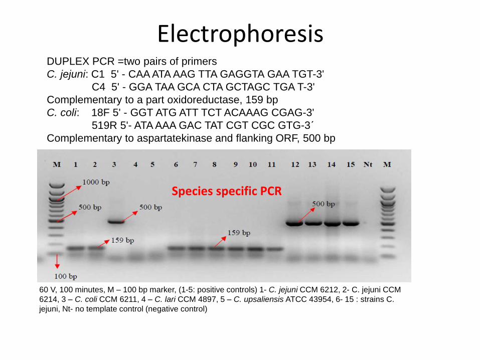
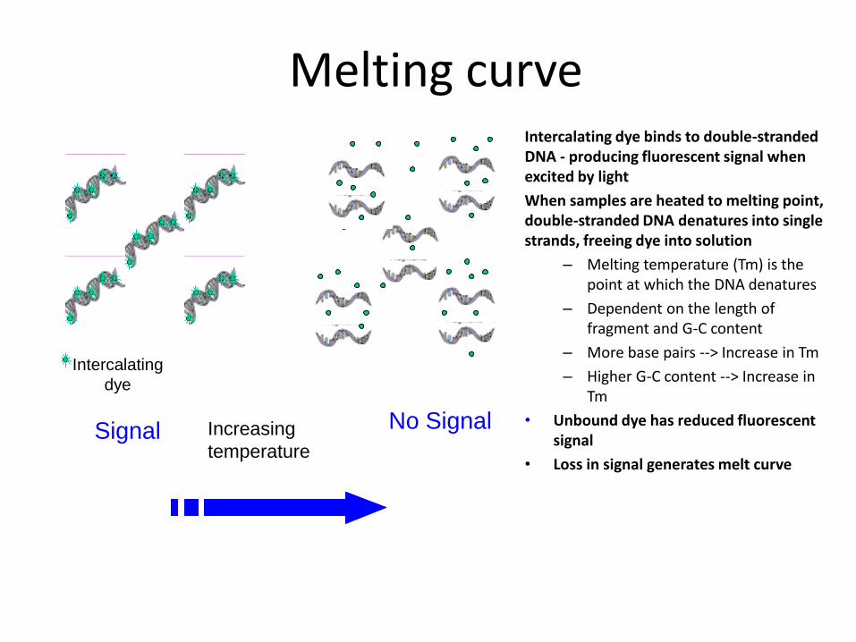
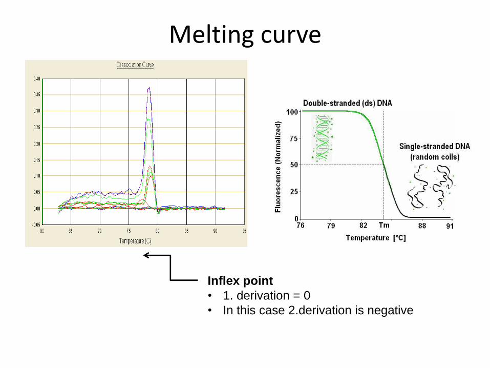
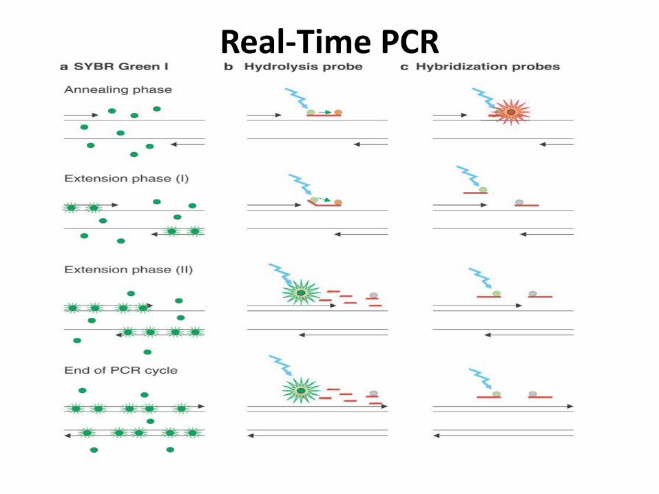
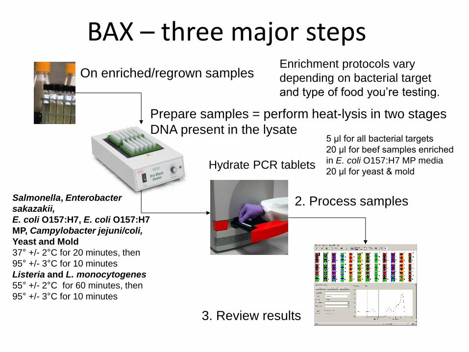
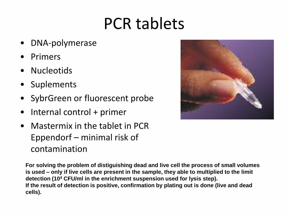
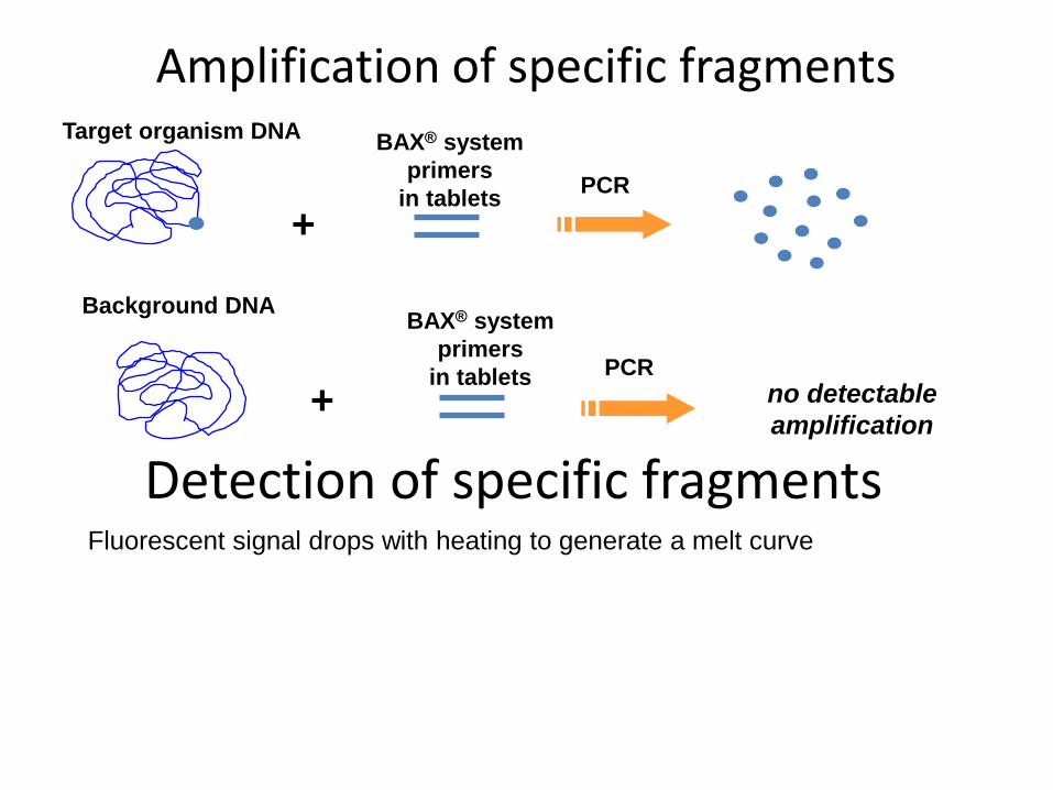
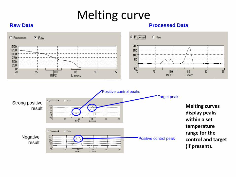
ATP bioluminiscence
httpwwwpromegacouk~mediaimagesresourcesfiguresenotesfe0027_fig1jpgla=en
httpwwwbiotekptassetstech_resources143clarity_atp_conc_fig2gif
Principle the detection of the present level of ATP (direct proportion to the presence of microorganisms or their products residues) The enzyme luciferase catalyzes the transformation of d-luciferin to oxyluciferin (indicated by emision of light at 560 nm)
ATP bioluminiscence
httpwwwbioxyscomimages2figure-B11gif
ATP Hygiene Monitoring System - SystemSURE II
httpwwwmicromaticcomdraft-keg-beerline-cleaning-hygiene-pid-ATP-100-tab-reviewshtml
Biosensors
60
httpsuploadwikimediaorgwikipediacommonseecBiosensor_Systemjpg
Biosenzor =the whole cellmicroorganism (CBS ndash cell-based sensors)
Cell-based sensors
Patogenic microorganisms interact during the infection with mammalian
cells ndash detection of cell or toxin by cell culture The damage of cell culture is measured optically or electrically
62
IDENTIFICATION OF MICROORGANISMS
Definition of species in bacteriaarchae
The criterion for a bacterial or archael species a collection of strains that have bull at least one diagnostic phenotypic trait bull ge70 DNAndashDNA hybridization of their purified molecules DNA
bull ~more than 97 16S rRNA sequence identity Under the same conditions some eucaryotic order would be a single bacterial species
The bacterial species definition bull eminent practical significance bull but a very difficult issue
bull very wide and often changes in DNA
bull mutations during replication of DNA
bull horizontal gene transfer through mobile elements plasmids also between species
Criterion for species in prokaryotic cells (bacteria and archea) Collection of strains characterised bull by at least one diagnostic feature bull their purified DNA molecules prove at least 70 hybridisation
which corresponds to at least 97 concordance in the sequence of 16S rRNA gene
Map of genome of B cereus ATCC 14579
httpwwwacgtinccomspecialty_dna_sequencinghtm
Identification based on genotype
64
DNA isolation PCR amplification of 16S rRNA gene (usually)
Sequencing of PCR product
Comparison to sequences in databases (BLAST)
Identification of bacteria PHENOTYPE METHODS bull Morphology of colony and cell (Gram staining shape) bull Requirements for temperature atmosphere nutrition bull Biochemical tests - presence of specific enzymes
bull catalase oxidase fermentation of glucose bull the way how they utilize different compounds
bull Immunochemical tests - specific antigens in the cell wall METHODS BASED ON PROTEIN ANALYSIS
bull MALDI-TOF MS METHODS BASED ON GENOTYPE bull looking for and comparing of specific DNA sequences
bull conserved universal genes (eg 16S rRNA) bull genes specific for genus species
bull done by PCR methods DNA chips sequencing etc
66
BIOCHEMICAL TESTING
Identification based on phenotype I Physiological + biochemical features
bull morfology of colonies and cells (Gram staining shape)
bull requirements for temperature atmosphere nutrient sources
bull basic biochemical tests oxidase catalase fermentationoxidation of glucose haemolysis etc
appearance of colonies on selective or selective-diagnostic agars (for other testing to be reinoculated on non-selective agar)
the classification of an isolate into a certain group
(eg for bacteria catalase positivenegative G+ cocci glucose fermentingnon-fermenting G- rods etc)
biochemical tests specific for certain groups of microorganisms
Identification based on phenotype I
68
Physiological + biochemical features Example Bacillus cereus
httpwwwfoodhaccpcommemberonlynewsletter164html
httpwwwnapavalleyedupeoplesrosePublishingImagesBacillus20cereus202(Gram20stain)jpg
Set of biochemical tests Microgenreg Bacillus ID Panel for the identification of Bacillus spp and related genera (time for performing 48 hours)
MYP agar (Mannitol Egg Yolk Polymyxin Agar) pink colonies with a precipitation zone (no fermentation of manitol + enzyme lecitinase)
catalase positive
httpwwwoxoidcomukblueprod_detailprod_detailasppr=CM0929ampc=ukamplang=ENamporg=9ampimg=CM0929ampsec=
G+ rod endspores forming
β-haemolysis moisty and easy to grow colonies
Principle of biochemical testing
to rank the bacterium to some group having the same basic features bull eg catalase positive gram positive cocci bull Family Enterobacteriaceae
the identification on the genus or species level
bull sets of biochemical tests specific for some group of bacteria
70
Gram staining procedure - principle
https40mediatumblrcom25da2e5b25bbe336819bf1715849d171tumblr_inline_ntrsgn9w1D1sv92o6_540jpg
Gram staining procedure - principle
httpsonlinesciencepsuedusitesdefaultfilesmicrb106CellStructurecow02354_03_15jpg
Gram staining procedure 1-2 drops of sterile water on a slide Spread culture in thin filter in this drop Let dry in airclose to fire Pass slide through flame 2-3x to fix Add crystal violet ndash 15-20 s to act Add Lugol solution (KII2) ndash 15-20 s to act Washing off with a mixture of
ethanolaceton (41) or acetone till the stain removes but max 15 s
Rinse with water decant excess liquid Add safranin 1 minute Rinse with water decant excess liquid Dry and put a drop of imersion oil use an
objective bdquoOILldquo
httpwwwtumblrcomtaggedgram-staining
G- red
G+ violet
Gram staining procedure + Identification
E coli
Family Enterobacteriaceae Gram-negative rods
Micrococcus luteus
Staphylococcus aureus
Gram-positive cocci Bacillus cereus
Gram-positive rods (with endospores)
Biochemical identification of G + bacteria
httpyoungeneticistblogspotcz201106diagnostic-microbiology-labhtml
G+ bacteria bull cocci ndash in clusters in pairs
diplococci bull bacilli
bull endospore bull no endospore
For G+ cocci bull Catalase positive ndash Staphylococcus
sp bull Coagulase + S aureus or some other few species bull Coagulase -
bull Novobiocin S bull S epidermidis etc
bull Novobiocin R bull S saprophyticus etc
bull Catalase negative Streptococcus sp Enterococcus sp
bull kind of haemolysis Other important G + group ndash filamentous bacteria
Catalase test Catalase test Catalase is a common enzyme found in nearly all living organisms that are exposed to oxygen where it catalyzes the decomposition of hydrogen peroxide to water and oxygen
2 H2O + O2 2 H2O2
catalase
bubbling when culture is add in peroxide
httpiws2collinedudcainCCCCD20Microcatalase_testhtm
Catalase testing into a drop of 3 peroxide to transfer a loop of bacterial culture catalase positive ndash releasing of air bubbles
Examples bull G+ cocci catalase positive Staphylococcus sp Micrococcus sp Kocuria sp Dermacoccus sp Macrococcus sp Rothia sp catalase negative Streptococcus sp Enterococcus sp bull G+ rods (bacilli) endospores forming catalase positive Bacillus sp catalase negative Clostridium sp
Resistance to ATBhaemolysis Resistance eg to novobiocin
httpquizletcom20077004mic-lecture-1-t2-flash-cards
Hemolysis on blood agar
Measuring the diameter of inhibition zone (less than 17 mm resistant more or equal to 17 mm sensitive) ndash differing in Staphylococcus sp (Clinical performance)
alpha-hemolysis - oxidizing the iron in the hemoglobin - green discoloration of the medium around beta-hemolysis - rupture of erythrocytes - complete clearing medium around gamma-hemolysis ndash no effect ndash no change
By cultivation on blood agar bacteria can be
differentiated based on their capacity to secrete
hemolysins The hemolysis will cause a clearing zone of
the blood agar around the colonies Bacteria can cause
different types of hemolysis
bullα-hemolysis which means an incomplete clearing
(green haemolysis)
bullβ-hemolysis which means a complete clearing
bullDouble hemolysis of some staphylococci consisting of
an inner β-hemolysis zone and an outer α-hemolysis
zone
bullNo hemolysis which is sometimes referred to as γ-
hemolysis which may seem illogical
Note that the α-hemolysin of staphylococci causes
complete hemolysis whereas their β-hemolysin causes
incomplete hemolysis
The capacity to produce hemolysins may vary between
different strains of a particular bacterial species
httpwwwvetbactorgvetbactdisplayextinfo=67
Identification of bacteria based on haemolysis
Streptococcal hemolysins α (alpha) hemolysis - ldquozone of green discolourationrdquo or ldquogreeningrdquo called partial or incomplete hemolysis produced by alpha-hemolytic streptococci such as Streptococcus suis This type of hemolysis on BA is due to bacterial production of hydrogen peroxide which oxidises hemoglobin to methemoglobin β (beta) hemolysis ndash a clear zone of complete hemolysis produced by beta-hemolytic streptococci such as S equi subsp equi This hemolysis is due to lysis of RBCs in the BA by the cytotoxin Streptolysin S
Source httppeopleupeicajlewishtmlhemolysinshtml
Bacterial hemolysins
Staphylococcal hemolysins Most coagulase-positive staphylococci (CPS) such as Staphylococcus aureus S pseudintermedius ( but not S hycius) typically produce an distinctive pattern of ldquodouble zonerdquo or ldquotargetrdquo hemolysis on blood agar Double-zone hemolysis indicates that the two hemolysins one which produces ldquocompleterdquo and another one that produces ldquoincompleterdquo hemolysis are present and this combined effect is visible due to different diffusion rates of these enzymes through the agar The staphylococcal α (alpha) hemolysin causes a complete type of hemolysis This zone of complete hemolysis is narrow and immediately adjacent to the bacterial colony on blood agar The staphylococcal β (beta) hemolysin causes an incomplete type of hemolysis at 37 degC This broader incomplete zone is outside of the inner complete zone and can be hard to see on early culture plates and also varies with the species of RBCs used to make the BA plate Its action is enhanced by chilling at 4 degC and the hemolysis becomes complete (this effect is referred to as ldquohot-coldrdquohemolysis) There are other staphylococcal cytotoxins that are also hemolysins but their lytic actions are not seen on blood agar
The confusion arises due to the NAMES of the two STAPHYLOCOCCAL HEMOLYSINS and the TYPE of hemolysis they produce These are the REVERSE of the two TYPES of STREPTOCOCCAL HEMOLYSIS Strictly speaking the descriptive terms ldquoalphardquo for ldquoincompleterdquo and ldquobetardquo for ldquocompleterdquo hemolysis apply only to the hemolytic toxins of Streptococcus species However other organisms beside Streptococcus and Staphylococcus species have cytotoxins that cause hemolysis and this hemolysis can be either complete or incomplete It is preferable to use the terms ldquocompleterdquo and ldquoincompleterdquo rather than ldquobetardquo or ldquoalphardquo to describe the hemolysis caused by these organisms but you will notice in bacteriology text books that the terms alpha and beta are commonly used
G ndash bacteria bull Cocci bull Bacilli (Rods) bull Oxidase positivenegative bull Glucose metabolism
bull fermentation bull oxidation bull Inactive
bull Lactose metabolism for coliform
bull Pigment formation for Pseudomonas sp
Biochemical identification of
G - bacteria
Oxidase test
Oxidase positive (OXI +) Aeromonas sp Pseudomonas sp Dark-blue to violet
Oxidase negative (OXI -) Enterobacteriaceae Bacillus sp Colourless
Oxidase bullabbreviated general name for some kind of cytochrome c oxidases (cytochrome oxidase or indophenol oxidase) enzymes involved in aerobic respiratory chain when oxygen is used as the final receptor for hydrogen bullOxidases are usually present only in aerobic organisms bullPresence or absence of oxidase(s) is an important feature of the bacterial metabolism used mainly in the identification process of gram-negative bacteria
Detection Reaction of the culture with redox indicator as eg NN-dimethyl-p-phenylenediamine (DMPD) dark-blue when oxidized and colorless when reduced Different test performances are possible
Fermentation of carbohydrates Type of utilisation of glucose or carbohydrates bull an important feature of the bacterial metabolism bull widely used in the identification process of bacteria bull fermentation strictly means to be under anaerobic conditions (but in bacterial
metabolism which can be but not have to be the same as the anaerobic conditions of cultivation) so very often is used in a general way like bdquoutilisationldquo
bull fermentation (like bdquoutilisationldquo) of carbohydrates to
bull acid and gas (Durham tubes) bull acid without gas bull non-utilised
bull Testing of fermentation or oxidation of carbohydrates
bull eg OF test for glucose bull by oxidation under aerobic conditions bull by fermenation under anaerobic conditions (an oil layer)
Fermentation of sugars to acid
Peptone Water wPhenol Red
(HiMedia M028I)
Ingredients gl
Peptic digest of animal tissue 10000
Sodium chloride 5000
Phenol red 0020
Final pH ( at 25degC) 68plusmn02
httpsuploadwikimediaorgwikipediacommons009Phenol_red_pH_60_-_80jpg
+ after sterilisation
at the room
temperature to add
aseptically sugar
discs and
inoculate
succrose trehalose rhamnose xylose
Incubation 30 degC 24 h
- +
Fermentation of carbohydrates
Casein enzymic hydrolysate and yeast extract provide nitrogenous compounds vitamin B complex and other growth nutrients Dextrose is the fermentable carbohydrate and bromocresol purple acts as the pH indicator with colour change from purple to yellow in acidic conditions Sodium chloride maintains osmotic equilibrium bdquoAnaerobicldquo conditions ndash inoculation by stabbing to used fresh prepared media (oxygen expelled by heating medium in boiling water or running steam for 15 min then to be cooled down quickly)
- +
Enterobacteriaceae ndash glucose fermentation
83
Tryptone Yeast Extract Agar Ingredients Gms Litre Casein enzymic hydrolysate 10000 Yeast extract 1500 Dextrose 10000 Sodium chloride 5000 Bromocresol purple 0015 Agar 15000 Final pH ( at 25degC) 70plusmn02
Test for oxidationfermentation of glucose (OF glucose test) detection of acid production by acidobazic indicator change to yellow (acid present) in lower pH from green (no acid) Anaerobic atmosph ndash covering by sterile mineral oil If fermented anaerobically then also runs aerobically
Fermentation of carbohydrates
Principle of biochemical tests bull The first step in identification is to rank the bacterium to some group of bacteria
having the same basic features ndash eg catalase positive gram positive cocci bull The identification on the genus or species level is finished by other biochemical tests
specific for each group of bacteria bull The tests can be done in tubes or miniaturized plate
ENTEROtest 24N (Erba Lachema) (24 means 24 biochemical tests N= new)
API 20E (bioMeacuterieux)
bull designed for routine identification of clinically important species of family Enterobacteriaceae without the use of reagents
bull A strip test OXItest or OFtest in microtitration wells can be used to confirm that the isolate belongs to Enterobacteriaceae family
bull The results are read visually or automatically using reader MIKROLAreg
bull The identification can be supplemented by diagnostic strip tests INDOLtest COLItest and VPtest
APIreg Gram negative Identification API 20E ndash Speciessubspecies identification of Enterobacteriaceae and groupspecies identification of non-fermenting Gram negative bacteria API Rapid 20E ndash 4-hour identification of Enterobacteriaceae API 20NE ndash 24 to 48-hour identification of Gram negative non-Enterobacteriaceae API Campy ndash 24-hour identification of Campylobacter species
Example Escherichia coli
ENTEROtest 24 N
Biochemical testing - Enterobacteriaceae bull TSI Triple Sugar Iron Agar (use a straight wire to stab the butt and streak the agar surface)
- to determine whether organisms can ferment glucose sucrose andor lactose with or without production of gas and the ability of the organism to produce hydrogen sulphide from thiosulphate in an acid environment is also tested
Formula Liter Enzymatic Digest of Casein 5 g Enzymatic Digest of Animal Tissue 5 g Yeast Enriched Peptone 10 g Dextrose 1 g Lactose 10 g Sucrose 10 g Ferric Ammonium Citrate 02 g Sodium Chloride 5 g Sodium Thiosulfate 03 g Phenol Red 0025 g Agar 135 g Final pH 73 plusmn 02 at 25 degC
inoculation in the butt by stab inoculation on the slant on the surface bull Fermentation of dextrose
(glucose) to acid bull anaerobically - yellow butt bull aerobically leads to products
which are later oxidised by air and pH remains alkaline (red) bull Fermentation of glucose to gas (anaerobically - in the stab) Cracks splits or bubbles in medium indicate gas production bull Production of hydrogen sulfide (H2S) ndash reaction with ferrous sulphate and sodium thiosulphate - a black precipitate in butt bull Lactose andor succrose
utilisation to acid Acid production ndash yellow butt and slant
Results
An alkaline slant-acid butt
(redyellow) - fermentation of
dextrose only
An acid slant-acid butt
(yellowyellow) - fermentation of
dextrose lactose andor sucrose
An alkaline slant-alkaline butt
(redred) - dextrose or lactose not fermented (non-fermenter)
Bacterium fermenting lactose andor succrose are able to ferment glucose
Phenol red ndash alkaline = red acid = yellow
bull TSI Triple Sugar Iron Agar (use a straight wire to stab the butt and streak the agar surface)
- to determine whether organisms can ferment glucose sucrose andor lactose with or without production of gas and the ability of the organism to produce hydrogen sulphide from thiosulphate in an acid environment is also tested
Biochemical testing - Enterobacteriaceae
Urea agar (Christensen) (use a 1 μl loop full)
- to determine whether an organism can split urea into ammonia and
carbon dioxide by the action of the enzyme urease
httpimageslidesharecdncomlab17-141201085153-conversion-gate0195-10-638jpgcb=1417424219
httpimageslidesharecdncomhelicobacterpylori-copy-110516201901-phpapp0195campylobacter-jejunihelicobacter-pylori-39-728jpgcb=1305577391
Phenol red
Medium is acidic - yellow
(pH 68plusmn02)
Alkaline reaction by urea hydrolysis - pink colour
Composition Gms Litre
Peptic digest of animal tissue
1000
Dextrose 1000
Sodium chloride 5000
Disodium phosphate 1200
Monopotassium phosphate 0800
Phenol red 0012
Agar 15000
Final pH ( at 25degC) 68plusmn02
Nutrition
Peptic digest of animal tissues dextrose
Sodium chloride - osmotic equilibrium
Phosphates - buffering of the medium
35-37 degC
Biochemical testing - Enterobacteriaceae
bull L-lysine decarboxylase (use a 1 μl loop full)
- to determine whether an organism can decarboxylate an amino acid (lysine) leading to formation of an
amine After inoculation both tubes are overlayed with sterile paraffin oil
httpfce-studynetdna-sslcomimagesupload-flashcards10295302332621_mjpg
+ CO2 + OH-
Lysine Decarboxylase Broth Typical Composition (glitre) Peptic digest of animal tissue 5000 Yeast extract 3000Dextrose 1000 (glucose prepared from starch chemically equivalent to glucose) L-Lysine hydrochloride 5000 Bromocresol purple 0020 Final pH ( at 25 degC) 68plusmn02 Inoculate below the surface cover by sterile paraffin oil
Two steps detection 1 step Dextrose is fermented ndash H+ is released Acidic conditions bull bromocresol purple changes colour to yellow bull in acidic conditions lysine decarboxylase is
activated 2 step L-lysine is decarboxylated and OH- released Alkaline conditions bull bromocresol purple changes colour back to
purple
Biochemical testing - Enterobacteriaceae
bull β-galactosidase (ONPG) (use a 1 μl loop full)
- to determine whether an organism has the enzyme szlig-galactosidase by using the compound o-
nitrophenyl-beta-D-galactopyranoside (ONPG)
- this test is used to differentiate between lactose-negative and lactose-delayed organisms
- ONPG is hydrolysed by the enzyme to o-nitrophenol which is yellow and galactose
O-Nitrophenyl-β-D-galactopyranoside (ONPG) is structurally similar to lactose (ie ONPG is an analog of lactose) except that orthonitrophenyl has been substituted for glucose ONPG is colorless compound O-nitrophenol is yellow visual evidence of hydrolysis
Lactose fermenting bacteria (ONPG positive) two enzymes Permease ndash transferring of the lactose molecule into the bacterial cell β-galactosidase - cleaving the galactoside bond producing glucose and galactose - inducible enzyme - made ONLY in the presence of the lactose substrate Coliform bacteria as Ecoli Klebsiella spp Enterobacter spp
hydrolysis
Late lactose fermentering bacteria Permease ndash not present delayed in the production of acid from lactose because of sluggish permease activity β-galactosidase ndash present - give a positive ONPG test Citrobacter spp Arizona spp
Non lactose fermenter (ONPG Negative) Permease ndash not present β-galactosidase ndash not present Salmonella spp Shigella spp Proteus spp Providencia spp and Morganella spp
Biochemical testing - Enterobacteriaceae
Procedure Culture Bacteria grown in medium containing lactose (to induce the production of the galactosidase enzyme) such as Kligler iron agar (KIA) or Triple sugar Iron (TSI) agar (Note β-galactosidase enzyme (inducible enzyme) is made ONLY in the presence of the lactose substrate) Inoculation 1A loopful of bacterial growth is emulsified in 005mL of physiologic saline to produce a heavy suspension 2One drop of toluene is added to the suspension and vigorously mixed for a few seconds to release the enzyme for bacterial cells 3An equal quantity of buffered ONPG solution is added to the suspension 4The mixture is placed in a 37 oC water bath 5When Using ONPG Tablets 6A loopful of bacterial suspension is added directly to the ONPG substrate resulting from adding 1mL of distilled water to a tablet in a test tube 7This suspension is also placed in a 37oC water bath
β-galactosidase (ONPG)
Results and Interpretations The rate of hydrolysis of ONPG to o-nitrophenol may be rapid for some organisms producing a visible yellow color reaction within 5 to 10 minutes Most tests are positive within 1 hour however reactions should not be interpreted as negative before 24 hours of incubation The yellow color is usually distinct and indicates that the organism has produced o-nitrophenol from the ONPG substrate through the action of β-galactosidase
Biochemical testing - Enterobacteriaceae
bull VogesndashProskauer (VP) test (use a 1 μl loop full)
- to determine whether an organism can produce acetylmethylcarbinol
(acetoin) from fermentation of glucose If present acetylmethyl carbinol is
converted to diacetyl in the presence of prop- naphthol (to be added first) strong
alkali (40 KOH) and atmospheric oxygen The diacetyl and quanidine-
containing compounds found in the peptones of the broth then condense to
form a pinkish red polymer
MRVP broth (pH 69)
Ingredients per liter of
deionized water
buffered peptone= 70 gm
glucose= 50 gm
dipotassium phosphate= 50 gm Voges-Proskauer Reagent A Barrittrsquos reagent A
Alpha-Naphthol 5 50 gm
Absolute Ethanol 1000
ml
Voges-Proskauer Reagent B Barrittrsquos reagent B
Potassium Hydroxide 400 gm
Deionized Water 1000 ml
Procedure of VogesndashProskauer (VP) Test
1Prior to inoculation allow medium to
equilibrate to room temperature
2Using organisms taken from an 18-24
hour pure culture lightly inoculate the
medium
3Incubate aerobically at 37 degC 24 hours
4Following 24 hours of incubation aliquot
2 ml of the broth to a clean test tube
5Re-incubate the remaining broth for an
additional 24 hours
6Add 6 drops of 5 alpha-naphthol and
mix well to aerate
7Add 2 drops of 40 potassium hydroxide
and mix well to aerate
8Observe for a pink-red color at the surface
within 30 min Shake the tube vigorously
during the 30-min period
Positive Reaction
A pink-red color at the surface
Examples Viridans group streptococci
(except Streptococcus vestibularis) Listeria
Enterobacter Klebsiella Serratia marcescens
Hafnia alvei Vibrio eltor Vibrio alginolyticus etc
Negative Reaction
A lack of a pink-red color
Examples Streptococcus mitis Citrobacter sp
Shigella Yersinia Edwardsiella Salmonella Vibrio
furnissii Vibrio fluvialis Vibrio vulnificus and Vibrio
parahaemolyticus etc
A copper color should be considered negative A
rust color is a weak positive reaction
Quality Control of VogesndashProskauer (VP) Test
VP positive Enterobacter aerogenes (ATCC13048)
VP negative Escherichia coli (ATCC25922)
Biochemical testing - Enterobacteriaceae
94
VogesndashProskauer (VP) test
Rapid test for confirmation of E coli ndash typical features
β-D-glucuronidase (97 strains) production of indole from tryptophan (typical for E coli)
Performance
Previous testing Gram-negative (eg able to grow on VRBD agar) oxidase negative
From pure culture to prepare suspension in saline solution (app 5 ml) at the density 3 McFarland
To transfer 05 ndash 1 ml of this suspension into a new tube ndash to add the strip of COLItest (chemicals ndash fluorogenic substrate for β-D-glucuronidase tryptophane are present in the pad at the end of the strip)
Incubation 37 degC 4 hours
Detecion of β-D-glucuronidase (to be done at first) UV (360 nm) ndash blue fluorescence (GLR + hellipβ-D-glucuronidase positive) ndash GLR test can be read after 1 hour
Detection of indole formation from tryptophane (to be done at second) to add Kovacs reagent (IND + hellipformation of red ring)
95
COLItest - Escherichia coli
Biochemical testing - Enterobacteriaceae
Fluorogenic culture medium
bull One or more fluorogenic substrates
bull For detection of specific enzyme which are able to cleave out the fluorogenic substrate that a designed
fluorophore (responsible for the UV fluorescence) is released
FLUOROGENIC MEDIUM (Colilert) E coli use β-glucuronidase to metabolize MUG and create fluorescence (UV 360 nm) Since most non-coliforms do not have these enzymes they are unable to grow and interfere
httpgawaterusgsgovprojectsbacteriapicturessummaryanalysisecolijpg
E coli (MPN method)
COLItest Biochemical testing - Enterobacteriaceae
COLItest bull Indole test (use a 1 μl loop full)
- to determine whether an organism can split indole from tryptophan
httpwwwmicrobiologyinfocomindole-test-principle-reagents-procedure-result-interpretation-and-limitations
indole production from tryptophan
Production of indole is detected by Kovacacutes reagent
Indole Kovacs Reagent
p-Dimethylaminobenzaldehyde 500 gm
Hydrochloric Acid 37 2500 ml
Amyl Alcohol 7500 ml
Biochemical testing - Enterobacteriaceae
INDOLe test ndash quick variant
Ecoli Citrobacter Enterobacter
Procedure bull on the filtration paper to add a drop of INDOL suspension bull to spread into by a loop some culture bullTo incubate ndash at the room temperature for 5 minutes bullTo read results
Biochemical testing - Enterobacteriaceae
ENTEROtest 24 ndash example of biochemical testing
routine identification of clinically important species of family Enterobacteriaceae the test is focused only on some species often isolated in clinical practis strains from other sources can be slightly different in the dominant biochemical profiles intended exclusively for the identification of family Enterobacteriaceae the preliminary ranking into family Enterobacteriaceae must be done (Gram staining oxidase glucose fermentation) the results of reactions obtained in this modified micromethod may differ from the results obtained using conventional tests and results available in literature sources in case of problem to control positivenegative reactions by control strains or to use for this tests tube variant to check the functionality of the kit not to check the accuracy or success of the identification
Preparation of suspension from over-night culture growing on non-selective agar (for staphylococci Columbia agar blood with 5 of sheep blood is recommended)
Inoculation of microtitre plates anaerobic reaction covered by mineral oil
Incubation
Reading results by both visual and instrumental mode respectively Standard off-line tests INDOLtest or COLItest Other off-line tests OXItest OFtest VPtest Acetoin test
ENTEROtest 24 N ndash example of biochemical testing
bull tube with 5 ml of sterile physiolsol bull suspension (1 McFarland) bull application 100 microl to each hole bull 2 drops of parafin oil into holes D-H of the first line (anaerobic conditions) bull Incubation 37degC 24h bull for check the purity of suspension to streak suspension on TSA (incubation 37 degC ndash 24 h
ENTEROtest 24N
ENTEROtest 24 N - evaluation
Escherichia coli
H 0+0+0=0 G 0+2+0=2 F 1+2+0=3 hellip Identification code 023426
1 1
2
4
ENTEROtest 24 ndash example of biochemical testing
The result = a set of biochemical properties (+-) which is compared to the sets of biochemical properties of different chosen genera and species for them the test is designed
ENTEROtest 24 N
T-index how is the likelihood of the different properties The combination of identication and T-index = the reliability of the result Excelent very good good (this case) acceptable intermediary strain needs some additional tests genus level non-existing taxon
Evaluation by using the identification code - By using a code book - By using software TNW
Identification score the measure of similarity to proposed taxon
The way of identification the obtained profile is compared with profiles in database bull The most similar profiles are found
bull Identification score = of similarity bull But what about the different reactions
bull T-index is calculated bull how is the probability of the different reactions
bull It depends on their importance ndash typical reactions should be the same
bull Reliability of the result bull the combination of identication score and T-index = bull Excelent very good good acceptable intermediary strain needs some
additional tests genus level non-existing taxon
ENTEROtest 24 ndash example of biochemical testing
Biochemical tests - divisions
ENTEROtest 24 species of family Enterobacteriaceae
NEFERMtest 24 Gramnegative non-fermenting bacteria and families Vibrionaceae Aeromonadaceae and Pleiomonas shigelloides present mainly in clinical material
ANAEROtest 23 ANAEROBIC BACTERIA (G + AND G-) routine identification of anaerobic bacteria (G + AND G-) in clinical material and in food
STREPTOtest 24 Streptococcus Enterococcus and related gram positive catalase negative cocci isolated from clinical material
STAPHYtest 24 genus Staphylococcus isolated from clinical material and for their distiguishing from related genera of other gram positive catalase positive cocci
Biochemical tests APIreg Gram positive Identification API Staph ndash Overnight identification of clinical staphylococci and micrococci API 20 Strep ndash 4 or 24-hour identification of streptococci and enterococci API Coryne ndash 24-hour identification of Corynebacteria and coryne-like organisms API Listeria ndash 24-hour identification of all Listeria species APIreg Gram negative Identification API 20E ndash Speciessubspecies identification of Enterobacteriaceae and groupspecies identification of non-fermenting Gram negative bacteria API Rapid 20E ndash 4-hour identification of Enterobacteriaceae API 20NE ndash 24 to 48-hour identification of Gram negative non-Enterobacteriaceae API Campy ndash 24-hour identification of Campylobacter species APIreg Anaerobe Identification API 20A ndash 24-hour identification of anaerobes Rapid ID 32 A ndash 4-hour identification of anaerobes
MicrogenTM GN-ID Convenient 12 substrate identification system for commonly encountered Enterobacteriaceae (GNA) Extended 24 substrate identification system for the complete range of Enterobacteriacea and oxidase positive non-fastidious Gram negative bacilli (GNA+B) MicrogenTM Listeria-ID Enhanced built-in haemolysis test MicrogenTM Bacillus-ID 24 substrate system for the identification of mesophilic Bacillus spp isolated from foods and related samples MicrogenTM Staph-ID 12 substrate system minimizes handling and manipulation Substrates selected specifically for Staphylococcus and related organisms MicrogenTM Strep-ID Substrates selected specifically for Streptococcus Enterococcus and related species Provides excellent differentiation of Enterococci spp especially E casseliflavus E mundtii and E gallinarum
Biochemical tests
Identification of microorganisms by MALDI-TOF MS
Department of Biochemistry and Microbiology University of Chemistry and Technology Prague
Sabina Purkrtovaacute1 Petra Junkovaacute2 1Laboratory of food microbiology 2Laboratory of applied proteomics
SabinaPurkrtovavschtcz PetraJunkovavschtcz
MALDI-TOF MS
1) sample preparation bull microbial culture or its
proteins extract is smearing onto a steel plate and covered over by matrix
bull matrix enables the sample (A) to be desorped and ionised as pseudomolecule ionts [A+H] +
2) MALDI-TOF MS analysis unique mass spectral fingerprint of desorbed microbial cell components (mainly intracellular proteins) different among genera species or also some strains
Matrix-Assisted Laser Desorption Ionization-Time of Flight Mass Spectrometry
httppubsrscorgencontentarticlehtml2014RAC4RA05604C
httpcmrasmorgcontent263547figures-only
3) identification comparison of mass spectrum to those of reference strains in database
MALDI-TOF MS PRINCIPLE
111
2
2
2L
teU
z
m
m mass z charge
L length of drifting zone e
elementary charge U
speeding voltage
Time of flight is a function of the specific ion mass (mz)
httpwwwsigmaaldrichcomtechnical-documentsarticlesbiologycustom-dna-oligos-qc-analysis-by-mass-spectrometryhtml
Soft ionisation method bull low level of sample fragmentation
Animation http cmgmstanfordedupansection_htmlMS
MALDI-TOF MS SAMPLE AND MATRIX
httpcmrasmorgcontent263547figures-only
Matrix bull able to absorb the energy of the laser (usually 337 nm) bull to crystalise with samples - usually acid
character (proton ionisation of sample) dissolved in organic solvent
Sample preparation Direct transfer bull intact cells spread directly onto a steel plate bull lysis of cells occurs during the contact with acid
matrix and by laser desorption bull most bacteria Proteins extraction bull previous extraction of proteins by organic acids
andor alcohol (eg ethanol and 70 formic acid) bull yeasts moulds some species of bacteria (depending
on the cell wall composition)
CHC α-Cyano-4-hydroxycinnamic acid (organic solvent 50 acetonitrile with 25 trifluoracetic acid) SA 35-Dimethoxy-4-hydroxycinnamic acid (sinapic acid)
DHB 25-Dihydroxybenzoic acid
httpwwwsigmaaldrichcomcatalogproductsigmac8982lang=enampregion=CZ
MALDI-TOF MS MICROORGANISMS
113
Mass spectrum protein profile z equals usally to 1+ (so mz usually corresponds to molecula mass) the range usually used for identification 2000 -20 000 mz the intensity of single peaks corresponds to the abundance of the protein Which proteins dominates in the protein profile abundant basic and mediumly hydrophobic mainly ribosomal proteins further cold-shock and heat-shock proteins chaperons etc
conserved house-keeping gene = conserved proteins = the acordance with identification based on DNA
Visualisation of mass spectrum protein profile ndash (software mMass 5 Strohalm et al 2010)
Bacillus cereus
Analysis is recomended (and validated) to be performed from colonies grown onto non-selective agar
MALDI-TOF MS ANALYSIS
114
Comparison of mass spectrum protein profile of unknown sample with these of reference strains present in database by software
Commercial databases from different MALDI-TOF MS producers Bruker Daltonics ndash MALDI BIOTYPER Shimadzu - Shimadzu Launchpad software + SARAMIS database Biomeacuterieux - VITEKreg MS Other databases compatible with different hardware systems (eg Andromas)
BioTyper The statistical analysis for correlation includes peak positions intensities and frequencies across the complete range of microorganisms Score value 0 (none similarity) - 1000 (absolute similarity) But it is expressed in decadic logarithm log(score value) 0-3
Range Description Symbols Color
2300 3000
highly probable species identification
( +++ ) green
2000 2299
secure genus identification probable species identification
( ++ ) green
1700 1999
probable genus identification ( + ) yellow
0000 1699
not reliable identification ( - ) red
Bacillus cereus
115
Range Description Symbols Color
2300 3000
highly probable species identification
( +++ ) green
2000 2299
secure genus identification probable species identification
( ++ ) green
1700 1999
probable genus identification ( + ) yellow
0000 1699
not reliable identification ( - ) red
Rank (Quality)
Matched Pattern Score Value
NCBI Identifier
1 ( +++ )
Bacillus cereus DSM 31T DSM 2554 1396
2 ( ++ )
Bacillus cereus 994000168 LBK 2203 1396
3 ( ++ )
Bacillus weihenstephanensis DSM 11821T DSM
2158 86662
4 ( ++ )
Bacillus mycoides DSM 2048T DSM 2155 1405
5 ( ++ )
Bacillus cereus 4080 LBK 2147 1396
6 ( + )
Bacillus thuringiensis DSM 2046T DSM 1975 1428
7 ( + )
Bacillus pseudomycoides DSM 12442T DSM 1787 64104
8 ( - )
Bacillus bataviensis DSM 15601T DSM 1369 220685
9 ( - )
Brevibacterium linens IMET 11075T HKJ 1347 1703
10 ( - )
Acinetobacter towneri DSM 14962T HAM 1345 202956
Bacillus cereus
Bacillus cereus
MALDI-TOF MS - PROCEDURE Direct method smearing sample in four parallels in lower and higher cells concentrations ndash after drying to cover over by matrix (1-2 microl) and let to crystallise at room temperature
Matrix solution of α-Cyano-4-hydroxycinnamic acid (10 mgml) in 50 acetonitrile with 25 trifluoroacetic acid (prepared with 10 TFA solution)
Protein standard (1 μl) Bruker Bacterial Test Standard (Bruker Daltonics SRN) ndash proteins extracted from z Escherichia coli DH5alpha BRL + some others
Equipment Bruker Autoflex Speed Database MALDI Biotyper 31
116
MALDI-TOF MSPROTEIN STANDARD
117
Analyte name Rank (Quality) Matched Pattern Score Value
F4 1 ( +++ ) Escherichia coli DH5alpha BRL 2439
Thank you for your attention
Questions to be sent to SabinaPurkrtovavschtcz
118
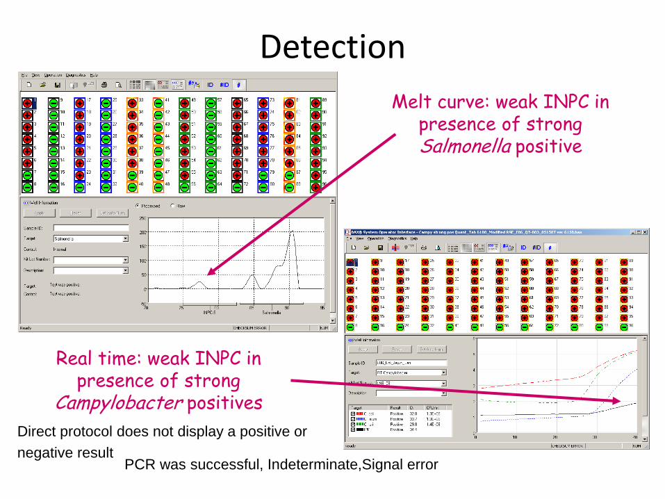
ATP bioluminiscence
httpwwwbioxyscomimages2figure-B11gif
ATP Hygiene Monitoring System - SystemSURE II
httpwwwmicromaticcomdraft-keg-beerline-cleaning-hygiene-pid-ATP-100-tab-reviewshtml
Biosensors
60
httpsuploadwikimediaorgwikipediacommonseecBiosensor_Systemjpg
Biosenzor =the whole cellmicroorganism (CBS ndash cell-based sensors)
Cell-based sensors
Patogenic microorganisms interact during the infection with mammalian
cells ndash detection of cell or toxin by cell culture The damage of cell culture is measured optically or electrically
62
IDENTIFICATION OF MICROORGANISMS
Definition of species in bacteriaarchae
The criterion for a bacterial or archael species a collection of strains that have bull at least one diagnostic phenotypic trait bull ge70 DNAndashDNA hybridization of their purified molecules DNA
bull ~more than 97 16S rRNA sequence identity Under the same conditions some eucaryotic order would be a single bacterial species
The bacterial species definition bull eminent practical significance bull but a very difficult issue
bull very wide and often changes in DNA
bull mutations during replication of DNA
bull horizontal gene transfer through mobile elements plasmids also between species
Criterion for species in prokaryotic cells (bacteria and archea) Collection of strains characterised bull by at least one diagnostic feature bull their purified DNA molecules prove at least 70 hybridisation
which corresponds to at least 97 concordance in the sequence of 16S rRNA gene
Map of genome of B cereus ATCC 14579
httpwwwacgtinccomspecialty_dna_sequencinghtm
Identification based on genotype
64
DNA isolation PCR amplification of 16S rRNA gene (usually)
Sequencing of PCR product
Comparison to sequences in databases (BLAST)
Identification of bacteria PHENOTYPE METHODS bull Morphology of colony and cell (Gram staining shape) bull Requirements for temperature atmosphere nutrition bull Biochemical tests - presence of specific enzymes
bull catalase oxidase fermentation of glucose bull the way how they utilize different compounds
bull Immunochemical tests - specific antigens in the cell wall METHODS BASED ON PROTEIN ANALYSIS
bull MALDI-TOF MS METHODS BASED ON GENOTYPE bull looking for and comparing of specific DNA sequences
bull conserved universal genes (eg 16S rRNA) bull genes specific for genus species
bull done by PCR methods DNA chips sequencing etc
66
BIOCHEMICAL TESTING
Identification based on phenotype I Physiological + biochemical features
bull morfology of colonies and cells (Gram staining shape)
bull requirements for temperature atmosphere nutrient sources
bull basic biochemical tests oxidase catalase fermentationoxidation of glucose haemolysis etc
appearance of colonies on selective or selective-diagnostic agars (for other testing to be reinoculated on non-selective agar)
the classification of an isolate into a certain group
(eg for bacteria catalase positivenegative G+ cocci glucose fermentingnon-fermenting G- rods etc)
biochemical tests specific for certain groups of microorganisms
Identification based on phenotype I
68
Physiological + biochemical features Example Bacillus cereus
httpwwwfoodhaccpcommemberonlynewsletter164html
httpwwwnapavalleyedupeoplesrosePublishingImagesBacillus20cereus202(Gram20stain)jpg
Set of biochemical tests Microgenreg Bacillus ID Panel for the identification of Bacillus spp and related genera (time for performing 48 hours)
MYP agar (Mannitol Egg Yolk Polymyxin Agar) pink colonies with a precipitation zone (no fermentation of manitol + enzyme lecitinase)
catalase positive
httpwwwoxoidcomukblueprod_detailprod_detailasppr=CM0929ampc=ukamplang=ENamporg=9ampimg=CM0929ampsec=
G+ rod endspores forming
β-haemolysis moisty and easy to grow colonies
Principle of biochemical testing
to rank the bacterium to some group having the same basic features bull eg catalase positive gram positive cocci bull Family Enterobacteriaceae
the identification on the genus or species level
bull sets of biochemical tests specific for some group of bacteria
70
Gram staining procedure - principle
https40mediatumblrcom25da2e5b25bbe336819bf1715849d171tumblr_inline_ntrsgn9w1D1sv92o6_540jpg
Gram staining procedure - principle
httpsonlinesciencepsuedusitesdefaultfilesmicrb106CellStructurecow02354_03_15jpg
Gram staining procedure 1-2 drops of sterile water on a slide Spread culture in thin filter in this drop Let dry in airclose to fire Pass slide through flame 2-3x to fix Add crystal violet ndash 15-20 s to act Add Lugol solution (KII2) ndash 15-20 s to act Washing off with a mixture of
ethanolaceton (41) or acetone till the stain removes but max 15 s
Rinse with water decant excess liquid Add safranin 1 minute Rinse with water decant excess liquid Dry and put a drop of imersion oil use an
objective bdquoOILldquo
httpwwwtumblrcomtaggedgram-staining
G- red
G+ violet
Gram staining procedure + Identification
E coli
Family Enterobacteriaceae Gram-negative rods
Micrococcus luteus
Staphylococcus aureus
Gram-positive cocci Bacillus cereus
Gram-positive rods (with endospores)
Biochemical identification of G + bacteria
httpyoungeneticistblogspotcz201106diagnostic-microbiology-labhtml
G+ bacteria bull cocci ndash in clusters in pairs
diplococci bull bacilli
bull endospore bull no endospore
For G+ cocci bull Catalase positive ndash Staphylococcus
sp bull Coagulase + S aureus or some other few species bull Coagulase -
bull Novobiocin S bull S epidermidis etc
bull Novobiocin R bull S saprophyticus etc
bull Catalase negative Streptococcus sp Enterococcus sp
bull kind of haemolysis Other important G + group ndash filamentous bacteria
Catalase test Catalase test Catalase is a common enzyme found in nearly all living organisms that are exposed to oxygen where it catalyzes the decomposition of hydrogen peroxide to water and oxygen
2 H2O + O2 2 H2O2
catalase
bubbling when culture is add in peroxide
httpiws2collinedudcainCCCCD20Microcatalase_testhtm
Catalase testing into a drop of 3 peroxide to transfer a loop of bacterial culture catalase positive ndash releasing of air bubbles
Examples bull G+ cocci catalase positive Staphylococcus sp Micrococcus sp Kocuria sp Dermacoccus sp Macrococcus sp Rothia sp catalase negative Streptococcus sp Enterococcus sp bull G+ rods (bacilli) endospores forming catalase positive Bacillus sp catalase negative Clostridium sp
Resistance to ATBhaemolysis Resistance eg to novobiocin
httpquizletcom20077004mic-lecture-1-t2-flash-cards
Hemolysis on blood agar
Measuring the diameter of inhibition zone (less than 17 mm resistant more or equal to 17 mm sensitive) ndash differing in Staphylococcus sp (Clinical performance)
alpha-hemolysis - oxidizing the iron in the hemoglobin - green discoloration of the medium around beta-hemolysis - rupture of erythrocytes - complete clearing medium around gamma-hemolysis ndash no effect ndash no change
By cultivation on blood agar bacteria can be
differentiated based on their capacity to secrete
hemolysins The hemolysis will cause a clearing zone of
the blood agar around the colonies Bacteria can cause
different types of hemolysis
bullα-hemolysis which means an incomplete clearing
(green haemolysis)
bullβ-hemolysis which means a complete clearing
bullDouble hemolysis of some staphylococci consisting of
an inner β-hemolysis zone and an outer α-hemolysis
zone
bullNo hemolysis which is sometimes referred to as γ-
hemolysis which may seem illogical
Note that the α-hemolysin of staphylococci causes
complete hemolysis whereas their β-hemolysin causes
incomplete hemolysis
The capacity to produce hemolysins may vary between
different strains of a particular bacterial species
httpwwwvetbactorgvetbactdisplayextinfo=67
Identification of bacteria based on haemolysis
Streptococcal hemolysins α (alpha) hemolysis - ldquozone of green discolourationrdquo or ldquogreeningrdquo called partial or incomplete hemolysis produced by alpha-hemolytic streptococci such as Streptococcus suis This type of hemolysis on BA is due to bacterial production of hydrogen peroxide which oxidises hemoglobin to methemoglobin β (beta) hemolysis ndash a clear zone of complete hemolysis produced by beta-hemolytic streptococci such as S equi subsp equi This hemolysis is due to lysis of RBCs in the BA by the cytotoxin Streptolysin S
Source httppeopleupeicajlewishtmlhemolysinshtml
Bacterial hemolysins
Staphylococcal hemolysins Most coagulase-positive staphylococci (CPS) such as Staphylococcus aureus S pseudintermedius ( but not S hycius) typically produce an distinctive pattern of ldquodouble zonerdquo or ldquotargetrdquo hemolysis on blood agar Double-zone hemolysis indicates that the two hemolysins one which produces ldquocompleterdquo and another one that produces ldquoincompleterdquo hemolysis are present and this combined effect is visible due to different diffusion rates of these enzymes through the agar The staphylococcal α (alpha) hemolysin causes a complete type of hemolysis This zone of complete hemolysis is narrow and immediately adjacent to the bacterial colony on blood agar The staphylococcal β (beta) hemolysin causes an incomplete type of hemolysis at 37 degC This broader incomplete zone is outside of the inner complete zone and can be hard to see on early culture plates and also varies with the species of RBCs used to make the BA plate Its action is enhanced by chilling at 4 degC and the hemolysis becomes complete (this effect is referred to as ldquohot-coldrdquohemolysis) There are other staphylococcal cytotoxins that are also hemolysins but their lytic actions are not seen on blood agar
The confusion arises due to the NAMES of the two STAPHYLOCOCCAL HEMOLYSINS and the TYPE of hemolysis they produce These are the REVERSE of the two TYPES of STREPTOCOCCAL HEMOLYSIS Strictly speaking the descriptive terms ldquoalphardquo for ldquoincompleterdquo and ldquobetardquo for ldquocompleterdquo hemolysis apply only to the hemolytic toxins of Streptococcus species However other organisms beside Streptococcus and Staphylococcus species have cytotoxins that cause hemolysis and this hemolysis can be either complete or incomplete It is preferable to use the terms ldquocompleterdquo and ldquoincompleterdquo rather than ldquobetardquo or ldquoalphardquo to describe the hemolysis caused by these organisms but you will notice in bacteriology text books that the terms alpha and beta are commonly used
G ndash bacteria bull Cocci bull Bacilli (Rods) bull Oxidase positivenegative bull Glucose metabolism
bull fermentation bull oxidation bull Inactive
bull Lactose metabolism for coliform
bull Pigment formation for Pseudomonas sp
Biochemical identification of
G - bacteria
Oxidase test
Oxidase positive (OXI +) Aeromonas sp Pseudomonas sp Dark-blue to violet
Oxidase negative (OXI -) Enterobacteriaceae Bacillus sp Colourless
Oxidase bullabbreviated general name for some kind of cytochrome c oxidases (cytochrome oxidase or indophenol oxidase) enzymes involved in aerobic respiratory chain when oxygen is used as the final receptor for hydrogen bullOxidases are usually present only in aerobic organisms bullPresence or absence of oxidase(s) is an important feature of the bacterial metabolism used mainly in the identification process of gram-negative bacteria
Detection Reaction of the culture with redox indicator as eg NN-dimethyl-p-phenylenediamine (DMPD) dark-blue when oxidized and colorless when reduced Different test performances are possible
Fermentation of carbohydrates Type of utilisation of glucose or carbohydrates bull an important feature of the bacterial metabolism bull widely used in the identification process of bacteria bull fermentation strictly means to be under anaerobic conditions (but in bacterial
metabolism which can be but not have to be the same as the anaerobic conditions of cultivation) so very often is used in a general way like bdquoutilisationldquo
bull fermentation (like bdquoutilisationldquo) of carbohydrates to
bull acid and gas (Durham tubes) bull acid without gas bull non-utilised
bull Testing of fermentation or oxidation of carbohydrates
bull eg OF test for glucose bull by oxidation under aerobic conditions bull by fermenation under anaerobic conditions (an oil layer)
Fermentation of sugars to acid
Peptone Water wPhenol Red
(HiMedia M028I)
Ingredients gl
Peptic digest of animal tissue 10000
Sodium chloride 5000
Phenol red 0020
Final pH ( at 25degC) 68plusmn02
httpsuploadwikimediaorgwikipediacommons009Phenol_red_pH_60_-_80jpg
+ after sterilisation
at the room
temperature to add
aseptically sugar
discs and
inoculate
succrose trehalose rhamnose xylose
Incubation 30 degC 24 h
- +
Fermentation of carbohydrates
Casein enzymic hydrolysate and yeast extract provide nitrogenous compounds vitamin B complex and other growth nutrients Dextrose is the fermentable carbohydrate and bromocresol purple acts as the pH indicator with colour change from purple to yellow in acidic conditions Sodium chloride maintains osmotic equilibrium bdquoAnaerobicldquo conditions ndash inoculation by stabbing to used fresh prepared media (oxygen expelled by heating medium in boiling water or running steam for 15 min then to be cooled down quickly)
- +
Enterobacteriaceae ndash glucose fermentation
83
Tryptone Yeast Extract Agar Ingredients Gms Litre Casein enzymic hydrolysate 10000 Yeast extract 1500 Dextrose 10000 Sodium chloride 5000 Bromocresol purple 0015 Agar 15000 Final pH ( at 25degC) 70plusmn02
Test for oxidationfermentation of glucose (OF glucose test) detection of acid production by acidobazic indicator change to yellow (acid present) in lower pH from green (no acid) Anaerobic atmosph ndash covering by sterile mineral oil If fermented anaerobically then also runs aerobically
Fermentation of carbohydrates
Principle of biochemical tests bull The first step in identification is to rank the bacterium to some group of bacteria
having the same basic features ndash eg catalase positive gram positive cocci bull The identification on the genus or species level is finished by other biochemical tests
specific for each group of bacteria bull The tests can be done in tubes or miniaturized plate
ENTEROtest 24N (Erba Lachema) (24 means 24 biochemical tests N= new)
API 20E (bioMeacuterieux)
bull designed for routine identification of clinically important species of family Enterobacteriaceae without the use of reagents
bull A strip test OXItest or OFtest in microtitration wells can be used to confirm that the isolate belongs to Enterobacteriaceae family
bull The results are read visually or automatically using reader MIKROLAreg
bull The identification can be supplemented by diagnostic strip tests INDOLtest COLItest and VPtest
APIreg Gram negative Identification API 20E ndash Speciessubspecies identification of Enterobacteriaceae and groupspecies identification of non-fermenting Gram negative bacteria API Rapid 20E ndash 4-hour identification of Enterobacteriaceae API 20NE ndash 24 to 48-hour identification of Gram negative non-Enterobacteriaceae API Campy ndash 24-hour identification of Campylobacter species
Example Escherichia coli
ENTEROtest 24 N
Biochemical testing - Enterobacteriaceae bull TSI Triple Sugar Iron Agar (use a straight wire to stab the butt and streak the agar surface)
- to determine whether organisms can ferment glucose sucrose andor lactose with or without production of gas and the ability of the organism to produce hydrogen sulphide from thiosulphate in an acid environment is also tested
Formula Liter Enzymatic Digest of Casein 5 g Enzymatic Digest of Animal Tissue 5 g Yeast Enriched Peptone 10 g Dextrose 1 g Lactose 10 g Sucrose 10 g Ferric Ammonium Citrate 02 g Sodium Chloride 5 g Sodium Thiosulfate 03 g Phenol Red 0025 g Agar 135 g Final pH 73 plusmn 02 at 25 degC
inoculation in the butt by stab inoculation on the slant on the surface bull Fermentation of dextrose
(glucose) to acid bull anaerobically - yellow butt bull aerobically leads to products
which are later oxidised by air and pH remains alkaline (red) bull Fermentation of glucose to gas (anaerobically - in the stab) Cracks splits or bubbles in medium indicate gas production bull Production of hydrogen sulfide (H2S) ndash reaction with ferrous sulphate and sodium thiosulphate - a black precipitate in butt bull Lactose andor succrose
utilisation to acid Acid production ndash yellow butt and slant
Results
An alkaline slant-acid butt
(redyellow) - fermentation of
dextrose only
An acid slant-acid butt
(yellowyellow) - fermentation of
dextrose lactose andor sucrose
An alkaline slant-alkaline butt
(redred) - dextrose or lactose not fermented (non-fermenter)
Bacterium fermenting lactose andor succrose are able to ferment glucose
Phenol red ndash alkaline = red acid = yellow
bull TSI Triple Sugar Iron Agar (use a straight wire to stab the butt and streak the agar surface)
- to determine whether organisms can ferment glucose sucrose andor lactose with or without production of gas and the ability of the organism to produce hydrogen sulphide from thiosulphate in an acid environment is also tested
Biochemical testing - Enterobacteriaceae
Urea agar (Christensen) (use a 1 μl loop full)
- to determine whether an organism can split urea into ammonia and
carbon dioxide by the action of the enzyme urease
httpimageslidesharecdncomlab17-141201085153-conversion-gate0195-10-638jpgcb=1417424219
httpimageslidesharecdncomhelicobacterpylori-copy-110516201901-phpapp0195campylobacter-jejunihelicobacter-pylori-39-728jpgcb=1305577391
Phenol red
Medium is acidic - yellow
(pH 68plusmn02)
Alkaline reaction by urea hydrolysis - pink colour
Composition Gms Litre
Peptic digest of animal tissue
1000
Dextrose 1000
Sodium chloride 5000
Disodium phosphate 1200
Monopotassium phosphate 0800
Phenol red 0012
Agar 15000
Final pH ( at 25degC) 68plusmn02
Nutrition
Peptic digest of animal tissues dextrose
Sodium chloride - osmotic equilibrium
Phosphates - buffering of the medium
35-37 degC
Biochemical testing - Enterobacteriaceae
bull L-lysine decarboxylase (use a 1 μl loop full)
- to determine whether an organism can decarboxylate an amino acid (lysine) leading to formation of an
amine After inoculation both tubes are overlayed with sterile paraffin oil
httpfce-studynetdna-sslcomimagesupload-flashcards10295302332621_mjpg
+ CO2 + OH-
Lysine Decarboxylase Broth Typical Composition (glitre) Peptic digest of animal tissue 5000 Yeast extract 3000Dextrose 1000 (glucose prepared from starch chemically equivalent to glucose) L-Lysine hydrochloride 5000 Bromocresol purple 0020 Final pH ( at 25 degC) 68plusmn02 Inoculate below the surface cover by sterile paraffin oil
Two steps detection 1 step Dextrose is fermented ndash H+ is released Acidic conditions bull bromocresol purple changes colour to yellow bull in acidic conditions lysine decarboxylase is
activated 2 step L-lysine is decarboxylated and OH- released Alkaline conditions bull bromocresol purple changes colour back to
purple
Biochemical testing - Enterobacteriaceae
bull β-galactosidase (ONPG) (use a 1 μl loop full)
- to determine whether an organism has the enzyme szlig-galactosidase by using the compound o-
nitrophenyl-beta-D-galactopyranoside (ONPG)
- this test is used to differentiate between lactose-negative and lactose-delayed organisms
- ONPG is hydrolysed by the enzyme to o-nitrophenol which is yellow and galactose
O-Nitrophenyl-β-D-galactopyranoside (ONPG) is structurally similar to lactose (ie ONPG is an analog of lactose) except that orthonitrophenyl has been substituted for glucose ONPG is colorless compound O-nitrophenol is yellow visual evidence of hydrolysis
Lactose fermenting bacteria (ONPG positive) two enzymes Permease ndash transferring of the lactose molecule into the bacterial cell β-galactosidase - cleaving the galactoside bond producing glucose and galactose - inducible enzyme - made ONLY in the presence of the lactose substrate Coliform bacteria as Ecoli Klebsiella spp Enterobacter spp
hydrolysis
Late lactose fermentering bacteria Permease ndash not present delayed in the production of acid from lactose because of sluggish permease activity β-galactosidase ndash present - give a positive ONPG test Citrobacter spp Arizona spp
Non lactose fermenter (ONPG Negative) Permease ndash not present β-galactosidase ndash not present Salmonella spp Shigella spp Proteus spp Providencia spp and Morganella spp
Biochemical testing - Enterobacteriaceae
Procedure Culture Bacteria grown in medium containing lactose (to induce the production of the galactosidase enzyme) such as Kligler iron agar (KIA) or Triple sugar Iron (TSI) agar (Note β-galactosidase enzyme (inducible enzyme) is made ONLY in the presence of the lactose substrate) Inoculation 1A loopful of bacterial growth is emulsified in 005mL of physiologic saline to produce a heavy suspension 2One drop of toluene is added to the suspension and vigorously mixed for a few seconds to release the enzyme for bacterial cells 3An equal quantity of buffered ONPG solution is added to the suspension 4The mixture is placed in a 37 oC water bath 5When Using ONPG Tablets 6A loopful of bacterial suspension is added directly to the ONPG substrate resulting from adding 1mL of distilled water to a tablet in a test tube 7This suspension is also placed in a 37oC water bath
β-galactosidase (ONPG)
Results and Interpretations The rate of hydrolysis of ONPG to o-nitrophenol may be rapid for some organisms producing a visible yellow color reaction within 5 to 10 minutes Most tests are positive within 1 hour however reactions should not be interpreted as negative before 24 hours of incubation The yellow color is usually distinct and indicates that the organism has produced o-nitrophenol from the ONPG substrate through the action of β-galactosidase
Biochemical testing - Enterobacteriaceae
bull VogesndashProskauer (VP) test (use a 1 μl loop full)
- to determine whether an organism can produce acetylmethylcarbinol
(acetoin) from fermentation of glucose If present acetylmethyl carbinol is
converted to diacetyl in the presence of prop- naphthol (to be added first) strong
alkali (40 KOH) and atmospheric oxygen The diacetyl and quanidine-
containing compounds found in the peptones of the broth then condense to
form a pinkish red polymer
MRVP broth (pH 69)
Ingredients per liter of
deionized water
buffered peptone= 70 gm
glucose= 50 gm
dipotassium phosphate= 50 gm Voges-Proskauer Reagent A Barrittrsquos reagent A
Alpha-Naphthol 5 50 gm
Absolute Ethanol 1000
ml
Voges-Proskauer Reagent B Barrittrsquos reagent B
Potassium Hydroxide 400 gm
Deionized Water 1000 ml
Procedure of VogesndashProskauer (VP) Test
1Prior to inoculation allow medium to
equilibrate to room temperature
2Using organisms taken from an 18-24
hour pure culture lightly inoculate the
medium
3Incubate aerobically at 37 degC 24 hours
4Following 24 hours of incubation aliquot
2 ml of the broth to a clean test tube
5Re-incubate the remaining broth for an
additional 24 hours
6Add 6 drops of 5 alpha-naphthol and
mix well to aerate
7Add 2 drops of 40 potassium hydroxide
and mix well to aerate
8Observe for a pink-red color at the surface
within 30 min Shake the tube vigorously
during the 30-min period
Positive Reaction
A pink-red color at the surface
Examples Viridans group streptococci
(except Streptococcus vestibularis) Listeria
Enterobacter Klebsiella Serratia marcescens
Hafnia alvei Vibrio eltor Vibrio alginolyticus etc
Negative Reaction
A lack of a pink-red color
Examples Streptococcus mitis Citrobacter sp
Shigella Yersinia Edwardsiella Salmonella Vibrio
furnissii Vibrio fluvialis Vibrio vulnificus and Vibrio
parahaemolyticus etc
A copper color should be considered negative A
rust color is a weak positive reaction
Quality Control of VogesndashProskauer (VP) Test
VP positive Enterobacter aerogenes (ATCC13048)
VP negative Escherichia coli (ATCC25922)
Biochemical testing - Enterobacteriaceae
94
VogesndashProskauer (VP) test
Rapid test for confirmation of E coli ndash typical features
β-D-glucuronidase (97 strains) production of indole from tryptophan (typical for E coli)
Performance
Previous testing Gram-negative (eg able to grow on VRBD agar) oxidase negative
From pure culture to prepare suspension in saline solution (app 5 ml) at the density 3 McFarland
To transfer 05 ndash 1 ml of this suspension into a new tube ndash to add the strip of COLItest (chemicals ndash fluorogenic substrate for β-D-glucuronidase tryptophane are present in the pad at the end of the strip)
Incubation 37 degC 4 hours
Detecion of β-D-glucuronidase (to be done at first) UV (360 nm) ndash blue fluorescence (GLR + hellipβ-D-glucuronidase positive) ndash GLR test can be read after 1 hour
Detection of indole formation from tryptophane (to be done at second) to add Kovacs reagent (IND + hellipformation of red ring)
95
COLItest - Escherichia coli
Biochemical testing - Enterobacteriaceae
Fluorogenic culture medium
bull One or more fluorogenic substrates
bull For detection of specific enzyme which are able to cleave out the fluorogenic substrate that a designed
fluorophore (responsible for the UV fluorescence) is released
FLUOROGENIC MEDIUM (Colilert) E coli use β-glucuronidase to metabolize MUG and create fluorescence (UV 360 nm) Since most non-coliforms do not have these enzymes they are unable to grow and interfere
httpgawaterusgsgovprojectsbacteriapicturessummaryanalysisecolijpg
E coli (MPN method)
COLItest Biochemical testing - Enterobacteriaceae
COLItest bull Indole test (use a 1 μl loop full)
- to determine whether an organism can split indole from tryptophan
httpwwwmicrobiologyinfocomindole-test-principle-reagents-procedure-result-interpretation-and-limitations
indole production from tryptophan
Production of indole is detected by Kovacacutes reagent
Indole Kovacs Reagent
p-Dimethylaminobenzaldehyde 500 gm
Hydrochloric Acid 37 2500 ml
Amyl Alcohol 7500 ml
Biochemical testing - Enterobacteriaceae
INDOLe test ndash quick variant
Ecoli Citrobacter Enterobacter
Procedure bull on the filtration paper to add a drop of INDOL suspension bull to spread into by a loop some culture bullTo incubate ndash at the room temperature for 5 minutes bullTo read results
Biochemical testing - Enterobacteriaceae
ENTEROtest 24 ndash example of biochemical testing
routine identification of clinically important species of family Enterobacteriaceae the test is focused only on some species often isolated in clinical practis strains from other sources can be slightly different in the dominant biochemical profiles intended exclusively for the identification of family Enterobacteriaceae the preliminary ranking into family Enterobacteriaceae must be done (Gram staining oxidase glucose fermentation) the results of reactions obtained in this modified micromethod may differ from the results obtained using conventional tests and results available in literature sources in case of problem to control positivenegative reactions by control strains or to use for this tests tube variant to check the functionality of the kit not to check the accuracy or success of the identification
Preparation of suspension from over-night culture growing on non-selective agar (for staphylococci Columbia agar blood with 5 of sheep blood is recommended)
Inoculation of microtitre plates anaerobic reaction covered by mineral oil
Incubation
Reading results by both visual and instrumental mode respectively Standard off-line tests INDOLtest or COLItest Other off-line tests OXItest OFtest VPtest Acetoin test
ENTEROtest 24 N ndash example of biochemical testing
bull tube with 5 ml of sterile physiolsol bull suspension (1 McFarland) bull application 100 microl to each hole bull 2 drops of parafin oil into holes D-H of the first line (anaerobic conditions) bull Incubation 37degC 24h bull for check the purity of suspension to streak suspension on TSA (incubation 37 degC ndash 24 h
ENTEROtest 24N
ENTEROtest 24 N - evaluation
Escherichia coli
H 0+0+0=0 G 0+2+0=2 F 1+2+0=3 hellip Identification code 023426
1 1
2
4
ENTEROtest 24 ndash example of biochemical testing
The result = a set of biochemical properties (+-) which is compared to the sets of biochemical properties of different chosen genera and species for them the test is designed
ENTEROtest 24 N
T-index how is the likelihood of the different properties The combination of identication and T-index = the reliability of the result Excelent very good good (this case) acceptable intermediary strain needs some additional tests genus level non-existing taxon
Evaluation by using the identification code - By using a code book - By using software TNW
Identification score the measure of similarity to proposed taxon
The way of identification the obtained profile is compared with profiles in database bull The most similar profiles are found
bull Identification score = of similarity bull But what about the different reactions
bull T-index is calculated bull how is the probability of the different reactions
bull It depends on their importance ndash typical reactions should be the same
bull Reliability of the result bull the combination of identication score and T-index = bull Excelent very good good acceptable intermediary strain needs some
additional tests genus level non-existing taxon
ENTEROtest 24 ndash example of biochemical testing
Biochemical tests - divisions
ENTEROtest 24 species of family Enterobacteriaceae
NEFERMtest 24 Gramnegative non-fermenting bacteria and families Vibrionaceae Aeromonadaceae and Pleiomonas shigelloides present mainly in clinical material
ANAEROtest 23 ANAEROBIC BACTERIA (G + AND G-) routine identification of anaerobic bacteria (G + AND G-) in clinical material and in food
STREPTOtest 24 Streptococcus Enterococcus and related gram positive catalase negative cocci isolated from clinical material
STAPHYtest 24 genus Staphylococcus isolated from clinical material and for their distiguishing from related genera of other gram positive catalase positive cocci
Biochemical tests APIreg Gram positive Identification API Staph ndash Overnight identification of clinical staphylococci and micrococci API 20 Strep ndash 4 or 24-hour identification of streptococci and enterococci API Coryne ndash 24-hour identification of Corynebacteria and coryne-like organisms API Listeria ndash 24-hour identification of all Listeria species APIreg Gram negative Identification API 20E ndash Speciessubspecies identification of Enterobacteriaceae and groupspecies identification of non-fermenting Gram negative bacteria API Rapid 20E ndash 4-hour identification of Enterobacteriaceae API 20NE ndash 24 to 48-hour identification of Gram negative non-Enterobacteriaceae API Campy ndash 24-hour identification of Campylobacter species APIreg Anaerobe Identification API 20A ndash 24-hour identification of anaerobes Rapid ID 32 A ndash 4-hour identification of anaerobes
MicrogenTM GN-ID Convenient 12 substrate identification system for commonly encountered Enterobacteriaceae (GNA) Extended 24 substrate identification system for the complete range of Enterobacteriacea and oxidase positive non-fastidious Gram negative bacilli (GNA+B) MicrogenTM Listeria-ID Enhanced built-in haemolysis test MicrogenTM Bacillus-ID 24 substrate system for the identification of mesophilic Bacillus spp isolated from foods and related samples MicrogenTM Staph-ID 12 substrate system minimizes handling and manipulation Substrates selected specifically for Staphylococcus and related organisms MicrogenTM Strep-ID Substrates selected specifically for Streptococcus Enterococcus and related species Provides excellent differentiation of Enterococci spp especially E casseliflavus E mundtii and E gallinarum
Biochemical tests
Identification of microorganisms by MALDI-TOF MS
Department of Biochemistry and Microbiology University of Chemistry and Technology Prague
Sabina Purkrtovaacute1 Petra Junkovaacute2 1Laboratory of food microbiology 2Laboratory of applied proteomics
SabinaPurkrtovavschtcz PetraJunkovavschtcz
MALDI-TOF MS
1) sample preparation bull microbial culture or its
proteins extract is smearing onto a steel plate and covered over by matrix
bull matrix enables the sample (A) to be desorped and ionised as pseudomolecule ionts [A+H] +
2) MALDI-TOF MS analysis unique mass spectral fingerprint of desorbed microbial cell components (mainly intracellular proteins) different among genera species or also some strains
Matrix-Assisted Laser Desorption Ionization-Time of Flight Mass Spectrometry
httppubsrscorgencontentarticlehtml2014RAC4RA05604C
httpcmrasmorgcontent263547figures-only
3) identification comparison of mass spectrum to those of reference strains in database
MALDI-TOF MS PRINCIPLE
111
2
2
2L
teU
z
m
m mass z charge
L length of drifting zone e
elementary charge U
speeding voltage
Time of flight is a function of the specific ion mass (mz)
httpwwwsigmaaldrichcomtechnical-documentsarticlesbiologycustom-dna-oligos-qc-analysis-by-mass-spectrometryhtml
Soft ionisation method bull low level of sample fragmentation
Animation http cmgmstanfordedupansection_htmlMS
MALDI-TOF MS SAMPLE AND MATRIX
httpcmrasmorgcontent263547figures-only
Matrix bull able to absorb the energy of the laser (usually 337 nm) bull to crystalise with samples - usually acid
character (proton ionisation of sample) dissolved in organic solvent
Sample preparation Direct transfer bull intact cells spread directly onto a steel plate bull lysis of cells occurs during the contact with acid
matrix and by laser desorption bull most bacteria Proteins extraction bull previous extraction of proteins by organic acids
andor alcohol (eg ethanol and 70 formic acid) bull yeasts moulds some species of bacteria (depending
on the cell wall composition)
CHC α-Cyano-4-hydroxycinnamic acid (organic solvent 50 acetonitrile with 25 trifluoracetic acid) SA 35-Dimethoxy-4-hydroxycinnamic acid (sinapic acid)
DHB 25-Dihydroxybenzoic acid
httpwwwsigmaaldrichcomcatalogproductsigmac8982lang=enampregion=CZ
MALDI-TOF MS MICROORGANISMS
113
Mass spectrum protein profile z equals usally to 1+ (so mz usually corresponds to molecula mass) the range usually used for identification 2000 -20 000 mz the intensity of single peaks corresponds to the abundance of the protein Which proteins dominates in the protein profile abundant basic and mediumly hydrophobic mainly ribosomal proteins further cold-shock and heat-shock proteins chaperons etc
conserved house-keeping gene = conserved proteins = the acordance with identification based on DNA
Visualisation of mass spectrum protein profile ndash (software mMass 5 Strohalm et al 2010)
Bacillus cereus
Analysis is recomended (and validated) to be performed from colonies grown onto non-selective agar
MALDI-TOF MS ANALYSIS
114
Comparison of mass spectrum protein profile of unknown sample with these of reference strains present in database by software
Commercial databases from different MALDI-TOF MS producers Bruker Daltonics ndash MALDI BIOTYPER Shimadzu - Shimadzu Launchpad software + SARAMIS database Biomeacuterieux - VITEKreg MS Other databases compatible with different hardware systems (eg Andromas)
BioTyper The statistical analysis for correlation includes peak positions intensities and frequencies across the complete range of microorganisms Score value 0 (none similarity) - 1000 (absolute similarity) But it is expressed in decadic logarithm log(score value) 0-3
Range Description Symbols Color
2300 3000
highly probable species identification
( +++ ) green
2000 2299
secure genus identification probable species identification
( ++ ) green
1700 1999
probable genus identification ( + ) yellow
0000 1699
not reliable identification ( - ) red
Bacillus cereus
115
Range Description Symbols Color
2300 3000
highly probable species identification
( +++ ) green
2000 2299
secure genus identification probable species identification
( ++ ) green
1700 1999
probable genus identification ( + ) yellow
0000 1699
not reliable identification ( - ) red
Rank (Quality)
Matched Pattern Score Value
NCBI Identifier
1 ( +++ )
Bacillus cereus DSM 31T DSM 2554 1396
2 ( ++ )
Bacillus cereus 994000168 LBK 2203 1396
3 ( ++ )
Bacillus weihenstephanensis DSM 11821T DSM
2158 86662
4 ( ++ )
Bacillus mycoides DSM 2048T DSM 2155 1405
5 ( ++ )
Bacillus cereus 4080 LBK 2147 1396
6 ( + )
Bacillus thuringiensis DSM 2046T DSM 1975 1428
7 ( + )
Bacillus pseudomycoides DSM 12442T DSM 1787 64104
8 ( - )
Bacillus bataviensis DSM 15601T DSM 1369 220685
9 ( - )
Brevibacterium linens IMET 11075T HKJ 1347 1703
10 ( - )
Acinetobacter towneri DSM 14962T HAM 1345 202956
Bacillus cereus
Bacillus cereus
MALDI-TOF MS - PROCEDURE Direct method smearing sample in four parallels in lower and higher cells concentrations ndash after drying to cover over by matrix (1-2 microl) and let to crystallise at room temperature
Matrix solution of α-Cyano-4-hydroxycinnamic acid (10 mgml) in 50 acetonitrile with 25 trifluoroacetic acid (prepared with 10 TFA solution)
Protein standard (1 μl) Bruker Bacterial Test Standard (Bruker Daltonics SRN) ndash proteins extracted from z Escherichia coli DH5alpha BRL + some others
Equipment Bruker Autoflex Speed Database MALDI Biotyper 31
116
MALDI-TOF MSPROTEIN STANDARD
117
Analyte name Rank (Quality) Matched Pattern Score Value
F4 1 ( +++ ) Escherichia coli DH5alpha BRL 2439
Thank you for your attention
Questions to be sent to SabinaPurkrtovavschtcz
118
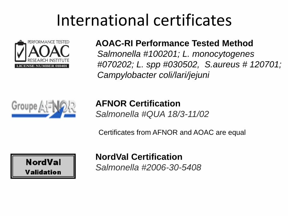
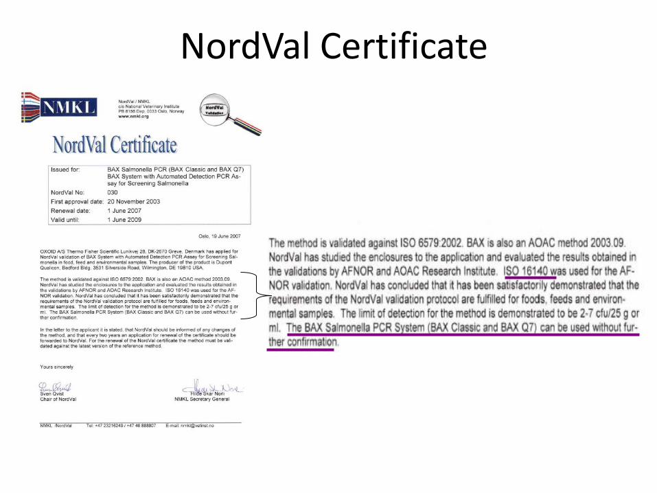
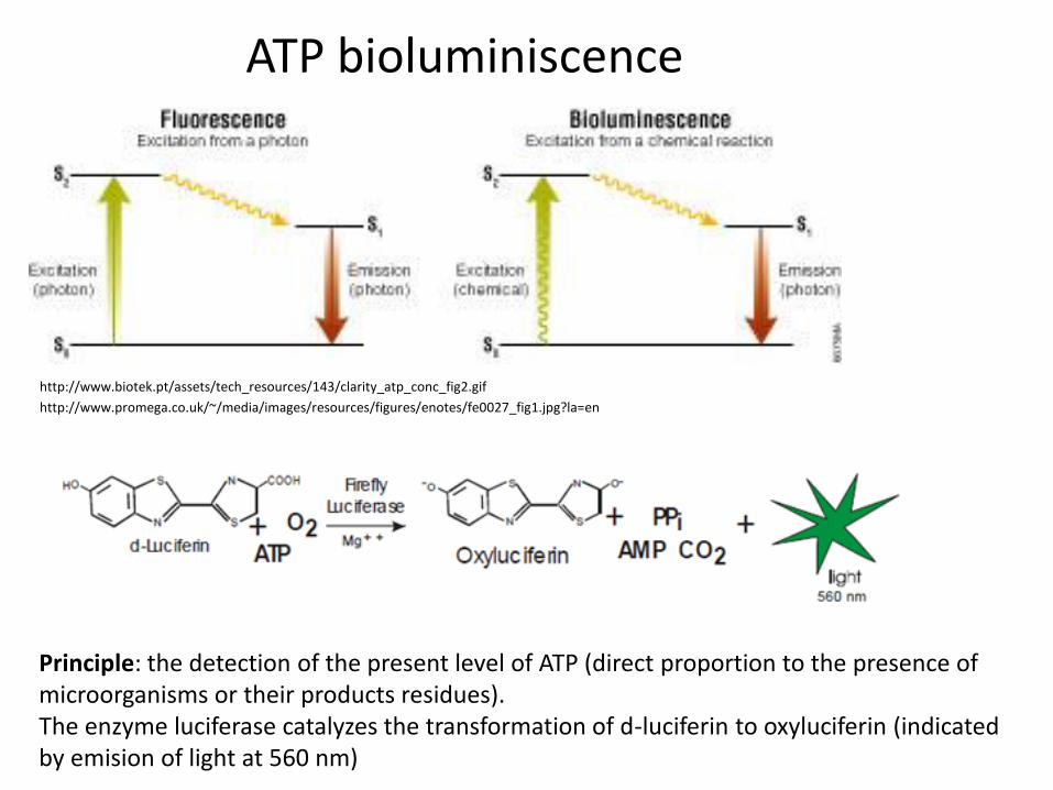
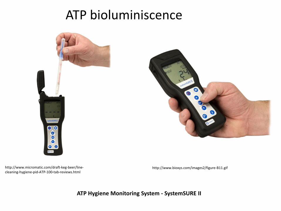
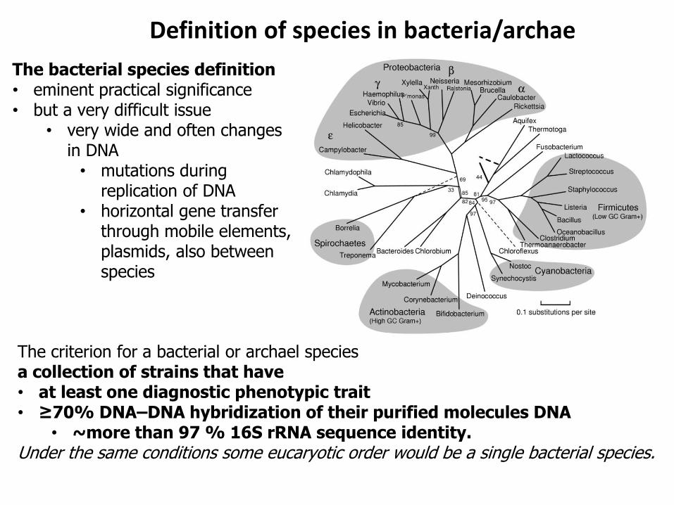
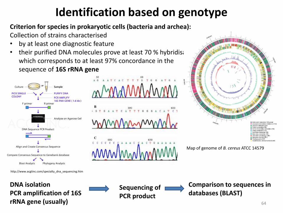
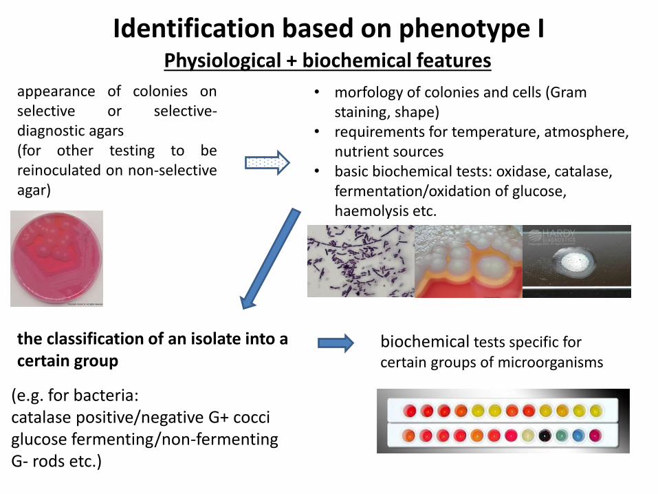
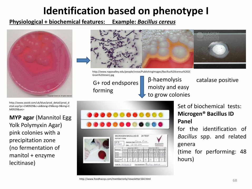
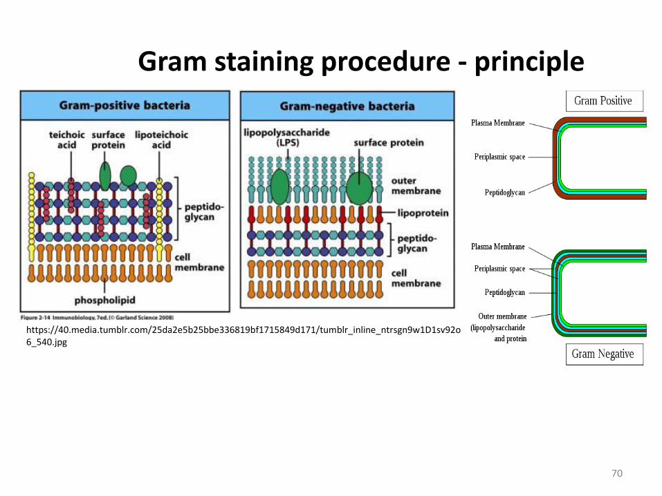
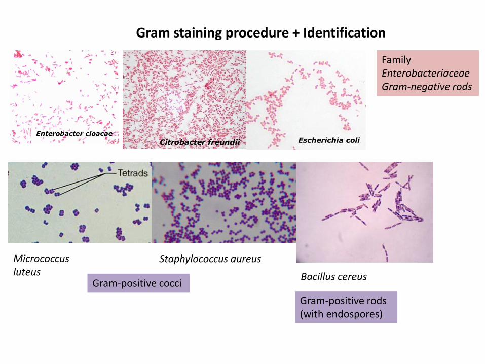
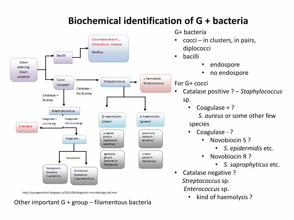
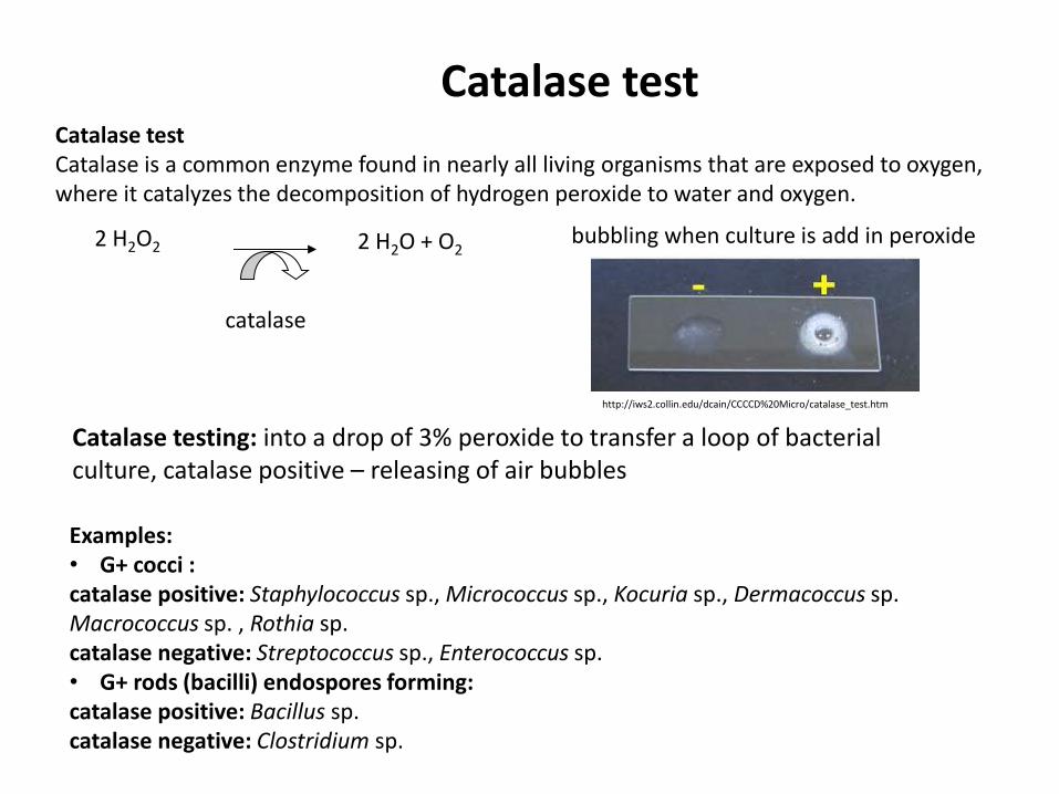
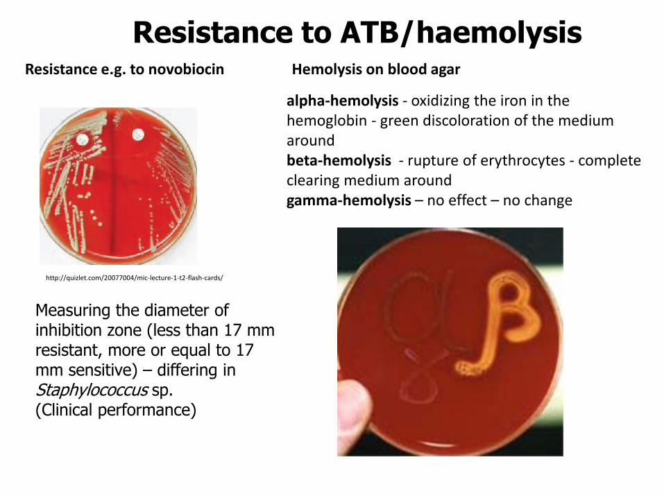
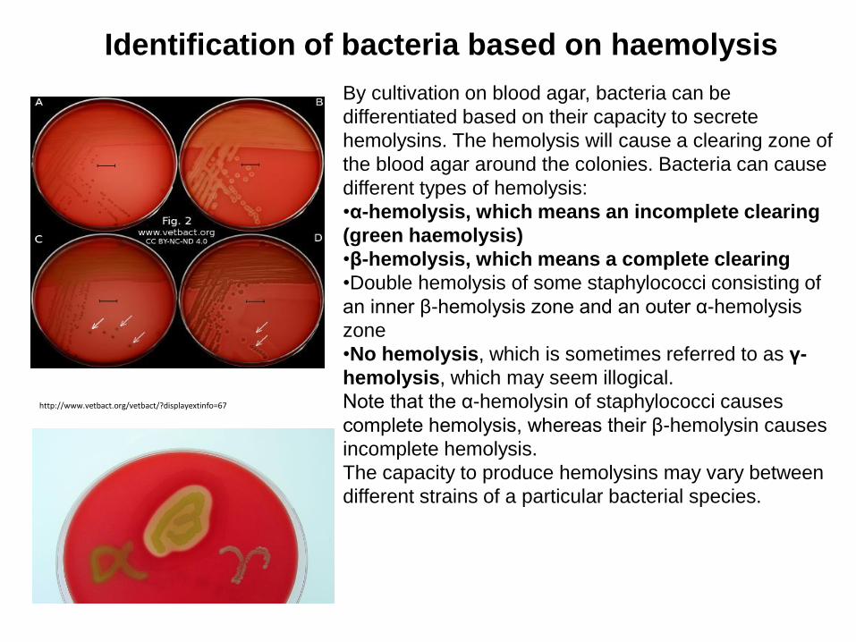
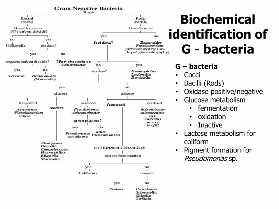
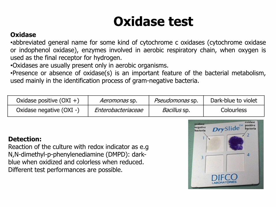
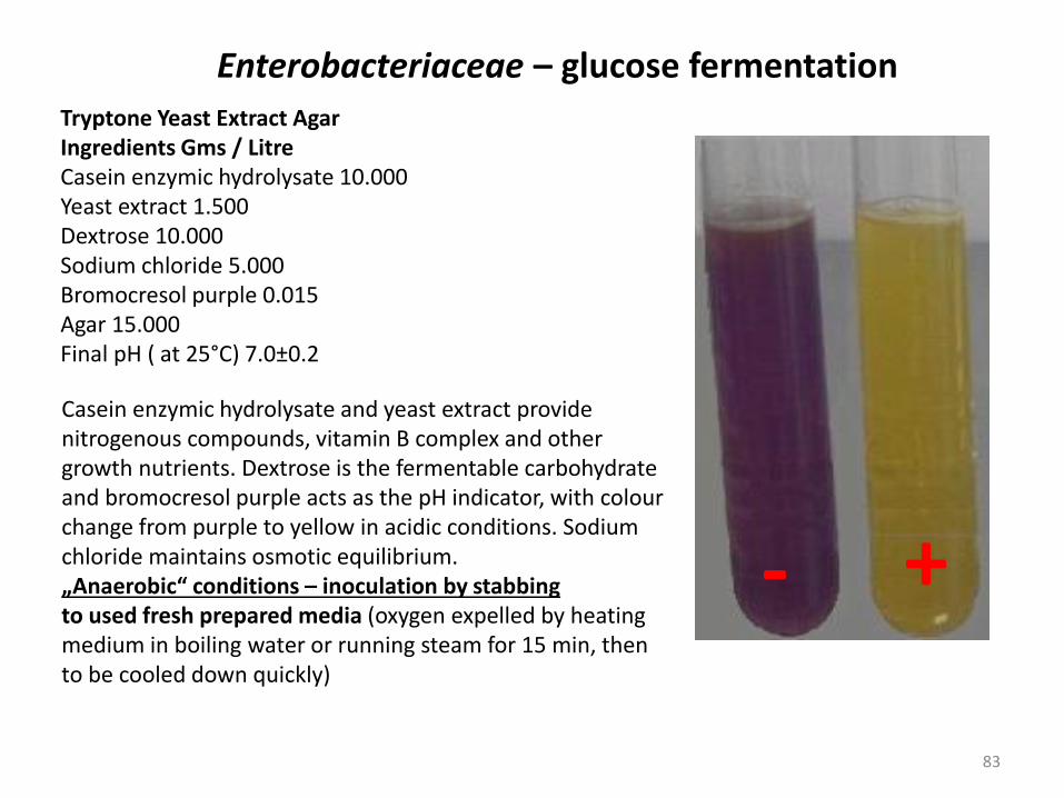
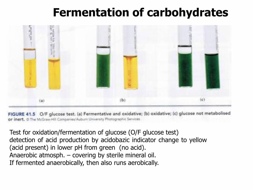
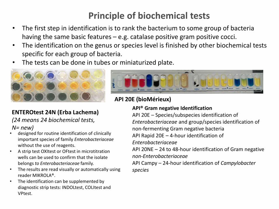
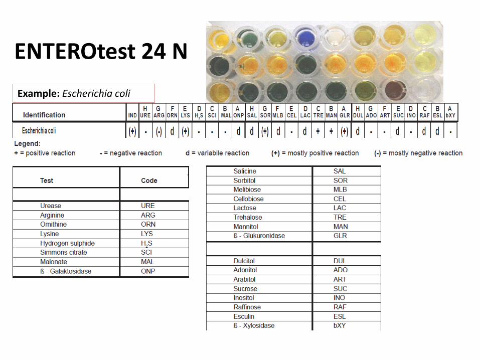
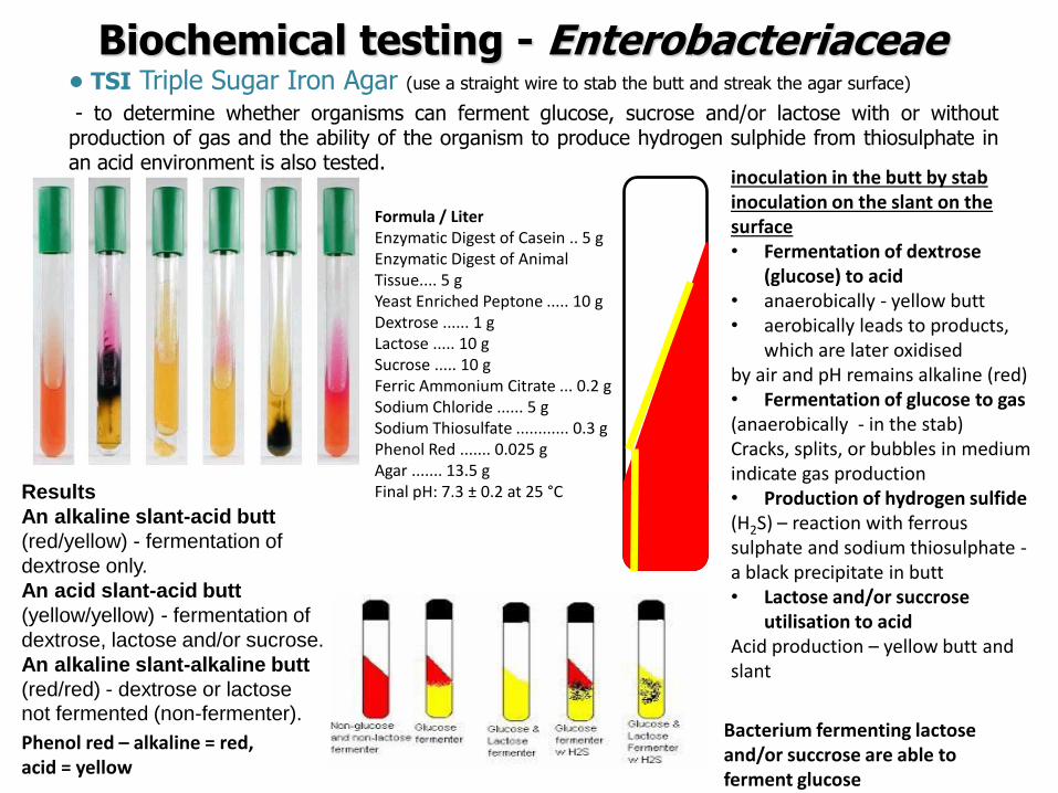
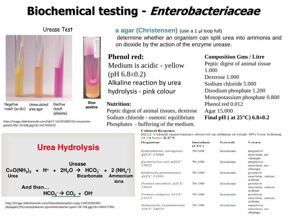
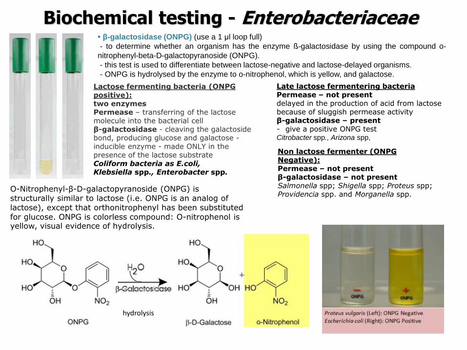
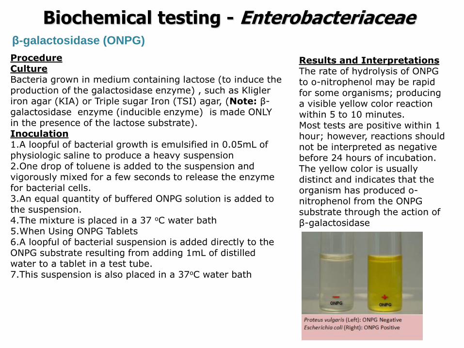
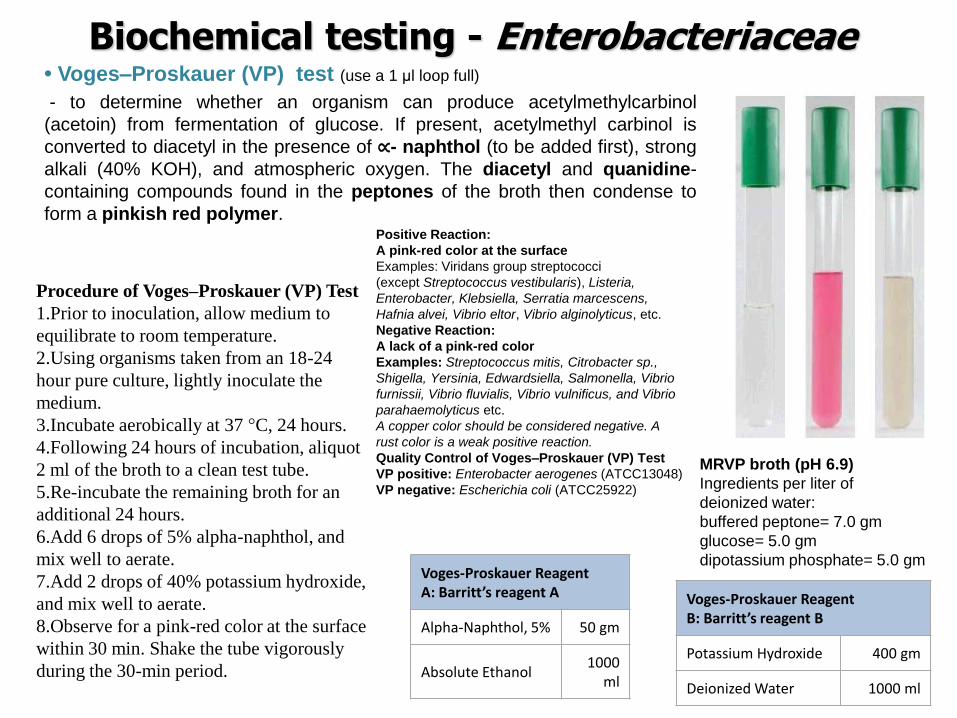
ENTEROtest 24 ndash example of biochemical testing
routine identification of clinically important species of family Enterobacteriaceae the test is focused only on some species often isolated in clinical practis strains from other sources can be slightly different in the dominant biochemical profiles intended exclusively for the identification of family Enterobacteriaceae the preliminary ranking into family Enterobacteriaceae must be done (Gram staining oxidase glucose fermentation) the results of reactions obtained in this modified micromethod may differ from the results obtained using conventional tests and results available in literature sources in case of problem to control positivenegative reactions by control strains or to use for this tests tube variant to check the functionality of the kit not to check the accuracy or success of the identification
Preparation of suspension from over-night culture growing on non-selective agar (for staphylococci Columbia agar blood with 5 of sheep blood is recommended)
Inoculation of microtitre plates anaerobic reaction covered by mineral oil
Incubation
Reading results by both visual and instrumental mode respectively Standard off-line tests INDOLtest or COLItest Other off-line tests OXItest OFtest VPtest Acetoin test
ENTEROtest 24 N ndash example of biochemical testing
bull tube with 5 ml of sterile physiolsol bull suspension (1 McFarland) bull application 100 microl to each hole bull 2 drops of parafin oil into holes D-H of the first line (anaerobic conditions) bull Incubation 37degC 24h bull for check the purity of suspension to streak suspension on TSA (incubation 37 degC ndash 24 h
ENTEROtest 24N
ENTEROtest 24 N - evaluation
Escherichia coli
H 0+0+0=0 G 0+2+0=2 F 1+2+0=3 hellip Identification code 023426
1 1
2
4
ENTEROtest 24 ndash example of biochemical testing
The result = a set of biochemical properties (+-) which is compared to the sets of biochemical properties of different chosen genera and species for them the test is designed
ENTEROtest 24 N
T-index how is the likelihood of the different properties The combination of identication and T-index = the reliability of the result Excelent very good good (this case) acceptable intermediary strain needs some additional tests genus level non-existing taxon
Evaluation by using the identification code - By using a code book - By using software TNW
Identification score the measure of similarity to proposed taxon
The way of identification the obtained profile is compared with profiles in database bull The most similar profiles are found
bull Identification score = of similarity bull But what about the different reactions
bull T-index is calculated bull how is the probability of the different reactions
bull It depends on their importance ndash typical reactions should be the same
bull Reliability of the result bull the combination of identication score and T-index = bull Excelent very good good acceptable intermediary strain needs some
additional tests genus level non-existing taxon
ENTEROtest 24 ndash example of biochemical testing
Biochemical tests - divisions
ENTEROtest 24 species of family Enterobacteriaceae
NEFERMtest 24 Gramnegative non-fermenting bacteria and families Vibrionaceae Aeromonadaceae and Pleiomonas shigelloides present mainly in clinical material
ANAEROtest 23 ANAEROBIC BACTERIA (G + AND G-) routine identification of anaerobic bacteria (G + AND G-) in clinical material and in food
STREPTOtest 24 Streptococcus Enterococcus and related gram positive catalase negative cocci isolated from clinical material
STAPHYtest 24 genus Staphylococcus isolated from clinical material and for their distiguishing from related genera of other gram positive catalase positive cocci
Biochemical tests APIreg Gram positive Identification API Staph ndash Overnight identification of clinical staphylococci and micrococci API 20 Strep ndash 4 or 24-hour identification of streptococci and enterococci API Coryne ndash 24-hour identification of Corynebacteria and coryne-like organisms API Listeria ndash 24-hour identification of all Listeria species APIreg Gram negative Identification API 20E ndash Speciessubspecies identification of Enterobacteriaceae and groupspecies identification of non-fermenting Gram negative bacteria API Rapid 20E ndash 4-hour identification of Enterobacteriaceae API 20NE ndash 24 to 48-hour identification of Gram negative non-Enterobacteriaceae API Campy ndash 24-hour identification of Campylobacter species APIreg Anaerobe Identification API 20A ndash 24-hour identification of anaerobes Rapid ID 32 A ndash 4-hour identification of anaerobes
MicrogenTM GN-ID Convenient 12 substrate identification system for commonly encountered Enterobacteriaceae (GNA) Extended 24 substrate identification system for the complete range of Enterobacteriacea and oxidase positive non-fastidious Gram negative bacilli (GNA+B) MicrogenTM Listeria-ID Enhanced built-in haemolysis test MicrogenTM Bacillus-ID 24 substrate system for the identification of mesophilic Bacillus spp isolated from foods and related samples MicrogenTM Staph-ID 12 substrate system minimizes handling and manipulation Substrates selected specifically for Staphylococcus and related organisms MicrogenTM Strep-ID Substrates selected specifically for Streptococcus Enterococcus and related species Provides excellent differentiation of Enterococci spp especially E casseliflavus E mundtii and E gallinarum
Biochemical tests
Identification of microorganisms by MALDI-TOF MS
Department of Biochemistry and Microbiology University of Chemistry and Technology Prague
Sabina Purkrtovaacute1 Petra Junkovaacute2 1Laboratory of food microbiology 2Laboratory of applied proteomics
SabinaPurkrtovavschtcz PetraJunkovavschtcz
MALDI-TOF MS
1) sample preparation bull microbial culture or its
proteins extract is smearing onto a steel plate and covered over by matrix
bull matrix enables the sample (A) to be desorped and ionised as pseudomolecule ionts [A+H] +
2) MALDI-TOF MS analysis unique mass spectral fingerprint of desorbed microbial cell components (mainly intracellular proteins) different among genera species or also some strains
Matrix-Assisted Laser Desorption Ionization-Time of Flight Mass Spectrometry
httppubsrscorgencontentarticlehtml2014RAC4RA05604C
httpcmrasmorgcontent263547figures-only
3) identification comparison of mass spectrum to those of reference strains in database
MALDI-TOF MS PRINCIPLE
111
2
2
2L
teU
z
m
m mass z charge
L length of drifting zone e
elementary charge U
speeding voltage
Time of flight is a function of the specific ion mass (mz)
httpwwwsigmaaldrichcomtechnical-documentsarticlesbiologycustom-dna-oligos-qc-analysis-by-mass-spectrometryhtml
Soft ionisation method bull low level of sample fragmentation
Animation http cmgmstanfordedupansection_htmlMS
MALDI-TOF MS SAMPLE AND MATRIX
httpcmrasmorgcontent263547figures-only
Matrix bull able to absorb the energy of the laser (usually 337 nm) bull to crystalise with samples - usually acid
character (proton ionisation of sample) dissolved in organic solvent
Sample preparation Direct transfer bull intact cells spread directly onto a steel plate bull lysis of cells occurs during the contact with acid
matrix and by laser desorption bull most bacteria Proteins extraction bull previous extraction of proteins by organic acids
andor alcohol (eg ethanol and 70 formic acid) bull yeasts moulds some species of bacteria (depending
on the cell wall composition)
CHC α-Cyano-4-hydroxycinnamic acid (organic solvent 50 acetonitrile with 25 trifluoracetic acid) SA 35-Dimethoxy-4-hydroxycinnamic acid (sinapic acid)
DHB 25-Dihydroxybenzoic acid
httpwwwsigmaaldrichcomcatalogproductsigmac8982lang=enampregion=CZ
MALDI-TOF MS MICROORGANISMS
113
Mass spectrum protein profile z equals usally to 1+ (so mz usually corresponds to molecula mass) the range usually used for identification 2000 -20 000 mz the intensity of single peaks corresponds to the abundance of the protein Which proteins dominates in the protein profile abundant basic and mediumly hydrophobic mainly ribosomal proteins further cold-shock and heat-shock proteins chaperons etc
conserved house-keeping gene = conserved proteins = the acordance with identification based on DNA
Visualisation of mass spectrum protein profile ndash (software mMass 5 Strohalm et al 2010)
Bacillus cereus
Analysis is recomended (and validated) to be performed from colonies grown onto non-selective agar
MALDI-TOF MS ANALYSIS
114
Comparison of mass spectrum protein profile of unknown sample with these of reference strains present in database by software
Commercial databases from different MALDI-TOF MS producers Bruker Daltonics ndash MALDI BIOTYPER Shimadzu - Shimadzu Launchpad software + SARAMIS database Biomeacuterieux - VITEKreg MS Other databases compatible with different hardware systems (eg Andromas)
BioTyper The statistical analysis for correlation includes peak positions intensities and frequencies across the complete range of microorganisms Score value 0 (none similarity) - 1000 (absolute similarity) But it is expressed in decadic logarithm log(score value) 0-3
Range Description Symbols Color
2300 3000
highly probable species identification
( +++ ) green
2000 2299
secure genus identification probable species identification
( ++ ) green
1700 1999
probable genus identification ( + ) yellow
0000 1699
not reliable identification ( - ) red
Bacillus cereus
115
Range Description Symbols Color
2300 3000
highly probable species identification
( +++ ) green
2000 2299
secure genus identification probable species identification
( ++ ) green
1700 1999
probable genus identification ( + ) yellow
0000 1699
not reliable identification ( - ) red
Rank (Quality)
Matched Pattern Score Value
NCBI Identifier
1 ( +++ )
Bacillus cereus DSM 31T DSM 2554 1396
2 ( ++ )
Bacillus cereus 994000168 LBK 2203 1396
3 ( ++ )
Bacillus weihenstephanensis DSM 11821T DSM
2158 86662
4 ( ++ )
Bacillus mycoides DSM 2048T DSM 2155 1405
5 ( ++ )
Bacillus cereus 4080 LBK 2147 1396
6 ( + )
Bacillus thuringiensis DSM 2046T DSM 1975 1428
7 ( + )
Bacillus pseudomycoides DSM 12442T DSM 1787 64104
8 ( - )
Bacillus bataviensis DSM 15601T DSM 1369 220685
9 ( - )
Brevibacterium linens IMET 11075T HKJ 1347 1703
10 ( - )
Acinetobacter towneri DSM 14962T HAM 1345 202956
Bacillus cereus
Bacillus cereus
MALDI-TOF MS - PROCEDURE Direct method smearing sample in four parallels in lower and higher cells concentrations ndash after drying to cover over by matrix (1-2 microl) and let to crystallise at room temperature
Matrix solution of α-Cyano-4-hydroxycinnamic acid (10 mgml) in 50 acetonitrile with 25 trifluoroacetic acid (prepared with 10 TFA solution)
Protein standard (1 μl) Bruker Bacterial Test Standard (Bruker Daltonics SRN) ndash proteins extracted from z Escherichia coli DH5alpha BRL + some others
Equipment Bruker Autoflex Speed Database MALDI Biotyper 31
116
MALDI-TOF MSPROTEIN STANDARD
117
Analyte name Rank (Quality) Matched Pattern Score Value
F4 1 ( +++ ) Escherichia coli DH5alpha BRL 2439
Thank you for your attention
Questions to be sent to SabinaPurkrtovavschtcz
118
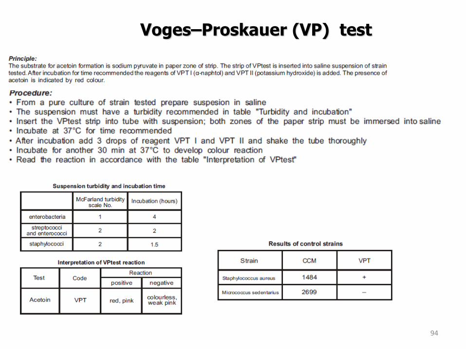
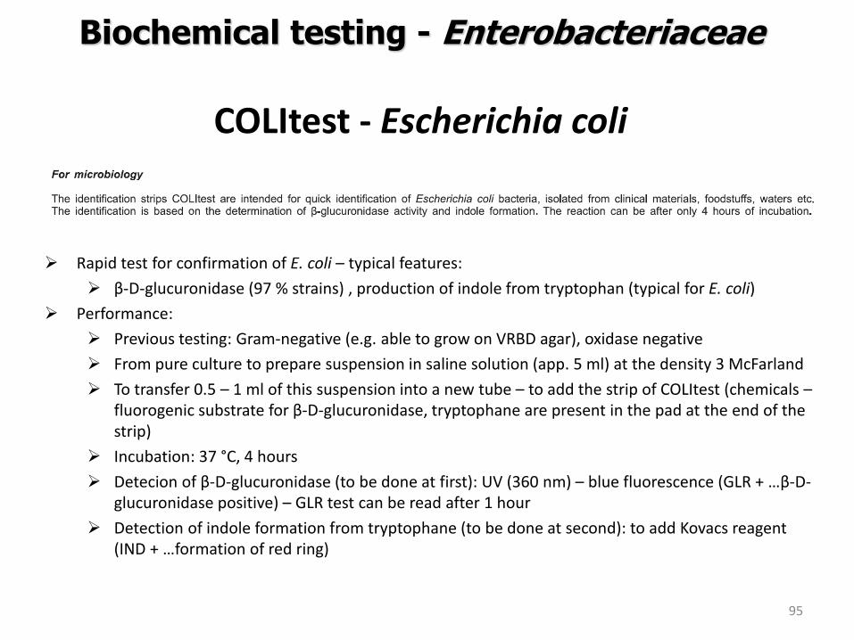
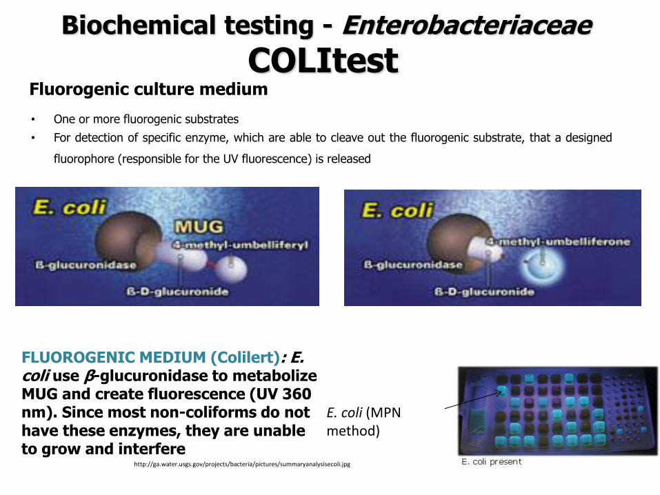
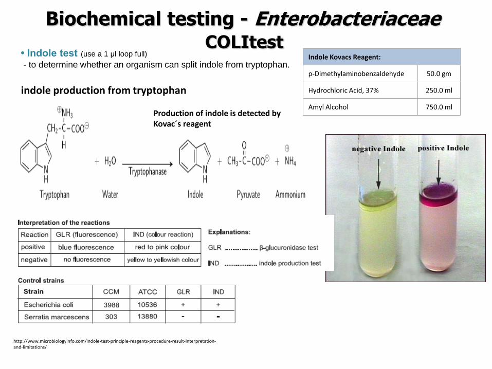
ENTEROtest 24 ndash example of biochemical testing
The result = a set of biochemical properties (+-) which is compared to the sets of biochemical properties of different chosen genera and species for them the test is designed
ENTEROtest 24 N
T-index how is the likelihood of the different properties The combination of identication and T-index = the reliability of the result Excelent very good good (this case) acceptable intermediary strain needs some additional tests genus level non-existing taxon
Evaluation by using the identification code - By using a code book - By using software TNW
Identification score the measure of similarity to proposed taxon
The way of identification the obtained profile is compared with profiles in database bull The most similar profiles are found
bull Identification score = of similarity bull But what about the different reactions
bull T-index is calculated bull how is the probability of the different reactions
bull It depends on their importance ndash typical reactions should be the same
bull Reliability of the result bull the combination of identication score and T-index = bull Excelent very good good acceptable intermediary strain needs some
additional tests genus level non-existing taxon
ENTEROtest 24 ndash example of biochemical testing
Biochemical tests - divisions
ENTEROtest 24 species of family Enterobacteriaceae
NEFERMtest 24 Gramnegative non-fermenting bacteria and families Vibrionaceae Aeromonadaceae and Pleiomonas shigelloides present mainly in clinical material
ANAEROtest 23 ANAEROBIC BACTERIA (G + AND G-) routine identification of anaerobic bacteria (G + AND G-) in clinical material and in food
STREPTOtest 24 Streptococcus Enterococcus and related gram positive catalase negative cocci isolated from clinical material
STAPHYtest 24 genus Staphylococcus isolated from clinical material and for their distiguishing from related genera of other gram positive catalase positive cocci
Biochemical tests APIreg Gram positive Identification API Staph ndash Overnight identification of clinical staphylococci and micrococci API 20 Strep ndash 4 or 24-hour identification of streptococci and enterococci API Coryne ndash 24-hour identification of Corynebacteria and coryne-like organisms API Listeria ndash 24-hour identification of all Listeria species APIreg Gram negative Identification API 20E ndash Speciessubspecies identification of Enterobacteriaceae and groupspecies identification of non-fermenting Gram negative bacteria API Rapid 20E ndash 4-hour identification of Enterobacteriaceae API 20NE ndash 24 to 48-hour identification of Gram negative non-Enterobacteriaceae API Campy ndash 24-hour identification of Campylobacter species APIreg Anaerobe Identification API 20A ndash 24-hour identification of anaerobes Rapid ID 32 A ndash 4-hour identification of anaerobes
MicrogenTM GN-ID Convenient 12 substrate identification system for commonly encountered Enterobacteriaceae (GNA) Extended 24 substrate identification system for the complete range of Enterobacteriacea and oxidase positive non-fastidious Gram negative bacilli (GNA+B) MicrogenTM Listeria-ID Enhanced built-in haemolysis test MicrogenTM Bacillus-ID 24 substrate system for the identification of mesophilic Bacillus spp isolated from foods and related samples MicrogenTM Staph-ID 12 substrate system minimizes handling and manipulation Substrates selected specifically for Staphylococcus and related organisms MicrogenTM Strep-ID Substrates selected specifically for Streptococcus Enterococcus and related species Provides excellent differentiation of Enterococci spp especially E casseliflavus E mundtii and E gallinarum
Biochemical tests
Identification of microorganisms by MALDI-TOF MS
Department of Biochemistry and Microbiology University of Chemistry and Technology Prague
Sabina Purkrtovaacute1 Petra Junkovaacute2 1Laboratory of food microbiology 2Laboratory of applied proteomics
SabinaPurkrtovavschtcz PetraJunkovavschtcz
MALDI-TOF MS
1) sample preparation bull microbial culture or its
proteins extract is smearing onto a steel plate and covered over by matrix
bull matrix enables the sample (A) to be desorped and ionised as pseudomolecule ionts [A+H] +
2) MALDI-TOF MS analysis unique mass spectral fingerprint of desorbed microbial cell components (mainly intracellular proteins) different among genera species or also some strains
Matrix-Assisted Laser Desorption Ionization-Time of Flight Mass Spectrometry
httppubsrscorgencontentarticlehtml2014RAC4RA05604C
httpcmrasmorgcontent263547figures-only
3) identification comparison of mass spectrum to those of reference strains in database
MALDI-TOF MS PRINCIPLE
111
2
2
2L
teU
z
m
m mass z charge
L length of drifting zone e
elementary charge U
speeding voltage
Time of flight is a function of the specific ion mass (mz)
httpwwwsigmaaldrichcomtechnical-documentsarticlesbiologycustom-dna-oligos-qc-analysis-by-mass-spectrometryhtml
Soft ionisation method bull low level of sample fragmentation
Animation http cmgmstanfordedupansection_htmlMS
MALDI-TOF MS SAMPLE AND MATRIX
httpcmrasmorgcontent263547figures-only
Matrix bull able to absorb the energy of the laser (usually 337 nm) bull to crystalise with samples - usually acid
character (proton ionisation of sample) dissolved in organic solvent
Sample preparation Direct transfer bull intact cells spread directly onto a steel plate bull lysis of cells occurs during the contact with acid
matrix and by laser desorption bull most bacteria Proteins extraction bull previous extraction of proteins by organic acids
andor alcohol (eg ethanol and 70 formic acid) bull yeasts moulds some species of bacteria (depending
on the cell wall composition)
CHC α-Cyano-4-hydroxycinnamic acid (organic solvent 50 acetonitrile with 25 trifluoracetic acid) SA 35-Dimethoxy-4-hydroxycinnamic acid (sinapic acid)
DHB 25-Dihydroxybenzoic acid
httpwwwsigmaaldrichcomcatalogproductsigmac8982lang=enampregion=CZ
MALDI-TOF MS MICROORGANISMS
113
Mass spectrum protein profile z equals usally to 1+ (so mz usually corresponds to molecula mass) the range usually used for identification 2000 -20 000 mz the intensity of single peaks corresponds to the abundance of the protein Which proteins dominates in the protein profile abundant basic and mediumly hydrophobic mainly ribosomal proteins further cold-shock and heat-shock proteins chaperons etc
conserved house-keeping gene = conserved proteins = the acordance with identification based on DNA
Visualisation of mass spectrum protein profile ndash (software mMass 5 Strohalm et al 2010)
Bacillus cereus
Analysis is recomended (and validated) to be performed from colonies grown onto non-selective agar
MALDI-TOF MS ANALYSIS
114
Comparison of mass spectrum protein profile of unknown sample with these of reference strains present in database by software
Commercial databases from different MALDI-TOF MS producers Bruker Daltonics ndash MALDI BIOTYPER Shimadzu - Shimadzu Launchpad software + SARAMIS database Biomeacuterieux - VITEKreg MS Other databases compatible with different hardware systems (eg Andromas)
BioTyper The statistical analysis for correlation includes peak positions intensities and frequencies across the complete range of microorganisms Score value 0 (none similarity) - 1000 (absolute similarity) But it is expressed in decadic logarithm log(score value) 0-3
Range Description Symbols Color
2300 3000
highly probable species identification
( +++ ) green
2000 2299
secure genus identification probable species identification
( ++ ) green
1700 1999
probable genus identification ( + ) yellow
0000 1699
not reliable identification ( - ) red
Bacillus cereus
115
Range Description Symbols Color
2300 3000
highly probable species identification
( +++ ) green
2000 2299
secure genus identification probable species identification
( ++ ) green
1700 1999
probable genus identification ( + ) yellow
0000 1699
not reliable identification ( - ) red
Rank (Quality)
Matched Pattern Score Value
NCBI Identifier
1 ( +++ )
Bacillus cereus DSM 31T DSM 2554 1396
2 ( ++ )
Bacillus cereus 994000168 LBK 2203 1396
3 ( ++ )
Bacillus weihenstephanensis DSM 11821T DSM
2158 86662
4 ( ++ )
Bacillus mycoides DSM 2048T DSM 2155 1405
5 ( ++ )
Bacillus cereus 4080 LBK 2147 1396
6 ( + )
Bacillus thuringiensis DSM 2046T DSM 1975 1428
7 ( + )
Bacillus pseudomycoides DSM 12442T DSM 1787 64104
8 ( - )
Bacillus bataviensis DSM 15601T DSM 1369 220685
9 ( - )
Brevibacterium linens IMET 11075T HKJ 1347 1703
10 ( - )
Acinetobacter towneri DSM 14962T HAM 1345 202956
Bacillus cereus
Bacillus cereus
MALDI-TOF MS - PROCEDURE Direct method smearing sample in four parallels in lower and higher cells concentrations ndash after drying to cover over by matrix (1-2 microl) and let to crystallise at room temperature
Matrix solution of α-Cyano-4-hydroxycinnamic acid (10 mgml) in 50 acetonitrile with 25 trifluoroacetic acid (prepared with 10 TFA solution)
Protein standard (1 μl) Bruker Bacterial Test Standard (Bruker Daltonics SRN) ndash proteins extracted from z Escherichia coli DH5alpha BRL + some others
Equipment Bruker Autoflex Speed Database MALDI Biotyper 31
116
MALDI-TOF MSPROTEIN STANDARD
117
Analyte name Rank (Quality) Matched Pattern Score Value
F4 1 ( +++ ) Escherichia coli DH5alpha BRL 2439
Thank you for your attention
Questions to be sent to SabinaPurkrtovavschtcz
118
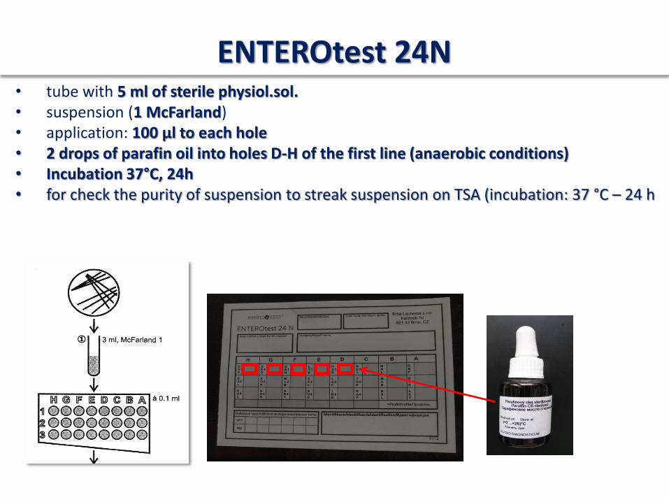
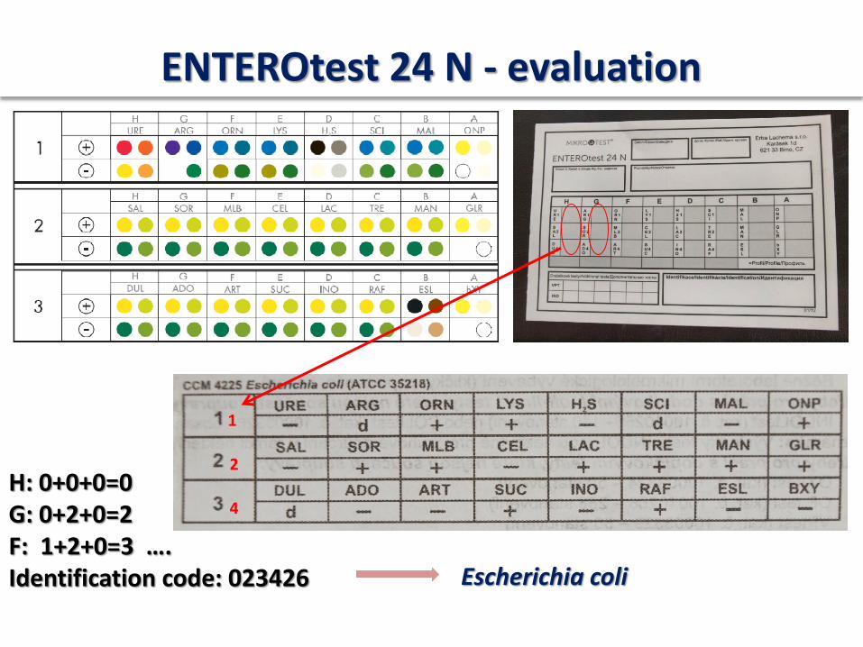
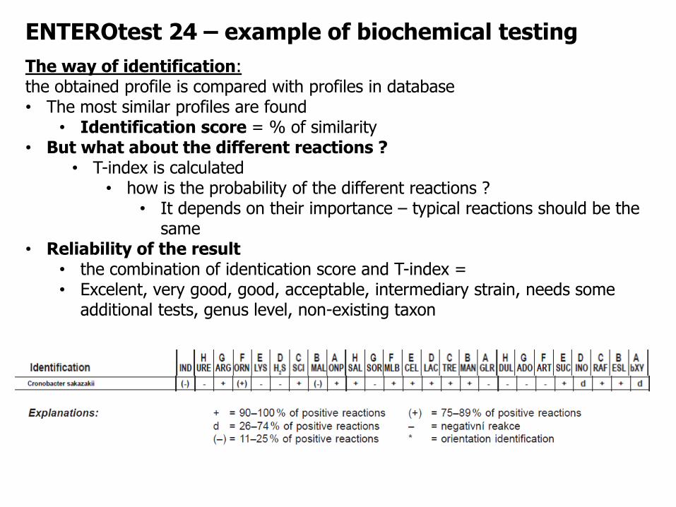
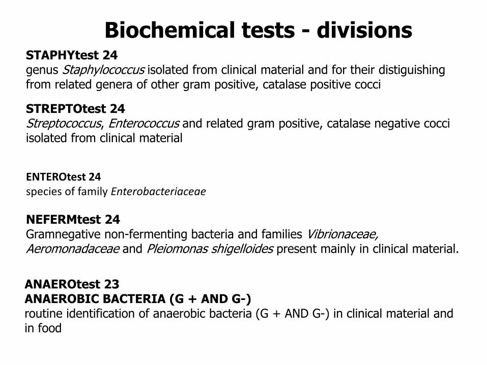
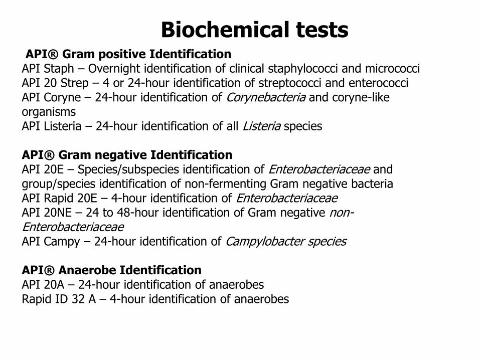
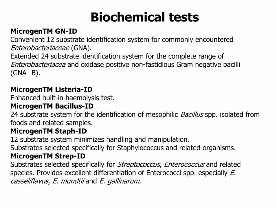
Bacillus cereus
115
Range Description Symbols Color
2300 3000
highly probable species identification
( +++ ) green
2000 2299
secure genus identification probable species identification
( ++ ) green
1700 1999
probable genus identification ( + ) yellow
0000 1699
not reliable identification ( - ) red
Rank (Quality)
Matched Pattern Score Value
NCBI Identifier
1 ( +++ )
Bacillus cereus DSM 31T DSM 2554 1396
2 ( ++ )
Bacillus cereus 994000168 LBK 2203 1396
3 ( ++ )
Bacillus weihenstephanensis DSM 11821T DSM
2158 86662
4 ( ++ )
Bacillus mycoides DSM 2048T DSM 2155 1405
5 ( ++ )
Bacillus cereus 4080 LBK 2147 1396
6 ( + )
Bacillus thuringiensis DSM 2046T DSM 1975 1428
7 ( + )
Bacillus pseudomycoides DSM 12442T DSM 1787 64104
8 ( - )
Bacillus bataviensis DSM 15601T DSM 1369 220685
9 ( - )
Brevibacterium linens IMET 11075T HKJ 1347 1703
10 ( - )
Acinetobacter towneri DSM 14962T HAM 1345 202956
Bacillus cereus
Bacillus cereus
MALDI-TOF MS - PROCEDURE Direct method smearing sample in four parallels in lower and higher cells concentrations ndash after drying to cover over by matrix (1-2 microl) and let to crystallise at room temperature
Matrix solution of α-Cyano-4-hydroxycinnamic acid (10 mgml) in 50 acetonitrile with 25 trifluoroacetic acid (prepared with 10 TFA solution)
Protein standard (1 μl) Bruker Bacterial Test Standard (Bruker Daltonics SRN) ndash proteins extracted from z Escherichia coli DH5alpha BRL + some others
Equipment Bruker Autoflex Speed Database MALDI Biotyper 31
116
MALDI-TOF MSPROTEIN STANDARD
117
Analyte name Rank (Quality) Matched Pattern Score Value
F4 1 ( +++ ) Escherichia coli DH5alpha BRL 2439
Thank you for your attention
Questions to be sent to SabinaPurkrtovavschtcz
118
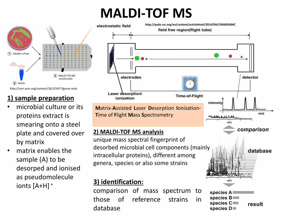
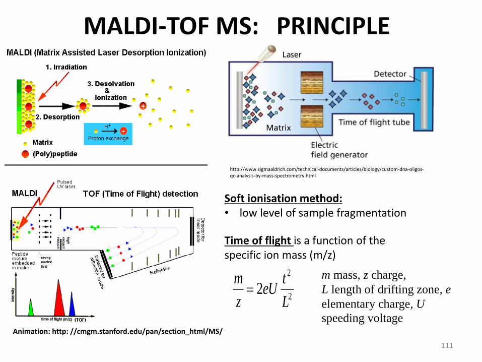
MALDI-TOF MSPROTEIN STANDARD
117
Analyte name Rank (Quality) Matched Pattern Score Value
F4 1 ( +++ ) Escherichia coli DH5alpha BRL 2439
Thank you for your attention
Questions to be sent to SabinaPurkrtovavschtcz
118
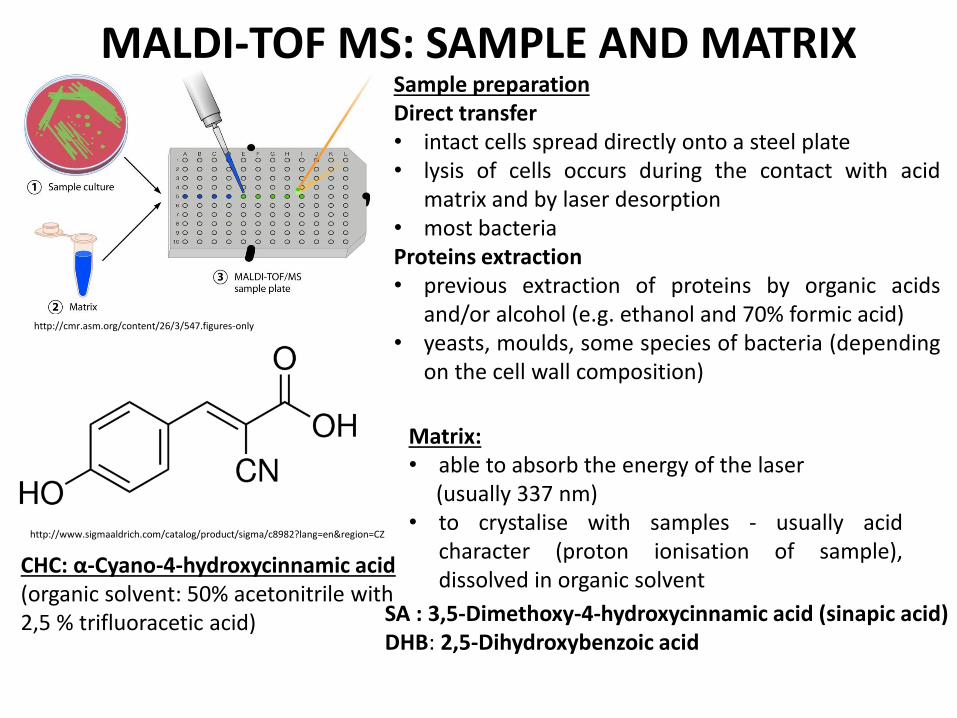
Thank you for your attention
Questions to be sent to SabinaPurkrtovavschtcz
118