
Randomized Controlled Trial of Ultrasound versus Palpation ...
3
*Corresponding author email: [email protected] Symbiosis Group Symbiosis www.symbiosisonline.org www.symbiosisonlinepublishing.com Randomized Controlled Trial of Ultrasound versus Palpation Method for Arterial Cannulation in Infants Less Than 24 Months of Age Tracy YS Tan 1 , Jens AK Petersen 2 , Xiuyan Zhao 3 and Katherine L Taylor 4 * 1 Department of Anesthesia, Kandang Kerbau Hospital, Singapore City, Singapore 2 Department of Anaesthesia, Aarhus University Hospital, Aarhus, Denmark 3 Department of Biostatistics, Hospital for Sick Children, Toronto, Canada 4 Department of Anesthesia, Hospital for Sick Children, Toronto, Canada SOJ Anesthesiology and Pain Management Open Access Research Article Abstract Introduction: Ultrasound guided peripheral arterial cannulation may increase success as the small diameter vessel can be visualized. Fewer attempts at arterial cannulation may reduce local vessel damage. However even the current small ultrasound probes provide additional bulk in a small area and palpation method may be preferred. Aim: To determine if ultrasound guided peripheral arterial catheter insertion results in fewer attempts, shorter time to cannulation and cost savings compared to palpation method in children <2 years of age. Methods: Patients ≤ 24 months scheduled for elective surgical procedures where arterial cannulation was planned were eligible for recruitment. Forty patients were randomized to receive either Ultrasound Assisted (US) or palpation method. The primary endpoint was time to successful cannulation between two groups using intention to treat analysis. After 3 failed attempts, providers could switch to alternative method. Secondary endpoints were number of attempted sites, number of attempts and cost of equipment. Results: Average weight of patients was 6.14 kg (95% CI 4.9-7.4 kg) for the US group and 5.5 kg (95% CI 4.1-6.9) for the palpation group. There was no age difference between groups. The initial randomized method was successful in 33/ 40 patients (17/ 20 Ultrasound and 16/ 20 Palpation method). Crossover to the other method resulted in an additional 4 ultrasound successes and 3 palpation successes. There was no difference in technique for rates of initial randomized method of success or time to successful insertion. Cost savings in disposable equipment were demonstrated. Conclusions: For infants and small children, ultrasound and palpation methods for arterial cannulation are similar with respect to time to secure arterial access. Keywords: Infant; Vascular Access device; Palpation; Ultrasound; Costs and Cost Analysis; Time Received: February 19, 2015; Accepted: April 15, 2015, Published: April 25, 2015 *Corresponding author: K L Taylor, Department of Anesthesia, Hospital for Sick Children, 555 University Avenue , Toronto, Canada,M5G 1X8, Tel: 1 416 813 7445; Fax: 416 813 7543; E- mail: [email protected] Introduction Arterial cannulation is a commonly performed invasive procedure in the operating room and intensive care unit. Traditionally, the artery is located by palpating the pulse of the patient. The catheter may not pass smoothly into the artery despite apparent blood return on initial puncture; resultant hematoma or arterial spasm hinders subsequent attempts. Furthermore, the pulse may be weak or absent in patients with hypotension, edema, obesity or local thrombosis due to previous arterial cannulation in the same location. The radial artery in children has a cross-sectional area of 1-4.2mm, [1] i.e. an inner diameter of 0.5-1.2mm [2]. Arterial catheter insertion can be challenging in the best of hands. While ultrasound (US) is being used with increasing frequency for central venous access, fewer clinicians are familiar with US- guided arterial cannulation. In adults, US was found to improve successful arterial cannulation at first attempt, thus decreasing the time to establish an arterial cannulation [1]. Schwemmer reported similar results in a small study in children (n=30, mean age 40 months) [2]. However, Ganesh et al. [3] reported no significant effect of US guidance compared with the palpation technique in a larger study of older children (n=152, mean age 99 months). Arterial cannulation in small children is difficult, with smaller arterial diameter and greater subcutaneous fat. The bulk of current US probes may hinder arterial catheter insertion. Our null hypothesis is that US does not facilitate arterial cannulation in small children compared with the palpation method. Materials and Methods This trial was registered with www.clinical trials. gov protocol # 1000030723. After Research Ethics Board approval and parental informed consent, children <24 months undergoing elective surgical procedures where indwelling Special Issue: Anesthesia & Postoperative Effects
Transcript of Randomized Controlled Trial of Ultrasound versus Palpation ...
*Corresponding author email: [email protected] Group
Symbiosis www.symbiosisonline.org www.symbiosisonlinepublishing.com
Randomized Controlled Trial of Ultrasound versus Palpation Method for Arterial Cannulation in Infants Less Than 24
Months of AgeTracy YS Tan1, Jens AK Petersen2, Xiuyan Zhao3 and Katherine L Taylor4*
1Department of Anesthesia, Kandang Kerbau Hospital, Singapore City, Singapore2Department of Anaesthesia, Aarhus University Hospital, Aarhus, Denmark
3Department of Biostatistics, Hospital for Sick Children, Toronto, Canada4Department of Anesthesia, Hospital for Sick Children, Toronto, Canada
SOJ Anesthesiology and Pain Management Open AccessResearch Article
Abstract
Introduction: Ultrasound guided peripheral arterial cannulation may increase success as the small diameter vessel can be visualized. Fewer attempts at arterial cannulation may reduce local vessel damage. However even the current small ultrasound probes provide additional bulk in a small area and palpation method may be preferred.
Aim: To determine if ultrasound guided peripheral arterial catheter insertion results in fewer attempts, shorter time to cannulation and cost savings compared to palpation method in children <2 years of age.
Methods: Patients ≤ 24 months scheduled for elective surgical procedures where arterial cannulation was planned were eligible for recruitment. Forty patients were randomized to receive either Ultrasound Assisted (US) or palpation method. The primary endpoint was time to successful cannulation between two groups using intention to treat analysis. After 3 failed attempts, providers could switch to alternative method. Secondary endpoints were number of attempted sites, number of attempts and cost of equipment.
Results: Average weight of patients was 6.14 kg (95% CI 4.9-7.4 kg) for the US group and 5.5 kg (95% CI 4.1-6.9) for the palpation group. There was no age difference between groups. The initial randomized method was successful in 33/ 40 patients (17/ 20 Ultrasound and 16/ 20 Palpation method). Crossover to the other method resulted in an additional 4 ultrasound successes and 3 palpation successes. There was no difference in technique for rates of initial randomized method of success or time to successful insertion. Cost savings in disposable equipment were demonstrated.
Conclusions: For infants and small children, ultrasound and palpation methods for arterial cannulation are similar with respect to time to secure arterial access.
Keywords: Infant; Vascular Access device; Palpation; Ultrasound; Costs and Cost Analysis; Time
Received: February 19, 2015; Accepted: April 15, 2015, Published: April 25, 2015
*Corresponding author: K L Taylor, Department of Anesthesia, Hospital for Sick Children, 555 University Avenue , Toronto, Canada,M5G 1X8, Tel: 1 416 813 7445; Fax: 416 813 7543; E- mail: [email protected]
IntroductionArterial cannulation is a commonly performed invasive
procedure in the operating room and intensive care unit. Traditionally, the artery is located by palpating the pulse of the patient. The catheter may not pass smoothly into the artery despite apparent blood return on initial puncture; resultant hematoma or arterial spasm hinders subsequent attempts. Furthermore, the pulse may be weak or absent in patients with hypotension, edema, obesity or local thrombosis due to previous arterial cannulation in the same location. The radial artery in children has a cross-sectional area of 1-4.2mm, [1] i.e. an inner diameter of 0.5-1.2mm [2]. Arterial catheter insertion can be challenging in the best of hands.
While ultrasound (US) is being used with increasing frequency for central venous access, fewer clinicians are familiar with US-guided arterial cannulation. In adults, US was found to improve successful arterial cannulation at first attempt, thus decreasing the time to establish an arterial cannulation [1]. Schwemmer reported similar results in a small study in children (n=30, mean age 40 months) [2]. However, Ganesh et al. [3] reported no significant effect of US guidance compared with the palpation technique in a larger study of older children (n=152, mean age 99 months).
Arterial cannulation in small children is difficult, with smaller arterial diameter and greater subcutaneous fat. The bulk of current US probes may hinder arterial catheter insertion. Our null hypothesis is that US does not facilitate arterial cannulation in small children compared with the palpation method.
Materials and MethodsThis trial was registered with www.clinical trials.
gov protocol # 1000030723. After Research Ethics Board approval and parental informed consent, children <24 months undergoing elective surgical procedures where indwelling
Special Issue: Anesthesia & Postoperative Effects
Page 2 of 3Citation: Tan TYS, Petersen JAK, Zhao X, Taylor KL (2015) Randomized Controlled Trial of Ultrasound versus Palpation Method for Arterial Cannulation in Infants Less Than 24 Months of Age. SOJ Anesthesiol Pain Manag, 2(2): 1-3.
Randomized Controlled Trial of Ultrasound versus Palpation Method for Arterial Cannulation in Infants Less Than 24 Months of Age
Copyright: © 2015 Taylor et al.
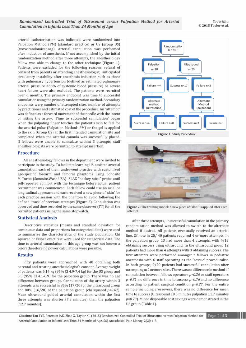
arterial catheterization was indicated were randomized into Palpation Method (PM) (standard practice) or US (group US) (www.randomizer.org). Arterial cannulation was performed after induction of anesthesia. If not accomplished by the initial randomization method after three attempts, the anesthesiology fellow was able to change to the other technique (Figure 1). Patients were excluded for the following reasons: refusal of consent from parents or attending anesthesiologist, anticipated circulatory instability after anesthesia induction such as those with pulmonary hypertension (defined as estimated pulmonary arterial pressure ≥66% of systemic blood pressure) or severe heart failure were also excluded. The patients were recruited over 6 months. The primary endpoint was time to successful cannulation using the primary randomization method. Secondary endpoints were number of attempted sites, number of attempts by practitioner and estimated cost of the procedure. An “attempt” was defined as a forward movement of the needle with the intent of hitting the artery. ‘Time to successful cannulation’ began when the palpating finger touches the patient’s skin to feel for the arterial pulse (Palpation Method- PM) or the gel is applied to the skin (Group US) at the first intended cannulation site and completed when the arterial cannula was successfully placed. If fellows were unable to cannulate withint 3 attempts, staff anesthesiologists were permitted to attempt insertion.
ProcedureAll anesthesiology fellows in the department were invited to
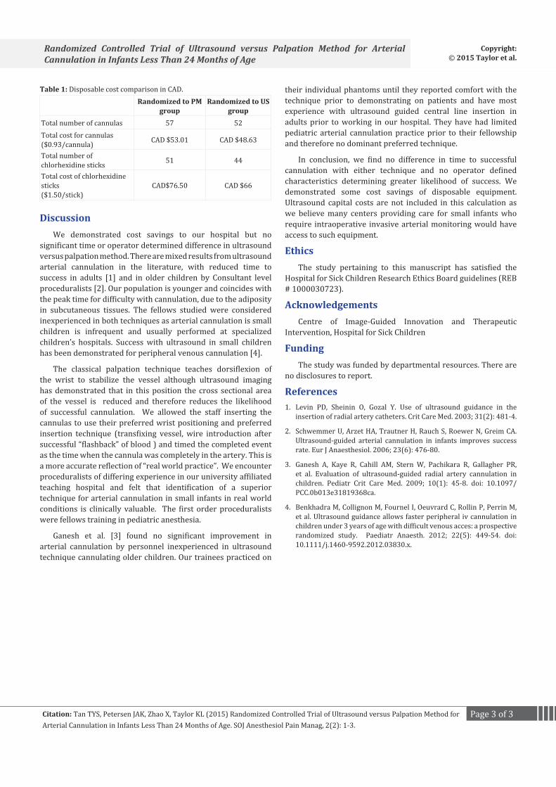
participate in the study. To facilitate learning US-assisted arterial cannulation, each of them underwent practice with customized age-specific forearm and femoral phantoms using Sonosite M-Turbo (Sonosite,Wash,USA) SLAX “hockey stick” probe until self-reported comfort with the technique before actual patient recruitment was commenced. Each fellow could use an axial or longitudinal approach and each received a new piece of ‘skin’ for each practice session with the phantom to avoid following the defined ‘track’ of previous attempts (Figure 2). Cannulation was observed and time recorded by the same observer (TT) for all the recruited patients using the same stopwatch.
Statistical AnalysisDescriptive statistics (means and standard deviation for
continuous data and proportions for categorical data) were used to summarize the characteristics of the study population. Chi squared or Fisher exact test were used for categorical data. The time to arterial cannulation in this age group was not known a priori therefore no power calculations were possible.
ResultsFifty patients were approached with 40 obtaining both
parental and treating anesthesiologist’s consent. Average weight of patients was 6.14 kg (95% CI 4.9-7.4 kg) for the US group and 5.5 (95% CI 4.1-6.9) for the palpation group. There was no age difference between groups. Cannulation of the artery within 3 attempts was successful in 85% (17/20) of the ultrasound group and 80% (16/20) of the palpation group (chi squared p=0.67). Mean ultrasound guided arterial cannulation within the first three attempts was shorter (7.8 minutes) than the palpation (12.7 minutes).
After three attempts, unsuccessful cannulation in the primary randomization method was allowed to switch to the alternate method if desired. All patients eventually received an arterial line. Of note in 25/ 40 patients required 4 or more attempts. In the palpation group, 13 had more than 4 attempts, with 4/13 obtaining success using ultrasound. In the ultrasound group 12 patients had more than 4 attempts with 3 obtaining success. The first attempts were performed amongst 7 fellows in pediatric anesthesia with 6 staff operating as the ‘rescue’ proceduralist. In both groups, 9/20 patients had successful cannulation after attempting at 2 or more sites. There was no difference in method of cannulation between fellows operators p=0.26 or staff operators p=0.31, no difference in time to success p=0.76 and no difference according to patient surgical condition p=0.27. For the entire sample including crossovers, there was no difference for mean time to success (Ultrasound 10.5 minutes palpation 11.7 minutes p=0.73). Minor disposable cost savings were demonstrated in the US group (Table 1).
Randomization N=40
Palpationn=20
Failure n=4
Alternate method
(ultrasound)
Success n=4 Failure n=0
Ultrasound n=20
Success n=17 Failure n=3
Alternate Method
(palpation)
Success n=3 Failure n=0
Figure 1: Study Procedure.
Figure 2: The training model: A new piece of “skin” is applied after each attempt.
Page 3 of 3Citation: Tan TYS, Petersen JAK, Zhao X, Taylor KL (2015) Randomized Controlled Trial of Ultrasound versus Palpation Method for Arterial Cannulation in Infants Less Than 24 Months of Age. SOJ Anesthesiol Pain Manag, 2(2): 1-3.
Randomized Controlled Trial of Ultrasound versus Palpation Method for Arterial Cannulation in Infants Less Than 24 Months of Age
Copyright: © 2015 Taylor et al.
DiscussionWe demonstrated cost savings to our hospital but no
significant time or operator determined difference in ultrasound versus palpation method. There are mixed results from ultrasound arterial cannulation in the literature, with reduced time to success in adults [1] and in older children by Consultant level proceduralists [2]. Our population is younger and coincides with the peak time for difficulty with cannulation, due to the adiposity in subcutaneous tissues. The fellows studied were considered inexperienced in both techniques as arterial cannulation is small children is infrequent and usually performed at specialized children’s hospitals. Success with ultrasound in small children has been demonstrated for peripheral venous cannulation [4].
The classical palpation technique teaches dorsiflexion of the wrist to stabilize the vessel although ultrasound imaging has demonstrated that in this position the cross sectional area of the vessel is reduced and therefore reduces the likelihood of successful cannulation. We allowed the staff inserting the cannulas to use their preferred wrist positioning and preferred insertion technique (transfixing vessel, wire introduction after successful “flashback” of blood ) and timed the completed event as the time when the cannula was completely in the artery. This is a more accurate reflection of “real world practice”. We encounter proceduralists of differing experience in our university affiliated teaching hospital and felt that identification of a superior technique for arterial cannulation in small infants in real world conditions is clinically valuable. The first order proceduralists were fellows training in pediatric anesthesia.
Ganesh et al. [3] found no significant improvement in arterial cannulation by personnel inexperienced in ultrasound technique cannulating older children. Our trainees practiced on
Randomized to PM group
Randomized to US group
Total number of cannulas 57 52Total cost for cannulas($0.93/cannula) CAD $53.01 CAD $48.63
Total number of chlorhexidine sticks 51 44
Total cost of chlorhexidine sticks($1.50/stick)
CAD$76.50 CAD $66
Table 1: Disposable cost comparison in CAD. their individual phantoms until they reported comfort with the technique prior to demonstrating on patients and have most experience with ultrasound guided central line insertion in adults prior to working in our hospital. They have had limited pediatric arterial cannulation practice prior to their fellowship and therefore no dominant preferred technique.
In conclusion, we find no difference in time to successful cannulation with either technique and no operator defined characteristics determining greater likelihood of success. We demonstrated some cost savings of disposable equipment. Ultrasound capital costs are not included in this calculation as we believe many centers providing care for small infants who require intraoperative invasive arterial monitoring would have access to such equipment.
EthicsThe study pertaining to this manuscript has satisfied the
Hospital for Sick Children Research Ethics Board guidelines (REB # 1000030723).
AcknowledgementsCentre of Image-Guided Innovation and Therapeutic
Intervention, Hospital for Sick Children
FundingThe study was funded by departmental resources. There are
no disclosures to report.
References1. Levin PD, Sheinin O, Gozal Y. Use of ultrasound guidance in the
insertion of radial artery catheters. Crit Care Med. 2003; 31(2): 481-4.
2. Schwemmer U, Arzet HA, Trautner H, Rauch S, Roewer N, Greim CA. Ultrasound-guided arterial cannulation in infants improves success rate. Eur J Anaesthesiol. 2006; 23(6): 476-80.
3. Ganesh A, Kaye R, Cahill AM, Stern W, Pachikara R, Gallagher PR, et al. Evaluation of ultrasound-guided radial artery cannulation in children. Pediatr Crit Care Med. 2009; 10(1): 45-8. doi: 10.1097/PCC.0b013e31819368ca.
4. Benkhadra M, Collignon M, Fournel I, Oeuvrard C, Rollin P, Perrin M, et al. Ultrasound guidance allows faster peripheral iv cannulation in children under 3 years of age with difficult venous acces: a prospective randomized study. Paediatr Anaesth. 2012; 22(5): 449-54. doi: 10.1111/j.1460-9592.2012.03830.x.


![DISTORTION OF THE MAXIMUM NORM OF THYROID VOLUME IN …thyroidvolume.com/articlebook2.pdf · thyroid gland [1, 2]. The use of ultrasound used instead of palpation should have simplified](https://static.fdocuments.in/doc/165x107/6053736a504df04b060bfb62/distortion-of-the-maximum-norm-of-thyroid-volume-in-thyroid-gland-1-2-the-use.jpg)
![Palpation [Kompatibilitási mód]](https://static.fdocuments.in/doc/165x107/61bd103e61276e740b0ef9f7/palpation-kompatibilitsi-md.jpg)


