
Ramón Roca-Tey Antonio Tombas Vascular access …
82
Vascular access handbook for people with kidney disease Through the collaboration of: Ramón Roca-Tey President of GEMAV (Spanish Multidisciplinary Vascular Access Group) Antonio Tombas President of ADER (Association of Renal Patients of Catalonia) Daniel Gallego President of ALCER (National Federation of Associations for the Fight Against Kidney Diseases)
Transcript of Ramón Roca-Tey Antonio Tombas Vascular access …

Vascular access handbook for people withkidney disease
Through the collaboration of:
Ramón Roca-TeyPresident of GEMAV
(Spanish MultidisciplinaryVascular Access Group)
Antonio TombasPresident of ADER
(Association of Renal Patients of Catalonia)
Daniel Gallego President of ALCER
(National Federation of Associations for the Fight Against Kidney Diseases)

Ramón Roca-Tey
Tamarit 144-146, 3º 3ª 08015 - Barcelona
+34 690352100
[email protected] [email protected]
Linkedin profile https://www.linkedin.com/in/ramon-roca-tey-77217b103/
ORCID ID https://orcid.org/0000-0003-2659-5578
Twitter @RocaTey
Vascular access handbook for people with
kidney disease
ISBN: 978-84-09-28494-8
e-ISBN: 978-84-09-28498-6
Legal deposit: DL B 4530-2021
© Ramón Roca Tey, on behalf of the Spanish Multidisciplinary Vascular Access Group (GEMAV). Deposit number: DEP637497908937247995

This Handbook has been edited with the scientific endorsement of:
Societies and Working Groups of Dialysis Access
Associations of people with kidney disease
Societies of Nephrology
Asian Pacific Society of Dialysis Access (APSDA)
www.apsda.info
Vascular Access Society(VAS)
www.vascularaccesssociety.com
American Society of Diagnostic and Interventional Nephrology (ASDIN)
www.asdin.org
Italian GDP of Vascular Access
www.accessivascolari.com
Japanese Society for Dialysis Access (JSDA)
www.jsda.net
European Kidney Patients’ Federation (EKPF)
Asociación Latina de Pacientes Renales (ALPAR)
www.ekpf.euwww.facebook.com/alparoficial
International Society of Nephrology (ISN)
www.theisn.org
European Renal Association-European Dialysis and Transplant Association
(ERA-EDTA)
www.era-edta.org
Sociedad Latinoamericana de Nefrologia e Hipertension (SLANH)
www.slanh.net
Asociación de Nefrología e Hipertensión Arterial de El Salvador
(ANHAES)
Asociación Hondureña de Nefrología y Trasplante (AHNT)
www.anhaes.org---
Asociación Nicaragüense de Nefrología (ANINEF)
---
Vascular Access Society of the Americas (VASA)
www.vasamd.org
Vascular Access Society of Britain&Ireland (VASBI)
www.vasbi.org.uk
Asociación Peruana del Acceso Vascular (APDAV)
www.apdav.com
Asociación Nacional de Pacientes en Diálisis y Trasplante de Perú
(ANPADYT-PERÚ)
www.facebook.com/anpadyt.peru
Asociación Centroamericana y del Caribe de Nefrología e Hipertensión
(ACECANH)
www.acecanh.org
Asociación Colombiana deNefrología e Hipertensión Arterial
Asociación Colombiana de Nefrologia e Hipertensión Arterial (ASOCOLNEF)
www.asocolnef.com
Asociación Guatemalteca de Nefrología (AGN)
www.facebook.com/asociacionguate-maltecadenefrologia/
Instituto Mexicano de Investigaciones Nefrológicas (IMIN)
www.imin.org.mx www.sbn.org.br www.san.org.ar
Sociedad Brasileña de Nefrologia (SBN)
Sociedad Argentina de Nefrología (SAN)
Societies of Vascular Surgery
Societies of Radiology, Ultrasound and Interventional Radiology
www.sedyt.org
Sociedad Española de Diálisis y Trasplante (SEDYT)
Sociedad Ecuatoriana de Nefrología (SEN)
www.sociedadecuatorianadenefrologia.com
Sociedad Española de Nefrologia (SEN)
www.senefro.org
Sociedad Paraguaya de Nefrología (SPN)
www.facebook.com/Socie-dad-Paraguaya-de-Nefrolo-
gia-110256457125498/
Sociedad Portuguesa de Nefrologia (SPN)
www.spnefro.pt
Sociedad Uruguaya de Nefrologia (SUN)
www.nefrouruguay.org.uy
Sociedad Venezolana de Nefrología (SVN)
www.svnefrologia.com
www.sld.cu/sitios/nefrologia
Sociedad Cubana de Nefrologia (SCN)
Asociación Latinoamericana de Cirugía Vascular y Angiología (ALCVA)
Asian Society for Vascular Surgery (ASVS)
www.alcva.org www.asvsurgery.com
Asociación Argentina de Angiología y Cirugía Cardiovascular
www.circv.com.ar
Colegio Argentino de Cirujanos Cardiovasculares (CACCV)
www.caccv.org.ar
Instituto Nacional de Angiología y Cirugía Vascular de Cuba (INACV)
www.instituciones.sld.cu/inacv/ www.sochivas.cl
Sociedad Chilena de Cirugía Vascular y Endovascular (SOCHIVAS)
www.seacv.es
Sociedad Española de Angiología y Cirugía Vascular (SEACV)
www.smacve.org.mx
Sociedad Mexicana de Angiología Cirugía Vascular y Endovascular A.C.
(SMACVE)
Sociedad Portuguesa de Angiología y Cirugía Vascular (SPACV)
www.spacv.org www.scacve.cat
Societat Catalana d’Angiologia i Cirurgia Vascular i Endovascular
(SCACVE)
Cardiovascular and Interventional Radiological Society of Europe (CIRSE)
www.cirse.org www.intervencionismosidi.org www.seram.es
Sociedad Iberoamericana de Intervencionismo (SIDI)
Sociedad Española de Radiología Médica (SERAM)
SOCIEDAD ESPAÑOLADE RADIOLOGÍAVASCULARE INTERVENCIONISTA
servei
www.servei.org
Sociedad Española de Radiología Vascular e Intervencionista (SERVEI)
www.seus.org
Sociedad Española de Ultrasonidos (SEUS)
Sociedad Portuguesa de Ecografia Médica (SPEM)
www.specom.pt
SOCIEDADE PORTUGUESA
DE ECOGRAFIA MÉDICA

Societies of Neprology Nursing
Associations, Government entities and Working Groups of Infectious Diseases Alliances of Kidney related Organizations and Specialist Nurses Organizations
Government Agencies, Academies of Medical Sciences and Kidney Foundations
The opinions expressed by the authors do not necessarily reflect the position of S.E.N.
ERA-EDTA’sendorsement is for the promotion of education in general, therefore the specific content of the publication is the responsibility of the authors.
ISN’sendorsement is for the promotion of education in general, therefore the specific content of the handbook and materials is the responsibility of GEMAV.
Sociedad de Radiología e imagenología del Uruguay (SRIU)
www.sriuy.org.uy www.radiolegs.org
Radiòlegs de Catalunya (RC)
The European Dialysis and Transplant Nurses Association/European Renal
Care Association (EDTNA/ERCA)
www.edtnaerca.org www.renalsociety.org www.slaen.org
Renal Society of Australasia (RSA)
Sociedad Latinoamericana de Enfermería Nefrológica (SLAEN)
Asociación Mexicana de Enfermeras en Nefrología (AMENAC)
www.amenac.org.mx
.
.
. .
Asociación Portuguesa de Enfermeros de Diálisis y Trasplante (APEDT)
Canadian Association of Nephrology Nurses and Technologists
l’Association canadienne des infirmières et infirmiers et des technologues de néphrologie.
(CANNT/ACITN)www.apedt.pt
www.saen.com.ar
Sociedad Argentina de Enfermería Nefrológica (SAEN)
Sociedad Española de Enfermería Nefrológica (SEDEN)
www.seden.orgwww.facebook.com/senferdialt
Sociedad Chilena de Enfermería en Diálisis y Trasplantes
(SENFERDIALT)
Sociedad de Enfermeras Especialistas en Nefrología del Perú (SEENP)
Associació Catalana d’Infermeria Nefrològica (ACIN)
www.acinefro.cat
European Kidney Health Alliance (EKHA)
European Specialist Nurses Organisation (ESNO)
www.ekha.eu www.esno.org
GEIRASGrupo de Estudio de Infecciones Relacionadas con la Asistencia Sanitaria
Asociación Catalana de Enfermeras del Control de Infección (ACICI)
Grupo de Estudio de Infecciones Relacionadas con la Asistencia
Sanitaria (GEIRAS)
Vigilància de les infeccions nosocomials als hospitals de Catalunya
(VINCat)
www.acici.cat/eswww.geiras-seimc.org https://catsalut.gencat.cat/ca/provei-dors-professionals/vincat/
www.fundacionrenal.com
Fundación Renal Íñigo Álvarez de Toledo (FRIAT)
Fundación Española de Diálisis (FED)
www.fedialisis.com
BC Renal Agency (BCRenal)
Salut de Catalunya
Salut/Organització Catalanade Trasplantaments
Organització Catalana de Trasplantaments (OCATT)
Academia de Ciencias Médicas de Bilbao (ACMB)
http://trasplantaments.gencat.cat www.acmbilbao.org
www.cannt.ca
Sociedad Española de Infusión y Acceso Vascular (SEINAV)
www.seinav.org
www.facebook.com/Sociedad-de-Enfermeras-Es-pecialistas-en-Nefrolog%C3%ADa-del-Per%-
C3%BA-SEENP-296003417580852/
www.BCRenal.ca

INDEX
SECTION 04Monitoring and surveillance of the arteriovenous fistula . . . . . . . . . 84
4.1. The importance of fistula surveillance . . . . . . . . . . . . . . . . . . . . . . . . . . . . . . . . . . 86 4.2. People in charge of fistula surveillance . . . . . . . . . . . . . . . . . . . . . . . . . . . . . . . . . 87 4.3. Fistula thrombosis . . . . . . . . . . . . . . . . . . . . . . . . . . . . . . . . . . . . . . . . . . . . . . . . 88 4.4. Fistula stenosis . . . . . . . . . . . . . . . . . . . . . . . . . . . . . . . . . . . . . . . . . . . . . . . . . . 89 4.5. Physical examination of the fistula . . . . . . . . . . . . . . . . . . . . . . . . . . . . . . . . . . . . . 91 4.6. Problems during the dialysis session . . . . . . . . . . . . . . . . . . . . . . . . . . . . . . . . . . . 94 4.7. Calculation of the fistula flow . . . . . . . . . . . . . . . . . . . . . . . . . . . . . . . . . . . . . . . . 95 4.8. Fistula exploration by using ultrasonography . . . . . . . . . . . . . . . . . . . . . . . . . . . . . 96 4.9. Fistula exploration by using fistulography. . . . . . . . . . . . . . . . . . . . . . . . . . . . . . . . 99
Frequently asked questions by the person with kidney disease (FAQs) . . . . . . . . . . . . . . 100
SECTION 05Complications of the arteriovenous fistula . . . . . . . . . . . . . . . . . . . . . . 102
5.1. Fistula complications . . . . . . . . . . . . . . . . . . . . . . . . . . . . . . . . . . . . . . . . . . . . . 104 5.2. Thrombosis and its treatment . . . . . . . . . . . . . . . . . . . . . . . . . . . . . . . . . . . . . . . 105 5.3. Stenosis and its treatment . . . . . . . . . . . . . . . . . . . . . . . . . . . . . . . . . . . . . . . . . 110 5.4. Management of the non-matured fistula . . . . . . . . . . . . . . . . . . . . . . . . . . . . . . . 117 5.5. Management of the infected fistula . . . . . . . . . . . . . . . . . . . . . . . . . . . . . . . . . . . 118 5.6. Fistula steal and its treatment . . . . . . . . . . . . . . . . . . . . . . . . . . . . . . . . . . . . . . . 120 5.7. Management of fistula aneurysms . . . . . . . . . . . . . . . . . . . . . . . . . . . . . . . . . . . . 123 5.8. Heart failure by high-flow . . . . . . . . . . . . . . . . . . . . . . . . . . . . . . . . . . . . . . . . . 125
Frequently asked questions by the person with kidney disease (FAQs) . . . . . . . . . . . . . . 126
SECTION 06Central venous catheters. . . . . . . . . . . . . . . . . . . . . . . . . . . . . . . . . . . . . . . . 128
6.1. What is a central venous catheter . . . . . . . . . . . . . . . . . . . . . . . . . . . . . . . . . . . . 130 6.2. Types of catheter . . . . . . . . . . . . . . . . . . . . . . . . . . . . . . . . . . . . . . . . . . . . . . . . 134 6.3. Catheter placement . . . . . . . . . . . . . . . . . . . . . . . . . . . . . . . . . . . . . . . . . . . . . . 137 6.4. Catheter handling . . . . . . . . . . . . . . . . . . . . . . . . . . . . . . . . . . . . . . . . . . . . . . . 140 6.5. Catheter complications . . . . . . . . . . . . . . . . . . . . . . . . . . . . . . . . . . . . . . . . . . . 146 6.6. The ten commandments of the catheter carrier . . . . . . . . . . . . . . . . . . . . . . . . . . 149
Frequently asked questions by the person with kidney disease (FAQs) . . . . . . . . . . . . . . 150
Bibliography . . . . . . . . . . . . . . . . . . . . . . . . . . . . . . . . . . . . . . . . . . . . . . . . . . . . 152
Videos of the handbook . . . . . . . . . . . . . . . . . . . . . . . . . . . . . . . . . . . . . . . . . 153
Glossary . . . . . . . . . . . . . . . . . . . . . . . . . . . . . . . . . . . . . . . . . . . . . . . . . . . . . . . . . 154
Editors and authors . . . . . . . . . . . . . . . . . . . . . . . . . . . . . . . . . . . . . . . . . . . . . . 10
Coordination, realization of videos, acknowledgments and dedication . . . . . . . . . . . . . . . . . . . . . . . . . . . . . . . . 11
PREFACE . . . . . . . . . . . . . . . . . . . . . . . . . . . . . . . . . . . . . . . . . . . . . . . . . . . . . . . . . 12
SECTION 01Procedures prior to vascular access creation . . . . . . . . . . . . . . . . . . . . . 16
1.1. Chronic kidney disease . . . . . . . . . . . . . . . . . . . . . . . . . . . . . . . . . . . . . . . . . . . . . 18 1.2. Hemodialysis treatment . . . . . . . . . . . . . . . . . . . . . . . . . . . . . . . . . . . . . . . . . . . . 19 1.3. The vascular access . . . . . . . . . . . . . . . . . . . . . . . . . . . . . . . . . . . . . . . . . . . . . . . 20 1.4. Native fistula . . . . . . . . . . . . . . . . . . . . . . . . . . . . . . . . . . . . . . . . . . . . . . . . . . . . 21 1.5. Prosthetic fistula (arteriovenous graft). . . . . . . . . . . . . . . . . . . . . . . . . . . . . . . . . . 23 1.6. Catheter . . . . . . . . . . . . . . . . . . . . . . . . . . . . . . . . . . . . . . . . . . . . . . . . . . . . . . . 24 1.7. Fistula placement procedure. . . . . . . . . . . . . . . . . . . . . . . . . . . . . . . . . . . . . . . . . 25 1.8. Pre-operative assessment. . . . . . . . . . . . . . . . . . . . . . . . . . . . . . . . . . . . . . . . . . . 26 1.9. When should the fistula be created? . . . . . . . . . . . . . . . . . . . . . . . . . . . . . . . . . . . 27 1.10. Looking after the veins . . . . . . . . . . . . . . . . . . . . . . . . . . . . . . . . . . . . . . . . . . . . . 28
Frequently asked questions by the person with kidney disease (FAQs) . . . . . . . . . . . . . . . 30
SECTION 02Arteriovenous fistula creation . . . . . . . . . . . . . . . . . . . . . . . . . . . . . . . . . . . 32
2.1. Vascular access on the podium . . . . . . . . . . . . . . . . . . . . . . . . . . . . . . . . . . . . . . . 34 2.2. Selection of the best vascular access . . . . . . . . . . . . . . . . . . . . . . . . . . . . . . . . . . . 35 2.3. Where should the native fistula be created. . . . . . . . . . . . . . . . . . . . . . . . . . . . . . . 36 2.4. Location of the native fistula . . . . . . . . . . . . . . . . . . . . . . . . . . . . . . . . . . . . . . . . . 37 2.5. Arteries and veins used in the upper limb . . . . . . . . . . . . . . . . . . . . . . . . . . . . . . . . 38 2.6. Types of native arteriovenous fistula in the upper limb . . . . . . . . . . . . . . . . . . . . . . 39 2.7. The prosthetic fistula in the upper limb . . . . . . . . . . . . . . . . . . . . . . . . . . . . . . . . . 42 2.8. Fall-back techniques. . . . . . . . . . . . . . . . . . . . . . . . . . . . . . . . . . . . . . . . . . . . . . . 43
Frequently asked questions by the person with kidney disease (FAQs) . . . . . . . . . . . . . . . 44
SECTION 03Arteriovenous fistula care . . . . . . . . . . . . . . . . . . . . . . . . . . . . . . . . . . . . . . . . 46
3.1. People in charge of fistula care . . . . . . . . . . . . . . . . . . . . . . . . . . . . . . . . . . . . . . . 48 3.2. When should you look after the fistula . . . . . . . . . . . . . . . . . . . . . . . . . . . . . . . . . . 49 3.3. Fistula care just after the operation . . . . . . . . . . . . . . . . . . . . . . . . . . . . . . . . . . . . 50 3.4. Fistula care during the maturation period. . . . . . . . . . . . . . . . . . . . . . . . . . . . . . . . 56 3.5. Care during the period of use of the fistula . . . . . . . . . . . . . . . . . . . . . . . . . . . . . . . 59
Frequently asked questions by the person with kidney disease (FAQs) . . . . . . . . . . . . . . . 80

EDITORSRamón Roca-Tey M.D., Ph.D., Nephrologist. Department of Nephrology, Hospital de Mollet, FundacióSanitària Mollet, Mollet del Vallès, Barcelona, Spain. President of the Spanish Multidisciplinary Vascular Access Group (GEMAV)
Antonio Tombas. President of the Association of Renal Patients of Catalonia (ADER)
Daniel Gallego. President of the National Federation of Associations for the Fight AgainstKidney Diseases (ALCER)
AUTHORSRamón Roca-Tey (1), Antonio Tombas (2), Daniel Gallego (3), Florentina Rosique (4), Inés Aragoncillo (5), Jose Ibeas (6), Marta Barrufet (7), Néstor Fontseré (8), David Hernán (9), Guillermo Moñux (10), Teresa Moreno (11), Joaquín Vallespín (12), Carolina Rubiella (6), Patricia Arribas (13), Dolores Arenas (14), Pilar Caro (15), Raúl Darbas (1), Dolores Ferrer (16), Natalia de la Fuente (17), Jorge Gómez (18), Fredzzia Graterol (19), Cristina López-Espada (20), Belén Moragrega (21), Alberto Sánchez (22), Amalia Talens (23)
(1) Department of Nephrology, Hospital de Mollet, Fundació Sanitària Mollet, Mollet del Vallès, Barcelona, Spain.
(2) President of the Association of Renal Patients of Catalonia (ADER). (3) President of the National Federation of Associations for the Fight Against Kidney Diseases
(ALCER). (4) Department of Nephrology, Hospital Clínico Universitario Virgen de la Arrixaca, Murcia, Spain. (5) Department of Nephrology, Hospital Gregorio Marañón, Madrid, Spain. (6) Department of Nephrology, Parc Taulí Hospital Universitari, Institut d’Investigació i
Innovació Parc Taulí I3PT, Universitat Autònoma de Barcelona, Sabadell, Barcelona, Spain. (7) Department of Radiology, Hospital Clínic, Universitat de Barcelona, Barcelona, Spain. (8) Department of Nephrology, Hospital Clínic, Universitat de Barcelona, Barcelona, Spain. (9) Íñigo Álvarez de Toledo Renal Foundation, Madrid, Spain. (10) Department of Angiology, Vascular and Endovascular Surgery. Hospital Universitario HM
Torrelodones, Spain. (11) Department of Radiology, Hospital Juan Ramón Jiménez, Huelva, Spain. (12) Department of Vascular Surgery, Hospital Parc Taulí, Universitat Autònoma de Barcelona,
Sabadell, Barcelona, Spain. (13) Department of Nephrology, Hospital Universitario Infanta Leonor, Madrid, Spain. (14) Department of Nephrology, Hospital del Mar, Barcelona, Spain. (15) Founding GEMAV member. (16) Department of Radiology, Hospital Universitario de La Ribera, Alzira, Spain. (17) Department of Vascular Surgery, Hospital Galdakao-Usansolo, Bizkaia, Spain. (18) Department of Radiology, Hospital Peset, Valencia, Spain. (19) Department of Nephrology. Hospital Germans Trias i Pujol, Badalona, Spain. (20) Department of Vascular Surgery, Complejo Hospitalario Universitario de Granada, Granada, Spain. (21) Department of Nephrology, Hospital San Juan de Dios de Zaragoza, Spain. (22) Department of Nephrology, Fundación Hospital de Jove, Gijón, Spain. (23) Department of Radiology, Consorcio Hospital General Universitario de Valencia, Spain.
GENERAL COORDINATIONRamón Roca-Tey
COORDINATION OF EACH SECTIONSection 01. Joaquín Vallespín
Section 02. Guillermo Moñux
Section 03. Néstor Fontseré and David Hernán
Section 04. Ramón Roca-Tey
Section 05. Marta Barrufet and Joaquín Vallespín
Section 06. Teresa Moreno
REALIZATION OF THE VIDEOSInés Aragoncillo, Patricia Arribas, Marta Barrufet, Carolina Rubiella and Ramón Roca-Tey
ACKNOWLEDGMENTSTo Carmen Contreras, Marta Ginel Ureña, Marat Sadovnicov and José Antonio Saura Soler for performing the illustrations.
To the “Madreams Creative” team for their patience and dedication in the layout process.
To Juan Carlos Julián Mauro (ALCER) for his determined support of this project.
To the European Kidney Patient’s Federation (EKPF) board for making possible the Handbook translation into English.
To Blanca Miranda and Ana Balseiro, from the Iñigo Álvarez de Toledo Renal Foundation (FRIAT), for their commitment to editing this Handbook.
To Avericum, Diaverum and Fresenius Medical Care for their collaboration in printing the Handbook
DEDICATIONIn loving memory of María Teresa González Álvarez MD (1948-2020), whose commitment and dedication contributed greatly to the birth of GEMAV as a society as well as to the development of this handbook.

PREFACEThe Spanish Multidisciplinary Vascular Access Group (GEMAV) is a transversal scientific society involving all professionals whose degree and professional dedication is performed in areas of health sciences related to vascular access for hemodialysis. The proof of the multidisciplinary profile of GEMAV is the current composition of its board with representation of nephrology, vascular surgery, interventional radiology and nephrological nursing.
The main objective of GEMAV is to promote and to inform about the adequate management of the vascular access for hemodialysis to optimize the care of the person with kidney disease. To the GEMAV board it is very clear that the attention shouldn’t focus on the vascular access in itself but on the person with kidney disease who has a vascular access. In this regard, there is a specific person in charge in the GEMAV board with the heading of “Member responsible for institutional relationships with others cientific societies and with associations of people with kidney disease”.
The GEMAV was born in October 2014, initially only as a working group, to elaborate the “Spanish Clinical Guidelines on Vascular Access for Hemodialysis” which was published in 2017.
Ramón Roca-TeyM.D., Ph.D., Nephrologist
President of the Spanish Multidisciplinary Vascular Access Group
(GEMAV)
The unprecedented success of these Guidelines encouraged the GEMAV to move forward and, as a result, it became a scientific society in 2019.The original idea of adapting the most important aspects of the Spanish Clinical Guidelines to the reality of people with kidney disease in the form of a handbook came from Mr. Antonio Tombas, president of the Association of Renal Patients of Catalonia (ADER) along with Mr. Daniel Gallego, president of the National Federation of Associations for the Fight Against Kidney Diseases (ALCER), who immediately joined this Project. On behalf of GEMAV, we must thank them both for the unconditional support to get this VASCULAR ACCESS HANDBOOK FOR PEOPLE WITH KIDNEY DISEASE accomplished. Of course, we are also very grateful to the Iñigo Alvarez de Toledo Renal Foundation (FRIAT) for having been in charge of the design and development of the digital format of this handbook, both the entire and the shortened version. Finally, but it is without a doubt the most important thing, we would like to thank the GEMAV professionals for their efforts to the contribution of the content of the handbook since, without them, it would have never come into being.
We have tried to develop a really useful and practical handbook for people with kidney disease. It is about transmitting information in
a simple and clear way to these people so that they can resolve any doubts they may have regarding vascular access for hemodialysis. For this reason, a minimum of textwritten in colloquial language has been included, we avoided technical words whenever It was possible and we also included a profusion of unpublished illustrations (as someone said: “an image is worth a thousand words”). Regarding the handbook structure, it consists of 6 Sections and, at the end of each Section, the “most frequent questions asked by the person with kidney disease regarding the vascular access” have been added (in total, 77 FAQs), 9 highly illustrative short videos linked to the text and a glossary with 61 items.
This Handbook, performed by GEMAV with the invaluable collaboration of ADER and ALCER, aims to help people with kidney diseases so that they can find the answers to some aspects of vascular access for hemodialysis once and for all. We hope we have achieved it.
13

SECTIONS
Proceduresprior to vascularaccess creation
Arteriovenous fistulacreation
Arteriovenous fistulacare
01
p. 16
02
p. 32
03
p. 46
Monitoringand surveillance of the arteriovenous fistula
Complications of the arteriovenous fistula
Central venous catheters
04
p. 84
05
p. 102
06
p. 128

PROCEDURES PRIOR TO VASCULAR ACCESS CREATION
01
AUTHORS
Joaquín VallespínDaniel GallegoRamón Roca-TeyFlorentina RosiqueAntonio Tombas

VA S C U L A R A C C E S S H A N D B O O K Procedures prior to vascular access creation
1918
1.1. Chronic kidney disease When kidney disease progresses and your kidneys start to cease functioning, you will have to go to the Advanced Chronic Kidney Disease (ACKD) outpatients’ clinic, where you will be given detailed information on the solutions available to you.
1.2. Hemodialysis treatmentThe kidneys are responsible for clearing toxins from blood and eliminating the re-maining liquid from the body. If the kid-neys don’t work correctly, alternatives are needed to carry out these functions. One of these alternatives is hemodialysis. Du-ring this treatment, your blood leaves the body to a dialysis machine, goes through a filter or dialyser in this machine (indicated by the box in the picture on the right) whe-re it is cleansed and goes back to the body without toxins.
HEMODIALYSIS MACHINE
FILTER ORDIALYSER

VA S C U L A R A C C E S S H A N D B O O K Procedures prior to vascular access creation
2120
To carry out this haemodialysis treat-ment, you need to have what is called a “vascular access for hemodialysis”.
This vascular access allows the blood to be forced out of the body into the hemo-dialysis machine and return unhindered to the body.
There are three types of vascular access: • Native arteriovenous fistula• Prosthetic arteriovenous fistula (arte-
riovenous graft)• Central venous catheter
Without none of these vascular access types, you CANNOT have hemodialysis treatment.
HEMODIALYSIS MACHINE
+ =Lackof vascular
accessHEMODIALYSIS
HEMODIALYSIS HEMODIALYSIS MACHINE
+ =Presence
of vascular access
1.3. The vascular access 1.4. Native fistulaThis is the vascular access recommen-ded for most people with ACKD becau-se it lasts longer than the others and has fewer complications. It consists of surgically creating a union under the skin between an artery and a vein in the upper limb, called an anastomosis (indicated in the picture on the right) (VideoEN 1.1). Once the union has been made, part of the blood circulating in the artery towards the hand is redirected to the vein through the anastomosis. The red and blue arrows in the picture on the right indicate the direction of the blood circulating inside the artery and the vein, respectively.
The result of the constant flow of blood from the artery to the vein through the anastomosis is that, after several weeks, this vein gets bigger and more resistant. In this way, after a certain period of time, this modified vein will be ready to need-le in order to supply the dialysis machi-ne with the required amount of blood to carry out the hemodialysis sessions. This is known as the maturation process of the fistula (VideoEN 1.1).
Anastomosis
ArteryVein

VA S C U L A R A C C E S S H A N D B O O K Procedures prior to vascular access creation
2322
When the vein has matured, 2 needles are usually inserted for performing the hemo-dialysis treatment, as shown in the following picture. Through the first needle, the blood is sent from the body to the dialysis machine and, once cleansed, it returns to the body through the second needle. The arrows in the picture indicate the direction of the blood (VideosEN 3.1, 3.2 and 3.3).
ARTERY
VEIN
This consists of surgically placing a tube of synthetic material (see the picture on the right) as a bridge between an artery and a vein under the skin, usually in the upper limb (see the following picture and Section 02). The 2 needles required to perform the hemodialysis session are inserted in this tube.The black arrows in the picture below indi-cate the direction of the blood.
ARTERY
VEIN
PROSTHETIC FISTULA PLACED BETWEEN AN ARTERY AND A VEIN
1.5. Prosthetic fistula (arteriovenous graft)

VA S C U L A R A C C E S S H A N D B O O K Procedures prior to vascular access creation
2524
1.6. CatheterThis is a plastic tube which is inserted into a large vein in the body, usually in the neck or the leg, and has 2 external limbs. The blood is sent through one of them to the hemo-dialysis machine and goes back through the other into the body once it has been clean-sed (see the following picture and Section 06).
HEART
EXTERNAL LIMBSOF CATHETER
CATHETER BODY
VEIN
1.7. Fistula placement procedureA small surgical intervention is needed to create the fistula. This operation is usually done in the outpatients’ clinic with local anaesthetic (you will be awake but the area where the fistula will be created will be asleep). This intervention is performed by a surgeon, called a Vascular Surgeon, who specialises in veins and arteries (VideoEN 1.1).

VA S C U L A R A C C E S S H A N D B O O K Procedures prior to vascular access creation
2726
To decide what kind of fistula (native or prosthetic) and where it has to be inserted (in the arm or, less frequently, in the leg), it is necessary to do a physical examination of the veins and arteries before the opera-tion.
Apart from this examination, it is also important to have a procedure known as “ultrasound vascular mapping”. Ultra-
sound is a harmless imaging technique for the body and can help a lot for choo-sing the best place to create the fistula. In the following picture, the physician is exploring the blood vessels in the arm using a white tool similar to a micropho-ne. This is the ultrasound probe or trans-ducer. Everything captured by the probe can be seen directly, in real time, on the ultrasound screen.
1.8. Pre-operative assessment
ULTRASOUND SCREEN
ULTRASOUNDPROBE
1.9. When should the fistula be created?
If hemodialysis treatment has not yet started (pre-dialysis stage), the nephrologist that regularly sees you in the ACKD outpatients’ clinic will tell you, usually based on the blood test results, the precise moment when the fistula must be created. This should be done at the earliest possible moment so you can start dialysis using a well-developed (mature) fistula.
If you have already started hemodialysis treatment through a catheter, the fistula should be created as soon as possible to avoid prolonging exposure to the catheter with its associated complications (see Section 06).

VA S C U L A R A C C E S S H A N D B O O K Procedures prior to vascular access creation
2928
The veins used to create a fistula in the arm are the same as those used when you have a blood test or when the nursing staff places an intravenous line. When this is done, there is always a risk of damaging these veins and if they are, they cannot be used to create a fistula. So it is very important to avoid needling in the veins of the arm where the fistula will be created and whenever possible, the veins in the hand must be used as shown in the following pictures.
1.10. Looking after the veins If you have already started hemodialysis treatment by using a catheter and are wai-ting for a fistula creation, it is no longer necessary to puncture veins for performing blood test or drug administration since, in general, this can all be done through the same catheter during the dialysis session.

3130
Frequently asked questions by the person with kidney disease (FAQs)
Where should I go when the kidney disease progresses and my kidney stops working?
• To the ACKD (Advanced Chronic Kidney Disease) outpatients’ clinic.
Which part of the haemodialysis machine cleans the blood?
• The filter or dialyzer.
Can haemodialysis be done without a vascular access?
• No, it can’t.
What different types of vascular access are used in haemodialysis?
• Native arteriovenous fistula, prosthetic arteriovenous fistula (graft) and central venous catheter.
What kind of vascular access is recommended for most people with chronic kidney disease?
• Native fistula.
What is the anastomosis of the native fistula?
• It is the union between an artery and a vein in the arm.
What is the name given to the vein development process that begins just after the creation of the fistula in the operating room?
• Fistula maturation.
Section 01
Is it forbidden to place a prosthetic fistula (graft) in the leg?
• No, it can be done in certain cases.
Where is a catheter placed?
• In a large vein in the body.
Is it usually necessary to be admitted to hospital and have general anaesthetic to create a native fistula?
• No, only local anaesthetic is used and hospitalisation is not required in most cases.
What is ultrasound vascular mapping?
• It is an examination of the arteries and veins by an imaging technique by using an ultrasound scanning device.
Why are blood tests not advised in the arm where my fistula will be created?
• Because the vein can be damaged to the extent that, once created, the fistula doesn’t mature enough .
If I have to be admitted to hospital and need a intravenous line, what is the best place to put it to reduce the risk of damaging the vein?
• In the arm hand where they will not perform the fistula.

ARTERIOVENOUS FISTULA CREATION
02AUTHORS
Guillermo MoñuxNatalia de la FuenteDaniel GallegoCristina LópezRamón Roca-TeyFlorentina RosiqueAntonio Tombas

Arteriovenous fistula creation
3534
VA S C U L A R A C C E S S H A N D B O O K
2.1. Vascular access on the podium The native fistula is the preferred choice among the three types of vascular access des-cribed (see Section 01), and takes first place on the podium as it lasts longer and has fewer complications than prosthetic fistula and catheter.
ACCEPTABLEPREFERRED
AVOIDAS MUCH AS
POSSIBLEPROSTHETIC FISTULA
NATIVEFISTULA
CATHETER
2.2. Selection of the best vascular access
As each person with kidney disease has its own characteristics that may be different from other people, the best type of vascular access to start the hemodialysis program must be personalized on a case-by-case basis. The final decision on the type and location of the best vascular access in a specific person must lie in the hands of the health professionals who look after you
and also the same person. This is known as the multidisciplinary team, as represented below this paragraph, from left to right: dialysis nursing staff, nephrologist, kidney disease person, vascular surgeon and interventional radiologist.

Arteriovenous fistula creation
3736
VA S C U L A R A C C E S S H A N D B O O K
2.3. Where should the native fistula be created
If the multidisciplinary team has decided to perform a native fistula in one of the upper extremities, it must be assessed whether it will be done on the right or left side. In equal conditions, that is to say, similar results of the physical examination and the vascular mapping by ultrasound (see Section 01), the fistula should be
performed in the non-dominant upper extremity for the comfort of the person with kidney disease. What does this mean? It means that if you are right-handed (see the following picture), the fistula should be done on the left upper limb and, on the other hand, the fistula must be created on the right side if you are left-handed.
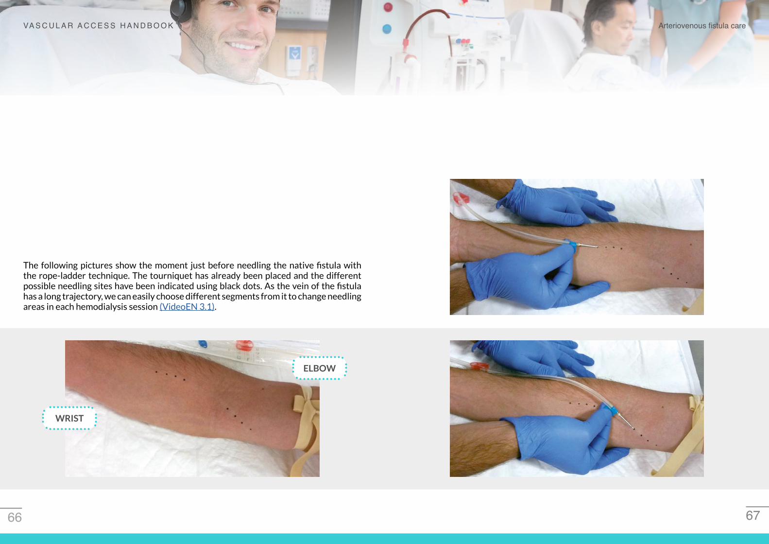
2.4. Location of the native fistulaEach upper limb is divided into two seg-ments: forearm (from the hand to the elbow) and arm (from the elbow to the shoulder). The creation of the native fis-tula is recommended as close to the hand as possible, that is, in the forearm at the wrist level in order to produce matura-tion of a long segment of the vein throu-ghout the upper limb, which will facilitate later the use of the rope-ladder needling technique to carry out hemodialysis (see Section 03 and VideoEN 3.1). The picture on the right shows a long segment of the vein throughout the forearm available for needling.
ELBOW
HAND

Arteriovenous fistula creation
3938
VA S C U L A R A C C E S S H A N D B O O K
2.5. Arteries and veins used in the upper limb
When the vascular surgeon creates a nati-ve fistula, she/he uses an artery and a vein in the upper limb, either in the forearm or the arm. The name given to the native fis-tula comes from the names of the artery and vein used to create it. The following
picture shows the main veins (intermittent line in blue dots) and arteries (solid line in red) in the forearm and arm that are used to create native fistulas.
BRACHIAL ARTERY
ULNAR ARTERY
RADIAL ARTERY
BASILIC VEIN
CEPHALIC VEIN
2.6. Types of native arteriovenous fistula in the upper limb
As shown by its name, the radio-cephalic fistula is formed by joining the radial artery and the cephalic vein in the forearm. The blood that circulates through the radial ar-tery will go straight into the cephalic vein through the anastomosis which, over some weeks, will dilate and get bigger and more resistant so that it can be needled and su-pply enough blood to the hemodialysis machine: this is what is called the maturation process of the radiocephalic fistula. It is considered the best choice for the native fis-tula as, once it has matured adequately, it lasts longer and has fewer complications than other fistulas. The pictures on the next page show the puncture process of a ra-diocephalic fistula for performing the dialysis treatment.

Arteriovenous fistula creation
4140
VA S C U L A R A C C E S S H A N D B O O K
The brachiocephalic and brachiobasilic fistulas are the result of the union or anastomosis at the elbow between the brachial artery and the cephalic vein and basilic vein, respectively. As a result of this union, dilation of both veins will occur at the arm level, which can later be used to carry out the dialysis treatment once the maturation period is over.
Even if the basilic vein has matured perfectly after performing the anastomosis, on occasions its location is too deep and this makes needling difficult, so a second intervention is required to bring it to the surface of the arm.

Arteriovenous fistula creation
4342
VA S C U L A R A C C E S S H A N D B O O K
2.7. The prosthetic fistula in the upper limb
Unlike the native fistula, the artery and the vein is not joined directly but is done through a tube of synthetic material (graft) which is placed under the skin of the upper limb so that it can be needled in the hemodialysis session (see Section 01 and the following pictures). The blue arrows in the first picture point the relief of the prosthetic fistula under the skin of the arm. The second picture shows this fistula already punctured with 2 needles just before connection with the dialysis machine.
SHOULDER
ELBOW
2.8. Fall-back techniques If all conventional vascular access options (native or prosthetic fistula) have been used up in both upper limbs, other solu-tions must be found so that the person with kidney disease can continue in the chronic hemodialysis programme. These are the so-called fall-back vascular acces-ses, among which the following two stand out:
THE PROSTHETIC OR GRAFT FISTULA IN THE LOWER LIMB. This involves placing a synthetic prosthe-sis (graft) in the thigh of the lower limb or leg, usually between the femoral artery and femoral vein. In the picture on the right, the synthetic material (graft) has already been placed and it can be seen in relief, just before needling, under the skin in the thigh of the lower left limb.
THE PROSTHESIS-CATHETER DEVICE (HeRo).
This combines the catheter and the pros-thetic fistula in the arm (see Sections 01 and 06).
KNEE

4544
Frequently asked questions by the person with kidney disease (FAQs)
What is the vascular access that must be avoided as much as possible due to associated complications?
• The catheter.
Who makes up the so-called multidisciplinary team?
• All the health professionals that look after you as well as you.
Which upper limb will be use to create the native fistula if I am left-handed?
• In the right arm as long as the results of the physical examination and ultrasound mapping are favourable.
Once the limb where the fistula will be created has been decided, which area is recommended for the operation?
• In the forearm, as close to the hand as possible.
What is the best type of native fistula?
• The radiocephalic fistula in the wrist.
What type of native fistula may require a second operation to bring it closer to the surface?
• The brachiobasilic fistula.
Section 02
What difference is there between a native fistula and a prosthetic fistula (graft)?
• Unlike the native fistula, the artery and the vein are not directly joined in the prosthetic fistula but through a tube made of synthetic material.
What are the so-called fall-back techniques?
• They are certain vascular accesses that can be used when all other possible conventional vascular accesses (native and prosthetic fistula) have been used up in both upper limbs.
Where is the prosthetic fistula placed in the lower limb?
• In the thigh.
What types of vascular access are combined to form the HeRO device?
• The catheter and the arm graft.

VA S C U L A R A C C E S S H A N D B O O K Procedures prior to vascular access creation
4746
ARTERIOVENOUS FISTULACARE
03AUTHORS
Néstor FontseréDavid HernánPatricia ArribasPilar CaroRaúl DarbasDaniel GallegoBelén MoragregaRamón Roca-TeyCarolina RubiellaAlberto SánchezAntonio Tombas

Arteriovenous fistula care
4948
VA S C U L A R A C C E S S H A N D B O O K
3.1. People in charge of fistula care
The multidisciplinary team is in charge of caring for your fistula (see Section 02). See that you, as a person with kidney disease, is included in the center of this team and, therefore, you are also responsible for this care.
3.2. When should you look after the fistula
Chronologically, the fistula must be looked after as follows:
• It must begin just after the vascular surgeon has created the fistula and you have left the operating room.
• It must continue while the fistula deve-lops (maturation).
• It must continue all the time the fistu-la is used to carry out the hemodialysis treatment.

Arteriovenous fistula care
5150
VA S C U L A R A C C E S S H A N D B O O K
3.3. Fistula care just after the operation
When you arrive home after the creation of the fistula, you must check the dressing that was put over the surgical wound. If you see that the dressing gets covered in more and more blood (bleeding), as in the following picture, you must immediately apply constant compression with the fingers of the other hand and go to the
Emergency Department of your hospital. At the same time, if you experience both intense pain in the hand and it also becomes cold and pale after having the fistula created, there may be insufficient blood reaching the hand, so you must go to the Emergency Department as well.
To make blood return to the heart more easily and avoid swelling (edema) of the limb where the fistula has been created, it is important to keep your arm raised, approximately at the height of the heart, by resting it on a cushion on the arm of the sofa or the chair where you are sitting or on a pillow when you are in bed.

Arteriovenous fistula care
5352
VA S C U L A R A C C E S S H A N D B O O K
If you experience pain in the surgical wound in the hours after the creation of the fistula, you must take the painkillers prescribed by your doctor.
Once you have a working fistula, it is im-portant to know that you must not take your blood pressure in the fistula-bearing arm, have an intravenous line or take blood for a blood test through one of the veins in this arm or directly through the fistula. Bear in mind that, from now on, the veins in this upper limb “must not be touched” and that the fistula must only be used to do the hemodialysis treatment.

Arteriovenous fistula care
5554
VA S C U L A R A C C E S S H A N D B O O K
The next two images show a person with kidney disease during the hemodialysis session. The yellow arrow points to the cuff used to take the blood pressure.
The watch is being worn on the right wrist and blood pressure is being taken on the right arm, which is not used for dialysis. CORRECT.
Blood pressure being taken in the left arm: the cuff has been placed on the fistula-bearing arm. NOT CORRECT.

Arteriovenous fistula care
5756
VA S C U L A R A C C E S S H A N D B O O K
3.4. Fistula care during the maturation period
It is recommended that you perform exer-cises before and after fistula creation, for example by compressing a rubber ball with your hand, as shownin the picture on the right (VideoEN 1.1). The aim of this exercises is to accelerate the fistula matu-ration process.
Once the surgical stitches have been removed and the professionals in charge of you give their approval, it is very important that you do these exercises. Bear in mind that the more time you spend doing them each day, the better the fistula maturation process will be.
If the fistula is not developing adequately (lack of maturation), it is essential to find out why not as soon as possible. For this, it is necessary to do a complete fistula exploration, which must include an ultrasound scan, in order to see where the exact problem lies that prevents its maturation (see Section 04). Once the problem has been detected, the fistula should be attempted to repair whenever possible (see Section 05).

Arteriovenous fistula care
5958
VA S C U L A R A C C E S S H A N D B O O K
Needling of the native fistula can start two weeks after its creation, never before, but the exact moment must be decided on a case-by-case basis from this date. In most case needling of this native fistula begins about a month after its creation.
To avoid the appearance of hematomas, needling of the prosthetic fistula should start between 2 and 4 weeks after its creation, when the swelling caused by the operation has gone down and the trajectory of the synthetic tube can be easily felt under the skin.
3.5. Care during the period of use of the fistula
No compression must be placed on the fistula-bearing limb as it can obstruct normal blood flow and cause the fistula to stop working (thrombosis) so that it can no longer be used for hemodialysis. Therefore, it is important not to wear tight-fitting clothes, watches, bracelets and occlusive bandages. It is advisable not to lift heavy
weights with the fistula-bearing arm or do brusque exercises and impact sports with it, either. You must not lie on the fistula-bearing arm to sleep. You must avoid sharp changes in temperature (saunas).

Arteriovenous fistula care
6160
VA S C U L A R A C C E S S H A N D B O O K
The fistula must be routinely needled by specialized nursing staff working in the hemodialysis units (never by a nursing staff with no knowledge or specific skill) or by yourself after a period of training.
Infections can get into the body when the fistula is needled. Thus, cleaning or asepsis measures of the fistula to eliminate microbes and avoid this must be stepped up.
To ensure this:
1. You must wash the fistula-bearing limb with soap and water before going into the dialysis room as shown in the picture on the right.
2. The nursing staff must disinfect the needling area using an antiseptic liquid that will be applied just before the needle’s insertion.

Arteriovenous fistula care
6362
VA S C U L A R A C C E S S H A N D B O O K
To make it easier to needling the native fistula, the vein must be dilated beforehand by compressing it above the fistula. This compression can be done by the person him/herself or by the application of a tourniquet (see the following pictures). This compression should not be performed to needle the prosthetic fistula.
COMPRESSIONBY TOURNIQUET
COMPRESSIONBY HAND
Some people with low pain tolerance can benefit from the use of local anaesthetic (cream or spray) before needling the fistula, as shown in the following picture.

Arteriovenous fistula care
6564
VA S C U L A R A C C E S S H A N D B O O K
THERE ARE THREE DIFFERENT FISTULA NEEDLING TECHNIQUES
1. Rope-ladder needling technique (VideoEN 3.1).
2. Area needling technique (VideoEN 3.2).
3. Buttonhole needling technique or constant needling in the same place (VideoEN 3.3).
1. ROPE-LADDER NEEDLING TECHNIQUE (VideoEN 3.1).This the method recommended for needling the fistula. Like the steps of a ladder, needling is spread out along the vein segment of the native fistula or the body of the prosthetic fistula which means that, in each dialysis session, two new sites are chosen to insert the needles. The greater the distance between the two needles, the more effective the hemodialysis treatment is. Although the insertion of the needle may hurt more on occasions, the advantage of using the rope-ladder technique is the absence or low development of aneurysms (large dilatations of the vein). The main problem is that this requires a length enough vein to allow to rotate needling.
Arteriovenous fistula care
6766
VA S C U L A R A C C E S S H A N D B O O K
The following pictures show the moment just before needling the native fistula with the rope-ladder technique. The tourniquet has already been placed and the different possible needling sites have been indicated using black dots. As the vein of the fistula has a long trajectory, we can easily choose different segments from it to change needling areas in each hemodialysis session (VideoEN 3.1).
ELBOW
WRIST

Arteriovenous fistula care
6968
VA S C U L A R A C C E S S H A N D B O O K
2. AREA NEEDLING TECHNIQUE (VideoEN 3.2). Although it is the needling method most frequently used in hemodialysis units, it should be avoided whenever possible. Through this technique, needling is repeatedly done in the same areas so that extremely close sites are used in every hemodialysis session to insert the needles. Although the pain caused by the needle as it enters is lower, the main drawback of this technique is that it progressively weakens the wall of the vein and aneurysms may develop, as shown in the following picture.
ANEURYSM
3. BUTTONHOLE NEEDLING TECHNIQUE OR CONSTANT NEEDLING IN THE SAME PLACE (VideoEN 3.3).
It is recommended that this technique be reserved for needling tortuous or deep native fistula or those with a short segment of vein available. The needles are always inserted exactly through the same hole in every hemodialysis session. The following picture shows the two entry sites of the blunt or rounded needles, so that a buttonhole is created for each needle. These needles go into the fistula vein through the tunnel previously formed under the skin.
NEEDLEENTRY HOLE

Arteriovenous fistula care
7170
VA S C U L A R A C C E S S H A N D B O O K
Whenever there is an easily fistula for needling, all highly motivated people being treated in a hemodialysis unit or at home can choose to needle themselves after a period of training (self-needling).
Once the needles have been inserted, they are securely fixed on the limb, as shown in the following picture, to prevent them from accidentally coming out during the dialysis session. This complication can be serious as it can cause an important bleeding.

Arteriovenous fistula care
7372
VA S C U L A R A C C E S S H A N D B O O K
The so-called hemodialysis lines, that is, the tubes of plastic that carry the blood between the needles and the dialysis machine, must be adequately fixed with enough space to avoid pulls and reduce the risk of accidentally pulling out the needles. For this reason, these should not be attached to anything movable, like the chair, the bed or the pillow.
To avoid these accidents, it is very important for nursing staff to always keep an eye on the needled limb throughout the whole hemodialysis session. In the following picture, a person is being treated by hemodialysis and has the body covered with a sheet except for the fistula-bearing limb, which must always be uncovered.

Arteriovenous fistula care
7574
VA S C U L A R A C C E S S H A N D B O O K
Once the hemodialysis session has finished, the needles must be removed and, to avoid bleeding from the holes, a compression must be made immediately with the fingers of the hand. During initial dialysis sessions, the nursing staff can carry out this compression but, later, the same person can do it with the other hand (see the following images).
COMPRESSION BY THE PERSON USING THE OTHER HAND
COMPRESSION BYTHE NURSING STAFF
Clamps must never be placed on the prosthetic fistula in order to compress it, and it is not advisable to use them in the native fistula. The compressive effect of the clamp may cause the fistula to stop working (thrombosis) and it may no longer be of use for hemodialysis.

Arteriovenous fistula care
7776
VA S C U L A R A C C E S S H A N D B O O K
After a period of time of manual compression on the needling sites, bandages or adhesive dressings will be placed on these same points that can be removed after 24 hours. The scab must never be taken off the needling wound.
If you observe bleeding through the dres-sing when you arrive home, the first thing you must do is keep calm and not panic. You must lift up the corresponding limb immediately and start re-compressing the needling site that is bleeding, just as you did in the hemodialysis room. If the bleeding does not stop after a reasona-ble amount of time despite this, go to the Emergency Department, maintaining ma-nual compression the whole time.
Arteriovenous fistula care
7978
VA S C U L A R A C C E S S H A N D B O O K
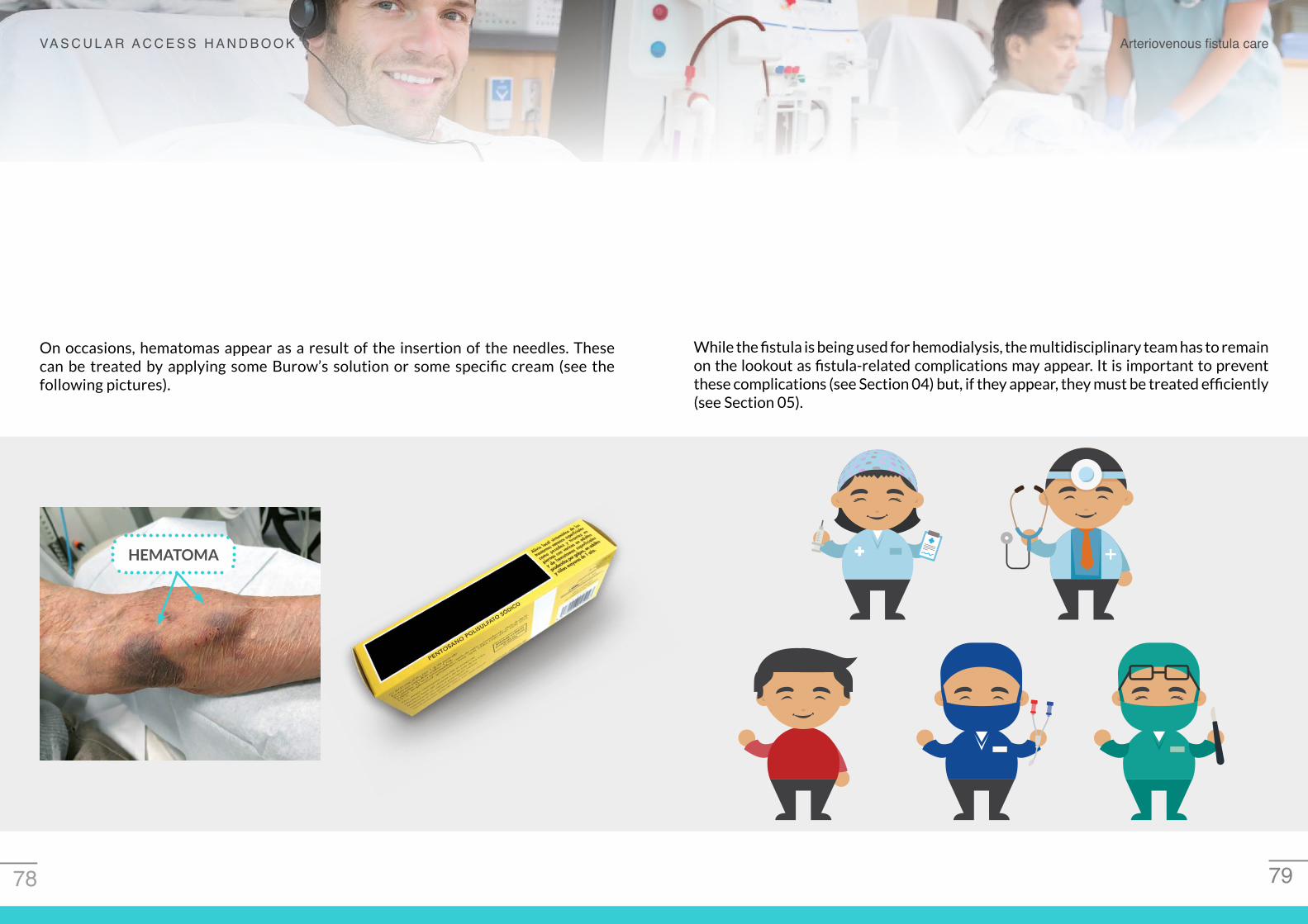
On occasions, hematomas appear as a result of the insertion of the needles. These can be treated by applying some Burow’s solution or some specific cream (see the following pictures).
HEMATOMA
While the fistula is being used for hemodialysis, the multidisciplinary team has to remain on the lookout as fistula-related complications may appear. It is important to prevent these complications (see Section 04) but, if they appear, they must be treated efficiently (see Section 05).
8180
Frequently asked questions by the person with kidney disease (FAQs)
In my dialysis unit there are very good professionals who look after my fistula. For this reason, I don’t really need to take care of it myself and I can stop worrying about it. Am I right?
• No, you are wrong. You form part of a multidisciplinary team and are also in charge of looking after the fistula.
Do I only have to worry about the fistula from the moment they have begun to needle it to do the haemodialysis session?
• No. Fistula care has to begin before, from the moment the vascular surgeon creates the fistula, and must continue throughout its development period. Then the care must continue while it is being used to carry out haemodialysis.
I had the fistula created this morning and left hospital with a clean dressing on the surgical wound. However, this afternoon I can see that as every hour passes, the dressing is getting more and more stained with blood. Is this normal?
• No, not at all. You must compress the dressing with the other hand and go to the Emergency Department immediately so that the wound can be checked.
I’ve had a fistula created today as my nephrologist told me that I would soon have to begin haemodialysis treatment. When I arrived home, the wound “woke up” and it started hurting. Can I take an ibuprofen tablets as painkillers?
• No. Bear in mind that you are in the pre-dialysis phase (ACKD) and cannot take anti-inflammatory tablets like ibuprofen as they can damage the kidneys even further and can speed up your entry into haemodialysis.
Section 03
Before leaving the hospital where I had the fistula created, the nurse took my blood pressure. I was told that it was fine, but it was taken in the opposite arm and I was told that my blood pressure couldn’t be taken in the fistula-bearing arm in the future. Why not?
• The external compression of the fistula may cause the blood to clot in the vein and the fistula to stop working (thrombosis). As a result, it may not be of use in the future for haemodialysis.
Yesterday I had the stitches taken out of the fistula in the wrist. The nurse gave me a rubber ball and told me to do exercises to help the development of the fistula. I am doing compression exercises with the ball half an hour every day. Is this enough?
• No, it’s very little time. Just think that the longer you spend doing exercises, the better and more quickly the fistula will mature.
Today I have been for a check-up at the ACKD outpatients’ clinic and I was told that my fistula is not maturing. Does this mean that I will have to start dialysis treatment through a catheter?
• Not necessarily. Some causes of lack of maturation can be corrected and, if this is your case, you will be able to begin haemodialysis using this repared fistula and avoid the use of the catheter.
Tomorrow I am having the stitches taken out of the fistula in my wrist. Will it be possible to start needling the fistula the day after tomorrow?
• No. The needling of the native fistula has to begin two weeks following its creation, never before. In most cases, the first needling of a mature native fistula is done around one month after its creation.

8382
This summer I want to get a suntan. Is it bad for me to allow direct sunlight on the fistula?
• Yes. Remember that you must avoid sharp changes in temperature.
Is it bad for my fistula to swim in the sea or a swimming pool?
• No, as long as the water temperature is no different from the environ-mental temperature.
Can any nurse needling my fistula on a routine basis?
• It is recommended that routine fistula needling should be performed by specialized nursing staff working in haemodialysis units with a specific level of knowledge and ability.
Today I have made the second session of hemodialysis and, because of the rotation in the rooms, a rookie nurse needled me. Is this right?
• No. The first cannulations of any new fistula must be carried out exclusively by experienced nursing staff in the haemodialysis unit.
Why do I have to wash the fistula-bearing arm with soap and water before going into the haemodialysis room if I have already had a shower at home?
• Because there is greater fistula hygiene and it reduces the risk of infection related with needling.
What is the best needling technique to cannulate the fistula?
• Rope-ladder needling.
What is the fistula needling method most frequently associated with the appearance of excessive vein dilatations, known as aneurysms?
• The area needling technique.
Frequently asked questions by the person with kidney disease (FAQs)
Section 03
My fistula has matured but there is only a short segment which can be needled. Which needling technique is recommended?
• The buttonhole technique .
Can I needling my own fistula?
• Yes, after a period of training, as long as the fistula is not difficult to needling.
When I am having haemodialysis, I often fall asleep and I like my whole body to be covered with a sheet, including the arm with the fistula in it. Is this right?
• No. The limb where the fistula is needled must always remain visible during the haemodialysis session in order to ensure the needles remain in place and don’t come out.
Can clamps be used after taking out the needles?
• Clamps must never be placed on a prosthetic fistulaand their use is not recommended in native fistula for the same reasonthat it is not advisable to take blood pressure in the fistula-bearing arm.
If I observe bleeding through the dressing when I arrive home after the haemodialysis session, I think it is best not to do anything and see how the bleeding develops. Is this right?
• No, not at all. What you must do is lift up your arm and compress the bleeding site again, as you did beforehand in the haemodialysis room. If bleeding continues after a reasonable amount of time despite everything, you must go to the Emergency Department of your hospitalbut continue compressing the bleeding site with your hand.

MONITORINGAND SURVEILLANCE OF THE ARTERIOVENOUS FISTULA
04
AUTHORS
Ramón Roca-TeyInés AragoncilloNéstor FontseréDaniel GallegoDavid HernánJose IbeasBelén MoragregaFlorentina RosiqueAntonio Tombas

Monitoring and surveillance of the arteriovenous fistula
8786
VA S C U L A R A C C E S S H A N D B O O K
4.1. The importance of fistula surveillance
In order to preserve the fistula for as long as possible to use it for haemodialysis, it is necessary to remain alert and keep an eye on it. Many people with kidney disease preserve their fistula in good conditions for years and do not need any other operation nor a catheter to be placed.
All the professionals looking after you and you, yourself, as the person with kidney disease, that is, the multidisciplinary team, are in charge of fistula surveillance (see Sections 02, 03 and 05).
4.2. People in charge of fistula surveillance

Monitoring and surveillance of the arteriovenous fistula
8988
VA S C U L A R A C C E S S H A N D B O O K
4.3. Fistula thrombosis
The main objective of fistula surveillance is to avoid its most frequent complication: thrombosis, which occurs when we say that “the fistula has stopped” and the blood can no longer flow.STOP
The most common cause of thrombosis is a narrowing (stenosis) in the vein of your fistula that gradually closes until the blood cannot flow and clots (stopped fistula).
This is the same as occurs when there is a narrowing in a tubing indicated by a yellow arrow in the following picture. Due to the presence of this stenosis, the liquid inside the tubing flows much slower than usual inside the tubing segment just before the narrow section (red colour). As a result, there is a high risk that the tubing will get jam and the liquid will not be able to get through. The black arrows indicate the direction in which the liquid flows inside the tubing.
STENOSIS
4.4. Fistula stenosis

Monitoring and surveillance of the arteriovenous fistula
9190
VA S C U L A R A C C E S S H A N D B O O K
Therefore, all professionals who take care of you and yourself should pay close attention to detect when this narrowing or stenosis occurs.
Once a stenosis has been detected in your fistula, the professionals of the multidisciplinary team of your Hospital will look for solutions to correct it and prevent fistula stopping.
?
The exploration or physical examination is very important to detect stenosis in the fistula. It is based on three basic aspects: inspection, palpation and auscultation (VideosEN 4.1, 4.2 and 4.3).
INSPECTIONThis consists of observing
the fistula
PALPATIONThis consists of touching the
fistula with the fingers
AUSCULTATIONThis consists of listening
to the fistula using a stethoscope
4.5. Physical examination of the fistula

Monitoring and surveillance of the arteriovenous fistula
9392
VA S C U L A R A C C E S S H A N D B O O K
The nursing staff must do a physical exami-nation of the fistula in every hemodialy-sis session just before needling begins (VideoEN 4.3).
The doctor in charge must perform the examination every month or when any pro-blem is detected.
You must check the fistula every day. Bear in mind that the fistula now forms part of your body and you must place the utmost importance on it. You must examine the whole fistula-bearing arm to see if there is anything abnormal (inspection) and touch the fistula to see if it is working (palpation). It
is also necessary to lift up the fistula-bearing arm above your heart for a few seconds every day and check to see if the blood empties from the fistula (collapse) because, if the fistula doesn’t empty completely, you may have a stenosis that is preventing this (VideosEN 4.1 and 4.2).
If you don’t notice this flow or vibration, the fistula may be stopped and is not working. In this case, you must phone your Hospital or Dialysis Centre and they will tell you what to do. Bear in mind that if your fistula has recently stopped, it might be possible to salvage it before the next dialysis session. Moreover, the longer the time that passes with a fistula stopped, the fewer the possibilities that exist to salvage it.
When you touch the fistula, you will notice a vibration which means blood flowing though the fistula that it is working.

Monitoring and surveillance of the arteriovenous fistula
9594
VA S C U L A R A C C E S S H A N D B O O K
The following problems may appear during the hemodialysis session and will make professionals suspect that there may be a stenosis in the fistula:
• Difficulty needling the fistula.
• Aspiration of clots during needling.
• Increase in venous pressure in the dialysis machine.
• Bleeding after withdrawing the needles despite having performed the usual compression on the needling sites.
4.6. Problems during the dialysis session
The force or quality of your fistula can be measured by calculating the amount of blood flowing through it. This is important for its surveillance. If this flow goes down over time (less force), it may indicate the progressive appearance of stenosis in your fistula. The flow can be calculated using devices located outside the dialysis machine (as in following picture) or already incorporated in the machine, as well as using an ultrasound device.
DIALYSISMACHINE
DEVICETO CALCULATEBLOOD FLOW
4.7. Calculation of the fistula flow

Monitoring and surveillance of the arteriovenous fistula
9796
VA S C U L A R A C C E S S H A N D B O O K
Ultrasonography is an imaging technique that does not harm the body, is painless and allows periodic fistula surveillance. It is a very important technique that has many advantages and which must be available in all hemodialysis rooms. Among other benefits, ultrasound allows the confirmation of a stenosis previously suspected by using other methods. The picture on the right shows a portable ultrasound machine where their probe and screen are highlighted (see Section 01).
4.8. Fistula exploration by using ultrasonography
ULTRASOUND SCREEN
ULTRASOUNDPROBE
In the picture on the right, a well-developed radiocephalic fistula is being explored by using the ultrasound probe. Everything captured by this probe can be seen directly, in real time, on the ultrasound screen.
ULTRASOUND SCREEN
ULTRASOUNDPROBE
WELL-DEVELOPED VEIN

Monitoring and surveillance of the arteriovenous fistula
9998
VA S C U L A R A C C E S S H A N D B O O K
In the following picture you can see the vein of native fistula (black colour) explo-red by using ultrasound that shows a stenosis between both yellow arrows.
In some cases, doubts still remain regarding the stenosis observed by ultrasonography, so a further exploration, called fistulography, will need to be done. This consists of injecting a contrast liquid into the fistula to be able to see the whole trajectory inside (VideoEN 5.1). In the following picture, you can see the contrast liquid that fills the fistula in black and an area of stenosis (or narrowing) indicated by the yellow arrows.
4.9. Fistula exploration by using fistulography

101100
Frequently asked questions by the person with kidney disease (FAQs)
Why do I have to surveillance my fistula?
• To preserve it and make sure I can have dialysis treatment through it for as long as possible.
How often do I have to fistula surveillance?
• Every day.
What is the most frequent complication of the fistula?
• Thrombosis, which occurs when we say the ”the fistula has stopped” and blood can no longer circulate through it.
What is the most frequent cause of fístula thrombosis in the fistula?
• It is a narrowing or stenosis in the fistula, which slowly closes until blood can no longer flow through it and finally clots (stopped fistula).
How can I detect myself this stenosis in my fistula?
• Through the exploration or physical examination.
What does the exploration or physical examination consist of?
• It is based on inspection, palpation and auscultation. But you should only do the inspection and palpation of the fistula.
What do you mean by inspection?
• It means you have to look the whole fistula-bearing arm to see if there is anything abnormal.
What do you mean by palpation?
• This involves touching the area of the fistula. Normally you will notice a vibration over the fistula.
Section 04
What does it mean if I don’t notice this vibration?
• It may be that the fistula has stopped and isn’t working.
What do I have to do if I am at home and I don’t notice this vibration?
• Call the hospital or hemodialysis unit and they will tell you what to do.
Although I don’t notice this vibration at home, can I wait until the next dialysis session to tell the doctor or nursing staff?
• No. Bear in mind that if your fistula has stopped recently, it may be possi-ble to salvage it before the next dialysis session. What’s more, the longer the fistula stops functioning, the fewer the possibilities to salvage it.
What is the raised arm test?
• It is a test that involves lifting up your fistula-bearing arm above the heart for a few seconds and then checking to see if the blood empties from the fistula (collapse). If the fistula is not completely empty, there may be a na-rrowing or stenosis that is preventing it from doing so.
How can I know if my fistula is good or bad?
• By measuring the amount of blood flowing through the fistula using some accessories located inside or outside the dialysis machine, or by using the ultrasound device.
Why is the ultrasound device important in the haemodialysis room?
• Because it allows us to confirm there is a stenosis in the fistula that has previously been suspected when using other surveillance methods.

Complications of the arteriovenous fistula
103102
VA S C U L A R A C C E S S H A N D B O O K
COMPLICATIONS OF THE ARTERIOVENOUS FISTULA
05AUTHORS
Marta BarrufetJoaquín VallespínDolores ArenasDaniel GallegoJorge GómezRamón Roca-TeyFlorentina RosiqueCarolina RubiellaAntonio Tombas

Complications of the arteriovenous fistula
105104
VA S C U L A R A C C E S S H A N D B O O K
5.1. Fistula complications An arteriovenous fistula does not have an expiry date and may be working for years without presenting any problem. However, complications that seriously affect fistula function can appear with the result that the fistula can no longer be used.
The multidisciplinary team (see Sections 02, 03 and 04) play a highly important role in detecting these complications and apply the appropriate treatment as soon as possible.
Anastomosis
ArteryVeinNATIVE FISTULA
5.2. Thrombosis and its treatmentThrombosis is the most frequent complication, both in the native fistula as well as the prosthetic fistula. Thrombosis occurs when a blood clot (thrombus) obstructs the inside of the fistula and the blood cannot flow (stopped fistula) (see Section 04).
As the thrombosed fistula can no longer be used to carry out hemodialysis, it is very important to do the treatment (thrombectomy) as soon as possible so that blood will flow inside the fistula again. The longer the fistula has stopped, the fewer the chances of salvaging it. Therefore, thrombosis must be considered a medical emergency.
Salvage treatment of the thrombosed fistula can be done in two different ways: through interventional radiology and through surgery.

Complications of the arteriovenous fistula
107106
VA S C U L A R A C C E S S H A N D B O O K
Thrombosis treatment through interventional radiologyThe vein is needled and a tube is inserted into it which allows the thrombus to be fragmented and aspirated.
Thrombosis treatment through surgery A small cut is made in the vein and the blood clot is removed.

Complications of the arteriovenous fistula
109108
VA S C U L A R A C C E S S H A N D B O O K
Fistulography of the stopped fistula (see the following picture on the right). The contrast liquid in black fills the artery and part of the fistula-bearing vein up to the yellow arrow where the thrombus (blood clot) obstructs the interior of the vein and prevents blood flow. As it is now, the fistula cannot be used to perform hemodialysis.
Anastomosis
ArteryVein
ARTERY
VEIN
ARTERY
Fistulography of a salvaged fistula following thrombosis (see the following picture on the right). An emergency intervention has been performed to salvage the fistula. This intervention has been a total success as it has eliminated all the clot (thrombus) and the blood can flow normally throughout the vein. Therefore, the fistula can now be used for hemodialysis. The yellow arrow on the fistulography shows the contrast (black) filling the whole segment of the salvaged vein and where the two needles can be inserted.
VEIN

Complications of the arteriovenous fistula
111110
VA S C U L A R A C C E S S H A N D B O O K
5.3. Stenosis and its treatmentFistula stenosis is the most frequent cause of thrombosis. As mentioned in Section 04, stenosis is a narrowing located at a specific point to the vein that gradually closes until blood can no longer get through and it clots inside the fistula (stopped fistula).
This is the same as the narrowing of a tubing: the narrow area of the tubing makes the liquid inside flow more slowly and there is a high risk of obstruction in it, as shown in the pictures on the next page.
Normal-sized tubing before the abnormal narrowing occurs. The black arrow shows the direction of the flow of the liquid inside.
Abnormal narrowed area of the tubing indicated by the yellow arrow. The liquid inside the tubing segment in front of the stenosis (red colour) flows much slower than usual and there is a high risk of obs-tructing the tubing.
The abnormal narrowing of the tubing has been repaired before it can obstruct the tubing (thrombosis) and the liquid inside can flow normally.

Complications of the arteriovenous fistula
113112
VA S C U L A R A C C E S S H A N D B O O K
Therefore, fistula stenosis must be treated before blood clots and thrombosis occur.
This corrective treatment of fistula stenosis can be done in two ways, depending on the location and extension of the stenosis: through surgery and through interventional radiology.
STENOSIS
STENOSIS
This involves creating a new anastomosis, that is, a new union to join the artery and vein, but further up, just above the problem area, thereby avoiding the stenosis.
Stenosis treatment through surgery

Complications of the arteriovenous fistula
115114
VA S C U L A R A C C E S S H A N D B O O K
Stenosis treatment though interventional radiology The vein is needled and a ball is inserted into the narrowed area of the vein (see the following picture and VideoEN 5.1). This ball acts like a balloon which is inflated in this area, thereby dilating the vein and thus opening up a new path for the blood to flow normally through the fistula.
Fistulography with two stenoses in the fistula, shown by the arrows (see the following picture on the right). The contrast goes through the two narrow areas in the fistula with difficulty. If nothing is done, these stenoses will gradually close up and the blood will stop flowing and clot, forming a blood clot or thrombus (stopped fistula).
Anastomosis
ArteryVein
VEIN
STENOSIS
ARTERY

Complications of the arteriovenous fistula
117116
VA S C U L A R A C C E S S H A N D B O O K
Fistulography of a fistula with two stenoses already repaired (see the following picture on the right). An intervention has been performed to fistula repair before it stops. This intervention has gone well and the two stenoses have been successfully dilated. Now, blood can now flow normally throughout the vein. As the two stenoses have disappeared, the contrast (black) in the fistulography easily fills the whole vein of the fistula.
VEIN
ARTERY
On occasions, there is no maturation of the fistula, that is, after creating the anastomosis, the vein does not develop enough and it cannot be used for dialysis. The most frequent cause is the presence of a stenosis in the trajectory of the vein, which can be treated as explained in Section 5.3. If the person with kidney disease is in this pre-dialysis stage, the aim is to repair the fistula before beginning the hemodialysis programme so that the first session can be done through a mature fistula.
5.4. Management of the non-matured fistula

Complications of the arteriovenous fistula
119118
VA S C U L A R A C C E S S H A N D B O O K
5.5. Management of the infected fistula
On occasions, an infection may appear in the needling area of the native or prosthetic fistula, as shown in the following picture indicated by the yellow arrow. In these cases, there may be fever associated with pain, heat, reddening and suppuration in the infected area. It is very important to identify the exact microbe responsible for the infection by taking a sample of the pus in order to provide the correct antibiotic treatment.
The presence of an infection in the fistula must always alert the multidisciplinary team, as the responsible microbe may pass into the blood and infect the whole body. Mild cases of infection can be treated with oral antibiotics and by changing the needling area. The most serious cases require hospital admission, intravenous administration of antibiotics and, in some cases, surgical treatment by closing the native fistula or taken out the infected graft in the case of prosthetic fistula.
MAXIMUM ALERT
MAXIMUM ALERT

Complications of the arteriovenous fistula
121120
VA S C U L A R A C C E S S H A N D B O O K
5.6. Fistula steal and its treatmentIn some cases of native fistula in the upper limb, a large amount of blood that goes through the artery to the hand is diverted towards the vein of the fistula, so that the fistula steals blood which should reach the hand and does not reach it. This complication appears with greater frequency when the brachial artery is used to create the fistula (see the picture on page 117).
This steal can cause a tingling sensation or numbness in some fingers and there may be a loss of strength and pain in the hand. In comparison to the other hand, the hand of the fistula-bearing limb is cold, pale and, in severe cases, one or several ulcers can be seen on the finger tips because of the lack of blood at this level (see the picture on page 118).
NATIVE FISTULA
VEIN
ARTERY

Complications of the arteriovenous fistula
123122
VA S C U L A R A C C E S S H A N D B O O K
Normal left handof a person
with kidney disease.
Right hand of the same person, corresponding to the fistula-bearing limb.
In relation to the other hand, it is pale, cold and has lost strength.
In more serious cases of intense pain and the appearance of ulcers, the treatment is generally surgical by using various techniques aimed at reducing the anastomosis size to decrease the amount of blood that passing directly through the artery to the vein and thus to increase the arrival of blood to the hand. These techniques aim to solve the problem by keeping the fistula functional but, sometimes, there is no other choice but to close it so that the blood can circulate to the hand.
• True aneurysm. This is an excessive dilatation of the vein in the native fistula. It appears more frequently when using the area needling technique (see Section 3). Although these aneurysms usually remain stable over time, the multidisciplinary team must be on the lookout just in case they begin to change.Treatment, usually surgical, must be performed when pain or skin changes appear above the aneurysm (thinning, redness, erosions, crusts) as they are associated with an increased risk of bleeding.
• False aneurysm (pseudoaneurysm). This is a false aneurysm because in reality it is a quickly-growing hematoma located beneath the skin. It is secondary to the bleeding from the hole in the vein wall caused by one of the needles used to puncture the fistula. Treatment is usually conservative through ultrasound-guided external manual compression and, if this is not enough, through local thrombin injection. Surgery must be reserved for cases where none of the other treatments have worked.
5.7. Management of fistula aneurysms

Complications of the arteriovenous fistula
125124
VA S C U L A R A C C E S S H A N D B O O K
FISTULA ANEURYSMS
5.8. Heart failure by high-flowThe presence of excessive flow (see Section 04) in an extremely developed fistula, particularly when the brachial artery is used to create the arteriovenous anastomosis, can cause an overload in the heart and heart failure.
Decompensation may also appear in the heart when there is a fistula with normal flow but with a previously weakened heart. For this reason, it is important to always have a check-up using heart ultrasound (echocardiography) before the surgeon creates the fistula.
Surgery is used to treat heart failure because of excess flow and consists of making the anastomosis smaller or totally closing the fistula in the most serious cases.

127126
Frequently asked questions by the person with kidney disease (FAQs)
Although the fistula has stopped, can I have a haemodialysis session through it?
• No, it is impossible. The blood has to flow freely through the fistula so that it can be used. Therefore, an urgent intervention is needed to salvage the fistula.
If a stenosis is detected in the fistula, is it always appropriate to adopt a conservative wait-and-see strategy to observe what happens?
• No, the multidisciplinary team must assess the risk of thrombosis in this stenosis and, if it is high, a corrective intervention of the stenosis must be done without delay.
If I have the stenosis repaired through dilation in the interventional radiology suite, the treatment definitive?
• It is not usually a definitive treatment. Typically, it lasts for many months but will depend on each case.
Will I have general anaesthetic to repair the stenosis in the fistula?
• It is not necessary. Local anaesthetic is usually enough.
Why is it important to carry out early treatment on the non-mature native fistula in the pre-dialysis stage?
• To be able to initiate the haemodialysis programme through this repaired fistula and thus avoid the use of catheter.
Yesterday, Friday, before needling, the nurse realised that I had a reddish spot on one of the areas of the vein that had been needled the previous week. When I got up this morning, I noticed that I didn’t feel well, I am
Section 05
feverish and there is pus in the spot. Do I have to go to the hospital or can I wait until the next haemodialysis session on Monday?
• What you explain could be a fistula infection and therefore, you have to go to the hospital today (not Monday) so that the nephrologist can examine the fistula, collect a sample of the pus and begin antibiotic treatment.
I am having dialysis through a catheter in the neck and last week I had a brachiocephalic fistula created. Since then, the hand of the fistula-bearing limb has lost strength and is cold and pale. Should I be worried?
• The fistula is ‘stealing’ some blood from your hand. It is very important that your nephrologist looks at the fistula and the hand in the next dialysis session. At the moment, you must increase the temperature in your hand by putting on a glove.
I have been having dialysis through the same fistula for a long time and it has 2 dilatations in it that have not grown. Does anything need to be done?
• Aneurysms in the fistula must not be treated unless you experience pain or there are changes in the skin above the aneurysm (reddening, thinning, erosions, crusts).
I had a heart attack 4 years ago and over the last 2 months I have been having dialysis through a tunnelled catheter. My nephrologist wants to create a fistula but has said that I need an echocardiography beforehand. Why do I need this exploration?
• As a fistula can overload the heart and cardiac failure, the nephrologist must check the state of the heart following the heart attack. If the heart has been weakened, the best thing to do is continue dialysis through the catheter.
Central venous catheters
129128
VA S C U L A R A C C E S S H A N D B O O K
CENTRALVENOUS CATHETERS
06AUTHORS
Teresa MorenoPatricia ArribasDolores FerrerDaniel GallegoJorge GómezFredzzia GraterolDavid HernánJose IbeasRamón Roca-TeyFlorentina RosiqueAmalia TalensAntonio Tombas

Central venous catheters
131130
VA S C U L A R A C C E S S H A N D B O O K
6.1. What is a central venous catheterThe catheter is a hollow, flexible, plastic tube that is placed in a large vein in the body, usually in the neck, although it can also be placed in the leg, as can be seen in the following pictures.
CATHETER
VEIN (BLUE)
KIDNEY
NECK LEG
As can be seen in the following picture, each catheter consists of three different parts:
• Tip. This is located internally. If the catheter is placed in the neck vein, the tip must be located just below the heart in an area of the so-called right atrium.
• Body. The blood flows inside it. The so-called tunnelled catheter incorporates a cuff.
• Limbs. These are external. In each dialysis session, the blood is sent to the dialysis machine through one of the limbs and goes back to the body once cleansed through the other. Between sessions, both limbs are sealed with caps.
CATHETER CUFF
CATHETER BODY
CATHETER LIMB
CATHETER TIP

Central venous catheters
133132
VA S C U L A R A C C E S S H A N D B O O K
The catheter allows the hemodialysis session to be done just after placement (it does not need a maturation period) and we avoid needling to access the blood. As the catheter is already placed inside the vein, only the caps must be taken out and the limbs connected to the hemodialysis machine to start the session.
However, in comparison to the fistula, the catheter usually creates more problems, dialysis treatment is lower in quality and has a greater risk of infection. Therefore, the information that the professionals of the multidisciplinary team have to transmit to the person with kidney disease is that a catheter is inferior to a fistula and does not substitute it. The order of preference of the three types of vascular access is shown on the next page (see Section 02).
ORDER OF PREFERENCE FORTHE THREE TYPES OF VASCULAR ACCESS
ACCEPTABLEPREFERRED
AVOID AS MUCHAS POSSIBLE
PROSTHETIC FISTULA
NATIVEFISTULA
CATHETER

Central venous catheters
135134
VA S C U L A R A C C E S S H A N D B O O K
There are 2 different types of catheter: the tunnelled and the non-tunnelled catheter.
• Tunnelled catheter. This is called as such because the body of the catheter is attached using a cuff in a tunnel located beneath the skin. In this way, the risk both of infection and movement of the catheter is reduced. This type of catheter should always be placed if it is impossible to create any kind of fistula, as the veins have been used up or in special circumstances like serious heart disease, which would get much worse after fistula creation. It is also placed awaiting the creation or maturation of a fistula when the person has had to start hemodialysis program without time for the fistula to perform or mature, respectively. The yellow arrow in the picture on the right shows the catheter body fixed in a tunnel beneath the skin.
EXIT SITE
CATHETER LIMBS
6.2. Types of catheter
TUNNELLED CATHETER
VEIN IN THE NECK
CATHETER BODY REPRESENTED INSIDE
THE VEIN WITHDOTTED LINES
CATHETER BODYBENEATH THE SKIN
(SUBCUTANEOUS TUNNEL)
RIGHT ATRIUM OFHEART WHERE THE CATHETER
TIP IS LOCATED
HEART
EXIT SITE
CATHETER LIMB

Central venous catheters
137136
VA S C U L A R A C C E S S H A N D B O O K
• Non-tunnelled catheter. There is no tunnel and the catheter body goes directly out of the body from the vein needling point. As a result, the risk of infection is higher than in the tunnelled catheter and it should not be left in place more than two weeks. It takes less time to place than the tunnelled catheter and it has been used to save many lives of people with kidney disease without a catheter in emergency life-threatening situations due to heart failure or potassium spikes. The yellow arrow in the image on the right shows the exit site of the catheter body, which leaves the vein directly without going through any tunnel. CATHETER
LIMBS
6.3. Catheter placementThe catheter is usually placed without hospitalisation (in the outpatients’ clinic) and with local anaesthetic (you will be awake but the area where the catheter will be placed is numb).
The process of placing the catheter can open the door to bodily infections. Therefore, a catheter should always be placed in an operating room or in a sterile room (total asepsis measures), that is, there are no microbe that can cause infection. The following picture shows an interventional radiology suite used for this end.

Central venous catheters
139138
VA S C U L A R A C C E S S H A N D B O O K
It is recommended the hemodialysis catheter be placed using ultrasound guidance and by qualified medical staff with experience.
ULTRASOUND PROBE
ULTRASOUND SCREEN
PORTABLE ULTRASOUND MACHINE
The following picture shows a physician (standing) just before an ultrasound-guided dialysis catheter placement in a person with kidney disease (lying down). Right before the local anesthesia is put in, the physician is looking for the vein in the right side of the person’s neck through the ultrasound probe and, everything that is captured by this probe, can be seen directly, in real time, on the ultrasound screen. It is important to highlight that both people in the image are wearing masks to prevent microbe transmission while the catheter is being placed.
ULTRASOUNDPROBE
ULTRASOUND SCREEN
PERSON WITHKIDNEY DISEASE

Central venous catheters
141140
VA S C U L A R A C C E S S H A N D B O O K
6.4. Catheter handlingThe haemodialysis catheter must only be used for performing the dialysis treatment and must not be used for other purposes (for example, to administer drugs outside the dialysis session).The catheter should be handled only when strictly necessary to avoid one of its complications, which is infection. The more the catheter is handled, the greater the risk of infection.
The connection and disconnection of the catheter to the dialysis machine in each session must be done exclusively by specialised staff in the dialysis unit (VideoEN 6.1).
As with its placement, the hemodialysis catheter handling must be carried out following strict asepsis measures (without microbes) to prevent infection. These measures include the professionals who handling the catheter, people around at that moment and the catheter carrier him/herself. For example, it is very important to wear a mask during the connection and disconnection of the catheter to the dialysis machine by both professionals as the person carrying it to prevent microbes from infecting the catheter (see the following picture and VideoEN 6.1).

Central venous catheters
143142
VA S C U L A R A C C E S S H A N D B O O K
Once the catheter has been connected to the dialysis machine, blood goes out from the body through one of the catheter limbs to the dialysis machine (red arrow in the following picture) and the cleansed blood goes back to the body from the ma-chine via the other limb (blue arrow in the following picture).
Once the dialysis session is over, you must check the dressing that has been put on it. This dressing must always be clean and dry and cover the whole catheter, as in the following picture. If you see at any time that this dressing is getting stained with blood or coming off and the catheter is visible, you must go to the Dialysis unit or Emergency Department at your hospital immediately.
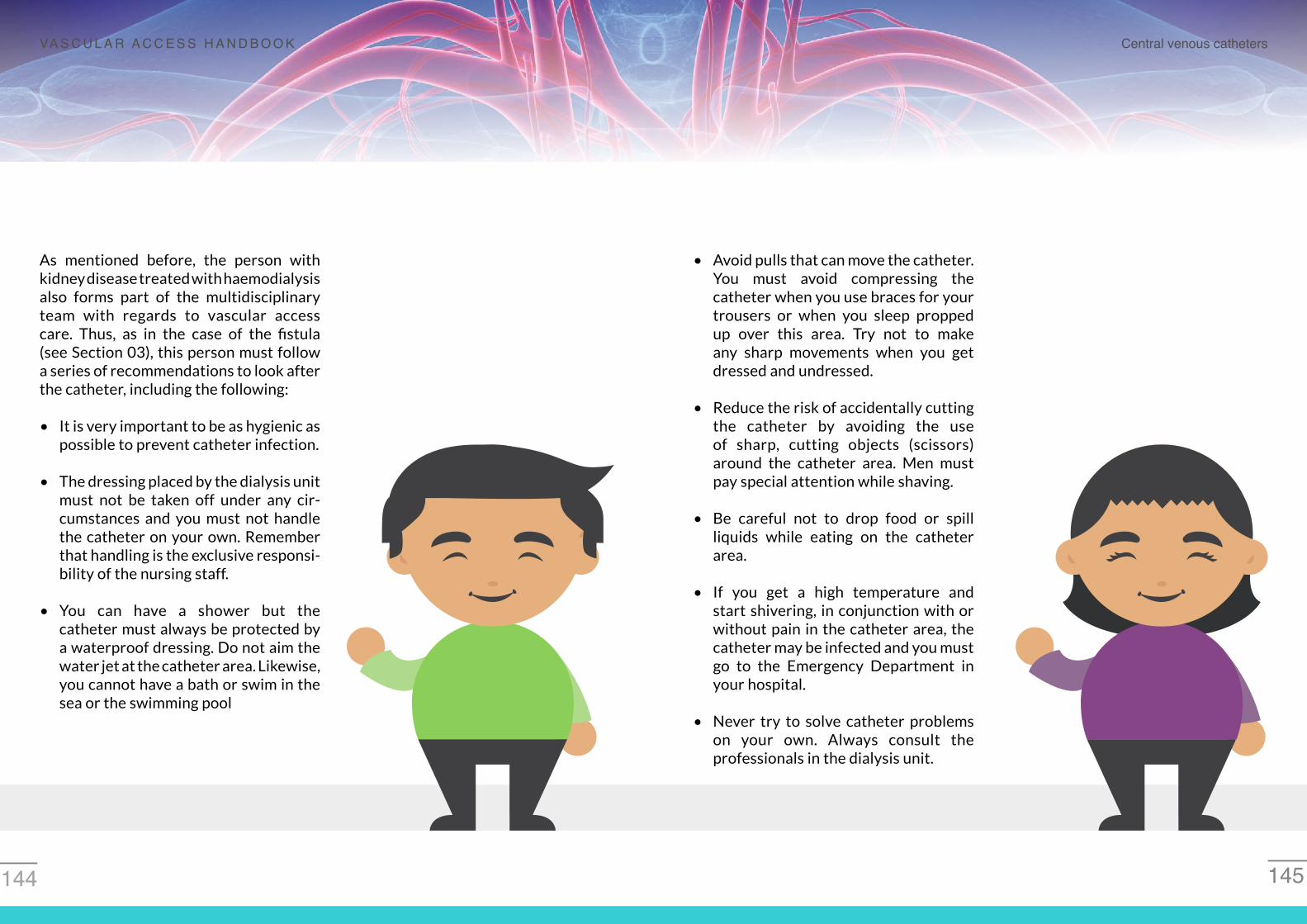
Central venous catheters
145144
VA S C U L A R A C C E S S H A N D B O O K
As mentioned before, the person with kidney disease treated with haemodialysis also forms part of the multidisciplinary team with regards to vascular access care. Thus, as in the case of the fistula (see Section 03), this person must follow a series of recommendations to look after the catheter, including the following:
• It is very important to be as hygienic as possible to prevent catheter infection.
• The dressing placed by the dialysis unit must not be taken off under any cir-cumstances and you must not handle the catheter on your own. Remember that handling is the exclusive responsi-bility of the nursing staff.
• You can have a shower but the catheter must always be protected by a waterproof dressing. Do not aim the water jet at the catheter area. Likewise, you cannot have a bath or swim in the sea or the swimming pool
• Avoid pulls that can move the catheter. You must avoid compressing the catheter when you use braces for your trousers or when you sleep propped up over this area. Try not to make any sharp movements when you get dressed and undressed.
• Reduce the risk of accidentally cutting the catheter by avoiding the use of sharp, cutting objects (scissors) around the catheter area. Men must pay special attention while shaving.
• Be careful not to drop food or spill liquids while eating on the catheter area.
• If you get a high temperature and start shivering, in conjunction with or without pain in the catheter area, the catheter may be infected and you must go to the Emergency Department in your hospital.
• Never try to solve catheter problems on your own. Always consult the professionals in the dialysis unit.

Central venous catheters
147146
VA S C U L A R A C C E S S H A N D B O O K
6.5. Catheter complications
1. Catheter dysfunction.On occasions, a blood clot is formed in the tip or the body of the catheter which totally or partially obstructs the passage of blood to the dialysis machine and prevents the session from progressing normally. Initial treatment consists of purging the limbs of the catheter with a syringe. If this is not enough, a drug used for dissolving the clot (for example, urokinase) must be put into the catheter body for a specific period of time to dissolve the clot. If the obstruction still persists, the catheter will need to be replaced by a new one. In the following picture, the black arrow shows a clot inside the catheter tip that is causing the dysfunction.
2. Catheter-related infection.If the catheter is infected, antibiotics must always be administered and, in some cases, the catheter must be taken out. There are three types of catheter infection:
• Infection of the exit site (see the next page). It is the mild type of infection but if it is not treated adequately, the microbe may propagate further away through the blood. There may be a slight fever and a secretion and even pus in the exit site.
• Infection of the subcutaneous tunnel (tunnellitis). This is associated with fever and referred pain in the catheter.
• Propagation of the bacteria to the blood that has infected the catheter (bacteraemia). It is the most serious catheter infection and may put the person’s life in danger. In this situation, there is high fever and shivering.
The most frequent tunnelled catheter-related problems are impaired function (dysfunction) and infection.
SUBCUTANEOUS TUNNEL
Infection located in the exit site of a tunnelled catheter. The yellow arrow shows the reddened exit site due to the infection.
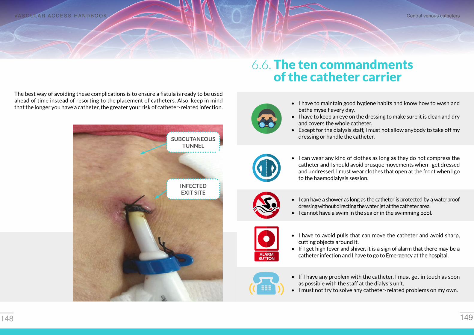
Central venous catheters
149148
VA S C U L A R A C C E S S H A N D B O O K
The best way of avoiding these complications is to ensure a fistula is ready to be used ahead of time instead of resorting to the placement of catheters. Also, keep in mind that the longer you have a catheter, the greater your risk of catheter-related infection.
SUBCUTANEOUS TUNNEL
INFECTEDEXIT SITE
6.6. The ten commandments of the catheter carrier
• I have to maintain good hygiene habits and know how to wash and bathe myself every day.
• I have to keep an eye on the dressing to make sure it is clean and dry and covers the whole catheter.
• Except for the dialysis staff, I must not allow anybody to take off my dressing or handle the catheter.
• I can wear any kind of clothes as long as they do not compress the catheter and I should avoid brusque movements when I get dressed and undressed. I must wear clothes that open at the front when I go to the haemodialysis session.
• I can have a shower as long as the catheter is protected by a waterproof dressing without directing the water jet at the catheter area.
• I cannot have a swim in the sea or in the swimming pool.
• I have to avoid pulls that can move the catheter and avoid sharp, cutting objects around it.
• If I get high fever and shiver, it is a sign of alarm that there may be a catheter infection and I have to go to Emergency at the hospital.
• If I have any problem with the catheter, I must get in touch as soon as possible with the staff at the dialysis unit.
• I must not try to solve any catheter-related problems on my own.

151150
Frequently asked questions by the person with kidney disease (FAQs)
Why do I need to have a catheter placed?
• Your kidneys have stopped working correctly and you need dialysis so that your blood can be cleaned and toxin-free. The catheter allows blood to be taken out of your body, passed through a dialysis machine where it is cleaned and sent back toxin-free to the body through the same catheter.
Are the catheter and the fistula the same? There are other persons who have hemodialysis with me that don’t have a catheter and are needled in the arm.
• The fistula is the best way of having dialysis but, in your case, as you don’t have a fistula yet or because it can’t be used at the moment, you need a catheter to do dialysis. When you have a fistula created or it can be used if already created, you will be needled in the arm and the catheter won’t be needed.
Where will the haemodialysis catheter be placed? A neighbour in my block is having dialysis through a catheter in her belly.
• Your neighbour is having peritoneal dialysis. You will have a catheter placed in a vein, usually in the neck, to do haemodialysis.
Can I have a shower with the haemodialysis catheter inside me?
• Yes, but you must always avoid getting the catheter wet. It must always protected by a waterproof dressing and you must not direct the water jet at the area where the catheter has been placed. If you notice that the dressing gets damp, you can use a hairdryer.
What clothes can I wear if I have a catheter?
• Whatever you like. Try not to make any brusque movements when you get dressed or undressed. But when you go for dialysis, we recommend wearing clothes that can be opened at the front to allow easy access to the catheter.
Section 06
This summer is very hot and I would like to have a swim in the sea or the swimming pool. I suppose that, even though I have a tunnelled catheter, there won’t be a problem. Is that right?
• No. Any kind of immersion in water is forbidden, even in the bath. The catheter may get wet and must always be covered and dry.
Most people have dialysis in my unit through a fistula with 2 needles in each session. I have dialysis through a tunnelled catheter and I avoid this needling, but my nephrologist wants me to have a fistula created. Why can’t I use the catheter all the time?
• The catheter is not as good as the fistula and does not substitute it. In comparison to the fistula, the catheter causes many problems, the blood is not cleansed as well and the risk of infection is much higher.
I have a catheter placed in my neck for haemodialysis. Yesterday, Friday, I had the dialysis session as usual but this morning I have had a high fever, shivering and pain in the catheter area. I have taken a tablet to bring down the temperature and I feel better now. What should I do? Can I wait till Monday’s session to tell the staff in the haemodialysis unit about the incident?
• No, you can’t wait. You may have a catheter infection and it is dangerous. You must go to Emergency at your hospital immediately.
Yesterday I went to both the cardiology outpatients’ clinic, as my heart is very weak, and the ACKD outpatients’ clinic in my hospital. My nephrologist told me I have to start haemodialysis soon and that I will have a tunnelled catheter inserted as a vascular access. Why can’t I have a fistula?
• Although the fistula is the best vascular access to begin hemodialysis sessions in most people, in your specific case creating a fistula may severely impair your heart disease and therefore, the catheter is the best option for you.
153152
Bibliography
Spanish Clinical Guidelines on Vascular Access for Hemodialysis. J. Ibeas, R. Roca-Tey, J. Vallespín, T. Moreno, G. Moñux, A. Martí-Monrós, et al. by the Spanish Multidis-ciplinary Vascular Access Group (GEMAV). Nefrología 2017; 37 (Suppl 1): 1-191. Available at www.gemav.org
Videos of the handbook
VideoEN 1.1. The arteriovenous fistula creationhttps://www.youtube.com/watch?v=yLyc52aABMIThe surgery to create an arteriovenous fistula is described. The exercises for fistula maturation are showing at the end of the video.
VideoEN 3.1. Rope ladder needling techniquehttps://www.youtube.com/watch?v=Qk7KHHsAgJ8Detailed description of the fistula needling process by using the ropeladder technique.
VideoEN 3.2. Area needling techniquehttps://www.youtube.com/watch?v=2yfK4dMp6D0Detailed description of the fistula needling process by using the area technique.
VideoEN 3.3. Buttonhole needling techniquehttps://www.youtube.com/watch?v=hAJnoFrOpUcDetailed description of the fistula needling process BY using the buttonhole technique.
VideoEN 4.1. Daily self-examination of the fistula: arteriovenous fistula without stenosishttps://www.youtube.com/watch?v=0RRpJ5xD6UYThe process of exploring a fistula is described step by step, in this case without any narrowing orstenosis, which must be performed daily by the person with kidney disease.
VideoEN 4.2. Daily self-examination of the fistula: arteriovenous fistula with stenosishttps://www.youtube.com/watch?v=jBNzN_am4sUThe process of exploring a fistula is described step by step, in this case with a narrowing or stenosis,which must be performed daily by the person with kidney disease.
VideoEN 4.3. Fistula exploration by the nursing staffhttps://www.youtube.com/watch?v=tVwyJqTmTrgDetailed description of the fistula exploration by the nursing staffin the dialysis room just before start to needling.
VideoEN 5.1. What is a fistulography?https://www.youtube.com/watch?v=hmxTCxJN6KcFistulography description procedure during the fistula stenosis treatment by using interventional radiology.
VideoEN 6.1. Dialysis connection through a catheterhttps://www.youtube.com/watch?v=k7456MZUbYwThe process of connecting a catheter to the hemodialysis machine to carry out a dialysis session is described in detail.

155154
Glossary
• ACKD (Advanced Chronic Kidney Disease) Outpatients’ Clinic. Outpatients’ clinic where you are given detailed information relating to the solutions that are available when your kidneys enter the ACKD phase.
• Advanced Chronic Kidney Disease (ACKD). Phase of chronic kidney disease in which the kidneys hardly function.
• Asepsis. Absence of microbe that can cause infection.
• Anastomosis. Specific point where the artery and the vein are joined to form the fistula.
• Aneurysm. Excessive dilatation of the vein in the fistula.
• Antibiotics. Drugs that destroys the microbe responsible for the infection.
• Antiseptic. Liquid disinfectant used to prevent infection.
• Area needling. Type of fistula needling in which the same areas of the vein in the native fistula or the body of the prosthetic fistula is needled repeatedly.
• Arm. Part of the upper limb of the person located between the elbow and the shoulder.
• Artery. Blood vessel in charge of taking blood from the heart to the person’s organs.
• Asepsis measures. Set of measures applied in hospitals and dialysis centres to prevent the introduction and transmission of the microbe.
• Bacteraemia. Presence of microbe in blood.
• Blood vessels. Hollow structures in the shape of a tube where blood is transported throughout the organism.
• Blood. Red-coloured liquid that flows inside all blood vessels.
• Brachiobasilic fistula. Type of native fistula resulting from the anastomosis between the brachial artery and the basilic vein.
• Buttonhole needling. Type of needling in the fistula in which the same exit site is needled in all haemodialysis sessions following the creation of a tunnel beneath the skin.
• Catheter limbs. Located in the outer part of the catheter. The blood is sent to the dialysis machine through one of the limbs and returns to the body toxin-free through the other in each haemodialysis session.
• Catheter body. It makes up most of the catheter and is where the blood flows constantly for the whole haemodialysis session.
• Catheter cuff. This is incorporated into the tunnelled catheter body and allows it to be attached to the subcutaneous tunnel.
• Catheter tip. Part of the catheter that remains inside the body of the person. If the tunnelled catheter is placed in the neck, the tip must be located in the right atrium of the heart.
• Central venous catheter. Type of vascular access that consists of a hollow, flexible, plastic tube placed in the large vein of the neck or the leg.
• Chronic kidney disease. Irreversible loss of kidney function. It is classified into different stages depending on its importance.
• Dialysis lines. Plastic tubes that transport the blood between the needles of the fistula or the catheter limbs and the dialysis machine.
• Dialysis machine. Machine used to do the haemodialysis treatment.
• Echocardiography. Ultrasound of the heart.
• Edema. Accumulation of liquid beneath the skin.
• Fall-back techniques. Vascular accesses used when a conventional vascular access is no longer viable.
• Filter or dialyser. Exact place in the dialysis machine where the blood cleanses the blood, which returns to the body toxin-free.
• Fistula auscultation. Part of the physical examination of the fistula that consists of listening to the fistula by using a stethoscope.
• Fistula inspection. Part of the physical examination of the fistula that consists of observing it.
• Fistula maturation. Process of development until the fistula is valid for cannulation with two needles and for use in the haemodialysis session.
• Fistula stenosis. Narrowing of the artery or vein in the fistula.
• Fistulography. Imaging technique that allows the exploration of blood vessels by administering intravenous contrast.

157156
Glossary
• Forearm. Part of the upper limb of the person located between the hand and the elbow.
• Hematoma. Accumulation of blood between the skin and the blood vessel.
• Hemodialyisis. Treatment that consists of eliminating toxins in the blood which are retained because of the chronic kidney disease by using a machine that is connected to the body through a vascular access.
• Hemodialysis machine. Machine used to carry out hemodialysis treatment.
• Heart. Vital organ that pumps blood through the arteries to distribute it throughout the person’s body.
• Mask or surgical mask. Type of mask used in hospitals and dialysis centres in order to avoid the microbe transmission from the nose and mouth.
• Microbe. A minute organism that can only be seen through a microscope, and which can cause infection.
• Microscope. Tool that allows us to see the microbe as they are too small to be observed with the naked eye.
• Multidisciplinary team. Group of people who look after the vascular access, including haemodialysis nursing staff, nephrologist, person with kidney disease, vascular surgeon and interventional radiologist.
• Native arteriovenous fistula. Type of vascular access that consists of joining an artery and vein so that, after a period of maturation, this vein can be cannulated with two needles to carry out the hemodialysis session.
• Palpation of the fistula. Part of the exploration of the fistula that consists of touching it with the fingers.
• Prosthesis-catheter device (HeRO). Fall-back device that combines the catheter and the prosthesis in the arm.
• Prosthetic arteriovenous fistula. Type of vascular access that consists of joining an artery and a vein though a synthetic tube (graft) so that, after a period of maturation, this graft can be cannulated by two needles to carry out a haemodialysis session.
• Radiocephalic fistula. Type of native fistula resulting from the anastomosis between the radial artery and the cephalic vein.
• Right atrium. Part of the heart where the tunnelled catheter tip is located when placed through the vein in the neck.
• Rope-ladder needling. Type of needling in the fistula in which needling is distributed along the whole trajectory of the vein of the native fistula or the body of the prosthetic fistula.
• Sterile. Object or substance that is microbe-free.
• Stethoscope. An apparatus used to auscultate or listen to the fistula.
• Thrombosis. A clot in the fistula that prevents the blood flowing normally inside it, thereby making it impossible to carry out haemodialysis treatment. Usually, the thrombosis is preceded by a stenosis.
• Thrombus. Blood clot responsible for thrombosis of the fistula.
• Tunnellitis. Infection of the subcutaneous tunnel where the catheter body is located.
• Ultrasonography. Imaging technique that is not painful or harmful and allows the blood vessels to be explored using the emission and reception of ultrasound echoes and their transmission into images.
• Ultrasound device. Machine used to perform the ultrasound scan.
• Ultrasound probe or transducer. Part of the ultrasound device, like a microphone, that allows the fistula to be explored or to look for a vein to place the catheter in.
• Ultrasound screen. Part of the ultrasound device, similar to a TV screen, which allows everything captured by the ultrasound probe to be seen directly, in real time.
• Urokinase. Drug that is used to dissolve a blood clot, which causes dysfunction, in the tip or body of the catheter.
• Vascular access. Access to the blood to carry out a haemodialysis session. It may be a native arteriovenous fistula, a prosthetic arteriovenous fistula (graft) or a central venous catheter.
• Vein. Blood vessel responsible for taking the blood back to the heart from the person’s organs.
The following companies have collaborated in printing the Handbook