Ralph Dürr - d-nb.info
168
Transcript of Ralph Dürr - d-nb.info


Ralph Dürr
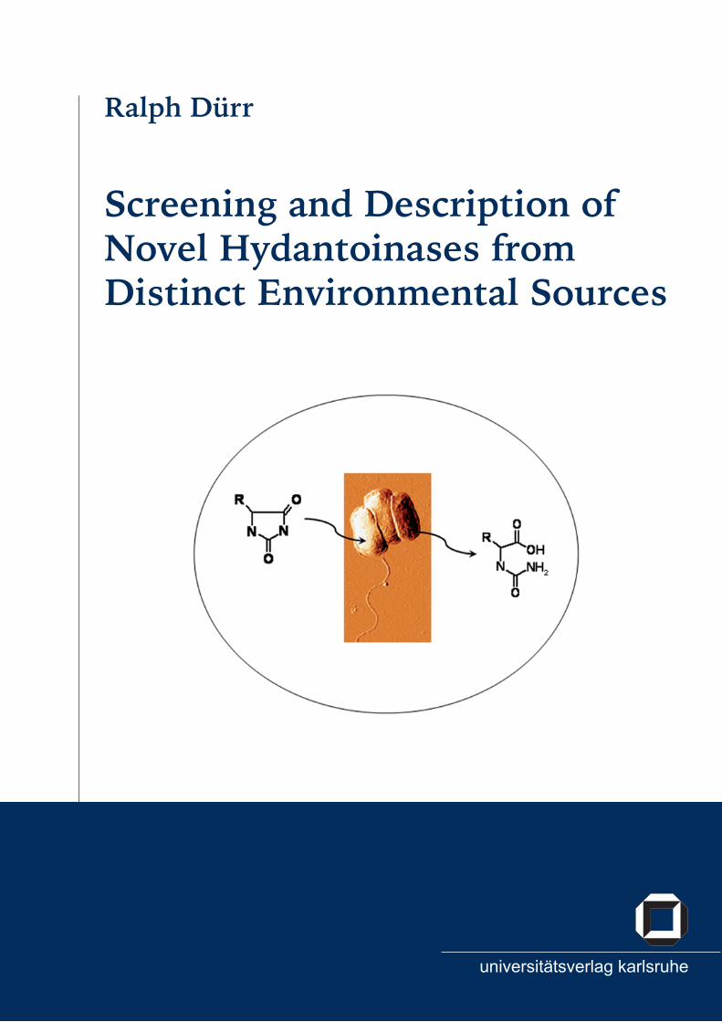
Screening and Description of Novel Hydantoinases from Distinct Environmental Sources


Screening and Description of Novel Hydantoinases from Distinct Environmental Sources
von Ralph Dürr

Universitätsverlag Karlsruhe 2007 Print on Demand
ISBN: 978-3-86644-173-6
Impressum
Universitätsverlag Karlsruhec/o UniversitätsbibliothekStraße am Forum 2D-76131 Karlsruhewww.uvka.de
Dieses Werk ist unter folgender Creative Commons-Lizenz lizenziert: http://creativecommons.org/licenses/by-nc-nd/2.0/de/
Dissertation, Universität Karlsruhe (TH)Fakultät für Chemieingenieurwesen und Verfahrenstechnik, 2007


SCREENING AND DESCRIPTION OF NOVELHYDANTOINASES FROM DISTINCT
ENVIRONMENTAL SOURCES
Von der Fakultät für Chemieingenieurswesen der Universität Karlsruhezur Erlangung der Würde eines Doktors der Ingenieurswissenschaften (Dr.-Ing.)
Genehmigte Dissertation
Vorgelegt von Ralph Dürr(Biologe t.o.)
aus Ulm/Donau
Referent: Prof. Dr. Christoph SyldatkKoreferent: Prof. Dr. Stephanie Burton
Tag des Kolloquiums: 05.07.2007


"Immer, wenn Dir eine Theorie als die wirklich einzig mögliche erscheint,nimm das als Zeichen, dass Du weder die Theorie noch das zu lösendeProblem verstanden hast."
Karl Popper in Objective Knowledge - an evolutionary approach (1972)
IX
Acknowledgment
I would like to thank the following persons for supporting the making of this Ph.D. thesis:
Prof. Dr. C. Syldatk for the provision and guidance through this Ph.D. project, for the op-portunity to conduct the work at his department, as well as for his help and the opportunityto realise my research visit in South Africa.
Prof. Dr. S.G. Burton and Prof. Dr. D.A. Cowan for the wonderful time in South Africa andfor the friendly and scientifically stimulating atmosphere at their departments.
Dr. Oliver Vielhauer from whom I learned so much about analytics, especially HPLC, andfor his guidance and friendship already throughout my diploma thesis – not to forget all therelaxed time we spent at the climbing wall.
Dr. Anke Neumann for her enormous help and introduction into molecular biology, as wellas for her patience and all her energy to show me that "genetics work".
Dr. J. Eberspächer for his deep knowledge of bacterial identification and our good discus-sions.
Dr. J. Altenbuchner for great help with molecular work.
Thanks to all friends and colleagues of the Chair of Technical Biology, University of Karls-ruhe, especially Sandra, Birgit and Vanessa, as well as Sebastian, Rudi, Frank, Ivana, Ana,Anja, Werner and Harald. I also acknowledge all the nice and helpful colleagues of the BioProcess Engineering Group, University of Cape Town, South Africa, and of the AdvancedResearch Centre for Applied Microbiology, University of the Western Cape, South Africa.
For the funding of this research and the scientific work in South Africa I gratefully acknowl-edge the Federal Ministry of Science and Education, Germany, and the National ResearchFoundation, South Africa, as well as the German Academic Exchange Service, Germany, andthe Federal State of Baden Württemberg, Germany.
The following are acknowledged for their support in providing access to soil samples:the South African National Parks, the joint Chinese-EU-SA MGATech project (QLRT-2001-01972), the Waikato University Antarctic Terrestrial Biology Program and Antarctica NZ.
My parents and my sister for their support and encouragement during all the times of mylife.
Special thanks to Karin for her never ending motivation and help throughout good and hardtimes of this thesis and in life.
X

Abstract
A screening program was conducted in order to find bacteria with "novel" hydantoinaseproperties. Bacteria with hydantoinase activity were recovered from terrestrial soil samplesof different geographic origins, including extreme environments using culture-basedscreening methods. All these isolates possessed the capability to transform the modelsubstrates D,L-5-benzylhydantoin and 5,6-dihydrouracil to the corresponding N-carbamoylamino acids. The recovered bacteria were identified based on 16S rRNA gene amplificationand were shown to belong to the genera Acinetobacter, Arthrobacter, Burkholderia, Bacillus,Delftia, Enterobacter, Flavobacterium, Microbacteriacae, Ochrobactrum, Pseudomonas, Staphylo-coccus, Stenotrophomonas and Streptomyces.These findings showed that microorganisms with hydantoinase activity are (i) distributedin various geographically distinct environmental habitats; (ii) distributed worldwide; (iii)found in certain bacterial genera. Furthermore, the presence of hydantoinase activity wasshown in genera in which hydantoinase activity has not previously been reported.
A greater selected number of the recovered bacterial isolates was used for further studies.The hydantoinases of these isolates showed a broad substrate spectrum in whole-cellactivity assays. No correlation between the hydantoinase specificity and a certain bacterialgenus and/ or a habitat could be obtained. Polymerase chain reaction (PCR) using degen-erate primer was applied for the amplification of a partial hydantoinase gene fragment.A successful hydantoinase amplification was not achieved for all strains tested, probablydue to the low sequence homology of hydantoinases. Phylogenetic analysis revealed thathydantoinases and dihyropyrimidinases can be clustered on the DNA-level accordingto their bacterial lineage. Two DIG-labelled DNA probes were used for the search ofhydantoinase genes in genomic DNA of the strains mentioned above and an Arthrobacterstrain collection. A positive hybridisation signal was obtained amongst others for A. ilicisDSM20138, A. psychrolactophilus DSM15612 and A. methylotrophus DSM14008 under verystringent hybridisation conditions. All three strains showed no hydantoinase activity inbiotransformations. Cryptic hydantoinase genes are assumed. They can be considered assilent/ cryptic because the hydantoinase is not expressed during growth, possibly due tothe absence of an appropriate inducer.
The wild-type strains Delftia sp. I24, Ochrobactrum sp. G21 and Bacillus sp. F18 were chosenfor further studies because all three strains were isolated from extreme habitats, eitherthermal spots or hypersaline lakes. The strains were described in detail using classical andmolecular biological identification methods as Delftia sp. I24, Ochrobactrum sp. G21 andBacillus megaterium F18. The methods used did not allow to identify the first two strains tothe genus level.The influence of bacterial growth on hydantoinase activity was investigated: Addition of
XI

NaCl to the growth media turned out to be favourable to achieve high enzyme activity forthe two halophilic strains Ochrobactrum sp. G21 and Bacillus megaterium F18. Salt stress isproposed as an activator for degradative enzymes and a co-regulation of the hydantoinasesystem, probably to get access to amino acids under hindered conditions. The hydantoinasefrom Ochrobactrum sp. G21 showed maximal induction by D,L-2-naphthylmethylhydantoin,from Bacillus megaterium F18 by D,L-5-tert-butylhydantoin and from Delftia sp. I24 withD,L-6-phenyl-5,6-dihydrouracil. Hydantoinase activity was highest in the late exponentialgrowth phase of Delftia sp. I24 when grown on growth medium (GM) supplementedwith or without 6-phenyl-5,6-dihydrouracil and for Bacillus megaterium F18 when grownin GM supplemented with 5% NaCl and D,L-5-tert-butylhydantoin. Highest activity ofOchrobactrum sp. G21 was obtainend in GM supplemented with 5% NaCl and D,L-2-naphtylmethylhydantoin, but growth was very slow.The biochemical properties of the hydantoinases were determined: The hydantoinase ofDelftia sp. I24 had a temperature and pH optimum of 30◦C and pH 9.0 and showed cofactorrequirement for Ni2+ and Co2+. The enzyme was stable at 4◦C and 20◦C for four days. Thehydantoinase of Ochrobactrum sp. G21 showed highest activity at 40◦C and pH 8.0 – 8.5.The hydantoinase was cofactor dependent, but no preference for one of the ions testedwas indicated. The enzyme lost activity during storage at all temperatures tested. Mostactivity remained after storage at 4◦C. The hydantoinase of Bacillus megaterium F18 hada temperature and pH optimum of 45 – 50◦C and pH 7.5 – 8.0. The hydantoinase showedcofactor dependency with preference to Mn2+ and Co2+. All three hydantoinases showed avery broad substrate range but with different activities regarding to the respective substrateand compared to each other.This is the first report on characterisation of a hydantoinase from the genus Delftia. Addi-tionally, no hydantoinase activity has been reported yet for halophilic Ochrobactrum and/or Bacillus species.
Of special interest was the elucidation of the genes being responsible for hydantoin anddihydropyrimidine degradation of the wild-type strains Delftia sp. I24 and Ochrobactrumsp. G21. The putative gene cluster of Delftia sp. I24 included four genes: an incompleteNADPH-dependent glutamate synthase (gltB), dihydropyrimidine dehydrogenase (pydA),permease (hyuP) and a D-hydantoinase (hyuH). The hydantoinase gene was expressed inE. coli and hydantoinase activity shown for D,L-5-(3-indolylmethyl) hydantoin and D,L-5-benzylhydantoin. The presence of a dihydropyrimidine dehydrogenase and the preferencein whole-cell biotransformations for 5,6-dihydrouracil rather than hydantoin indicates thatthese enzymes are involved in the pyrimdine reduction pathway. The putative gene clusterof Ochrobactrum sp. G21 comprised nine ORFs, six being potentially involved in hydantoin-hydrolysation: carbamoylase (hyuC), D-hydantoinase (hyuH), two transporters (OrfS1 andOrfS2) and two permeases (hyuP1 and hyuP2). The hydantoinase was as well expressed in E.
XII

coli and activity shown for the same substrates as for the cloned hydantoinase of Delftia sp.I24. This is the first report on the genetical organisation of hydantoin degradation for mem-bers of the genus Delftia and Ochrobactrum. Both, but especially the hydantoin-hydrolysinggene cluster of Delftia sp. I24 showed a high similarity with putative gene clusters fromclosely related bacteria. This included the presence of certain genes, gene pattern and geneorientation.Phylogenetic analysis on a protein level of the two "novel" hydantoinases, known hyd-antoinases and dihydropyrimidinases, including putative protein sequences, revealed thatthese enzymes can be clustered, with some exceptions, in the following groups: Rhizobialesfamily (Rhizo-Fam), Comamonadacae family (Com-Fam), Pseudomonas family (Pseud-Fam),Bacilli family (Bac-Fam) and Agrobacterium family (Agro-Fam). The highly conserved "his-tidine motif" of the superfamily of amidohydrolases as well as the conserved amino acidsforming the active site can be found for all protein sequences used in this study. A differencewas found in the substrate recognition sites, whereas some of the groups mentioned aboveshowed to posses the same recognition sites as known hydantoinases. All these findingsstrongly support the hypothesis of a common ancestor for all members of the superfamilyof amidohydrolases.
XIII

Zusammenfassung
Um Bakterien mit "neuen" Hydantoinaseeigenschaften zu finden, wurde ein Screeningdurchgeführt. Bakterien mit Hydantoinaseaktivität wurden mittels klassischer Anre-icherungsverfahren aus Bodenproben von geographisch verschiedenen Ökosystemenisoliert, inklusive extremer Habitate. Die gewonnenen bakteriellen Isolate besaßendie Fähigkeit, die Modellsubstrate D,L-5-Benzylhydantoin und 5,6-Dihydrouracil zuder jeweils korrespondierenden Carbamoylaminosäure umzuwandeln. Die Analyseder 16S rRNA Gensequenz zeigte, dass die gewonnenen Wildstämme den folgendenGattungen zuzuordnen sind: Acinetobacter, Arthrobacter, Burkholderia, Bacillus, Delftia,Enterobacter, Flavobacterium, Microbacteriacae, Ochrobactrum, Pseudomonas, Staphylococcus,Stenotrophomonas und Streptomyces.Hieraus konnte gefolgert werden, dass Mikroorganismen mit Hydantoinaseaktivität (i) indiversen geographisch verschiedenen Ökosystemen verteilt sind; (ii) weltweit vorkommen;(iii) hauptsächlich in gewissen Gattungen vorkommen. Dennoch konnten neue Gattungengefunden werden, für die bisher noch keine Hydantoinaseaktivität beschrieben wurde.
Für weiter führende Studien wurde eine größere Anzahl der isolierten Bakterienstämmeausgewählt. Die Mehrzahl der Hydantoinasen dieser Stämme zeigte in Ganzzell-Aktivitätstests ein breites Substratspektrum. Jedoch konnte kein Zusammenhang zwischender Substratspezifität der Hydantoinase und der Artzugehörigkeit und/ oder des Habi-tats des jeweiligen Vertreters gefunden werden. Zur Amplifizierung von bestimmten Hy-dantoinasegenabschnitten wurde die Polymerase Kettenreaktion (PCR) mit degeneriertenPrimern angewandt. Es konnte nicht für alle getesteten Bakterien ein Genabschnitt ampli-fiziert werden. Phylogenetische Studien mit den gefundenen partiellen Hydantoinasegenenund einigen in Datenbanken abgelegten Hydantoinasegenen zeigte, dass Hydantoinasenund Dihydropyrimidinasen in Abhängigkeit Ihrer Abstammung gruppiert werden können.Zwei DNA-Sonden wurden für die Suche nach Hydantoinasegenen in genomischer DNAder oben genannten Bakterien und einer Arthrobacter Stammsammlung verwendet. U.a.wurde ein positives Hybridisierungssignal, das die Präsenz einer möglichen Hydantoinaseandeutet, für die Stämme A. ilicis DSM20138, A. psychrolactophilus DSM15612 and A. methy-lotrophus DSM14008 gefunden. In Aktivitätstests mit ganzen Zellen zeigten alle drei Stämmekeine Aktivität für verschiedene Hydantoine und Dihydropyrimidine. So genannte "kryp-tische" oder "stille" Hydantoinasegene werden vermutet. Diese Gene werden möglicher-weise während des Zellwachstums nicht abgelesen, da ein geeigneter Induktor für die jew-eilige Hydantoinase nicht vorhanden war.
Die Wildstämme Delftia sp. I24, Ochrobactrum sp. G21 und Bacillus sp. F18 wurden fürweitere Untersuchungen herangezogen, da diese drei Stämme aus extremen Habitaten(heißen Quellen oder hypersalinen Seen) isoliert worden waren. Die drei Wildstämmekonnten mittels klassischer und molekularbiologischer Bestimmungsmethoden den fol-
XIV
genden Spezies zugeordnet werden: Delftia sp. I24, Ochrobactrum sp. G21 und Bacillusmegaterium sp. F18. Es war mit den angewandten Methoden nicht möglich, die Art derbeiden erstgenannten Stämme zu identifizieren.Weiterhin wurde der Einfluss von verschiedenen Wachstumsfaktoren auf die Hydan-toinaseaktivität untersucht: Die Zugabe von Salz zum Medium führte zu einer signifikan-ten Erhöhung der Hydantoinaseaktivität der halophilen Stämme Ochrobactrum sp. G21und Bacillus megaterium sp. F18. Möglicherweise veranlasst Salzstress die Expressionabbauender Enzyme und des Hydantoinasesystems, letzteres um Zugang zu Aminosäurenfür den Metabolismus unter erschwerten Bedingungen zu erhalten. Einen großer Effektauf die Hydantoinaseaktivität hatte die Zugabe von Induktoren zum Wachstumsmedium.Die höchste Aktivität wurden jeweils mit D,L-6-Phenyl-5,6-Dihydrouracil für die Hyd-antoinase von Delftia sp. I24, mit D,L-2-Naphtylmethylhydantoin für Ochrobactrum sp.G21 und D,L-5-tert-Butylhydantoin für Bacillus megaterium sp. F18 erzielt. Die bestenMedien bezüglich Hydantoinaseaktivität waren: Wachstumsmedium (GM) mit und ohneD,L-6-Phenyl-5,6-Dihydrouracil für die Hydantoinase von Delftia sp. I24, GM ergänzt mit5% NaCl und D,L-5-tert-Butylhydantoin für Bacillus megaterium sp. F18 und GM ergänztmit 5% NaCl und D,L-2-Naphtylmethylhydantoin für Ochrobactrum sp. G21. Für diebeiden erstgenannten Stämme wurde die maximale Hydantoinaseaktivität in der spätenexponentiellen Phase festgestellt. Dahingegen erreichte Ochrobactrum sp. G21 bei geringemZellwachstum die maximale Hydantoinaseaktivität erst nach einer Wachstumszeit von 96Stunden.Die Hydantoinasen der einzelnen Stämme besaßen die folgenden biochemischen Eigen-schaften: Die Hydantoinase von Delftia sp. I24 zeigte ein Temperatur- und pH-Optimumvon 30◦C und pH 9.0. Biochemische Studien weisen darauf hin, dass diese Hydantoinaseabhängig von den Cofaktoren Ni2+ und Co2+ ist. Stabilitätstests zeigten, dass dieseHydantoinase bei 4◦C and 20◦C stabil ist. Die Hydantoinase von Ochrobactrum sp. G21wies die höchste Aktivität bei 40◦C und pH 8.0 – 8.5 auf. Cofaktor-Abhängigkeit konntenachgewiesen werden, jedoch konnte keine Präferenz für eines der getesteten Ionenfestgestellt werden. Die Hydantoinase war bei keiner Lagertemperatur über vier Tagehin stabil. Die höchste Aktivität blieb bei einer Lagertemperatur von 4◦C erhalten. DieHydantoinase von Bacillus megaterium F18 wies ein Temperatur- und pH-Optimum von 45 –50◦C und pH 7.5 – 8.0 auf. Als benötigte Cofaktoren werden Mn2+ oder Co2+ angenommen,da bei Zugabe dieser Ionen in biochemischen Studien die höchste Aktivität erzielt wurde.Alle Hydantoinasen zeigten ein breites Substratspektrum, jeweils mit unterschiedlichenAktivitäten.Dies ist die erste Beschreibung und Charakterisierung einer Hydantoinase eines Vertretersder Gattung Delftia. Ebenso wurde in der Literatur bisher noch nicht über Hydantoinaseak-tivität von halophilen Ochrobactrum- und Bacillus-Spezies berichtet.
XV
Von speziellem Interesse war die Aufklärung der für den Hydantoinabbau verantwortlichenGene der Stämme Delftia sp. I24 und Ochrobactrum sp. G21. Das putative Genclustervon Delftia sp. I24 beinhaltete vier Gene: eine unvollständige NADPH-abhängige Gluta-matsynthase (gltB), eine Dihydropyrimidin-Dehydrogenase (pydA), eine Permease (hyuP)und eine D-Hydantoinase (hyuH). Das Hyantoinasegen wurde in E. coli exprimiert unddie Umsetzung von D,L-5-(3-Indolylmethyl) Hydantoin und D,L-5-Benzylhydantoin kon-nte nachgewiesen werden. Das Vorhandensein einer Dihydropyrimidin-Dehydrogenaseund die bevorzugte Umsetzung von 5,6-Dihydrouracil vor Hydantoin in Ganzzell-Biotransformationen zeigt an, dass diese Enzyme eine Funktion im Pyrimidineabbau haben.Das Gencluster von Ochrobactrum sp. G21 umfasste neun offene Leserahmen, wobei sechsdavon eventuell am Hydantoinabbau beteiligt sind: Carbamoylase (hyuC), D-Hydantoinase(hyuH), zwei Transporter (OrfS1 und OrfS2) und zwei Permeasen (hyuP1 und hyuP2). DasHydantoinasegen wurde ebenfalls in E. coli exprimiert und Aktivität wurde für dieselbenHydantoine wie für die Hydantoinase aus Delftia sp. I24 gezeigt. In der Literatur wurdennoch keine für den Hydantoinabbau verantwortlichen Gencluster für beide oben genan-nten Gattungen dargestellt. Beide, aber hauptsächlich das Gencluster von Delftia sp. I24,zeigten eine sehr hohe Ähnlichkeit mit putativen Genclustern nahe verwandter Arten. Dieszeigte sich in der Abfolge und Orientierung von Genen, die vermutlich im Hydantoinabbaubeteiligt sind.Phylogenetische Studien mittels der Aminosäuresequenzen der in dieser Arbeit gewonnen"neuen" Hydantoinasen, bekannten Hydantoinasen und Dihydropyrimidinasen, zeigten,dass diese Enzyme mit wenigen Ausnahmen gattungs- oder ordungsspezifisch in die fol-genden Gruppen eingeteilt werden konnten: Rhizobiales Familie (Rhizo-Fam), Comamonada-cae Familie (Com-Fam), Pseudomonas Familie (Pseud-Fam), Bacilli Familie (Bac-Fam) undAgrobacterium Familie (Agro-Fam). Alle verwendeten Enzyme wiesen hierbei das kon-servierte "Histidin-Motif" der Superfamilie der Amidohydrolasen auf. Ebenso wurden füralle Vertreter die konservierten Aminosäuren gefunden, die für die Bildung des aktiven Zen-trums verantwortlich sind. Ein großer Unterschied wurde für die Aminosäuren gezeigt, dievermutlich für die Substraterkennung verantwortlich sind. Bei manchen der oben genan-nten Familien zeigte sich hierbei eine hohe Übereinstimmung mit Substraterkennungsmo-tiven von beschriebenen Hydantoinasen. Die Gesamtheit dieser Ergebnisse bestätigt dieHypothese, dass alle Mitglieder der Superfamilie der Amidohydrolasen, und damit ebensoder Hydantoinasen, von einem gemeinsamen Vorfahren abstammen.
XVI

Contents
Contents
1 List of Publications 3
2 Introduction 52.1 Hydantoin Cleaving Enzymes . . . . . . . . . . . . . . . . . . . . . . . . . . . . 52.2 Screening for Hydantoinases . . . . . . . . . . . . . . . . . . . . . . . . . . . . 72.3 Hydantoinases – Classification and Properties . . . . . . . . . . . . . . . . . . 92.4 The Hydantoin Hydrolysing Gene Cluster . . . . . . . . . . . . . . . . . . . . . 132.5 Industrial Applications . . . . . . . . . . . . . . . . . . . . . . . . . . . . . . . . 15
3 Research Proposal 21
4 Summary Chapters I – IV 234.1 Summary Chapter I: Distribution of Hydantoinases . . . . . . . . . . . . . . . 234.2 Summary Chapter II: Biodiversity of Hydantoinases . . . . . . . . . . . . . . . 274.3 Summary Chapter III: Properties of Selected Hydantoinases . . . . . . . . . . 304.4 Summary Chapter IV: Gene Clusters for Hydantoin Degradation . . . . . . . 37
5 Future Development 41
6 References 43
7 Publications and Manuscripts 517.1 Chapter I: Distribution of Hydantoinases . . . . . . . . . . . . . . . . . . . . . 517.2 Chapter II: Biodiversity of Hydantoin Cleaving Enzymes . . . . . . . . . . . . 697.3 Chapter III: Properties of Selected Hydantoinases . . . . . . . . . . . . . . . . 997.4 Chapter IV: Gene Clusters for Hydantoin Degradation . . . . . . . . . . . . . 123
1

Whitepage
2

1 List of Publications
This Ph.D. is based on the following publications and manuscripts (see section 7.1 – 7.4),being summarized in section 4.1 – 4.4:
Chapter I:
Running title: Distribution of HydantoinasesDürr R., Vielhauer O., Burton S.G., Cowan D.A., A. Puñal, Brandão P.F.B., Bull A.T. and Syl-datk C. (2006) Distribution of hydantoinase activity in bacterial isolates from geographicallydistinct environmental sources.Journal of Molecular Catalysis B: Enzymatic 39: 160-165.
Chapter II:
Running title: Biodiversity of HydantoinasesDürr R., Biodiversity of hydantoin cleaving enzymes.(not published)
Chapter III:
Running title: Properties of Selected HydantoinasesDürr R., Brucher B., Eberspächer J., Vielhauer O., Burton S.G., Cowan D.A. and SyldatkC. Description and characterisation of the D-hydantoinases from three microorganismsisolated from extreme environments.(Submitted to Applied and Environmental Microbiology)(Part of the experimental work was done within the diploma thesis of Birgit Brucher.)
Chapter IV:
Running title: Gene Clusters for Hydantoin DegradationDürr R., Neumann A., Vielhauer O., Altenbuchner J., Burton S.G., Cowan D.A. and SyldatkC. Genes responsible for hydantoin degradation of a halophilic Ochrobactrum sp. G21 andDelftia sp. I24 – new insight into relation of D-hydantoinases and dihydropyrimidinases.(Submitted to Journal of Molecular Catalysis B: Enzymatic)
3

Whitepage
4

2 Introduction
2.1 Hydantoin Cleaving Enzymes
Concerning the IUPAC-nomenclature the chemical compound hydantoin is declared as"imidazolidin-2,4-dione", a 5-membered ring bearing two nitrogen atoms and two carbonylgroups (Fig. 1).
NHNH
O
OR
NH2
O
NH2
NHNH
O
OR
NH N
O
O
OH
O
NH
O
NH2
OH
NH2
O
O
ROH
NH2
O
R
OH
D-hydantoin
D-hydantoinase D-carbamoylase
D-N-carbamoyl amino acid D-amino acid
racemase or pH > 8.0
L-hydantoin
+ H2O
- H2O + H2O
+ H2O
- H2O
5,6-dihydrouracil
dihydropyrimidinase
ß-ureidopropionic acid ß-alanine
3-ureidopropionase
+ H2O
+CO2+ NH3
+CO2+ NH3
Figure 1: Reaction scheme of enzymes involved in hydantoin and dihydropyrimidine con-version.
Microbial enzymes are known to cleave hydantoins as follows (for details see: Syldatk et al.,1992; Syldatk and Pietsch 1995; Ogawa and Shimizu 1997; Syldatk et al., 1999): First, the hy-dantoin is hydrolysed by a hydantoinase by a ring opening step to form the correspondingN-carbamoyl amino acid. Second, a so-called "N-carbamoylase" (N-carbamoyl amino acidamidohydrolases) is able to cleave this intermediary product to the respective amino acid.Additionally, a racemase is able to racemise 5-monosubstituted hydantoins. This can alsooccur chemically under basic conditions (Fig. 1). In Fig. 1 only the D-specific cleavage isshown since almost all hydantoinases obtained in the present work are D-selective. Obvi-ously, the L-route of hydantoin cleavage is analogous to the D-route shown. Hydantoinasescan be grouped due to their stereospecifity in D-, L- and non-selective hydantoinases.
5
2 INTRODUCTION
Hydantoinases together with dihydropyrimidinases belong to the EC group of hydrolases,in detail to the EC 3.5.2 group of cyclic amidases. In the EC nomenclature hydantoinaseis an alternative name for dihydropyrimidinase (EC 3.5.2.2). Various enzymes with theability to catalyse the cleavage of dihydropyrimidines and hydantoins are known: e.g.Arthrobacter crystallopoietes DSM20117 (Siemann et al., 1999), Bacillus sp. AR9 (Sharma andVohra, 1997), Bacillus stearothermophilus SD-1 (Lee et al., 1994; Lee et al., 1995), Pseudomonassp. NCIM5109 (Sudge et al., 1998). The function of dihydropyrimidinases is the hydrolysisof dihydrouracil derivatives (Fig. 1), a reaction involved in the reductive pathway ofpyrimidine degradation. Dihydropyrimidinase and hydantoinase are not necessarily thesame enzyme (Syldatk et al., 1999). Thus, the name hydantoinase should be employed for allenzymes that hydrolyse hydantoin and/or 5-monosubstituted hydantoin derivatives andnot as a synonym for dihydropyrimidinases as stated in the EC-nomenclature (Syldatk et al.,1999). This was suggested since an Agrobacterium sp. and Arthrobacter aurescens DSM3745were able to cleave hydantoin and 5-monosubstituted hydantoin derivatives but notdihydropyrimidines (Runser and Meyer, 1993; May et al., 1998d). Additionally, an imidasefrom Blastobacter sp. was able to convert dihydropyrimidines as well as hydantoin, but herethe metabolic function was different compared to dihydropyrimidinases. Nevertheless, inthis study we will not strictly distinguish between these two enzymes because only a fewreal hydantoinases are known and, except of biochemical data, no further differentiation isknown.Besides dihydropyrimidinases and hydantoinases different hydantoin cleaving enzymesare known however with other metabolic functions (see Syldatk et al., 1999):
• Allantoinase (allantoin amidohydrolase, EC 3.5.2.5): Allantoin is part of the purine de-gradation pathway of plants and microorganisms. This enzyme hydrolysis allantoin.Most allantoinases have a high substrate specificity and low enantioselectivity.
• Carboxymethylhydantoinase (L-carboxymethylhydantoin amidohydrolase,EC 3.5.2.4): Carboxymethylhydantoinase enables the cleavage of L-carboxy-methylhydantoin to N-carbamoyl-L-aspartic acid, a reaction involved in thepyrimidine degradation pathway.
• Carboxyethylhydantoinase (not included in the EC nomenclature): This enzyme catal-yses the hydrolysis of hydantoin propionic acid to N-carbamoyl-L-glutamic acid. It isinvolved in histidine degradation.
• N-Methylhydantoinase (N-methylhydantoin amidohydrolase, EC 3.5.2.14): Thisenzyme is involved in the microbial degradation of creatinine, hydrolyses N-methylhydantoin to N-carbamoylsarcosine and is ATP-dependent.
6
2.2 Screening for Hydantoinases
• Imidase (not included in the EC nomenclature): This enzyme is probably involvedin the degradation of succinimide to succinamic acid. An imidase from Blastobactersp. A17p-4 had a broad substrate spectrum including imides, hydantoin, dihydro-pyrimidine but not 5-monosubsubstituted hydantoins.
From recent investigations on DNA and amino acid sequences of different amidohydrolasesand cyclic amidases with subsequent phylogenetic analysis it is known today that mostenzymes of that group not only share a number of highly conserved regions and invariantamino acid residues but are a product of a divergent evolution (May et al., 1998e; Holm andSander, 1997). Using structure and sequence homology, hydantoinases can be grouped inthe following two families.The superfamily of "amidases involved in nucleotide metabolism" including theL-hydantoinase from A. aurescens DSM3745, dihydropyrimidinase, allantoinase, dihy-droorotase, urease and others (like adenine deaminase, phosphotriesterase, not beingfound to be hydantoinases) and the family of "ATP - dependent cyclic amidases" includingN-methylhydantoinase and L-oxoprolinase. Not included are imidase, carboxymethyl-hydantoinase and carboxyethylhydantoinase because there is no sequence informationavailable for these enzymes. It is suggested that the urease-related amidases have evolvedfrom a common ancestor. This theory is confirmed by the findings of Taillades et al. (1998):in the primitive hydrosphere the formation of N-carbamoyl amino acids and hydantoinswas more effective than of α-amino acids, assuming a higher carbon dioxide concentrationthan that of formaldehyde. It implements that the microorganisms of that earlier times hadto use N-carbamoyl amino acids and hydantoins as C- and/ or N-source generating thehydantoinase and N-carbamoylase system.
2.2 Screening for Hydantoinases
It is only possible to cultivate a limited number of bacteria whereas the total amount ofbacteria is much higher. The extent of microbial and molecular diversity was shown byTorsvik et al. (2002) and Venter et al. (2004). By using a "whole-genome shotgun sequencingapproach" to microbial populations from seawater samples from the Sarragosa Sea nearBermuda the second group found DNA sequences attributed to 1800 genomic species with148 previously unknown bacterial phylotypes. More than 1.2 million unknown genes wherefound.Hydantoinases are known to be present in certain microorganisms. Two alternative wayscan be used to access hydantoinases. One possibility is to isolate microorganisms withhydantoinase activity and another to access directly the hydantoinase gene followed bycloning and expression of the enzyme. The first question is how it is possible to accessmicroorganisms with the ability to cleave hydantoins from this great number of bacteria as
7

2 INTRODUCTION
described above.Up to now, only culture based methods are described in literature with exception of a fewother approaches. There are two main ways of screening for bacteria with hydantoinase/carbamoylase activity: The most common method is the use of enrichment cultures inwhich an environmental sample (soil, sediment, water) is inoculated in a special mediacontaining the desired hydantoin as sole C- and/or N-source. Only bacteria with the abilityto use hydantoins are able to grow and can be isolated on agar plates in the next step.The other method is the direct use of bacterial isolates from culture collections. For bothways the hydantoinase activity of pure cultures needs to be proven by activity assays andtherefore the following methods can be used.Overlay assays in which bacterial colonies are grown on agar plates and hydantoinaseactivity is detected by a spot-test based on the direct detection of carbamoyl amino acidsin agar using the Ehrlich Reagent (Morin et al., 1986a). A simple method is the addition ofthe unsubstituted hydantoin and phenol red to the media for agar plates. The conversionof hydantoin leads to a decrease of the pH initiating a colour change in the agar. Thiswas used to isolate hydantoinase positive E. coli transformants and for the screening ofhydantoinase-producing bacteria from soil (Kim et al., 1997b). The disadvantage of thismethod is that it is not sensitive enough for L-selective hydantoinases which can notcleave unsubstituted hydantoins (May et al., 1998d). Chien and Hsu (1996) developed amicrotiter plate assay based on the detection of the produced carbamoyl amino acid bythe addition of dimethylaminobenzaldehyde to the reaction. This reagent is also used inthe so-called Ehrlich Test. The authors stated that this method is simple, rapid and highlysensitive in comparison to other methods. An analogous detection method was used toshow hydantoinase activity in crude extracts of microorganisms run on polyacrylamidegels under non-denaturing conditions.Nevertheless, these methods do not give evidence on the selectivity of the occurringreaction. A sensitive method for the differentiation of D- and L-selective hydantoinases isthe use of acrylamide gels with crude extracts from microorganisms. The backward enzymereaction is used for in situ product precipitation, which allows the location of the hydanto-inase on a polyacrylamide gel. This is possible because the soluble substrate N-carbamoyltryptophan will be converted to the poorly soluble indolylmethylhydantoin. The selectivityof hydantoinases can be detected by reactions either with D- or L-N-carbamoyl tryptophan(May et al., 1998c).Other detection methods are thin-layer chromatography (Lee et al., 1994), a spectropho-tography assay (Chevalier et al., 1989) and polarimetric methods for enzyme catalysedenantioselective reactions (Teves et al., 1999). The most commonly used analytical methodis HPLC (High Performance Liquid Chromatography). Whole cells, resting cells or thepure enzymes can be used in activity assays. The advantage of HPLC-analytics is that boththe substrate (hydantoins) and the corresponding products (carbamoyl amino acids and
8

2.3 Hydantoinases – Classification and Properties
amino acids) can be detected in a single run. As well the chiral separation of enantiomers isfeasible.Besides the detection methods mentioned above immunological and molecular detectionmethods were reported as well. Especially molecular detection methods will gain moreinterest since during the last decade much more genetical information of hydantoinases andthe corresponding enzymes is reported. From particular interest would be the developmentof genetical methods to access hydantoinase genes directly from strain collections, soil orgenomic libraries. Advantages are cost reduction and the loss of time intensive isolationmethods.Polyclonal antibodies were successfully developed for the detection of the L-hydantoinaseof Arthrobacter aurescens but they were not specific for the detection of the D-hydantoinaseof Agrobacterium sp. (Siemann et al., 1993a and b). LaPointe et al. (1994) developed a 1.5 kbDIG-labelled DNA probe derived from a recombinant hydantoinase gene of Pseudomonasputida DSM 84. The probe only hybridised with total DNA from Pseudomonas strains ofrRNA group I showing hydantoinase activity. More successful was the development of a122 bp DNA probe from the same organism as mentioned above to detect D-hydantoinasegenes in other bacterial genera by DNA and by colony hybridisation. The probe was specificwhile allowing 32% mismatch to detect D-hydantoinase activity in a range of bacteria (LaPointe et al., 1995).Recently, degenerate primers were reported for the amplification of a 330 bp dihydro-pyrimidinase gene fragment of Bacillus sp. TS-23 (Lin et al., 2005). The amplified 330 bpDNA fragment was used as a probe to find the dihydropyrimidinase gene within a genomiclibrary of the same strain. These degenerate primers were as well used in this study toamplify hydantoinase gene sequences from different environmental bacterial isolates. Oneproblem of the development of universal primers for a successful amplification of thehydantoinase gene is the low sequence homology of hydantoinases.
2.3 Hydantoinases – Classification and Properties
Hydantoinases can be grouped according to their stereospecificity into D-, L- andnon-selective hydantoinases. However, some hydantoinases showed a change instereoselectivity for different substrates. The L-hydantoinase from A. aurescensDSM3745 showed to be L-selective for D,L-benzylhydantoin whilst conversion of D,L-methylthioethylhydantoin was D-selective (May, 1998f). Similar observations were obtainedfor Flavobacterium sp. AJ-3912. D-specificity was only obtained for benzyloxymethylhyd-antoin, whereas the other substrates tested were cleaved L-specific (Yokozeki et al., 1987).Since almost all of the hydantoinases obtained in this study were D-selective the main fo-cus was on D-hydantoinases. L- and non-selective hydantoinases were less reported thanD-hydantoinases and found within the following species: Arthrobacter aurescens DSM3745
9

2 INTRODUCTION
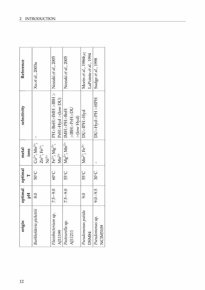
and DSM3747 (May, 1998f; Gross et al., 1990), Arthrobacter sp. DSM7330 (Völkel and Wagner,1995), Arthrobacter sp. DSM9771 (Wagner et al., 1996), Microbacterium liquefaciens (formerlyFlavobacterium sp.) AJ-3912 (Yokozeki et al., 1987; cited in Nozaki et al., 2005), a hyperther-mophilic archeon Methanococcus jannaschii DSM2661 (Chung et al., 2002), Pseudomonas sp.NS671 (Ishikawa et al., 1993, 1997; Watabe et al., 1992a) and Pseudomonas sp. RU-KM3s (Bur-ton et al., 1998; Buchanan et al., 2001; Matcher et al., 2004).Hydantoinase activity from Ochrobactrum anthropi (Pozo et al., 2002) has to be mentioned.Unfortunately the authors do not report on the stereospecificity of this enzyme. The strainwas able to cleave D,L-(2-methylthioetyl)-hydantoin to methionine and was induced by thishydantoin. The enzyme had a pH optimum of 9.0 and activity was significantly increasedby the addition of Ca2+, Na+, Cu2+, Co2+, Mg2+, Zn2+ and Fe3+.D-hydantoinases were reported for numerous microorganisms. A selection is shown in Ta-ble 1.
10

2.3 Hydantoinases – Classification and Properties
Tabl
e1:
Bioc
hem
ical
prop
erti
esof
sele
cted
D-h
ydan
toin
ase
enzy
mes
.(o
pt.:
opti
mal
;m
etal
ions
:in
crea
sed
acti
vity
inth
epr
esen
ceof
the
resp
ecti
vem
etal
ion
obse
rved
;B
nH:5
-ben
zylh
ydan
toin
;B
uH:5
-(se
c)-b
utyl
hyda
ntoi
n;D
U:5
,6-d
ihyd
rour
acil;
HPH
:5-h
ydro
xyph
enyl
hyda
ntoi
n;IB
H:5
-iso
buty
lhyd
anto
in;I
PH:5
-iso
-pr
opyl
hyda
ntoi
n;M
H:
5-m
ethy
lhyd
anto
in;
MPH
:5-
met
hyox
yphe
nylh
ydan
toin
;M
TH
:5-
(2-m
ethy
lthi
oeth
yl)
hyda
ntoi
n;PH
:5-
phen
ylhy
dant
oin;
PrH
:5-p
ropy
lhyd
anto
in;T
HE:
5-th
ieny
lhyd
anto
in)
orig
inop
tim
alop
tim
alm
etal
sele
ctiv
ity
Ref
eren
cepH
Tio
ns
Agr
obac
teri
umsp
.10
.060
◦ CN
i2+,M
g2+
BnH
>H
PH>
IPH
Run
ser
and
Ohl
eyer
IPI-
671
>M
TH
>Bu
H>
IBH
IBH
1990
;Run
ser
and
>M
H>
Hyd
(No
DU
)M
eyer
,199
3A
grob
acte
rium
9.0
60◦ C
Mn2
+T
HE>
MPH
>PH
Gri
fant
inie
tal.,
1998
;tu
mef
acie
ns>
BuH
>H
PG>
MH
Oliv
ieri
etal
.,19
81;
NR
RLB
1129
1A
char
yet
al.,
1997
Agr
obac
teri
umtu
me-
9.0
40–
60◦ C
-M
H>
HPH
>H
ydH
artl
eyet
al.,
1998
;fa
cien
sR
U-O
RBu
rton
etal
.,19
98A
rthr
obac
ter
crys
tal-
8.0
50◦ C
Zn2
+H
yd>
PH>
MTH
Siem
ann
etal
.,19
99lo
poie
tes
DSM
2011
7>
HPH
>Bn
H>
DU
Baci
llus
sp.A
R9
9.5
65◦ C
Mg2
+,N
i2+,
Hyd
>PH
>D
USh
arm
aan
dVo
hra
Mn2
+,C
o2+
>H
PH19
97Ba
cillu
sci
rcul
ans
8.0
–10
.075
◦ CM
n2+,N
i2+,
Hyd
>PH
>M
TH
>Lu
ksa
etal
.,19
97C
o2+
PrH
>IB
HBa
cillu
sst
earo
ther
mo-
8.0
65◦ C
Mn2
+H
yd>
DU
>PH
Kim
etal
.,19
97a;
Lee
philu
sSD
-1>
HPH
etal
.,19
94,1
995
Baci
llus
stea
roth
erm
o-9.
560
◦ CM
n2+,C
o2+,
MH
>M
TH
>IB
H>
Muk
ohar
aet
al.,
1994
;ph
ilus
NS1
122A
Ni2
+IP
H>
BnH
Ishi
kaw
aet
al.,
1994
11
2 INTRODUCTIONor
igin
opti
mal
opti
mal
met
alse
lect
ivit
yR
efer
ence
pHT
ions
Burk
hold
eria
pick
ettii
8.0
50◦ C
Co2
+,M
n2+,
-X
uet
al.,
2003
aZ
n2+,F
e2+,
Ni2
+
Flav
obac
teri
umsp
.7.
5–
9.0
60◦ C
Fe2+,M
g2+,
PH>
BnH
>IM
H>
IBH
>N
ozak
ieta
l.,20
05A
J111
99M
n2+
PrH
>H
yd>
(low
DU
)Pa
steu
rella
sp.
7.5
–9.
055
◦ CM
g2+,M
n2+
IMH
>PH
>Bn
HN
ozak
ieta
l.,20
05A
J112
11>
IBH
>Pr
H>
DU
>(l
owH
yd)
Pseu
dom
onas
putid
a9.
055
◦ CM
n2+,F
e2+
DU
>IP
H>
Hyd
Mor
inet
al.,
1986
b,c;
DSM
84La
Poin
teet
al.,
1994
Pseu
dom
onas
sp.
9.0
–9.
530
◦ C-
DU
>H
yd>
PH>
HPH
Sudg
eet
al.,
1998
NC
IM51
09
12

2.4 The Hydantoin Hydrolysing Gene Cluster
D-selective hydantoinases are predominantly found in bacteria belonging to the generaArthrobacter, Agrobacterium, Bacillus, Flavobacterium and Pseudomonas (Table 1). This doesnot mean that hydantoinases are special or unique enzymes of these genera. An indicationthat hydantoinases are widespread bacterial enzymes is the observation that within the lastyears the number of putative hydantoinases increased significantly due to the complete se-quencing of bacterial genomes. Hydantoinases are predominantly known for the generamentioned above probably because these genera can be cultured best in the screening andgrowth media used.Regarding the biochemical properties, the pH-optimum of hydantoinases is found at analkaline pH 7.5 – 10.0 (Table 1) and the optimal temperature lies in most cases between 50 –65◦C. Exceptions are the temperature optima of the hydantoinase from a halophilic Pseudo-monas sp. NCIM5109 (30◦C) and from Bacillus circulans (75◦C, see Table 1). For industrialuse, a higher temperature for the hydantoinase used and therefore the process temperatureis desirable since the solubility of hydantoins increases at higher temperatures.Hydantoinases require as well divalent metal ions as cofactor (Table 1), preferred ions areMg2+, Zn2+ and Mn2+. Metal ion requirements can be shown by biochemical sensitivity testsusing ion metal chelators like EDTA. It has to be mentionend that biochemical studies donot provide a definite evidence of the metal ions associated with the enzyme. This wasreported for the L-hydantoinase of A. aurescens DSM3745 which showed in first reactiva-tion experiments activation with Mn2+- or Co2+-ions. In contrast, by atom absorption spec-troscopy (AAS) and inductive coupled plasma-atomic emission spectrometry (ICP-AES) itwas shown that the enzyme contained 2.5 mol Zn2+/mol subunit (May et al., 1998a and b).This was confirmed by crystallographic analysis (Abendroth et al., 2002b). Additionally, thepresence of metal ions for the D-hydantoinases from Thermus sp. (Abendroth et al., 2002a),Burkholderia pickettii (Xu et al., 2003), and B. stearothermophilus SD-1 (Cheon et al., 2002) wereshown by crystallographic analysis.
2.4 The Hydantoin Hydrolysing Gene Cluster
The genes encoding for the proteins involved in hydantoin and/or dihydropyrimidineconversion have been described for different microorganisms (Fig. 2). All of the obtainedgene cluster have in common genes encoding for hydantoinase/ dihydropyrimidinase andcarbamoylase/ β-ureidopropionase. A pattern was obtained in the orientation of thesegenes being homogeneous in certain bacterial families. In the case of Arthrobacter aurescensDSM3747, reported to be L-specific, the genes for hydantoin conversion are transcribed inthe same direction and were as follows: hyuP encoding for a putative transport protein;hyuA for the hydantoin racemase; hyuH for the hydantoinase gene; hyuC for the carba-moylase (Wiese et al., 2001). The name hyu stands for hydantoin utilization. Interestingly,in the hydantoin gene cluster of Arthrobacter crystallopoietes DSM20117, the genes show aswell one orientation and consist of genes encoding for a hydantoinase, one D-carbamoylase
13
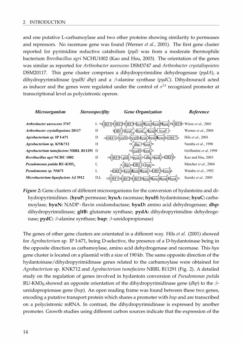
2 INTRODUCTION
and one putative L-carbamoylase and two other proteins showing similarity to permeasesand repressors. No racemase gene was found (Werner et al., 2001). The first gene clusterreported for pyrimidine reductive catabolism (pyd) was from a moderate thermophilebacterium Brevibacillus agri NCHU1002 (Kao and Hsu, 2003). The orientation of the geneswas similar as reported for Arthrobacter aurescens DSM3747 and Arthrobacter crystallopoietesDSM20117. This gene cluster comprises a dihydropyrimidine dehydrogenase (pydA), adihydropyrimidinase (pydB/ dhp) and a β-alanine synthase (pydC). Dihydrouracil actedas inducer and the genes were regulated under the control of σ54 recognized promoter attranscriptional level as polycistronic operon.
Microorganism Gene Organization ReferenceStereospecifity
Arthrobacter aurescens 3747
Arthrobacter crystallopoietes 20117
Agrobacterium sp. IP I-671
Agrobacterium sp. KNK712
Agrobacterium tumefaciens NRRL B11291
Brevibacillus agri NCHU 1002
Pseudomonas putida RU-KM3S
Pseudomonas sp. NS671
Microbacterium liquefaciens AJ 3912
L
D
D
D
D
D
L
L
D,L
Wiese et al., 2001
Werner et al., 2004
Hils et al., 2001
Namba et al., 1998
Griffantini et al.,1998
Kao and Hsu, 2003
Matcher et al., 2004
Watabe et al., 1992
Suzuki et al., 2005
ORF1 ORF2 ORF3 ORF8hyuPhyuAhyuH hyuC
ORF1 hyuCL hyuCD hyuH hyuP
hyuN hyuH hyuChyuDhyuAORF1 ORF7
dhp hyuC
hyuH hyuC
ORF3 gltB pydA ORF8dhp pydC
dhp ORF1 bup
ORF5 ORF5hyuHhyuHhyuC hyuA
ORF1 ORF2 ORF3 hyuP hyuAhyuH hyuC
Figure 2: Gene clusters of different microorganisms for the conversion of hydantoins and di-hydropyrimidines. (hyuP: permease; hyuA: racemase; hyuH: hydantoinase; hyuC: carba-moylase; hyuN: NADP - flavin oxidoreductase; hyuD: amino acid dehydrogenase; dhp:dihydropyrimidinase; gltB: glutamate synthase; pydA: dihydropyrimidine dehydroge-nase; pydC: β-alanine synthase; bup: β-ureidopropionase)
The genes of other gene clusters are orientated in a different way. Hils et al. (2001) showedfor Agrobacterium sp. IP I-671, being D-selective, the presence of a D-hydantoinase being inthe opposite direction as carbamoylase, amino acid dehydrogenase and racemase. This hyugene cluster is located on a plasmid with a size of 190 kb. The same opposite direction of thehydantoinase/dihydropyrimidinase genes related to the carbamoylase were obtained forAgrobacterium sp. KNK712 and Agrobacterium tumefaciens NRRL B11291 (Fig. 2). A detailedstudy on the regulation of genes involved in hydantoin conversion of Pseudomonas putidaRU-KM3S showed an opposite orientation of the dihydropyrimidinase gene (dhp) to the β-ureidopropionase gene (bup). An open reading frame was found between these two genes,encoding a putative transport protein which shares a promoter with bup and are transcribedon a polycistronic mRNA. In contrast, the dihydropyrimidinase is expressed by anotherpromoter. Growth studies using different carbon sources indicate that the expression of the
14

2.5 Industrial Applications
hydantoin-hydrolysing enzymes dhp and bup are regulated by carbon catabolite repression(Matcher et al., 2004).Watabe et al. (2002) described the hydantoin converting system of Pseudomonas sp. NS671,whereas the genes encoding for the hydantoinase (hydH), carbamoylase (hyuC) and race-mase (hyuA) were located on a 172 kb plasmid and were orientated in the same direction.The classification of the ATP-dependent hydantoinase of Pseudomonas sp. NS671 is still un-clear because the enzyme shows no enantioselectivity and therefore seemed to be differentto known L-specific N-methylhydantoinases (Ishikawa et al., 1993, 1997). It was not tested,whether N-methylhydantoin could be cleaved by the enzyme.
2.5 Industrial Applications
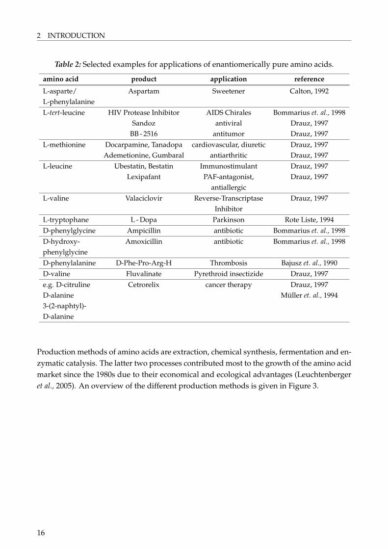
Within the last 20 years the market for the so called feed amino acids L-lysine, D,L-methinonine, L-threonine and L-tryptophane increased dramatically. The food sector is ofhigh importance with the demand for the flavor enhancer L-glutamic acid from sodiumglutamate and L-aspartic acid and the sweetener L-aspartyl L-phenylalanine methyl ester(Aspartam) from L-phenylalanine. Other proteinogenic amino acids are needed in thepharmaceutical and cosmetic industry (Leuchtenberger et al., 2005). A brief summary of theuse of amino acids is given in Table 2.
15
2 INTRODUCTION
Table 2: Selected examples for applications of enantiomerically pure amino acids.
amino acid product application reference
L-asparte/ Aspartam Sweetener Calton, 1992L-phenylalanineL-tert-leucine HIV Protease Inhibitor AIDS Chirales Bommarius et. al., 1998
Sandoz antiviral Drauz, 1997BB - 2516 antitumor Drauz, 1997
L-methionine Docarpamine, Tanadopa cardiovascular, diuretic Drauz, 1997Ademetionine, Gumbaral antiarthritic Drauz, 1997
L-leucine Ubestatin, Bestatin Immunostimulant Drauz, 1997Lexipafant PAF-antagonist, Drauz, 1997
antiallergicL-valine Valaciclovir Reverse-Transcriptase Drauz, 1997
InhibitorL-tryptophane L - Dopa Parkinson Rote Liste, 1994D-phenylglycine Ampicillin antibiotic Bommarius et. al., 1998D-hydroxy- Amoxicillin antibiotic Bommarius et. al., 1998phenylglycineD-phenylalanine D-Phe-Pro-Arg-H Thrombosis Bajusz et. al., 1990D-valine Fluvalinate Pyrethroid insectizide Drauz, 1997e.g. D-citruline Cetrorelix cancer therapy Drauz, 1997D-alanine Müller et. al., 19943-(2-naphtyl)-D-alanine
Production methods of amino acids are extraction, chemical synthesis, fermentation and en-zymatic catalysis. The latter two processes contributed most to the growth of the amino acidmarket since the 1980s due to their economical and ecological advantages (Leuchtenbergeret al., 2005). An overview of the different production methods is given in Figure 3.
16

2.5 Industrial Applications
resolution, enzymes,
diastereomers,
chromatography
D,L-amino acidsStrecker Synthesis
all kinds of
L-amino acids
all kinds of
D-amino acids
proteinogenic,
natural
L-amino acids
protein hydrolysis
and extraction
fermentationenzymatic synthesis,
amino acid transformation,
asymetric synthesis
Figure 3: Basic methods for amino acid synthesis (from Drauz, 1997).
For the production of amino acids by means of the hydantoinase process amino acids areformed by an enzymatic step using hydantoins as starting material. Hydantoins can besynthesised comparatively easily by chemical methods (Bucher and Steiner, 1934). Differentways can be employed to get access to hydantoins as a starting material (Fig. 4).
NHNH
O
OR
R COOH
NH2
R COOEt
NCO
NHNH
O
O
R-CHO + HCN +
(NH4)2CO3
Strecker SynthesisH+
+OCN-
+NH3
+R-CHOH+
1.OH-
2.H2/Kat.
*L, D or D,L
*
*
*
Figure 4: Methods for the chemical synthesis of hydantoins.
17

2 INTRODUCTION
An advantage of the hydantoinase process is that hydantoins can racemise under certainchemical conditions (see Fig. 1) or enzymatically by a so-called hydantoin racemase. Hence,the substrate can theoretically be converted with 100% yield. Another advantage of thehydantoinase process is the broad substrate spectrum of hydantoinases and carbamoylases(Hils, 1998).A problem of the hydantoin process is the instability of the carbamoylase which can lowerthe space-time yield in the process (Kim et al., 1994). Recently the development of newmolecular methods and extended insight in substrate accessibility helped to overcomethis problem and may improve the industrial processes of amino acid production byhydantoinases and carbamoylases:
1. Directed evolution was used to convert the hydantoinase from Arthrobacter sp.DSM9771 from D-specificity to L-specificity with a five-fold increase in total activity(May et al., 2000).
2. The oxidative stability and thermostability of a N-carbamoylase from Agrobacteriumtumefaciens NRRL B11291 was improved by directed evolution using DNA shuffling(Oh et al., 2002).
3. The construction of whole-cell tailor-made biocatalysts by co-expression of the genesencoding for the L-hydantoinase, the L-N-carbamoylase and the hydantoin racemasefrom Arthrobacter aurescens DSM3747 in E. coli by using vectors with different copynumbers (Wilms et al., 2001).
4. The development of bifunctional hydantoinase/ carbamoylase fusion proteins withenhanced performance by the use of DNA-shuffling (Kim et al., 2000).
5. The use of two hydantoinases and one carbamoylase made it possible to producehighly lipophilic, silicon-containing amino acids (Smith et al., 2001). This is an ex-ample of the broad substrate range of hydantoinases and carbamoylases and for theirversatility in industrial application.
Economical important targets being produced using hydantoinases and carbamoylases in-clude aromatic D-amino acids (D-phenylglycine, p-hydroxy-D-phenylglycine), D-serine, L-methinonie or L-phosphinotricine (Leuchtenberger et al., 2005). The precursor of amoxicilin,p-hydroxy-D-phenylglycine is one of the most important compounds produced via the hyd-antoinase process. There are three different approaches for the industrial production startingwith D,L-5-p-hydroxyphenylhydantoin (from Drauz et al., 1991):
18

2.5 Industrial Applications
Snamprogetti-processImmobilised dihydropyrimidinase from calf liver at pH 8.0, 30◦C. The intermediary productN-carbamoyl-D-p-hydroxyphenylglycin is chemically decarboxylated with HNO2 to thecorresponding amino acid.
Kaneka-processImmobilised, resting cells from Bacillus brevis with D-hydantoinase activity at pH 9.0, 30◦C.Chemical decarboxylation is employed as well.
Recordati-processImmobilised, resting cells from Agrobacterium radiobacter with D-hydantoinase and D-carbamoylase activity at pH 9.0, 30◦C.
An industrial process was established by Rütgers-Biotech in the early 1990ies for the pro-duction of non-natural aromatic L-amino acids by whole cells of Arthrobacter aurescensDSM3745 and DSM3747 containing a racemase, L-hydantoinase and L-carbamoylase.The L-amino acids produced were L-tryptophan, L-phenylalanine, L-O-benzylserine,L-p-chloro-phenylalanine, L-p-fluoro-phenylalanine, L-p-nitro-phenylalanine, L-1-naphtyl-alanine, L-2-naphtylalanine, L-3,4-dimethoxy-phenylalanine and L-2-thienylalanine (Syl-datk and Pietsch, 1995).
19

Whitepage
20

3 Research Proposal
Hydantoin-hydrolysing enzymes have been studied for a long time and were shown tobe present in various microorganisms. Much is known about the biochemical propertiesof different hydantoinases, the reaction mechanism of the enzyme; crystal structures weredetermined and hydantoin-hydrolysing gene clusters elucidated. As well, hydantoinasesare used in industry for the production of various amino acids. Nevertheless, there arestill many questions remaining. For example, the question of the "natural function" is notsolved, including the distribution of microorganisms with hydantoinase activity in nature,and little is known about the phylogenetic relationship between hydantoinases themselves.Moreover, due to the increasing biotechnological market new enzymes with special sub-strate specificities are needed. Therefore the present work focused on the following mainquestions:
• Is it possible to find hydantoinases in bacteria in which hydantoinase activity was notshown before?
• Is there a preferred environmental natural habitat for bacteria with the ability to cleavehydantoins?
• If there are bacteria with non-described hydantoinases, do they exhibit special bio-chemical properties, probably due to adaption to a certain environmental habitat?
• What are the differences of "novel" hydantoinases on a molecular level?
To solve these questions the present work is concentrated on the screening, characterisationand comparison of "novel" hydantoinases as follows:
• Recovery of microorganisms with the ability to cleave hydantoins to the correspondingcarbamoyl amino acids from geographically distinct environmental habitats includingextreme habitats.
• Phylogenetic characterisation of the bacterial isolates.
• Characterisation of growth conditions of selected strains from extreme environments.
• Biochemical characterisation of hydantoinases.
• Investigation of the phylogenetic relationship of hydantoinases.
• Isolation of the gene clusters responsible for hydantoin conversion of selected strains.
21

Whitepage
22

4 Summary Chapters I – IV
4.1 Summary Chapter I: Distribution of Hydantoinases
Complete title:
DISTRIBUTION OF HYDANTOINASE ACTIVITY IN BACTERIAL ISOLATES
FROM GEOGRAPHICALLY DISTINCT ENVIRONMENTAL SOURCES
A screening program was conducted with the aim to find novel hydantoinases frommicroorganisms of different geographical origins and to investigate their distribution innature. This included terrestrial soil samples from various sites in South Africa, fromhot springs and salt-lakes in China and environmental samples from the cold Antarctica.Thirty-two bacterial strains were isolated from these samples by enrichment techniquesusing D,L-5-(3-indolylmethyl) hydantoin as carbon- and nitrogen source. All microorg-anisms showed the ability to cleave the model substrates D,L-5-benzylhydantoin and5,6-dihydrouracil. In biotransformations with D,L-5-benzylhydantoin all of them showedformation of N-carbamoyl-D-phenylalanin. Therefore these hydantoinases were classifiedas D-hydantoinases, except for the unclassified Microbacteriacae sp. K3 and Burkholderia sp.M3. They were either non-selective or L-specific. All bacterial isolates were identified at thegenus level by amplification and sequencing of the 16S rRNA gene. Figure 5 and Figure 6show the geographical origin of all strains being recovered.
23

4 SUMMARY CHAPTERS I – IV
South Africa
See Figure 6
Antarctica
Pseudomonassp. N7
and G7, Arthrobacter sp.
E7 and F7
China, Long Pu,
hot springs
Delftia sp. I24
China, Inner Mongolia,
saline lakes
Bacillussp. F18 and K18,
Ochrobactrumsp. G21,
Streptomycessp. I20
Figu
re5:
Wor
ldw
ide
dist
ribu
tion
ofba
cter
iali
sola
tes
poss
essi
nghy
dant
oina
seac
tivi
ty.(
Phot
ogra
phs
are
exam
ples
ofth
eco
llect
ing
area
sbu
tdo
nots
how
the
orig
inal
plac
eof
sam
plin
g.Ph
otog
raph
sw
ere
take
nw
ithi
nth
eC
hine
se-E
U-S
AM
GA
Tech
proj
ecta
ndW
aika
toU
nive
rsit
yA
ntar
ctic
Terr
estr
ialB
iolo
gyPr
ogra
m(p
rovi
ded
byPr
of.D
.A.C
owan
)and
byth
eau
thor
.)
24

4.1 Summary Chapter I: Distribution of Hydantoinases
Stellenbosch, compost company &
pinewood forest
Acinetobactersp. K5,Burkholderiasp. K3,
Enterobactersp. J7,Flavobacteriumsp. F8,
Microbacteriaceasp. M3,Ochrobactrum sp. C15,
I21 and J24,Pseudomonas sp. G6, N8 and P8,
Staphylococcus sp. H7
West Coast N. P. and
Veldrift
Bacillussp. A16, D17 and F16,
Ochrobactrum sp. F21
TsitsikammaN. P.
Pseudomonassp. M18,
Bacillussp. H20Lesotho
Arthrobacter sp. K20
Cape Town, Table
Mountain
Stenotrophomonassp.N1
Worcester, Distillery
wastewater
Ochrobactrumsp. D24
CederbergMountains
Pseudomonas sp. L9
Swaziland
Bacillus sp. G18
Figu
re6:
Dis
trib
utio
nof
bact
eria
liso
late
spo
sses
sing
hyda
ntoi
nase
acti
vity
inSo
uth
Afr
ica,
Leso
tho
and
Swaz
iland
.(Ph
otog
raph
sar
eex
ampl
esof
the
colle
ctin
gar
eas
butd
ono
tsho
wth
eor
igin
alpl
ace
ofsa
mpl
ing.
Phot
ogra
phs
wer
eta
ken
byth
eau
thor
.)
25
4 SUMMARY CHAPTERS I – IV
The screening and isolation for various microorganisms with hydantoinase activity is de-scribed in literature, but usually no precise data are available on the exact origin and natureof the environmental samples used in the screening experiments. In this study it is shownthat microorganisms with the ability to metabolise hydantoins are widely dispersed in re-spect to the nature of the soil sample and to geographical origin. Nevertheless, most bac-teria were found in environmental samples with a high amount of degrading material (e.g.compost soil or guano) which confirms the hypothesis that hydantoinases are involved incatabolic pathways to access amino acids as metabolic substrates. Interestingly, microorg-anisms with hydantoinase activity were also found in soil samples originating from extremeenvironments. Growth under different conditions (as temperature and media composition)showed that the two Arthrobacter sp. F7 and G7 and the two Pseudomonas sp. N7 and G7could be designated as psychrotrophic. This is the first report of psychrotrophic microorg-anisms from Antarctica with hydantoinase activity. In literature only one halophilic strainwith hydantoinase activity, Pseudomonas sp. NCIM5109 is reported. This study describesmore halophilic bacteria with hydantoinase activity but from different genera: Bacillus sp.F18 and K18, Ochrobactrum sp. G21 and Streptomyces sp. I20. All of them were isolatedfrom a saline environment and were designated as moderate halophilic due to their abilityto grow in media supplemented with up to 10% NaCl.Microorganisms with hydantoinase activity were predominantly found in the genera Arthro-bacter, Bacillus, Ochrobactrum and Pseudomonas but we also can report hydantoinase activityin genera for which no hydantoinase activity was known before: Stenotrophomonas, one "Mi-crobacteriacae", Staphylococcus, Acinetobacter, Delftia and Streptomyces.In summary, it was shown that microorganisms with hydantoinase activity are:
(I) distributed in various geographically distinct environmental habitats,
(II) distributed worldwide and
(III) found in certain bacterial genera.
(IV) Furthermore, the presence of hydantoinase activity was demonstrated for genera inwhich hydantoinase activity has not previously been reported.
26

4.2 Summary Chapter II: Biodiversity of Hydantoinases
4.2 Summary Chapter II: Biodiversity of Hydantoinases
Complete title:
BIODIVERSITY OF HYDANTOIN CLEAVING ENZYMES
The UNCED (1992) defined the term "biological diversity" or "biodiversity" to be the varietyof genes, species and ecosystems found on our planet. It embraces all life forms – from plantand animal life to microorganisms – and the water and land in which they interact. Re-garding the "biodiversity of hydantoinases" that would mean the variety of hydantoinases.It is not possible to measure this but the following questions can try to highlight the term"variety of hydantoinases":
1. It is known that hydantoinases have a broad and diverse substrate spectrum (see In-troduction, Table 1). The question remains if there is a correlation between substratespectrum and origin of hydantoinases concerning bacterial genera and/ or ecologicalhabitat.
2. Can the hydantoinase gene sequence of different microorganisms be clustered intospecific groups either related to a genus and/ or to a certain habitat?
3. Is there a correlation between the biochemical properties of hydantoinases and certainhydantoinase groups?
4. Are hydantoinase genes also present in microorganisms without any hydantoinaseactivity?
To answer these points several bacterial isolates from distinct environmental habitats werechosen for biochemical and genetic characterisation. The selection criteria were (i) if a strainshowed high activity towards the substrates D,L-5-benzylhydantoin and 5,6-dihyrouracil(Dürr et al., 2006); (ii) if a strain was isolated from an extreme or unique environmentalhabitat like a hot spring or a biofilm; (iii) if no hydantoinase activity had been reported inliterature for that certain genus before. The investigation on these bacterial isolates led tothe following conclusions:
1. The hydantoinases selected for this study showed a broad substrate spectrum but nocorrelation between a certain hydantoinase activity and certain bacterial genera and/or ecological habitat could be obtained. However we propose that environmental con-ditions like a high organic content of the soil can evoke hydantoinase activity, sincemany strains were recovered from soil samples like guano and compost.The preference of most of the bacterial strains for higher conversion of 5,6-dihydrouracil than for the non-substituted hydantoin lead to the conclusion thatthese enzymes can be considered as dihydropyrimidinases and are part of the re-ductive pathway of pyrimidine degradation. Syldatk et al. (1999) demonstrated
27

4 SUMMARY CHAPTERS I – IV
that D-hydantoinases are not necessarily identical to dihydropyrimidinases and theterm hydantoinase should be used for enzymes being able to hydrolyse hydan-toin or 5-monosubstituted hydantoins. Since all strains were able to cleave hydan-toin or 5-monosubstituted hydantoins they were declared as hydantoinases. Re-garding the other substrates tested most enzymes expressed higher activity to-wards the 5-monosubstituted hydantoin 5-benzylhydantoin than for the dihydro-pyrimidine counterpart D,L-6-phenyl-5,6-dihydrouracil. The other substrates D,L-(5-tert-butylhydantoin, D,L-5-(3-indolylmethyl) hydantoin) were hydrolysed with lowerrates.A range of Arthrobacter strains was tested for the ability to cleave the above mentioneddihydropyrimidines and hydantoins. Two different groups were obtained: In whole-cell biotransformation the first group showed hydantoinase activity with a higher pref-erence for 5,6-dihydrouracil and D,L-6-phenyl-5,6-dihydrouracil than for hydantoins.They seem to be quite different from the hydantoinases of other Arthrobacter strainsdescribed in literature. The other group showed no activity. No activity could be ex-plained by the absence of hydantoin-hydrolysing enzymes or to their non-expressiondue to the absence of an adequate inducer.
2. Degenerate primers were developed or retrieved from a recent publication (Lin et al.,2005) for the amplification of a partial hydantoinase gene sequence. The primer pairswere not specific enough for the amplification of hydantoinases of all strains tested.One reason could be the low sequence homology of hydantoinases. A hydantoinasegene fragment could be amplified from a few bacteria like Delftia sp. I24, Burkholde-ria sp. M3, Bacillus sp. G18, Pseudomonas sp. M18, Arthrobacter citreus DSM20133 andseveral Ochrobactrum isolates. The gene fragments obtained had different sizes due tothe two primer pairs used. Phylogenetic analysis was conducted using hydantoinasegene fragments from the above mentioned strains and hydantoinase and dihydropy-rimdine genes from the database. For a confident analysis a certain DNA-region ofeach hydantoinase was used namely the part of the smallest amplified fragment (app.320 bp). In a phylogenetic tree the hydantoinase genes were predominantly found tobe clustered according to their phylogenetic origin. Interestingly, A. citreus DSM20133,Delftia sp. I24 and the Ochrobactrum hydantoinases were not clustered together withknown hydantoinases but with putative hydantoinases. These results could have beenobtained because each genus showed a specific nucleotide usage (which is seen e.g. indifferent GC contents). However, the data obtained above is supported by a phylo-genetic tree of protein sequences (Chapter IV).In summary, we found that hydantoinases can be clustered according to their bacterialorigin.
28
4.2 Summary Chapter II: Biodiversity of Hydantoinases
3. No correlation was obtained between the biochemical properties of hydantoinaseswithin a certain cluster of the phylogenetic tree. One reason could be that not all nu-cleotide positions encoding the catalytic sites and/ or functional recognition sites areincluded in the gene sequence used for the phylogenetic analysis.
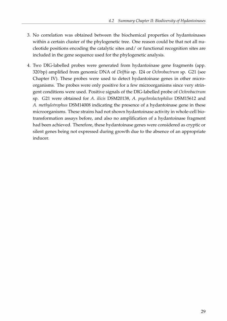
4. Two DIG-labelled probes were generated from hydantoinase gene fragments (app.320 bp) amplified from genomic DNA of Delftia sp. I24 or Ochrobactrum sp. G21 (seeChapter IV). These probes were used to detect hydantoinase genes in other micro-organisms. The probes were only positive for a few microorganisms since very strin-gent conditions were used. Positive signals of the DIG-labelled probe of Ochrobactrumsp. G21 were obtained for A. ilicis DSM20138, A. psychrolactophilus DSM15612 andA. methylotrophus DSM14008 indicating the presence of a hydantoinase gene in thesemicroorganisms. These strains had not shown hydantoinase activity in whole-cell bio-transformation assays before, and also no amplification of a hydantoinase fragmenthad been achieved. Therefore, these hydantoinase genes were considered as cryptic orsilent genes being not expressed during growth due to the absence of an appropriateinducer.
29

4 SUMMARY CHAPTERS I – IV
4.3 Summary Chapter III: Properties of Selected Hydantoinases
Complete title:
DESCRIPTION AND CHARACTERISATION OF THE D-HYDANTOINASES
FROM THREE MICROORGANISMS ISOLATED FROM EXTREME
ENVIRONMENTS.
In Chapter I and II it was shown that it is still possible to find microorganisms with hyd-antoinase activity not having been described before, and from habitats in which the pres-sure of survival is high due to extreme conditions. The amplification and comparison ofpartial hydantoinase gene sequences showed that hydantoinases are closely related to theirphylogenetic origin. Some hydantoinases are clustered apart from known hydantoinases.This makes them attractive for further characterisation e.g. the hydantoinases from var-ious Ochrobactrum species or the one from Delftia sp. I24. The questions arise if "novel"hydantoinases of microorganisms or hydantoinases of bacteria from extreme environmentsshow unique growth- and hydantoinase properties or any adaption to the special environ-mental conditions. Therefore three bacterial strains were selected for:
1. further identification of the microbial strains;
2. characterisation and optimisation of growth conditions in relation to hydantoinaseactivity;
3. characterisation of the biochemical properties of the hydantoinases.
The selection criteria for the microorganisms used included the environment of isolation,type of bacteria, hydantoinase activity and/ or substrate range. The first strain, Delftia sp.I24, was selected because this strain was isolated from a Chinese hot spring at a screeningtemperature of 50◦C (Fig. 5 & 7b), and no hydantoinase activity had been reported for thisgenus before. Ochrobactrum sp. G21 and Bacillus sp. F18 were selected because they bothwere found in soil from hypersaline lakes, Inner Mongolian Autonomous Region, China(Fig. 5 & 7a). The hydantoinase of each strain showed good activity and had a broadsubstrate spectrum.
Bacterial Identification
The three bacterial isolates were further classified using molecular biological and classicalbacterial identification methods. They could be characterised as follows:
1. Comparison of the 16S rDNA sequence of strain Delftia sp. I24 to known sequencesshowed the highest similarity to Delftia acidovorans or Delftia tsuruhatensis at the samesimilarity level. Colonies of Delftia sp. I24 were rod shaped with a size of 4 – 6 µm (Fig.8a), motile, Gram-staining negative, aerobic, catalase and oxidase positive. Delftia sp.
30

4.3 Summary Chapter III: Properties of Selected Hydantoinases
(a) Unnamed Mongolian salt lake (b) Algal mat from site LP4
Figure 7: Environmental sites from which the bacterial strains Ochrobactrum sp. G21 andBacillus sp. F18 (subfigure a), Delftia sp. I24 (subfigure b) were isolated. (Photographsare examples of the collecting areas but do not show the original place of sampling. Pho-tographs were taken within the Chinese-EU-SA MGATech project and Waikato UniversityAntarctic Terrestrial Biology Program and provided by Prof. D.A. Cowan.)
I24 could not be identified to the species level because the biochemical criteria didnot show an exact concordance to one of the two Delftia species mentioned above.Therefore this strain was assigned as Delftia sp. I24.
2. The 16S rDNA sequence of Ochrobactrum sp. G21 showed highest homology tothree type strains Ochrobactrum anthropi, Ochrobactrum lupine or Ochrobactrum triciti.Ochrobactrum sp. G21 cells were coccoide rods with a size of 2 µm (Fig. 8b), coloniesyellow, motile, Gram-staining negative, aerobic, catalase and oxidase positive. Con-cerning different characteristics, Ochrobactrum sp. G21 could not be assigned with theutmost probability to one of the type strains mentioned above.
3. Bacillus sp. F18 could be undoubtedly assigned as Bacillus megaterium concerning the16S rRNA gene sequence. Therefore this strain was designated as Bacillus megateriumF18. The colonies of this strain were rod shaped with a size of 5 – 6 µm (Fig. 8c), Gram-staining positive, facultative anaerob, catalase positive and oxidase negative.
All three strains were deposited at the German Collection of Microorganisms and CellCultures: Delftia sp. I24: DSM18833; Ochrobactrum sp G21: DSM18828; Bacillus megateriumF18: DSM18825.
31

4 SUMMARY CHAPTERS I – IV
2.9µm
(a) Delftia sp. I24
2.8µm
(b) Ochrobactrum sp. G21
2.8µm
(c) Bacillus megaterium F18
Figure 8: Atomic force microscopy (AFM) images of the three bacterial isolates being identi-fied.
Growth Characteristics of the Three Bacterial Isolates in Relation toHydantoinase ActivityGross et al. (1990) showed that the treatment of cells of A. aurescens DSM3747, a Gram-staining positive bacteria, with sodium-desoxycholate led to enhanced cell permeabilisationand therefore higher hydantoinase and carbamoylase activity. To overcome cell membranetransport problems for substrates and products Ochrobactrum sp. G21 was chosen as amodel strain and were treated with different methods. Addition of toluene (20 µl/mL) tothe biotransformation solution led to highest hydantoinase activity.Concerning the carbon source required for growth no optimisation of the media wasnecessary for the strains Ochrobactrum sp. G21 and B. megaterium F18 since they grew verywell on glucose, whereas Delftia sp. I24 showed low growth on this carbon source. Additionof fructose to the media led to high growth of this strain. Concerning the origin of thestrain, a Chinese hot spring, this strain should be able to grow at high temperatures whichcould not be confirmed. However, this strain was isolated at a screening temperature of50◦C and therefore declared as thermophilic. It is supposed that Delftia sp. I24 might be acontamination of this habitat, only possessing the ability to survive at high temperatures.The influence of salt on growth and hydantoinase activity were investigated for the strainsOchrobactrum sp. G21 and B. megaterium F18, because they were isolated from hypersalinelakes and previously shown to be halophilic (Chapter I). For both strains the growthbehavior was found to be opposite compared to hydantoinase activity characteristics.Whilst growth was highest at a low salt concentration of the medium, hydantoinase activitywas stimulated in media with higher salt concentrations (Table 3). An attractive hypothesisfor Ochrobactrum sp. G21 and B. megaterium F18 is, that the hydantoin degrading systemof these bacteria is co-regulated with salt stress proteins and other metabolic pathwaystriggered by salt stress presumably to get access to amino acids under limited conditions.This was derived from studies on the effect of salt stress on microorganisms: Salt stress had
32

4.3 Summary Chapter III: Properties of Selected Hydantoinases
an important effect on the synthesis of degradative enzymes in Bacillus subtilis (Kunst et al.,1995). Another study on Listeria monocytogenes showed that the synthesis of 40 proteins wasrepressed or induced during salt stress. This includes general stress proteins, transportersand general metabolism proteins (Duché et al., 2002).
Table 3: Influence of different parameters on growth and hydantoinase activity. For eachparameter the optimal conditions concerning growth or hydantoinase activity are given.(I24: Delftia sp. I24; G21: Ochrobactrum sp. G21; F18: B. megaterium F18; GM: growthmedia; IMH: D,L-5-(3-indolylmethyl) hydantoin; NMH: D,L-2-naphtylmethylhydantoin;PheDU: D,L-6-phenyl-5,6-dihydrouracil; t- BH: D,L-5-tert-butylhydantoin)
I24 G21 F18
carbon source fructose glucose glucosegrowth medium GM/ GM+PheDU GM GM+2%NaClNaCl conc. for hyd. activity not tested 5% 5%inductor IMH/ PheDU NMH t-BHMedium for hyd. activity GM GM+5%NaCl
+NMHGM+5%NaCl+t-BH
The addition of different inducers to the growth media had a significant effect on hyd-antoinase activity. The dissimilarity of each hydantoinase was shown: The hydantoinaseof Delftia sp. I24 showed highest induction with the addition of the polycyclic aro-matic hydantoin D,L-5-(3-indolylmethyl) hydantoin or the aromatic dihydropyrimidinederivative D,L-6-phenyl-5,6-dihydrouracil to the growth media (Table 3). In case of thehydantoinase of Ochrobactrum sp. G21 addition of the polycyclic aromatic hydantoinD,L-2-naphtylmethylhydantoin led to a dramatic increase of hydantoinase activity and thehydantoinase of B. megaterium F18 was stimulated highest with the aliphatic hydantoinD,L-5-tert-butylhydantoin (Table 3).Hydantoinase activity was highest in the late exponential phase of growth for severalPseudomonas species (Morin et al., 1986b, c; Gokhale et al., 1996; Sudge et al., 1998), Agrobac-terium (Kim and Kim 1993) and in the exponential phase of Arthrobacter sp. DSM3747(Syldatk et al., 1990).All previous results were obtained neglecting the variation of hydantoinase activity duringgrowth since harvesting was done routinely at a certain point of time. In order to findthe best media and growth phase for highest hydantoinase activity different media werecompared and hydantoinase activity was determined during growth (Table 3).Hydantoinase activity of Delftia sp. I24 was found to be highest in the exponential phasewhen grown on pure GM – or slightly lower on GM supplemented with 0.2 g/L D,L-6-phenyl-5,6-dihydrouracil. The favourable media for B. megaterium F18 hydantoinase
33

4 SUMMARY CHAPTERS I – IV
activity was GM supplemented with 5%NaCl and 0.2 g/L D,L-5-tert-butylhydantoin, aswell with highest activity in the late exponential phase. Highest hydantoinase activityof Ochrobactrum sp. G21 was obtained when grown on GM supplemented with 5%NaCland 0.2 g/L D,L-2-naphtylmethylhydantoin after 96 h but growth was very little. Theresults of the halophilic strains Ochrobactrum and Bacillus showed that highest hydantoinaseexpression could be achieved by adjustment of the salt concentration of the medium andwith the choice of the appropriate inductor.
Properties of Hydantoinases
In order to determine the biochemical properties of the hydantoinases bacterial strains weregrown in their favourable media as described in the previous section.
(I) Optimal temperature and pH (Table 4)The temperature optimum of the hydantoinase of Delftia sp. I24 was very low with30◦C. This is in accordance to the results obtained for the growth characteristics of thisstrain (see previous section) and confirms the hypothesis that Delftia sp. I24 is probablya contamination of this habitat. Together with the D-hydantoinase from a halophilicPseudomonas sp. NCIM5109 (Sudge et al., 1998) this is the lowest temperature optimum ofD-hydantoinases reported. The optima of temperature and pH for Ochrobactrum sp. G21were 40◦C and pH 8.0 – 8.5, respectively. The hydantoinase of the only Ochrobactrum straindescribed in literature, Ochrobactrum anthropi, showed a pH-optimum of pH 9.0 (Pozo et al.,2002). Unfortunately the influence of temperature was not tested in this study. The highestoptimal temperature was obtained for the hydantoinase of B. megaterium F18 at 50 – 55◦Cand a pH-optimum of 7.5 – 8.0. The optimal temperature of this hydantoinase is quite lowsince hydantoinases from other Bacilli showed higher optimal temperatures: Bacillus sp.AR9, B. stearothermophilus SD-1 and B. thermocatenulatus GH-2 at 65◦C (Sharma and Vohra,1997; Lee et al., 1994, 1995; Park et al., 1998; Kim et al., 1997a), B. stearothermophilus NS1122Aat 60 – 70◦C (Ishikawa et al., 1994) and B. circulans at 75◦C (Luksa et al., 1997).
Table 4: Properties of the hydantoinases from Delftia sp. I24, Ochrobactrum sp. G21 and B.megaterium F18. The optimal value of each parameter is shown. (I24: Delftia sp. I24; G21:Ochrobactrum sp. G21; F18: B. megaterium F18)
I24 G21 F18
T [◦C] 30 40 50 – 55pH 9.0 8.0 – 8.5 7.5 – 8.0cofactor Ni2+/ Co2+ no preference Mn2+/ Co2+
salt conc. no influence 0 – 2.5% no influence
34

4.3 Summary Chapter III: Properties of Selected Hydantoinases
(II) Cofactor requirements (Table 4)Hydantoinases are known to be dependent on cofactors, e.g. the L-hydantoinase fromArthrobacter aurescens DSM3745 contains about 2.5 mol Zn2+/mol subunit (May et al., 1998a,b) and the hydantoinase of Bacillus sp. AR9 contains Mn2+ in the active centre (RhadaKishan et al., 2005). It has to be mentioned that biochemical studies can give a hint on thecofactor being present in the enzyme but for verification complex methods such as atomicabsorption spectroscopy, inductive coupled plasma-atomic emission spectrometry and/ orX-ray crystallography have to be used.Biochemical studies on the cofactor requirements of the three strains showed that allhydantoinases are cofactor dependent because addition of EDTA inhibited or completelyremoved hydantoinase activity. Since the hydantoinase activities of Delftia sp. I24 andB. megaterium F18 were enhanced by the addition of Ni2+ or Co2+ and Mn2+ or Co2+,respectively, it is supposed that one of these ions is present in one of the hydantoinaseenzymes. In contrast, the hydantoinase from Ochrobactrum sp. G21 showed no enhancedactivity caused by one of the ions tested. The hydantoinase from Ochrobactrum anthropi wasdescribed in literature to show increased activity by addition of Ca2+, Na+, Cu2+ or Co2+
(Pozo et al., 2002). Unfortunately the D-hydantoinases of Ochrobactrum sp. G21 describedin this study and of Ochrobactrum anthropi cannot be compared in detail because bothhydantoinases were studied in other aspects except for pH and cofactor requirements.Taking into account these two aspects, or particularly the unequal cofactor requirements,we can conclude that these hydantoinases are rather different enzymes.
(III) Influence of salt concentration on hydantoinase activity (Table 4)The hydantoinase activities of Delftia sp. I24 and Bacillus megaterium F18 remained the sameat salt concentrations ranging from 2.5 to 20% NaCl, whereas the hydantoinase activityof Ochrobactrum sp. G21 decreased with raising NaCl-concentrations. This seem to becontrary to the growth experiments using different salt concentrations of the media inwhich hydantoinase activity was enhanced in media with higher salt concentrations. Wehave to consider that we were using growing cells on the one hand and resting cells onthe other. Growing cells are able to respond to factors like salt stress for example withaltered enzyme expression, whereas resting cells remain in the current-state of harvest.Consequently, in assays using resting cells only enzymes or enzyme systems can directly beaffected by the salt concentration, e.g. inhibited or enhanced. In the present study only thehydantoinase of Ochrobactrum sp. G21 was inhibited by higher salt concentrations.
(IV) Enzyme stabilityThe hydantoinase enzyme of whole cells of Delftia sp. I24 did not loose activity at a storagetemperature of 4◦C and 20◦C within four days, whereas activity was almost lost at 40◦Cafter one day of storage. Hydantoinase in cells treated with toluene showed lower stability
35
4 SUMMARY CHAPTERS I – IV
since the activity decreased at all storage temperatures after one day. The hydantoinaseactivity of whole cells and toluene treated cells of Ochrobactrum sp. G21 decreased at allstorage conditions significantly. Best storage was achieved at 4◦C.
(V) Substrate range of the hydantoinasesAll three strains showed a broad substrate range and were able to cleave the following sub-strates: 5,6-dihydrouracil, hydantoin, D,L-5-tert-butylhydantoin, D,L-5-benzylhydantoin,D,L-5-(3-indolylmethyl) hydantoin and D,L-6-phenyl-5,6-dihydrouracil (except of Delftiasp. I24 which was not able to cleave the latter). The hydantoinase from Delftia sp. I24displayed for all substrates tested a significantly lower specific activity than the ones fromthe two other strains. The hydantoinase showed highest activity for 5,6-dihydrouracilindicating that this enzyme could be involved in the pyrimidine reductive catabolism.Interestingly, the hydantoinase of Delftia sp. I24 was not able to cleave the best inducer ingrowth experiments being D,L-6-phenyl-5,6-dihydrouracil. The highest specific activitieswere obtained for the hydantoinase of Bacillus megaterium F18. This enzyme showedpreference to 5-monosubstituted hydantoins or dihydropyrimidines bearing an arylic sidechain compared to the corresponding aliphatic derivatives.
In summary this section describes microorganisms with novel hydantoinase properties: aD-hydantoinase from Delftia sp. I24 as well as the D-hydantoinases of the two halophilicstrains Ochrobactrum sp. G21 and Bacillus megaterium F18. All of the hydantoinases showedunique properties regarding salt and inductor requirements during growth for enzyme ex-pression and they displayed different properties like variation in pH and temperature op-tima and cofactor requirements.
36

4.4 Summary Chapter IV: Gene Clusters for Hydantoin Degradation
4.4 Summary Chapter IV: Gene Clusters for Hydantoin Degradation
Complete title:
GENES RESPONSIBLE FOR HYDANTOIN DEGRADATION OF A
HALOPHILIC Ochrobactrum SP. G21 AND Delftia SP. I24 – NEW INSIGHT
INTO RELATION OF D-HYDANTOINASES AND DIHYDROPYRIMIDINASES.
Different gene clusters responsible for the cleavage of hydantoins and dihydropyrimidinesare elucidated for a range of microorganisms (Fig. 2). Nevertheless, no gene clusterresponsible for hydantoin degradation has been reported for any halophilic species or forbacteria belonging to the genus Ochrobactrum and Delftia.In the preceding chapter growth characteristics in relation to hydantoinase activity and thebiochemical properties of the hydantoinases of the two moderate halophiles Ochrobactrumsp. G21 and Bacillus megaterium F18 as well as of Delftia sp. I24 were accurately described.The genes responsible for hydantoin hydrolysing activity were of particular interest sincethe wild type strains originated from extreme environments and the biochemical propertiesof the hydantoinases described showed unique properties. The amplification of a hyd-antoinase gene fragment using degenerate primers was possible for Ochrobactrum sp. G21and Delftia sp. I24 but not for Bacillus megaterium F18 (see Chapter II). The hydantoinasegene clusters were identified by screening of genomic libraries with DIG-labelled probes.These probes were derived from hydantoinase gene fragments amplified by PCR. Thegenomic libraries were obtained by digestion of genomic DNA using selected restrictionenzymes and cloning of the respective DNA fragments, bearing the hydantoinase gene intoE. coli. Clones bearing the hydantoinase gene were obtained by probe hybridisation. Thecomplete DNA sequences were received by sequencing of the plasmids. Since amplificationof a partial hydantoinase gene sequence was crucial to prepare a DIG-labelled probe,Ochrobactrum sp. G21 and Delftia sp. I24 were chosen for characterisation of the hydantoinhydrolysing gene cluster.
The gene clusters responsible for hydantoin hydrolysation are shown in Figure 9. The clus-ter of Delftia sp. I24 consisted of four genes: A hydantoinase gene (hyuH), a putative di-hydroprimidine dehydrogenase gene (pydA), a putative permease gene (hyuP) and an in-complete glutamate synthase gene (gltB). HyuH was first incomplete but completed by PCRamplification of the missing DNA fragment. The deduced amino acid sequence showedhighest similarity to the hydantoinase of Pseudomonas putida DSM84. The hydantoinasewas expressed in E. coli, and activity was shown for D,L-5-benzylhydantoin and D,L-5-(3-indolylmethyl) hydantoin. The putative dihydroprimidine dehydrogenase and the putativepermease were located upstream of the hydantoinase gene. The permease was probably re-sponsible for hydantoin or dihydropyrimidine transport. The presence of pydA and the pref-erence for dihydrouracil in activity tests (see Chapter III) indicated that the gene cluster is
37
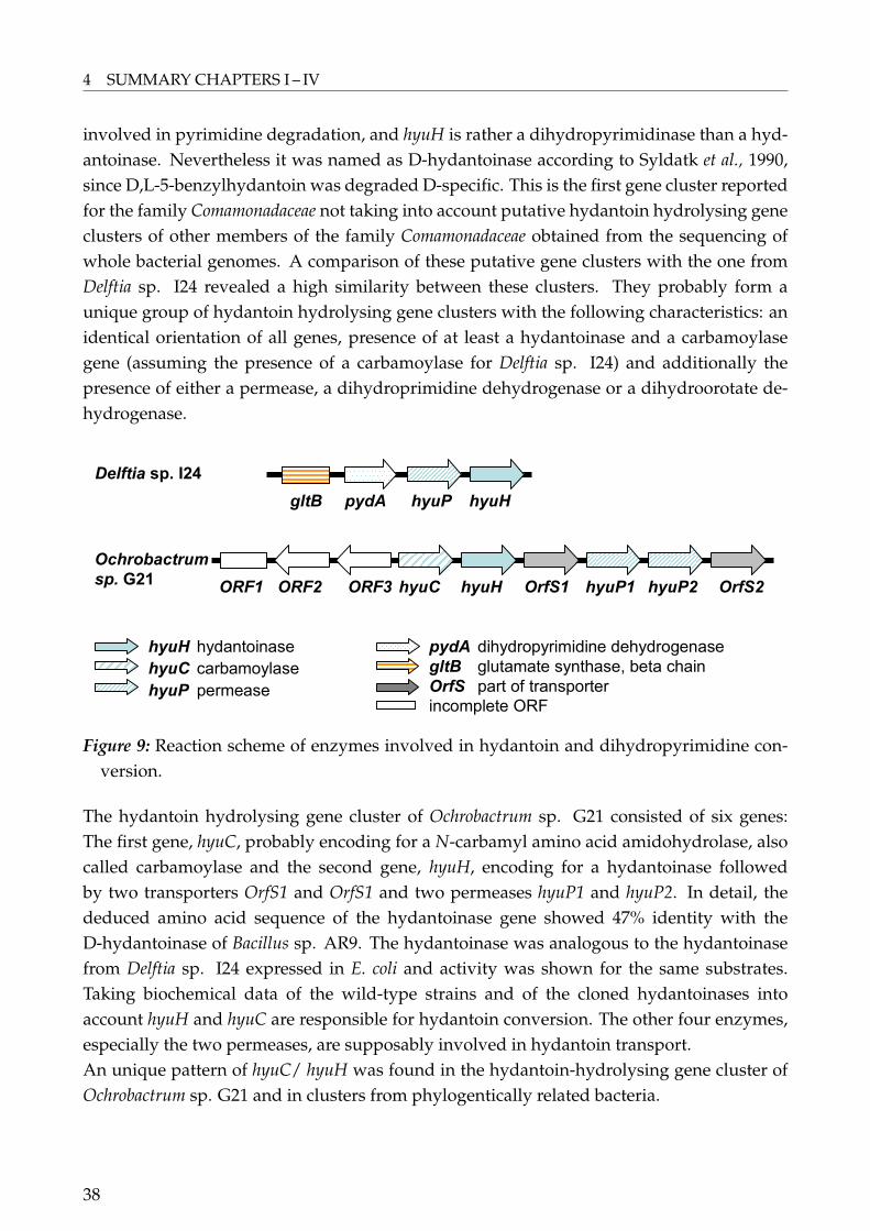
4 SUMMARY CHAPTERS I – IV
involved in pyrimidine degradation, and hyuH is rather a dihydropyrimidinase than a hyd-antoinase. Nevertheless it was named as D-hydantoinase according to Syldatk et al., 1990,since D,L-5-benzylhydantoin was degraded D-specific. This is the first gene cluster reportedfor the family Comamonadaceae not taking into account putative hydantoin hydrolysing geneclusters of other members of the family Comamonadaceae obtained from the sequencing ofwhole bacterial genomes. A comparison of these putative gene clusters with the one fromDelftia sp. I24 revealed a high similarity between these clusters. They probably form aunique group of hydantoin hydrolysing gene clusters with the following characteristics: anidentical orientation of all genes, presence of at least a hydantoinase and a carbamoylasegene (assuming the presence of a carbamoylase for Delftia sp. I24) and additionally thepresence of either a permease, a dihydroprimidine dehydrogenase or a dihydroorotate de-hydrogenase.
Delftia sp. I24
ORF3ORF1 hyuC
Ochrobactrum
sp. G21hyuH OrfS1 hyuP1 hyuP2 OrfS2
gltB pydA hyuP hyuH
hyuH hydantoinase
hyuC carbamoylase
hyuP permease
pydA dihydropyrimidine dehydrogenase
gltB glutamate synthase, beta chain
OrfS part of transporter
incomplete ORF
ORF2
Figure 9: Reaction scheme of enzymes involved in hydantoin and dihydropyrimidine con-version.
The hydantoin hydrolysing gene cluster of Ochrobactrum sp. G21 consisted of six genes:The first gene, hyuC, probably encoding for a N-carbamyl amino acid amidohydrolase, alsocalled carbamoylase and the second gene, hyuH, encoding for a hydantoinase followedby two transporters OrfS1 and OrfS1 and two permeases hyuP1 and hyuP2. In detail, thededuced amino acid sequence of the hydantoinase gene showed 47% identity with theD-hydantoinase of Bacillus sp. AR9. The hydantoinase was analogous to the hydantoinasefrom Delftia sp. I24 expressed in E. coli and activity was shown for the same substrates.Taking biochemical data of the wild-type strains and of the cloned hydantoinases intoaccount hyuH and hyuC are responsible for hydantoin conversion. The other four enzymes,especially the two permeases, are supposably involved in hydantoin transport.An unique pattern of hyuC/ hyuH was found in the hydantoin-hydrolysing gene cluster ofOchrobactrum sp. G21 and in clusters from phylogentically related bacteria.
38

4.4 Summary Chapter IV: Gene Clusters for Hydantoin Degradation
Hydantoinases are known to belong to the protein superfamily of amidohydrolases relatedto ureases. Enzymes of this superfamily have evolved from a common ancestor and are aproduct of a divergent evolution (May et al., 1998e, Holm and Sander 1997).The question of the phylogenetic relation of hydantoinases/ dihydropyrimidinases wasraised. Therefore a pylogenetic tree was built using protein sequences of hydantoinasesand dihydropyrimidinases including putative sequences. It is remarkable, that as a result,predominantly hydantoinases from related bacterial families were grouped together inthis tree. Taking into account functional recognition sites and the formed branches of thephylogenetic tree, six major groups were obtained and specified in families as follows:1) L-hydantoinases, disjoined from the major tree;2) Rhizobiales family (Rhizo-Fam), the most inconsistent group comprising hydantoinasesfrom bacteria of the order Rhizobiales or of the same bacterial class, except of one Pseudo-monas; Ochrobactrum sp. G21 is included in this family;3) Comamonadacae family (Com-Fam) including Delftia sp. I24;4) Pseudomonas family (Pseud-Fam);5) Bacilli family (Bac-Fam), including Brevibacillus agri and several Bacillus species;6) Agrobacterium family (Agro-Fam), being separated from the other Rhizobiales and includ-ing two hydantoinases from Agrobacterium species and a hydantoinase from Burkholderiapickettii.
A deeper insight into amino acids being involved in the cleavage of hydantoins revealedthat all hydantoinases used in the phylogenetic tree showed the highly conserved aminoacid motif of the superfamily of amidohydrolases (GxxDxHxH). This motif is suggested tobe involved in metal centre assembly (Holm and Sander, 1997). The motif known to form theactive site of hydantoinases consisting of His58, His60, Lys150, His183, His239 and Asp315(Cheon et al., 2002, 2003; Rhada Kishan et al., 2005; Abendroth et al., 2002a, b; Xu et al., 2003)is also found in all amino acid sequences.The substrate recognition sites of several hydantoinases belonging to different bacteria areknown. These known amino acid positions involved in the substrate recognition sites areinvestigated within the families obtained in the phylogenetic tree mentioned above. Interest-ingly, most families of this tree exhibited conserved amino acids residues at positions form-ing functional recognition sites of known hydantoinases. In detail, members of the Bac-Famand Rhizo-Fam showed a similarity in amino acids involved in the substrate recognitionsites of Bacillus stearothermophilus SD-1 (Met63, Leu65, Phe152, Tyr155 and Phe159; Cheon etal., 2002). The Bac-Fam showed only a difference at position 65 in which Phe can be foundinstead of Leu. No effect on activity and substrate selectivity of this substitutions for thehydantoinase of Bacillus stearothermophilus SD-1 could be obtained (Cheon et al., 2004). TheRhizo-Fam showed an additional substitution at position 159 (Leu instead of Phe). The samesubstitution for the hydantoinase of Bacillus steraothermophilus SD-1 led to lower activity
39
4 SUMMARY CHAPTERS I – IV
(Cheon et al., 2004).Completely identical were the amino acids of the exocyclic substituent recognition site ofBurkholderia pickettii (Xu et al., 2003) and the other members of the Agro-Fam being deter-mined as Thr62, Ser64, Gln93, Phe150, Tyr153 and Asn157. We propose that the aminoacid residues of the members of the Bac-, Rhizo- and Agro-Fam possibly form the substraterecognition sides as shown by comparison to known recognition sites. This assumption canbe used concerning the Bac- and Rhizo-Fam since activity was not lost for substitutions atrelevant amino acid positions at the hydantoinase of B. stearothermophilus. The Com- andPseud-Fam shared the same amino acid residues at relevant amino acids residues but didnot share similarity like the families mentioned above to any known substrate recognitionsite.The family-/ order-specific clusters obtained in the phylogenetic study and the strong simi-larity of the proposed families absolutely confirms the theory of a common ancestor and thatthe members of the superfamily of amidohydrolases are product of a divergent evolution.Still the question of the differences of hydantoinases and dihydropyrimidinases remains.Up to now it is only possible to distinguish between them by using biochemical assays. Oneidea is to differentiate between them on a genetical level or at the genetical organisation ofthe gene clusters. No certain gene pattern or organisation of the gene clusters was observedthat allows to differentiate between hydantoinases and dihydropyrimidinases. For exam-ple, a permease should only been found in connection with hydantoinases to act as trans-membrane transporter to access hydantoins (as in the case of the hydantoin hydrolysinggene cluster of A. aurescens DSM3747). A permease would not be necessary for dihydropy-riminases since the preferred substrate is a cellular compound. However, the hydantoinhydrolysing gene cluster of Delftia sp. I24 is supposed to be involved in the pyrimidine de-gradation pathway and the "hydantoinase" is rather a dihydropyrimidinase. But this genecluster exhibit a permease (Figure 9). That means that the dramatically increasing num-ber of putative hydantoinase and dihydropyrimidinase gene sequences will not solve thequestions of the differentiation of these enzymes. Additionally, the question of the naturalfunction of hydantoinases cannot be answered since exact biochemical data is missing.
40
5 Future Development
Further studies should foccus on the novel hydantoin hydrolysing gene clusters introducedin the previous chapter. There are still some investigations to be accomplished to confirm allthe results obtained in this study:
• We suppose that the hydantoin-hydrolysing gene cluster of Delftia sp. I24 also includesa carbamoylase downstream the hydantoinase. Experiments should be conducted toconfirm its presence.
• It is remarkable that permeases were found in most of the hydantoin hydrolysing geneclusters, but in literature nothing is reported on the substrate specificity and mecha-nism of these enzymes. Further investigations should concentrate on these enzymeswhich can provide insight into the transmembrane transport (uptake and release) ofhydantoins and carbamoyl amino acids. These findings would be of special interestfor industrial production of enantiomerically pure amino acids by the hydantoinaseprocess.
• To clarify the amino acids involved in functional and catalytic recognition of the pro-posed hydantoinase families at least the hydantoinase of Ochrobactrum sp. G21 andDelftia sp. I24 should be purified and crystallised. This would provide insight into thecatalytic and functional recognition sites of the two hydantoinases. As well, this couldgive further information on the presence of metal ions in these enzymes.
41

Whitepage
42
6 References
Achary A., Hariharan K.A., Bandhyopadhyaya S., Ramachandran R. and Jayaraman K.(1997) Application of numerical modelling for the development of optimised complexmedium for D-hydantoinase production from Agrobacterium radiobacter NRRL B 11291.Biotech. Bioeng. 55: 148-154.
Abendroth J., Niefind K. and Schomburg D. (2002a) X-ray structure of a dihydro-pyrimidinase from Thermus sp. at 1.3 A resolution. J. Mol. Biol. 320: 143-156.
Abendroth J., Niefind K., May O., Siemann M., Syldatk C. and Schomburg D. (2002b)The structure of L-hydantoinase from Arthrobacter aurescens leads to an understanding ofdihydropyrimidinase substrate and enantioselectivity. Biochem. 41: 8589-8597.
Bajusz S., Szell E., Bagdy D., Barabas E., Horvath G., Dioszegi M., Fittler Z., Szabo G.,Juhasz A., Tomori E. and Szilagyi G. (1990) Highly active and selective anticoagulants: D-Phe-Pro-Arg-H, a free tripeptide aldehyde prone to spontaneous inactivation, and its stableN-methylderivate, D-MePhe-Pro-Arg-H. J. Med. Chem. 33: 1729-1735.
Bommarius A.S., Schwarm M. and Drauz K. (1998) Biocatalysis to amino acid-based chiralpharmaceuticals – examples and perspectives. J. Mol. Catal. B: Enzymatic 5: 1-11.
Buchanan K., Burton S.G., Dorrington R.A., Matcher G.F. and Skepu Z. (2001) A novelPseudomonas putida strain with high levels of hydantoin-converting activity, producing L-amino acids. J. Mol. Catal. B: Enzymatic 11: 397-406.
Bucher H.T. and Steiner W.J. (1934) Über Reaktionen der α-oxy- und α-Aminonitrile. Syn-these von Hydantoinen. J. Prakt. Chem. 140: 291-316.
Burton S.G., Dorrington R.A., Hartley C., Kirchmann S., Matcher G. and Phehane V.(1998) Production of enantiomerically pure amino acids: characterisation of South Africanhydantoinases and hydantoinase-producing bacteria. J. Mol. Catal. B: Enzymatic 5: 301-305.
Calton J.C. (1992) in J.D. Rozell and F. Wagner. The enzymatic production of L-aspartic acidin biocatalytic production of amino acids and derivates. Hanser Publishers, New York, 3-21.
Cheon Y.H., Kim H.S., Han K.H., Abendroth J., Niefind K., Schomburg D., Wang J. andKim Y. (2002) Crystal structure of D-hydantoinase from Bacillus stearothermophilus: Insightinto the stereochemistry of enantioselectivity. Biochem. 41: 9410-9417.
Cheon Y.-H., Park H.-S., Lee S.-C., Lee D.-E. and Kim H.-S. (2003) Structure based muta-tional analysis of the active state site residues of D-hydantoinase. J. Mol. Catal. B: Enzymatic26: 217-222.
Cheon Y.H., Park H.S., Kim J.H., Kim Y. and Kim H.S. (2004) Manipulation of the activesite loops of D-hydantoinase, a (β/α)8-barrel protein, for modulation of the substrate speci-ficity. Biochem. 43: 7413-7420.
43

6 REFERENCES
Chevalier P., Roy D. and Morin A. (1989) Hydantoinase activity of immobilized non-growing Pseudomonas putida cells. Appl. Microbiol. Biotechnol. 30: 482-486.
Chien H.R. and Hsu W.-H. (1996) Rapid and sensitive detection of D-hydantoinase produc-ing microorganisms by using microtiter plate assay. Biotechnol. Techniques 10: 879-882.
Chung J., Back J., Lim J., Park Y. and Han Y. (2002) Thermostable hydantoinase from a hy-perthermophilic archeon, Methanococcus jannaschii. Enzyme Microbial Technol. 30: 867-874.
Dürr R., Vielhauer O., Burton S.G., Cowan D.A., A. Punal, Brandao P.F.B., Bull A.T. andSyldatk C. (2006) Distribution of hydantoinase activity in bacterial isolates from geogra-phically distinct environmental sources. J. Mol. Catal. B: Enzymatic 39: 160-165.
Drauz K., Kottenhahn M., Makryaleas K., Klenk H. and Bernd M. (1991) Chemoenzymaticsynthesis of D-ω-ureidoaminoacids. Angew. Chemie. 103: 704-706.
Drauz K.H. (1997) Chiral amino acids: a versatile tool in the synthesis of pharmaceuticalsand fine chemicals. Chimia 51: 310-314.
Duché O., Tremoulet F., Glaser P. and Labadie J. (2002) Salt stress proteins induced in Lis-teria monocytogenes. Appl. Environ. Microbiol. 68: 1491-1498.
Gokhale D.V., Bastawde K.B., Patil S.G., Kalkote U.R., Joshi R.A., RavindranathanT., Gaikwad B.G., Jogdand V.V. and Nene S. (1996) Chemoenzymatic synthesis of D-phenylglycine using hydantoinase of Pseudomonas desmolyticum resting cells. Enzyme Micro-bial Technol. 18: 353-357.
Grifantini R., Galli G., Carpani G., Pratesi C., Frascotti G. and Grandi G. (1998) Efficientconversion of 5-substituted hydantoins to D-α-amino acids using recombinant Escherichiacoli strains. Microbiology 144: 947-954.
Gross C., Syldatk C., Mackowiak V. and Wagner F. (1990) Production of L-tryptophanfrom D,L-5-indolylmethylhydantoin by resting cells of a mutant of Arthrobacter species(DSM3747). J. Biotechnol. 14: 363-376.
Hartley C.J., Kirchmann S., Burton S.G. and Dorrington R.A. (1998) Production of D-aminoacids from D,L-5-substituted hydantoins by an Agrobacterium tumefaciens strain and isola-tion of a mutant with inducer independent expression of hydantoin-hydrolysing activity.Biotechnol. Letters 20: 707-711.Hils M. (1998) Mutanten der D-Carbamoylase zur Bildung aktiven Enzyms bei Expressiondes Genes in Eschericha coli für die Enzyme des Hydantoin-Abbaus aus Agrobacterium sp. IPI-671. Dissertation, University of Stuttgart.
Hils M., Münch P., Altenbuchner J., Syldatk C. and Mattes R. (2001) Cloning and charac-terization of genes from Agrobacterium sp. IP I-671 involved in the hydantoin metabolism.Appl. Microbiol. Biotechnol. 57: 680-688.
44

Holm L. and Sander C. (1997) An evolutionary treasure: Unification of a broad set of amido-hydrolase related to urease. Proteins 28: 72-82.
International Union of Biochemistry and Molecular Biology. (1993) Enzyme nomencla-ture, Academic Press Inc., New York. Ishikawa T., Watabe K., Mukohara Y., Kobayashi S.and Nakamura H. (1993) Microbial conversion of D,L-5-substituted hydantoins to the cor-responding L-amino acids by Pseudomonas sp. strain NS671. Biosci. Biotechnol. Biochem. 57:982-986.
Ishikawa T., Mukohara Y., Watabe K., Kobayaschi S. and Nakamura H. (1994) Microbialconversion of D,L-5-substituted hydantoins to the corresponding L-amino acids by Bacillusstearothermophilus NS1122A. Biosci. Biotechnol. Biochem. 58: 265-270.
Ishikawa T., Watabe K., Mukohara Y. and Nakamura H. (1997) Mechanism of stereospecificconversion of D,L-5-substituted hydantoins to the corresponding L-amino acids by Pseudo-monas sp. strain NS671. Biosci. Biotechnol. Biochem. 61: 185-187.
Kao C. and Hsu W. (2003) A gene cluster involved in pyrimidine reductive catabolism fromBrevibacillus agri NCHU1002. Biochem. Biophys. Res. Commun. 303: 848-854.
Kim D.M. and Kim H.S. (1993) Enzymatic synthesis of D-p-hydroxy-phenylglycine fromD,L-hydroxyphenylhydantoin. Enzyme Microb. Technol. 15: 530-534.
Kim D.M., Kim G.J. and Kim H.S. (1994) Enhancement of operational stability of immobi-lized whole cell D-hydantoinase. Biotechnol. Lett. 16: 11-16.
Kim G.-J., Park J.-H., Lee D.-C., Ro H.-S. and Kim H.-S. (1997a) Primary structure, sequenceanalysis, and expression of the thermostable D-hydantoinase from Bacillus stearothermophilusSD-1. Mol. Gen. Genet. 255: 152-156.
Kim G.-J., Lee S.-G., Park J.-H. and Kim H.-S. (1997b) Direct detection of the hydantoinaseactivity on solid agar plates and electrophoretic acrylamide gels. Biotechnol. Tech. 11: 511-513.
Kim G.-J. and Kim H.-S. (1998) Identification of the structural similarity in the functionallyrelated amidohydrolases acting on the cyclic amide ring. Biochem. J. 330: 295-302.Kim G.-J., Cheon Y.-H. and Kim H.-S. (2000) Direct evolution of a novel N-carbamoylase/D-hydantoinase fusion enzyme for functional expression with enhanced stability. Biotechnol.Bioeng. 68: 211-217.
Kunst F. and Rapoport G. (1995) Salt stress is an environmental signal affecting degradativeenzyme synthesis in Bacillus subtilis. J. Bacteriol. 177: 2403-2407.
LaPointe G., Viau S., Leblanc D., Robert N. and Morin A. (1994) Cloning, sequencing andexpression in Escherichia coli of the D-hydantoinase gene of Pseudomonas putida and distrib-ution of homologous genes in other microorganisms. Appl. Environ. Microbiol. 60: 888-895.
45

6 REFERENCES
LaPointe G., Leblanc D. and Morin A. (1995) Use of a polymerase-chain-reaction-amplifiedDNA probe from Pseudomonas putida to detect D-hydantoinase-producing microorganismsby direct colony hybridization. Appl. Environ. Microbiol. 42: 895-900.
Lee S.G., Lee D.C., Sung M.H. and Kim H.S. (1994) Isolation of thermostable D-hydantoinase-producing thermophilic Bacillus sp. SD-1. Biotechnol. Lett. 16: 461-466.
Lee S.-G., Lee D.-C., Hong S.-P., Sung M.-H. and Kim H.-S. (1995) Thermostable D-hydantoinase from thermophilic Bacillus stearothermophilus SD-1: characteristics of purifiedenzyme. Appl. Microbiol. Biotechnol. 43: 270-276.
Leuchtenberger W., Huthmacher K. and Drauz K. (2005) Biotechnological production ofamino acids and derivates: current status and prospects. Appl. Microbiol. Biotechnol. 69: 1-8.
Lin L.-L., Hsu W.-H., Hsu W.-Y., Kan S.-C. and Hu H.-Y. (2005) Phylogenetic analysis andbiochemical characterization of a thermostable dihydropyrimidinase from alkaliphilic Bacil-lus sp. TS-23. Antonie van Leeuwenhoek 88: 189-197.
Luksa V., Starkuviene V., Starkuviene B. and Dagys R. (1997) Purification and characteri-zation of the D-hydantoinase from Bacillus circulans. Appl. Biochem. Biotechnol. 62: 219-231.
Matcher G.F., Burton S.G. and Dorrington R.A. (2004) Mutational analysis of the hydantoinhydrolysis pathway in Pseudomonas putida RU-KM3s. Appl. Microbiol. Biotechnol. 65: 391-400.
May O., Siemann M.G. and Syldatk C. (1998a) The hydantoin amidohydrolase from Arthro-bacter aurescens DSM3745 is a zinc metalloenzyme. J. Mol. Catal. B. Enzymatic 5: 367-370. MayO., Siemann M.G. and Syldatk C. (1998b) Catalytic and structural function of zinc for thehydantoinase from Arthrobacter aurescens DSM3745. J. Mol. Catal. B. Enzymatic 4: 211-218.
May O., Siemann M. and Syldatk C. (1998c) A new method for the detection of hyd-antoinases with respect to their enantioselectivity on acrylamide gels based on enzymeactivity stain. Biotechnol. Tech. 12: 309-312.
May O., Siemann M., Pietzsch M., Kiess M., Mattes R. and Syldatk C. (1998d) Substrate-dependent enantioselectivity of a novel hydantoinase from Arthrobacter aurescens DSM3745:Purification and characterization among other cyclic amidases. J. Biotechnol. 61: 1-13.
May O., Habenicht A., Mattes R., Syldatk C. and Siemann M. (1998e) Molecular evolutionof hydantoinases. Biol. Chem. 379: 743-747.
May O. (1998f) The hydantoinase from Arthrobacter aurescens DSM3745 and its relation toother hydantoinases. Dissertation, University of Stuttgart.
May O., Nguyen P. and Arnold F. (2000) Inverting enantioselectivity by direct evolution ofhydantoinase for improved production of L-methionine. Nat. Biotechnol. 18: 317-320.
46

Morin A., Hummel W. and Kula M.R. (1986a) Rapid detection of microbial hydantoinaseson solid medium Biotechnol. Lett. 8: 571-576.
Morin A., Hummel W. and Kula M. (1986b) Production of hydantoinase from Pseudomonasfluorescens strain DSM 84. Appl. Microbiol. Biotechnol. 25: 91-96.
Morin A., Hummel W., Schutte H. and Kula M. (1986c) Characterization of hydantoinasefrom Pseudomonas fluorescens strain DSM84. Biotechnol. Appl. Biochem. 8: 564-574.
Müller A., Busker E., Engel J., Kutscher B., Bernd M. and Schally A.V. (1994) Structuralinvestigation of Cetrorelix, a new potent and long-acting LH-RH antagonist. Int. J. PeptideProtein Res. 43: 264-270.
Mukohara Y., Ishikawa T., Watabe K. and Nakamura H. (1994) A thermostable hydanto-inase of Bacillus stearothermophilus NS1122A: Cloning, sequencing, and high expression ofthe enzyme gene, and some properties of the expressed enzyme. Biosci. Biotechnol. Biochem.58: 1621-1626.
Nanba H., Ikenaka Y., Yamada Y., Yajima K., Takano M. and Takahashi S. (1998) Isolationof Agrobacterium sp. strain KNK712 that produces N-carbamyl-D-amino acid amidohydro-lase, cloning of the gene for this enzyme, and properties of the enzyme. Biosci. Biotech.Biochem. 62: 875-881. Nozaki H., Kira I., Watanabe K. and Yokozeki K. (2005) Purificationand properties of D-hydantoin hydrolase and N-carbamoyl-D-amino acid amidohydrolasefrom Flavobacterium sp. AJ11199 and Pasteurella sp. AJ11221. J. Mol. Catal. B: Enzymatic 32:205-211.
Olivieri R., Fascetti E., Angelini L. and Degan L. (1981) Microbial transformation of racemichydantoins to D-amino acids. Biotechnol. Bioeng. 23: 2173-2183.
Ogawa J. and Shimizu S. (1997) Diversity and versatility of microbial hydantoin-transforming enzymes. J. Mol. Catal. B: Enzymatic 2: 163-176.
Oh K.-H., Nam S.-H. and Kim S.-H. (2002) Improvement of oxidative and thermostabilityof N-carbamyl-D-amino acid amidohydrolase by directed evolution. Protein Eng. 15: 689-695.
Park J.-H., Kim G.-J., Lee S.-G. and Kim H.S. (1998) Biochemical properties of thermostableD-hydantoinase from Bacillus thermocatenulatus GH-2. Annals New York Acad. Sci. 864: 337-340.
Pozo C., Rodelas B., de la Escalera S. and Gonzalez-Lopez J. (2002) D,L-hydantoinaseactivity of an Ochrobactrum anthropi strain. J. Appl. Microbiol. 92: 1028-1034.
Rahda Kishan K.V., Vohra R.-K., Ganesan K., Sharma V. and Sharma R. (2005) Molecularstructure of D-hydantoinase from Bacillus sp. AR9: Evidence of mercury inhibition. J. Mol.Biol. 347: 95-105.
47

6 REFERENCES
Rote Liste (1994). Bundesverband der Pharmazeutischen Industrie e.V., ECV Editio Cantor,Aulendorf (Germany).
Runser S. and Ohleyer E. (1990) Properties of the hydantoinase from Agrobacterium sp. IPI-671. Biotechnol. Lett. 12: 259-264.
Runser S. and Meyer P. (1993) Purification and biochemical characterization of thehydantoin-hydrolyzing enzyme from Agrobacterium sp. A hydantoinase with no 5,6-dihydropyrimidine amidohydrolase activity. Eur. J. Biochem. 213: 1315-24.
Sharma A. and Vohra R.M. (1997) A thermostable D-hydantoinase isolated from amesophilic Bacillus sp. AR9. Biochem. Biophys. Res. Commun. 234: 485-488.
Siemann M., Syldatk C. and F. Wagner (1993a) Characterization of serological propertiesof polyclonal antibodies produced against enzymes involved in the L-selective cleavage ofhydantoins. Biotechnol. Lett. 1: 1-6.
Siemann M., Syldatk C. and Wagner F. (1993b) Detection and comparison of productionstrains with selective L-selective hydantoin-cleaving activity using polyclonal antibodies.Biotechnol. Tech. 5: 361-366.
Siemann M., Alvarado-Marin A., Pietzsch M. and Syldatk C. (1999) A D-specific hydantoinamidohydrolase: properties of the metalloenzyme purified from Arthrobacter crystallopoietes.J. Mol. Catal. B: Enzymatic 6: 387-397.
Smith R.J., Pietsch M., Waniek T., Syldatk C. and Bienz S. (2001) Enzymatic synthesisof enantiomerically enriched D- and L-3-silylated alanines by deracemization of D,L-5-silylmethylated hydantoins. Tetrahedron: Asymmetry 12: 157-165.
Sudge S.S., Bastawde K.B., Gokhale D.V., Kalkote U.R. and Ravindranathan T. (1998)Production of D-hydantoinase by halophilic Pseudomonas sp NCIM 5109. Appl. Microbiol.Biotechnol. 49: 594-599.
Suzuki S., Takenaka Y., Onishi N. and Yokozeki K. (2005) Molecular cloning and expres-sion of the hyu genes from Microbacterium liquefaciens AJ 3912, responsible for the conversionof 5-substituted hydantoins to α-amino acids, in Escherichia coli. Biosci. Biotechnol. Biochem.69: 1473-1482.
Syldatk C., Mackowiak V., Höke H., Gross C., Dombach G. and Wagner F. (1990) Cellgrowth and enzyme synthesis of a mutant of a Arthrobacter sp. (DSM3747) used for the pro-duction of L-amino acids from D,L-5-monosubstituted hydantoins. J. Biotech. 14: 345-362.
Syldatk C., Müller R., Siemann M. and Wagner F. (1992) in J.D. Rozzel and F. Wagner. Bio-catalytic production of amino acids and derivates. Hanser publishers, New York, 75-176.
Syldatk C. and Pietsch M. (1995) Hydrolysis and formation of hydantoins. In: Drauz K. andWaldmann H. (Eds), Enzyme catalysis in organic synthesis. VCH Publishers Inc Weinheim,Germany, 409-431.
48

Syladtk C., May O., Altenbuchner J., Mattes R. and Siemann M. (1999) Microbial hyd-antoinases – industrial enzymes from the origin of life? Appl. Microbiol. Biotechnol. 51: 293-309.
Taillades J., Beuzelin I., Garrel L., Tabacik V., Bied C. and Commeyras A. (1998). Origins ofLife and Evolution of the Biosphere, Kluwer Academic Publishers, Netherlands, 28: 61-77.
Teves H., Waniek T., Pietsch M., Syldatk C. and Reuss M. (1999) Application of differentpolarimetric methods for the analysis of enzyme-catalyzed enantioslective reactions. Frese-nius J. Anal. Chem. 363: 738-743.
Torsvik V., Ovreas L. and Thingstad T.F. (2002) Prokaryotic diversity-magnitude, dynam-ics, and controlling factors. Science 296: 1064-1066.
UNCED: United Nations Conference on Environment and Development (1992) The GlobalPartnership for Environmental and Development: A Guide to Agenda 21. UNCED, Geneva,Switzerland, 116.
Venter J.C., Remington K., Heidelberg J.F., Halpern A.L., Rusch D., Eisen J.A., Wu D.,Paulsen I., Nelson K.E., Nelson W., Fouts D.E., Levy S., Knap A.H., Lomas M.W., NealsonK., White O., Peterson J., Hoffman J., Parsons R., Baden-Tillson H., Pfannkoch C., RogersY.H. and Smith H.O. (2004) Environmental genome shotgun sequencing of the Sargasso Sea.Science 304: 58-60.
Völkel D. and Wagner F. (1995) Reaction mechanism for the conversion of 5-monosubstituted hydantoins to enantiomerically pure L-amino acids. Ann. NY Acad. Sci.750: 1-9.
Wagner T., Hantke B. and Wagner F. (1996) Production of L-methionine from D,L-5-(2-methylthioethyl) hydantoin by resting cells of a new mutant strain of Arthrobacter speciesDSM7330. J. Biotechnol. 46: 63-68.
Watabe K., Ishikawa T., Mukohara Y. and Nakamura H. (1992) Cloning and sequencingof the genes involved in the conversion of 5-substituted hydantoins to the correspondingL-amino acids from the native plasmid of Pseudomonas sp. strain NS671. J. Bacteriol. 174:962-969.
Werner M., Las Heras Vazques F. J., Fritz C., Vielhauer O., Siemann- Herzberg M., Al-tenbuchner J. and Syldatk C. (2004) Cloning of D-specific hydantoin utilization genes fromArthrobacter crystallopoietes. Eng. Life Sci. 4: 563-572.
Wiese A., Syldatk C., Mattes R. and Altenbuchner J. (2001) Organization of genes responsi-ble for the stereospecfic conversion of hydantoins to α-amino acids in Arthrobacter aurescensDSM3747. Arch. Microbiol. 176: 187-196.
49

6 REFERENCES
Wilms B., Wiese A., Syldatk C., Mattes R. and Altenbuchner J. (2001) Development of anEscherichia coli whole cell biocatalyst for the production of L-amino acids. J. Biotechnol. 86:19-30.
Xu Z., Liu Y., Yang Y., Jiang W., Arnold E. and Ding J. (2003) Crystal structure of D-hydantoinase from Burkholderia pickettii at a resolution of 2.7 angstroms: Insights into themolecular basis of enzyme thermostability. J. Bacteriol. 185: 4038-4049.
Yokozeki K., Yoshiteru H. and Kubota K. (1987) Mechanism of asymmetric production ofL-aromatic amino acids from the corresponding hydantoins by Flavobacterium sp. Agric. Biol.Chem. 51: 737-746.
50

7 Publications and Manuscripts
7.1 Chapter I: Distribution of Hydantoinases
DISTRIBUTION OF HYDANTOINASE ACTIVITY INBACTERIAL ISOLATES FROM GEOGRAPHICALLY DISTINCT
ENVIRONMENTAL SOURCES
published in:Journal of Molecular and Catalysis B: Enzymatic:
39 (2006) 160-165.
R. Dürr1∗, O. Vielhauer1, S.G. Burton2, D.A. Cowan3, A. Puñal1, P.F.B. Brandão1, A.T. Bull4,C. Syldatk1
1 Department of Chemical Engineering, Chair of Technical Biology, University of Karlsruhe(TH), Germany2 Department of Chemical Engineering, University of Cape Town, South Africa3 Department of Biotechnology, University of the Western Cape, South Africa4 Research School of Biosciences, University of Kent, Canterbury, Kent, United Kingdom
∗ Corresponding author: R. DürrTel: +49 (0) 721 608 6736Fax: +49 (0) 721 608 4881e-mail: [email protected]: hydantoinase, screening, extreme environment
51

7 PUBLICATIONS AND MANUSCRIPTS
AbstractHydantoin cleaving bacterial isolates were recovered from terrestrial soil samples originat-ing from different geographic sources (Antarctica, South Africa, China) using culture-basedscreening methods (selective agar plates and shake flask cultures supplemented with hydan-toins). Thirty-two bacterial isolates possessing the capability to transform the model sub-strates benzylhydantoin and dihydrouracil to the corresponding N-carbamoyl-amino acidswere successfully cultured. Amplification and sequencing of the 16S rDNA revealed that theisolates belonged to the genera Arthrobacter, Burkholderia, Bacillus, Delftia, Enterobacter, Flavo-bacterium, Ochrobactrum, Pseudomonas and Stenotrophomonas, with one isolate assigned to thefamily Microbacteriacae. We have shown that microorganisms with hydantoinase activityare
• distributed in various geographically distinct environmental habitats,
• distributed worldwide,
• found in certain bacterial genera. Furthermore, we have demonstrated the presenceof hydantoinase activity in genera in which hydantoinase activity has not previouslybeen reported.
52

7.1 Chapter I: Distribution of Hydantoinases
Introduction
Enzymes classed as "hydantoinases" catalyse the hydrolysis of hydantoins in a ring-openingstep after which a further enzymatic or chemical hydrolytic step can lead to the formationof amino acids. These enzymes may have different substrate specificities and in general areselective in forming either L-or D-N-carbamoyl amino acids. Hydantoinases are usuallygrouped, according to their stereospecificity, as D-, L- or non-selective hydantoinases.D-hydantoinase, together with N-carbamoyl-D-amino acid hydrolase [1] is used in the pro-duction of D-amino acids for the synthesis of semi-synthetic antibiotics, peptide hormones,pyrethroids, and pesticides [2].Hydantoinases are classed as cyclic amidases (EC 3.5.2). In this study, in accordance withSyldatk et al., [3], the name hydantoinase will be employed for all enzymes that hydrolysehydantoin and/or 5-monosubstituted hydantoin derivatives and not as a synonym fordihydropyrimidinases, as stated in the EC-nomenclature. It has been shown that dihy-dropyrimidinases and hydantoinases are not necessarily the same enzyme [3]. The functionof dihydropyrimidinases is the hydrolysis of dihydrouracil derivatives, a reaction involvedin the reductive pathway of pyrimidine degradation. The in vivo metabolic function ofmany hydantoinases is still unknown. For example, the hydantoinase from Agrobacteriumsp. [4] is not able to hydrolyse dihydropyrimidines, but can hydrolyse hydantoin and5-monosubstituted hydantoins.On the basis of amino acid sequence determinations and phylogenetic analyses, it hasbeen shown that the L-hydantoinase from Arthrobacter aurescens DSM3745 belongs to aprotein superfamily which includes dihydropyrimidinases, collapsin response mediatorproteins, allantoinase, dihydroorotase and ureases [5]. It is suggested that hydantoinasesare members of a very old protein family and have evolved from a common ancestor, andthat these first primitive, ancient microorganisms were able to use abiotically synthesisedhydantoins and N-carbamoyl-α-amino acids as C- and/ or N-sources [3]. Gojkovic etal. [6] demonstrated that dihydropyrimidinases, and thus the reductive catabolism ofpyrimidines, are present in all major eukaryotic kingdoms; a comparison of dihydro-pyrimidinase like enzymes showed that the majority of bacterial hydantoinases belong tothe same group as the eukaryotic dihydropyrimidinases, and the authors suggest that theancient progenitor of this group was likely to be a catabolic enzyme.The question of the natural function, and origins, of hydantoinases remains. This studywill not resolve this question, but will give more detailed information on the occurrence ofhydantoin cleaving enzymes in nature. Hydantoinase activity has been found in a widespectrum of microorganisms belonging to, amongst others, the genera Arthrobacter, Pseudo-monas, Bacillus and Flavobacterium [7, 8]. However, no precise data are available of the exactorigins and nature of the environmental sample used for the isolation of microorganismswith hydantoinase activity. Thus, we have conducted a screening program for bacteriawith hydantoinase activity from terrestrial soil samples of different geographic regions,
53

7 PUBLICATIONS AND MANUSCRIPTS
including extreme habitats. This study has focused on the questions: are microorganismswith hydantoinase activity
(i) only found in a small range of similar environmental habitats;
(ii) are they distributed worldwide; and
(iii) are hydantoinases limited to certain bacterial genera?
54

7.1 Chapter I: Distribution of Hydantoinases
Experimental ProceduresAbbreviationsBnH: benzylhydantoin; CA: Corynebacterium medium; CA-plates: Corynebacterium agarplates; C-Ala: N-carbamoyl-β-alanine; D-/ L-C-Phe: D-/ L-carbamoyl-phenylalanin; DU:dihydrouracil; GM: growth medium; HMH: D,L-5-(hydroxymethyl) hydantoin; IMH:D,L-5-(3-indolylmethyl) hydantoin; RBC: Rotating Biological Contactor, SA: South Africa;TBE: tris-borate buffer.
ChemicalsHydantoins were kindly supplied by Degussa AG, Germany. All other chemicals used wereobtained from commercial sources and were of reagent grade.
Soil SamplesSoil and sediment samples for enrichment experiments were collected in South Africa,Lesotho, Swaziland, China (Yunnan Province, and the Inner Mongolian AutonomousRegion), and the Miers Valley, McMurdo Dry Valleys, Eastern Antarctica. Soil samplesfrom South African National Parks were collected with the permission of the South AfricanNational Parks (SANP). Soil samples from China and Antarctica were provided underthe auspices of the EU-MGATech and UWC-Waikato University-Antarctica NZ researchprograms, respectively (DAC).
MediaThe following media were used: Medium I, according to [9], used for the screening exper-iments, contained: 10.0 g/L HMH or IMH, 0.2 g/L fructose, 3.9 g/L (NH4)2SO4, 0.95 g/LKH2PO4, 2 g/L K2HPO4·3H2O, 0.02 g/L MgSO4·7H2O, 0.02 g/L CaCl2·2H2O and 10 mL ofa trace element solution at pH 7.0. The trace element solution contained: 50 mg/L H3BO3,40 mg/L MnSO4·2H2O, 40 mg/L ZnSO4·7H2O, 20 mg/L (NH4)6Mo7O24·4H2O, 20 mg/LFeCl3, 10 mg/L KI, 4 mg/L CuSO4·5H2O. CA-plates, used for the isolation and/or storageof bacterial isolates, contained: 10 g/L casein peptone, 5 g/L yeast extract, 5 g/L glucose,5 g/L NaCl, 15 g/L agar, pH 7.2; (Media 53, German Collection of Microorganisms andCell Cultures). CA, used for the storage of bacterial isolates and for the starter cultures,contained: 10 g/L casein peptone, 5 g/L yeast extract, 5 g/L glucose, 5 g/L NaCl. GM,according to [9], used as growth medium for biotransformation assays, contained: 10 g/Lglucose, 6.5 g/L (NH2)4SO2, 0.2 g/L MgSO4, 0.02 g/L MnCl2·4H2O, 0.02 g/L FeSO4,0.02 g/L CaCl2·2H2O, 5.54 g/L KH2PO4, 7.6 g/L K2HPO4·3H2O, 0.32 g/L citrate-1-hydrate,1 g/L IMH at pH 6.8. TBE (10x), used for agarose gel electrophoresis, was preparedas follows: 108 g Tris and 55 g boric acid were dissolved in 900 mL water. 40 mL 0.5 MNa2EDTA (pH 8.0) was added and the volume adjusted to 1 liter.
55

7 PUBLICATIONS AND MANUSCRIPTS
Isolation of Hydantoin Cleaving IsolatesTwo methods for the isolation of microorganisms possessing hydantoin cleaving enzymeswere used. First, soil samples were enriched aerobically in shake flasks, at 30◦C, 160 rpm onmedium supplemented with either IMH or HMH. Chinese soil samples were also incubatedat 40◦C and 50◦C. Bacterial isolates were obtained by spreading the enriched mediumon agar plates (selective plates) based on Medium I, containing the enrichment substratewith addition of 15 g/L agar. Pure isolates were obtained by picking single colonies andreplating onto fresh agar plates. Finally, isolates were transferred onto CA-plates. In asecond screening method, approx. 1 g of soil sample was added to 10 mL sterile water,containing 0.0005 g nystatin (as fungicide) and 10 µL Triton R©X100, mixed well and 100 µL ofthe resulting suspension was spread on selective plates (see above). Further isolation stepswere conducted as described above. Chinese thermal pool sediment samples were alsoincubated at 40◦C and Antarctic soil samples at 4◦C. Isolates were stored for short periodsat 4◦C on CA-plates or long term at -70◦C in a sterile solution of 20% glycerol in CA.
Assay of Enzyme ActivityFor the detection of hydantoinase activity in the bacterial isolates, biotransformation exper-iments were conducted as follows: A loopfull of bacterial biomass was inoculated into 5 mLCA at 30◦C and incubated over night as a starter culture. 2 mL starter culture was added to50 mL of GM (supplemented with IMH as inducer for enzyme expression) and incubated at30◦C and 160 rpm. Cells were harvested after 48 h by centrifugation (10 min, 8,000 rpm and4◦C; Beckman, AvantiTM J-25). Resting cells were obtained by washing twice with 100 mMK-phosphate buffer (pH 8), followed by centrifugation and resuspension in the same buffer.The substrate BnH was dissolved in 100 mM K-phosphate buffer (pH 8.0), assisted by a30 min sonication. The assay substrates (50 mM DU or 10 mM BnH, dissolved in 100 mMK-phosphate buffer (pH 8.0)) were pre-incubated at 40◦C. The reaction was started by addi-tion of 1.5 mL resting cells to 1.5 mL pre-incubated substrate solution and was conducted at40◦C. The reaction was stopped by addition of 100 µL 33% trifluoroacetic acid to 500 µL ofthe reaction solution and centrifugation at 13,000 rpm for 7 min (Heraeus, Biofuge pico).
56
7.1 Chapter I: Distribution of Hydantoinases
AnalysisThe concentrations of BnH and L- and/or D-C-Phe were determined by HPLC analysis(Merck Hitachi, La Chrom) according to [10], using a Nucleodex β-PM-column (Macherey-Nagel, Germany). The mobile phase contained 20% MeOH/ 80% (0.1% H3PO4) solution,pH 3.7 (NaOH). The flow rate was 0.2 mL/min. Detection was carried out at 210 nm. Theproduct of DU-biotransformations, N-carbamoyl-β-alanine, was determined photometri-cally after derivatisation. 300 µL of the cell free biotransformation supernatant was addedto 800 µL Ehrlich Reagent (1 g 4-dimethylaminobenzaldehyde, 5 mL H2O, 5 mL 6 M HCl)and 900 µL water. The quantification of the yellow product was performed photometricallyat 430 nm (Unicam, Heλos a).
16S rDNA Preparation and SequencingGenomic DNA from the bacterial isolates was extracted and purified using the QuiagenDNeasy Tissue Kit (Quiagen), following the manufacturers instructions for Gram-positivebacteria. DNA was checked for quality and quantity using 1% agarose gel electrophoresisin TBE buffer stained with ethidium bromide (10 µl/100 mL) and quantified under UV-lightin comparison to standard gene fragments of 10 ng and 25 ng. Polymerase chain reaction(PCR) was used for the amplification of the 16S rRNA gene and performed using a Thermo-Hybaid PCRsprint Thermocycler under the following conditions: 0.2 mM dNTPs (Promega,USA), 5 µL Taq-Polymerase, 10x-reaction buffer (Promega, USA), 0.5 mM forward primer,0.5 mM reverse primer, 1.5 mM MgCl2 (Promega, USA), 1 µl genomic DNA, and PCR wa-
ter combined to a total volume of 50 µl. The following universal bacterial oligonucleotideprimer sequences were used: forward primer E9F (5‘- GAG TTT GAT CCT GGC TAG; [11])and reverse primer U151OR (5‘- GGT TAC CTT GTT ACG ACTT; [12]). For the amplifica-tion the following cycles were used: Initial denaturation at 94◦C for 1 min followed by thirtyamplification cycles with template DNA denaturation at 94◦C for 1 min, primer annealing at51◦C for 1 min and primer extension at 72◦C for 1 min and final extension at 72◦C for 10 min.Amplified DNA was purified from the reaction mixture by agarose gel purification us-ing the GFXTM PCR-DNA and gel band purification kit (Amersham Bioscience, NewYork). PCR products were sequenced by the Department of Molecular and Cell Biol-ogy, University of Cape Town, South Africa, using the oligonucleotide primer E9F. Theresulting nucleotide sequences were analysed using the BioEdit Sequence Alignment Ed-itor software (Copyright R© 1997-2001 Tom Hall, Department of Microbiology, North Car-olina State University). Nucleotide sequence homology searches were carried out usingthe BlastN electronic mail server from the National Centre for Biotechnology Information(http://www.ncbi.nlm.nih.gov/blast/).
57

7 PUBLICATIONS AND MANUSCRIPTS
Results and DiscussionBacterial isolates with the ability to grow on IMH as a nutritional source were recoveredfrom enrichment experiments, but interestingly, no hydantoinase-positive bacteria werefound in enrichment cultures using HMH. Since all bacterial isolates were isolated usingIMH as an enrichment substrate, this hydantoin was routinely used as an inducer for hyd-antoinase expression in bioconversion experiments. Isolates with activity towards BnH andDU were characterised using 16S rRNA gene amplification, sequencing and database com-parisons. The isolates were assigned according to the closest 16S rDNA match (Table 1).Further characterisation via classical bacterial identification is in progress.In total, thirty-two bacterial strains were isolated from terrestrial soil samples of different en-vironmental sources (Table 1). Pseudomonas putida strains and an Agrobacterium tumefaciensstrain have previously been isolated from South Africa and characterised [13]. Pseudomon-ads have been isolated from a wide variety of sources, including soils, fresh or sea water,sewage, foodstuffs and food industry wastes [14].Various members of the genus Pseudomonas with the ability to produce carbamoyl-aminoacids and/ or amino acids have been reported, including a Pseudomonas desmolyticum iso-late with the capability to produce D-phenylglycine [15] and various P. putida strains [16,17, 18]. In this study, bacterial isolates with hydantoinase activity, belonging to the genusPseudomonas, were found in a variety of soil samples including dry alpine soils, compost andoligotrophic gravels from Antarctica. All isolates showed degradation of the test substratesDU and BnH, but at different levels of conversion (Table 1).
58

7.1 Chapter I: Distribution of Hydantoinases
Tabl
e1:
Hyd
anto
inas
epo
siti
veba
cter
iali
sola
tes.
(%ho
mol
ogy/
bp:h
omol
ogy
toth
ebe
stm
atch
inth
eda
taba
se,a
ndth
enu
mbe
rof
nucl
eoti
des
used
inth
ese
quen
ceco
mpa
riso
nus
ing
E9F
asse
quen
cing
prim
er.C
onve
rsio
nda
taar
esh
own
as:(
DU
toN
-C-A
la:+
<1
mM
;++
>1
mM
<10
mM
;+++
>10
mM
)and
BnH
-bio
tran
sfor
mat
ion
D/L
-C-P
he(–
nopr
oduc
t;+
<0.
5m
M;+
+>
0.5
mM
))
desc
ript
ion
ofso
ilsa
mpl
eis
olat
ege
nus
%ho
mol
ogy
DU
-bio
trB
nH-b
iotr
no.
/bp
C-A
laL-
C-P
D-C
-P
SA,T
able
Mou
ntai
n,70
0m
a.s.
l.,so
ilN
1St
enot
roph
omon
assp
.99
%/4
72+
-++
SA,S
telle
nbos
ch,p
inew
ood
fore
st,s
oil
K3
Und
.Mic
roba
cter
iaca
e10
0%/4
45++
++
M3
Burk
hold
eria
sp.
98%
/513
++
-SA
,Wor
ches
ter,
Dis
tell
Bran
dyD
isti
llery
,bi
ofilm
ofa
RBC
for
win
edi
still
ery
was
tew
ater
D24
Och
roba
ctru
msp
.98
%/7
49++
+-
+
SA,S
telle
nbos
ch,R
elia
nce
Com
post
Com
pany
,G
6Ps
eudo
mon
assp
.98
%/6
92++
-+
com
post
ofw
ine
yard
sC
15O
chro
bact
rum
sp.
99%
/530
+++
-++
H7
Stap
hylo
cocc
ussp
.99
%/5
92++
-+
SA,
Stel
lenb
osch
,R
elia
nce
Com
post
Com
pany
,co
mpo
stof
fiber
mat
eria
lF8
Flav
obac
teri
umsp
.98
%/5
63++
-+
SA,S
telle
nbos
ch,R
elia
nce
Com
post
Com
pany
,K
5A
cine
toba
cter
sp.
98%
/356
++-
+sh
redd
erm
ater
ial(
five
wee
ksol
d)I2
1O
chro
bact
rum
sp.
99%
/585
+++
-+
J24
Och
roba
ctru
msp
.10
0%/5
86++
-+
SA,S
telle
nbos
ch,R
elia
nce
Com
post
Com
pany
,J7
Ente
roba
cter
sp.
97%
/384
+-
+sh
redd
erm
ater
ial(
twel
vew
eeks
old)
N8
Pseu
dom
onas
sp.
99%
/492
+++
-+
P8Ps
eudo
mon
assp
.99
%/7
54++
-+
SA,W
estC
oast
N.P
.,gu
ano
from
asm
allp
ond
atLa
ngeb
aan
Lago
onA
16Ba
cillu
ssp
.99
%/7
27++
+-
++
SA,
Veld
rift
,"g
uano
"fr
oma
salt
lake
wit
hbi
rddu
ngF1
6Ba
cillu
ssp
.10
0%/5
20++
-++
SA,W
estC
oast
N.P
.,al
galm
ater
ialo
fD
17Ba
cillu
ssp
.10
0%/6
88++
-+
59
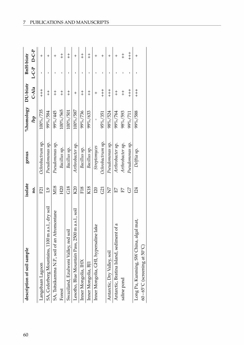
7 PUBLICATIONS AND MANUSCRIPTSde
scri
ptio
nof
soil
sam
ple
isol
ate
genu
s%
hom
olog
yD
U-b
iotr
BnH
-bio
trno
./b
pC
-Ala
L-C
-PD
-C-P
Lang
ebaa
nLa
goon
F21
Och
roba
ctru
msp
.10
0%/7
35++
+-
+SA
,Ced
erbe
rgM
ount
ains
,110
0m
a.s.
l.,dr
yso
ilL9
Pseu
dom
onas
sp.
99%
/584
++-
+SA
,Tsi
tsik
amm
aN
.P.,
soil
ofan
Afr
omon
tane
M18
Pseu
dom
onas
sp.
99%
/445
++-
+Fo
rest
H20
Baci
llus
sp.
100%
/565
++-
++Sw
azila
nd,E
zulw
eniV
alle
y,re
dso
ilG
18Ba
cillu
ssp
.10
0%/5
01++
-++
Leso
tho,
Blue
Mou
ntai
nPa
ss,2
500
ma.
s.l.,
soil
K20
Art
hrob
acte
rsp
.10
0%/5
87+
-+
Inne
rM
ongo
lia,I
HX
F18
Baci
llus
sp.
99%
/736
++-
++In
ner
Mon
golia
,BJ1
K18
Baci
llus
sp.
99%
/633
++-
++In
ner
Mon
golia
,GH
I,hy
pers
alin
ela
keI2
0St
rept
omyc
es-
+-
+G
21O
chro
bact
rum
sp.
95%
/351
+++
-+
Ant
arct
ic,D
ryV
alle
y,so
ilN
7Ps
eudo
mon
assp
.98
%/5
24++
+-
+A
ntar
ctic
,Bra
tina
Isla
nd,s
edim
ento
faE7
Art
hrob
acte
rsp
.99
%/7
64++
-+
salin
epo
ndF7
Art
hrob
acte
rsp
.98
%/5
93++
-++
G7
Pseu
dom
onas
sp.
99%
/711
+++
-++
+Lo
ngPu
,Kun
min
g,SW
Chi
na,a
lgal
mat
,60
–65◦ C
(scr
eeni
ngat
50◦ C
)I2
4D
elfti
asp
.99
%/5
88++
+-
+
60
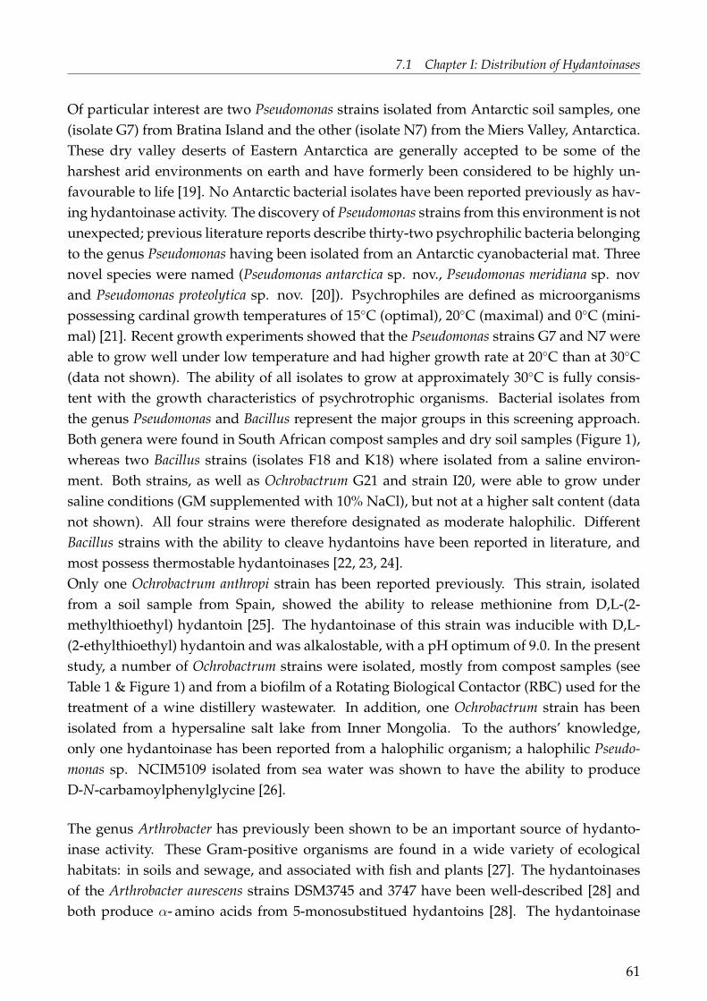
7.1 Chapter I: Distribution of Hydantoinases
Of particular interest are two Pseudomonas strains isolated from Antarctic soil samples, one(isolate G7) from Bratina Island and the other (isolate N7) from the Miers Valley, Antarctica.These dry valley deserts of Eastern Antarctica are generally accepted to be some of theharshest arid environments on earth and have formerly been considered to be highly un-favourable to life [19]. No Antarctic bacterial isolates have been reported previously as hav-ing hydantoinase activity. The discovery of Pseudomonas strains from this environment is notunexpected; previous literature reports describe thirty-two psychrophilic bacteria belongingto the genus Pseudomonas having been isolated from an Antarctic cyanobacterial mat. Threenovel species were named (Pseudomonas antarctica sp. nov., Pseudomonas meridiana sp. novand Pseudomonas proteolytica sp. nov. [20]). Psychrophiles are defined as microorganismspossessing cardinal growth temperatures of 15◦C (optimal), 20◦C (maximal) and 0◦C (mini-mal) [21]. Recent growth experiments showed that the Pseudomonas strains G7 and N7 wereable to grow well under low temperature and had higher growth rate at 20◦C than at 30◦C(data not shown). The ability of all isolates to grow at approximately 30◦C is fully consis-tent with the growth characteristics of psychrotrophic organisms. Bacterial isolates fromthe genus Pseudomonas and Bacillus represent the major groups in this screening approach.Both genera were found in South African compost samples and dry soil samples (Figure 1),whereas two Bacillus strains (isolates F18 and K18) where isolated from a saline environ-ment. Both strains, as well as Ochrobactrum G21 and strain I20, were able to grow undersaline conditions (GM supplemented with 10% NaCl), but not at a higher salt content (datanot shown). All four strains were therefore designated as moderate halophilic. DifferentBacillus strains with the ability to cleave hydantoins have been reported in literature, andmost possess thermostable hydantoinases [22, 23, 24].Only one Ochrobactrum anthropi strain has been reported previously. This strain, isolatedfrom a soil sample from Spain, showed the ability to release methionine from D,L-(2-methylthioethyl) hydantoin [25]. The hydantoinase of this strain was inducible with D,L-(2-ethylthioethyl) hydantoin and was alkalostable, with a pH optimum of 9.0. In the presentstudy, a number of Ochrobactrum strains were isolated, mostly from compost samples (seeTable 1 & Figure 1) and from a biofilm of a Rotating Biological Contactor (RBC) used for thetreatment of a wine distillery wastewater. In addition, one Ochrobactrum strain has beenisolated from a hypersaline salt lake from Inner Mongolia. To the authors’ knowledge,only one hydantoinase has been reported from a halophilic organism; a halophilic Pseudo-monas sp. NCIM5109 isolated from sea water was shown to have the ability to produceD-N-carbamoylphenylglycine [26].
The genus Arthrobacter has previously been shown to be an important source of hydanto-inase activity. These Gram-positive organisms are found in a wide variety of ecologicalhabitats: in soils and sewage, and associated with fish and plants [27]. The hydantoinasesof the Arthrobacter aurescens strains DSM3745 and 3747 have been well-described [28] andboth produce α- amino acids from 5-monosubstitued hydantoins [28]. The hydantoinase
61

7 PUBLICATIONS AND MANUSCRIPTS
0
2
4
6
8
10
12
14
16[number of bacteria]
compost SA dry
soil
Mongolia
(saline)
Antarctic Thermal
Arthrobacter Ochrobactrum Pseudomonas
Bacillus Others
Figure 1: Distribution of isolated bacterial genera in relation to environmental type. (Soilsamples (described in Table 1) are grouped by the dominant property: compost: derivedfrom compost or from habitats with a high amount of degrading material; SA dry soil:samples from South African desert and montane sites; Mongolia (saline): all saline Mon-golian sediment samples; Antarctic: all Antarctic soil samples; Thermal: Sediment sam-ples from habitats with elevated temperature (more than 50◦C).)
from A. aurescens DSM3745 belongs to the amidohydrolase superfamily [5] and has beenshown to be a Zn2+-metalloenzyme [29, 30] with L-selectivity in the conversion of D,L-5-(3-indolylmethyl) hydantoin. The hydantoinase and carbamoylase genes from Arthrobacteraurescens DSM3747 and DSM3745 have each been cloned and their nucleotide sequencesdetermined. These two enzymes show a high degree of nucleotide and amino acid sequenceidentity (96 – 98%) [31, 32].In this study Arthrobacter strains were found both in hot desert (African) and cold desert(Antarctic) soils. Two other Arthrobacter strains (F7 and G7) were found in sediment ofa saline pond from Bratina Island, Antarctica. These bacterial isolates showed the samegrowth characteristics as the Pseudomonas strains N7 and G7 (data not shown), and havebeen designated as psychrotrophic. The isolation of Arthrobacter strains from this environ-ment is not unexpected, since psychrophilic and psychrotrophic Arthrobacter strains have
62

7.1 Chapter I: Distribution of Hydantoinases
been reported from samples of subterranean cave silts [33], glacier silts [34] and the soilsof Antarctica [35, 36, 37, 38, 39]. Psychrotrophic bacteria isolated from cyanobacterial matsin the McMurdo Dry Valleys, Antarctica, have been recently characterised as Arthrobacterflavus sp. nov. [40] and Arthrobacter roseus sp. nov. [41]. Two isolates from penguin rookerysoil samples in Antarctica have been proposed as Arthrobacter gangotriensis sp. nov. andArthrobacter kerguelensis sp. nov. [42].
We also report the isolation at a growth temperature of 50◦C of a Delftia sp. strain from a hotspring algal mat (Long Pu, China) with activity towards BnH and DU. To our knowledge,no hydantoinase from this genus has been reported previously. However, thermophilichydantoinases have been described. A thermophilic D-hydantoinase from the moderatethermophile Bacillus stearothermophilus SD-1 was shown to have pH and temperature optimaof approximately 8.0 and 65◦C, respectively [22]. The most thermophilic hydantoinase re-ported to date (Topt = 80◦C) is derived from the hyperthermophilic archaeon, Methanococcusjannaschii; isolated from a submarine hydrothermal vent [43]. Thermostable hydantoinaseshave also been isolated from a mesophilic Bacillus sp. AR9 [23] and a moderate thermophilicBacillus stearothermophilus NS1122A, with optimal temperatures of between 60◦C and 70◦C[24].D-hydantoinase activity has been reported in Burkholderia pickettii [44], towards D,L-5-mercaptoethyl-hydantoin in Enterobacter cloacae [45], and in several different Flavobacteriumspecies [46, 47]. Bacterial isolates recovered from South African soil and belonging to thesegenera were also found in our study.We also report the detection of hydantoinase activity in bacterial genera where this enzymehas not been reported previously. These include Stenotrophomonas sp. N1, Microbacteriacaesp. K3, Staphylococcus sp. H7, Acinetobacter sp. K5, Delftia sp. I24 and an unidentified isolateI20 (possibly Streptomyces; Dr. Eberspächer pers. communication).
The aim of this study was the recovery of microorganisms with the ability to cleave hydan-toins. The standard condition for principal isolation of microorganisms was Medium I (pH7.0) at 30◦C. Apart from Delftia sp. I24, all bacterial isolates were recovered under standardconditions. However, isolates originating from saline and cold environments (Mongolia andAntarctica, respectively) were not isolated under conditions exactly reflecting their environ-mental origins. After subsequent tests for their ability to grow under saline and/ or coldconditions, these strains were designated as psychrotrophic or moderately halophilic, asappropriate.
63
7 PUBLICATIONS AND MANUSCRIPTS
ConclusionsWe conclude on the basis of our isolation data and published literature that(i) hydantoinase-positive bacterial isolates are very widely dispersed, both geographicallyand with respect to environmental conditions, and(ii) among culturable aerobes, hydantoinases are predominantly found in certain genera(Pseudomonas, Ochrobactrum, Bacillus, Arthrobacter).(iii) We have also described microorganisms for which no hydantoinase activity wasreported previously.With respect to the natural function of hydantoinases, it may be significant that the majorityof the isolates recovered were derived from samples with a high organic load, such ascompost soil and guano. These findings support the hypothesis that hydantoinases areinvolved in catabolic pathways to access hydantoins and hydantoin-like molecules asmetabolic substrates.
AcknowledgmentThe authors would like to thank the Federal Ministry of Science and Education (BMBF),Germany, and the National Research Foundation (NRF), South Africa, for the fundingof this bilateral project. The German Academic Exchange Service (DAAD) and the Stateof Baden Württemberg are gratefully acknowledged for supporting the correspondingauthor´s research in South Africa. The following are specifically acknowledged for supportand/ or approval in providing access to samples: the South African National Parks for theopportunity to collect soil samples in the national parks, the joint Chinese-EU-SA MGATechproject (QLRT-2001-01972) for access to Chinese samples, the Waikato University AntarcticTerrestrial Biology Program and Antarctica NZ for access to soils from the Antarctic DryValleys, and Prof. Gideon Wolfaardt (University of Stellenbosch) for collection of biofilmsamples from the RBC of a brandy distillery.
64

7.1 Chapter I: Distribution of Hydantoinases
References
1. A. Louwrier, C.J. Knowles. Enzyme Microb. Technol. 19 (1996) 562-571.
2. C. Syldatk, A. Läufer, R. Müller, H. Höke. Adv. Biochem. Eng. Biotechnol. 41 (1990)29-75.
3. C. Syladtk, O. May, J. Altenbuchner, R. Mattes, M. Siemann. Appl. Microbial. Biotechnol.51 (1999) 293-309.
4. S. Runser, P.C. Meyer. Eur. J. Biochem. 213 (1993) 1315-1324.
5. O. May, A. Habenicht, R. Mattes, C. Syldatk, M. Siemann. Biol. Chem. 379 (1998)743-747.
6. Z. Gojkovic, L. Rislund, B. Andersen, M.P.B. Sandrini, P.F. Cook, K.D. Schnackerz, J.Pikur. Nucleic. Acids. Res. 31 (2003) 1683-1692.
7. C. Syldatk, D. Cotoras, G. Dombach, C. Gross, H. Kallwass, F. Wagner. Biotechnol. Lett.9 (1987) 25-30.
8. R. Tsugawa, S. Okumura, T. Ito, N. Katsuga. Agric. Biol. Chem. 30 (1966) 27-34.
9. C. Syldatk, V. Mackowiak, H. Höke, C. Gross, G. Dombach, F. Wagner. J. Biotech. 14(1990) 345-362.
10. H. Teves, T. Waniek, M. Pietzsch, C. Syldatk, M. Reuss., J. Fresenius. Anal. Chem. 363(1999) 738-743.
11. V. Farrelly, F.A. Rainey, E. Stackebrandt. Appl. Environ. Microbiol. 61 (1995) 2798-2801.
12. A.L. Reysenbach, N.R. Pace. In. Archaea. A laboratory manual. Thermophiles. 1995by Cold Spring Harbour.
13. S.G. Burton, R.A. Dorrington, C. Hartley, S. Kirchmann, G. Matcher, V. Phehane. J. Mol.Catal. B: Enzymatic 5 (1998) 301-305.
14. A. Balows, H.G. Trüper, M. Dworkin, W. Harder, K.H. Schleifer. The Prokaryotes,Second Edition, Springer Verlag, New York, 1992, p. 3074.
15. D.V. Gokhale, K.B. Bastawde, S.G. Patil, U.R. Kalkote, R.A. Joshi, T. Ravindranathan,B.G. Gaikwad, V.V. Jogdand, S. Nene. Enzyme Microb. Technol. 18 (1996) 353-357.
16. H.R. Chien, W.-H Hsu. Biotechnol. Tech. 10 (1996) 879-882.
17. H.R. Chien, Y.L. Jih, W.Y. Yang, W.H. Has. Biochim. Biophys. Acta 1395 (1998) 68-77.
65

7 PUBLICATIONS AND MANUSCRIPTS
18. G. La Pointe, S. Viau, D. LeBlanc, N. Robert, A. Morin. Appl. Environ. Microbiol. 60(1994) 888- 895.
19. W.L. Boyd, J.T. Staley, J.W. Boyd. Antarctic. Res. Ser. 8 (1996) 125-159.
20. G.S.N. Reddy, G.I. Matsumoto, P. Schumann, E. Stackebrandt, S. Shivaji. Int. J. Syst.Evol. Microbiol. 54 (2004) 713-719.
21. R.Y. Morita. Bacteriological Reviews 39 (1975) 144-167.
22. S.G. Lee, D.C. Lee, S.P. Hong, M.H. Sung, H.S Kim. Appl. Microbial. Biotechnol. 43(1995) 270-276.
23. A. Sharma, R.M. Vohra. Biochem. Biophys. Res. Commun. 234 (1997) 485-488.
24. T. Ishikawa, Y. Mukohara, K. Watabe, S. Kobayaschi, H. Nakamura. Biosci. Biotechnol.Biochem. 58 (1994) 265-270.
25. C. Pozo, B. Rodelas, S. de la Escalera, J. González-López. J. Appl. Microbiol. 92 (2002)1028-1034.
26. S.S. Sudge, K.B. Bastawde, D.V. Gokhale, U.R. Kalkote, T. Ravindranathan. Appl.Microbiol. Biotechnol. 49 (1998) 594-599.
27. A. Balows, H.G. Trüper, M. Dworkin, W. Harder, K.H. Schleifer, The Prokaryotes, Sec-ond Edition, Springer Verlag, New York, 1992, 1285-1286.
28. C. Gross, C. Syladtk, F. Wagner. Biotechnol. Tech. 1 (1987) 85-90.
29. O. May, M. Siemann, M. G. Siemann C. Syldatk. J. Mol. Catal B: Enzymatic 5 (1998)367-370.
30. O. May, M. Siemann, C. Syldatk. J. Mol. Catal B: Enzym 4 (1998) 211-218.
31. B. Wilms, A. Wiese, C. Syldatk, R. Mattes, J. Altenbuchner, M. Pietsch. J. Biotechnol. 68(1999) 101-113.
32. A. Wiese, B. Wilms, C. Syldatk, R. Mattes, J. Altenbuchner. Appl. Microbiol. Biotechnol.55 (2001) 750-757.
33. A.M. Gounot. Ann. Inst. Pasteur Paris 113 (1967) 923-945.
34. A. Moiroud, A.M. Gounot. Hebd. Seances Acad. Sci. Ser. D. Sci. Nat. 269 (1969) 2150-2152.
35. R.M. Johnson, M. Inai, S. McCarthy. J. Ariz-Nev. Acad. Sci. 16 (1981) 51-60.
36. R.M. Johnson, R.D. Bellinoff. Antarct. Res. Ser. 30 (1981) 169-184.
66

7.1 Chapter I: Distribution of Hydantoinases
37. J.M. Madden, S.K. Siegel, R.M. Johnson Antarct. Res. Ser. 30 (1979) 77-103.
38. J. Siebert, P. Hirsch. Polar Biol. 9 (1988) 37-44.
39. S. Shivaji, N.S. Rao, L. Saisree, G.S.N. Reddy, G. Seshu Kumar, P.M. Bhargava. PolarBiol. 10 (1989) 225-229.
40. G.S.N. Reddy, R.K. Aggarwal, G.I. Matsumoto, S. Shivaji. Int. J. Syst. Evol. Microbiol.50 (2000) 1553-1561.
41. G.S.N. Reddy, J.S.S. Prakash, G.I. Matsumoto, E. Stackebrandt, S. Shivaji. Int. J. Syst.Evol. Microbiol. 52 (2002) 1017-1021.
42. P. Gupta, G.S.N. Reddy, D. Delille, S. Shivaji. Int. J. Syst. Evol. Microbiol. 54 (2004)2375-2378.
43. J.-H. Chung, J.-H. Back, J.-H. Lim, Y.I. Park, Y.S. Han. Enzyme Microbial. Technol. 30(2002) 867-874.
44. Z. Xu, Y. Liu, Y. Yang, W. Jiang, E. Arnold, J. Ding. J. Bacteriol. 185 (2003) 4038-4049.
45. G. LaPointe, D.Leblanc, A. Morin. Appl. Microbiol. Biotechnol. 42 (1995) 895-900.
46. K. Yokozeki, K. Sano, C. Eguchi, K. Yamada, K. Mitsugi. Agric. Biol. Chem. 51 (1987)363-369.
47. Y. Nishida, K. Nakamichi, K. Nabe, T. Tosa. Enzyme Microb. Technol. 9 (1987) 721-725.
67

Whitepage
68

7.2 Chapter II: Biodiversity of Hydantoin Cleaving Enzymes
7.2 Chapter II: Biodiversity of Hydantoin Cleaving Enzymes
BIODIVERSITY OF HYDANTOIN CLEAVING ENZYMES
69

7 PUBLICATIONS AND MANUSCRIPTS
Introduction"Biogeography" is the "science that attempts to document and understand spatial patternsof biodiversity, distribution of organisms, both past and present, and of related patternsof variation over the earth in the numbers and kinds of living things" (Brown and Lo-molino, 1998). Baas-Becking (1934) postulated that microorganisms are highly dispersedresulting in a cosmopolitan microbial world ("everything is everywhere") and a lack ofbiogeographical patterns for free living bacteria. This theory is still under strong debatewithin the microbiologist community. Recent investigations demonstrated biogeographicalpatterns for some bacterial taxa. Whitaker et al. for example (2003) found a correlationbetween the geographical and genetic distance of hot spring archaean Sulfolobus isolatesfrom geographically distinct regions.
The United Nations Conference on Environment and Development, UNCED (1992) definedthe term "biological diversity" or "biodiversity" to be the variety of genes, species, andecosystems found on our planet. It embraces all life forms – from plant and animal lifeto microorganisms – and the water and land in which they interact. An introduction onhydantoinases as well as the distribution and occurrence of hydantoinase possessing micro-organisms was shown by Dürr et al. (2006). In summary, hydantoinase-positive bacterialisolates are very widely dispersed, both geographically and with respect to environmentalconditions. Among culturable aerobes hydantoinase-positive bacteria are predominantlyfound in certain genera: Pseudomonas, Ochrobactrum, Bacillus and Arthrobacter.This chapter will focus on the biodiversity of hydantoin cleaving enzymes meaning the vari-ety of hydantoinases hereby referring to the UNCED definition. Therefore a detailed inves-tigation of the following topics was conducted for the hydantoinases previously described(Dürr et al., 2006):
1. It is known that hydantoinases have a broad and diverse substrate spectrum (com-pare Introduction, Table 1). The remaining question is if there is a correlation betweensubstrate spectrum and origin of hydantoinases concerning bacterial genera and/ orecological habitat?
2. Can the hydantoinase gene sequence of different microorganisms be clustered intospecific groups either related to a genus and/ or to a certain habitat?
3. Is there a correlation between the biochemical properties of hydantoinases and certaingroups of hydantoinase gene sequences?
4. Are hydantoinase genes as well present in microorganisms for which hydantoinaseactivity could not be shown, yet?
The screening for hydantoinases is normally done by classical ways using enrichmenttechniques which enable the isolation of microorganisms with hydantoinase activity.
70
7.2 Chapter II: Biodiversity of Hydantoin Cleaving Enzymes
Additionally, molecular and immunological attempts are described to get access to newenzymes being able to cleave dihydropyrimidines/ hydantoins.Hydantoinase gene sequences were found by searching for hydantoinase positive clones ina genomic library either by activity assays (LaPointe et al., 1994; Kim et al., 1997) or growthon hydantoin as the sole nitrogen source (Mukohara et al., 1994; Chien et al., 1998). Poly-clonal antibodies were successfully developed for the detection of the L-hydantoinase fromArthrobacter aurescens but they were not specific for the detection of the D-hydantoinasefrom Agrobacterium sp. (Siemann et al., 1993 a, b). LaPointe et al. (1994) developed a 1.5 kbDIG-labelled DNA probe derived from a recombinant hydantoinase gene from Pseudomonasputida DSM84. The probe only hybridised with total DNA from Pseudomonas strains ofrRNA group I showing hydantoinase activity. No hybridisation occurred with DNA frombacteria outside rRNA group I indicating the divergence of genes responsible for hydantoinconversion. The authors used a 122 bp DNA probe obtained from the same organismmentioned above to detect D-hydantoinase genes in other bacterial genera by DNA andby colony hybridisation. The probe was specific, allowing 32% mismatch, to detect knownD-hydantoinase activity in microorganisms belonging to the genera Pseudomonadaceae,Agrobacterium, Serratia, Corynebacterium and Arthrobacter. In an experiment under environ-mental conditions also bacteria belonging to the genera Pseudomonas, Klebsiella, Enterobacterand Enterococcus showed a positive signal towards the probe. Hydantoinase activity wasshown for the mentionend bacteria (LaPointe et al., 1995).Recently, degenerate primers were reported for the amplification of a 330 bp dihydro-pyrimidinase gene fragment of Bacillus sp. TS-23 (Lin et al., 2005). They were designedfrom conserved motifs obtained from the comparison of the amino acid sequence ofhydantoinases/ dihydropyrimidinases from Bacillus stearothermophilus, Pseudomonas putida,Agrobacterium tumefaciens and Streptomyces coelicor. The amplified 330 bp DNA fragmentwas used as a probe to identify the dihyropyrimidinase gene within a genomic library. Theisolated dihyropyrimidinase gene consisted of 1422 bp and was deduced to contain 472amino acids sharing 40 – 87% sequence identity with Arthrobacter aurescens, Thermus sp.,Burkholderia picketii and Bacillus stearothermophilus.
71

7 PUBLICATIONS AND MANUSCRIPTS
Materials and MethodsAbbreviationsBnH: 5-benzylhydantoin; CA: Corynebacterium medium; CA-plates: Corynebacteriumagar plates; C-Ala: N-carbamoyl-β-alanine; C-Gly: N-carbamoyl-glycine; CH3-IMH: D,L-5-(3-indolylmethyl)-3-N-methylhydantoin; D-/L-C-Phe: N-carbamoyl-D/ L-phenylalanin; D-/L-C-t-Leu: N-carbamoyl-D/L-tert-leucine; D-/L-C-Try: N-carbamoyl- D-/L-tryptophane;N-C-βPhe: N-carbamoyl-β-phenylalanine; DIG-G21: random primed DNA probe labeledwith digoxigenin-dUTP, DNA derived from hydantoinase amplification of genomic DNAfrom Ochrobactrum sp. G21; DIG-I24: see DIG-G21, genomic DNA from Delftia sp. I24; DU:5,6-dihydrouracil; GM: growth medium; Hyd: hydantoin; IMH: D,L-5-(3-indolylmethyl)hydantoin; SA: South Africa; t-BH: D,L-5-tert-butylhydantoin; PheDU: D,L-6-phenyl-5,6-dihydrouracil.
ChemicalsAll chemicals were obtained from commercial sources and were of reagent grade exceptfor hydantoins, dihydropyrimidines and their derivatives. They were kindly supplied byDegussa AG, Germany. If not otherwise stated enzymes, chemicals and kits for geneticalexperiments were obtained from Fermentas, Germany. PheDu and N-C-βPhe were synthe-sised using an optimised method (modified following instructions of Dakin and Dudley,1914): N-C-βPhe: 200 mg 3-amino-3-phenylpropionic acid, 120 mg KNCO and 10 mL waterwere poured in a 25 mL round-bottomed flask and allowed to react at 80◦C with a condenserconnected to the flask. As the reaction proceeded the initial white suspension became aclear solution. After one hour the flask was cooled down to room temperature and the pHwas adjusted to about 3 with 4N HCl. The precipitate was left 30 min to settle down. Thenit was filtered through a Buchner funnel, washed with 5 mL of water and dried. PheDu:150 mg N-C-Phe were poured in a 25 mL round-bottomed flask, 4 mL of water were addedand 4 mL of HCl 32% were slowly dropped into the milky mixture. The reaction was carriedout for approximately 2 hours at 80◦C with a condenser connected to the flask. The flaskwas cooled to 4◦C; after 30 min the precipitate was collected by filtration, washed with 5 mLof cold water and dried. Both reactions were followed by HPLC.
Bacterial StrainsIsolation and characterisation of the bacterial isolates used was described elsewhere (Dürret al., 2006).
72
7.2 Chapter II: Biodiversity of Hydantoin Cleaving Enzymes
Media and General Growth ConditionsThe media used in this study were: CA, used for the starter cultures and growth medium(Dürr et al., 2006); GM according to Syldatk et al. (1990), used as growth medium forbiotransformation assays either with 0.2 – 1 g/L IMH or CH3-IMH as inducer. Bacterialstrains were routinely maintained at CA-plates at 30◦C and stored at 4◦C (Dürr et al., 2006).For long term storage bacterial isolates were kept on 20% glycerol at – 80◦C.
Cell Growth and BiotransformationsCell growth and biotransformations were performed as described elsewhere (Dürr et al.,2006) but with the following modifications: 200 mM K-phosphate buffer (pH 8.0) was usedinstead of 100 mM; the total volume of the biotransformation reaction was 1 mL and thesubstrates were dissolved by heating if necessary (40 – 50◦C). Biotransformations werestopped by addition of 100 µL of 50% H3PO4. The final concentrations of the substrateswere as follows: 25 mM DU, 20 mM Hyd, 8 mM t-BH, 5 mM BnH, 2 mM IMH, 4 mM PheDU.
Determination of Dry WeightA volume of 0.5 mL – 1 mL of the cell suspension used for biotransformation assayswas centrifuged (10 min, 13,000 rpm), the supernatant discarded and the pellet driedover night at 100◦C. The biomass dry weight was determined by weighing of the driedpellet. The average of three measurements was used for the determination of the dry weight.
AnalysisHPLCHPLC-analysis (High Performance Liquid Chromatography) was the general detection andquantification method for hydantoins and their corresponding derivatives except for DUand Hyd (see below). HPLC-analysis was routinely carried out on an Agilent 1100 seriesHPLC System (Agilent Technologies, Germany). In most cases substrates and productswere first analysed using non-chiral separation. For selected samples chiral analysis of thecarbamoyl amino acid followed. HPLC analysis was conducted as follows:
73

7 PUBLICATIONS AND MANUSCRIPTS
1a) Non-chiral analysis of D,L-5-tert-butylhydantoin (t-BH) andN-carbamoyl-D/L-tert-leucin (C-t-Leu)
column Thermo Hypersil-Keystone BDS C18(250 x 4.6 mm, 5 µm)
mobile phase 20% MeOH/ 80% (0.1% H3PO4)flow rate 1 mL/mindetection wavelength 210 nmretention times t-BH: 15.7 min
C-t-Leu: 13.5 min
1b) Chiral analysis of N-carbamoyl-D/L-tert-leucin (D- or L-C-t-Leu)
column Macherey-Nagel, Nucleodex β-PM(150 x 4 mm, 5 µm)
mobile phase 5% MeOH/ 95% (0.1% H3PO4), pH 3.0flow rate 0.5 mL/mindetection wavelength 200 nmretention times L-C-t-Leu: 9.2 min
D-C-t-Leu: 10.2 min
2a) Non-chiral analysis of D,L-5-benzylhydantoin (BnH) andN-carbamoyl-D/L-phenylalanin (C-Phe)
column Thermo Hypersil-Keystone BDS C18(250 x 4.6 mm, 5 µm)
mobile phase 20% MeOH/ 80% (0.1% H3PO4)flow rate 1 mL/mindetection wavelength 210 nmretention times BnH: 22.8 min
C-Phe: 25.5 min
2b) Chiral analysis of N-carbamoyl-D/L-phenylalanin (D- or L-C-Phe)
column Macherey-Nagel, Nucleodex β-PM(150 x 4 mm, 5 µm)
mobile phase 10% MeOH/ 90% (0.1% H3PO4), pH 3.0flow rate 0.5 mL/mindetection wavelength 210 nmretention times L-C-Phe: 18.0 min
D-C-Phe: 14.6 min
74

7.2 Chapter II: Biodiversity of Hydantoin Cleaving Enzymes
3a) Non-chiral analysis of D,L-5-(3-indolylmethyl) hydantoin (IMH) andN-carbamoyl-D-/L-tryptophan (C-Try)
column Thermo Hypersil-Keystone BDS C18(250 x 4.6 mm, 5 µm)
mobile phase 20% MeOH/ 80% (0.1% H3PO4)flow rate 1 mL/mindetection wavelength 210 nmretention times IMH: 37.0 min
C-Try: 29.0 min
3b) Chiral analysis of N-carbamoyl-D-/L-tryptophan (D- or L-C-Try)
column Chiral Technology Europe, Chiralpak QN-AX(150 x 4 mm, 5 µm)
mobile phase 50% MeOH/ 50% (0.2M AcOH); pH 6.0 (25% NH3)flow rate 0.8 mL/mindetection wavelength 280 nmretention times D-C-Try: 32.8 min
L-C-Try: 52.9 min
4) Non-chiral analysis of D,L-6-phenyl-5,6-dihydrouracil (PheDU) andN-carbamoyl-β-phenylalanine (C-βPhe)
column Thermo Hypersil-Keystone BDS C18(250 x 4.6 mm, 5 µm)
mobile phase 20% MeOH/ 80% (0.1% H3PO4)flow rate 1 mL/mindetection wavelength 210 nmretention times PheDU: 19.0 min
C-βPhe: 16.0 min
75
7 PUBLICATIONS AND MANUSCRIPTS
Ehrlich TestThe Ehrlich Test (described by Dürr et al., 2006) was applied for detection and quantitativedetermination of C-Ala and C-Gly, the products of DU- and Hyd-biotransformations,respectively.
DNA Manipulation MethodsGeneral recombinant DNA techniques were carried out by standard procedures (Sambrocket al., 1989). Chromosomal bacterial DNA was extracted using the Qiagen DNeasy Tissue kit(Qiagen, Germany) following the manufacturer’s protocol. DNA fragments were purifiedfrom standard agarose gels by a DNA extraction kit. Pure plasmids were obtained by aGeneJETTM Plasmid Mini Prep Kit. Sequencing was done commercially (JenaGen GmBH,Germany) and DNA-analysis was carried out using BlastN (Altschul et al., 1997) andClustalW (Chenna et al., 2003). Phylogenetic and molecular evolutionary analyses wereconducted using MEGA version 3.1 (Kumar et al., 2004).
Amplification of a Hydantoinase DNA-FragmentPolymerase chain reaction (PCR) was applied for the amplification of a hydantoinase genefragment using genomic bacterial DNA as template. Amplification was carried out onone of the following thermocyclers: Eppendorf Mastercycler gradient (Germany) or MJResearch PTC-200 (USA). The conditions were: 10 – 100 ng template DNA, 1x-Taq bufferwith (NH4)2SO4, 1.5 U Taq DNA Polymerase, 50 pmol upstream primer and downstreamprimer each, 0.2 mM dNTP´s, and PCR-water, combined to a volume of 20 µL. The followingdegenerate oligonucleotide primers were used synthesised by MWG-Biotech, Germany:dhpf: 5´-GCSGCVTTYGGNGGNACNAC-3´,dhpr: 5´-TCNCCRTTYTCNGCRTGNAC-3´ (Lin et al., 2005) orHyd-f: 5´-GCCGCAGCATGCGGNGGNACNAC-3´,Hyd-r: 5´-GAYGCNGAYHTAGTCATATGGGACCCTAATGGTG-3´ (provided by Dr. A.Puñal, University of Karlsruhe).PCR-amplification was initiated with DNA denaturation at 95◦C for 5 min, followed by30 cycles of denaturation at 94◦C for 1 min, annealing at 40 – 60◦C for 2 min and primerextension at 72◦C for 1 min. The cycles were finalised by 7 min extension at 72◦C. Afteramplification PCR-products of 320 – 330 bp or approximately 950 bp were purified. PurifiedPCR-products were either sequenced directly or cloned.
Cloning of PCR-ProductsTo obtain an adequate amount of DNA for sequencing the purified PCR-products werecloned using the Strata CloneTM PCR Cloning kit (Stratagene, USA). The inserts of thepurified plasmids were sequenced.
76

7.2 Chapter II: Biodiversity of Hydantoin Cleaving Enzymes
Dot Blot AnalysisDot blot analysis was carried out using the DIG DNA Labeling and Detection Kit (Roche,Germany) using standard procedures. A volume of 1 µL of pure genomic DNA was usedfor dot blot analysis. The probes used for DIG-labelling and detection were DIG-I24 andDIG-G21 (see Chapter 4).
77

7 PUBLICATIONS AND MANUSCRIPTS
Results and DiscussionThe discovery of various microorganisms with hydantoinase activity from different geo-graphical origins was described previously (Dürr et al., 2006). Several bacterial isolates fromthis screening program were selected for further biochemical and genetical characterisation.The selection criteria were:(i) high activity of the hydantoinase of a strain towards the substrates BnH and DU;(ii) isolation of the strain from an extreme or unique environmental habitat like hot springsor biofilms;(iii) no report in literature on hydantoinase activity of the chosen bacterial genus.
Substrate Spectrum of HydantoinasesDifferent hydantoins and dihydropyrimidines were used as a substrate to determine thesubstrate spectrum of the hydantoinases from the chosen bacterial strains. The chemicalstructures of the six substrates tested are shown in Figure 1.
N
N
O
O
NH NH
O
O
NHNH
O
O
NH NH
O
O
NN
O
O
NNH N
O
O
5,6-dihydrouracil (DU)
hydantoin (Hyd)
5-tert-butylhydantoin (t-BH)
5-benzylhydantoin (BnH)
D,L-5-(3-indolylmethyl) hydantoin IMH
6-phenyl-5,6-dihydrouracil (PheDU)
Figure 1: Chemical structure of hydantoins and dihydropyrimidines used as substrates inthis study.
Looking at the wild-type strains isolated from different geographical regions (South Africa,China and Antarctica) it is noticeable that nearly all strains are able to cleave the followingsubstrates (Table 1): the non-substituted hydantoin Hyd (with the exception of Microbacteri-acae sp. K3) and the dihydropyrimidine derivative DU, as well as BnH being a compoundbearing an aryl-side chain. The hydantoinase activities were higher for the non-substituted
78

7.2 Chapter II: Biodiversity of Hydantoin Cleaving Enzymes
substrate DU than for Hyd for almost all strains tested for a similar substrate concentration.The opposite result is obtained for Bacillus sp. A16 and F18. The preference towards DUfor most of the bacteria tested in this study leads to the assumption that the enzymesbeing responsible for hydantoin and dihydropyrimidine conversion can be considered asdihydropyrimidinases and are part of the reductive pathway of pyrimidine degradation.Syldatk et al. (1999) demonstrated that D-hydantoinases are not necessarily identical todihydropyrimidinases and the term "hydantoinase" should be used for enzymes being ableto hydrolyse hydantoin or 5-monosubstituted hydantoins. Consequently, enzymes beingresponsible for hydantoin and dihydropyrimidine conversion are named as hydantoinasesin this study.
79

7 PUBLICATIONS AND MANUSCRIPTS
Tabl
e1:
Subs
trat
esp
ectr
umof
hyda
ntoi
nase
sin
who
le-c
ellb
iotr
ansf
orm
atio
ns.
The
spec
ific
hyda
ntoi
nase
acti
vity
for
each
subs
trat
eis
show
nas
the
conc
entr
atio
nof
the
form
edca
rbam
oyla
min
oac
idin
rela
tion
toth
ebi
omas
sus
edan
dti
me
[mM
/(h*
g)].
Diff
eren
tsub
stra
teco
ncen
tra-
tion
sw
ere
used
beca
use
ofdi
ffer
enta
queo
usso
lubi
lity
ofhy
dant
oins
and
dihy
ropy
rim
idin
es.I
fdet
erm
ined
the
confi
gura
tion
ofth
efo
rmed
prod
ucte
nant
iom
eris
give
nin
brac
kets
.(*
grow
thin
CA
med
ium
supp
lem
ente
dw
ith
0.2
g/L
CH
3-IM
H;S
A:S
outh
Afr
ica)
Isol
ate
subs
trat
eD
UH
ydt-
BH
BnH
IMH
PheD
U(2
5mM
)(2
0mM
)(8
mM
)(5
mM
)(2
mM
)(4
mM
)-
orig
inpr
oduc
tC
-Ala
C-G
lyC
-t-L
euC
-Phe
C-T
ryC
-ßPh
e
Del
ftia
sp.I
24C
hina
,hot
spri
ngs
54.5
413
3.45
*2.
80*
(D)
0.97
(D)
5.37
*0
Mic
roba
cter
iaca
esp
.K3
SApi
new
ood
fore
st60
.02
00
0.43
(D,L
)0
0.90
Burk
hold
eria
sp.M
3pi
new
ood
fore
stSA
57.8
310
.76
0.33
(D)
0.13
(L)
0.35
0.10
Flav
obac
teri
umsp
.F8
SA,c
ompo
st68
.04
11.2
70.
33(D
)1.
58(D
)0.
560.
62St
rept
omyc
essp
.I20
Mon
golia
,sal
ine
lake
90.5
55.
060
0.32
(D)
00
Baci
llus
sp.A
16SA
,gua
noof
apo
nd30
.57
45.6
5.69
(D)
0.76
(D)
2.70
(D)
1.49
Baci
llus
sp.F
18M
ongo
lia,s
alin
ela
ke61
.77
85.9
12.
53(D
)0.
72(D
)1.
470.
30Ba
cillu
ssp
.G18
Swaz
iland
,red
soil
0.81
96.4
80.
124.
26(D
)0.
54(D
)1.
70Ba
cillu
ssp
.H20
SAfo
rest
soil
37.4
82.
220
-0
0Ps
eudo
mon
assp
.G7
Ant
arct
ica
51.4
84,
630.
85(D
)0.
93(D
)0
0.33
Pseu
dom
onas
sp.M
18SA
,for
ests
oil
40,1
850
.83
0.72
1.69
(D)
2.1
(D)
0.05
Pseu
dom
onas
sp.L
9SA
,mou
ntai
nso
il11
.23
4.73
00
00
Och
roba
ctru
msp
.C15
SA,c
ompo
st69
.48
7.48
03.
11(D
)0.
440.
51O
chro
bact
rum
sp.G
21M
ongo
lia,s
alin
ela
ke63
.91
34.9
90
2.33
(D)
0.48
(D)
0.82
Och
roba
ctru
msp
.D24
SA,b
iofil
m41
.93
58.9
90.
131.
21(D
)0,
44(D
)0
Och
roba
ctru
msp
.F21
SA,a
lgal
mat
eria
l37
.71
38.4
30.
183.
12(D
)0,
23(D
)0.
30O
chro
bact
rum
sp.I
21SA
,com
post
21.8
526
.95
0.42
2.18
(D)
0.38
(D)
0.40
Art
hrob
acte
rsp
.E7
Ant
arct
ica
24.2
51,
440
-0
0.51
Art
hrob
acte
rsp
.K20
Leso
tho,
mou
ntai
nso
il18
.74
1.31
1.05
(D)
0.09
02.
38A
.pol
ychr
omog
enes
DSM
2013
6D
SMZ
32.7
70.
680
00
4.86
A.p
olyc
hrom
ogen
esD
SM34
2D
SMZ
38.2
90.
300
00
5.73
80
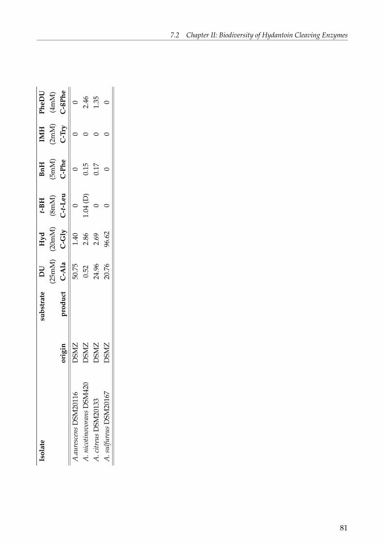
7.2 Chapter II: Biodiversity of Hydantoin Cleaving EnzymesIs
olat
esu
bstr
ate
DU
Hyd
t-B
HB
nHIM
HPh
eDU
(25m
M)
(20m
M)
(8m
M)
(5m
M)
(2m
M)
(4m
M)
-or
igin
prod
uct
C-A
laC
-Gly
C-t
-Leu
C-P
heC
-Try
C-ß
Phe
A.a
ures
cens
DSM
2011
6D
SMZ
50.7
51.
400
00
0A
.nic
otin
ovor
ans
DSM
420
DSM
Z0.
522.
861.
04(D
)0.
150
2.46
A.c
itreu
sD
SM20
133
DSM
Z24
.96
2.69
00.
170
1.35
A.s
ulfu
reus
DSM
2016
7D
SMZ
20.7
696
.62
00
00
81
7 PUBLICATIONS AND MANUSCRIPTS
Comparing the conversion of the substituted hydantoin BnH and the substituted di-hydropyrimidine PheDu (both bearing an aryl-side chain and consisting either of a 5- or6-membered ring, Fig. 1) with DU and Hyd the opposite behaviour could be observedfor most of the strains tested: BnH was converted by more strains and with a higherhydantoinase activity compared to PheDU conversion. This shows that substrate bindingand turnover are not only dependent on interactions between enzyme and substrate ring,but also on interactions between enzyme and substrate side chain ( additionally see ChapterIV). The substrates t-BH and IMH in comparison to the substrates mentioned above werecleaved by less bacteria.In the case of IMH the latter result attracts attention: All strains should be able to convertthe 5-monosubstituted hydantoin IMH since they were isolated on media containing IMHas C- and N-source. It has to be mentioned that fructose and ammonia were as well presentin the screening media (see Dürr et al., 2006) but nevertheless strains possessing an activehydantoinase were screened.A second observation is that almost all bacteria screened are D-specific documented by theformation of either D-C-t-Leu, D-C-Phe and/ or D-C-Try (Table 1). L-specific hydantoinaseswould be expected since other microorganisms were isolated on IMH like Flavobacteriumsp. I-3 (Nishida et al., 1987), Flavobacterium sp. AJ-3912 (Sano et al., 1977; Yokozeki et al.,1987a, b) and Arthrobacter aurescens 3747 (Gross et al., 1987) and were reported to formL-tryptophan from IMH. However, stereoselectivity can vary depending on the substrate:The L-hydantoinase from A. aurescens DSM3745 showed to be L-selective for D,L-BnHwhilst conversion of D,L-methylthioethylhydantoin was D-specific (May, 1998). Similarresults were obtained for Flavobacterium sp. AJ-3912. D-specificity was only obtained forbenzyloxymethylhydantoin whereas the other substrates tested were cleaved L-selectively(Yokozeki et al., 1987b). In this study a change of selectivity for different substrates wasobtained for Burkholderia sp. M3 (Table 1).
The enzymes involved in hydantoin degradation of Arthrobacter aurescens DSM3747, Arthro-bacter aurescens DSM3745 and Arthrobacter crystallopoietes DSM20117 are well described (e.g.May, 1998; Waniek, 2000; Werner, 2001 and Wiese, 2000). The question arises as to whethermore members of the genus Arthrobacter possess hydantoinase activity? Since only threeArthrobacter species were isolated in the previously reported screening program (Dürr etal., 2006) more Arthrobacter species were included for hydantoinase screening. Thereforevarious Arthrobacter strains were obtained from the German Collection of Microorganismsand Cell Cultures (DSMZ) and were tested for the ability to cleave various hydantoinsand dihydropyrimidines (Table 1). The ability to cleave BnH was formerly reported for A.citreus DSM20133/ ATCC11624 (LaPointe et al., 1995) which could be confirmed within thisstudy (Table 1).It is remarkable that almost all Arthrobacter strains from the DSMZ with hydantoinase
82
7.2 Chapter II: Biodiversity of Hydantoin Cleaving Enzymes
activity preferred DU and PheDu as substrates and were able to cleave non-substitutedhydantoin and in some cases 5-monosubstituted hydantoin derivatives with very lowactivity. This indicates that the enzymes from these Arthrobacter species can be consideredas dihydropyrimidinases and are quite different from the known hydantoin hydrolysingenzymes like Arthrobacter aurescens DSM3747, DSM3745 and Arthrobacter crystallopoietesDSM20117 since these enzymes were able to cleave various 5-monosubstituted hydantoins.None of the following Arthrobacter strains obtained from DSMZ showed conversion of thehydantoins and dihydropyrimidines tested: A. agilis DSM20550, A. atrocyaneus DSM20127,A. chlorophenolicus DSM12829, A. ilicis DSM20138, A. methylotrophus DSM14008, A. nasipho-cae DSM13988, A. nicotianae DSM20123, A. oxydans DSM20119, A. pascens DSM20545,A. protomorphiae DSM20644, A. psychrolactophilus DSM15612, A. roseus DSM14508, A.sulvonivorans DSM14002, A. uratoxydans DSM20647 and A. ureafaciens DSM20126. Noconversion of hydantoins/ dihydropyrimidines by these strains could be observed becauseeither they all do not possess a hydantoinase and/or a carbamoylase, or hydantoinase/carbamoylase genes are present but they were not induced with the appropriate inducer.For all biotransformation assays IMH was added to the growth medium as inducer. Variousinducers were reported in literature for hydantoin-hydrolysing activity in bacteria forexample: uracil for Agrobacterium sp. IP-671 (Runser et al., 1990; Runser and Meyer, 1993),D,L-5-(3-indolylmethyl) methylhydantoin and D,L-5-(3-indolylmethyl)-3-N-hydantoin forArthrobacter aurescens DSM3747 and Arthrobacter aurescens DSM3745 (Syldatk et al., 1990;Syldatk and Pietsch, 1995), hydantoin and isopropylhydantoin for Pseudomonas fluorescensDSM84 (Morin et al., 1986), hydantoin for Pseudomonas sp. RUKM1 and hydantoin, dihydro-uracil and thiouracil for Pseudomonas sp. RUKM3s (Burton et al., 1998). Nevertheless,no inducer was needed for hydantoin-hydrolysing activity in Agrobacterium radiobacterNRRLB11291 (Oliveri et al., 1981; Deepa et al., 1993). In addition, the activity of thehydantoinase of a halophilic Pseudomonas sp. NCIM5109 was not increased by the additionof different hydantoins or dihydropyrimidines to the media (Sudge et al., 1998).
The question still remains whether there is a correlation between the origin of the strain and"good" hydantoinase activity represented by activity and a broad substrate spectrum. Thisquestion cannot be easily answered since strains with good activity originated from variousdifferent environmental habitats (Table 1). For example: Bacillus sp. F18 from a saline lake,Pseudomonas sp. M18 from forest soil and Ochrobactrum sp. D24 from a biofilm. The authorreported (Dürr et al., 2006) that the bacteria with hydantoinase activity mentioned abovewere predominantly recovered from soil or samples with a high organic load. Probablysuch soil conditions promote "good" hydantoinase activity since many of the respectivemicroorganisms originated from compost soil, guano or wastewater biofilm.
83
7 PUBLICATIONS AND MANUSCRIPTS
Amplification of Hydantoinase Gene FragmentsLin et al. (2005) reported degenerate primers to amplify a gene fragment encoding for di-hydropyrimidinase. In this study the same degenerate primers called dhpf/dhpr were usedin order to amplify hydantoinase gene fragments from the genomic DNA of various envi-ronmental isolates with and without hydantoinase activity (Table 2).It was not feasible to amplify all potential hydantoinase genes by this primer pair.Hence, degenerate primers (Hyd-f/Hyd-r) which were designed for the amplification ofD-hydantoinase gene fragments were used subsequently (provided by Dr. A. Puñal). Theprimer pairs tested were not able to amplify a hydantoinase gene fragment from all strainstested. No amplification was obtained for the following environmental isolates: Microbacte-riacae sp. K3, Flavobacterium sp. F8, Streptomyces sp. I20, Bacillus sp. F18, Bacillus sp. H20,Pseudomonas sp. L9, Arthrobacter sp. E7 and Arthrobacter sp. K20; and for the followingArthrobacter strains obtained from the DSMZ: A. polychromogenes DSM20136, A. polychromo-genes DSM342, A. aurescens DSM20116, A. nicotinovorans DSM420, A. sulfureus DSM20167,A. nasiphocae DSM13988, A. chlorophenolicus DSM12829, A. sulfonivorans DSM14002, A. roseusDSM14508, A. oxydans DSM20119, A. nicotianae DSM20123, A. ureafaciens DSM20126, A. atro-cyaneus DSM20127, A. pascens DSM20545, A. agilis DSM20550, A. protophormiae DSM20644and A. uratoxydans DSM20647.The primer pair dhpf/dhpr was primarily designed for the amplification of the dihydro-pyrimidinase of Bacillus sp. TS-23 (Lin et al., 2005). In this study only one hydantoinase genefragment of a member of the genus Bacillus, namely Bacillus sp. G18, could be amplified. Theamplified gene fragment showed 80% homology to the hydantoinase of Bacillus sp. KNK245(Table 2). No hydantoinase gene of any other Bacillus could be amplified. Similar resultswere obtained for the genus Pseudomonas. A gene fragment of the only one hydantoinase,namely from Pseudomonas sp. M18, was amplified. More successful was the amplification ofseveral hydantoinase gene fragments from Ochrobactrum species. Four hydantoinase genesequences were obtained using the primer dhpf/dhpr and one with Hyd-f/Hyd-r. They allshowed highest homology to a putative hydantoinase from Brucella suis 1330. A partial hyd-antoinase homolog gene sequence could be obtained for Delftia sp. I24 and Burkholderia sp.M3. It is notable that in literature neither gene nor protein sequences involved in hydantoindegradation have been reported for the genus Delftia or Ochrobactrum.
84

7.2 Chapter II: Biodiversity of Hydantoin Cleaving EnzymesTa
ble
2:A
mpl
ifica
tion
ofhy
dant
oina
sege
nefr
agm
ents
and
Dot
-Blo
tana
lysi
s(L
abel
ling
see
next
page
).
isol
ate
PCR
-id
enti
tiy
high
estm
atch
:ac
cess
ion
DIG
DIG
prim
erpa
ir%
/bp
orga
nism
/enz
yme
num
ber
-I24
-G21
(bp
alig
ned)
Del
ftia
sp.I2
4dh
pf/d
hpr1
82%
/48
5(1
140)
Pseu
dom
onas
putid
a/D
-Hyd
L241
57+
-Bu
rkho
lder
iasp
.M3
Hyd
-f/H
yd-r
88%
/91
4(9
57)
Burk
hold
eria
xeno
vora
nsC
P000
270.
1-
-LB
400/
putD
HP
Baci
llus
sp.G
18dh
pf/d
hpr
80%
/24
8(3
23)
Baci
llus
sp.K
NK
245/
Hyd
AB2
2297
9-
-Ps
eudo
mon
assp
.M18
dhpf
/dhp
r83
%/
284
(320
)Ps
eudo
mon
aspu
tida/
D-H
ydU
8419
7-
-P.
putid
aR
U-K
M1
--
--
++
Och
roba
ctru
msp
.C15
dhpf
/dhp
r86
%/
263
(317
)Br
ucel
lasu
is13
30/
putD
-Hyd
AE0
1429
1.4
-+
Och
roba
ctru
msp
.G21
dhpf
/dhp
r186
%/
1347
(145
5)Br
ucel
lasu
is13
30/
putD
-Hyd
AE0
1429
1.4
-+
Och
roba
ctru
msp
.D24
dhpf
/dhp
r93
%/
293
(317
)Br
ucel
lasu
is13
30/
putD
-Hyd
AE0
1429
1.4
++
Och
roba
ctru
msp
.F21
dhpf
/dhp
r92
%/
293
(318
)Br
ucel
lasu
is13
30/
putD
-hyd
AE0
1429
1.4
++
Och
roba
ctru
msp
.I21
Hyd
-f/H
yd-r
90%
/88
4(9
58)
Bruc
ella
suis
1330
/pu
tD-h
ydA
E014
291.
4-
+88
%/
409
(958
)Ps
eudo
mon
assp
.A
B232
933
KN
K00
3A/
Hyd
A.a
ures
cens
DSM
3745
--
--
-+
A.c
ryst
allo
poie
tes
dhpf
/dhp
r-
--
--
DSM
2011
7A
.citr
eus
DSM
2013
3H
yd-f
/Hyd
-r84
%/
269
(967
)2Fr
anki
aal
ni/
putD
-Hyd
,DH
P-
-+
A.il
icis
DSM
2013
8-
--
--
+A
.psy
chro
lact
ophi
lus
--
--
-+
DSM
1561
2A
.met
hylo
trop
hus
--
--
-+
DSM
1400
8
85
7 PUBLICATIONS AND MANUSCRIPTS
Additional description of Table 2: Each hydantoinase gene fragment is shown in relation toits organism, to the primer pair used for PCR and the size of the obtained gene frag-ment (bp aligned). Further, consistency of the hydantoinase gene fragment is givenas the highest match obtained by comparison to enzymes from the database. Thelast two rows show the detection of a possible hydantoinase using two DIG-labelledprobes derived from the putative hydantoinase gene of Delftia sp. I24 (DIG-I24) andOchrobactrum sp. G21 (DIG-G21), respectively (for comparison see Chapter IV). (1) Se-quences obtained as described in Chapter IV; (2) Partial deduced amino acid sequence(322 amino acids) of the putative hydantoinase of A. citreus DSM20133 showed 47% se-quence similarity to the hydantoinase of Bacillus sp. TS-23 by 321 amino acids alignedand 44% sequence similarity to the D-hydantoinase of A. crystallopoietes DSM20117.
Unexpected results were obtained for strains from the genus Arthrobacter. No amplificationwas obtained for the L-hydantoinase of A. aurescens DSM3745 with all primer pairs whereasthe D-hydantoinase of A. crystallopoietes DSM20117 could be amplified by dhpf/dhpr. Agene fragment was obtained with the primer pair Hyd-f/Hyd-r for A. citreus DSM20133with highest sequence similarity to a putative hydantoinase of Frankia alni with 84% identityat 269 bp of a total of 967 bp aligned (Table 2). Nevertheless, the deduced amino acid se-quence of this gene fragment revealed 47% identity to the hydantoinase of Bacillus sp. TS-23and 44% identity to the D-hydantoinase of A. crystallopoietes DSM20117. This indicates thatthe amplified DNA-fragment encodes for a hydantoinase of A. citreus DSM20133.
It is remarkable that not all hydantoinases could be amplified by using the primer pairdhpf/dhpr. This primer pair was designed based on the amino acid motifs AAAFGGand VHAENG obtained by the alignment of the amino acid sequences of dihydropyrim-idinases/ hydantoinases from Bacillus stearothermophilus NS1122A, Burkholderia pickettii,Pseudomonas putida, Agrobacterium tumefaciens and Streptomyces coelicolor (Lin et al., 2005).Comparing the amino acid sequences of other hydantoinases and dihydropyrimidinasesat the positions mentioned above we found that the amino acids were not identical (datanot shown, amino acid sequences used are from microorganisms shown in Fig. 3, ChapterIV). Moreover, comparing the nucleotides sequence of a greater number of hydantoinases/dihydropyrimidinases and the corresponding gene sequence of the degenerate primer atthe appropriate position the sequences are nearly identical for a small majority of sequencesbut not for all sequences. This explains why not all hydantoinases used in this studycould be amplified. The major problem in hydantoinase primer design is the low sequencehomology of hydantoinases and the lack of conserved regions not allowing successfuldesign of degenerate primer.
86
7.2 Chapter II: Biodiversity of Hydantoin Cleaving Enzymes
Phylogenetic Relation of D-HydantoinasesIn literature the evolutionary relationship of hydantoinases and related enzymes was in-vestigated based on amino acid sequence and structure homology data. Hence, the L-hydantoinase of A. aurescens DSM3745, dihyropyrimidinase, allantoinase, dihydroorotase,urease and others such as adenine deaminase, adenosine aminoacilase, AMP deaminase,arylphosphatase, cytosine deaminase, chlorohydrolase, formylmethyldehydrogenase, imi-dazolonepropionase and phosphotriesterase are shown to belong to a superfamily of cyclicamidases (for review see Syldatk et al., 1999). All these enzymes share a low sequence simi-larity. They have evolved from a common ancestor by divergent evolution and are membersof a very old protein family. This is in agreement with the speculation that in the primitivehydrosphere the formation of hydantoins and N-carbamoyl amino acids was higher thanthat of α-amino acids (Talliades et al., 1998). Thus, first primitive microorganisms on earthhad to develop an enzyme system to use hydantoins and N-carbamoyl amino acids as C-and/ or N-source.To our knowledge no study has focussed on the comparison of a larger number of hyd-antoinases and dihydropyrimidinases and on the phylogenetic and evolutionary relationof these enzymes. Therefore, a phylogenetic evolutionary analysis was performed (Fig. 2)in this study using hydantoinase DNA sequences obtained beforehand (Table 2), describedhydantoinase sequences and putative hydantoinase sequences. For the analysis the distancebased method Minimum Evolution was used since it provides a fast analysis and a modelfor sequence evolution (Knoop and Müller, 2006). In total, 36 gene fragments were alignedand used for phylogenetic analysis with the aim to classify the gene sequences obtained inthis study (Fig. 2).
87

7 PUBLICATIONS AND MANUSCRIPTS
gi|Burxeno put
gi|Art 20133
gi|Scoe put
gi|Save put
gi|Achr
gi|Aaur L-Hyd
gi|Asp L-Hyd
gi|Pput2440 put
gi|PputYZ put
gi|PputCCRC
gi|Pput84
gi|RU3s DHP
gi|Pseu M18
gi|Pflu-1 put
gi|Del I24
gi|Bur M3
gi|Bur383 put
gi|Mloti put
gi|Spom put
gi|Brajap put
gi|Och C15
gi|Och I21
gi|Och D24
gi|Bsuis put
gi|Och G21
gi|Och F21
gi|Ctet put
gi|Efae put
gi|Bac245
gi|Bac G18
gi|BacPS3
gi|Gbacst
gi|Gbackau put
gi|Rpick
gi|Ag-671
gi|Agtume
100
91
100
64
100
100
69
100
70
90
99
74
93
34
37
80
87
47
95
100
78
60
56
100
43
42
91
92
13
13
35
58
99
0.1
Figure 2: Phylogenetic tree of partial gene fragments of hydantoinases and dihydropyrimid-inases
88

7.2 Chapter II: Biodiversity of Hydantoin Cleaving Enzymes
Additional description of Figure 2: Phylogenetic tree of partial gene fragments of hyd-antoinases and dihydropyrimidinases (called nucleotide phylogenetic tree). The genesequences used were amplified from wild type strains (Table 2) or were from hyd-antoinases obtained from databases. Since most of the obtained gene sequences hada different length a gene fragment for each hydantoinase was determined spanningthe region between the primer pair dhpf/dhpr, primers included. The tree was con-structed using MEGA with the minimum evolution method (Kumar et al., 2004). Boot-strap values represent 1000 replicates. (put: putative enzyme; Ac: accession numbersfor the nucleotide sequence; the size of the obtained hydantoinase gene fragment isshown in brackets)The hydantoinases sequences used were: Art 20133: A. citreus DSM20133 (320 bp); BacG18: Bacillus sp. G18 (323 bp); Bur M3: Burkholderia sp. M3 (321 bp); Del I24: Delftiasp. I24 (320 bp); Och G21: Ochrobactrum sp. G21 (317 bp); Och D24: Ochrobactrumsp. D24 (318 bp); Och F21: Ochrobactrum sp. F21 (318 bp); Och I21: Ochrobactrum sp.I21 (320 bp); Och C15: Ochrobactrum sp. C15 (317 bp); Pseu M18: Pseudomonas sp.M18 (320 bp); Asp L-Hyd: L-Hyd from Arthrobacter sp. BT801, Ac: AY069990 (320 bp);Achr: D-Hyd from Arthrobacter crystallopoietes DSM20117, Ac: AY185303 (318 bp); AaurL-Hyd: L-Hyd from Arthrobacter aurescens DSM3747, Ac: AF146701 (320 bp); Ag-671:D-Hyd from Agrobacterium sp. IP I-671, Ac: AF335479 (320 bp); Agtume: D-Hyd fromAgrobacterium tumefaciens, Ac: X91070 (320 bp); Bac245: Hyd from Bacillus sp. KNK245,Ac: AB222979 (323 bp); BacPS3: D-Hyd from Bacillus sp. PS3, Ac: E01160 (323 bp);Brajap put: putative DHP from Bradyrhizobium japonicum USDA110; Ac: NC004463(323 bp); Bsuis put: putative Hyd from Brucella suis 1330, Ac: NP697312 (317 bp);Burxeno put: put DHP from Burkholderia xenovorans LB400, Ac: CP000270 (323 bp);Bur383 put: putative Hyd from Burkholderia sp. 383, Ac : CP000150 (320 bp); Ctetput: putative DHP from Clostridium tetani E88, Ac: NP782372 (317 bp); Efae put:putative Hyd from Enterococcus faecalis V583, Ac: AE016830 (317 bp); Gbacst: Hydfrom Geobacillus stearothermophilus NS1122A, Ac: S73773 (323 bp); Gbackau put: pu-tative Hyd from Geobacillus kaustophilus HTA426, Ac: BA000043 (323 bp); Mloti put:put DHP from Mesorhizobium loti MAFF303099, Ac : NC 002678 (320 bp); PputCCRC:D-Hyd from Pseudomonas putida isolate CCRC 12857, Ac: U84197 (320 bp); Pput84:D-Hyd from Pseudomonas putida DSM84, Ac: L24157 (322 bp); Pput2440 put: puta-tive Hyd from Pseudomonas putida KT2440, Ac: NC 002947 (320 bp); PputYZ put: pu-tative D-Hyd from Pseudomonas putida strain YZ-II6, Ac: AY387829 (320 bp); Pflu-1put: put Hyd from Pseudomonas fluorescens PfO-1, Ac: CP000094 (320 bp); Rpick: D-Hyd from Ralstonia/ Burkholderia pickettii, Ac: AF320814 (320 bp); RU3s L-DHP: L-DHP from Pseudomonas putida RU-KM3S, (sequence not in database, personal com-munication) (320 bp), Save put: putative Hyd from Streptomyces avermitilis MA-4680,Ac: NC 003155 (320 bp); Scoe put: putative Hyd from Streptomyces coelicolor A3(2),
89

7 PUBLICATIONS AND MANUSCRIPTS
Ac: AL939127 (320 bp); Spom put: putative Hyd from Silicibacter pomeroyi DSS-3, Ac:CP000031 (320 bp); Stok put: put DHP from Sulfolobus tokodaii str. 7, Ac: NC 003106(311 bp).
In the resulting tree (Fig. 2, in this context called nucleotide phylogenetic tree) it isnoteworthy that hydantoinases were in most cases grouped according to their phylogeneticorigin. That means that hydantoinases from bacteria belonging either to the same genus orto the same order/ family were grouped together. A genus specific grouping was obtainedfor hydantoinases from Bacillus species and Pseudomonas species. Order or family specificgrouping was obtained for the Burkholderiales Delftia sp. I24 and the two Burkholderias,Burkholderia sp. 383 and Burkholderia M3 as well as for the Brucellaceae including fiveOchrobactrums species and Brucella suis 1330. These results were obtained probably due tothe problem that each genus showed a specific nucleotide usage which is seen e.g. in differ-ent GC contents. The results obtained in the nucleotide phylogenetic tree are supported by aphylogenetic tree of protein sequences of various hydantoinases and dihydropyrimidinases(see Fig. 3, chapter IV, in this context called protein phylogenetic tree). Consequently,applying the mentioned theory of a common ancestor of the protein superfamily of cyclicamidases on hydantoinases the observed behaviour would be expected. Thus it is assumedthat all D-hydantoinases evolved from one ancestral hydantoinase/ dihydropyrimidinaseby divergent evolution excluding L-hydantoinases. The L-hydantoinases from Arthrobactersp. BT801 and Arthrobacter aurescens DSM3747 were shown to be closely related to theD-hydantoinase from A. crystallopoietes DSM20117 (Fig. 2). This is in contrast to the resultsobtained in the protein phylogenetic tree for hydantoinases and dihyropyrimidinases (Fig.3, chapter IV). The location of the L-hydantoinases in a separate branch of the proteinphylogenetic tree is definitely correct since the information used comprises the completeamino acid sequence of the enzymes. This includes all amino acids being involved in theactive site and as well in the specific exocyclic recognition of hydantoins. Precisely becausethis information is not included in the sequences used for the nucleotide phylogenetic treean incorrect classification of the different Arthrobacter hydantoinases may be obtained.All hydantoinase gene sequences from the wild-type strains obtained in this study can be re-lated to putative and certain hydantoinase/ dihydropyrimidinase gene or protein sequencesfrom closely related bacteria. One exception is the hydantoinase/ dihydropyrimidinasefrom Arthrobacter citreus DSM20133 being related to two putative hydantoinases fromStreptomyces avermilitis and S. coelicolor. We assume that this enzyme is a novel type ofArthrobacter hydantoinase due to its substrate spectrum with preference to dihydropyrim-idines (Table 1) and its location in the phylogenetic tree (Fig. 2).
90

7.2 Chapter II: Biodiversity of Hydantoin Cleaving Enzymes
Detection of Hydantoinase Genes Using Dot Blot Analysis
Two different DIG-labelled DNA probes were used for the detection of hydantoinasegenes. One probe was derived from a 320 bp-hydantoinase gene fragment amplified fromthe genomic DNA of Delftia sp. I24, so called DIG-I24, and one from Ochrobactrum sp.G21, so called DIG-G21 (for details see Chapter IV). The probe DIG-I24 showed only apositive hybridisation signal for P. putida RU-KM1 and for two Ochrobactrum species. P.putida RU-KM1 exhibited hydantoinase activity towards hydantoin, methylhydantoin andhydroxyphenylhydantoin (Burton et al., 1998) and was used in this study as a control strainfor Pseudomonads. The other Pseudomonas strains tested such as Pseudomonas sp. G7 or M18showed no positive signal.
More interesting were the hybridisation signals obtained for DIG-G21 which was positivefor all Ochrobactrum species. This was expected since all hydantoinase gene fragments ofthis genus are closely related and share high homology as shown in Table 2 and Fig. 2.Further, the L-specific hydantoinase gene of Arthrobacter aurescens DSM3745 was detectedbut not the D-specific hydantoinase gene of A. crystallopoietes DSM20117. By using theDIG-labelled probe DIG-G21 the four Arthrobacter species A. citreus DSM20133, A. ilicisDSM20138, A. psychrolactophilus DSM15612 and A. methylotrophus DSM14008 showedpositive hybridisation signals. Except for A. citreus DSM20133 none of these four strainsshowed hydantoinase activity. For repetition, A. citreus DSM20133 showed hydantoinaseactivity (see Table 1) and a positive hydantoinase gene fragment amplification by PCR.This was confirmed by literature. A 120 bp DNA probe derived from P. putida DSM84 wasspecific for A. citreus DSM20133 in hybridisation experiments (LaPointe et al.,1995).Hall et al. (1987) defined the term cryptic genes as "phenotypically silent DNA sequencesnot normally expressed during the life cycle of an individual but capable of activationas a rare event in a few members of a large population by mutation recombination,insertion elements, or other genetic mechanisms". In many cases cryptic genes are silentat experimental conditions and their expression is induced in the natural environment(Tamburine and Mastromei, 2000). Cryptic genes can be classed in two groups. One groupencodes for a known function not being expressed and the other is expressed but theirfunction is unknown or the protein product is not active for example because of misfolding(Innes et al., 2001). One example for cryptic genes are nitrile hydratase genes which wereconsidered to be cryptic genes because they were amplified from eight R. erythropolis strainsbut the strains were not able to grow on acetonitrile and benzonitrile (Brandão et al., 2003).Hence, the possible presence of hydantoinase genes for the three strains A. ilicis DSM20138,A. psychrolactophilus DSM15612 and A. methylotrophus DSM14008 is shown by positivehybridisation signals with the DNA-probe DIG-G21 (Table 2). The hydantoinase genes wereconsidered as cryptic genes because no activity for the substrates tested could be obtained(Table 1). We assume that the hydantoinase genes are not expressed due to the absence of
91

7 PUBLICATIONS AND MANUSCRIPTS
an adequate inducer during growth.
Genomic DNA of strains with hydantoinase activity did not show hybridisation for oneof the digoxigenin labelled probes (Table 2). One reason could be that the conditions usedfor hybridisation were too stringent to allow pairing with distantly related sequences. Thisis supported by the fact that hybridisation was predominantly found for closely relatedspecies as for the Delftia sp. I24 probe with P. putida RU-KM1 which is a member of theclosely related Pseudomonas group (see Fig. 1). Another example is the hybridisation of theOchrobactrum sp. G21 probe to Ochrobactrum species. In general, lowering of the very highhybridisation temperature (68◦C) would tolerate mismatches and would allow the detectionof more hydantoinases. However, hybridisation conditions need to be adjusted adequatelysince not desired sequences can lead to undesired signals. LaPointe et al. (1995) showedthat the adequate adjustment of hybridisation conditions for a 120 bp DNA-probe from P.putida DSM84 was necessary. Since the hybridisation conditions of the digoxigenin probeswere very stringent they were only specific to similar hydantoinase gene sequences anddid not show hybridisation for all hydantoinase positive strains. To use them as universalprobes to detect hydantoinase genes in microorganisms or even in environmental samplesthe hybridisation conditions need to be optimised.
ConclusionAfter some decades of research on hydantoinases, novel enzymes can still be discoveredwhich differ from already known enzymes. These enzymes can be considered as novelwhen differences in biochemical and genetical characterisation are obtained. For exampleseveral bacteria with hydantoinase activity recovered in this study showed a broad sub-strate spectrum and are phylogenetically distantly related to known enzymes. Examplesare Delftia sp. I24 and the Ochrobactrum isolates. This makes them potential candidates forindustrial use e.g. for the production of β-amino acids. The main issue of this chapter wasthe biodiversity of hydantoin cleaving enzymes resulting in the following conclusions:
1. Hydantoinases obtained in this study showed a broad substrate spectrum. No corre-lation between hydantoinase properties and a certain bacterial genera and/ or originof an ecological habitat could be observed.
2. Hydantoinases can be clustered on the DNA-level according to their bacterial lineage.
3. No pattern for the biochemical properties of hydantoinases within a certain cluster(point II) was found. One reason could be that not all nucleotide positions encodingfor active site and/ or functional recognition are included in the 320 bp DNA fragmentused for clustering.
92

7.2 Chapter II: Biodiversity of Hydantoin Cleaving Enzymes
4. Cryptic hydantoinase genes are assumed for A. ilicis DSM20138, A. psychrolactophilusDSM15612 and A. methylotrophus DSM14008. They can be considered as silent sincethe hydantoinase is not expressed during growth possibly due to the absence of anappropriate inducer.
One remaining question about hydantoinases is that the physiological function is stillunknown. We showed in this chapter that the enzymes used cleave dihydropyrimidinesand hydantoins, highlighting that these enzymes probably are involved in pyrimidinedegradation. Besides these findings, their diverse biochemical properties as indicatedby different substrate specificities show that they also have another unknown metabolicfunction. Nevertheless, the biochemical experiments conducted have not answered all thequestions of this problem.
93

7 PUBLICATIONS AND MANUSCRIPTS
ReferencesAltschul S.F., Madden T.L., Schaffer A.A., Zhang J., Zhang Z., Miller W. and Lipman D.J.(1997) Gapped BLAST and PSI-BLAST: a new generation of protein database search pro-grams. Nucleic Acids Res. 25: 3389-3402.
Baas-Becking L.G.M. (1934). Geologie of Inleiding Tot de Milieukunde. The Hague, TheNetherlands: W. P. Van Stokum.
Brandão P.F.B., Clapp J.P., and Bull A.T. (2003) Diversity of nitrile hydratase and amidaseenzyme genes in Rhodococcus erythropolis recovered from geographically distinct habitats.App. Env. Microbiol. 69: 5754-5766.
Brown J.H. and Lomolino M.V. (1998) Biogeograhpy, 2nd Ed., Sinauer Associates, Inc.,Sunderland, USA.
Burton S.G., Dorrington R.A., Hartley C., Kirchmann S., Matcher G. and Phehane V.(1998) Production of enantiomerically pure amino acids: characterization of South Africanhydantoinases and hydantoinase-producing bacteria. J. Mol. Cat. B: Enzymatic 5: 301-305.
Chenna R., Sugawara H., Koike T., Lopez R., Gibson T.J., Higgins D.G. and ThompsonJ.D. (2003) Multiple sequence alignment with the Clustal series of programs. Nucleic AcidsRes. 31: 3497-3500.
Chien H.R., Jih Y.L., Yang W.Y. and Hsu W.H. (1998) Identification of the open readingframe for the Pseudomonas putida D-hydantoinase gene and expression of the gene in Es-cherichia coli. Biochim. Biophys. Acta. 1395: 68-77.
Dakin H. D. and Dudley H. W. (1914) The resolution of inactive uramido-acids and hy-dantoins into active components and their conversion to amino-acids I. beta-phenyl-alpha-uramidopropionic acids, benzylhydantoin and phenylalanine. J. Biol. Chem. 17: 29-36.
Deepa S., Sivasankar B., Jayararman K., Prabhakaran K., George S., Palani P., RameshK.S., Srinivasan C.V., Kandasamy N.R. and Sadhukhan A.K. (1993) Enzymatic productionand isolation of D-amino acids from the corresponding 5-substituted hydantoins. Process.Biochem. 28: 447-452.
Dürr R., Vielhauer O., Burton S.G., Cowan D.A., Puñal A., Brandão P.F.B., Bull A.T., andSyldatk C. (2006) Distribution of hydantoinase activity in bacterial isolates from geogra-phically distinct environmental sources. J. Mol. Cat. B: Enzymatic 39: 160-165.
Gross C., Syldatk C. and Wagner F. (1987) Screening method for microorganisms producingL-amino acids from D,L-5-monosubstituted hydantoins. Biotechnol. Tech. 1: 85-90.
Hall B.G., Yokoyama S. and Calhoun D.H. (1987) Role of cryptic genes in microbial evolu-tion. Mol. Biol. Evol. 1: 109-124.Innes D., Beacham I.R., Beven C.-A., Douglas M., Laird M.W., Joly J. C. and Burns D.M.(2001) The cryptic ushA gene (ushAc) in natural isolates of Salmonella enterica (serotypeTyphimurium) has been inactivated by a single missense mutation. Microbiol. 147: 1887-1896.
94

7.2 Chapter II: Biodiversity of Hydantoin Cleaving Enzymes
Kim G.-J., Park J.-H., Lee D.C., Ro H.S. and Kim H.-S. (1997) Primary structure, sequenceanalysis, and expression of the thermostable D-hydantoinase from Bacillus stearothermophilusSD-1. Mol. Gen. Genet. 255: 152-156.
Knoop V. and Müller K. (2006) Gene und Stammbäume. Ein Handbuch zur molekularenPhylogenetic. Elsevier Verlag, Spektrum Akademischer Verlag.
Kumar S., Tamura K., and Nei M. (2004) MEGA3: Integrated software for molecular evolu-tionary genetics analysis and sequence alignment. Briefings in Bioinformatics 5: 150-163.
LaPointe G., Viau S., Leblanc D., Robert N. and Morin A. (1994) Cloning, sequencing andexpression in Escherichia coli of the D-hydantoinase gene of Pseudomonas putida and distrib-ution of homologous genes in other microorganisms. App. Environ. Microbiol. 60: 888-895.
LaPointe G., Leblanc D. and Morin A. (1995) Use of a polymerase-chain-reaction-amplifiedDNA probe from Pseudomonas putida to detect D-hydantoinase producing microorganismsby direct colony hybridization. Appl. Microbiol. Biotechnol. 42: 895-900.
Lin L.-L., Hsu W.-H., Hsu W.-Y., Kan S.-C. and Hu H.-Y. (2005) Phylogenetic analysis andbiochemical characterization of a thermostable dihydropyrimidinase from alkaliphilic Bacil-lus sp. TS-23. Antonie van Leeuwenhoek 88: 189-197.
May O. (1998) The hydantoinase from Arthrobacter aurescens DSM3745 and its relation toother hydantoinases. Dissertation, University of Stuttgart.
Nishida Y., Nakamichi K., Nabe K. and Tosa T. (1987) Enzymatic production of L-tryptophan from D,L-5-indolylmethylhydantoin by Flavobacterium sp. Enzyme Microb. Tech-nol. 9: 721-725.
Morin A., Hummel W., Schutte H. and Kula M. R. (1986) Characterization of hydantoinasefrom Pseudomonas fluorescens strain DSM84. Biotechnol. Appl. Biochem. 8: 564-574.
Mukohara Y., Ishikawa T., Watabe K. and Nakamura H. (1994) A thermostable hydanto-inase of Bacillus stearothermophilus NS1122A: Cloning, sequencing, and high expression ofthe enzyme gene, and some properties of the expressed enzyme. Biosci. Biotech. Biochem. 58:1621-1626.
Oliveri R., Fascetti E., Angelini L. and Degen L. (1981). Microbial transformation of racemichydantoins to D-amino acids. Biotechnol. Bioeng. 13: 2173-2183.Runser S.M., Chinski N. and Ohleyer E. (1990) D-p-hydroxyphenylglycine productionfrom D,L-p-hydroxyphenylhydantoin by Agrobacterium species. Appl. Microbiol. Biotechnol.33: 382-388.
Runser S.M. and Meyer P.C. (1993) Purification and characterization of the hydantoin-hydrolyzing enzyme from Agrobacterium species. Eur. J. Biochem. 213: 1315-1324.
Sambrook J., Fritsch E.F. and Maniatis T. (1989) Molecular Coning: A laboratory manual,Cold Spring Harbor Laboratory Press, ColdSpring Harbor, New York.
95

7 PUBLICATIONS AND MANUSCRIPTS
Sano R., Yokozeki K., Eguchi C., Kagawa T., Noda I. and Mitsugi K. (1977) Enzymaticproduction of L-tryptophan from L- and D,L- indolylmethylhydantoin by newly isolatedbacterium. Agric. Biol. Chem. 41: 819-825.
Siemann M., Syldatk C. and Wagner F. (1993a) Characterization of serological propertiesof polyclonal antibodies produced against enzymes involved in the L-selective cleavage ofhydantoins. Biotechnol. Lett. 1: 1-6.
Siemann M., Syldatk C. and Wagner F. (1993b) Detection and comparison of productionstrains with selective L-selective hydantoin-cleaving activity using polyclonal antibodies.Biotechnol. Tech. 5: 361-366.
Sudge S.S., Bastawde K.B., Gokhale D.V., Kalkote U.R., Ravindranathan T. (1998) Produc-tion of D-hydantoinase by halophilic Pseudomonas sp. NCIM5109. Appl. Microbiol. Biotechnol.49: 594-599.
Syldatk C., Mackowiak V., Höke H., Gross C., Dombach G. and Wagner F. (1990) Cellgrowth and enzyme synthesis of a mutant of Arthrobacter sp. (3747) used for the productionof L-amino acids from D,L-5-monosubstituted hydantoins. J. Biotechnol. 14: 345-362.
Syldatk C. and Pietsch M. (1995) Hydrolysis and formation of hydantoins. In: Drauz, K.and Waldmann H. (Eds), Enzyme catalysis in organic synthesis. VCH, Weinheim, Germany.409-431.
Syldatk C., May O., Altenbuchner J., Mattes R. and Siemann M. (1999) Microbial hyd-antoinases – industrial enzymes from the origin of life? Appl. Microbiol. Biotechnol. 51:293-309.
Taillades J., Beuzelin I., Garrel L., Tabacik V., Bied C. and Commeyras A. (1998) N-carbamoyl-α-amino acids rather than free α-amino acids formation in the primitive hy-drosphere: A novel proposal for the emergence of prebiotic peptides. Origins Life Evol.Biosphere 28:61-77.
Tamburini E. and Mastromei G. (2000) Do bacterial cryptic genes really exist? Res. Micro-biol. 151: 179-182.United Nations Conference on Environment and Development (1992) The Global Part-nership for Environmental and Development: A Guide to Agenda 21. UNCED, Geneva,Switzerland, pp.116.
Waniek T. (2000) Untersuchungen zur Substratspezifität und Enantioselektivität mikro-bieller Hydantoinasen. Dissertation, University of Stuttgart.
Werner A. (2001) Klonierung der D-Carbamoylase aus Arthrobacter crystallopoietesDSM20117. Dissertation, University of Stuttgart.
Whitaker R.J., Grogan D.W. and Taylor J. T. (2003) Geographic barriers isolate endemicpopulations of hyperthermophilic archaea. Science 301: 976-978.
Wiese A. (2000) Molekulargenetische und funktionelle Untersuchung des Hydantoin-Operons aus Arthrobacter aurescens DSM3747. Dissertation, University of Stuttgart.
96

7.2 Chapter II: Biodiversity of Hydantoin Cleaving Enzymes
Yokozeki K., Sano K., Eguchi C., Yamada K. and Mitsugi K. (1987a) Enymatic productionof L-tryptophan from D,L-5-indolylmethylhydantoin by mutants of Flavobacterium sp.T-523. Agric. Biol. Chem. 51: 363-369.
Yokozeki K., Yoshiteru H. and Kubota K. (1987b) Mechanism of asymmetric productionof L-aromatic amino acids from the corresponding hydantoins by Flavobacterium sp. Agric.Biol. Chem. 51: 737-746.
97

Whitepage
98

7.3 Chapter III: Properties of Selected Hydantoinases
7.3 Chapter III: Properties of Selected Hydantoinases
DESCRIPTION AND CHARACTERISATION OF THED-HYDANTOINASES FROM THREE MICROORGANISMS
ISOLATED FROM EXTREME ENVIRONMENTS
submitted toApplied and Environmental Microbiology
R. Dürr1∗, B. Brucher1, J. Eberspächer2, O. Vielhauer1, S.G. Burton3, D.A. Cowan4, C.Syldatk1
1 Department of Chemical Engineering, Chair of Technical Biology, University of Karlsruhe(TH), Germany2 Institute of Microbiology, University of Hohenheim, Germany3 Department of Chemical Engineering, University of Cape Town, South Africa4 Department of Biotechnology, University of the Western Cape, South Africa
∗ Corresponding author: R. DürrFax: +49 (0) 721 608 4881e-mail: [email protected]: D-hydantoinase, halophiles, induction, enzyme optima, stability, Delftia,Ochrobactrum
99
7 PUBLICATIONS AND MANUSCRIPTS
AbstractThree microorganisms with hydantoinase activity were previously isolated from extremeenvironments (saline lakes and a hot spring). In this study, growth requirements for highhydantoinase activity and the biochemical properties of the hydantoinases are described.Based on 16S rDNA and classical identification methods the bacterial isolates were iden-tified as Delftia sp. strain I24, as Ochrobactrum sp. strain G21 and as Bacillus megateriumstrain F18, respectively. This is the first report on a hydantoinase from a bacterium ofthe genus Delftia and on a hydantoinase from a moderately halophilic Ochrobactrum sp.and a moderately halophilic Bacillus megaterium species. The influence of bacterial growthon hydantoinase activity was investigated. In the case of the two halophilic strains theaddition of NaCl turned out to be favourable to achieve an efficient conversion of the modelsubstrate D,L-5-benzylhydantoin. We propose an activation of degradative enzymes andalso the hydantoinase system by salt stress. The hydantoinase of Ochrobactrum sp. G21showed maximal induction by D,L-2-naphthylmethylhydantoin and the enzyme of Bacillusmegaterium F18 by D,L-tert-butylhydantoin. Growth of Delftia sp. I24 was improved byreplacing glucose with fructose, and the highest enzyme activity levels were found duringthe end of the exponential growth phase. The same results were obtained for Bacillusmegaterium F18. Temperature and pH optima of the hydantoinases were 30◦C and pH 9.0for Delftia sp. I24, 40◦C and pH 8.0 – 8.5 for Ochrobactrum sp. G21 and 45 – 50◦C and pH7.5 – 8.0 for Bacillus megaterium F18, respectively. All three hydantoinases are found to becofactor dependent with preference to Ni2+ and Co2+ for Delftia sp. I24, Mn2+ and Co2+
for Bacillus megaterium F18. No preference to a cofactor was obtained for the hydantoinaseof Ochrobactrum sp. G21. All hydantoinases had a broad substrate spectrum being able tocleave various hydantoins and dihydropyrimidines.
100
7.3 Chapter III: Properties of Selected Hydantoinases
Introduction
The biotechnological production of amino acids and derivates is of economical interest. Inthe list of important fermentation products demanded by the global market amino acids fol-low immediately behind ethanol and antibiotics. Amino acids can be produced by extrac-tion, chemical synthesis, fermentation and enzymatic catalysis. Among others, the followingenzyme systems are examples for enzymatic production of amino acids by the chemical in-dustry: (i) Aspartase from E. coli and aspartate β-decarboxylase from Pseudomonas decunhaefor the production of L-aspartate and L-alanine; (ii) Leucine dehydrogenase with cofactorrecycling using formate dehydrogenase for the production of L-tert-leucine from trimethylpyruvate. Of considerable interest is the use of the hydantoinase/ carbamoylase system.The targets are D-serine, L-methionine and L-phosphinocitrine; of special significance areD-phenylglycine and p-hydroxy-D-phenylglycine as building blocks for the production ofthe semisynthetic antibiotics ampicillin and amoxicillin (16).Hydantoins are converted to the corresponding amino acids in a two step reaction: Hydro-lysis of hydantoins via a hydantoinase which forms the corresponding N-carbamoyl aminoacid, followed by the action of a N-carbamoyl amino acid amidohydrolase (so-called carba-moylase), leading to the respective amino acid. Hydantoinases belong to the group of cyclicamidases (E.C. 3.5.2) including dihyropyrimidinases, dihydroorotases and allantoinases(38). Hydantoinase activity has been found for a wide spectrum of microorganisms belong-ing to, amongst others, the genera Arthrobacter, Pseudomonas, Bacillus and Flavobacterium (34,38).In the present work we describe the identification of bacteria with hydantoinase activity, theinfluence of growth parameters on hydantoinase activity and the biochemical characterisa-tion of D-hydantoinase from three different microorganisms isolated from geographicallydistinct environmental habitats. The isolates are the two moderately halophilic speciesOchrobactrum sp. G21 and Bacillus sp. F18 and a Delftia sp. I24 (6).Hydantoinase activity has been reported for an Ochrobactrum anthropi strain (25) isolatedfrom soil. The strain was able to cleave D,L-(2-methylthioetyl)-hydantoin to methionineand was induced by this hydantoin compound. The enzyme had a pH optimum of 9.0 andactivity was significantly increased by the addition of Ca2+, Na2+, Cu2+, Co2+, Mg2+, Zn2+ andFe3+.In literature several Bacillus strains with the ability to cleave hydantoins have been reported,most of them possessing thermostable hydantoinases: Bacillus stearothermophilus SD-1, Bacil-lus sp. AR9, Bacillus stearothermophilus NS1122A (15, 29, 10). No halophilic Bacillus strainwith hydantoinase activity has been reported. The only halophilic strain with hydantoinaseactivity reported so far was Pseudomonas sp. NCIM5109 isolated from sea water (32). Thebest medium for growth and enzyme production was nutrient broth with 2% NaCl. The D-hydantoinase was not inducible by addition of different hydantoins, dihydrouracil, uracil orβ-alanine to the growth medium. The D-hydantoinase had a broad substrate spectrum and
101

7 PUBLICATIONS AND MANUSCRIPTS
was able to convert dihydrouracil, hydantoin, allantoin and phenylhydantoin. The optimalpH and temperature were 9.0 – 9.5 and 30◦C, respectively.Delftia sp. I24, isolated by our group (6), is the only species known of this genus to beable to convert hydantoins. The gram-negative genus Delftia belongs to the Comamonadaceaelineage in the β-subclass of Proteobacteria together with other genera including Acidovorax,Brachymonas, Hydrogenophaga, Polaromonas, Rhodoferax, Variovorax, Xenophilus (41). In litera-ture some type strains of Delftia are described: Delftia tsuruhatensis sp. nov. with the abil-ity to assimilate terephtalate was isolated from activated sludge of a domestic wastewatertreatment plant in Japan (30), Delftia tsuruhatensis AD 9 isolated from soil surrounding atextile dyeing plant and having the ability of aniline degradation (17) and Delftia acidovo-rans bearing a stereospecific D-amino acid amidase exhibiting high enzyme activity againstD-phenylalanine amide, D-tryptophan amide and D-tyrosine amide (9).
102

7.3 Chapter III: Properties of Selected Hydantoinases
Materials and MethodsAbbreviationsAFM: atomic force microscopy; BnH: D,L-5-benzylhydantoin; t-BH: D,L-tert-butylhydantoin; CA: Corynebacterium medium; CA-plates: Corynebacterium agar plates;CH3-IMH: D,L-5-(indolylmethyl)-3-N-methylhydantoin; C-Ala: N-carbamoyl-β-alanine;C-Gly: N-carbamoyl-glycine; D-/L-C-Phe: N-carbamoyl-D-/L-phenylalanin; N-C-βPhe:N-carbamoyl-β-phenylalanine; DU: 5,6-dihydrouracil; GM: growth medium; GMF: growthmedium with fructose; Hyd: hydantoin; HMH: D,L-5-(hydroxymethyl) hydantoin; IMH:D,L-5-(3-indolylmethyl) hydantoin; Methyd: D,L-5-(2-methylthioethyl) hydantoin; NMH:D,L-2-naphthylmethylhydantoin; PheDU: D,L-6-phenyl-5,6-dihydrouracil; PN: productiv-ity number.
ChemicalsAll chemicals used were obtained from commercial sources and were of reagent gradeexcept for hydantoins, dihydropyrimidines and their derivates. They were kindly suppliedby Degussa AG, Germany. PheDu and N-C-βPhe were synthesised using a optimisedmethod (modified after (4)) as follows:N-C-βPhe: 200 mg 3-amino-3-phenylpropionic acid, 120 mg KNCO and 10 mL water werepoured in a 25 ml round-bottomed flask and allowed to react at 80◦C with a condenserconnected to the flask. As the reaction proceeded the initial white suspension became aclear solution. After one hour the flask was cooled down to room temperature, and the pHwas adjusted to about 3 with 4N HCl. The precipitate was left 30 min to settle down. Thenit was filtered through a Buchner funnel, washed with 5 ml of water and dried.PheDu: 150 mg N-C-βPhe were poured in a 25 ml round-bottomed flask, 4 ml of water wereadded and 4 ml of HCl 32% were slowly dropped into the milky mixture. The reaction wascarried out for approximately 2 hours at 80◦C with a condenser connected to the flask. Theflask was cooled to 4◦C, after 30 min the precipitate collected by filtration, washed with 5 mlof cold water and dried. Both reactions were followed by HPLC.
Bacterial StrainsThe bacterial isolates used in this study were the two moderately halophilic strainsOchrobactrum sp G21, Bacillus sp. F18 and a "thermophilic" strain Delftia sp. I24. Thebacterial strains were deposited at DSMZ (German Collection of Microorganisms and CellCultures) under the following accession numbers: Delftia sp. I24: DSM18833; Ochrobactrumsp G21: DSM18828; Bacillus sp. F18: DSM18825. The isolates Ochrobactrum sp. G21 andBacillus sp. F18 were isolated from hypersaline lakes, Inner Mongolian Autonomous Re-gion, China and the isolate Delftia sp. I24 from an algal mat of a hot spring (60 – 65◦C), LongPu, Yunnan Province, China (6). Since this strain was obtained at a screening temperatureof 50◦C it was assumed to be thermophilic. The bacterial strains were routinely maintained
103

7 PUBLICATIONS AND MANUSCRIPTS
at CA-plates at 30◦C (6).
MediaThe media used in this study were: CA for the starter cultures (6); GM according to (35)as growth medium for Ochrobactrum sp. G21 and Bacillus sp. F18. GM contained: 10 g/Lglucose, 6.5 g/L (NH2)4SO2, 0.2 g/L MgSO4, 0.02 g/L MnCl12·4H2O, 0.02 g/L FeSO4, 0.02g/L CaCl2·2H2O, 5.54 g/L KH2PO4, 7.6 g/L K2HPO4, 0.32 g/L citric acid, 0.2 – 1 g/L IMHor another hydantoin compound as inductor. The pH of the medium was adjusted to pH6.8; GMF, used as growth medium for Delftia sp. I24, contained the same ingredients as GMbut 9 g/L fructose instead of glucose.
Phylogenetic Description16S rDNA amplification, sequence determination and database analysis were conducted asdescribed by (6). Classical bacterial identification was carried out as described by (33).
Cell Growth and HarvestingAn overnight culture of cells in CA-medium was added to 100 mL of growth medium inan Erlenmeyer flask (300 mL). Delftia sp. I24 was first cultured in GM, later routinely inGMF, Ochrobactrum sp. G21 and Bacillus sp. F18 in GM at 30oC and 140 rpm. For growthexperiments, GM and GMF were supplemented with different concentrations of NaCl and/or with different hydantoins or dihydropyrimidines (0.2 g/L). Whole cells were harvestedby centrifugation (10,000 rpm, 10 min, 4oC; AvantiTM J-30 I, Beckmann Coulter). Restingcells were obtained by washing twice with 0.2 M K-phosphate buffer (pH 8.0) followed bycentrifugation and resuspension in the same buffer.
BiotransformationThe substrates DU, Hyd, t-BH, BnH, IMH and PheDU were dissolved in 0.2 M potassium-phosphate buffer (pH 8.0) assisted, if necessary, by heating to 40 – 50◦C. For biotransforma-tion assays 500 µL (200 µL) of resting cell suspension were added to 500 µL (800 µL) of thesubstrate solution (pre-incubated at 40◦C) and usually allowed to react over 20 to 24 hoursat 40oC and 1,100 rpm (Thermomixer compact, Eppendorf). BnH was used as a modelsubstrate. Toluene (20 µL/mL) was added to the biotransformations in order to enhancecell permeability. The reaction was stopped by addition of 100 µL of 50% H3PO4 followedby centrifugation at 13,000 rpm for 7 min. For pH-assays the pH was adjusted using thefollowing buffer solutions: pH 6.0 to 8.0, 0.2 M potassium-phosphate buffer; pH 7.5 to9.5, 0.2 M Tris-HCl-buffer; pH 10, 0.2 M carbonate buffer. For detection of the temperatureoptimum the biotransformations were carried out between 20◦C and 60◦C. For testing theinfluence of metal-ions 10 mM EDTA and the following compounds (final concentration1 mM each) were added to the biotransformation solutions in form of their sulphates or
104
7.3 Chapter III: Properties of Selected Hydantoinases
chlorides: Ca2+, Mn2+, Ni+, Zn2+, Na2+, Co2+, Cu2+, Fe3+, Mg2+.
AnalysisThe analysis of most of the substrates was performed using High Performance LiquidChromatography (HPLC) on an Agilent 1100 series instrument (Agilent, Germany). Theconcentrations of BnH, IMH, PheDU, t-BH and their corresponding carbamoyl aminoacids were determined using a Hypersil-Keystone BDS C18 column (Thermo, Germany).The mobile phase contained 20% MeOH/ 80% (0.1% H3PO4) solution. The flow rate was1 mL/min. Detection was carried out at 210 nm or 280 nm, respectively. The products ofDU- and Hyd-conversion C-Ala and C-Gly were analysed using the Ehrlich Test (6).
CalculationsIt is not possible to define the catalytic activity of enzymes involved in a reaction for wholecell systems. However, the efficiency of the conversion of substances using microorganismscan be characterised by the so-called productivity number (PN) modified after (31):
PN =amount of product [mM]DWB [g/mL] * time [min]
(1)
DWB representing the dry weight of biocatalyst per reaction volume in this equation[g/mL]. The PN resembles the specific activity as defined for pure enzymes but instead ofreferring to the turnover of one single enzyme in a certain time it implies the dry weight ofthe utilised cells and also includes other factors such as inhibition and transport phenomena.Hence, hydantoinase activity is given as the amount of carbamoyl amino acid producedin relation to the dry weight of the catalyst used and time. If not stated otherwise, PNis referred to the amount of D-/L-C-Phe produced, since BnH was used as a model substrate.
105

7 PUBLICATIONS AND MANUSCRIPTS
Results and DiscussionPhylogenetic Characterisation16S rDNA Amplification
Phylogenetic characterisation using 16S rDNA amplification to the genus level was de-scribed previously for all three strains (6). More detailed 16S rDNA comparison revealedthat Delftia sp. I24 could be either classified as Delftia tsuruhatensis or Delftia acidovorans,Ochrobactrum sp. G21 as Ochrobactrum anthropi, O. lupine or O. triciti and Bacillus sp. F18 asBacillus megaterium (Table 1).
Table 1: 16S rDNA amplification with the primer pair U151OR and E9F of the previouslyisolated bacterial strains.
bacterial isolate closest % identity16S rDNA match (identical bp/aligned bp)
Delftia sp. I24 Delftia tsuruhatensis 99.4% (1393/1402)Comamonas (Delftia) 99.1%(1393/1405)
cf. acidovoransOchrobactrum sp. G21 Ochrobactrum anthropi 99.4% (1081/1087)
Ochrobactrum tritici 99.4% (1081/1087)Ochrobactrum lupine 99.4% (1081/1087)
Bacillus sp F18 Bacillus megaterium 99.9% (1315 /1316)
Classical Bacterial Identification
Colonies of the strain Delftia sp. I24 were rod shaped with a size of 4 – 6 µm (see Fig. 1a),motile, Gram-staining negative, aerob, catalase and oxidase positive. Classical identificationshowed that the strain Delftia sp. I24 could not be assigned undoubtedly to Delftia acidovo-rans or tsuruhatensis, since not all criteria tested accorded to one of these strains (data notshown, references were taken from (40)). Ochrobactrum sp. G21 cells were coccoide rodswith a size of 2 µm (Fig. 1b), colonies yellow, motile, Gram-staining negative, aerob, cata-lase and oxidase positive. Concerning different characteristics Ochrobactrum sp. G21 couldnot be assigned with assurance to one of the known Ochrobactrum species (data not shown,references from (2)). For exact classification of the strains Delftia sp. I24 and Ochrobactrumsp. G21 at least DNA-DNA hybridisation or further classification methods should be used.Bacillus sp. F18 were rod shaped with a size of 5 – 6 µm (see Fig. 1c), Gram-staining positive,facultative anaerob, catalase positive and oxidase negative. 16S rRNA gene amplificationand classical bacterial identification showed that this strain could undoubtedly be assignedas Bacillus megaterium.Resuming the phylogenetic characterisation, the strains were designated as Delftia sp. I24,Ochrobactrum sp. G21 and Bacillus megaterium F18.
106
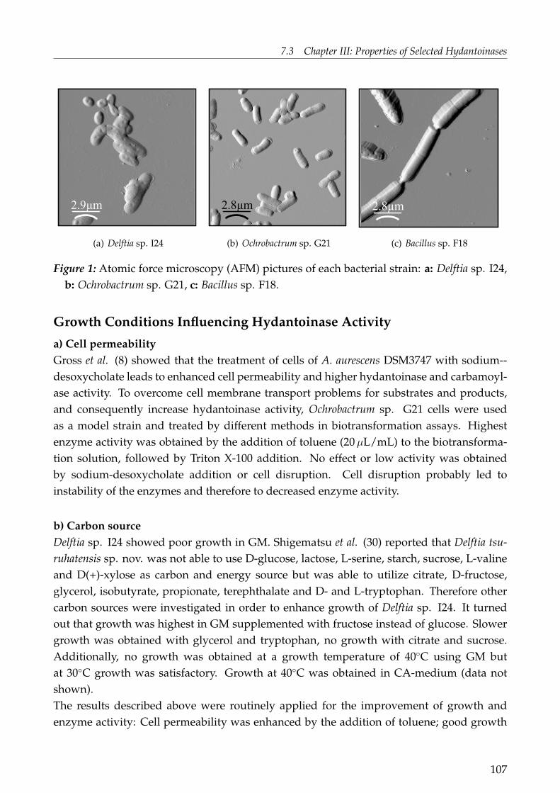
7.3 Chapter III: Properties of Selected Hydantoinases
2.9µm
(a) Delftia sp. I24
2.8µm
(b) Ochrobactrum sp. G21
2.8µm
(c) Bacillus sp. F18
Figure 1: Atomic force microscopy (AFM) pictures of each bacterial strain: a: Delftia sp. I24,b: Ochrobactrum sp. G21, c: Bacillus sp. F18.
Growth Conditions Influencing Hydantoinase Activity
a) Cell permeabilityGross et al. (8) showed that the treatment of cells of A. aurescens DSM3747 with sodium--desoxycholate leads to enhanced cell permeability and higher hydantoinase and carbamoyl-ase activity. To overcome cell membrane transport problems for substrates and products,and consequently increase hydantoinase activity, Ochrobactrum sp. G21 cells were usedas a model strain and treated by different methods in biotransformation assays. Highestenzyme activity was obtained by the addition of toluene (20 µL/mL) to the biotransforma-tion solution, followed by Triton X-100 addition. No effect or low activity was obtainedby sodium-desoxycholate addition or cell disruption. Cell disruption probably led toinstability of the enzymes and therefore to decreased enzyme activity.
b) Carbon sourceDelftia sp. I24 showed poor growth in GM. Shigematsu et al. (30) reported that Delftia tsu-ruhatensis sp. nov. was not able to use D-glucose, lactose, L-serine, starch, sucrose, L-valineand D(+)-xylose as carbon and energy source but was able to utilize citrate, D-fructose,glycerol, isobutyrate, propionate, terephthalate and D- and L-tryptophan. Therefore othercarbon sources were investigated in order to enhance growth of Delftia sp. I24. It turnedout that growth was highest in GM supplemented with fructose instead of glucose. Slowergrowth was obtained with glycerol and tryptophan, no growth with citrate and sucrose.Additionally, no growth was obtained at a growth temperature of 40◦C using GM butat 30◦C growth was satisfactory. Growth at 40◦C was obtained in CA-medium (data notshown).The results described above were routinely applied for the improvement of growth andenzyme activity: Cell permeability was enhanced by the addition of toluene; good growth
107
7 PUBLICATIONS AND MANUSCRIPTS
was achieved for Delftia sp. I24 in GM supplemented with fructose instead of glucose.
c) Salt concentrations during growthOchrobactrum sp. G21 and Bacillus megaterium F18 were able to grow at NaCl concentrationsof 5% and 7%, respectively, but with lower growth rates compared to NaCl free conditions.Hydantoinase activity, however, was stimulated by higher salt concentrations (Table 2).
Table 2: Influence of the NaCl content of the medium on hydantoinase activity. (Cells ofOchrobactrum sp. G21 and Bacillus megaterium sp. F18 were grown in GM supplementedwith 0.2 g/L IMH and at different salt concentrations. Biotransformations were conductedat standard conditions.)
strain NaCl content PN[%] [nM/(min * g)]
F18 0 39.8F18 2 39.2F18 5 36.7F18 7 46.0G21 0 7.35G21 2 9.59G21 5 40.92
It has been shown that salt stress has an important effect on the synthesis of degradativeenzymes in Bacillus subtilis (14). The synthesis of 40 proteins was found to be repressedor induced during salt stress in Listeria monocytogenes cells which can tolerate certain en-vironmental stress. The 40 proteins include general stress proteins, transporters and pro-teins involved in general metabolism (5). An attractive hypothesis for Ochrobactrum sp. G21and Bacillus megaterium F18 is a co-regulation of the hydantoin degrading system and othermetabolic pathways together with salt stress proteins, triggered by salt stress. The func-tion of the co-regulation of the hydantoinase system could be the supply of α- and/ or β-amino acids for the metabolism during salt stress. This could be verified by investigatingthe enzyme expression using 2-D-gel electrophoresis or gene expression using DNA-array-techniques of cells grown under normal and salt stress conditions.d) Effect of inducers on hydantoinase activityVarious hydantoin- and dihydropyrimidine derivatives were added as potential inducersto the growth medium (Fig. 2).
All three strains tested showed hydantoinase activity in the absence of any inducer, nev-ertheless the hydantoinase activity could be increased by the presence of inducers. Thehydantoinase of Delftia sp. I24 showed highest activity in the presence of the aromatic hy-
108

7.3 Chapter III: Properties of Selected Hydantoinases
I24 G21 F18
no hyd 0,00265122 0,00344045 0,0047
CH3-IMH 0 0,0071706 0
IMH 0,00466494 0,00735274 0,03984441
DU 0,00313089 0,00670111 0,05039574
Hyd 0,00296252 0,00559281 0,04380681
t-BH 0,00193602 0,00167404 0,06240928
HMH 0 0,00330337 0,03896739
BnH 0,00191638 0,00261524 0,04541509
PheDU 0,00488977 0,00286125 0,04195694
Methyd 0,00282173 0,00216043 0,04832776
NMH 0,00311731 0,01924634 0,03853745
erste Reihe G21
zweite Reihe I24
dritte F18
no hyd
CH3-IMH
IMH
DU
Hyd
t-BH
HMH
BnH
PheDU
Methyd
NMH
I24
G21
F18
0
0,01
0,02
0,03
0,04
0,05
0,06
0,07
PN [mM/(min*g)]
Figure 2: Effect of inducers on hydantoinase activity. (Cells were grown in the presenceof different inducers. Biotransformations were conducted with 2.5 mM BnH as substratefor Ochrobactrum sp. G21 and Delftia sp. I24 and 5 mM BnH for Bacillus megaterium F18.no hyd: no inducer added to the growth medium; for abbreviations of the inducers seeMaterials and Methods.)
dantoin IMH and the aromatic dihyropyrimidine PheDU. A dramatic increase in activitywas obtained by addition of NMH for Ochrobactrum sp. G21 (Fig. 2). At least a doubleincrease in activity was obtained by addition of CH3-IMH and IMH, followed by DU andHyd. In contrast, the hydantoinase of Bacillus megaterium F18 showed highest activity in thepresence of t-BH, a hydantoin with a sterically demanding side chain, followed by DU andMethyd.These results are not surprising since a positive effect of various inducers for hydantoincleaving activity has repeatedly been reported in literature. An optimal induction canvary widely between species and strains: Induction by 2-thiouracil was found for Agrobac-terium sp. IP-671 (27, 28), by D,L-5-(3-indolylmethyl)-3-N-methylhydantoin and D,L-5-(3-indolylmethyl)-3-N-hydantoin for Arthrobacter aurescens DSM3747 and 3745 (35, 36), byhydantoin and isopropylhydantoin for Pseudomonas fluorescens DSM84 (21), by hydantoinfor Pseudomonas sp. RUKM1 and by hydantoin, dihydrouracil and thiouracil for Pseudo-
109

7 PUBLICATIONS AND MANUSCRIPTS
monas sp. RUKM3S (3). The activity of the hydantoinase of a halophilic Pseudomonas sp.NCIM5109 (32) was not increased by addition of different hydantoins or dihydropyrim-idines to the media. In contrast, the three strains used in this study did not show the maxi-mum induction with one of the hydantoins or dihydropyrimidines reported above showingthat induction could be strain or species specific and indicating that the three describedhydantoinases could have novel unique properties.
e) Enzyme activity during growthHydantoinase activity was found to be highest in the late exponential phase of growthfor different Pseudomonas species (22, 7, 32), for Agrobacterium (11) and in the exponentialphase for Arthrobacter sp. (DSM3745) (35). In the present study we investigated thehydantoinase activity during growth for different growth media (data not shown): Delftiasp. I24 grew equally well in GMF and GMF + PheDU. Enzyme activity was highest at themid exponential phase when grown in GMF. Ochrobactrum sp. G21 was grown in GM, GM+ 5% NaCl and GM + 5% NaCl + NMH. Growth was very good in GM, as seen before,lower in GM + 5% NaCl and very slow in GM + 5% NaCl + NMH. Interestingly, at agrowth time of 94 h hydantoinase activity was highest in GM+ 5% NaCl + NMH. This isprobably an effect of hydantoinase induction by NMH and the assumed co-regulation ofthe hydantoinase system with salt stress proteins due to salt stress, resulting in increasedlevels of hydantoinases as potential amino acid suppliers. Similar results were obtained forBacillus megaterium F18 showing highest enzyme activity during the late exponential phasewith the preferred medium for highest enzyme production being GM + 5% NaCl or GM +5% NaCl + t-BH.
Summary of growth conditionsThe growth and hydantoin enzyme synthesis of all three isolates studied varied con-siderably depending on composition of the growth medium and cultivation time. Theadjustment of different parameters such as media composition and cultivation time cansignificantly influence hydantoinase activity. The two halophilic strains Ochrobactrum sp.G21 and Bacillus megaterium F18 were able to grow in media with higher salt concentrationsbut high growth rates are obtained for media with a low NaCl content. High levels ofenzyme activity can be achieved by the addition of NaCl and/ or hydantoins to the growthmedia.
Properties of Hydantoinases
The hydantoinase enzymes of the three bacterial isolates were characterised in biotrans-formation assays using resting cells. Cells were grown under optimised conditions, asdescribed above. Delftia sp. I24 was grown to mid exponential phase in GMF, Bacillusmegaterium F18 in GM supplemented with 5% NaCl and 0.2 g/L t-BH and Ochrobactrumsp. G21 in GM supplemented with 5% NaCl; For Ochrobactrum sp. G21 GM with 5% NaCl
110
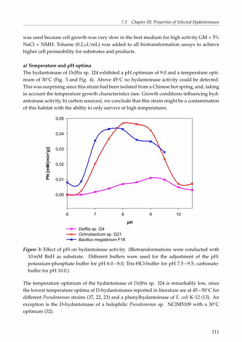
7.3 Chapter III: Properties of Selected Hydantoinases
was used because cell growth was very slow in the best medium for high activity GM + 5%NaCl + NMH. Toluene (0.2 µl/mL) was added to all biotransformation assays to achievehigher cell permeability for substrates and products.
a) Temperature and pH optimaThe hydantoinase of Delftia sp. I24 exhibited a pH optimum of 9.0 and a temperature opti-mum of 30◦C (Fig. 3 and Fig. 4). Above 45◦C no hydantoinase activity could be detected.This was surprising since this strain had been isolated from a Chinese hot spring, and, takingin account the temperature growth characteristics (see: Growth conditions influencing hyd-antoinase activity, b) carbon sources), we conclude that this strain might be a contaminationof this habitat with the ability to only survive at high temperatures.
pH
6 7 8 9 10
PN [mM/(min*g)]
0,00
0,01
0,02
0,03
0,04
0,05
Delftia sp. I24
Ochrobactrum sp. G21
Bacillus megaterium F18
Figure 3: Effect of pH on hydantoinase activity. (Biotransformations were conducted with10 mM BnH as substrate. Different buffers were used for the adjustment of the pH:potassium-phosphate buffer for pH 6.0 – 8.0; Tris-HCl-buffer for pH 7.5 – 9.5; carbonate-buffer for pH 10.0.)
The temperature optimum of the hydantoinase of Delftia sp. I24 is remarkably low, sincethe lowest temperature optima of D-hydantoinases reported in literature are at 45 – 50◦C fordifferent Pseudomonas strains (37, 22, 23) and a phenylhydantoinase of E. coli K-12 (13). Anexception is the D-hydantoinase of a halophilic Pseudomonas sp. NCIM5109 with a 30◦Coptimum (32).
111
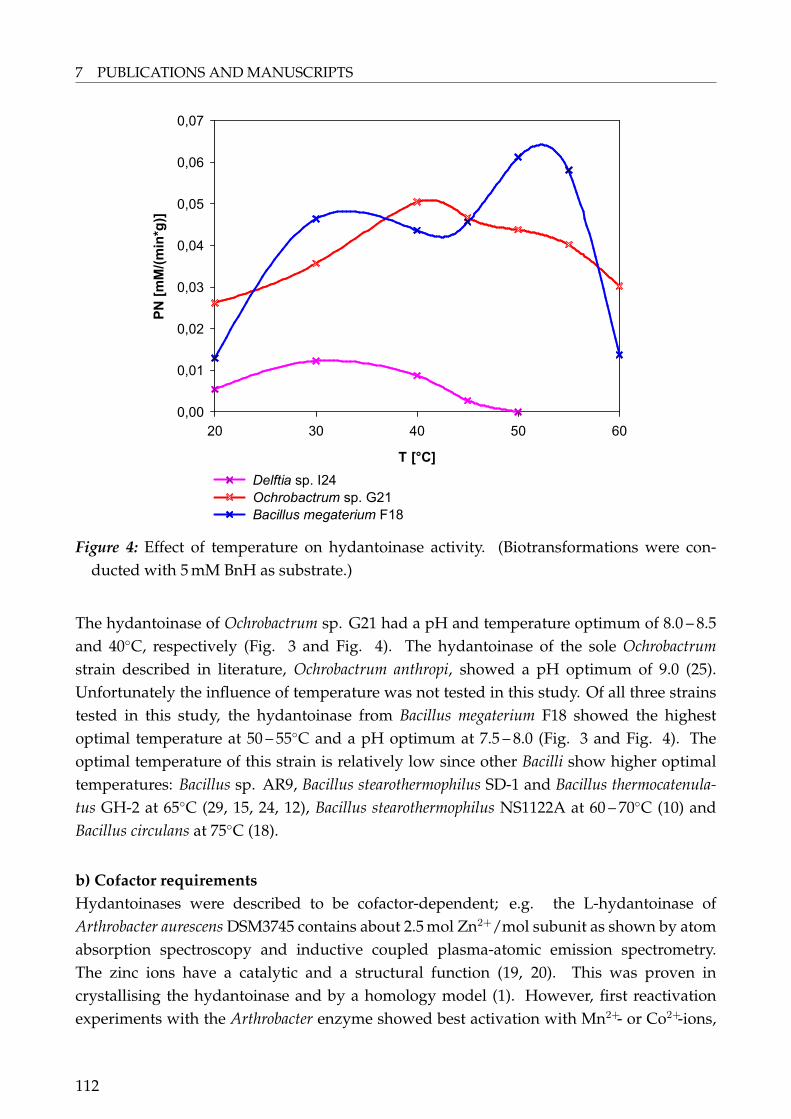
7 PUBLICATIONS AND MANUSCRIPTS
T [°C]
20 30 40 50 60
PN [mM/(min*g)]
0,00
0,01
0,02
0,03
0,04
0,05
0,06
0,07
Delftia sp. I24
Ochrobactrum sp. G21
Bacillus megaterium F18
Figure 4: Effect of temperature on hydantoinase activity. (Biotransformations were con-ducted with 5 mM BnH as substrate.)
The hydantoinase of Ochrobactrum sp. G21 had a pH and temperature optimum of 8.0 – 8.5and 40◦C, respectively (Fig. 3 and Fig. 4). The hydantoinase of the sole Ochrobactrumstrain described in literature, Ochrobactrum anthropi, showed a pH optimum of 9.0 (25).Unfortunately the influence of temperature was not tested in this study. Of all three strainstested in this study, the hydantoinase from Bacillus megaterium F18 showed the highestoptimal temperature at 50 – 55◦C and a pH optimum at 7.5 – 8.0 (Fig. 3 and Fig. 4). Theoptimal temperature of this strain is relatively low since other Bacilli show higher optimaltemperatures: Bacillus sp. AR9, Bacillus stearothermophilus SD-1 and Bacillus thermocatenula-tus GH-2 at 65◦C (29, 15, 24, 12), Bacillus stearothermophilus NS1122A at 60 – 70◦C (10) andBacillus circulans at 75◦C (18).
b) Cofactor requirementsHydantoinases were described to be cofactor-dependent; e.g. the L-hydantoinase ofArthrobacter aurescens DSM3745 contains about 2.5 mol Zn2+/mol subunit as shown by atomabsorption spectroscopy and inductive coupled plasma-atomic emission spectrometry.The zinc ions have a catalytic and a structural function (19, 20). This was proven incrystallising the hydantoinase and by a homology model (1). However, first reactivationexperiments with the Arthrobacter enzyme showed best activation with Mn2+- or Co2+-ions,
112
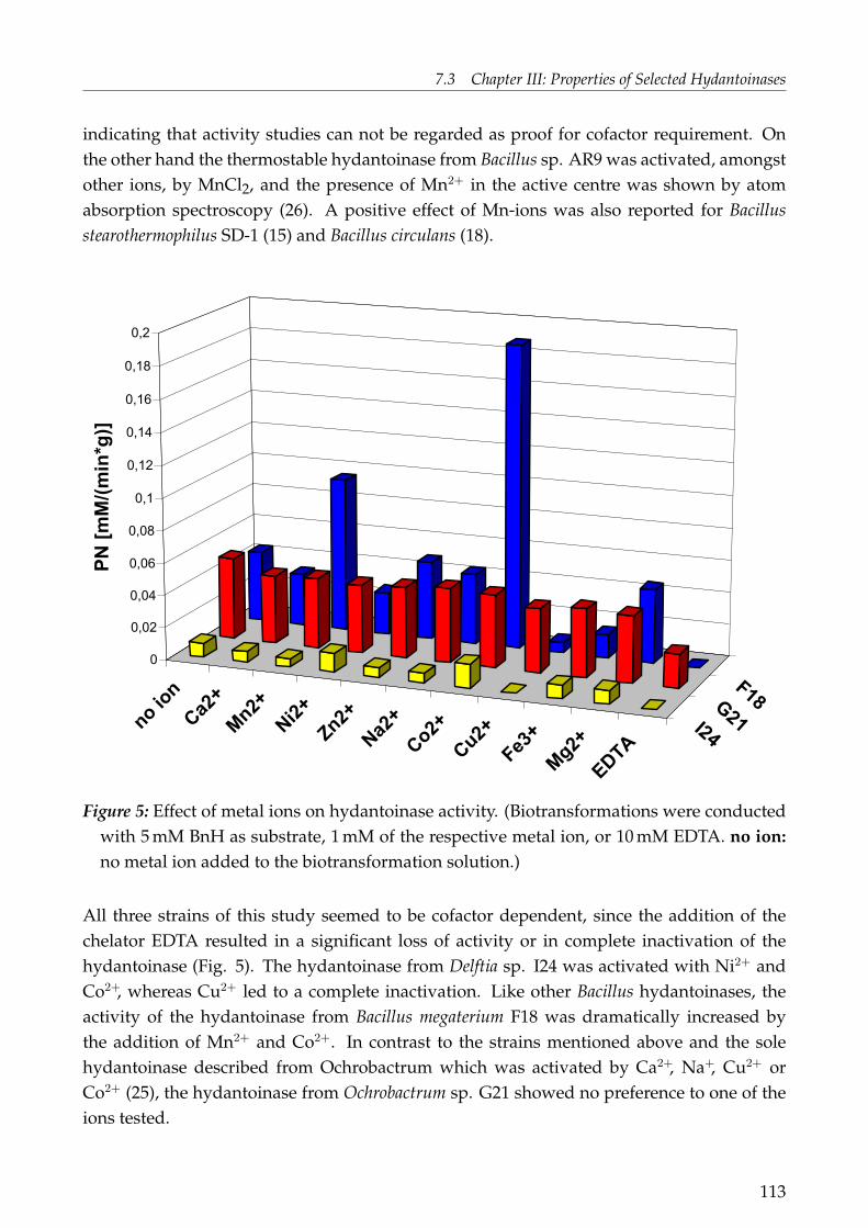
7.3 Chapter III: Properties of Selected Hydantoinases
indicating that activity studies can not be regarded as proof for cofactor requirement. Onthe other hand the thermostable hydantoinase from Bacillus sp. AR9 was activated, amongstother ions, by MnCl2, and the presence of Mn2+ in the active centre was shown by atomabsorption spectroscopy (26). A positive effect of Mn-ions was also reported for Bacillusstearothermophilus SD-1 (15) and Bacillus circulans (18).
no io
n
Ca2
+
Mn2
+
Ni2+
Zn2+
Na2
+
Co2
+
Cu2
+
Fe3+
Mg2
+
EDTA
I24
G21
F18
0
0,02
0,04
0,06
0,08
0,1
0,12
0,14
0,16
0,18
0,2
PN
[m
M/(m
in*g
)]
Figure 5: Effect of metal ions on hydantoinase activity. (Biotransformations were conductedwith 5 mM BnH as substrate, 1 mM of the respective metal ion, or 10 mM EDTA. no ion:no metal ion added to the biotransformation solution.)
All three strains of this study seemed to be cofactor dependent, since the addition of thechelator EDTA resulted in a significant loss of activity or in complete inactivation of thehydantoinase (Fig. 5). The hydantoinase from Delftia sp. I24 was activated with Ni2+ andCo2+, whereas Cu2+ led to a complete inactivation. Like other Bacillus hydantoinases, theactivity of the hydantoinase from Bacillus megaterium F18 was dramatically increased bythe addition of Mn2+ and Co2+. In contrast to the strains mentioned above and the solehydantoinase described from Ochrobactrum which was activated by Ca2+, Na+, Cu2+ orCo2+ (25), the hydantoinase from Ochrobactrum sp. G21 showed no preference to one of theions tested.
113

7 PUBLICATIONS AND MANUSCRIPTS
c) Enzyme stabilityThe stability of the three hydantoinases was investigated at different storage temperatures(Fig. 6): In the initial activity test no difference was observed between whole cells andtoluene treated cells of Delftia sp. I24 which was in contrast to the results obtained forOchrobactrum sp. G21. Hydantoinase in whole cells of Delftia sp. I24 did not loose activityat 4◦C and 20◦C during a four days storage, quite contrary to toluene treated cells. Toluenetreated cells showed a significant loss in activity during storage at 4◦C and 20◦C either dueto the negative effect of toluene on the hydantoinase at long term storage times or becausethe hydantoinase was no longer in the appropriate environment due to the permeabilisedcell membrane. The influence of toluene to cell membrane permeabilisation was higher forOchrobactrum sp. G21, indicated by higher initial activity in toluene treated cells as for un-treated cells. Enzyme activity decreased under all storage conditions and almost all of theactivity was lost at 40◦C storage in both strains. Hydantoinase enzyme stability was alsotested for Bacillus megaterium F18 (data not shown). For this strain activity increased dur-ing a four days storage. One explanation could be that the cells still had enough nutrientsto grow because it was not possible to wash the cells properly due to the slimy and densecharacter of the cells.
114

7.3 Chapter III: Properties of Selected Hydantoinases
initial activity
4ºC
20ºC
40ºC
4ºC
20ºC
40ºC
I24-toluene
I24-cells
G21-toluene
G21-cells
0,000
0,005
0,010
0,015
0,020
0,025
0,030
0,035
0,040
PN
[m
M/(m
in*g
)]
No storage 1 day storage 4 days storage
Figure 6: Effect of storage on hydantoinase activity. (Biotransformations were conductedwith 5 mM BnH as substrate. Cells were stored at 4◦C, 20◦C and 40◦C with or without20 µL/mL toluene for 1 to 4 days. initial activity (no storage): Biotransformations weredirectly conducted after harvesting of cells.)
d) Influence of NaCl on hydantoinase activityThe effect of NaCl on hydantoinase activity in biotransformations was tested in the con-centration range from zero to 20% NaCl (data not shown). No influence or only a veryslight decrease of hydantoinase activity for high NaCl concentrations on the hydantoinasesfrom Delftia sp. I24 and Bacillus megaterium F18 was observed. The hydantoinase fromOchrobactrum sp. G21 was approximately twice as active when tested in 2.5% NaCl thanin 20% NaCl. These results seem to be contradictory to the observations of growing cells.Obviously growing cells respond to salt stress resulting in activation of certain enzymesystems, e.g. the hydantoinase system; whereas resting cells do not show response to highersalt concentrations and only the enzymes can be affected.
e) Substrate spectrumAn Ochrobactrum anthropi isolate is reported to hydrolyse D,L-(2-methylthioethyl) hydantointo methionine (25). Unfortunately no other hydantoins were tested. The hydantoinase from
115

7 PUBLICATIONS AND MANUSCRIPTS
Ochrobactrum sp. G21 described within this study showed a broad substrate spectrum andwas able to cleave all substrates tested in this study (Figure 7 and Table 3).
Table 3: Substrate range of the D-hydantoinases from Delftia sp. I24 (I24), Ochrobactrum sp.G21 (G21) and Bacillus megaterium F18 (F18). (Cells were grown as follows: Delftia sp. I24in GMF for 62 h or in CA supplemented with 0.2 g/L CH3-IMH as inducer (1), Ochrobac-trum sp. G21 in GM supplemented with 2.5% NaCl for 48 h and Bacillus megaterium F18 inGM supplemented with 5% NaCl and 0.2 g/L t-BH for 14 h at standard conditions. Bio-transformations were carried out at optimised conditions: Delftia sp. I24 at pH 9.0, 30◦Cand 1 mM Co2+, Ochrobactrum sp. G21 at pH 8.0 and 40◦C and Bacillus megaterium F18 atpH 8.0, 50◦C and 1 mM Co2+; all assays were supplemented with 20 µL/mL toluene, ex-cept for (1). Biotransformations were conducted at initial conditions. The chemical struc-tures of the substrates used are shown in Table 7.)
PN [mM/(min*g)]substrate conc. [mM] I24 G21 F18
DU 25 1.8 2.52 1.26Hyd 20 0.45 2.18 3.90t-BH 8 0.05(1) 0.11 0.26IMH 2 0.09(1) 0.09 0.17BnH 5 0.01 0.25 0.44
PheDU 4 0 0.08 0.39
The hydantoinase from Bacillus megaterium F18 had a similar substrate range as the onefrom Ochrobactrum sp. G21 but showed higher enzyme activity than the other two strainstested. The higher enzyme activity for BnH and PheDU in comparison to t-BH indicatesthat 5-monosubstituted hydantoins or dihydropyrimidines with an aromatic side chainsare favoured over aliphatic ones. As well, hydantoin was hydrolysed at higher rates thanDU. The same result was reported for the purified hydantoinase enzyme from Bacillusstearothermophilus SD-1 (15) and Bacillus sp. AR9 (26). The hydantoinase of Delftia sp. I24showed the highest activity for DU, indicating that this enzyme might be involved inpyrimidine reductive catabolism. Other substrates were cleaved but at much lower reactionrates compared to Ochrobactrum sp. G21 and Bacillus megaterium F18.Comparing enzyme induction (Fig. 2) and enzyme activities towards different substrates(Table 3) it is surprising that high induction by a certain inducer is not correlated with highenzyme activity towards this compound.
Many different microorganisms with the ability to cleave hydantoins and/or dihydropy-rimidines were isolated and characterised during the last decades. In conclusion we
116

7.3 Chapter III: Properties of Selected Hydantoinases
N
N
O
O
NH NH
O
O
NHNH
O
O
NH NH
O
O
NN
O
O
NNH N
O
O
5,6-dihydrouracil (DU)
hydantoin (Hyd)
5-tert-butylhydantoin (t-BH)
5-benzylhydantoin (BnH)
D,L-5-(3-indolylmethyl) hydantoin IMH
6-phenyl-5,6-dihydrouracil (PheDU)
Figure 7: Chemical structures of hydantoins and dihydropyrimidines used for determinationof the substrate range.
showed that it is still possible to find and describe new microorganisms with hydantoinaseactivity and as well with novel properties. In our particular case we described a novelD-hydantoinase from Delftia sp. I24, isolated from a Chinese hot spring, as well as theD-hydantoinases of two halophilic strains, Ochrobactrum sp. G21 and Bacillus megateriumF18, respectively. All of the hydantoinases showed unique properties regarding salt andinductor requirements during growth for enzyme expression and they displayed differentproperties like variation in pH and temperature optima and co-factor requirements. For acomparison with known hydantoinases it is still necessary to obtain more information onthe amino acid or gene sequence of these enzymes.
117

7 PUBLICATIONS AND MANUSCRIPTS
AcknowledgmentThe authors would like to thank the Federal Ministry of Science and Education (BMBF),Germany and the National Research Foundation (NRF), South Africa for the funding ofthis bilateral project. The State of Baden Württemberg is gratefully acknowledged for sup-porting the author’s doctoral thesis. Specifically acknowledged is the joint Chinese-EU-SAMGATech project (QLRT-2001-01972) for providing access to Chinese samples. We wouldlike to thank Mr. Martin Ploß at the laboratory of Prof. Dr. Andreas Kuhn, University ofHohenheim, Germany, for the conduction of atomic force microscopy.
118
7.3 Chapter III: Properties of Selected Hydantoinases
References
1. Abendroth J., K. Niefind, O. May, M. Siemann, C. Syldatk, and D. Schomburg. 2002The structure of L-hydantoinase from Arthobacter aurescens leads to an understandingof dihydropyrimidinase substrate and enantiospecificity. Biochemistry 41:8589-8597.
2. Bergey’s Manual of Systematic Bacteriology. 2005 Volume Two: The Proteobacte-ria Part C Alpha-, Beta-, Delta-, and Epsilonproteobacteria; 2nd Edition, pp. 389-392,Editor-in-Chief G.M. Garrity. Springer.
3. Burton S.G., R.A. Dorrington, C. Hartley, S. Kirchmann, G. Matcher, and V. Phe-hane. 1998 Production of enantiomerically pure amino acids: characterization of SouthAfrican hydantoinases and hydantoinase-producing bacteria. J. Mol. Catalysis B: Enzy-matic 5:301-305.
4. Dakin H. D., and H. W. Dudley. 1914 The resolution of inactive uramido-acids andhydantoins into active components and their conversion to amino-acids I. beta-phenyl-alpha-uramidopropionic acids, benzylhydantoin and phenylalanine. J. Biol. Chem.17:29-36.
5. Duché O., F. Trémoulet, P. Glaser, and J. Labadie. 2002 Salt stress proteins induced inListeria monocytogenes. Appl. Environ. Microbiol. 68:1491- 1498.
6. Dürr R., O. Vielhauer, S.G. Burton, D.A. Cowan, A. Puñal, P.F.B. Brandão, A.T. Bull,and C. Syldatk. 2006 Distribution of hydantoinase activity in bacterial isolates fromgeographically distinct environmental sources. J. Mol. Catal. B. Enzymatic 39:160-165.
7. Gokhale D.V., K.B. Bastawde, S.G. Patil, U.R. Kalkote, R.A. Joshi, T. Ravin-dranathan, B.G. Gaikwad, V.V Jogdand., and S. Nene. 1996 Chemoenzymatic synthe-sis of D-phenylglycine using hydantoinase of Pseudomonas desmolyticum resting cells.Enzyme Microbial. Technol. 18:353-357.
8. Gross C., C. Syldatk, V. Mackowiak, and F. Wagner. 1990 Production of L-tryptophanfrom D,L-5-indolylmethylhydantoin by resting cells of a mutant of Arthrobacterspecies (DSM 3747). J. Biotechnol. 14:363- 376.
9. Hongpattarakere T., H. Komeda, and Y. Asano. 2005 Purification, characterization,gene cloning and nucleotide sequencing of D-stereospecific amino acid amidase fromsoil bacterium: Delftia acidovorans. J. Ind. Microbiol. Biotechnol. 32:567-76.
10. Ishikawa T., Y. Mukohara, K. Watabe, S. Kobayashi, and H. Nakamura. 1994 Micro-biol conversion of D,L-5-substituted hydantoins to the corresponding L-amino acidsby Bacillus stearothermophilus NS1122A. Biosci. Biotechnol. Biochem. 58:265-270.
119

7 PUBLICATIONS AND MANUSCRIPTS
11. Kim D.M., and H.S. Kim. 1993 Enzymatic synthesis of D-p-hydroxy-phenylglycinefrom D,L-hydroxyphenylhydantoin. Enzyme Microbial. Technol. 15:530-534.
12. Kim G.-J., J.-H. Park D.-C. Lee, H.-S. Ro, and H.S. Kim. 1997 Primary structure,sequence analysis, and expression of the thermostable D-hydantoinase from Bacillusstearothermophilus SD-1. Mol. Gen. Genet. 255:152-156.
13. Kim G.-J., D.-E. Lee, and H.S. Kim. 2000. Functional expression and characterizationof the two cyclic amidohydrolase enzymes, allantoinase and a novel phenylhydan-toinase, from Escherichia coli. J. Bacteriol. 182:7021-7028.
14. Kunst F., and G. Rapoport. 1995 Salt stress is an environmental signal affectingdegradative enzyme synthesis in Bacillus subtilis. J. Bacteriol. 177:2403-2407.
15. Lee S.G., D.C. Lee, S.P. Hong, M.H. Sung, and H.S Kim. 1995 Thermostable D-hydantoinase from thermophilic Bacillus stearothermophilus SD-1: characteristics ofpurified enzyme. Appl. Microbiol. Biotechnol. 43:270-276.
16. Leuchtenberger W., K. Huthmacher, and K. Drauz. 2005 Biotechnological productionof amino acids and derivatives: current status and prospects. Appl. Microbiol. Biotech-nol. 69:1-8.
17. Liang Q., M. Takeo, M. Chen, W. Zhang, Y. Xu , and M. Lin. 2005 Chromosome-encoded gene cluster for the metabolic pathway that converts aniline to TCA-cycleintermediates in Delftia tsuruhatensis AD9. Microbiology 151:3435-3446.
18. Luka V., V. Starkuviené, B. Starkuviené, and R. Dagys. 1997 Purification and char-acterization of the D-hydantoinase from Bacillus circulans. Appl. Biochem. Biotechnol.62:219-231.
19. May O., M. G. Siemann, and C. Syldatk. 1998 The hydantoin amidohydrolase fromArthrobacter aurescens DSM3745 is a zinc metalloenzyme. J. Mol. Catal. B. Enzymatic.5:367-370.
20. May O., M. G. Siemann, and C. Syldatk. 1998 Catalytic and structural function of zincfor the hydantoinase from Arthrobacter aurescens DSM3745. J. Mol. Catal. B. Enzymatic.4:211-218.
21. Morin A., W. Hummel, and M.R. Kula. 1986 Production of hydantoinase from Pseudo-monas flurescens DSM84. Appl. Microbiol. Biotechnol 25:91-96.
22. Morin A., W. Hummel, H. Schutte, and M.R. Kula. 1986 Characterization of hydanto-inase from Pseudomonas fluorescens strain DSM84. Biotechnol. Appl. Biochem. 8:564-574.
120
7.3 Chapter III: Properties of Selected Hydantoinases
23. Morin A., D. Leblanc, A. Paleczek, W. Hummel, and M.R. Kula. 1990. Comparisonof seven microbial hydantoinases. J. Biotechnol. 16:37-48.
24. Park J.-H., G.-J. Kim, S.-G. Lee, and H.S. Kim. 1998 Biochemical properties of ther-mostable D- hydantoinase from Bacillus thermocatenulatus GH-2. Ann. NY. Acad. Sci.864:337-340.
25. Pozo C., B. Rodelas, S. de la Escalera, and J. González-López. 2002 D,L-hydantoinaseactivity of an Ochrobactrum antrophi strain. J. Appl. Microbiol. 92:1028-1034.
26. Radha Kishan K. V., M. R.-K. Vohra, K. Ganesan, V. Sharma, and R. Sharma. 2005.Molecular structure of D-hydantoinase from Bacillus sp. AR9: Evidence of mercuryinhibition. J. Mol. Biol. 347:95-105.
27. Runser S.M., N. Chinski, and E. Ohleyer. 1990 D-p-hydroxyphenylglycine produc-tion from D,L-p-hydroxyphenylhydantoin by Agrobacterium species. Appl. Microbiol.Biotechnol. 33:382-388.
28. Runser S.M., and P.C. Meyer. 1993 Purification and characterization of the hydantoin-hydrolyzing enzyme from Agrobacterium species. Eur. J. Biochem. 213:1315- 1324.
29. Sharma A., and R.M. Vohra. 1997 A thermostable D-hydantoinase isolated from amesophilic Bacillus sp. AR9. Biochem. Biophys. Res. Commun. 234:485-488.
30. Shigematsu T., K. Yumihara, Y. Ueda, M. Numaguchi, S. Morimura, and K. Kida.2003 Delftia tsuruhatensis sp. nov., a terephthalate-assimilating bacterium isolated fromactivated sludge. Int. J. Syst. Evol. Microbiol. 53:1479-1483.
31. Simon H., J. Bader, H. Günther, S. Neumann, and J. Thanos. 1985 Chiral compoundssynthesized by biocatalytic reductions. Angew. Chem. Int. Ed. Engl. 24:539-553.
32. Sudge S.S., K.B. Bastawde, D.V. Gokhale, U.R. Kalkote, and T. Ravindranathan.1998 Production of D- hydantoinase by halophilic Pseudomonas sp. NCIM5109. Appl.Microbiol. Biotechnol. 49:594-599.
33. Süßmuth R., J. Eberspächer, and R. Haag, W. Springer. 1999 Mikrobiologisch-Biochemisches Praktikum. Thieme Verlag.
34. Syldatk C., D. Cotoras, G. Dombach, C. Gross, H. Kallwass, and F. Wagner. 1987Substrate- and stereospecificity, induction and metallodependence of a microbial hyd-antoinase. Biotechnol. Lett. 9:25-30.
35. Syldatk C., V. Mackowiak, H. Höke, C. Gross, G. Dombach, and F. Wagner. 1990Cell growth and enzyme synthesis of a mutant of a Arthrobacter sp. (DSM3747) usedfor the production of L-amino acids from D,L-5-monosubstituted hydantoins.J. Biotech.14:345-362.
121

7 PUBLICATIONS AND MANUSCRIPTS
36. Syldatk C., and M.Pietsch 1995 Hydrolysis and formation of hydantoins. p. 409-431In: Drauz, K. and Waldmann H. (Eds), Enzyme catalysis in organic synthesis. VCH,Weinheim, Germany.
37. Takahashi S., Y. Kii, H. Kumagai, and H. Yamada. 1978 Purification, crystallisationand properties of hydantoinase from Pseudomonas striata. J. Ferment. Technol. 56:492-498.
38. Tsugawa R., S. Okumura, T. Ito, and N. Katsuga. 1966 Production of L-glutamic acidfrom D,L-5-hydantoin propionic acid by microorganisms. Agric. Biol. Chem. 30:27-34.
39. Webbs EC Enzyme nomenclature. 1992 San Diego, USA: Academic Press.
40. Wen A., M. Fegan, C. Hayward, S. Chakraborty, and L.I. Sly. 2003 Phylogenetic re-lationships among members of the Comamonadaceae, and description of Delftia acidovo-rans (den Dooren de Jong 1926 and Tamaoka et al. 1987) gen. nov., comb. nov.Int. J.Syst. Evol. Microbiol. 53:1479-1483.
41. Willems A., and P. de Vos 2002 Comamonas In The Prokaryotes: An Evolving Elec-tronic Resource for the Microbiological Community, 3rd edition Springer-Verlag, NewYork.
122

7.4 Chapter IV: Gene Clusters for Hydantoin Degradation
7.4 Chapter IV: Gene Clusters for Hydantoin Degradation
GENES RESPONSIBLE FOR HYDANTOIN DEGRADATION OF AHALOPHILIC Ochrobactrum SP. G21 AND Delftia SP. I24 –
NEW INSIGHT INTO RELATION OF D-HYDANTOINASES ANDDIHYDROPYRIMIDINASES
submitted toJournal of Molecular Catalysis B: Enzymatic
R. Dürr1∗, A. Neumann1, O. Vielhauer1, J. Altenbuchner2, S.G. Burton3, D.A. Cowan4, C.Syldatk1
1 Department of Chemical Engineering, Chair of Technical Biology, University of Karlsruhe(TH), Germany2 Institute of Industrial Genetics, University of Stuttgart, Germany3 Department of Chemical Engineering, University of Cape Town, South Africa4 Department of Biotechnology, University of the Western Cape, South Africa
∗ Corresponding author: R. DürrFax: +49 (0) 721 608 4881e-mail: [email protected]: extreme environment, hydantoinase operon, substrate recognition,D-hydantoinase families
123

7 PUBLICATIONS AND MANUSCRIPTS
AbstractDelftia sp. I24 and a moderately halophilic Ochrobactrum sp. G21 are able to hydrolyse di-hydropyrimidines and hydantoins D-specific. The genes being with the utmost probabilityinvolved in dihydropyrimidine and hydantoin degradation of these two microorganismswere cloned into an appropriate vector and transformed into E. coli. The putative genecluster of Delftia sp. I24 included four genes: an incomplete NADPH-dependent gluta-mate synthase gene (gltB), dihydropyrimidine dehydrogenase gene(pydA), permease gene(hyuP) and an incomplete D-hydantoinase gene (hyuH). The hydantoinase gene sequencewas completed by PCR amplification. The putative gene cluster of Ochrobactrum sp. G21comprised nine ORFs, six being potentially involved in hydantoin-hydrolysation: carba-moylase (hyuC), D-hydantoinase (hyuH), two transporters (OrfS1 and OrfS2) and two per-meases (hyuP1 and hyuP2). Expression of the D-hydantoinases from Delftia sp. I24 and fromOchrobactrum sp. G21 in E. coli, followed by biotransformation assays confirmed hydanto-inase activity. This is the first report of the genetical organisation of hydantoin-degradationwithin the genera Delftia and Ochrobactrum. Phylogenetic analysis of the two "novel" hyd-antoinases and known hydantoinases and dihydropyrimidinases, including putative pro-tein sequences, revealed that they can be classed with some exceptions in the followinggroups: L-Hydantoinases (L-Hyd), Rhizobiales family (Rhizo-Fam), Comamonadacae family(Com-Fam), Pseudomonas family (Pseud-Fam), Bacilli family (Bac-Fam) and Agrobacteriumfamily (Agro-Fam). The highly conserved "histidine motif" for the superfamily of amidohy-drolases could be found for all hydantoinases of this study but differences were found in thesubstrate recognition sites, whereas some of the above mentioned groups showed to possesthe same recognition sites as known hydantoinases.
124
7.4 Chapter IV: Gene Clusters for Hydantoin Degradation
Introduction
Hydantoinases can be found, amongst other organisms, in microorganisms and are classedas cyclic amidases (EC 3.5.2). Hydantoinases are able to hydrolyse the five-membered ringof hydantoins or/and 5-monosubstituted hydantoin derivatives forming N-carbamoyl α-amino acids. A carbamoylase can transform this intermediate product to the correspondingamino acid. The natural function of hydantoinases is still unknown. In comparison, dihy-dropyrimidinases are involved in the reductive pathway of pyrimidine degradation andtherefore are able to cleave 5,6-dihydrouracil to the corresponding N-carbamoyl β-aminoacid. Various enzymes with the ability to catalyse the cleavage of dihydropyrimidines andhydantoins are known: e.g. Arthrobacter crystallopoietes DSM20117 [1], Bacillus sp. AR9 [2],Bacillus stearothermophilus SD-1 [3,4], Pseudomonas sp. NCIM5109 [5]. It is notable that in theEC-nomenclature the name hydantoinase is used as a synonym for dihydropyrimidinase.Contrary, the hydantoinases of Agrobacterium IP-671 and Arthrobacter aurescens DSM3745are able to cleave the five-membered ring of hydantoin but not dihydrouracil [6, 7]. Dueto this fact, Syldatk et al. [8] proposed that the name hydantoinase should be used forall enzymes that hydrolyse hydantoin or/and 5-monosubstituted derivatives and not assynonym for dihydropyrimidinases. However, in this study we will not strictly distinguishbetween these two enzymes because only a few real hydantoinases are known and exceptfor biochemical data no further differentiation is known. Therefore the focus of this workwill be on hydantoinases and dihydropyrimidinases.The hydantoinase process is widely used in industry for production of D- and L-aminoacids, e.g. for the production of the aromatic amino acids D-phenylglycine and p-hydroxyl-D-phenylglycine as building blocks for semisynthetic antibiotics (ampicillin andamoxicillin). Economic targets are as well D-serine, L-methinonine, L-phosphinotricine [9].
During recent years the gene clusters of different microorganisms, encoding for the enzymesbeing involved in hydantoin and/ or dihydropyrimidine conversion, have been elucidated(Fig. 1). The genes of hydantoinase/ dihydropyrimidinase and carbamoylase have beenfound to form part of an unique operon, whereas other genes coding for e.g. racemase andpermease are not commonly present. However, the genetic organisation within the clusterhas been found to be very diverse in positioning and transcriptional orientation. Neverthe-less, the only pattern observed is a similar transcriptional orientation in bacteria from thesame genus or suborder (Fig. 1): All Agrobacterium species show an opposite orientationof the hydantoinase and carbamoylase genes. A similar orientation of all genes involvedin hydantoin conversion was obtained for the two Arthrobacter species and Microbacteriumliquefaciens. In spite of this, the two Pseudomonas gene clusters show a complete differentorganisation.Different techniques have been used to identify these clusters. A genomic phage library ofAgrobacterium sp. IP-I671 was screened by plaque hybridisation using a DIG-labelled hyuC-
125

7 PUBLICATIONS AND MANUSCRIPTS
DNA fragment. The hyuC-DNA was obtained by amplification of the carbamoylase gene bydegenerate primers. These primers were derived by consensus analysis using known carba-moylase sequences from Agrobacterium strains [12]. Other clusters were obtained in a similarmanner using information derived from the amino acid sequence of purified proteins [10,14, 17]. The hydantoinase gene cluster from A. crystallopoietes DSM20117 was identifiedby cloning the hydantoinase and expanding the flanking DNA regions by inverse PCR.Thus, the complete hydantoinase, D-N-carbamoylase and a putative L-N-carbamoylasegene could be found [11]. Recently, the dihyropyrimidinase and β-ureidopropionase geneof Pseudomonas putida RU-KM3S were identified by transposon mutagenesis and selectionof altered growth phenotypes [16].
microorganism gene organisation Ref.
Arthrobacter aurescens 3747
Arthrobacter crystallopoietes 20117
Agrobacterium sp. IP I-671
Agrobacterium sp. KNK712
Agrobacterium tumefaciens NRRL B11291
Brevibacillus agri NCHU 1002
Pseudomonas putida RU-KM3S
Microbacterium liquefaciens AJ 3912
[10]
[11]
[12]
[13]
[14]
[15]
[16]
[17]
ORF1 ORF2 ORF3 ORF8hyuP hyuA hyuH hyuC
ORF1 hyuCLhyuCD hyuH hyuP
hyuN hyuH hyuC hyuD hyuAORF1 ORF7
dhp hyuC
hyuH hyuC
ORF3 gltB pydA ORF8dhp pydC
dhp ORF1 bup
ORF1 ORF2 ORF3 hyuP hyuA hyuH hyuC
Figure 1: Genetic organisation of genes encoding for the enzymes involved in hydantoin-and/ or dihydropyrimidine hydrolysis from different microbial sources. (hyuP: perme-ase; hyuA: racemase; hyuH: hydantoinase; hyuC: carbamoylase; hyuN: NADP-flavin ox-idoreductase; hyuD: amino acid dehydrogenase; dhp: dihydropyrimidinase; gltB: glu-tamate synthase; pydA: dihydropyrimidine dehydrogenase; pydC: β-alanine synthase;bup: β-ureidopropionase. Incomplete ORFs are shown in rectangles.)
In the present study we describe the detection and characterisation of the genes involvedin hydantoin cleavage of Ochrobactrum sp. G21 and Delftia sp. I24. This was achieved byscreening a genomic library of each strain with the aid of a DIG-labelled hydantoinase DNA-fragment. The genomic library was obtained by digestion of the genomic DNA of each strainand cloning and transformation of the resulting DNA-fragments into an appropriate vectorand competent E. coli cells.Ochrobactrum sp. G21 was isolated from a hypersaline lake, Inner Mongolian AutonomousRegion, China and Delftia sp. I24 from an algal mat of hot springs (60 – 65◦C) Long Pu, Yun-nan Province, China [18]. Growth conditions and hydantoinase activity of both strains havebeen studied intensively (manuscript in preparation). In literature only one Ochrobactrumanthropii with D,L- hydantoinase activity has been reported, isolated from a soil sample in
126

7.4 Chapter IV: Gene Clusters for Hydantoin Degradation
Spain [19]. No hydantoinase activity from the family Delftia was reported before. As well,none is known about the genetical organisation of hydantoin cleaving enzymes in these twofamilies.
127

7 PUBLICATIONS AND MANUSCRIPTS
Materials and MethodsAbbreviationsAgro-Fam: Agrobacterium family; Bac-Fam: Bacilli family; BnH: D,L-5-benzylhydantoin;Com-Fam: Comamonadacae family; DIG-G21: random primed DNA probe labeled withdigoxigenin-dUTP, DNA derived from hydantoinase amplification of genomic DNA fromOchrobactrum sp. G21; DIG-I24: see DIG-G21, genomic DNA from Delftia sp. I24; dhp: di-hydropyrimidinase; DIG: digoxigenin; gltB: glutamate synthase; D-Hyd: D-Hydantoinase;L-Hyd: L-Hydantoinase; hyu: hydantoin utilization; hyuH: hydantoinase; hyuP: per-mease; IMH: D,L-5-(3-indolylmethyl) hydantoin; LB: Luria-Bertani medium; LB-Amp:Luria-Bertani medium supplemented with ampicillin; ORF: open reading frame; pydA:dihydroprimidine dehydrogenase; pydO: dihydroorotate dehydrogenase; Pseud-Fam:Pseudomonas familiy; Rhizo-Fam: Rhizobiales family; SGLs: stereochemistry gate loops.
ChemicalsAll chemicals used were obtained from commercial sources and were of reagent grade.If not stated otherwise, enzymes, chemicals for molecular work and molecular kits wereobtained from Fermentas, Germany.
Bacterial Strains and MediaThe bacterial isolates used within this study were a halophilic Ochrobactrum sp. G21(DSM18828) and a Delftia sp. I24 (DSM18833) described by Dürr et al. [18]. The mediaand growth conditions of Ochrobactrum sp. G21 and Delftia sp. I24 have been describedpreviously [18]. For cloning E. coli XL-1 blue and E. coli JM109 was used routinely, beingcultivated in LB medium at 37◦C. Ampicillin (100 µg/mL) was added (LB-Amp) if E. colicells were cultured carrying recombinant plasmids.
DNA Manipulation MethodsGeneral recombinant DNA techniques were carried out by standard procedures [20].Chromosomal bacterial DNA was extracted using the Qiagen DNeasy Tissue kit (Qiagen,Germany) following the manufacturer’s protocol for gram-negative bacteria. DNA frag-ments were purified from standard agarose gels by a DNA extraction kit. Pure plasmidswere obtained by a GeneJETTM Plasmid Mini Prep Kit. Sequencing was done commercially(JenaGen GmBH, Germany) and DNA and protein sequence analysis were carried out usingBlastN and BlastP [21], ORF finder (at the National Centre for Biotechnology Informationwebsite), ClustalW [22], BPROM (available at www.softberry.com) and NEBcutter [23].Phylogenetic and molecular evolutionary analyses were conducted using MEGA version3.1 [24].
128
7.4 Chapter IV: Gene Clusters for Hydantoin Degradation
Preparation of a 320 bp DNA FragmentTwo homologous probes for hydantoinases were generated by polymerase chain reaction(PCR) using genomic DNA from Ochrobactrum sp. G21 or Delftia sp. I24 as template.Amplifications were carried out either on a Eppendorf Mastercycler gradient (Germany) orMJ Research PTC-200 (USA) at the following conditions: 50 – 150 ng template DNA, 1 x Taqbuffer with (NH4)2SO4, 1.5 U Taq DNA Polymerase, 50 pmol upstream primer and down-stream primer each, 0.2 mM dNTP´s, and PCR-water, combined to volume of 20 µL. Thefollowing degenerate oligonucleotide primers were used [25], synthesised by MWG-Biotech,Germany: 5´-GCSGCVTTYGGNGGNACNAC-3´ and 5´-TCNCCRTTYTCNGCRTGNAC-3´. Amplification was initiated with DNA denaturation at 95◦C for 5 min, followed by 30cycles of denaturation at 94◦C for 1 min, annealing at 60◦C for 2 min and primer extension at72◦C for 1 min. The PCR reaction was completed at 72◦C for 7 min. After PCR amplificationa 320 bp DNA fragment was purified for each strain. In order to verify hydantoinase am-plification purified PCR products were sequenced and compared to known hydantoinasesequences. To obtain an adequate amount of DNA for DIG-labelling the purified PCRproducts were cloned using the Strata CloneTM PCR Cloning kit (Stratagene, USA). Theobtained plasmid inserts were excited by different restriction enzymes and purified.
Preparation of DIG-Labelled ProbesThe DIG DNA Labeling and Detection Kit (Roche, Germany) was used for DIG-randomprimed DNA labelling of the 320 bp DNA fragments following the manufacturer’s instruc-tions. This results in two probes: DIG-I24 obtained from hydantoinase DNA amplificationof Delftia sp. I24 and DIG-G21 of Ochrobactrum sp. G21.
DNA Digest and Southern BlotChromosomal DNA was digested using different restriction enzymes (EcoRI, HindIII) andseparated on standard agarose gels. The DNA was blotted on a nylon membrane (Roti R©
Nylon plus, Roth, Germany) using standard methods. The nylon membrane was hybridisedwith the DIG-labelled probe by 68◦C followed by immunological detection applying theDIG DNA Labeling and Detection Kit (Roche, Germany). NBT/BCIP was used for thecolour reaction. By this reaction DNA fragments possessing the hydantoinase genes couldbe located. The DNA fragments were afterwards purified.
Plasmid Preparation, Cloning and ScreeningPlasmid DNA (pUC19) was digested using either EcoRI or HindIII. Subsequent dephospho-rylation was conducted by addition of shrimp alkaline phosphatase. Ligation was carriedout under the following conditions: 50 ng vector DNA (pUC19/EcoRI or pUC19/HindIII),2 x molar excess of the purified DNA fragment, 1 x ligation buffer, T4-DNA-ligase and water(nuclease free), combined to a volume of 20 µL. The ligated DNA was transformed into E.
129

7 PUBLICATIONS AND MANUSCRIPTS
coli XL-1 [26], plated on LB-Amp supplemented with 38.4 µg/ml IPTG and 40 µg/ml X-Gal(in DMSO) and incubated at 37◦C. E. coli XL-1 cells bearing recombinant plasmids wereobtained by blue/white screening. Positive clones were picked and transferred on a masterplate and on a nylon membrane on LB-Amp plates. Cells were grown overnight at 37◦C.DNA transfer from E. coli colonies to the nylon membrane were conducted as followed(modified after [27]): the membrane was transferred on 3MM Whatman paper saturatedwith denaturation solution (0.5 M NaOH, 1.5 M NaCl, 0.1% SDS) and incubated for 15 minfollowed by neutralisation on 3MM Whatman paper saturated with neutralisation solution(1.5 M NaCl, 1 M TRIS/ HCl, pH7.5) for 5 min. The nylon membrane was transferredon 3MM Whatman paper saturated with 2 x SSC for 15 min. Cell debris were washedout with 2 x SSC. DNA fixation on the nylon membrane was carried out for 2 h at 80◦C.Hybridisation with the DIG-labelled probe and immunological detection was conductedas described before. DIG-labelled clones bearing a plasmid containing an insert with thehydantoinase gene were re-plated on LB-Amp. Plasmid DNA was isolated. For verificationof the correct insert and to estimate the location of the hydantoinase gene in the DNAfragment PCR reactions were carried out as described above using the plasmid DNA astemplate. In addition, plasmid DNA was digested by the same restriction enzymes as usedfor cloning in combination with restriction enzymes cutting in the hydantoinase fragment.These restriction enzymes were PauI for Ochrobactrum sp. G21, Eco72I and Eco52I for Delftiasp. I24. The digested plasmids were run on an agarose gel, bloted on a nylon membrane,hybridised and the immunological detection performed as described above. To obtain highconcentration of plasmid DNA, plasmid DNA was isolated by the method of [23] or by aQIAfilter Plasmid Maxi Kit (Qiagen, Germany). The obtained plasmids were sequencedand analysed (see above).
130
7.4 Chapter IV: Gene Clusters for Hydantoin Degradation
Completion of the Hydantoinase Gene from Delftia sp. I24 andHeterologous Expression of the Hydantoinases in E. coliThe hydantoinase gene sequence from Delftia sp. I24 was completed by PCR amplificationusing chromosomal DNA from Delftia sp. I24. The complete hydantoinase gene was ampli-fied with the primers s4740 (5´-AAAAAACATATGGGTCAATCACAGGGTTCGGTAT-3´)and s4741 (5´-AAAAAGCTTAGCGCGCCACGGCCGTGGGCT-3´), the resulting genefragment cloned into the vector pJOE4786.1 which was cut by SmaI. The resulting plasmidwas cut by BamH1 and NdeI and the hydantoinase gene inserted into the expression vectorpJOE5697.2 resulting in the plasmid pJOE5704.1. The hydantoinase gene from Ochrobactrumsp. G21 was amplified by PCR using chromosomal Ochrobactrum sp. G21 DNA andthe primers s4738 (5´-AAAAACATATGGCAAAGGTCATCAAAGGCGGA-3´) and 4739(5´-AAAAAAAGCTTAGACCCCTATCGGCATGTGTTCGGCGCT-3´). The fragment wascut by HindIII and NdeI and inserted into the expression vector pJOE5427.5 resulting in thevector pJOE5702.1. Both plasmids pJOE5704.1 and pJOE5702.1 were transformed into E. coliJM109 using standard procedures.
Biotransformation ExperimentsE. coli cells for biotransformation experiments were grown in LB-AMP with 0.2% rham-nose as inducer for 12 h at 30◦C. Cell harvesting and biotransformations were performedas described by [18]. 3 mM BnH and IMH were used as substrates. The analysis of thesubstrates and products was performed using High Performance Liquid Chromatography(HPLC) on an Agilent 1100 series instrument (Agilent, Germany). The concentrations ofBnH and IMH and their corresponding carbamoyl amino acids were determined using aHypersil-Keystone BDS C18 column (Thermo, Germany). The mobile phase contained 20%MeOH/ 80% (0.1% H3PO4) solution. The flow rate was 1 mL/min. Detection was carriedout at 210 nm or 280 nm, respectively.
131
7 PUBLICATIONS AND MANUSCRIPTS
Results and DiscussionAmplification of a 320 bp Hydantoinase FragmentA 320 bp DNA fragment was amplified for both strains, Ochrobactrum sp. G21 and Delftiasp. I24, using degenerate primers. The DNA sequence obtained for Delftia sp. I24 showedthe highest homology to a putative D-hydantoinase of Burkholderia sp. 383 (85% similarityof 167 bp), and the DNA sequence for Ochrobactrum sp. G21 showed the highest homologyto a putative D-hydantoinase of Brucella suis 1330 (93% similarity of 248 bp). This indicatedthe amplification of a hydantoinase DNA fragment for both strains. The DNA fragmentswere used for preparation of a DIG-labelled probe.
Cloning and Screening of a DNA Fragment Containing the Gene ClusterResponsible for Hydantoin Degradation
The DIG-labelled probes were used to screen a genomic library of each of the two strains.These genomic libraries were derived from digested genomic DNA followed by cloning andtransformation into pUC19 and E. coli XL-1. Four plasmids, two for each strain, carryingthe genetic information likely to code for enzymes involved in hydantoin conversion wereobtained. Restriction enzyme and DNA sequence analysis revealed that only a partialputative hydantoinase gene sequence of Delftia sp. I24 was cloned into each of the twoplasmids. Therefore approximately 4 kb of a 15 kb plasmid, designated pUC19-M50, weresequenced. In the case of Ochrobactrum sp. G21 two plasmids with a size of approximately6.6 kb (pUC19-H1-10) and 11.1 kb (pUC19-L1-61) containing the hydantoinase homologgene were obtained. DNA analysis showed that both plasmids contained the same DNAinformation, except for pUC19-L1-61 more in downstream direction. Hence, the plasmidpUC19-L1-61 was completely sequenced.
Sequence Analysis of pUC19-M50 (Delftia sp. I24)The sequencing and analysis of 4469 bp of the plasmid pUC-M50, containing the hydanto-inase homolog gene sequence of Delftia sp. I24, revealed two complete open reading frames(ORFs) and two incomplete ORFs (Table 1). Since the hydantoin encoding sequence wasfirst incomplete it was completed by PCR amplification resulting in known 4790 bp of thegene cluster. Primers were designed from the putative dihydropyrimidinase sequence ofDelftia acidovorans SPH-1. All ORFs are transcribed in the same direction and the overallGC-content of the obtained DNA fragment was 67%. This is in concordance to other speciesof this genus since a GC content of 66 – 69 mol% is reported for other Delftia species [29,30]. Analogously to other hydantoin gene clusters the genes involved in hydantoin degra-dation were abbreviated as hyu "hydantoin utilization" [31]. The ORFs can be attributed tothe following proteins (Table 1 and for overview Figure 2):
132

7.4 Chapter IV: Gene Clusters for Hydantoin Degradation
Table 1: Overview of ORFs obtained from plasmid pUC19-M50 (Delftia sp. I24). (The puta-tive Shine Dalgarno sequence is underlined and italics. The start codons of the ORFs arebold. * ORF not complete.)
gene start end length GC putative Shine assumed[bp] [%] Dalgarno sequence function
gltB* - 319 - - - glutamatesynthase
pydA 354 1673 1320 67 GAGAAGGAGCTTCGCAATG dihydro-pyrimidine
dehydrogenasehyuP 1806 3299 1494 65 AGAAGGAGCTTCGCAATG permeasehyuH 3330 4790 1461 68 CAGGAGGCATGACGCTATG hydantoinase
gltB: The deduced amino acid sequence of this ORF could be assigned to the family ofNADPH-dependent glutamate synthases (beta chain) and related oxidoreductases by com-parison to a conserved domain database [32].pydA: This gene was named pydA, since the deduced protein sequence showed the high-est homology to the dihydropyrimidine dehydrogenase (pydA) of Brevibacillus agri [15] with59% identity. This group of enzyme is known to catalyse the reduction of uracil and thymineto the corresponding 5,6-dihydropyrimidines.hyuP: The highest homology of the obtained amino acid sequence of this ORF to a "non-putative" protein was to the allantoin permease Dal4 from Saccharomyces cerevisiae with 28%identity. A comparison to a conserved database showed that this ORF can be associated tothe family of cytosine/ uracil/ thiamine/ allantoin permeases [32]. Additionally, a Kyte-Doolittle hydropathy plot [33] revealed a hydrophobic character of this protein (data notshown) indicating a transmembrane protein. Resuming all the data mentioned above, weassume that this ORF encodes for a permease with its function in hydantoin transport andtherefore named as hyuP.hyuH: The highest identity (66%) of the deduced amino acid sequence of this ORF to a"non-putative" enzyme was obtained to the D-hydantoinase of Pseudomonas putida DSM84[34, 35]. Interestingly, the highest homologies (compare Fig. 2) of the deduced proteinsequence was found with 98% to a putative dihydropyrimidinase of Delftia acidovoransSPH-1 (accession ZP01581256), with 84% to a putative D-hydantoinase of Acidovorax avenaesubsp. citrulli AAC00-1 (accession ZP01403855), with 81% to a putative dihyropyrimidi-nase of Verminephrobacter eiseniae EF01-2 (accession EAT75980) and with 78% to a putativeD-hydantoinase of Polaromonas naphthalenivorans CJ2 (accession ZP01019452). All these or-ganisms, including Delftia sp. I24, belong to the lineage of proteobacteria, burkholderiales,family of comamonadacaea. The expression of this gene in E. coli followed by biotransfor-mations showed the conversion of the substrates BnH and IMH (Table 2). This proofs the
133
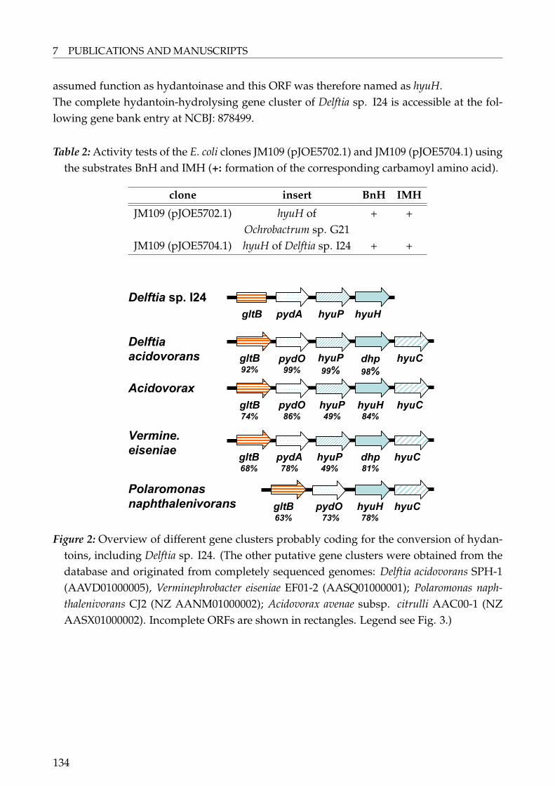
7 PUBLICATIONS AND MANUSCRIPTS
assumed function as hydantoinase and this ORF was therefore named as hyuH.The complete hydantoin-hydrolysing gene cluster of Delftia sp. I24 is accessible at the fol-lowing gene bank entry at NCBJ: 878499.
Table 2: Activity tests of the E. coli clones JM109 (pJOE5702.1) and JM109 (pJOE5704.1) usingthe substrates BnH and IMH (+: formation of the corresponding carbamoyl amino acid).
clone insert BnH IMH
JM109 (pJOE5702.1) hyuH of + +Ochrobactrum sp. G21
JM109 (pJOE5704.1) hyuH of Delftia sp. I24 + +
Delftia sp. I24
gltB pydA hyuP hyuH
Vermine.
eiseniae
Polaromonas
naphthalenivorans
Acidovorax
Delftia
acidovorans dhp
98%
pydO99%
hyuH84%
pydO86%
hyuP49%
hyuC
gltB68%
pydA78%
hyuP49%
dhp81%
hyuC
gltB63%
pydO73%
hyuH78%
hyuC
gltB92%
gltB74%
hyuP
99%hyuC
Figure 2: Overview of different gene clusters probably coding for the conversion of hydan-toins, including Delftia sp. I24. (The other putative gene clusters were obtained from thedatabase and originated from completely sequenced genomes: Delftia acidovorans SPH-1(AAVD01000005), Verminephrobacter eiseniae EF01-2 (AASQ01000001); Polaromonas naph-thalenivorans CJ2 (NZ AANM01000002); Acidovorax avenae subsp. citrulli AAC00-1 (NZAASX01000002). Incomplete ORFs are shown in rectangles. Legend see Fig. 3.)
134
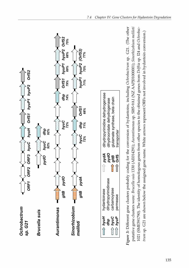
7.4 Chapter IV: Gene Clusters for Hydantoin Degradation
Bru
cella
suis
Aura
ntim
onas
Sin
orh
izobiu
m
melilo
ti
OR
F1
OR
F3
hyuC
Ochro
bactr
um
sp. G
21
hyuH
Orf
S1
hyuP
1hyuP
2O
rfS
2
hyuC
92%
dhp
92%
pydO
hyuC
72%
dhp
74%
pydO
gltB
hyuC
72%
dhp
71%
Orf
S1
68%
hyuP
275%
hyuP
171%
gltB
pydA
Orf
S1
79%
hyuP
169%
hyuP
268%
Orf
S2
75%
(Orf
S2)
77%
hyuH
hydantoinase
dhp
dihydropyrimidinase
hyuC
carbamoylase
hyuP
permease
pydA
dihydropyrimidine dehydrogenase
pydO
dihydroorotatedehydrogenase
gltB
glutamatesynthase, betachain
OrfS
transporter
OR
F2
Figu
re3:
Diff
eren
tge
necl
uste
rspr
obab
lyco
ding
for
the
conv
ersi
onof
hyda
ntoi
ns,i
nclu
ding
Och
roba
ctru
msp
.G
21.
(The
othe
rpu
tati
vege
necl
uste
rsw
ere:
Bruc
ella
suis
1330
(AE0
1429
1);A
uran
timon
assp
.SI8
5-9A
1(N
ZA
APJ
0100
0001
);Si
norh
izob
ium
mel
iloti
1021
(SM
E591
790)
.The
iden
tity
ofho
mol
ogou
sge
nes
from
othe
rsp
ecie
sto
the
obta
ined
gene
sfr
omD
elfti
asp
.I24
and
Och
roba
c-tr
umsp
.G21
are
show
nbe
low
the
assi
gned
gene
nam
e.W
hite
arro
ws
repr
esen
tOR
Fsno
tinv
olve
din
hyda
ntoi
nco
nver
sion
.)
135

7 PUBLICATIONS AND MANUSCRIPTS
Sequence Analysis of pUC19-L1-61 (Ochrobactrum sp. G21)The cloned chromosomal DNA in the plasmid pUC19-L1-61 comprised 8453 bp with anoverall GC content of 56%. In total eight complete ORFs and one incomplete ORFs were ob-tained coding for the proteins listed (Table 3 and Fig 3). Except for hyuC and hyuH all otherdeduced protein sequences are classified by comparison to a Conserved Domain Database[32]:ORF1: The first ORF, incomplete, probably belongs to the family of ABC-type sugar trans-port systems, ATPase components.ORF2: The second ORF, oppositely directed to ORF1, shows identity to the MutT/ Nudixfamily of proteins.ORF3: This second, oppositely directed ORF could be assigned as a bacterial regulatory pro-tein belonging to the TetR-family or as transcriptional regulator AcrR. With high probabilityall first three ORFs mentioned are not involved in hydantoin conversion.hyuC: Highest similarity of the deduced amino acid sequence was obtained to the N-carbamoyl-L-amino acid amidohydrolase, also called L-carbamoylase, of Bacillus stearother-mophilus strain NS1122A with 34% identity, followed by the L-carbamoylase of ArthrobacterBT801 (32%). We assume that this ORF is responsible for carbamoyl amino acid degradationsince Ochrobactrum sp. G21 was able to degrade carbamoyl phenylalanin to phenylalanine(data not shown) and therefore named as carbamoylase (hyuC).hyuH: The deduced amino sequence consists of 484 amino acids and showed highest homol-ogy to a putative hydantoinase of Brucella suis 1330 and to a putative dihydropyrimidinaseof Aurantimonas sp. SI85-9A1 with 92% and 74% identity, respectively. Both strains, likeOchrobactrum belong to the order of rhizobiales. An identity of 47% was obtained for theD-hydantoinase of Bacillus sp. AR9 as first "non-putative" enzyme. This ORF is responsiblefor hydantoin conversion and was named hyuH since the expressed gene in E.coli showedconversion of BnH and IMH (Table 2).OrfS1: This 867 bp long gene fragment shows to encode for an ATP-binding subunit of thebacterial ABC-type nitrate and sulfonate transport system, respectively. These enzymes areinvolved in the transport of different compounds like sugars, ions, peptides and more com-plex organic molecules.hyuP1 and hyuP2: Both ORFs can be associated to a family of permease ABC transporterproteins. Homologies were only obtained to putative enzymes (Fig. 3). Probably they areacting as permeases being involved in the transport of hydantoins and therefore named ashyuP.OrfS2: The last ORF can be assigned to the family of TauA, ABC-type nitrate/ sulfonate/bicarbonate transport systems.
136

7.4 Chapter IV: Gene Clusters for Hydantoin Degradation
Table 3: Overview of ORFs obtained from plasmid pUC19-L1-61 (Ochrobactrum sp. G21).(The putative Shine Dalgarno sequence is underlined and italics. The start codons of theORFs are bold. * ORF not complete.)
gene start end length GC putative Shine assumed[bp] [%] Dalgarno sequence function
ORF1* - 207 - - - glucosetransporter
ORF2 602 204 399 56 CGAGAATTCGTCATCATG Nudix-proteinORF3 1297 599 699 53 AAAGGTGTCAAATG regulatorhyuC 1517 2752 1239 58 GCGGAGGCAGAGCGCATG carbamoylasehyuH 2827 4281 1455 57 AAGGGGAACGACGAACAATG hydantoinaseOrfS1 4285 5151 867 55 ATAGGGGTCTGAGACATG part of
transporterhyuP1 5188 6081 894 57 GCTGGAGATCGTCCATG permeasehyuP2 6078 6938 861 54 AAAGGGCAGCCACATG permeaseOrfS2 7052 8050 999 56 AGAGGAGAACTGAAATG part of
transporter
The complete gene cluster which is responsible for hydantoin-hydrolysis of Ochrobactrumsp. G21 is accessible at the following gene bank entry at NCBJ: 883016.Promoter regions (-10 and -35 respectively) were found in front of the hydantoinaseand carbamoylase genes. This leads to the suggestion that hyuC and hyuH are regu-lated independently. The control of the expression of dihydropyrimidinase (dhp) andβ-ureidopropionase (bup) with two promoters is as well found for P. putida RU-KM3s [16],but the dhp and bup genes are orientated oppositely.In conclusion, this is the first report on genes responsible for hydantoin and dihydro-pyrimidine degradation of members of the genera Delftia and Ochrobactrum.
Comparison to Other Hydantoin Cleaving Gene ClustersThe highest homology of hyuH in Delftia sp. I24 was obtained to putative hydantoinases/dihydropyrimidinases of other members of the family Comamonadaceae (see previouschapter). Therefore the obtained gene cluster of Delftia sp. I24 was primarily comparedto gene clusters from other members of the family Comamonadaceae, obtained by completesequencing of the genomes, and to known hydantoin-hydrolysing gene clusters (seeFig. 1 and Fig. 2). Interestingly, the Comamonadaceae gene clusters showed a very highconservancy of enzymes involved in hydantoin degradation:(a) All ORFs were shown to be orientated in the same direction.(b) The identity of the deduced amino acid sequences of pydA, hyuP and hyuH from Delftiasp. I24 in comparison to the other enzymes was quite high (Fig. 2).(c) In all clusters hydantoinase and carbamoylase homologous genes were present, all of
137
7 PUBLICATIONS AND MANUSCRIPTS
them in the same orientation. To date, the presence of a carbamoylase in Delftia sp. I24 hasnot been shown but it is very likely that a carbamoylase gene is present downstream thehydantoinase.(d) The general structure of the gene clusters is pydA/pydO – hydH – "hyuC". This is sup-ported by the observation that the identity of these enzymes is quite high (Fig. 2). Thepresence of pydA together with hyuH and hyuC and the possibility of Delftia sp. I24 totransform dihydrouracil [18] indicate that the hydantoin metabolising enzymes of Delftiasp. I24 could be part of the pyrimidine reductive catabolism.(e) The presence of gltB in most of the gene clusters shown in Fig. 2 and in the genecluster of Brevibacillus agri (Fig. 1) raises the question of whether there is any association ofhydantoinase-cleaving enzymes with glutamate metabolism and/or nitrogen metabolism.Comparing the above-mentioned gene clusters (Fig. 2) to other clusters shown in Fig. 1,no conserved pattern could be observed for this group, excepting Brevibacillus agri. Takinginto account all of these observations we can assume that gene clusters of these members ofthe family comamonadacae and Brevibacillus agri form their own class of hydantoinase geneclusters with a highly conserved organisation.Completely different gene organisation could be observed for the gene cluster of Ochrobac-trum sp. G21 and putative gene clusters of related microorganisms (Fig. 3). Very obviously,and unique for all these gene clusters is that the carbamoylase gene is located upstreamin front of the hydantoinase gene, forming a hyuC/ hyuH motif. This organisation is onlyfound in the known hydantoinase gene cluster of Arthrobacter crystallopoietes DSM20117.Other analogies could not be obtained for all of these gene clusters. The only concordancecould be demonstrated for the gene clusters of Ochrobactrum sp. G21, Aurantimonas andof Sinorhizobium meliloti in which hypothetical transport proteins and permeases couldbe observed downstream of hyuC/ hyuH. Notably, gltB genes could be observed in theupstream direction of hyuC/ hyuH for all bacteria, except for Ochrobactrum sp. G21.
Phylogenetic and Structural Comparison of Hydantoinases
The non-ATP dependent L-hydantoinase from A. aurescens DSM3745 belongs, together withdihydropyrimidinase/ D-hydantoinase and allantoinase, to the superfamily of amidohydro-lases related to ureases [36, 37]. N-methylhydantoinase or 5-oxoprolinase are not associatedto this family. The authors suggested that the enzymes of this superfamily have evolvedfrom a common ancestor and are the product of a divergent evolution. The amino acid se-quence of members of this superfamily show a low sequence identity (always less 30%) butthe tertiary structure always consists of the same TIM (triose phosphate isomerase) barrelfold [38, 39, 40, 41, 42, 43, 44, 45] indicating a very similar central (α/β)4-barrel with strongvariation in the β-sheet. They all belong to a metalloenzyme family with a significant con-served binuclear metal centre. The active site of known hydantoinases is located at oneside of the barrel distant from the β-rich domain at the end of a hydrophobic cleft. In all
138

7.4 Chapter IV: Gene Clusters for Hydantoin Degradation
of the enzymes of this superfamily the structure responsible for the activation of the hydro-lytic reaction is highly conserved: A water molecule is situated between the two metal ionsresulting in a reduction of the pKa of this water molecule. The ligands of this metal ion(either zinc or nickel) are two histidines each, one aspartate and one carboxylated lysine(summarised from [38]).As described above hydantoinases are members of a superfamily of amidohydrolases re-lated to ureases but one remaining question concerns the phylogenetic relationship of hyd-antoinases/ dihydropyrimidinases themselves. In order to investigate this a phylogenetictree was built in this study using different D- and L-hydantoinases/ dihydropyrimidinasesfrom various bacterial genera, including putative D-hydantoinases (Figure 4). Clearly, theL-hydantoinases, including the L-hydantoinase from Arthrobacter aurescens DSM3745, weredisjoined from the other D-hydantoinases. The major tree consists of five branches eachbranch accommodating hydantoinases of phylogenetically related bacteria disregarded sev-eral exceptions. Six major groups can be distinguished by this phylogenetic tree (Fig. 3) andan alignment of all hydantoinases used (data not shown):The Rhizobiales family (Rhizo-Fam) is the most inconsistent group in which most bacteria be-long to the order Rhizobiales or to the same bacterial class with exception of one Pseudomonas.This Pseudomonas, Pseudomonas sp. KNK003A, is more likely to be an Agrobacterium species(personal communication). The hydantoinase of Ochrobactrum sp. G21 can be located in thisgroup. More unique and limited are the other groups in which only hydantoinases frombacteria of a certain class or family can be found: Comamonadacae family (Com-Fam) in-cluding Delftia sp. I24, Pseudomonas family (Pseud-Fam), Bacilli family (Bac-Fam), includingBrevibacillus agri and several Bacillus species. Interestingly, two hydantoinases from Agrobac-terium species were separated from the other Rhizobiales, together with one hydantoinasefrom Burkholderia and due to the higher number of hydantoinases from the genus Agrobac-terium designated as Agrobacterium family (Agro-Fam). One reason could be that these threehydantoinases are plasmid-encoded of the respective wild-type strains and the other hyd-antoinases from the Rhizo-Fam are found in the chromosomal DNA. The other strains re-maining can not be classed into a unique group. In summary, we have shown that hyd-antoinases can be clustered according to their phylogenetic origin and form unique families.This is not surprising since members of the superfamily of amidohydrolases are a product ofa divergent evolution [36]. We assume, since hydantoinases are members of this family theyare expected to show an evolution similar to the superfamily because divergent evolutioncan lead to a close relationship of phylogenetic related species. In this study we confirmedthe theory of the common ancestor for this superfamily, since the same was observed forhydantoinases.In literature several crystal structures and the respective catalytic and functional sites of hyd-antoinases/ dihydropyrimidinases are reported: Bacillus stearothermophilus SD1 [42], Bacillussp. AR9 [39], Arthrobacter aurescens DSM3745 [38], Burkholderia pickettii [47] and Thermus sp.
139

7 PUBLICATIONS AND MANUSCRIPTS
[39]. A highly conserved motif for the superfamily of amidohydrolases is the motif GxxDx-HxH, suggested to be involved in metal centre assembly [37]. The histidines of this motif areknown to be ligands for metal binding of ureases and dihydroorotate synthases [48, 49]. Theactive site of hydantoinases consists of His58, His60, Lys150 (in most cases carboxylated),His183, His239 and Asp315 [38, 39, 42, 46, 47]. It was shown by Cheon et al. [42] that thecatalytic structure of hydantoinases is highly conserved, whereas the substrate recognitionis not. The hydantoinase from Bacillus stearothermophilus SD-1 has been well studied [42, 50,51] and it was shown that the recognition of the exocyclic site chain of 5-D-monosubstitutedhydantoins takes place in a completely buried hydrophobic substrate binding pocket. Thispocket is formed by the hydrophobic and bulky residues of three loops, so called stereo-chemistry gate loops (SGLs; see Figure 5), whereas the side chains of the residues Met63,Leu65, Phe152, Tyr155 and Phe159 play the mayor role. In particular, the hydrophobic andbulky residues of amino acids in SGL-3, Phe152, Tyr155 and Phe159 are responsible for inter-action with the chiral exocyclic substituent of the substrate. Mutagenesis studies of residuesinvolved in the hydrophobic substrate binding pocket showed that only a small effect onhydantoinase activity or substrate specificity was observed when Leu65 was changed toPhe65. A predominant role has been proposed for Tyr155 for substrate binding and for thetransition state [39, 42]. Phe159 is also part of the hydrophobic lid and interacts strongly withthe exocyclic subsitutent of the substrate. Mutagenesis studies showed that this residue canaffect the size of the binding pocket and modulate the substrate specificity [51].
140

7.4 Chapter IV: Gene Clusters for Hydantoin Degradation
gi|Pseud put RUKM3s
gi|Pseud putida DSM84
gi|Pseud ent
gi|Pseud aer
gi|Pseud fluor1
gi|Pseud fluor5
Pseud-Fam
gi|Pol napht
gi|Delftia I24
gi|V eiseniae
gi|Acidovorax
Com-Fam
gi|Mes BNC1
gi|Pseud sp KNK003A
gi|Och G21
gi|Bru suis 1330
gi|Aur SI85
gi|Mes lot 303
gi|Agr tum C58
gi|Sin mel
gi|Silicibacter
gi|Roseobacter
Rhizo-Fam
gi|Thermus
gi|Entero fae
gi|Art crys 20117
gi|Bur pic
gi|Agr IP-I671
gi|Agr tum B-11291
Agro-Fam
gi|Brevibac agri
gi|Bac sp AR9
gi|Bac clausii
gi|Bac B-14911
gi|Bac sp TS-23
gi|Bac KNK245
gi|Bac stearSD1
gi|Bac stear
Bac-Fam
gi|Art aur 3745
gi|Art BT801L-Hyd
100
64
100
86
84
100
100
95
76
100
100
100
100
44
55
99
98
99
100
73
94
100
100
28
12
53
100
33
54
99
73
41
100
0.1
Figure 4: Phylogenetic tree of hydantoinases and dihydropyrimidinases.
141

7 PUBLICATIONS AND MANUSCRIPTS
Additional description of Figure 4: The sequences were aligned using MEGA, version 3.1based on ClustalX (Gonnet-matrix). The tree was constructed using the same programselecting the minimum evolution method [24]. Bootstrap analysis was performed with1000 replicates. The hydantoinases described in this study are highlighted in boxes.Sources of amino acid sequences were: Acidovorax: putative D-Hyd, Acidovorax ave-nae subsp. citrulli AAC00-1 (EAT96435.1); Agr IP-I671: D-Hyd, Agrobacterium sp. IPI-671 (AF335479 3); Agr tum B-11291: D-Hyd, Agrobacterium tumefaciens NRRL B-11291 (Q44184); Agr tum C58: putative DHP; Agrobacterium tumefaciens str. C58 (NP533058.1); Art BT801: L-Hyd, Arthrobacter sp. BT801 (AAL55412.1); Art aur 3745: L-Hyd, Arthrobacter aurescens DSM3745 (P81006); Art crys 20117: D-Hyd, Arthrobactercrystallopoietes DSM20117 (AAO24771.1); Aur SI85: putative DHP, Aurantimonas sp.SI85-9A1 (ZP 01226320.1); Bac sp AR9: D-Hyd, Bacillus sp. AR9 (AAV65953.1); BacB-14911: putative DHP, Bacillus sp. NRRL B-14911 (ZP 01170481.1); Bac KNK245: pu-tative DHP, Bacillus sp. KNK245 (BAE16757.1); Bac sp TS-23: DHP, Bacillus sp. TS-23(AAY18594.1); Bac clausii: putative DHP, Bacillus clausii KSM-K16 (YP 177275.1); Bacstear: D-Hyd, Bacillus stearothermophilus NS1122A (AAC60487.1); Bac stearSD1: D-Hyd Bacillus stearothermophilus SD-1 (1K1D E); Brevibac agri: DHP, Brevibacillus agri(AAO66292.1); Bru suis 1330: putative D-Hyd, Brucella suis 1330 (AAN29227.1); Burpic: D-Hyd, Burkholderia/ Ralstonia pickettii (1NFG D); Delftia I24: D-Hyd, Delftia sp.I24 (this study); Entero fae: putative DHP, Enterococcus faecalis V583 (NP 816221.1);Mes BNC1: putative DHP, Mesorhizobium sp. BNC1 (YP 675206.1); Mes lot 303: puta-tive DHP, Mesorhizobium loti MAFF303099 (BAB48959.1); Och G21: D-Hyd, Ochrobac-trum sp. G21 (this study); Pol napht: D-Hyd, Polaromonas naphthalenivorans CJ2 (ZP01019452.1); Pseud sp KNK003A: Hyd, Pseudomonas sp. KNK003A (BAE20330.1);Pseud aer: putative DHP, Pseudomonas aeruginosa PAO1 (NP 249132.1); Pseud ent:putative DHP, Pseudomonas entomophila L48 (YP 608802.1); Pseud fluor1: putativeHyd, Pseudomonas fluorescens PfO-1 (YP 349170.1); Pseud fluor5: putative Hyd, Pseudo-monas fluorescens Pf-5 (YP 259655.1); Pseud putida DSM84: D-Hyd, Pseudomonas putidaDSM84 (Q59699); Pseud put RUKM3s: L-Hyd, Pseudomonas putida RUKM3S(sequencenot in database, personal communication); Roseobacter: putative DHP, Roseobac-ter sp. MED193 (ZP 01055868.1); Silicibacter: D-Hyd, Silicibacter sp. TM1040 (YP613492.1); Sin mel: putative DHP, Sinorhizobium meliloti (ABG76935.1); Thermus: D-Hyd, Thermus Sp. (1GKP); V eiseniae: putative DHP, Verminephrobacter eiseniae EF01-2(EAT75980.1).
142
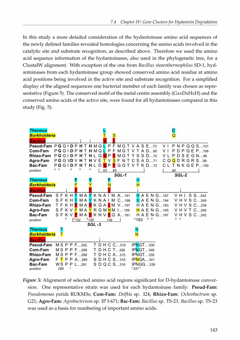
7.4 Chapter IV: Gene Clusters for Hydantoin Degradation
In this study a more detailed consideration of the hydantoinase amino acid sequences ofthe newly defined families revealed homologies concerning the amino acids involved in thecatalytic site and substrate recognition, as described above. Therefore we used the aminoacid sequence information of the hydantoinases, also used in the phylogenetic tree, for aClustalW alignment. With exception of the one from Bacillus stearothermophilus SD-1, hyd-antoinases from each hydantoinase group showed conserved amino acid residue at aminoacid positions being involved in the active site and substrate recognition. For a simplifieddisplay of the aligned sequences one bacterial member of each family was chosen as repre-sentative (Figure 5). The conserved motif of the metal centre assembly (GxxDxHxH) and theconserved amino acids of the active site, were found for all hydantoinases compared in thisstudy (Fig. 5).
41
Thermus L C 765 Burkholderia T S Q 766 Bacillus ↓ ↓ M L 767 Pseud-Fam P G G I D P H T H M Q L P F M G T V A S E…73 V I P N P Q Q S…101 768 Com-Fam P G G I D P H T H M Q L P F M G T V T A D…80 V I P D P Q E P…108 769 Rhizo-Fam P G G I D P H T H L Q M P F M G T Y S S D…70 V L P D S E G N…98 770 Agro-Fam P G G VD V H T H V E T V S F N T C S A D…71 C Q Q D R G R S…99 771 Bac-Fam P G G I D P H T H L D M P F G G T V T K D…72 C L T N K G E P…100 772 position * * * * * | 63 65 | | 95 | 773 SGL-1 SGL-2 774 Thermus F Y F H 775 Burkholderia F Y N H 776 Bacillus ↓ F Y F ↓ ↓ 777 Pseud-Fam S F K H F M A Y K N A I M A…161 H A E N G…187 V H I S S…242 778 Com-Fam S F K H F M A Y K N A I M C…168 H A E N G…194 V H V S C…249 779 Rhizo-Fam T F K H F M A Y K G A L M V…157 H A E N G…183 V H V S C…238 780 Agro-Fam S F K V F M A Y R G M N M I… 159 H A E N G…185 V H V T C…240 781 Bac-Fam S F K V F M A Y K N V F Q A…161 H A E N G…187 V H V S C…242 782 position * |*152 *155 159 | *183 * * * * 783 SGL -3 784 Thermus T N 785 Burkholderia T N 786 Bacillus ↓ 787 Pseud-Fam M S P P F…292 T D H C C…319 IPNGT…339 788 Com-Fam M S P P F…299 T D H C T…326 IPNGT…346 789 Rhizo-Fam M S P P F…288 T D H C A…315 IPNGT…335 790 Agro-Fam F T P P A…289 S D H C S…316 IPNGA…341 791 Bac-Fam W S P P L…291 S D Q C S…318 IPNGG…339 792 position 288 * * *337 * 793 794
Figure 5: Alignment of selected amino acid regions significant for D-hydantoinase conver-sion. One representative strain was used for each hydantoinase family: Pseud-Fam:Pseudomonas putida RUKM3s; Com-Fam: Delftia sp. I24; Rhizo-Fam: Ochrobactrum sp.G21; Agro-Fam: Agrobacterium sp. IP I-671; Bac-Fam: Bacillus sp. TS-23. Bacillus sp. TS-23was used as a basis for numbering of important amino acids.
143

7 PUBLICATIONS AND MANUSCRIPTS
Additional description of Figure 5: Identical amino acids of all 36 D-hydantoinasesaligned are indicated by asterisks (*). The GxxDxHxH-motif is bold. Amino acids asso-ciated with the catalytic centre are highlighted by an arrow from the top. Amino acidsinvolved in recognition sites of hydantoinases described by crystal structure deter-mination and structural analysis are shown on top of the alignment. Bacillus: Bacillussteraothermophillus SD1 [42, 51]; Burkholderia: Burkholderia pickettii [47], Thermus: Ther-mus sp. [39]. The stereochemistry gate loops (SGL) obtained for the hydantoinase ofBacillus stearothermophillus SD1 are shown below the sequence at the appropriate posi-tions. Amino acids being part of the functional recognition sites of hydantoinases withdetermined crystal structures and shown to be identical within the proposed familiesare highlighted by different colours.
Hydantoinases of the Bac-Fam and Rhizo-Fam showed high similarity with respect to theamino acids involved in the functional recognition sites of Bacillus stearothermophilus SD-1,as described above. The Bac-Fam showed only one difference in position 65, in whichPhe can be found instead of Leu (Fig. 5). Only a minor effect on activity and substrateselectivity of this substitution for the hydantoinase of Bacillus stearothermophilus SD-1 wasobserved [51]. These authors also reported the influence of a mutation at position 159 ofthe hydantoinase of Bacillus stearothermophilus SD-1 from Phe to Leu. This mutation led tolower activity for all substrates tested but no loss in activity [51]. As seen in Figure 5 thesame substitutions were obtained for all members of the Rhizo-Fam.Quite prominent is the identity of all amino acids involved in the recognition sites ofBurkholderia pickettii and the other members of the Agro-Fam (Fig. 3). The crystal structureof the D-hydantoinase of Burkholderia pickettii has been solved and the overall structureand catalytic site was found to be similar to other hydantoinases [47]. A significant featurewas found in the functional recognition site of the D-hydantoinase of Burkholderia pickettii:Tyr153, His 181, Thr286 and Asn335 are responsible for recognition of the amide group ofhydantoins by formation of hydrogen bonds. It is notable that the substitution of Thr286 bySer, as found in the hydantoinase of Thermus sp., does not affect the interaction with the sub-strate. The exocyclic substituent recognition sites were determined as Thr62, Ser64, Gln93,Phe150, Tyr153 and Asn157. Comparing amino acids involved in the substrate recognitionof the hydantoinase from B. pickettii and the other Agro-Fam members, absolutely identicalamino acids were obtained (Fig. 5).In literature the structures of the dihydropyrimidinase of Thermus sp. and the L-hydantoinase of Arthrobacter aurescens DSM3745 were described, too [38, 39]. In both studiesthe importance of the hydrophobic pocket, consisting of Leu64, Cys95, Phe152, Tyr155and Phe159, of the D-hydantoinase of Thermus sp. on substrate specificity was shown.A remarkable role was shown for Tyr155 which is part of this hydrophobic pocket andstabilises the transition state with its hydroxyl group. In this transition state additionallySer288, His183 and Asn336 were involved.
144
7.4 Chapter IV: Gene Clusters for Hydantoin Degradation
Comparing all these amino acid positions to the equivalent amino acid residues of theremaining two families, the Com-Fam and Pseud-Fam, no significant homology couldbe obtained. In fact, there are conserved amino acids but no high concordance, as seenfor the other hydantoinase families described above. Nevertheless, the amino acids ofhydantoinases of the Com-Fam and Pseud-Fam showed 100% identity in positions shownto be involved in recognition sites of the hydantoinases mentioned above.Summarising the comparison of structurally important amino acids we can conclude thatwe have found a very high similarity in "functional" amino acids of the Bac-Fam andRhizo-Fam to the ones of Bacillus stearothermophilus SD-1 as well as identical amino acidsof the Agro-Fam and Burkholderia pickettii. An attractive hypothesis for these three groupswould be that the conserved amino acids described form the substrate recognition sites ofall members of each family. However, this has to be proved at least by modelling of thehydantoinases. Taking into account the two other families, we are at least able to state thateach of the proposed families is highly conserved as a result of parallel evolution of thefamilies.
ConclusionsWe have elucidated two gene clusters responsible for the hydantoin degradation in Delftiasp. I24 and a moderate halophilic Ochrobactrum sp. G21. No information on the geneticorganisation of hydantoin degradation of members of these genera has been described inliterature previously.We have shown a high similarity of these gene clusters to putative gene clusters fromrelated bacteria. Phylogenetic comparison of different hydantoinases and grouping of theminto specific families with identical sites for the recognition of hydantoins showed that inmost cases hydantoinases from related bacteria share the highest homology. This supportsthe theory that hydantoinases evolved from a common ancestor and their evolution takesplace parallel to phylogenetic evolution for each genus.
AcknowledgmentThe authors would like to thank the Federal Ministry of Science and Education (BMBF),Germany, and the National Research Foundation (NRF), South Africa, for the funding of thisbilateral project. The State of Baden Württemberg is gratefully acknowledged for supportingthe corresponding author‘s Ph.D. within the "Landesgraduiertenförderungsgesetz".
145

7 PUBLICATIONS AND MANUSCRIPTS
References
1. Siemann M., Alvarado-Marin A., Pietzsch M. and Syldatk C. (1999) J. Mol. Catal. B:Enzymatic 6:387-397.
2. Sharma A. and Vohra R.M. (1997) Biochem. Biophys. Res. Commun. 234:485-488.
3. Lee S.G., Lee D.C., Sung M.H. and Kim H.S. (1994) Biotechnol. Lett. 16:461-466.
4. Lee S.-G., Lee D.-C., Hong S.-P., Sung M.-H. and Kim H.-S. (1995) Appl. Microbial.Biotechnol. 43:270-276.
5. Sudge S.S., Bastawde K.B., Gokhale D.V., Kalkote U.R., Ravindranathan T. (1998)Appl. Microbiol. Biotechnol. 49:594-599.
6. Runser S. and Meyer P. (1993) Eur. J. Biochem. 213:1315-1324.
7. May O., Siemann M., Pietzsch M., Kiess M., Mattes R. and Syldatk C. (1998) J.Biotechnol. 61:1-13.
8. Syldatk C., May O., Altenbuchner J., Mattes R. and Siemann M. (1999) Appl. Micro-biol. Biotechnol. 51:293-309.
9. Leuchtenberger W., Huthmacher K. and Drauz. K. (2005) Appl. Microbiol. Biotechnol.69:1-8.
10. Wiese A., Syldatk C., Mattes R. and Altenbuchner J. (2001) Arch. Microbiol. 176:187-196.
11. Werner M., Vazques F. J. L, Fritz C., Vielhauer O., Siemann- Herzberg M., Altenbuch-ner J. and Syldatk C. (2004) Engineering in Life Science 4:563-572.
12. Hils M., Münch P., Altenbuchner J., Syldatk C. and Mattes R. (2001) Appl. Microbiol.Biotechnol. 57:680-688.
13. Nanba H., Ikenaka Y., Yamada Y., Yajima K., Takano M. and Takahashi S. (1998)Biosci. Biotech. Biochem. 62:875-881.
14. Grifantini R., Galli G., Pratesi C., Frascotti G. and Grandi G. (1998) Microbiol.144:947-954.
15. Hsu W.H. and Kao C.H. (2003) Biochem. Biophys. Res. Commun. 303:848-854.
16. Matcher G.F., Burton S.G. and Dorrington R.A. (2004) Appl. Microbiol. Biotechnol.65:391-400.
146

7.4 Chapter IV: Gene Clusters for Hydantoin Degradation
17. Suzuki S., Takenaka Y., Onishi N. and Yokozeki K. (2005) Biosci. Biotechnol. Biochem.69:1473-1482.
18. Dürr R., Vielhauer O., Burton S.G., Cowan D.A., Puñal A., Brandão P.F.B., Bull A.T.and Syldatk C. (2006) J. Mol. Cat. B: Enzymatic 39:160-165.
19. Pozo C., Rodelas B., de la Escalera S. and González-López J. (2002) J. Appl. Microbiol.92:1028-1034.
20. Sambrook J., Fritsh E.F., Maniatis T. (1989) Molecular Cloning: A Laboratory Manual,Cold Spring Harbor Laboratory Press, Cold Spring Harbor, New York.
21. Altschul S.F., Madden T.L., Schaffer A.A., Zhang J., Zhang Z., Miller W. and LipmanD.J. (1997) Nucleic Acids Res. 25:3389-3402.
22. Chenna R., Sugawara H., Koike T., Lopez R., Gibson T.J., Higgins D.G. and Thomp-son J.D. (2003) Nucleic Acids Res. 31:3497-3500.
23. Vincze T., Posfai J. and Roberts R.J. (2003) Nucleic Acids Res. 31:3688-3691.
24. Kumar S., Tamura K. and Nei M. (2004) Briefings in Bioinformatics 5:150-163.
25. Lin L.-L., Hsu W.-H., Hsu W.-Y., Kan S.-C and Hu H.-Y. (2005) Antonie van Leeuwenhoek88:189-197.
26. Inoue H., Nojima und Okayama H. (1990) Gene 96:23-28.
27. Grunstein M. and Hogness D.S. (1975) Proc. Natl. Acad. Sci. USA 72:3961- 3965.
28. Lee S.Y. and Rasheed S. (1990) Biotechniques 9:676-9.
29. Shigematsu T, Yumihara K., Ueda Y., Numaguchi M., Morimura S. and Kida K.(2003) Int. J. Syst. Evol. Microbiol. 53:1479-1483.
30. Wen A., Fegan M., Hayward C., Chakraborty S. and Sly L.I. (1999) Int. J. Syst. Evol.Microbiol. 49:567-576.
31. Watabe K., Ishikawa T., Mukohara Y. and Nakamura H. (1992) J. Bacteriol. 174:962-969.
32. Marchler-Bauer A, Anderson J.B., Cherukuri P.F., DeWeese-Scott C., Geer L.Y.,Gwadz M., He S., Hurwitz D.I., Jackson J.D., Ke Z., Lanczycki C.J., Liebert C.A.,Liu C., Lu F., Marchler G.H., Mullokandov M., Shoemaker B.A., Simonyan V., SongJ.S., Thiessen P.A., Yamashita R.A., Yin J.J., Zhang D. and Bryant S.H. (2005) NucleicAcids Res. 33:192-6.
33. Kyte J. and Doolittle R. (1982) J. Mol. Biol. 157:105-132.
147

7 PUBLICATIONS AND MANUSCRIPTS
34. LaPointe G., Viau S., LeBlanc D., Robert N. and Morin A. (1994) J. Appl. Environ.Microbiol. 60:888-895.
35. Chien H.R., Jih Y.L., Yang W.Y. and Hsu W.H. (1998) Biochim. Biophys. Acta. 1395:68-77.
36. May O., Habenicht A. Mattes R., Syldatk S. and Siemann M. (1998) Biol. Chem. Vol.379:743-747.
37. Holm L. and Sander C. (1997) Proteins 28:72-82.
38. Abendroth J., Niefind K., May O., Siemann M., Syldatk C. and Schomburg D. (2002)Biochem. 41:8589-8597.
39. Abendroth J, Niefind K. and Schomburg D. (2002) J. Mol. Biol. 28:143-156.
40. Benini S., Rypniewski W. R., Wilson K. S., Miletti S., Ciurli S. and Mangani S. (1999)Structure 7:205-216.
41. Benini S., Rypniewski W.R., Wilson K.S., Miletti S., Ciurli S. and Mangani S. (2000)J. Biol. Inorg. Chem. 5:110-118.
42. Cheon Y.H., Kim H.S., Han K.H., Abendroth J., Niefind K., Schomburg D., Wang J.and Kim Y.S. (2002) Biochemistry 41:9410-9417.
43. Jabri E., Carr M.B., Hausinger R.P. and Karplus P.A. (1995) Science 268:998-1004.
44. Thoden J.B., Phillips G.N., Neal T.M., Raushel F.M. and Holden H.M. (2001) Bio-chemistry 40:6989-6997.
45. Vanhooke J.L., Benning M. M., Raushel F.M. and Holden H.M. (1996) Biochemistry35:6020-6025.
46. Radha Kishan K.V., Vohra R.M., Ganesan K., Agrawal V., Sharma V.M. and SharmaR. (2005) J. Mol. Biol. 347:95-105.
47. Xu Z., Liu Y., Yang Y., Jiang W., Arnold E. and Ding J. (2003) J. Bacteriol. 185:4038-4049.
48. Park I.S. and Hausinger R.P. (1993) Protein Science 2:1034-1041.
49. Zimmermann B.H., Kemling N.M. and Evans D.R. (1995) Biochemistry 34:7038-7046.
50. Cheon Y.-H., Park H.-S., Lee S.-C., Lee D.-E. and Kim H.-S. (2003) J. Mol. Catal. B:Enzymatic 26:217-222.
51. Cheon Y.H., Park H.S., Kim J.H., Kim Y. and Kim H.S. (2004) Biochemistry 43:7413-7420.
148