RADT 1542 SKULL RADIOGRAPHY Wynn Harrison, Med, RT(R)(N)
77
RADT 1542 RADT 1542 SKULL RADIOGRAPHY SKULL RADIOGRAPHY Wynn Harrison, Med, RT(R) Wynn Harrison, Med, RT(R) (N) (N)
-
Upload
conrad-griscom -
Category
Documents
-
view
219 -
download
1
Transcript of RADT 1542 SKULL RADIOGRAPHY Wynn Harrison, Med, RT(R)(N)

RADT 1542RADT 1542SKULL RADIOGRAPHYSKULL RADIOGRAPHY
Wynn Harrison, Med, RT(R)(N)Wynn Harrison, Med, RT(R)(N)

What a Bad Day!!!!What a Bad Day!!!!

Section 1: The CraniumSection 1: The Cranium
New Words:New Words:
HEMANGIOMA: tumor containing blood HEMANGIOMA: tumor containing blood vessels and fibrous tissuevessels and fibrous tissue
TRAUMATIC PNEUMOCEPHALUS: TRAUMATIC PNEUMOCEPHALUS: injury causing gas/air in the extracranial injury causing gas/air in the extracranial cavitycavity

HYDROCEPHALUS: increased HYDROCEPHALUS: increased cerebrospinal fluid in the ventriclescerebrospinal fluid in the ventricles
PAGET’S DISEASE: overproduction of PAGET’S DISEASE: overproduction of bonebone
CRANIOTOMY: Incision into the headCRANIOTOMY: Incision into the head

SUBDURAL HEMATOMA: Blood SUBDURAL HEMATOMA: Blood collected beneath the duracollected beneath the dura
TERATOMA: Congenital tumor containing TERATOMA: Congenital tumor containing 1+ embryonic germ layers (hair/teeth/skin 1+ embryonic germ layers (hair/teeth/skin may be present)may be present)

ENCEPHALITIS: Inflammation of the ENCEPHALITIS: Inflammation of the brainbrain
GLIOMA: Primary tumor of the brainGLIOMA: Primary tumor of the brain
MENINGIOMA: Slow growing benign MENINGIOMA: Slow growing benign tumor in the meningestumor in the meninges

ACOUSTIC NEUROMA: Benign tumor ACOUSTIC NEUROMA: Benign tumor involving hearinginvolving hearing
CRANIO-PHARYNGIOMA: Benign tumor CRANIO-PHARYNGIOMA: Benign tumor above the sella turcicaabove the sella turcica
CONCUSSION: Impact injuryCONCUSSION: Impact injury

ANENCEPHALUS: Congenital absence of the ANENCEPHALUS: Congenital absence of the brainbrain
ACEPHALUS: Absence of headACEPHALUS: Absence of head
MICROCEPHALIC: Small headMICROCEPHALIC: Small head
BURR HOLES: Holes bore into skull for BURR HOLES: Holes bore into skull for pressure reliefpressure relief

Why Dogs Bite Their OwnersWhy Dogs Bite Their Owners

POSITIONING CONCERNSPOSITIONING CONCERNS
Some people don’t like to have their face Some people don’t like to have their face touchedtouched
Wash your hands FIRSTWash your hands FIRST CLEAN BUCKY or TABLE (and have them CLEAN BUCKY or TABLE (and have them
watch you)watch you) Remember body habitusRemember body habitus
The body is attached to the head!!!!The body is attached to the head!!!!
Lines/Landmarks/PlanesLines/Landmarks/Planes
Median saggitalMedian saggital CoronalCoronal

CompositionComposition
Composed of 22 Composed of 22 BonesBones Eight Cranial Bones Eight Cranial Bones Fourteen Facial BonesFourteen Facial Bones

Radiographic LandmarksRadiographic Landmarks
GlabellaGlabella: Triangular area : Triangular area between and slightly between and slightly superior to the eyebrows superior to the eyebrows and above the bridge of and above the bridge of the nosethe nose
NasionNasion: Depression at : Depression at the bridge of the nosethe bridge of the nose
Junction of the two nasal Junction of the two nasal bones and the frontal bonebones and the frontal bone

Radiographic LandmarksRadiographic Landmarks Inner CanthusInner Canthus: Where : Where
the eyelids meet near the the eyelids meet near the nosenose
Outer CanthusOuter Canthus: Lateral : Lateral junction of the eyelidsjunction of the eyelids
AcanthionAcanthion: Midline point : Midline point at the junction of the at the junction of the upper lip and the nasal upper lip and the nasal septum. Point where the septum. Point where the nose and the upper lip nose and the upper lip meetmeet

Radiographic LandmarksRadiographic Landmarks
GonionGonion: Angle of the mandible (Jaw): Angle of the mandible (Jaw)
Mental PointMental Point: A triangular area projects : A triangular area projects forward as the chin (mentum). The center forward as the chin (mentum). The center of the mentum is the mental pointof the mentum is the mental point

Radiographic LandmarksRadiographic Landmarks
EAREAR Auricle/Pinna:Auricle/Pinna:
External portion of the External portion of the ear. Large flap of ear ear. Large flap of ear made of cartilagemade of cartilage
TEATEA: Top of the ear : Top of the ear attachment. Superior attachment. Superior attachment of the attachment of the auricle. Level of the auricle. Level of the petrous ridge on each petrous ridge on each sideside

Radiographic LinesRadiographic Lines
Radiographic lines are Radiographic lines are important to skull important to skull positioningpositioning
Certain lines are formed Certain lines are formed from anterior structures from anterior structures on the face and connect on the face and connect to the EAMto the EAM
EAMEAM: External acoustic : External acoustic (auditory) meatus, (auditory) meatus, Opening of the external Opening of the external ear canalear canal

Radiographic LandmarksRadiographic Landmarks
EyeEye Supraorbital Margin (SOML):Supraorbital Margin (SOML): Superior rim of Superior rim of
the bony orbit of the eyethe bony orbit of the eye
Infraorbital Margin (IOML):Infraorbital Margin (IOML): Inferior rim of the Inferior rim of the bony orbit of the eyebony orbit of the eye
Midlateral Orbital Margin:Midlateral Orbital Margin: (OML) Portion of (OML) Portion of the lateral rim near the outer canthus of the the lateral rim near the outer canthus of the eyeeye

Radiographic LinesRadiographic Lines
Orbitomeatal LineOrbitomeatal Line (OML):(OML): Located between Located between the outer canthus the outer canthus (midlateral orbital margin) (midlateral orbital margin) and the EAMand the EAM
Infraorbitomeatal LineInfraorbitomeatal Line (IOML):(IOML): Formed by Formed by connecting the middle of connecting the middle of the infraorbital margin to the infraorbital margin to the EAMthe EAM

Radiographic PlanesRadiographic Planes
Interpupillary Line Interpupillary Line (IPL): (IPL): A line connecting A line connecting
either the pupils or the either the pupils or the outer canthi of the outer canthi of the patient’s eyespatient’s eyes
The IPL must be The IPL must be exactly perpendicular exactly perpendicular to the IR in a TRUE to the IR in a TRUE LATERAL position LATERAL position

Talk about TOO MUCH RAIN~Talk about TOO MUCH RAIN~


Radiographic LandmarkRadiographic Landmark
Inion or EOPInion or EOP:: External occipital protuberanceExternal occipital protuberance Rise or bump along the midline of the lower Rise or bump along the midline of the lower
back of the head near the junction of the head back of the head near the junction of the head and neckand neck
Extension of the IOML posteriorly Extension of the IOML posteriorly approximates the location of the inionapproximates the location of the inion

Radiographic LandmarkRadiographic Landmark
VertexVertex Top portion of the Top portion of the
headhead

Sutures of the SkullSutures of the Skull
Joints formed Joints formed between the cranial between the cranial bones are known as bones are known as suturessutures
Fibrous connective Fibrous connective tissue hold bones tissue hold bones tightly togethertightly together
Synarthrodial – Don’t Synarthrodial – Don’t permit movementpermit movement

Sutures of the SkullSutures of the Skull
Coronal SutureCoronal Suture: :
Sagittal SutureSagittal Suture: :
Lambdoidal SutureLambdoidal Suture::
Squamosal SutureSquamosal Suture::

ArthrologyArthrology
Temporomandibular Joint:Temporomandibular Joint:
Only movable joints in the craniumOnly movable joints in the cranium
Formed by the mandibular fossa on each Formed by the mandibular fossa on each temporal bone with corresponding condyle of temporal bone with corresponding condyle of the mandiblethe mandible

FontanelsFontanels
Soft spotsSoft spots Present at birthPresent at birth Unossified connective Unossified connective
tissuetissue Where three or more Where three or more
bones ajoinbones ajoin Six FontanelsSix Fontanels Gradually replaced with Gradually replaced with
bonebone Allow for skull Allow for skull
compression during birthcompression during birth
Most prominent are the Most prominent are the anterior and posterior anterior and posterior fontanelsfontanels
Located on the anterior Located on the anterior and posterior ends of the and posterior ends of the sagittal suturesagittal suture

FontanelsFontanels
Articulation between the Articulation between the frontal and both parietal frontal and both parietal bones at the anterior end bones at the anterior end of the sagittal suture is of the sagittal suture is the the bregmabregma
Articulation between the Articulation between the occipital bone and both occipital bone and both parietal bones at the parietal bones at the posterior end is the posterior end is the lambdalambda
Anterolateral (sphenoid) Anterolateral (sphenoid) fontanel is the fontanel is the pterionpterion
Posterolateral fontanel is Posterolateral fontanel is the the asterionasterion

FontanelsFontanels

FontanelsFontanels

FontanelsFontanels

CalvariumCalvarium
Skull CapSkull Cap
Composed of four bonesComposed of four bones Frontal boneFrontal bone Right Parietal boneRight Parietal bone Left Parietal boneLeft Parietal bone Occipital boneOccipital bone

CalvariumCalvarium

CalvariumCalvarium

Cranial FloorCranial Floor
Composed of four bonesComposed of four bones Ethmoid boneEthmoid bone Sphenoid boneSphenoid bone Right Temporal boneRight Temporal bone Left Temporal boneLeft Temporal bone

Cranial FloorCranial Floor

Skull MorphologySkull Morphology
MesocephalicMesocephalic: Average shaped head, the : Average shaped head, the petrous ridges lie at a 47 degree angle petrous ridges lie at a 47 degree angle with the MSPwith the MSP
BrachycephalicBrachycephalic: Short, broad, shallow : Short, broad, shallow head. Petrous ridges form a 54 degree head. Petrous ridges form a 54 degree angle with the MSPangle with the MSP
DolichocephalicDolichocephalic: Long, narrow, deep : Long, narrow, deep head. Petrous ridges form a 40 degree head. Petrous ridges form a 40 degree angle with the MSPangle with the MSP

Occipital BoneOccipital Bone

Occipital BoneOccipital Bone

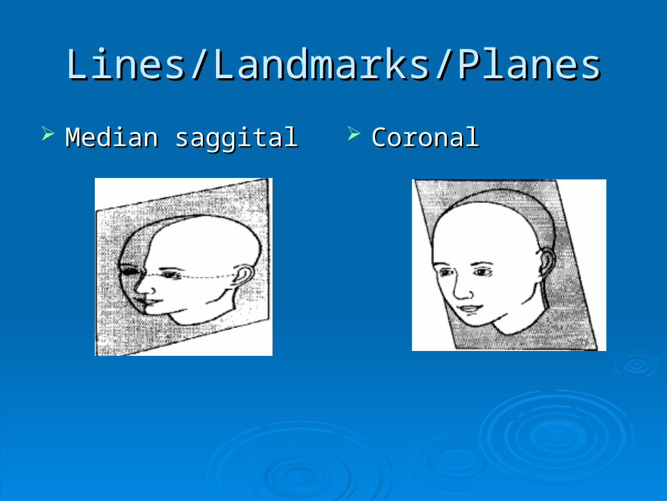
Sphenoid BoneSphenoid Bone
Location and Location and purposepurpose Midline of cranial floorMidline of cranial floor Anchor to hold the 8 Anchor to hold the 8
cranial bones togethercranial bones together Articulates with all Articulates with all
cranial bonescranial bones Forms the base of Forms the base of
skull, small portion of skull, small portion of each lateral wall, and each lateral wall, and posterior wall of each posterior wall of each orbitorbit
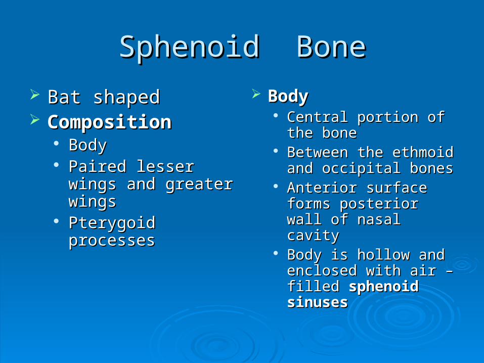
Sphenoid BoneSphenoid Bone
Bat shapedBat shaped CompositionComposition
BodyBody Paired lesser wings Paired lesser wings
and greater wingsand greater wings Pterygoid processesPterygoid processes
BodyBody Central portion of the Central portion of the
bonebone Between the ethmoid Between the ethmoid
and occipital bonesand occipital bones Anterior surface forms Anterior surface forms
posterior wall of nasal posterior wall of nasal cavitycavity
Body is hollow and Body is hollow and enclosed with air – enclosed with air – filled filled sphenoid sphenoid sinusessinuses

Sphenoid BoneSphenoid Bone
Body of sphenoidBody of sphenoid Sella TurcicaSella Turcica
• Saddle – shaped depressionSaddle – shaped depression• Superior surface of the bodySuperior surface of the body• Houses pituitary glandHouses pituitary gland• Sella turcica is localized for a radiographic Sella turcica is localized for a radiographic
exam by centering ¾ inches anterior and ¾ exam by centering ¾ inches anterior and ¾ superior to the EAM on a lateral projectionsuperior to the EAM on a lateral projection

Body of SphenoidBody of Sphenoid
Dorsum SellaeDorsum Sellae: is the posterior portion of : is the posterior portion of the sella turcicathe sella turcica
Posterior ClinoidPosterior Clinoid ProcessesProcesses: Extend : Extend superiorly from the lateral margin of the superiorly from the lateral margin of the dorsum sellaedorsum sellae
Optic GrooveOptic Groove: Depression that runs : Depression that runs horizontally across the bodyhorizontally across the body
Optic ChiasmOptic Chiasm: Formed by crossing of the : Formed by crossing of the optic nerves, situated in the optic grooveoptic nerves, situated in the optic groove

Sphenoid BoneSphenoid Bone
Lesser Wings of the SphenoidLesser Wings of the Sphenoid Extends laterally and horizontally across the Extends laterally and horizontally across the
anterosuperior aspect of the bodyanterosuperior aspect of the body Junction at the midline is the Junction at the midline is the sphenoid ridgesphenoid ridge Anterior clinoid processesAnterior clinoid processes extend from the extend from the
posterior portion of each lesser wingposterior portion of each lesser wing

Sphenoid BoneSphenoid Bone
Greater Wings of SphenoidGreater Wings of Sphenoid Posterior to the lesser wingsPosterior to the lesser wings Arise from the lateral surfaces of the body and Arise from the lateral surfaces of the body and
extend outwardextend outward Openings on each greater wing serve as Openings on each greater wing serve as
passageways for the nerves and blood passageways for the nerves and blood vessels supplying the orbits and facevessels supplying the orbits and face

Sphenoid BoneSphenoid Bone

Sphenoid BoneSphenoid Bone
Pterygoid ProcessesPterygoid Processes Extensions of bone from under the sphenoid Extensions of bone from under the sphenoid
bone at the junction of the body and greater bone at the junction of the body and greater wingswings
Articulates anteriorly with the Articulates anteriorly with the palatine bonepalatine bone and and vomervomer
Comprised of two plates of bone fused Comprised of two plates of bone fused togethertogether
Medial plate is called the Medial plate is called the pterygoid hamuluspterygoid hamulus because it has a small hook – like processbecause it has a small hook – like process

Sphenoid BoneSphenoid Bone

Sphenoid BoneSphenoid Bone

Sella Turcica Sphenoid BoneSella Turcica Sphenoid Bone

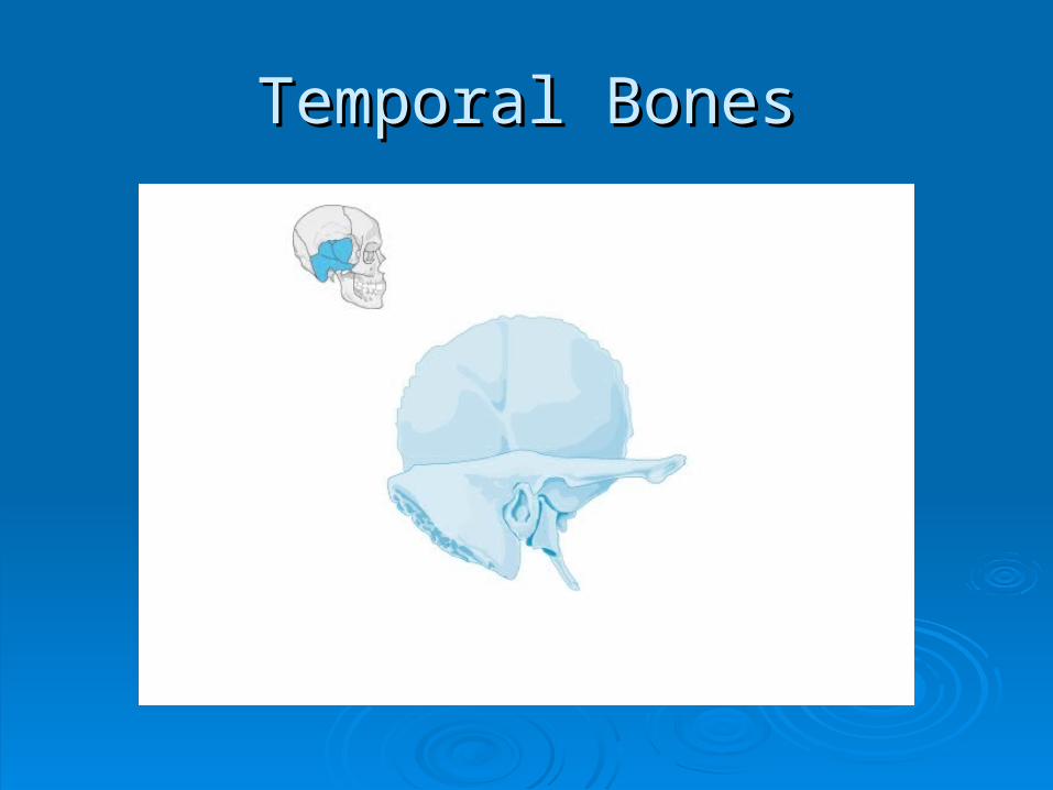
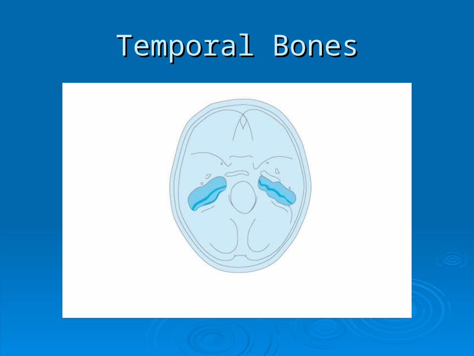
Temporal BonesTemporal Bones

Temporal BonesTemporal Bones
Named for the region Named for the region around the templesaround the temples
LocationLocation Below the parietal Below the parietal
bonebone Articulates with the Articulates with the
greater wing of the greater wing of the sphenoid and the sphenoid and the occipital bonesoccipital bones
Contains 4 RegionsContains 4 Regions SquamousSquamous TympanicTympanic MastoidMastoid PetrousPetrous
Temporal BonesTemporal Bones

Temporal BonesTemporal Bones
Temporal BonesTemporal Bones

Group Photo Anyone?Group Photo Anyone?

AP and PA projectionsAP and PA projections
Petrous pyramids fill the orbit if there is no Petrous pyramids fill the orbit if there is no angulationangulation
Petrous pyramid will lie in lower 1/3 of orbit Petrous pyramid will lie in lower 1/3 of orbit with 15 degree angulation with 15 degree angulation
if AP is it caudal or cephalic?if AP is it caudal or cephalic?

ROUTINE PROCEDURESROUTINE PROCEDURESAP ViewAP View
OML and sagittal OML and sagittal plane perpendicular plane perpendicular to the cassetteto the cassette
Petrous pyramids fillPetrous pyramids fill
the orbitsthe orbits

AP Axial (Townes)AP Axial (Townes)
OML = 30 degreesOML = 30 degrees IOML = 37 degreesIOML = 37 degrees
Posterior clinoidsPosterior clinoids
HAAS is reverse HAAS is reverse Townes viewTownes view

PA SkullPA Skull
OML = perpendicular OML = perpendicular to cassetteto cassette
0 degree angulation0 degree angulation
Petrous pyramids fill Petrous pyramids fill the orbitsthe orbits

PA CaldwellPA Caldwell
Patient PA with 12 – Patient PA with 12 – 15 degree caudal 15 degree caudal angulation of CRangulation of CR
CR exits nasionCR exits nasion
Petrous pyramids in Petrous pyramids in bottom 1/3 of orbitbottom 1/3 of orbit

Lateral SkullLateral Skull
Interpupillary line Interpupillary line perpendicular to perpendicular to cassettecassette
Midsagittal plane Midsagittal plane parallel to cassetteparallel to cassette
(Look at patient body (Look at patient body habitus)habitus)
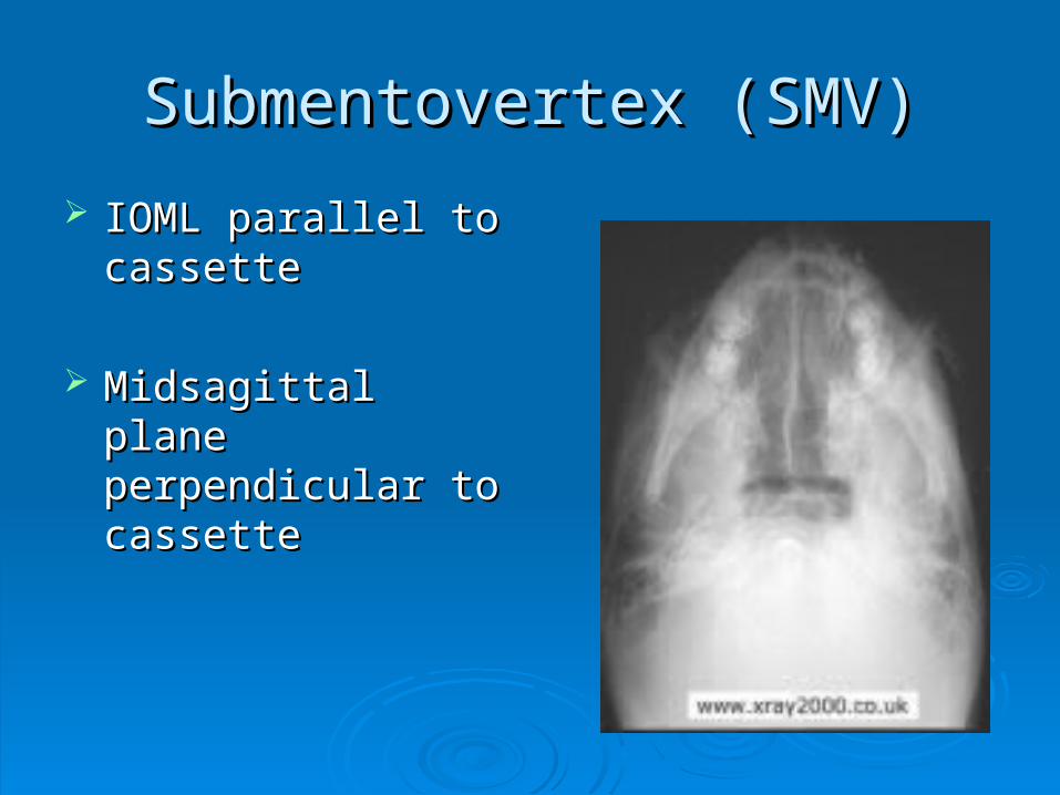
Submentovertex (SMV)Submentovertex (SMV)
IOML parallel to IOML parallel to cassettecassette
Midsagittal plane Midsagittal plane perpendicular to perpendicular to cassettecassette

Positioning ErrorsPositioning Errors
Rotation occurs Rotation occurs when the median when the median Saggital plane is not Saggital plane is not parallel to the film.parallel to the film.


Tilt occurs when the Tilt occurs when the interpupilary line is interpupilary line is not at 90 to the film not at 90 to the film


Trauma and PathologyTrauma and Pathology


Golf clubGolf club

Paget’s DiseasePaget’s Disease

Pituitary tumorPituitary tumor

Film critiqueFilm critique

Critical thinkingCritical thinking