Radiology An Introduction. 2 Learning Objectives List the Properties of X-rays Understand the need...
46
Radiology Radiology An Introduction An Introduction
-
Upload
ashlee-dina-nicholson -
Category
Documents
-
view
222 -
download
6
Transcript of Radiology An Introduction. 2 Learning Objectives List the Properties of X-rays Understand the need...

RadiologyRadiology
An IntroductionAn Introduction

22
Learning ObjectivesLearning Objectives
List the Properties of X-raysList the Properties of X-rays
Understand the need for radiologyUnderstand the need for radiology
Explain Radiation ProtectionExplain Radiation Protection

33
IntroductionIntroduction
Discovery of the x-rayDiscovery of the x-ray
X-ray picture/ roentgenograph/radiographX-ray picture/ roentgenograph/radiograph
44
Radiation, Radiology and Radiation, Radiology and RadiographyRadiography
RadiationRadiation = = Emission of energy in the Emission of energy in the form of EMR or particulate radiation.form of EMR or particulate radiation.
RadiographyRadiography = the techniques involved = the techniques involved in producing radiographs.in producing radiographs.
Radiology Radiology = = interpretation of radiographs interpretation of radiographs and other images.and other images.
55
Dental RadiologyDental Radiology
Dental exams one of most commonDental exams one of most common
1997-8 survey in UK found:1997-8 survey in UK found: 19 million intra-oral radiographs taken19 million intra-oral radiographs taken 2.9 million panoramic radiographs taken2.9 million panoramic radiographs taken
In Sweden 15 millions/yIn Sweden 15 millions/y
66
Properties of x-raysProperties of x-rays InvisibleInvisible
Have no chargeHave no charge
Travel at speed of lightTravel at speed of light
Have no mass or weight Have no mass or weight
Travel in straight linesTravel in straight lines

77
Properties of x-raysProperties of x-rays
Can cause ionizationCan cause ionization
Can affect photographic film emulsionCan affect photographic film emulsion
Can affect living tissueCan affect living tissue
88
Properties of x-raysProperties of x-rays
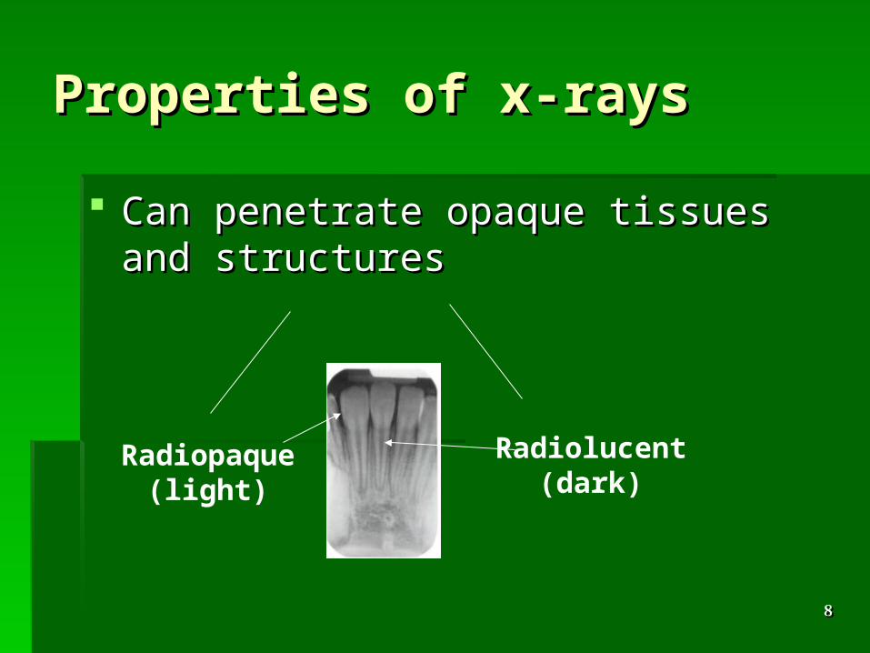
Can penetrate opaque tissues and Can penetrate opaque tissues and structuresstructures
Radiopaque(light)
Radiolucent(dark)

99
Conventional Conventional Radiography and Digital Radiography and Digital ImagingImaging
Conventional radiographyConventional radiography uses uses radiographic film as the image radiographic film as the image detector/sensor.detector/sensor.

1010
Conventional Conventional Radiography and Digital Radiography and Digital ImagingImaging
Digital imagingDigital imaging uses a charged coupled uses a charged coupled device (CCD) as the image device (CCD) as the image detector/sensor.detector/sensor.
1111
Advantages of digital imagingAdvantages of digital imaging
No use of films, intensifying screens, cassettes No use of films, intensifying screens, cassettes etc.etc.
Computer hardware and software allows you to Computer hardware and software allows you to view and store images.view and store images.
Multiple images are obtained without changing Multiple images are obtained without changing film holder or using new film.film holder or using new film.
Less exposure to radiationLess exposure to radiation
1212
Why do we take Why do we take radiographs?radiographs?
1313
Role of radiographsRole of radiographs
Clinical examination phaseClinical examination phase Diagnosis (confirm / exclude)Diagnosis (confirm / exclude) Treatment planningTreatment planning During treatment During treatment Follow up after various treatment Follow up after various treatment
proceduresprocedures
1414
Are all radiographs Are all radiographs necessary?necessary?
1515
Must justify taking any radiographs; not a Must justify taking any radiographs; not a blanket screening for all patients.blanket screening for all patients.
Radiographs do have limitations and should Radiographs do have limitations and should never replace a thorough clinical examination.never replace a thorough clinical examination.
There are disadvantages and risks - must There are disadvantages and risks - must weigh up benefits against risks.weigh up benefits against risks.
Radiographs can indicate the need for further Radiographs can indicate the need for further investigation.investigation.

1616
The uses of radiographsThe uses of radiographs
1717
General dentistry:General dentistry:
Loss of tooth structureLoss of tooth structure
Carious (occlusal, proximal)Carious (occlusal, proximal)
Non - carious (attrition, abrasion, erosion, fracture)Non - carious (attrition, abrasion, erosion, fracture)
Periodontal diseasePeriodontal disease
Endodontic diseaseEndodontic disease
Impacted teethImpacted teeth
Trauma (root and alveolar fractures, foreign bodies)Trauma (root and alveolar fractures, foreign bodies)
Other pathology affecting boneOther pathology affecting bone
1818
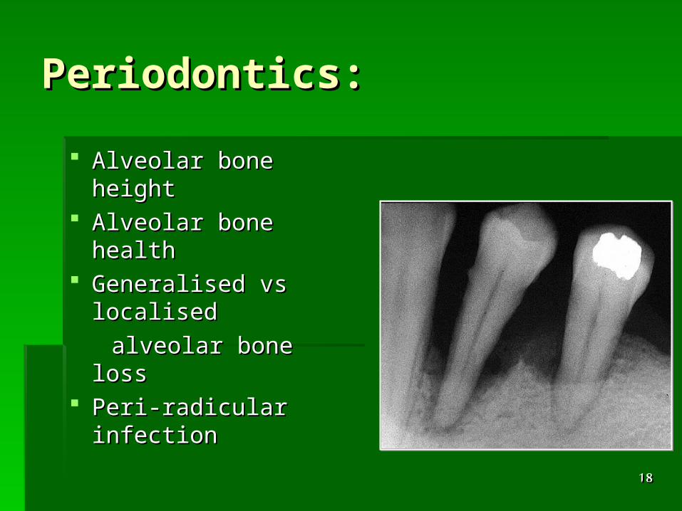
Periodontics:Periodontics:
Alveolar bone heightAlveolar bone height Alveolar bone healthAlveolar bone health Generalised vs Generalised vs
localised localised
alveolar bone lossalveolar bone loss Peri-radicular Peri-radicular
infectioninfection
1919
Orthodontics:Orthodontics:
General growth and developmentGeneral growth and development Delayed eruptionDelayed eruption Ectopic teethEctopic teeth Eruption pathsEruption paths Impacted teethImpacted teeth Supernumerary teethSupernumerary teeth
2020
Oral medicine and oral Oral medicine and oral surgery:surgery:
ExtractionsExtractions Jaw fracturesJaw fractures TumoursTumours InfectionsInfections Foreign bodiesForeign bodies
2121
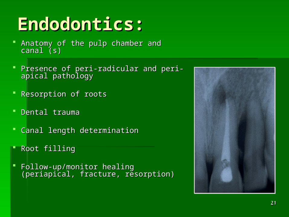
Endodontics:Endodontics: Anatomy of the pulp chamber and canal (s)Anatomy of the pulp chamber and canal (s)
Presence of peri-radicular and peri-apical Presence of peri-radicular and peri-apical pathologypathology
Resorption of rootsResorption of roots
Dental traumaDental trauma
Canal length determinationCanal length determination
Root fillingRoot filling
Follow-up/monitor healing (periapical, Follow-up/monitor healing (periapical, fracture, resorption)fracture, resorption)

2222
RadiologyRadiology
3 common dental X-rays:3 common dental X-rays:
BitewingsBitewings PeriapicalPeriapical PanoramicPanoramic
(Univ. Manitoba, 2005)(Univ. Manitoba, 2005)

2323
Types of RadiographsTypes of Radiographs
Intra-oralIntra-oral
Extra-oralExtra-oral
Other technologies/imaging modalitiesOther technologies/imaging modalities
2424
Intra-oral radiographyIntra-oral radiography
Radiographic film/detector is exposed Radiographic film/detector is exposed whilst inside the patient’s mouthwhilst inside the patient’s mouth
Image of a small area - a few teeth and Image of a small area - a few teeth and adjacent supporting structuresadjacent supporting structures

2525
Bitewing:Bitewing:
Indications:Indications: baseline examinationbaseline examination detection of:detection of:
- - dental caries dental caries
- non-carious tooth loss- non-carious tooth loss- monitoring the progress - monitoring the progress of any loss of tooth structureof any loss of tooth structure- assessing existing restorations - assessing existing restorations (defects, contacts)(defects, contacts)- assessment of periodontal status.- assessment of periodontal status.

2626
Periapical:Periapical:
Indications:Indications: detection of apical infection/inflammationdetection of apical infection/inflammation dental trauma (to the tooth and associated dental trauma (to the tooth and associated
alveolar bone)alveolar bone) assessment of root morphologyassessment of root morphology endodontic diagnosis, planning,endodontic diagnosis, planning,
treatment and monitoringtreatment and monitoring

2727
Occlusal:Occlusal:
Indications:Indications: presence/absence of developing teethpresence/absence of developing teeth supernumerary teethsupernumerary teeth impacted teethimpacted teeth pathology not fully demonstrated in an intraoral viewpathology not fully demonstrated in an intraoral view contour of buccal and lingual cortical platecontour of buccal and lingual cortical plate localisation technique (used with another film) localisation technique (used with another film) when unable to take intra-oral radiographswhen unable to take intra-oral radiographs
- limited opening of mouth- limited opening of mouth- uncooperative child.- uncooperative child.

2828
Occlusal:Occlusal:
2929
Extra-oral radiographyExtra-oral radiography
Radiographic film/detector positioned Radiographic film/detector positioned outside the patient’s mouth.outside the patient’s mouth.
Can image larger areas of the mandible Can image larger areas of the mandible and maxilla, face, skull.and maxilla, face, skull.
3030
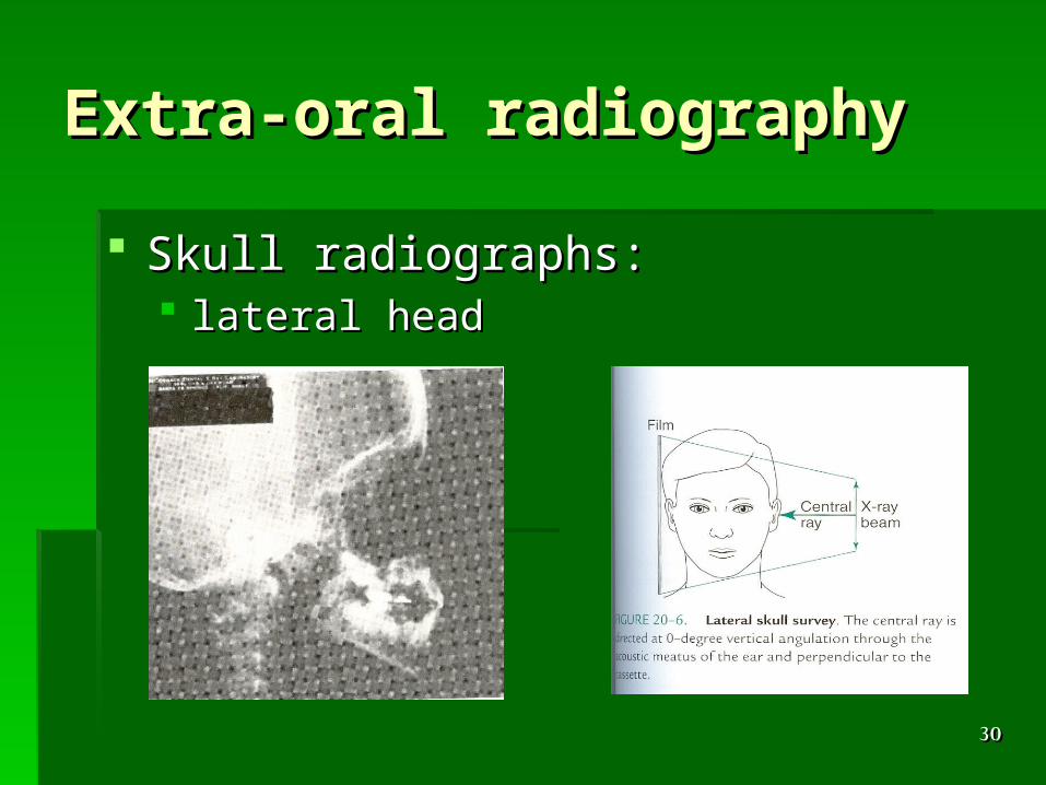
Extra-oral radiographyExtra-oral radiography
Skull radiographs: Skull radiographs: lateral headlateral head
3131
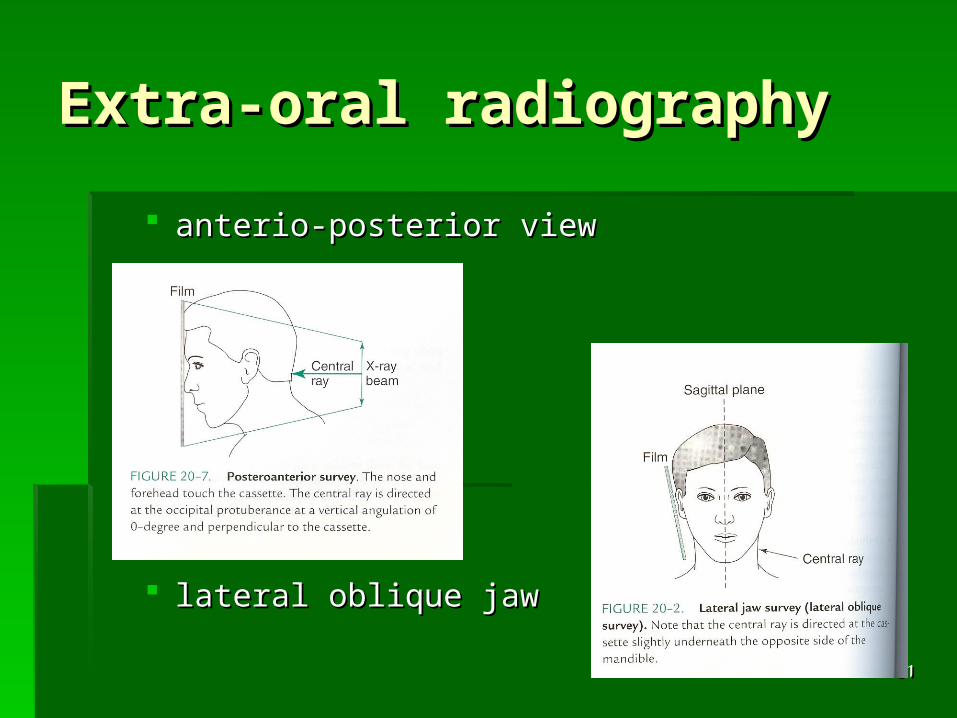
Extra-oral radiographyExtra-oral radiography
anterio-posterior viewanterio-posterior view
lateral oblique jawlateral oblique jaw
3232
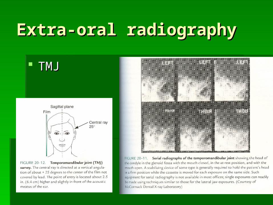
Extra-oral radiographyExtra-oral radiography
TMJTMJ
3333
Extra-oral radiographyExtra-oral radiography
TomographyTomography Panoramic radiographPanoramic radiograph
orthopantomograph (OPG)orthopantomograph (OPG) TMJTMJ maxillary sinus viewsmaxillary sinus views
Modern OPG machines can do a range of skull Modern OPG machines can do a range of skull views by altering the extent of the tomographic views by altering the extent of the tomographic layer used and by altering the shape of the layer used and by altering the shape of the tomographic layer usedtomographic layer used

3434
Indications for traditional Indications for traditional OPGOPG
Assessment of Assessment of
-- wisdom teethwisdom teeth
-- TMJ pathologyTMJ pathology
-- maxillary sinusmaxillary sinus
-- jaw bone jaw bone pathologiespathologies
-- orthodontic orthodontic diagnosisdiagnosis
-- jaw bone jaw bone fractures.fractures.

3535
Other Technologies and Other Technologies and Imaging ModalitiesImaging Modalities
Computerised tomography (CT)Computerised tomography (CT)
Magnetic resonance imaging (MRI)Magnetic resonance imaging (MRI)
UltrasoundUltrasound
3636
Computerised Computerised tomography (CT)tomography (CT)
Radiographic cutting of a region/structure Radiographic cutting of a region/structure into thin slicesinto thin slices
Fairly high dosesFairly high doses Good diagnostic informationGood diagnostic information Used in oral maxillofacial surgery Used in oral maxillofacial surgery
(diagnosis and treatment planning of (diagnosis and treatment planning of tumours, fractures and neuropathies)tumours, fractures and neuropathies)

3737
Magnetic resonance Magnetic resonance imaging (MRI)imaging (MRI)
Gives better soft tissue images than CT scansGives better soft tissue images than CT scans No radiation - uses magnetic field and sound No radiation - uses magnetic field and sound
waveswaves Very good diagnostic informationVery good diagnostic information Used in oral maxillofacial surgery (diagnosis Used in oral maxillofacial surgery (diagnosis
and treatment planning of tumours, fractures and treatment planning of tumours, fractures and neuropathies, gold standard for TMJ and neuropathies, gold standard for TMJ imaging) imaging)
3838
UltrasoundUltrasound
Limited uses for dental careLimited uses for dental care
Salivary gland tumoursSalivary gland tumours
Possibly TMJPossibly TMJ
Locating foreign object in soft tissueLocating foreign object in soft tissue
Soft tissue cystsSoft tissue cysts
3939
Radiation UnitsRadiation Units
ExposureExposure Absorbed doseAbsorbed dose Dose EquivalentDose Equivalent
4040
Radiation UnitsRadiation Units
Exposure: Exposure: amount of radiation in a beam of x-raysamount of radiation in a beam of x-rays ionization in airionization in air Coulomb per kgCoulomb per kg
4141
Radiation UnitsRadiation Units
Absorbed dose: Absorbed dose: amount of radiation absorbed by the amount of radiation absorbed by the
tissuestissues amount of energy deposited in 1 kgamount of energy deposited in 1 kg Gray, Gy (1 joule/kg)Gray, Gy (1 joule/kg) Rad (radiation absorbed dose) equal to Rad (radiation absorbed dose) equal to
0.01 J/kg0.01 J/kg
4242
Radiation UnitsRadiation Units
Dose Equivalent: Dose Equivalent: amount of radiation absorbed by the amount of radiation absorbed by the
tissues x Quality Factor (QF)tissues x Quality Factor (QF) QF relates to biological damageQF relates to biological damage Sievert, Sv equals 1 GySievert, Sv equals 1 Gy

4343
Radiation ProtectionRadiation Protection
Good technique to avoid re-takes:Good technique to avoid re-takes: use of correct film for the view intendeduse of correct film for the view intended use of appropriate film holderuse of appropriate film holder correct film placement within film holdercorrect film placement within film holder correct placement (angulation) of film holder correct placement (angulation) of film holder
in patient’s mouthin patient’s mouth correct tube angulationcorrect tube angulation correct exposure timecorrect exposure time

4444
Radiation ProtectionRadiation Protection
Protect patient, public and staffProtect patient, public and staff Remember dose is cumulativeRemember dose is cumulative Benefit/risk ratioBenefit/risk ratio Dose reduction = Dose reduction = time, distance, shieldingtime, distance, shielding
High speed filmHigh speed film Long exp. cableLong exp. cable Lead coats toLead coats toreduced exp. time so dentist steps away stop scatter radiationreduced exp. time so dentist steps away stop scatter radiation
4545
ConclusionConclusion
X-rays is essential in dentistryX-rays is essential in dentistry Dose is cumulativeDose is cumulative Follow radiation protection principlesFollow radiation protection principles Each X-ray exam should be justifiedEach X-ray exam should be justified
4646
ReferencesReferences
Whaites E (1996) Whaites E (1996) Essentials of dental Essentials of dental radiography and radiology,radiography and radiology, 2 2ndnd edition, edition, Churchill LivingstoneChurchill Livingstone
de Lrye and Johnson, de Lrye and Johnson, Essentials of Essentials of dental radiography for dental assistantsdental radiography for dental assistants
Goaz and White, Goaz and White, Oral radiologyOral radiology: : Principles and interpretation,Principles and interpretation, CV Mosby CV Mosby


![A Text-book of radiology [X-rays] - orau.org](https://static.fdocuments.in/doc/165x107/589ad0d81a28ab82468ba4bc/a-text-book-of-radiology-x-rays-orauorg.jpg)