Radiologic Evaluation of a Patient with Telangiectatic Osteosarcoma...
51
Morgan Chessia, HMS III Gillian Lieberman, MD BIDMC, December 2009 Radiologic Evaluation of a Patient with Telangiectatic Osteosarcoma
Transcript of Radiologic Evaluation of a Patient with Telangiectatic Osteosarcoma...

Morgan Chessia, HMS IIIGillian Lieberman, MDBIDMC, December 2009
Radiologic Evaluation of a
Patient with Telangiectatic
Osteosarcoma

ObjectivesReview the radiologic evaluation of osteosarcoma.
Demonstrate classic findings of telangiectatic osteosarcoma on plain film, MRI, skeletal scintigraphy, and chest CT.
Discuss two important sequelae of osteosarcoma: joint invasion and pneumothorax.

Our Patient: HistoryJC is a 13 year old boy who presents with two months of leg pain.
He is an active kid who enjoys “sword play” and running.
He does not recall any injury.
The pain is increasing; it now wakes him at night and he can’t bear weight on his leg.
He denies fever, fatigue, or weight loss.

Our Patient: Physical ExamOn exam, JC appears healthy and in no acute distress.
There is a firm mass and swelling over the posteromedial aspect of his left proximal tibia.
The left knee joint is stable with full range of motion; there is no evidence of an effusion.

Anatomy of the Lower Extremity
Fibula
Tibia
Adapted from: © Elselvier. Moses et al. Clinical Gross Anatomy. www.netteranatomy.com.
Fibula
Tibia
Posterior Tibial Vessels
Adapted from: Internet Pathology Lab for Medical Education. http://library.med.utah.edu/WebPath/webpath.html# MENU
Axial View of the Lower Extremity
Plain Film of Lower Extremity AP View

Differential Diagnosis of Leg Pain in Adolescents
TumorPrimary Bone TumorsNeuroblastomaLeukemia
Trauma/OveruseFractureSoft tissue injuryOsgood-SchlatterHypermobility
InfectionOsteomyelitisSeptic arthritisRheumatic feverLyme diseaseToxic synovitis
Orthopedic/MechanicalSlipped capital femoral epiphysisLegg-Calvé-Perthes Disease
Hematologic DisordersHemophiliaSickle Cell
InflammationJuvenile arthritisReactive arthritisSLEHSP
Other/NoninflammatoryGrowing painsFibromyalgiaReflex sympathetic dystrophyConversion reaction

Our Patient: Refined Differential Diagnosis Based on the H&P
Bone Tumor
Osteomyelitis
Trauma/OveruseFractureSoft tissue injuryOsgood-Schlatter
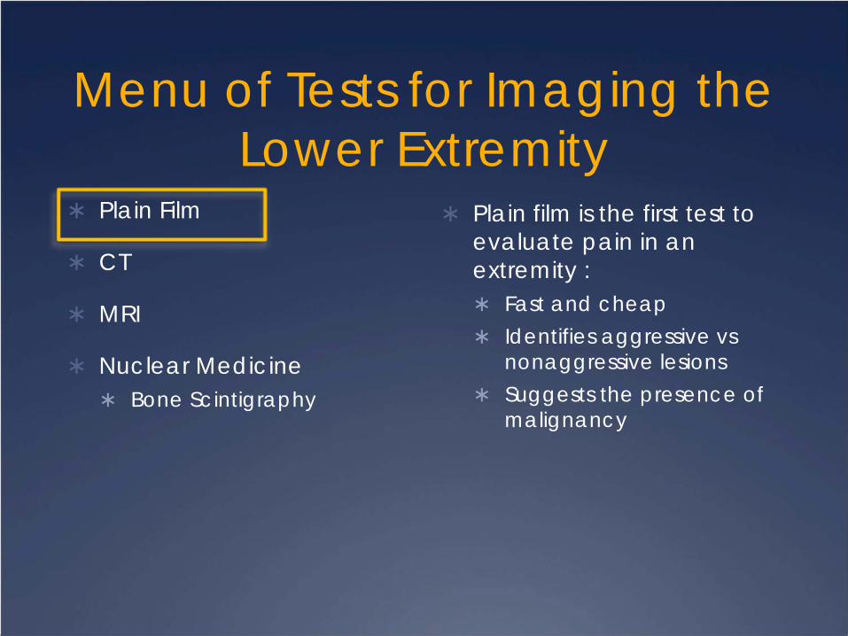
Menu of Tests for Imaging the Lower Extremity
Plain Film
CT
MRI
Nuclear MedicineBone Scintigraphy
Plain film is the first test to evaluate pain in an extremity :
Fast and cheapIdentifies aggressive vs nonaggressive lesionsSuggests the presence of malignancy
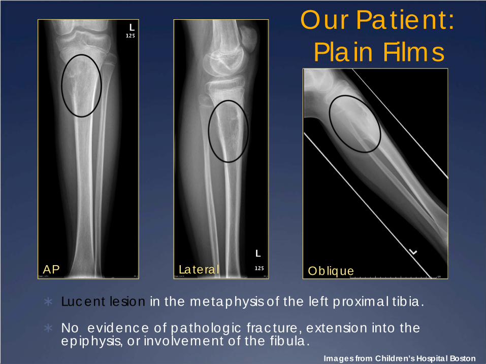
Our Patient: Plain Films
Lucent lesion in the metaphysis of the left proximal tibia.
No evidence of pathologic fracture, extension into the epiphysis, or involvement of the fibula.
Images from Children’s Hospital Boston
ObliqueAP Lateral

Benign MalignantOsteosarcoma
Ewing sarcoma
Leukemia
Metastastatic disease (rare)
Neuroblastoma
Retinoblastoma
Rhabdomyosarcoma
Hodgkin lymphoma
Miller TT et al. 2008.
Lesions of Bone under 20 Years of Age
Presenter
Presentation Notes
Specific to certain age groups Immature vs. mature bones Occur in characteristic locations Long bones vs. flat bones Intramedullary vs cortical Epiphysis vs. metaphysis vs. diaphysis

Strategy for Evaluating Bone Lesions on Plain Film
We will narrow our differential by examining particular characteristics of the lesion on plain film.
These characteristics include:Location,Margins,Trabecular Pattern,Periosteal Reaction, andSoft tissue component

BenignSimple unicameral bone cysts
Aneurysmal bone cysts
Localized langerhans cell histiocytosis
Chondromyxoid fibroma
Fibrous dysplasia
Enchondroma
MalignantOsteosarcoma
Chondrosarcoma
Hodgkin Lymphoma
Metastastatic disease
Miller, TT. et al.
Lesions Common to the Metaphysis

Evaluation of Bone Lesions on Plain Film: Margins and Zone of Transition
“Geographic 1c”Ill-defined marginsWide zone of transition
Aggressive lesion
Images from Children’s Hospital Boston
Lateral
Presenter
Presentation Notes
Geographic=focal lesion

Evaluation of Bone Lesions on Plain Film: Trabecular Pattern
AP
Image from Children’s Hospital Boston
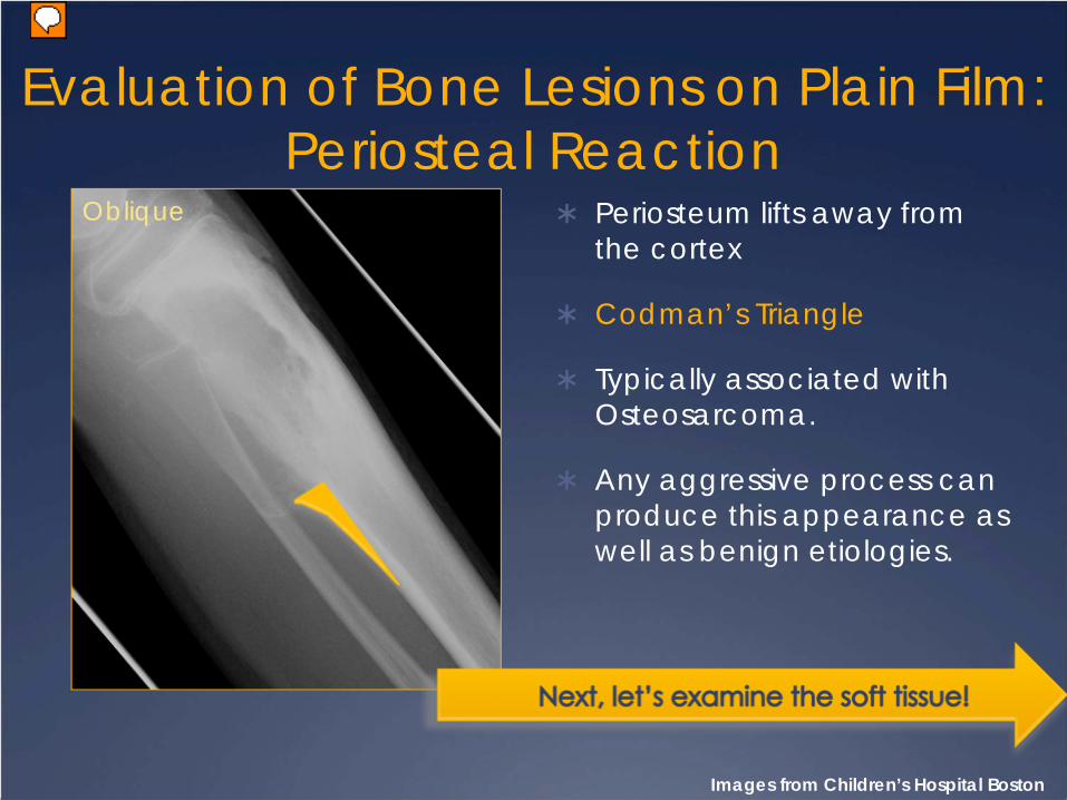
Evaluation of Bone Lesions on Plain Film: Periosteal Reaction
Oblique
Images from Children’s Hospital Boston
Periosteum lifts away from the cortex
Codman’s Triangle
Typically associated with Osteosarcoma.
Any aggressive process can produce this appearance as well as benign etiologies.
Presenter
Presentation Notes
Pathologic fracture of a cyst Infection Subdural hematoma

Evaluation of Bone Lesions on Plain Film: Soft Tissue Component
Soft tissue mass displaces adjacent fat
Highly suggestive of malignancy
Differential diagnosis of bone lesion with mass:
OsteosarcomaEwing sarcomaLymphoma
AP
Image from Children’s Hospital Boston Miller, TT et al. 2008.

OSTEOSARCOMA !
Plain Film Features of Osteosarcoma
Most common primary bone tumor of adolescence.Peak incidence ages 8-25, and 50+ secondary to Paget’s Disease.
Commonly found in the metaphyses of long bones.
Appearance can be lytic, as in our patient, sclerotic or mixed.
Trabecular pattern typically permeative with wide zone of transition. Our patient showed a geographic pattern…more about this later!
Periosteal reactions include: sunburst, hair on end, and Codman’s triangle.
Typically associated with a soft tissue mass.
Kim, HJ et al. 2010.

Diagnosis of OsteosarcomaPlain films are the first step in the radiologic evaluation of lower extremity pain in adolescents.
Obtaining a radiograph at initial presentation decreases the delay to pathologic diagnosis by 11 weeks.
Osteosarcoma can be accurately diagnosed by clinical presentation and radiographs in 66% of patients.
Tissue biopsy is considered the definite diagnosis of OS however, OS resembles some tumors histologically and could be misdiagnosed if radiographic features are not appreciated.
Miller, TT et al. 2001.
Presenter
Presentation Notes
Take home message…get the plain film! e.g. osteoblastoma and osteoid osteoma

Imaging of Bone Tumors: MRI

Imaging of Bone Tumors: CT vs MRI
MRI and CT are equally accurate for local staging of malignant bone and soft tissue tumors.
No significant difference between CT and MRI for determining local tumor extent.
MRI has superior sensitivity to soft tissue contrast.
MRI is multi-planar and can image the involved bone in the long axis and assess the joint adjacent to the tumor.
MRI avoids radiation exposure.
Meyer , JS et al. 2008
Presenter
Presentation Notes
A multi- institutional study of 387 patients that included both children and adults found that magnetic resonance imaging (MRI) and computed tomography (CT) were equally accurate for local staging of malignant bone and soft tissue tumors. Furthermore, this study found that there was no statistically significant difference between CT and MRI for determining local tumor extent. While we recognize that CT provides adequate information, we require MRI of the primary tumor due to its sensitivity to soft tissue contrast and multiplanar capabilities, which provide direct imaging of the involved bone in the long axis and direct assessment of the joint adjacent to the tumor. Furthermore, MRI avoids the radiation that is used when performing at CT.
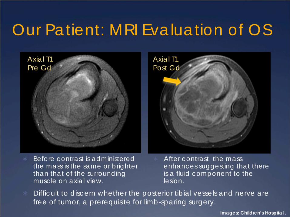
Our Patient: MRI Evaluation of OS
Before contrast is administered the mass is the same or brighter than that of the surrounding muscle on axial view.
After contrast, the mass enhances suggesting that there is a fluid component to the lesion.
Axial T1 Pre Gd
Axial T1 Post Gd
Images: Children’s Hospital .
Difficult to discern whether the posterior tibial vessels and nerve are free of tumor, a prerequisite for limb-sparing surgery.

Peripheral Enhancement on T1Peripheral enhancement of the lesion on sagittalview.
Marked cortical destruction and invasioninto soft tissue.
Epiphysis is apparently free of disease.
There do not appear to be any “skip lesions” distally.
Sagittal T1 Post
Image: Children’s Hospital Boston.

Enhancement on STIR
Bright signal suggests presence of abnormal tissue within the lesion.
Shows invasion into the muscle.
No apparent involvement of the epiphysis.
STIR doesn’t differentiate well between tumor and edema and can overestimate the extent of the tumor.
Sagittal MRI STIR
Image: Children’s Hospital Boston.

Axial T2 FS
Cystic Components of Lesion
Image: Children’s Hospital Boston

Fluid-Fluid Levels
Cystic spaces with fluid-fluid levels, likely hemorrhage and necrotic debris.
Differential diagnosis of this appearance:
TelangiectaticOsteosarcomaAneurysmal Bone Cyst
Image: Children’s Hospital Boston.
Axial T2 FS

Telangiectatic Osteosarcoma vs Aneurysmal Bone Cyst
Telangiectatic Osteosarcoma
Geographic pattern with wide margins.
Periosteal reaction
Associated soft tissue mass.
Thick ring of enhancement surrounding hemorrhagic and necrotic cystic spaces that corresponds to sarcomatoustissue.
Matrix mineralization is present, reflecting underlying osteoidproducing tumor.
Aneurysmal Bone CystWell-defined encapsulated lesion with narrow margins.
Intact periosteum.
No soft tissue component.
Thin peripheral ring reflects the limited thickness of reactive tissue surrounding the cystic spaces.
No mineralization present.
Murphy MD et al. 2003.

Classification of Osteosarcoma
SurfaceHigh-grade surfaceParostealPeriosteal
IntramedullaryConventional(75%)
OsteoblasticFibroblasticChondroblastic
Telangiectatic (4.5-11%)Small CellMultifocalMalignant fibrous histiocytoma-like
Murphy MD et al. 2007.
Presenter
Presentation Notes
High grade surface=Conventional OS that develops on the surface Parosteal OS=Surface lesion composed of low-grade fibroblastic cells Periosteal OS=Surface lesion composed of moderate-grade chondroblastic cells High-grade/conventional Osteoblastic=osteoid in a fine lacelike pattern around tumors Fibroblastic=focal osteoid production Chondroblastic=cartilagenous matrix Telangiectatic=cystic Round cell=Small blue-staining cells on H&E Multifocal=arise many places at once, poor prognosis Malignant fibrous=no osteoid

Our Patient: Diagnosis Confirmed with Fluoroscopy Guided Bx
Biopsy confirmed that this patient had TelangiectaticOsteosarcoma as predicted by plain film and MRI findings.
Image: Children’s Hospital Boston.
Flouroscopy

Imaging of Bone Tumors

Our Patient: Detection of Osseous Metastases
Heterogeneous uptake of radionuclide in left proximal tibia.
Central area of photopenia “donut sign” characteristic of Telangiectatic Osteosarcoma.
Areas of increased uptake in the left calcaneus and distal femur.
Image: Children’s Hospital Boston. Murphy, MD et al. 2003.
Tc 99-m Skeletal Scintigraphy
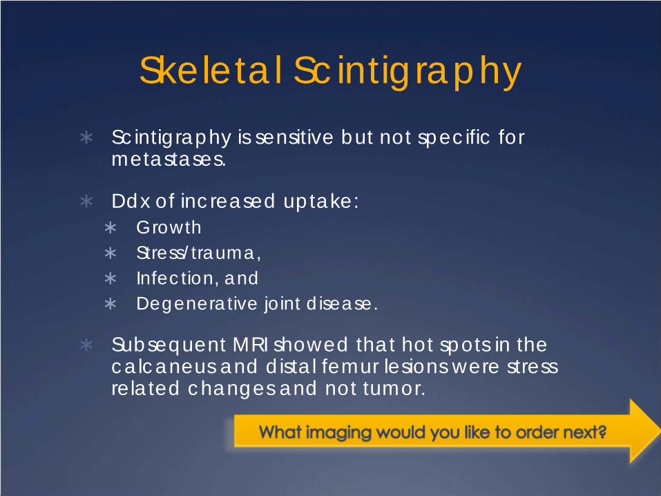
Skeletal ScintigraphyScintigraphy is sensitive but not specific for metastases.
Ddx of increased uptake: GrowthStress/trauma, Infection, andDegenerative joint disease.
Subsequent MRI showed that hot spots in the calcaneus and distal femur lesions were stress related changes and not tumor.

Imaging of Bone Tumors

Out Patient: Detection of Lung Metastases
Eighty percent of osteosarcomas metastasize to the lung.
Chest CT has 75% sensitivity and 100% specificity for the evaluation of metastases in osteosarcoma.
Four calcified lung nodules were identified on our patient’s chest CT.
Images: Children’s Hospital Boston Franzius C et al. 2001.
Axial Chest CT C+ Axial Chest CT C+

Our Patient: Plain Film Evaluation of Hardware
Our patient underwent two cycles of Ifosfamide and Etoposidefollowed by a successful limb-sparing surgery.
Images: Children’s Hospital Boston.
AP AP Oblique
Presenter
Presentation Notes
Allograft=dead donor bone

Companion Patient #1Our first companion patient is a 28 year old woman who presents with two months of pain in her right thigh and knee.
She cannot bear weight on her right leg.
On exam she has fullness, effusion, and decreased range of motion in her right knee joint.
A soft mass is palpated posteriorly.

Companion Patient #1: Plain Films
Ill-defined sclerotic lesion involving the distal femur without evidence of cortical breakthrough, periosteal reaction, or pathologic fracture.
Associated soft tissue mass.
AP Lateral Oblique
Images: BIDMC PACS

Companion Patient #1: Plain Film Two Months Later
Soft tissue mass displaces fat posteriorly and anteriorly.
Codman’s triangle.
Osteoid deposition in soft tissues.
No pathologic fracture.
No new focal lytic or sclerotic lesions.
LateralImages: BIDMC PACS
Presenter
Presentation Notes
From the plain film you can’t really see if there is involvement of the joint space.

Companion Patient #1: Joint Involvement on MRI
Low-signal mass involving the distal femur.
Soft tissue component.
Extension into knee joint.
Tibia does not appear to be involved.
Images: BIDMC PACS
Coronal MRI 3D Reconstruction
*

Companion Patient #1: MRI Evaluation of Neurovascular Bundle
Tumor invades into surrounding muslce.
Does not appear to invade the poplitealvessels or sciatic nervewhich is a prerequisite for limb-sparing surgery.
Sagittal view showed no evidence of skip metastases.
Axial T1 MRI C-
*
Images: BIDMC PACS

Companion Patient #1: Evaluation of
Hardware
Patient underwent a successful limb-sparing surgery.
Images: BIDMC PACS
AP

Companion Patient #2
Companion patient #2 is a 25 year-old male from Costa Rica who presents with two months of leg pain.
He reports decreased appetite, 20 lb weight loss, and night sweats.
He walks with an antalgic gate.
He has a large palpable mass around the distal femur.
Small effusion of the knee joint.

Companion Patient #2: Plain Films
Scerotic, permeative lesion of the right mid and distal femur.
Large soft tissue massdisplacing posterior fat.
Periosteal reaction.
No evidence of pathologic fractures.
Lateral
Images: BIDMC PACS

Companion Patient #2: Plain Film 3 Months Later
Sclerotic, permeativelesion of the mid and distal femur extending to the articular surface of the knee joint.
Cortical destruction.
Hair-on-end periostealreaction.
APImages: BIDMC PACS

Companion Patient #2: Lung Nodules on CT Chest
Patient has numerous calcified nodules on CT.
Two of the largest nodules are identified here.
Our differential diagnosis includes calcified granulomatousdisease. Biopsy is required to confirm the presence of OS metastases.
Images: BIDMC PACS
Axial Chest CT C- Axial Chest CT C-

Companion Patient #2: Chest Radiograph
While admitted for treatment with high dose methotrexate our patient developed pleuritic chest pain on the right side.
Pneumothorax occurs in 5% of people with lung metastases who undergo chemotherapy.
May be caused by fistula between parenchyma and pleura that develops as nodules necrose.
Upright AP
Images: BIDMC PACSRastogi, R et al. 2008.
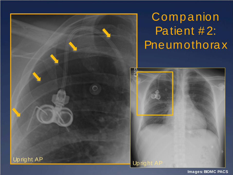
Companion Patient #2:
Pneumothorax
Upright APImages: BIDMC PACS
Upright AP

Compation Patient #2: Follow- Up
Companion Patient #2 was treated with chemotherapy and underwent a successful limb-sparing surgery.
His prognosis is guarded given his extensive lung metastases.

SummaryRadiologic evaluation of leg pain begins with plain film.
Age of patient, location, margins, trabecular pattern, periosteal reaction, and the presence of a soft tissue mass can help narrow the differential diagnosis of a bone lesion.
OS has several subtypes, such as Telangiectic, with different radiologic and histologic appearances.
Definitive diagnosis of suspected OS requires fluoroscopy-guided bone biopsy, however radiography is essential because some bone tumors appear similar histologically.
Work-up for OS includes MRI; skeletal scintigraphy; and chest CT.
Complications of OS included invasion into joint space and pneumothoraces.

References1. Franzius, C et al. FDG-PET for detection of pulmonary metastases from malignant primary bone
tumors: comparison with spiral CT. Annals of Oncology 2001; 12:479–486.
2. Kim, HJ et al. Pediatric osteosarcomas. Current opinion in pediatrics. 2010; 22:00-00.
3. Meyer , JS et al. Imaging Guidelines for Children With Ewing Sarcoma and Osteosarcoma. Pediatr Blood Cancer 2008;51:163–170.
4. Miller, SL. Malignant and benign bone tumors. Rad Clinics of North America. 2001; 39(4).
5. Miller, TT. Bone Tumors and Tumorlike Conditions: analysis with Conventional Radiography. Radiology 2008; 246: 662-674.
6. Murphy, MD et al. The Many Faces of Osteosarcoma. Radiographics. 2007; 17(5):1205-1231.
7. Murphy MD, et al. Telangiectatic Osteosarcoma: Radiologic-Pathologic Comparison. Radiology. 2003; 229:545-553.
8. Rastogi, R et al. Unusual thoracic CT manifestations of osteosarcoma: review of 16 cases. Pediatr Radiol. 2008 May;38(5):551-8.
9. Tse, SML and Laxer, RM. Approach to Acute Limb Pain in Childhood. Pediatrics in Review. 2006; 27: 170-180.

Acknowledgements
Dr. Gillian Lieberman
Dr. Jean-Marc Gauguet
Dr. James Knutson
Maria Levantakis