Radiographic Risk Factors for Interprosthetic Femur Fractures fileinterprosthetic fractures of the...
17
Radiographic Risk Factors for Interprosthetic Femur Fractures Jason S Lipof, MD Ari D Amitai, MD John P Ketz, MD Kyle T Judd, MD Gillian L Soles, MD Catherine A Humphrey, MD John T Gorczyca, MD
Transcript of Radiographic Risk Factors for Interprosthetic Femur Fractures fileinterprosthetic fractures of the...

Radiographic Risk Factors for Interprosthetic Femur Fractures
Jason S Lipof, MD Ari D Amitai, MD John P Ketz, MD Kyle T Judd, MD
Gillian L Soles, MD Catherine A Humphrey, MD
John T Gorczyca, MD

The authors of this presentation have no financial conflicts or
disclosures
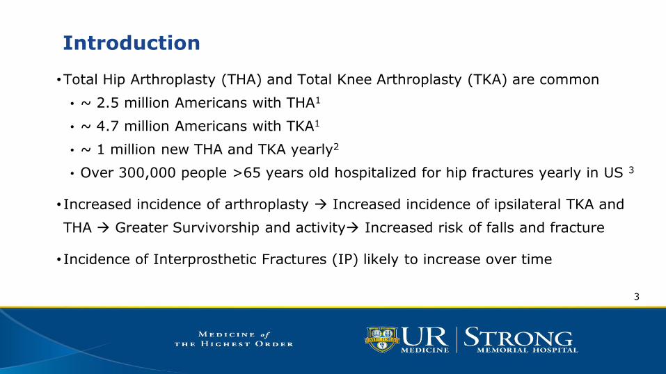
Introduction
•Total Hip Arthroplasty (THA) and Total Knee Arthroplasty (TKA) are common
• ~ 2.5 million Americans with THA1
• ~ 4.7 million Americans with TKA1
• ~ 1 million new THA and TKA yearly2
• Over 300,000 people >65 years old hospitalized for hip fractures yearly in US 3
• Increased incidence of arthroplasty Increased incidence of ipsilateral TKA and
THA Greater Survivorship and activity Increased risk of falls and fracture
• Incidence of Interprosthetic Fractures (IP) likely to increase over time
3

Incidence of Periprosthetic Fractures6,7,8
THA 0.1%-6%
TKA 0.3%-5.5%
4
IP Femur
Fx
?

Introduction
•Limited clinical data evaluating potential risks for fracture
• Interprosthetic distance, Cortical Width, Medullary
Diameter
•Bone strength and cortical width may be more predictive
than interprosthetic distance4,5
5

Hypothesis
A difference in cortical width and medullary diameter will exist
between two groups of patients; those sustaining interprosthetic
femur fractures, and those with ipsilateral implants and intact
femurs.
6

Materials and Methods
•CPT codes to identify patients undergoing treatment of periprosthetic femur fx
•CPT codes used to find cohort having undergone ipsilateral THA/TKA
•Chart review for age, sex, diagnosis of osteoporosis
• Intact femoral isthmus identified
• Medullary Diameter (MD) & Cortical Width (CW) measured using PACS
• Independent Sample T-test performed
• Mean MD, CW, and ratio of MD/CW
•P<0.05
7

Materials and Methods
8
Cortical width measured at isthmus by summing the thickness of medial and lateral cortices.
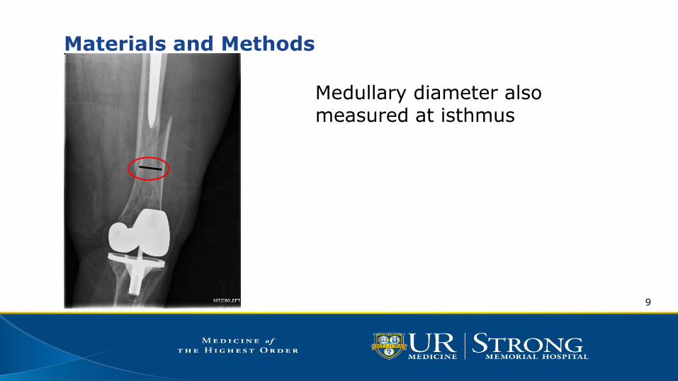
Materials and Methods
9
Medullary diameter also measured at isthmus

Results
Interprosthetic Fractures (n=23)
Intact Cohort (n=25)
Age 82 72
Sex Female 18 Male 5
Female 17 Male 8
Cemented Implant 6 (26%) 1 (4%)
Dx of Osteoporosis 7 (30%) 6 (24%)
10

Results Interprosthetic
Fractures (n=23)
Intact Cohort (n=25)
Mean SD Mean SD P-value 95% of
difference
Cortical Width (mm)
12.26 2.55 16.74 3.9 <0.0001 (-6.42, -2.55)
Medullary Diameter (mm)
21.3 4.15 14.81 3.19 <0.0001 (4.34, 8.62)
MD:CW 1.86 0.76 0.96 0.41 <0.0001
(0.55, 1.25)
11

Results
Intraclass Correlation Coefficient
CW Intraclass
Correlationb
95% Confidence Interval MD
Intraclass Correlation
95% Confidence Interval
Lower Bound Upper Bound Lower Bound Upper Bound
Single Measures .669 0.362 0.845 .871 0.721 0.943
Average Measures .801 0.532 0.916 .931 0.838 0.971
12

Conclusion
• Cortical Width and Medullary Diameter may be predictive of increased risk for
interprosthetic fractures of the femur
• Radiographic measures may help identify at risk patients for inclusion in
fracture prevention programs and treatment of osteoporosis
• Age-dependent risk – increased survivorship, aging population
• Few patients in either group had been given a formal diagnosis of osteoporosis
• May indicate need for increased surveillance
13

Limitations
• Radiographs not calibrated
• Unable to measure interprosthetic distance
• Retrospective review
14
References
1Steiner C, Andrews R, Barrett M, Weiss A. HCUP ProjectionsMobility/Orthopedic Procedures 2003 to 2012. 2012. HCUP Projections Report # 2012-03.
2012 Sep 20. U.S. Agency for Healthcare Research and Quali: ty.
2Prevalence of Total Hip and Knee Replacement in the United States Hilal Maradit Kremers, Dirk R. Larson, Cynthia S. Crowson, Walter K. Kremers,
Raynard E. Washington, Claudia A. Steiner, William A. Jiranek, Daniel J. Berry J Bone Joint Surg Am Sep 2015, 97 (17) 1386-1397
3HCUPnet. Healthcare Cost and Utilization Project (HCUP). 2012. Agency for Healthcare Research and Quality, Rockville, MD. http://hcupnet.ahrq.gov.
Accessed 5 August 2016
4Lehmann, Wolfgang, et al. "Biomechanical evaluation of peri-and interprosthetic fractures of the femur." Journal of Trauma and Acute Care Surgery
68.6 (2010): 1459-1463.
5Iesaka, Kummer, et al. Stress risers between two ipsilateral intramedullary stems: a finite element and biomechanical analysis. Journal of Arthroplasty. 2005; 20(3):386-391.
6Berry DJ. Epidemiology of periprosthetic fractures after major joint replacement: hip and knee. Orthop Clin North Am. 1999;30:183–190
7Rorabeck CH, Taylor JW. Periprosthetic fractures of the knee complicating total knee arthroplasty. Orthop Clin North Am. 1999;30:265–277.
8Kenny P, Rice J, Quinlan W. Interprosthetic fracture of the femoral shaft. J Arthroplasty. 1998;13:361–364
15

Thank You
16

17