RADIATION SAFETY MANUAL - Tulane University
113
RADIATION SAFETY MANUAL TULANE UNIVERSITY HEALTH SCIENCES CENTER 1430 TULANE AVENUE NEW ORLEANS, LA 70112 REVIEWED AND APPROVED 13 September 2007 LOUISIANA LICENSE NO.: LA-0004-L01 _______________________________ Chairman, Radiation Safety Committee _________________________________ Radiation Safety Officer
Transcript of RADIATION SAFETY MANUAL - Tulane University

RADIATION SAFETY MANUAL
TULANE UNIVERSITY HEALTH SCIENCES CENTER1430 TULANE AVENUE
NEW ORLEANS, LA 70112
REVIEWED AND APPROVED
13 September 2007
LOUISIANA LICENSE NO.: LA-0004-L01
_______________________________Chairman, Radiation Safety Committee
_________________________________Radiation Safety Officer

TABLE OF CONTENTS
I. Objectives of Radiation Protection
A. General. . . . . . . . . . . . . . . . . . . . . . . . . . . . . . . . . . . . . . . . . . . . . . . . . . . . . . . . . . . . . . . . . . . . . . . . . . . . 7
B. ALARA. . . . . . . . . . . . . . . . . . . . . . . . . . . . . . . . . . . . . . . . . . . . . . . . . . . . . . . . . . . . . . . . . . . . . . . . . . . . 7
C. ALARA Review Form. . . . . . . . . . . . . . . . . . . . . . . . . . . . . . . . . . . . . . . . . . . . . . . . . . . . . . . . . . . . . . . . 9
II. Radiation Safety Committee. . . . . . . . . . . . . . . . . . . . . . . . . . . . . . . . . . . . . . . . . . . . . . . . . . . . . . . . . . . . . . . 10
III. Radiation Safety Officer. . . . . . . . . . . . . . . . . . . . . . . . . . . . . . . . . . . . . . . . . . . . . . . . . . . . . . . . . . . . . . . . . . 11
IV. Guidelines for Nuclear Medicine Activities Involving Technologists & Other Paramedical Personnel. . . . . 12
V. Nuclear Medicine Routine for Ordering, Receiving, Opening Packages Containing Radioactive Material;Procedure for Documenting Use of Material. . . . . . . . . . . . . . . . . . . . . . . . . . . . . . . . . . . . . . . . . . . . . . . . . . 13
VI. Instructions for Administration of Radiopharmaceuticals for Diagnostic and Therapeutic Procedures.. . . . . 16
VII. Laboratory Rules for Use of Radioactive Material. . . . . . . . . . . . . . . . . . . . . . . . . . . . . . . . . . . . . . . . . . . . . . 17
VIII. Restriction and Labeling of Radiation Areas. . . . . . . . . . . . . . . . . . . . . . . . . . . . . . . . . . . . . . . . . . . . . . . . . . 19
IX. Personnel Monitoring Policy
A. Requirements for Monitoring Individuals.. . . . . . . . . . . . . . . . . . . . . . . . . . . . . . . . . . . . . . . . . . . . . . . . . 20
B. Location of Personnel Monitoring Device. . . . . . . . . . . . . . . . . . . . . . . . . . . . . . . . . . . . . . . . . . . . . . . . . 20
C. Ring Badges. . . . . . . . . . . . . . . . . . . . . . . . . . . . . . . . . . . . . . . . . . . . . . . . . . . . . . . . . . . . . . . . . . . . . . . . 20
D. Exchange. . . . . . . . . . . . . . . . . . . . . . . . . . . . . . . . . . . . . . . . . . . . . . . . . . . . . . . . . . . . . . . . . . . . . . . . . . . 20
E. Issue of Personnel Monitoring Devices; Maintenance of Records.. . . . . . . . . . . . . . . . . . . . . . . . . . . . . . 20
F. Thyroid Monitoring.. . . . . . . . . . . . . . . . . . . . . . . . . . . . . . . . . . . . . . . . . . . . . . . . . . . . . . . . . . . . . . . . . . 20
X. Limits for Exposure to Ionizing Radiation. . . . . . . . . . . . . . . . . . . . . . . . . . . . . . . . . . . . . . . . . . . . . . . . . . . . 22
XI. Radiation and Contamination Area Surveys, Nuclear Medicine.. . . . . . . . . . . . . . . . . . . . . . . . . . . . . . . . . . . 24
XII. Decontamination Procedures . . . . . . . . . . . . . . . . . . . . . . . . . . . . . . . . . . . . . . . . . . . . . . . . . . . . . . . . . . . . . . 25
XIII. Contaminated Equipment. . . . . . . . . . . . . . . . . . . . . . . . . . . . . . . . . . . . . . . . . . . . . . . . . . . . . . . . . . . . . . . . . 26
XIV. Emergency Procedures
A. Whom to Call. . . . . . . . . . . . . . . . . . . . . . . . . . . . . . . . . . . . . . . . . . . . . . . . . . . . . . . . . . . . . . . . . . . . . . . 27
B. Loss of Source.. . . . . . . . . . . . . . . . . . . . . . . . . . . . . . . . . . . . . . . . . . . . . . . . . . . . . . . . . . . . . . . . . . . . . . 27
C. Storage in Anticipation of Natural Catastrophy. . . . . . . . . . . . . . . . . . . . . . . . . . . . . . . . . . . . . . . . . . . . . 28

D. Minor Spills. . . . . . . . . . . . . . . . . . . . . . . . . . . . . . . . . . . . . . . . . . . . . . . . . . . . . . . . . . . . . . . . . . . . . . . . 28
E. Major Spills.. . . . . . . . . . . . . . . . . . . . . . . . . . . . . . . . . . . . . . . . . . . . . . . . . . . . . . . . . . . . . . . . . . . . . . . . 28
F. Accident Involving Radioactive Dusts, Mists, Fumes, Organic Vapors and Gases. . . . . . . . . . . . . . . . . . 29
G. Injuries to Personnel Involving Radiation Hazard. . . . . . . . . . . . . . . . . . . . . . . . . . . . . . . . . . . . . . . . . . . 29
H. Fires . . . . . . . . . . . . . . . . . . . . . . . . . . . . . . . . . . . . . . . . . . . . . . . . . . . . . . . . . . . . . . . . . . . . . . . . . . . . 29
XV. Instructions for Maintenance.. . . . . . . . . . . . . . . . . . . . . . . . . . . . . . . . . . . . . . . . . . . . . . . . . . . . . . . . . . . . . . 30
XVI. Instructions for Housekeeping.. . . . . . . . . . . . . . . . . . . . . . . . . . . . . . . . . . . . . . . . . . . . . . . . . . . . . . . . . . . . . 31
XVII. Escort Personnel. . . . . . . . . . . . . . . . . . . . . . . . . . . . . . . . . . . . . . . . . . . . . . . . . . . . . . . . . . . . . . . . . . . . . . . . 32
XVIII. Instructions for Visitors.. . . . . . . . . . . . . . . . . . . . . . . . . . . . . . . . . . . . . . . . . . . . . . . . . . . . . . . . . . . . . . . . . . 33
XIX. Storage of Radionuclides
A. Liquids and Solids.. . . . . . . . . . . . . . . . . . . . . . . . . . . . . . . . . . . . . . . . . . . . . . . . . . . . . . . . . . . . . . . . . . . 34
B. Gases . . . . . . . . . . . . . . . . . . . . . . . . . . . . . . . . . . . . . . . . . . . . . . . . . . . . . . . . . . . . . . . . . . . . . . . . . . . . 34
XX. Radionuclide Disposal
A. General Guidance. . . . . . . . . . . . . . . . . . . . . . . . . . . . . . . . . . . . . . . . . . . . . . . . . . . . . . . . . . . . . . . . . . . . 35
B. Procedure for Disposal of Liquid and Gases. . . . . . . . . . . . . . . . . . . . . . . . . . . . . . . . . . . . . . . . . . . . . . . 35
C. Procedure for Disposal by Decaying-In-Storage. . . . . . . . . . . . . . . . . . . . . . . . . . . . . . . . . . . . . . . . . . . . 36
D. Procedure for Transfer for Burial. . . . . . . . . . . . . . . . . . . . . . . . . . . . . . . . . . . . . . . . . . . . . . . . . . . . . . . . 36
E. Procedure for Returning Generators to the Manufacturer. . . . . . . . . . . . . . . . . . . . . . . . . . . . . . . . . . . . . 37
F. Transfer to Unit Dose Pharmacy or Commercial Disposal Agency. . . . . . . . . . . . . . . . . . . . . . . . . . . . . . 37
G. Specific Wastes. . . . . . . . . . . . . . . . . . . . . . . . . . . . . . . . . . . . . . . . . . . . . . . . . . . . . . . . . . . . . . . . . . . . . . 37
H. Infectious, Highly Toxic, Hazardous Substances. . . . . . . . . . . . . . . . . . . . . . . . . . . . . . . . . . . . . . . . . . . . 38
I. Incineration. . . . . . . . . . . . . . . . . . . . . . . . . . . . . . . . . . . . . . . . . . . . . . . . . . . . . . . . . . . . . . . . . . . . . . . . . 38
XXI. Safety Rules: Fixed Radiographic. . . . . . . . . . . . . . . . . . . . . . . . . . . . . . . . . . . . . . . . . . . . . . . . . . . . . . . . . . . 39
XXII. Safety Rules: Fixed Fluoroscopic. . . . . . . . . . . . . . . . . . . . . . . . . . . . . . . . . . . . . . . . . . . . . . . . . . . . . . . . . . . 40
XXIII. Safety Rules: Operators of Mobile Radiographic Equipment. . . . . . . . . . . . . . . . . . . . . . . . . . . . . . . . . . . . . . 41
XXIV. Safety Rules: Operators of Mobile Fluoroscopic Equipment. . . . . . . . . . . . . . . . . . . . . . . . . . . . . . . . . . . . . . 42

XXV. Safety Rules: Persons in the Vicinity of Mobile X-Ray Equipment. . . . . . . . . . . . . . . . . . . . . . . . . . . . . . . . . 43
XXVI. Specific Instructions for Allied Medical Workers. . . . . . . . . . . . . . . . . . . . . . . . . . . . . . . . . . . . . . . . . . . . . . 44
XXVII. Radiation and Pregnancy, Radiation and Allied Medical Staff.. . . . . . . . . . . . . . . . . . . . . . . . . . . . . . . . . . . . 45
XXVIII. Diagnostic Radiology and the Pregnant Patient. . . . . . . . . . . . . . . . . . . . . . . . . . . . . . . . . . . . . . . . . . . . . . . . 46
XXIX. Patient Pregnancy Screening. . . . . . . . . . . . . . . . . . . . . . . . . . . . . . . . . . . . . . . . . . . . . . . . . . . . . . . . . . . . . . . 47
XXX. Nuclear Medicine and the Pregnant Patient. . . . . . . . . . . . . . . . . . . . . . . . . . . . . . . . . . . . . . . . . . . . . . . . . . . 48
XXXI. Quality Control Procedures
A. Calibration of Dose Calibrator. . . . . . . . . . . . . . . . . . . . . . . . . . . . . . . . . . . . . . . . . . . . . . . . . . . . . . . . . . 49
B. Quality Control, Scintillation Camera. . . . . . . . . . . . . . . . . . . . . . . . . . . . . . . . . . . . . . . . . . . . . . . . . . . . 51
C. Quality Control of Radiopharmaceuticals. . . . . . . . . . . . . . . . . . . . . . . . . . . . . . . . . . . . . . . . . . . . . . . . . 53
D. Sealed Sources; Leakage/Contamination .. . . . . . . . . . . . . . . . . . . . . . . . . . . . . . . . . . . . . . . . . . . . . . . . . 53
E. Procedures for Calibration of Survey Instruments. . . . . . . . . . . . . . . . . . . . . . . . . . . . . . . . . . . . . . . . . . . 53
XXXII. Procedures for Nursing Staff and Patient Care
A. Diagnostic Procedures.. . . . . . . . . . . . . . . . . . . . . . . . . . . . . . . . . . . . . . . . . . . . . . . . . . . . . . . . . . . . . . . . 54
B. Therapeutic Procedures.. . . . . . . . . . . . . . . . . . . . . . . . . . . . . . . . . . . . . . . . . . . . . . . . . . . . . . . . . . . . . . . 54
XXXIII. Training Program: Personnel Working in the Vicinity of Radioactive Material. . . . . . . . . . . . . . . . . . . . . . . 56
XXXIV. Procedure for Monitoring or Checking Trap Effluent.. . . . . . . . . . . . . . . . . . . . . . . . . . . . . . . . . . . . . . . . . . . 57

APPENDIX
Appendix A. Radiation Safety Committee.. . . . . . . . . . . . . . . . . . . . . . . . . . . . . . . . . . . . . . . . . . . . . . . . . . . . . . . . 58
Appendix B. Technologists Approved For Radionuclide Injections. . . . . . . . . . . . . . . . . . . . . . . . . . . . . . . . . . . . . 59
Appendix C. Daily Incoming/Outgoing Shipment Inspection Log. . . . . . . . . . . . . . . . . . . . . . . . . . . . . . . . . . . . . . 60
Appendix D. Radionuclide Distribution Log. . . . . . . . . . . . . . . . . . . . . . . . . . . . . . . . . . . . . . . . . . . . . . . . . . . . . . . 61
Appendix E. Radiation Exposure History Request Form. . . . . . . . . . . . . . . . . . . . . . . . . . . . . . . . . . . . . . . . . . . . . 62
Appendix F. Dosimeter Badge Monitoring Service Request. . . . . . . . . . . . . . . . . . . . . . . . . . . . . . . . . . . . . . . . . . 63
Appendix G. Radionuclide Disposal Form. . . . . . . . . . . . . . . . . . . . . . . . . . . . . . . . . . . . . . . . . . . . . . . . . . . . . . . . 64
Appendix H. Notice to Employees.. . . . . . . . . . . . . . . . . . . . . . . . . . . . . . . . . . . . . . . . . . . . . . . . . . . . . . . . . . . . . . 65
Appendix I. Diagnostic And/Or Therapeutic Procedure Misadministration Report. . . . . . . . . . . . . . . . . . . . . . . . 66
Appendix J. Therapeutic Radionuclide Consultation Form. . . . . . . . . . . . . . . . . . . . . . . . . . . . . . . . . . . . . . . . . . . 67
Appendix K. Quality Management Program. . . . . . . . . . . . . . . . . . . . . . . . . . . . . . . . . . . . . . . . . . . . . . . . . . . . . . . 68
Appendix L. Annual Review Form; Quality Management Program. . . . . . . . . . . . . . . . . . . . . . . . . . . . . . . . . . . . . 69
Appendix M. Quality Management Checklist for Administration of Sodium Iodide ( I, I); Activities Greater than125 131
30 ìCi. . . . . . . . . . . . . . . . . . . . . . . . . . . . . . . . . . . . . . . . . . . . . . . . . . . . . . . . . . . . . . . . . . . . . . . . . . 70
Appendix N. Instructions for Brachytherapy. . . . . . . . . . . . . . . . . . . . . . . . . . . . . . . . . . . . . . . . . . . . . . . . . . . . . . . 71
Appendix O. Operating Room Care; Brachytherapy Sources. . . . . . . . . . . . . . . . . . . . . . . . . . . . . . . . . . . . . . . . . . 72
Appendix P. Nursing Care; Brachytherapy Sources/Radiopharmaceutical Therapy. . . . . . . . . . . . . . . . . . . . . . . . 73
Appendix Q. Radionuclide Therapy Survey. . . . . . . . . . . . . . . . . . . . . . . . . . . . . . . . . . . . . . . . . . . . . . . . . . . . . . . 75
Appendix R. Activities and Dose Rates For Authorizing Patient Release. . . . . . . . . . . . . . . . . . . . . . . . . . . . . . . . 76

Appendix S. Activities and Dose Rates Above Which Instructions Should Be Given When Authorizing PatientRelease. . . . . . . . . . . . . . . . . . . . . . . . . . . . . . . . . . . . . . . . . . . . . . . . . . . . . . . . . . . . . . . . . . . . . . . . . 77
Appendix T. Activities of Radiopharmaceuticals That Require Instructions And Records When Administered ToPatients Who Are Breast-Feeding An Infant or Child. . . . . . . . . . . . . . . . . . . . . . . . . . . . . . . . . . . . . 78
Appendix U. Home Instruction, Radiopharmaceutical Therapy. . . . . . . . . . . . . . . . . . . . . . . . . . . . . . . . . . . . . . . . 79
Appendix V. Home Instructions.. . . . . . . . . . . . . . . . . . . . . . . . . . . . . . . . . . . . . . . . . . . . . . . . . . . . . . . . . . . . . . . . 80
Appendix W. 90Sr Ophthalmic Applicator Therapy. . . . . . . . . . . . . . . . . . . . . . . . . . . . . . . . . . . . . . . . . . . . . . . . . . 81
Appendix X. Instructions For Family of Released Patient. . . . . . . . . . . . . . . . . . . . . . . . . . . . . . . . . . . . . . . . . . . . 82
Appendix Y. Report of Radioactivity of Cadaver. . . . . . . . . . . . . . . . . . . . . . . . . . . . . . . . . . . . . . . . . . . . . . . . . . . 83
Appendix Z. Instructions To Funeral Director For Embalming Body Containing Radioactive Material. . . . . . . . . 84
Appendix AA. Autopsy Or Surgery Precautions. . . . . . . . . . . . . . . . . . . . . . . . . . . . . . . . . . . . . . . . . . . . . . . . . . . . . 85
Appendix BB. Application for Authorization to Use Radionuclides/Radiation Sources for In Vivo Human Use. . . 86
Appendix CC. Application for Use of Radioactive Material/Radiation Sources.. . . . . . . . . . . . . . . . . . . . . . . . . . . . 90
Appendix DD. Management Of Victims Of Radioactive Contamination. . . . . . . . . . . . . . . . . . . . . . . . . . . . . . . . . . 94
Appendix EE. Condensed Research Laboratory Rules. . . . . . . . . . . . . . . . . . . . . . . . . . . . . . . . . . . . . . . . . . . . . . . . 109

Page 7
I. OBJECTIVE OF RADIATION PROTECTION
A. General
The specific objectives of radiation protection are: (1) to prevent, to the extent practicable, the occurrence ofsevere radiation-induced nonstochastic diseases by adhering to dose equivalent limits that are below theapparent practical threshold dose equivalent levels; and (2) to limit risk of the stochastic effects, fatal cancerand genetic effects, to a reasonable level in comparison with non-radiation risks and in relation to societalneeds, benefits gained and economic factors. These objectives are achieved by applying individual doseequivalent limits for occupational and nonoccupational (general public) exposures.
It is emphasized that for the purposes of radiation protection, a cautious assumption is made, the reliability ofwhich has not been established. This is the assumption that the dose-risk relationship is strictly proportional(linear) without threshold throughout the range of radiation protection. Furthermore, doses and theprobability of response (risk) are assumed to accumulate linearly. At higher doses, received acutely, such asin accidents, more complex (non-linear) dose-risk relationships may apply.
Under these assumptions, any selected dose equivalent limit will have an associated level of risk. TulaneUniversity Health Sciences Center endorses the following: (1) the need to justify any activity which involvesradiation exposure on the basis that the expected benefits exceed the predicted cost (justification); (2) theneed to reduce the total radiation detriment from such justifiable activities or practices to AS LOW AS ISREASONABLY ACHIEVABLE (ALARA), economic and social factors being taken into account and (3) theneed to apply individual effective dose equivalent limits to ensure that the procedures for justification andALARA do not result in individuals or groups of individuals exceeding levels of acceptable risk.
B. ALARA
Tulane University Health Sciences Center is committed to keeping exposures ALARA. The Radiation SafetyCommittee (RSC) will perform an annual review of the radiation safety program. This shall include reviewof summaries of the types and amounts of radioactive material used, occupational dose reports, andcontinuing education and training for all personnel who work with or in the vicinity of radioactive material. The purpose of the review is to ensure that individuals make every reasonable effort to maintain occupationaldoses, doses to the general public and releases of radioactive material ALARA, taking into account the stateof technology and the cost of improvements in relation to benefits. Modification to operating procedures orto equipment and facilities will be made where they will reduce exposures unless the cost is considered to beunjustified. In addition to maintaining doses to individuals as far below the limits as is reasonablyachievable, the sum of the doses received by all exposed individuals will also be maintained at the lowestpracticable level.
The RSC will delegate authority to the Radiation Safety Officer (RSO) for enforcement of the ALARAconcept.
The RSC will support the RSO in those instances where it is necessary for the RSO to assert his authority. Where the RSO has been overruled, the RSC will record the basis for its action in the minutes of the RSC’squarterly meeting.

Page 8
ALARA LEVELSInvestigational Levels (mrems per calendar quarter)
Level I Level II
Whole body; head and trunk; 125 375active blood-forming organs;lens of eye; gonads
Hands and forarms; feet and 375 1125ankles
Skin of whole body* 1250 3750
*Not normally applicable to nuclear medicine operations except thoseusing significant quantities of beta emitting nuclides.
____________________________________________
The following actions will be taken at the Investigational Levels stated in the ALARA LEVELS Table above.
1. QUARTERLY EXPOSURE OF INDIVIDUALS TO LESS THAN INVESTIGATIONAL LEVEL I.
Except when deemed appropriate by the RSO, no further action will be taken in those cases where an individual'sexposure is less than values for Investigational Level I.
2. PERSONNEL EXPOSURE EQUAL TO OR GREATER THAN INVESTIGATIONAL LEVEL I, BUT LESSTHAN INVESTIGATIONAL LEVEL II.
The RSO will review the exposure of each individual whose quarterly exposures equal or exceed InvestigationalLevel I. He will report the results of his review at the first RSC meeting following the quarter when the exposurewas recorded. If the exposure does not equal or exceed Investigational Level II, no action related specifically tothe exposure is required unless deemed appropriate by the RSC. The RSC will, however, consider each exposurein comparison with those of others performing similar tasks as an index of ALARA program quality and willrecord the review in the RSC minutes. No written notification of the exposure will be forwarded to theindividual.
3. PERSONNEL EXPOSURE GREATER THAN INVESTIGATIONAL LEVEL II.
The RSO will review techniques/procedures and make recommendations for reducing exposure. The RSC willreview the recommendations and indicate appropriate follow-up. A written notification of the exposure will beforwarded to the individual.

Page 9
C. ALARA REVIEW FORM
Name:_____________________________________________________ SS No.: _________________________
Date:___________________________
This individual has exceeded the doses listed below.
ALARA Dose in mRem (mSv)/quarter
Level I Level II Dose ReceivedWhole body (Including Gonads, Lensof Eyes, Red Bone Marrow) 125 (1.25) 375 (3.75) _____________
Hands, Feet 375 (3.75) 1125 (11.25) _____________
Skin of Whole Body 1250 (12.5) 3750 (37.5) _____________
Reasons for excessive exposure: _______________________________________________________________________
_________________________________________________________________________________________________
Protective Considerations: Yes No N/A
1. Can time in the work area be reduced? ___ ___ ___
2. Will special dosimetry or area monitoring be required? ___ ___ ___
3. Can special tools/equipment be employed? ___ ___ ___
4. Can additional shielding/distance be employed? ___ ___ ___
Corrective action(s) taken:____________________________________________________________________________
_________________________________________________________________________________________________
____________________ ________________________________Date Radiation Safety Officer
________________________________Acknowledgment
THIS REPORT IS FURNISHED TO YOU UNDER THE PROVISIONS OF THE LOUISIANA RADIATIONPROTECTION REGULATIONS. YOU SHOULD RETAIN THIS REPORT FOR FURTHER REFERENCE.

Page 10
II. RADIATION SAFETY COMMITTEE (RSC)
The control of radionuclides and radiation safety at Tulane University Health Sciences Center is theresponsibility of the RSC. Refer to Appendix A for a listing of the RSC members.
The RSC is responsible for ensuring that all individuals who work with or in the vicinity of sources of radiationhave sufficient training and work experience to enable them to perform their duties safely and in accordance withregulations and conditions of the license. The RSC is also responsible for ensuring that all use of sources ofradiation is conducted in a safe manner and in accordance with regulations and conditions of the license.
The RSC shall:
a. Be familiar with all pertinent regulations, the terms of the license, and information in support of the requestfor the license and its amendments.
b. Review the training and experience of any individual who uses radioactive material (including physicians,technologists, physicists, and pharmacists) and determine that the qualifications are sufficient to enable themto perform their duties safely and in accordance with regulations and conditions of the license.
c. Establish a program to ensure that all individuals whose duties may require them to work in the vicinity ofradioactive material (e.g., nursing, security and housekeeping personnel) are properly instructed.
d. Review and approve all requests for use of radioactive material within the institution.
e. Prescribe special conditions that will be required during a proposed use of radioactive material such asrequirements for bioassays, physical examinations of users and special monitoring procedures.
f. Review the entire radiation safety program at least annually to determine that all activities are beingconducted safely and in accordance with regulations and conditions of the license. The review shall includeexamination of all records, reports from the RSO, results of inspections, written safety procedures andmanagement control systems.
g. Recommend remedial action to correct any deficiencies identified in the radiation safety program.
h. Maintain written records of all committee meetings, actions, recommendations and decisions. Minutes of theRSC meetings shall include: the date of the meeting, listing of the members present, listing of the membersabsent, a summary of the deliberations, a record of the recommended actions and the numerical results of anyballots.
i. Ensure that the radioactive material license is amended when necessary, prior to any changes in facilities,equipment, policies, procedures and personnel.
j. Include representatives from Nuclear Medicine, Radiology/Radiation Oncology, Nursing, Research andManagement. The RSO shall also be a member of the RSC. A quorum of the RSC must include: the RSO, arepresentative from management and fifty percent (50%) of all RSC members.
The RSC shall meet as often as necessary to conduct its business, but not less than quarterly, or as often as theJoint Commission of Accreditation of Healthcare Organizations requires (JCAHO).

Page 11
III. RADIATION SAFETY OFFICER (RSO)
The RSO will be responsible for radiological safety. General surveillance over all activities involving radioactivematerial and determining compliance with rules and regulations, license conditions and conditions or projects asapproved by the RSC are the responsibilities of the RSO.
The RSO is responsible for providing advice regarding procurement, safe handling, monitoring, use and disposalof all radioactive sources. He will furnish in-service education on all aspects of radiation protection to personnelat all levels of responsibility.
The RSO will maintain records of personnel exposure, and will notify individuals of exposures approachingmaximum permissible amounts. An annual inventory of all radionuclides shall be maintained in order to assurethe quantity on hand has been authorized by the license.
The RSO shall be notified in case of accidents and shall be responsible for the primary considerations involved inthe prevention of spread of contamination. The RSO shall have one or more deputies.
The RSO will investigate all overexposures, accidents, losses, misadministrations or other excursions from goodradiation safety. The maintenance of a procedure file on all matters relating to the radionuclide program fromreceipt to final disposition is the responsibility of the RSO. This also includes performance checks on surveyequipment as well as in-service education. The RSO will review the radiation safety program in its entirety onceper year.
The RSO is also responsible for the accuracy and completeness of other tasks required by regulation and willverify review by his signature on key documents. This does not mean that the RSO performs tasks, but ratherthat the record has been reviewed. Documents requiring the signature of the RSO:
A. Sealed source inventory.
B. Sealed source wipe/leak test.
C. Survey of sealed source storage areas.
D. Dose calibrator linearity.
E. Dose calibrator accuracy.
F. Dose calibrator geometry.
G. The RSO will:
1. Ensure that surveys shall be conducted in unrestricted areas.
2. Maintain records of radionuclide disposal by release to sewer which shall include (a) log of sewerdisposal quantities by type of radionuclide (b) the daily effluent rate (c) monthly average concentration.
3. Maintain records of misadministrations of radionuclides and of the corrective actions taken.
4. Maintain records of semi-annual fume hood flow velocity calibrations.

Page 12
IV. GUIDELINES FOR NUCLEAR MEDICINE ACTIVITIES INVOLVING TECHNOLOGISTS AND OTHERPARAMEDICAL PERSONNEL
A. An authorized physician may permit technologists and other paramedical personnel to perform the followingactivities:
1. Preparation and quality control testing of radiopharmaceutical sources of radiation.
2. Measurements of radiopharmaceutical doses prior to administration.
3. Use of appropriate instrumentation for the collection of data to be used by the physician.
4. Administration of radiopharmaceuticals from radionuclide sources to patients, within the limits permittedunder applicable laws. Whenever a technologist or other paramedical person administers aradiopharmaceutical to a patient by injection, a physician (not necessarily the authorized user ofradionuclides) shall be immediately accessible.
B. Authorized physicians who permit activities to be performed by technologists and other paramedicalpersonnel shall:
1. Prior to such permission, determine that such technologists and other paramedical personnel have beenproperly trained to perform their duties. This training shall include training in the following subjects asapplicable to the duties assigned:
a. General characteristics of radiation and radioactive material.
b. Physical, chemical and pharmaceutical characteristics of each radiopharmaceutical to be used.
c. Mathematics and calculations basic in the use and measurement of radioactivity, including units ofquantity of radioactivity (Curies, millicuries, microcuries, Becquerels) and units of radiation doseand radiation exposure (Roentgens, Rad, Rem, Gray and Sievert).
d. Use of radiation instrumentation for measurements and monitoring, including operating procedures,calibration of instruments and limitation of instruments.
e. Principles and practices of radiation protection.
f. Additional training in the above subjects, as appropriate, when new duties are added.
2. Assure that such technologists and other paramedical personnel receive appropriate retraining in thesubjects listed to maintain proficiency and to keep abreast of developments in the field of nuclearmedicine.
3. Keep records showing the bases for such determinations of proper retraining.
4. Retain responsibility as authorized user for the satisfactory performance of such activities. Certificationin Nuclear Medicine Technology by the American Registry of Radiologic Technology (ARRT) or theNuclear Medicine Technology Certification Board (NMTCB) will satisfy the above trainingrequirements.
C. Personnel (Technologists and other paramedical) approved for radionuclide injections are listed in Appendix B.

Page 13
V. NUCLEAR MEDICINE ROUTINE FOR ORDERING, RECEIVING, OPENING PACKAGES CONTAININGRADIOACTIVE MATERIAL; PROCEDURE FOR DOCUMENTING USE OF MATERIAL
A. ORDERING
1. Nuclear Medicine Technologists of the Nuclear Medicine Department will place all orders forradioactive material and will ensure that the requested materials and quantities are authorized by theRadioactive Material License. Possession limits are not to be exceeded.
2. A written record that identifies the radionuclide, chemical form and activity level shall be maintained.
3. A written request will be obtained from the physician who ordered the procedure. If a therapeuticprocedure has been ordered, a written request will be obtained as well from the physician who willperform the procedure.
B. RECEIVING
1. During normal working hours, carriers will be instructed to deliver radioactive packages directly to theNuclear Medicine Department.
2. During off-duty hours, security personnel or other designated individuals will accept delivery ofradioactive packages in accordance with the procedures outlined in the sample memorandum, SectionV.E.
C. MONITORING
Special requirements must be followed for packages containing quantities of radioactive material in excess ofthe Type A quantity limits, as defined in Radiation Regulations (e.g. more than 20 Curies of Mo, Tc,99 99m
uncompressed Xe or more than 3 Curies (3 Ci) of Xe, I or I. The licensee shall make arrangements133 133 131 125
to receive:
1. the package when the carrier offers it for delivery, or
2. the notification of the arrival of the package at the carrier's terminal and to take possession of the packageexpeditiously.
Such packages must be monitored for external radiation levels and surface contamination within three (3)hours after receipt if received during working hours or within eighteen (18) hours if received after workinghours. The Division must be notified if removable contamination exceeds 0.01 ìCi (22,000 dpm)/100 cm .2
D. PROCEDURE FOR OPENING PACKAGES CONTAINING RADIOACTIVE MATERIAL
1. Put on impermeable disposable gloves to prevent hand contamination.
2. Visually inspect packages for any sign of damage (e.g. wetness, crushed). If damaged is noted, stopprocedure and notify RSO.
Radiation Safety Officer: Charles F. ReindlOffice: 988-5486
Deputy Radiation Safety Officer: Jay FolseOffice: 988-5486

Page 14
3. Measure the exposure rate from the package at one meter (1m) and at the package surface. If it is higherthan expected, stop and notify the RSO. (The transport index noted on packages with Yellow II orYellow III labels is the approximate dose rate, in mR/hr at one meter (1m) from the package surface). The surface dose rate for such packages should not exceed 200 mR/hr. The dose rate from packages with"White I" labels should be less than 0.5 mR/hr at the package surface.
4. Open the package with the following precautionary steps:
a. Remove the packing slip.
b. Open the outer package following the suppliers' instructions, if provided.
c. Open the inner package and verify that the contents agree with the packing slip.
d. Check the integrity of the final source container. Look for broken seals or vials, loss of liquid,condensation or discoloration of the packing material.
e. If anything is other than expected, stop and notify the RSO.
5. If there is any reason to suspect contamination, wipe the external surface of the final source container andremove the wipe sample to a low-background area. Assay the wipe sample to determine if there is anyremovable radioactivity. The licensee should specify in the procedure manual which instrument (e.g. athin-window G-M survey meter, a Sodium Iodide Thallium activated crystal and ratemeter, a liquidscintillation counter, or a proportional flow counter) should be used for these assays. The detectionefficiency must be determined to convert wipe sample counts per minute (cpm) to disintegrations perminute (dpm). Note that a dose calibrator is not sufficiently sensitive for this measurement. Takeprecautions against the potential spread of contamination.
6. Check the user request to ensure that the material received is the material that was ordered.
7. Monitor the packing material and empty packages for contamination with a survey meter prior todiscarding.
a. If contaminated, treat this material as radioactive waste.
b. If not contaminated, remove or obliterate the radiation labels before discarding as in-house trash.
8. Record receipt of radioactive material on the proper form.

Page 15
E. MEMO TO SECURITY
TO: Director of Security
FROM: Radiation Safety Officer
SUBJECT: RECEIPT OF PACKAGES CONTAINING RADIOACTIVE MATERIAL
Any packages containing radioactive material that arrive between 1630 hours and 0700 hours orduring the weekend shall be accepted by the Security guard on duty and taken immediately to theNuclear Medicine Department. Unlock the door, place the package on top of the counter and relockthe door.If the package is wet or appears to be damaged, immediately contact the Radiation Safety Officer. Ask the carrier to remain at the hospital until it can be determined that neither he/she nor the deliveryvehicle is contaminated.
RADIATION SAFETY OFFICER: Charles F. Reindl, M.S.
Office: 988-2867
Home: 837-8516
NUCLEAR MEDICINE PHYSICIAN: Harold Neitzschman, M.D.
Office: 988-7627
Home: 593-9257
NUCLEAR MEDICINE TECHNOLOGIST: Cheryl Albert, R.T.(N)
Office: 988-5715
Home: 785-8336
F. PROCEDURES FOR DOCUMENTING USE OF RADIOACTIVE MATERIAL
1. A record of receipt, use, transfer, disposal and assay of all radioactive material shall be maintained forthree (3) years.
2. See Appendix C for appropriate form.

Page 16
VI. INSTRUCTIONS FOR ADMINISTRATION OF RADIOPHARMACEUTICALS FOR DIAGNOSTIC ANDTHERAPEUTIC PROCEDURES
A. Before writing a prescription, the authorized user or physician under the supervision of an authorized userwill personally review the patient's case to establish that the medical use is indicated for the patient.
B. Before administering a radiopharmaceutical, the authorized user or the physician under the supervision of anauthorized user will personally make and date a prescription. If changes are required, they will be recordedin writing in the patient's chart or in another appropriate record, and will be dated and signed.
C. Before administering a radiopharmaceutical, the identity of the patient, the radiopharmaceutical, and thedosage will be confirmed by the person administering the radiopharmaceutical to establish agreement withthe prescription. Any dose that differs from the prescribed dose by more than ten percent (10%) shall not beadministered.
D. ASSAY OF RADIOPHARMACEUTICAL DOSAGES
1. Assay, within thirty (30) minutes before medical use, the activity of each radiopharmaceutical dosagethat contains more than 10 ìCi (370 kBq) of a photon-emitting radionuclide.
E. RECORD REQUIREMENTS
Retain a record of assays three (3) years. To satisfy this requirement, the record shall contain the following:
1. The patient’s name and identification number (if one has been assigned).
2. The generic name or trade name, radiopharmaceutical abbreviation, lot number and expiration date of theradiopharmaceutical.
3. The prescribed dosage and activity of the dosage at the time of assay, or a notation that the total activityis less than 10 ìCi (370 kBq).
4. The date and time of administration of the radiopharmaceutical.
5. The initials of the individual who performed the assay.

Page 17
VII. LABORATORY RULES FOR USE OF RADIOACTIVE MATERIAL
A. Wear laboratory coats or other protective clothing at all times in areas where radioactive materials are used.
B. Wear impermeable disposable gloves at all times while handling radioactive material.
C. Monitor hands and clothing for contamination after each procedure or before leaving the area.
D. Use syringe shields for preparation of patient doses and administration to patients, except in circumstances(e.g. pediatric cases) where their use would compromise the patient's well-being.
E. Do not eat, drink, smoke or apply cosmetics in any area where radioactive material is being stored or used.
F. Do not pipette by mouth.
G. Assay each patient dose in dose calibrator prior to administration. Do not use any dose that differs from theprescribed dose by more than ten percent (10%). Check the patient's name and identification number and theprescribed radionuclide, chemical form and dosage prior to administration.
H. Wear personnel monitoring devices (dosimeter badge or thermoluminescent dosimeter [TLD]) at all timeswhile in areas where radioactive materials are used or stored. These should be worn on the lapel. When notbeing worn to monitor occupational exposures, personnel monitoring devices should be stored in the workplace in a designated low-background area.
I. Wear a ring badge when:
1. Eluting a generator
2. Preparing Kits (Radionuclide labeling)
3. Injecting mCi activities
4. When holding patients during procedures
J. Dispose of radioactive waste only in specifically designated, labeled and properly shielded receptacles.
K. Use plastic backed absorbent paper to cover the work area to absorb radioactive material in the event of aspill.
L. Confine the radioactive solutions in shielded containers that are clearly labeled. Radiopharmaceuticalmultidose diagnostic vials and therapy vials should be labeled with the isotope, the name of the compound,the date and time of receipt or preparation. A log book should be used to record the preceding informationand total prepared activity, assay in mCi/cc at a specific time, total volume prepared, total volume remaining,the measured activity of each patient dosage and any other appropriate information. Syringes and unitdosages should be labeled with the radiopharmaceutical name or abbreviation, type of study or patient's nameand identification number.
M. Always transport radioactive material in shielded containers.
N. Always keep flood sources, syringes, waste and other radioactive material in shielded containers.
O. Perform required Radiation Area and Contamination Surveys.
Page 18
P. The spread of contamination is a matter of good housekeeping.
1. Keep the laboratory neat and clean. Keep the work area free of equipment and material not required forthe immediate procedure.
2. Wash hands and arms thoroughly before handling any object which goes to the mouth, nose or eyes. Monitor the hands whenever contamination is suspected and decontaminate immediately.
3. Keep fingernails short and clean. Do not work with radioactive material if there is a break in the skinbelow the wrist unless the wound is so protected that radioactive material cannot gain access to the body. Cover the break with an appropriate bandage (plastic or adhesive) and wear impermeable disposablegloves.
4. Food containers are not permitted in the laboratory. Refrigerators should not be used jointly for food andradioactive material storage.
Q. Radioactive Specimens, Excreta or Body Fluids
1. Excreta and Body Fluids may be disposed in the sanitary sewer.
2. Specimens shall be labeled with the radionculide, activity in ìCi, date and special instructions to thepathologist.
3. All waste shall be disposed in accordance with Section XX of the RSM.

Page 19
VIII. RESTRICTION AND LABELING OF RADIATION AREAS
A. All radiation areas are to be properly labeled and as such are to be restricted from entrance by unauthorizedpersonnel.
B. A sign bearing the radiation caution symbol and the words "Caution High Radiation Area" will be postedwhen the level is such that a major portion of the body could receive in any one (1) hour a dose in excess of100 mR (1 mSv).
C. A sign bearing the radiation caution symbol and the words "Caution Radiation Area" will be posted when thelevel is such that a major portion of the body could receive in any one (1) hour a dose in excess of 5 mR(0.05 mSv) or 100 mR/five (5) days.
D. A sign bearing the radiation caution symbol and the words "Caution Airborne Radioactivity Area" will beposted in any room, enclosure or operating area which has airborne radioactive materials in excess of theamounts specified in radiation regulations.
E. A sign bearing the radiation caution symbol and the words "Caution Radioactive Materials" will be displayedin all rooms and on containers in which radioactive material is stored or used.
F. Notice to Employees will be posted in areas utilizing radioactive materials. See Appendix H.

Page 20
IX. PERSONNEL MONITORING POLICY
A. REQUIREMENTS FOR MONITORING INDIVIDUALS
Personnel Monitoring is recommended for individuals for whom there is a reasonable probability ofexceeding ten percent (10%) of the occupational dose equivalent limit of 5 rems/yr (50 mSv/yr) in the courseof their work.
Personnel who work with radiation sources and may exceed ten percent (10%) of the occupational doseequivalent limit shall wear a personnel monitoring device (dosimeter badge or TLD) to assess actualexposure during work or as a check against unplanned exposures.
B. LOCATION OF PERSONNEL MONITORING DEVICE
All personnel monitoring devices are to be worn at the lapel. Whenever protective lead aprons are worn, thepersonnel monitoring devices shall be worn on the outside of the apron at the lapel.
For DECLARED PREGNANT WOMEN, a second personnel monitoring device shall be issued. Thepersonnel monitoring device shall be worn at the waist under any protective apron in order to monitorembryo/fetal radiation dose.
C. RING BADGES
Any individual eluting a generator, preparing kits, injecting doses in the mCi or larger range or individualsperforming invasive radiological procedures in which the hands of the individual could inadvertently becomeexposed to direct radiation shall be issued a ring badge for extremity (hand) monitoring.
D. EXCHANGE
Hospital/clinic monitoring devices and ring badges shall be exchanged at monthly intervals. Researchpersonnel may exchange monitoring devices at quarterly intervals. All monitoring devices shall be returnedno later than two (2) days after issue of new monitoring devices. New monitoring devices shall be wornwithin ±two (2) days of issue date.
E. ISSUE OF PERSONNEL MONITORING DEVICES; MAINTENANCE OF RECORDS
The RSO or Deputy RSO shall issue all personnel monitoring devices and (2) maintain results of monthlyand annual dose summaries for all monitored individuals
F. THYROID MONITORING
Individuals involved in vented operations which utilize, at any one time, more than one millicurie (1 mCi) ofI and/or I or unvented laboratory operations involving 0.1 mCi of I and/or I in an aqueous form shall125 131 125 131
have bioassays performed within 72 hours following a single operation and every two (2) weeks if use ofthese amounts continue. Records of the bioassay shall be maintained for inspection by the RSO and theaction point listed below shall be observed.
INITIAL ACTION LEVEL: Greater than 0.12 ìCi of I or 0.04 ìCi of I.125 131
IF INITIAL ACTION LEVEL IS EXCEEDED
1. An investigation of the operations involved, including air sampling surveys to determine the causes ofexposure and to evaluate the potential for further exposures.

Page 21
2. If investigation indicates, the licensee shall restrict the worker from further exposure until the source ofexposure is discovered and corrected.
3. Corrective actions that will eliminate or lower the potential for further exposures shall be implemented.
4. A repeat bioassay shall be taken within two (2) weeks of the previous measurement in order to confirmthe effectiveness of the corrective action taken and to obtain an estimate of effective half-life.
5. Reports or notification shall be provided as required by Radiation Regulations.
FINAL ACTION LEVEL: Greater than 0.5 ìCi of I or 0.14 ìCi of I.125 131
IF FINAL ACTION LEVEL IS EXCEEDED
1. Prevent the individual from any further handling of I or I until the thyroid burden is below the limits.125 131
2. As soon as possible, refer the case to appropriate medical consultant for recommendations regardingtherapeutic procedures in order to accelerate removal of radioactive iodine from the body. This shouldbe done within two to three (2-3) hours after exposure when the time of exposure is known so that anyprescribed thyroid blocking agent would be effective.
3. Carry out repeated measurements at approximately one (1) week intervals until the thyroid is less than0.12 ìCi of I or 0.04 ìCi of I.125 131
Individuals involved in administration of encapsulated I and/or I shall not require thyroid monitoring125 131
unless the integrity of the capsule is broken. If this occurs, Section F.1 shall be observed.

Page 22
X. LIMITS FOR EXPOSURE TO IONIZING RADIATION:
Summary of Recommendationsab
A. Occupational exposures (annual)c
1. Effective dose equivalent limit (stochastic effects) 50 mSv (5 rem)
2. Dose equivalent limits for tissues and organs (nonstochastic effects)
a. Lens of eye 150 mSv (15 rem)
b. All others (e.g. red bone marrow, breast, lung, gonads, skin andextremities)
500 mSv (50 rem)
3. Guidance: Cumulative exposure 10 mSv x age (1 rem x age)
B. Planned special occupational exposure, effective dose equivalent limit 100 mSv (10 rem)
C. Guidance for emergency occupational exposure 100 mSv (10 rem)
D. Public exposures (annual)
1. Effective dose equivalent limit, continuous or frequent exposure 1 mSv (0.1 rem)c
2. Effective dose equivalent limit, infrequent exposure 5 mSv (0.5 rem)c
3. Remedial action recommended when:
a. Effective dose equivalent >5 mSv (>0.5 rem)
b. Exposure to radon and its decay products >0.007Jhm-3 (>2 WLM)
4. Dose equivalent limit for lens of eye, skin and extremities 50 mSv (5 rem)
E. Education and training exposures (annual)c
1. Effective dose equivalent limit 1 mSv (0.1 rem)
2. Dose equivalent limits for lens of eye, skin and extremities 50 mSv (5 rem)
F. Declared Pregnant Females (Embryo-fetus exposures)
1. Total dose equivalent limit 5 mSv (0.5 rem)
2. Dose equivalent limit in a month 0.5 mSv (0.05 rem)
G. Negligible Individual Risk Level (annual)c
1. Effective dose equivalent per source or practice 0.01 mSv (0.001 rem)
__________________________________________________________________
Excluding medical exposures.a
See below for recommendations on Q.b
Sum of external and internal exposures.c
Including background but excluding internal exposures.d

Page 23
RECOMMENDED VALUES OF Q FOR VARIOUS TYPES OF RADIATION
Type of radiation Approximate value of Q
X rays, á rays, â particles and electrons 1
Thermal neutrons 5
Neutrons (other than thermal), protons, alpha particles and multiple-charged particles of unknown energy 20

Page 24
XI. RADIATION AND CONTAMINATION AREA SURVEYS, NUCLEAR MEDICINE
A. All elution, preparation, and injection areas will be surveyed with a low range thin window G-M surveymeter and decontaminated if necessary. NOTE: Each survey meter instrument shall be checked for properoperation with a dedicated check source before each use. Records of these checks are not required.
B. All areas where radionuclides are routinely prepared for use or administered shall be surveyed at the end ofeach day of use.
C. All areas where radionuclides are routinely prepared for use or administered including radionuclide storagelocations shall be surveyed for removable contamination at the end of each week of use.
D. Measurement of radiation levels with the survey meter shall be sufficiently sensitive to detect 0.1 mR/hr. The method for performing wipe tests will be sufficiently sensitive to detect 37 Bq/100 cm (.001 ìCi) for the2
contaminant involved.
E. A record will be kept of all survey results, including negative results. The record shall be maintained forthree (3) years and will include:
1. Location, date and type of equipment used.
2. Name of person conducting the survey.
3. Drawing of area surveyed, identifying relevant features such as active storage areas, active waste areas,etc.
4. Measured exposure rates keyed to location on the drawing and at least one reading in an unrestrictedarea.
5. Detected contamination levels, keyed to locations on the drawing.
6. Corrective action taken in the case of contamination or excessive exposure rates, reduced contaminationlevels or exposure rates after corrective action, and any appropriate comments.
F. The area shall be considered contaminated if the ACTION LEVELS below are exceeded. The RSO shall benotified immediately if direct survey or contamination action levels are exceeded.
ACTION LEVEL Direct Survey: 2 X Bkg. @ Surface
ACTION LEVEL Removable Contamination: 37 Bq(0.001 ìCi)

Page 25
XII. DECONTAMINATION PROCEDURES
A. GENERAL CONSIDERATIONS
1. Prevent spread of contamination: The RSO should be called for assistance as soon as possible whenevera spill occurs. The first consideration shall include tracking by persons, movement by air currents(hoods, fans, etc.), water, mopping and other physical actions. To confine it, decontaminate spill fromoutside toward center.
2. Monitoring: Make full use of instruments and available assistance. Each step of the decontaminationshould be monitored. One person should be kept clean to operate instruments and do other monitoring.When instruments become contaminated, any progress is hopeless. Protective clothing, footwear, glovesand assault masks should be used as needed.
3. Records: Complete records should be made of each action. Copies should be sent to the RSO. In mostcases, the RSO will be involved in the clean-up, thus a joint report can be filed.
4. Waste disposal: Provisions must be made for disposal of cleaning solutions and contaminated articles. Insome instances, it may be judged better to dispose of a contaminated article rather than to attempt todecontaminate.
B. SPECIFIC PROCEDURES
1. Skin and hands as contaminated areas.
a. Decontaminating agent - mild soap and water or detergent and water. If necessary, follow by softbrush, heavy lather and tepid water.
b. Remarks - Wash two to three (2 - 3) minutes and monitor. Do not wash over three (3) or four (4)times. Use light pressure with heavy lather. Wash for two (2) minutes, three times. Rinse andmonitor. Use care not to scratch or erode skin.
c. Maximum permissible levels of contamination:
Alpha - 150 dpm/100 cm2
Beta-Gamma - Average less than 0.3 mR/hr for each hand surface or 100 cm of skin surface, using2
GM survey meter.
2. Wounds (cuts and breaks in skin)
a. Decontaminating agent - running tap water. Report to physician and RSO.
b. Remarks - wash wound with large volumes of running water. Spread wound to permit flushingaction by water.
c. Maximum permissible levels of contamination - keep wound contamination as low as possible.
3. Ingestion by swallowing
a. Decontaminating agent - immediately induce vomiting. Drink large quantities of liquids to diluteactivity.
b. Remarks - urine and feces analysis will be necessary to determine amount of radionuclides in body.

Page 26
XIII. CONTAMINATED EQUIPMENT
A. Radioactive contamination is defined as the deposition of radioactive material in any place where it is notdesired and particularly in any place where its presence may be harmful. Under no circumstances shallcontaminated equipment be in the laboratory or be returned to a stock room.
B. Equipment that may be reused should be decontaminated.
C. Contaminated equipment which is no longer of any use may be discarded in the dry active waste can. If toolarge for such disposal, request a survey and disposal information from the RSO.
D. Equipment to be repaired by shop and maintenance personnel or by a commercial contractor shall bedemonstrated to be free of contamination prior to servicing.
E. If it becomes necessary to make emergency repairs on contaminated equipment, the work will be supervisedby the RSO who will assure that the necessary safeguards are taken. It is the responsibility of the laboratorypersonnel to request this supervision.

Page 27
XIV. EMERGENCY PROCEDURES
A. WHOM TO CALL
In the event of an emergency, i.e., spills, bodily injury and contamination involving a radiation source, fires,etc., notify the RSO.
RSO: Charles F. Reindl, M.S.Office: 988-5486
Deputy RSO: Jay FolseOffice: 988-5486
B. LOSS OF SOURCE
Immediately upon discovery of a loss of a sealed source, an appropriate plan of action should be initiated. An example of such a plan would be as follows:
1. Call the RSO immediately.
2. Make a list of all possible places in which the source might have been and where it might be found.
3. Choose the most sensitive and appropriate portable survey instruments (e.g. mR meters or portablescintillation detectors for gamma or high energy beta emitters) for conducting the search.
4. If the source had been transported, check the entire route of travel;
5. If the source had been used with a patient, survey the patient, the patient's room and all bandages, linen,bedding and trash from the patient's room.
6. Survey the entire route from the patients room to the laundry and the laundry facility.
7. Survey the entire route from the patient's room to the incinerator, the incinerator, trash awaitingincineration and the incinerator ash.
8. Survey the entire route from the patient's room to the dumpster and the trash in the dumpster. If needed,request Security to impound the dumpster until the search can be completed.
9. If instruments had been used with the patient, survey the entire route from the patient’s room to theinstrument cleaning and sterilization areas.
10. Survey all areas where the source might be found, such as sink drains or plumbing fixtures, elevatorshafts, waste cans, trash bins and vacuum cleaners or house vacuum systems.
11. Continue the search until the source is found or the search is terminated by the RSO.
12. The RSO shall notify the Radiation Protection Division.

Page 28
C. STORAGE IN ANTICIPATION OF NATURAL CATASTROPHY
In the event of hurricane, flooding or other disaster, all radioactive material should be returned to the storagesite. Individual amounts of material should be stored in double containers and sealed as well as possible toprevent leakage. Each container should be labeled with the name of the radionuclide, its chemical form andactivity present on a specified date. The storage safe or cabinet should be locked and sealed with waterprooftape. If time permits, a list of the radionuclides placed in the storage area should be posted with the date andactivity present. If a suitable storage area does not exist, contact the RSO.
D. MINOR SPILLS
1. NOTIFY: All persons in the area that a spill has occurred.
2. PREVENT THE SPREAD: Cover the spill with absorbent paper.
3. CLEAN UP: Use disposable gloves and remote handling tongs. Carefully fold the absorbent paper andpad. Insert into a plastic bag and dispose of in the radioactive waste container. Include all othercontaminated materials such as impermeable disposable gloves.
4. SURVEY: With a G-M survey meter, check the area around the spill, hands, clothing and shoes forcontamination.
5. REPORT: Report incident to the RSO.
E. MAJOR SPILLS
1. CLEAR THE AREA: Notify all persons not involved in the spill to vacate the room.
2. PREVENT THE SPREAD: Cover the spill with absorbent pads; do not attempt to clean. Confine themovement of all personnel potentially contaminated to prevent the spread.
3. SHIELD THE SOURCE: If possible, the spill should be shielded, but only if it can be done withoutfurther contamination or increased radiation exposure.
4. VENTILATION SYSTEM: Switch off all fans and air conditioners.
5. CLOSE THE ROOM: Leave the room and lock the door(s) to prevent entry.
6. CALL FOR HELP: Notify the RSO immediately.
7. PERSONNEL DECONTAMINATION: Contaminated clothing should be removed and stored for furtherevaluation by the RSO. If the spill is on the skin, flush thoroughly and wash with mild soap and tepidwater.

Page 29
F. ACCIDENT INVOLVING RADIOACTIVE DUSTS, MISTS, FUMES, ORGANIC VAPORS AND GASES
1. NOTIFY all other persons to vacate the room immediately.
2. HOLD BREATH and close return air vents, switch off air circulating devices, etc., if time permits.
3. VACATE the room.
4. NOTIFY the RSO.
5. Ascertain that all DOORS GIVING ACCESS TO THE ROOM ARE CLOSED and post conspicuouswarnings or guards to prevent accidental opening of doors.
6. REPORT at once all known or suspected inhalations of radioactive material.
G. INJURIES TO PERSONNEL INVOLVING RADIATION HAZARD
1. WASH MINOR WOUNDS immediately under running water while spreading the edges of the wound.
2. REPORT all radiation accidents involving personnel (wounds, overexposures, ingestion, inhalation) tothe RSO.
3. CALL A PHYSICIAN qualified to treat radiation injuries.
4. Permit no person involved in a radiation injury to return to work without approval of the RSO andattending physician.
H. FIRES
1. FOLLOW "FIRE" PROCEDURE in Emergency Preparedness Manual.
2. NOTIFY the RSO.
3. GOVERN THE FIRE-FIGHTING OR OTHER EMERGENCY EQUIPMENT observing restrictions ofthe RSO.
4. Following the emergency, monitor the area and determine the protective devices necessary for safedecontamination.
5. Decontamination shall be supervised by the RSO.

Page 30
XV. INSTRUCTIONS FOR MAINTENANCE
A. Maintenance personnel should enter the laboratories employing radioactive sources only for authorized andnecessary purposes.
B. When radioactive sources are properly stored, it is not dangerous to enter these areas. If in doubt concerninghazards present, contact the RSO.
C. General maintenance work may be performed only when all radioactive materials have been returned to theirshielded containers. Contact the technologist before initiation of cleaning or general maintenance work.
D. If sign below is posted, entry is prohibited.
E. Maintenance personnel shall notify the RSO before any alterations to shielding or to shielded areas.
DO NOT ENTER

Page 31
XVI. INSTRUCTIONS FOR HOUSEKEEPING
Radiation, as we know it today, is found in many forms and amounts. Radiation has always been present to somedegree in nature, our food, building materials and our bodies. Even though levels of radioactivity in most areasare very low, personnel should use caution and have respect for the possible hazard. As part of this caution:
A. DO observe warning signs.
B. DO report to your supervisor anything you think is not right.
C. DO NOT empty waste cans labeled with the radiation sign.
D. DO NOT dispose of any packages or other containers labeled with an undefiled radiation sign. If you are indoubt, contact your supervisor.
E. DO NOT clean any spills, either wet or dry, in areas that use radioactive material, until you have beenassured that the spill is not radioactive.
F. DO NOT handle or move containers with the radiation sign.
G. DO contact the RSO if you have any questions or concerns.

Page 32
XVII. ESCORT PERSONNEL
Prior to transporting a patient with an implanted radioactive source, the escort should be informed of the locationof the implanted source. The escort should be given a personnel monitoring device to wear and should beinstructed on its use. Escort personnel should observe the following:
A. Minimize their exposure by staying as far from the source as is possible while transporting the patient (unlessotherwise advised by the RSO or the medical staff individual assisting the patient).
B. Use designated patient elevators.
C. If public elevators are used, the general public should be excluded.
D. The least crowded corridors should be selected for passage.

Page 33
XVIII. INSTRUCTIONS FOR VISITORS
A. No visitors are permitted in any laboratory using a radiation source unless accompanied by a qualifiedindividual familiar with the hazards involved.
B. All visitors shall be issued a personal monitoring device when they enter an area in which radioactivematerials are located in such amounts that they constitute a potential personal hazard or increase thepossibility of spread of contamination. Accumulated doses shall be recorded for the visitor along with theindividual's name, age and address. This information shall be sent in a written memorandum to the RSO tobe kept on file.
C. Pregnant female visitors shall not be permitted in laboratories using a radiation source.
Page 34
XIX. STORAGE OF RADIONUCLIDES
All areas where radioactive materials are used and stored shall be locked when not attended by authorizedpersonnel.
A. LIQUIDS AND SOLIDS
1. All radioactive samples must be clearly labeled at all times with pertinent information about the contents,such as the name of the isotope, its chemical form and the quantity of radioactive material as well as thename of the responsible individual.
2. Storage sites for large amounts of radioactive material should be as remote from occupied areas as ispractical.
3. The background radiation in unrestricted areas shall be such that individuals continuously present in thearea will not receive a dose in excess of 2 mR in any one hour or will not receive a dose in excess of 100mR in any seven (7) consecutive days. The whole body exposure in unrestricted areas shall be such thatany individual will not receive a dose in excess of 0.5 rem in any period of one (1) calendar year.
4. The storage place should be chosen as to minimize risk from fire. The storage place should have asuitable means of egress.
5. The storage areas shall be well marked with a "Caution Radioactive Materials" sign. If necessary,entrance requirements shall be posted.
B. GASES
1. The general storage requirements listed above apply as well as the following considerations:
a. Radioactive solutions that emit gases shall be labeled and kept in approved hoods which are providedwith filters and have adequate ventilation.
b. In general, only such amounts of material as are necessary for immediate experiments or diagnosticexams should be stored in the laboratory area.
c. For maximum permissible concentrations in air, consult the RSO.

Page 35
XX. RADIONUCLIDE DISPOSAL
The following general guideline and procedure may be used for disposal of radioactive waste.
There are four (4) commonly used methods of waste disposal: (1) release to the environment through the sanitarysewer or by evaporative release (2) decay-in-storage (DIS) (3) transfer to a burial site or back to the manufacturerand (4) release to in-house waste. With the exception of patient excreta and generally licensed in vitro kitexemptions, nothing in these guidelines relieves the licensee from maintaining records of the disposal of licensedmaterial.
A. GENERAL GUIDANCE
1. Follow "UNIVERSAL PRECAUTIONS" while handling all waste.
2. Records of all amounts in ìCi of all radionuclides must be maintained.
3. All radioactivity labels must be defaced or removed from containers and packages prior to in-house wastedisposal. If waste is compacted, all labels that are visible in the compacted mass must be defaced orremoved.
4. Remind employees that nonradioactive waste such as leftover reagents, boxes and packaging materialshould not be mixed with radioactive waste.
5. Occasionally monitor all procedures to ensure that radioactive waste is not created unnecessarily. Review all new procedures to ensure that waste is handled in a manner consistent with establishedpolicies.
6. In all cases, consider the entire impact of various available disposal routes. Consider occupational andpublic exposure to radiation, other hazards associated with the material and routes of disposal (e.g,toxicity, carcinogenicity, pathogenicity, flammability and expense).
B. PROCEDURE FOR DISPOSAL OF LIQUIDS AND GASES
Liquids may be disposed of by release to the sanitary sewer or evaporative release to the atmosphere. Thisdoes not relieve licensees from complying with other regulations regarding toxic or hazardous properties ofthese materials.
1. Material must be readily water soluble or readily dispersible biological material in water. There aremonthly limits based on the total sanitary sewerage release of your facility. (Excreta from patientsundergoing medical diagnosis or therapy is exempt from all the above limitations). Make a record of thedate, radionuclide and estimated activity that was release in ìCi or mCi and of the sink or toilet at whichthe material was released.
2. Limits on permissible concentrations in effluents to unrestricted areas are enumerated in the regulations. These limits apply at the boundary of the restricted area. Make a record of the date, radionuclide,estimated activity that was released in ìCi or mCi and of the vent site at which the material was released.

Page 36
C. PROCEDURE FOR DISPOSAL BY DECAY-IN-STORAGE (DIS)
Short-lived material may be disposed of by DIS. If you use this procedure, keep material separated accordingto half-life.
1. Use separate containers for different types of waste, e.g., capped needles and syringes in one container,other injection paraphernalia such as swabs and gauze in another, and unused dosages in a thirdcontainer. Smaller departments may find it easier to use just one container for all DIS waste. Becausethe waste will be surveyed with all shielding removed, the containers in which waste will be disposed ofmust not provide any radiation shielding for material.
2. When the container is full, seal it with tape and attach an identification tag that includes the date sealed,the longest-lived radioisotope in the container and the initials of the individual sealing the container. Thecontainer may then be transferred to the DIS area.
3. Decay the material for at least ten (10) half-lives.
4. Prior to disposal as in-house waste, monitor each container as follows:
a. Check your radiation detection survey meter for proper operation.
b. Plan to monitor in a low-level (<0.05 mR/hr) area.
c. Remove any shielding from around the container.
d. Monitor all surfaces of each individual container.
e. Discard as in-house waste only those containers that cannot be distinguished from bkg. Record thedate on which the container was sealed, the disposal date and type of material (e.g., paraphernalia,unused dosages). Check to be sure no radiation labels are visible.
f. Containers that can be distinguished from background radiation levels must be returned to the storagearea for further decay or transferred for other disposal.
5. If possible, Mo/ Tc generators should be held 60 days before being dismantled due to the occasional99 99m
presence of a long-lived contaminant. When dismantling generators, keep a radiation detection surveymeter (preferable with a speaker) at the work area. Dismantle the oldest generator first, then workforward chronologically. Hold each individual column in contact with the radiation detection surveymeter in a low-background (<0.05 mR/hr) area. Log the generator date and disposal date for your wastedisposal records. Remove or deface the radiation labels on the generator shield.
D. PROCEDURE FOR TRANSFER FOR BURIAL
Except for material suitable for DIS and some animal carcasses, solids must be transferred to a burial site. Follow the packaging instructions you received from the transfer agent and the burial site operator. For yourrecords of disposal, keep the consignment sheet that the transfer agent remitted to you.

Page 37
E. PROCEDURE FOR RETURNING GENERATORS TO THE MANUFACTURER
Used Mo/ Tc generators may be returned to the manufacturer. This permission does not relieve the99 99m
licensee from the requirement to comply with Department of Transportation (DOT) regulations.
1. Retain the records needed to demonstrate that the package qualifies per DOT regulations.
2. Assemble the package in accordance with the manufacturer's instructions.
3. Perform the dose rate and removable contamination measurements.
4. Label the package and complete the shipping papers in accordance with the manufacturer's instructions.
F. TRANSFER TO UNIT DOSE PHARMACY OR COMMERCIAL DISPOSAL AGENCY
1. Only unused doses, oral therapy doses and containers and bulk Tc may be returned to the pharmacy.99m
2. Brachytherapy sources may be transferred to supplier provided DOT regulations are satisfied.
3. Records shall be maintained of amounts of radioactive materials transferred to commercial disposalagency licensed to receive radioactive waste materials.
G. SPECIFIC WASTES
1. BACTEC C TEST VIALS14
a. Autoclave all vials to destroy pathogens.
b. Liquids may be disposed via sanitary sewer system. On no single day will more than 1 mCi bereleased in the sewer system. Over a period of one (1) month, the activity released, when diluted bythe average monthly quantity of water, will not exceed a concentration of 3X10 ìCi/ml of C.-4 14
c. After autoclaving and rinse, the vials shall be placed in plastic bags for disposal with other laboratorywaste material.
d. Care should be exercised to protect vials from breakage during autoclaving and rinsing procedures.
e. Vials may alternatively be incinerated.
2. H, C3 14
a. 0.05 ìCi or less of the above radioactive material/g of medium used for scintillation counting may bedisposed without regard to radioactivity providing that all regulations governing any other toxic orhazardous property of these materials are observed.
Page 38
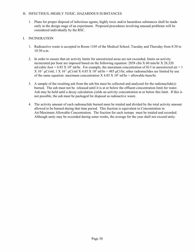
H. INFECTIOUS, HIGHLY TOXIC, HAZARDOUS SUBSTANCES
1. Plans for proper disposal of infectious agents, highly toxic and/or hazardous substances shall be madeearly in the design stage of an experiment. Proposed procedures involving unusual problems will beconsidered individually by the RSC.
I. INCINERATION
1. Radioactive waste is accepted in Room 1105 of the Medical School, Tuesday and Thursday from 8:30 to10:30 a.m.
2. In order to ensure that air activity limits for unrestricted areas are not exceeded, limits on activityincinerated per hour are imposed based on the following equation: 2858 cfm X 60 min/hr X 28,320ml/cubic foot = 4.85 X 10 ml/hr. For example, the maximum concentration of H-3 in unrestricted air = 19
X 10 ìCi/ml; 1 X 10 ìCi/ml X 4.85 X 10 ml/hr = 485 ìCi/hr; other radionuclides are limited by use-7 -7 9
of the same equation: maximum concentration X 4.85 X 10 ml/hr = allowable burn/hr.9
3. A sample of the resulting ash from the ash bin must be collected and analyzed for the radionuclide(s)burned. The ash must not be released until it is at or below the effluent concentration limit for water. Ash may be held until a decay calculation yields an activity concentration at or below this limit. If this isnot possible, the ash must be packaged for disposal as radioactive waste.
4. The activity amount of each radionuclide burned must be totaled and divided by the total activity amountallowed to be burned during that time period. This fraction is equivalent to Concentration inAir/Maximum Allowable Concentration. The fraction for each isotope must be totaled and recorded. Although unity may be exceeded during some weeks, the average for the year shall not exceed unity.

Page 39
XXI. SAFETY RULES: FIXED RADIOGRAPHIC
A. Particular care should be taken to limit the useful beam to the smallest area consistent with clinicalrequirements and to align accurately the X-ray beam with the patient and film.
B. Gonadal shielding should be used for the patient when appropriate, but never as a substitute for adequatebeam collimation and alignment.
C. When a patient must be held in position for radiography, mechanical supporting or restraining devices shouldbe used. If the patient must be held by an individual, that individual shall be protected with appropriateshielding devices, such as protective gloves and apron. The individual should be so positioned that no part ofhis body will be struck by the useful beam and that his body is as far as possible from the edge of the usefulbeam.
D. Special precautions, consistent with clinical needs, should be taken to minimize exposures of the embryo orfetus in patients known to be or suspected of being pregnant.
E. Use the maximum source-skin distance consistent with the conditions of the examination.
F. Only persons whose presence is necessary shall be in the radiographic room during exposure. All suchpersons shall be protected.
G. The radiographer shall stand behind the barrier provided for his/her protection during radiographicexposures.
H. Special care shall be taken to insure adequate filtration in multi-purpose machines. Particular care shall betaken to insure adequate filtration in any machine equipped with a beryllium window tube.

Page 40
XXII. SAFETY RULES: FIXED FLUOROSCOPIC
A. Particular care should be taken to limit the useful beam to the smallest area consistent with clinicalrequirements and to align accurately the X-ray beam with the patient and film.
B. Gonadal shielding should be used for the patient when appropriate, but never as a substitute for adequatebeam collimation and alignment.
C. When a patient must be held in position for radiography, mechanical supporting or restraining devices shouldbe used. If the patient must be held by a individual, that individual shall be protected with appropriateshielding devices, such as protective lead gloves and an apron. The individual should be so positioned thatno part of his body will be struck by the useful beam and that his body is as far a possible from the edge ofthe useful beam.
D. Special precautions, consistent with clinical needs, should be taken to minimize exposures of the embryo orfetus in patients know to be or suspected of being pregnant.
E. Use the maximum source-skin distance consistent with the conditions of the examination.
F. Protective aprons of at least 0.5 mm lead equivalent should be worn in the fluoroscopy room by each person(except the patient). X-ray monitoring devices shall be worn by all persons in the X-ray room (except thepatient) on the outside of the protective apron on the lapel.
G. Only persons whose presence is required should be in the fluoroscopic room.
H. The hand of the fluoroscopist should not be placed in the useful beam unless the beam is attenuated by thepatient and a protective glove of at least 0.5 mm lead equivalent.
I. Fluoroscopy should not be utilized as a substitute for radiography. Fluoroscopy is to be reserved for thestudy of dynamics, special relationships or guidance in spot filming of critical details.
J. In cineradiography, special care must be taken to limit patient exposure when, as is often the case, tubecurrents and potentials employed are higher than those normally used in fluoroscopy.
K. Image intensification shall always be provided on mobile fluoroscopic equipment. It shall be impossible tooperate mobile fluoroscopic equipment unless the useful beam is intercepted by the image intensifier.

Page 41
XXIII. SAFETY RULES: OPERATORS OF MOBILE RADIOGRAPHIC EQUIPMENT
A. Particular care should be taken to limit the useful beam to the smallest area consistent with clinicalrequirements and to align accurately the X-ray beam with the patient and film.
B. Gonadal shielding should be used for the patient when appropriate, but never as a substitute for adequatebeam collimation and alignment.
C. When a patient must be held in position for radiography, mechanical supporting or restraining devices shouldbe used. If the patient must be held by an individual, that individual shall be protected with appropriateshielding devices, such as protective lead gloves and an apron. The individual should be so positioned thatno part of his body will be struck by the useful beam and that his body is as far as possible from the edge ofthe useful beam.
D. Special precautions, consistent with clinical needs, should be taken to minimize exposures of the embryo orfetus in patients known to be or suspected of being pregnant.
E. Use the maximum source-skin distance consistent with the conditions of the examination.
F. Mobile X-ray equipment shall not be used for fluoroscopy, unless it meets the requirements for mobilefluoroscopes.
G. Mobile equipment should be used only for examinations where it is impractical to transfer patients topermanent radiographic installations.
H. Patients in adjoining beds should be at least twelve (12) feet away from the central ray of the primary beam. If the beds cannot be moved, adjacent patients shall be furnished with a 0.5 mm lead equivalent apron.
I. Prior to making the X-ray exposure, the technologist will announce his/her intention to do so. No exposure isto be made if any person, other than the patient is within a twelve (12) foot radius of the X-ray beam and isnot properly shielded.
Page 42
XXIV. SAFETY RULES: OPERATORS OF MOBILE FLUOROSCOPIC EQUIPMENT
A. Particular care should be taken to limit the useful beam to the smallest area consistent with clinicalrequirements and to align accurately the X-ray beam with the patient and film.
B. Gonadal shielding should be used for the patient when appropriate, but never as a substitute for adequatebeam collimation and alignment.
C. When a patient must be held in position for radiography, mechanical supporting or restraining devices shouldbe used. If the patient must be held by an individual, that individual shall be protected with appropriateshielding devices, such as protective lean gloves and an apron. The individual should be so positioned thatno part of his body will be struck by the useful beam and that his body is as far as possible from the edge ofthe useful beam.
D. Special precautions, consistent with clinical needs, should be taken to minimize exposures of the embry orfetus in patients known to be or suspected of being pregnant.
E. Use the maximum source-skin distance consistent with the conditions of the examination.
F. Stand as far as possible from the patient, the tube and the useful beam.. Wear a protective apron or standbehind a suitable shield of at least 0.5 mm lead equivalent.

Page 43
XXV. SAFETY RULES: PERSONS IN THE VICINITY OF MOBILE X-RAY EQUIPMENT
A. Vacate the room if possible.
B. If in the room, stand as far as possible from the patient, the X-ray tube and the useful beam.
C. Declared pregnant women shall not hold patients.
D. Follow all instructions given by the X-ray technologist.
E. When a patient must be held in position for radiography, mechanical supporting or restraining devices shouldbe used. If the patient must be held by an individual, that individual shall be protected with appropriateshielding devices, such as protective lead gloves and an apron. The individual should be so positioned thatno part of his body will be struck by the useful beam and that his body is as far as possible from the edge ofthe useful beam.

Page 44
XXVI. SPECIFIC INSTRUCTIONS FOR ALLIED MEDICAL WORKERS
Housekeeping Personnel:
All housekeeping personnel should be aware of the locations of all restricted area in order to practice goodradiation protection measures.
The measures are:
1) Get user permission and instructions from the RSO before cleaning any spill in restricted area.
2) Do not clean counter tops, hoods, refrigerators or sinks in restricted areas unless specifically requestedand instructed by the area supervisor or RSO.
3) Do not remove bedclothes, dishes, trash or other items from rooms posted with radiation signs unlessspecifically instructed by a member of the radiation safety staff.
Security Personnel:
All security personnel should be aware of the locations of all restricted areas and be able to recognize packagescontaining radioactive material in order to practice good radiation protection measures.
Maintenance Personnel:
All maintenance personnel should be aware of the locations of all restricted areas so that they may practice goodradiation protection measures.
These measures are:
1) Obtain permission before working in an area that is in or adjacent to a restricted area.
2) Be aware of hoods, sinks, refrigerators and storage areas used for radioactive materials or sources.
Clerical Personnel:
All clerical personnel in the departments that use radiation should be aware of the locations of restricted areas sothat they may practice good radiation protection measures.
Good practice includes:
1) Do not eat, drink, smoke or apply cosmetics in areas where radioactive materials are used.
2) Do not store food or drink in refrigerators where radioactive materials are stored.

Page 45
XXVII. RADIATION AND PREGNANCY, RADIATION AND ALLIED MEDICAL STAFF
Radiation and allied medical staff who may be exposed to radiation should contact the RSO if they becomepregnant or are planning to become pregnant. The mother assumes all risk until she specifically declares herpregnancy in a written and signed statement to her supervisor or the RSO. At that time, the hospital isresponsible for assuring that the duties of a female staff member will not result in a dose equivalent that is morethan 500 mrem to the fetus.
THE MOST RADIOSENSITIVE PERIOD FOR THE EMBRYO/FETUS IS FROM EIGHT TO FIFTEEN (8-15)WEEKS GESTATION AGE.
Guidelines for protecting the embryo/fetus are: (Observe Checked Items)
___ 1. Distance protection shall be practiced at all times.
___ 2. At no time shall any part of the body be so positioned that allows exposure from the primary beamfrom X-ray sources or unshielded radioactive sources.
___ 3. Holding patients for immobilization shall be prohibited.
___ 4. Mobile X-ray or Fluoroscopy work must be performed with protection of a 0.5 mm lead equivalentwrap-around apron.
___ 5. The personal monitoring device (dosimeter badge) shall be worn at the lapel outside of the protectiveapron.
___ 6. An additional monitoring device shall be worn at waist level under the protective apron (when apronis worn) to determine exposure directly to the fetus.
___ 7. Impermeable disposable gloves shall be worn and pipetting device used while performingmanipulations involving radionuclides.
___ 8. Generator elution shall not be performed during the first trimester.
___ 9. Kit preparation shall not be performed during the first trimester.
___ 10. Therapeutic amounts of radionuclides shall not be administered.
___ 11. Ventilation imaging shall not be performed.
___ 12. Millicurie amounts of H, C, I, or I shall not be used.3 14 125 131
___ 13. Extra care shall be observed to avoid spillage, vaporization and internal/external contamination.
I SHALL OBSERVE ITEMS CHECKED ABOVE.
________________________________________EMPLOYEE ___________________________DATE
________________________________________RSO
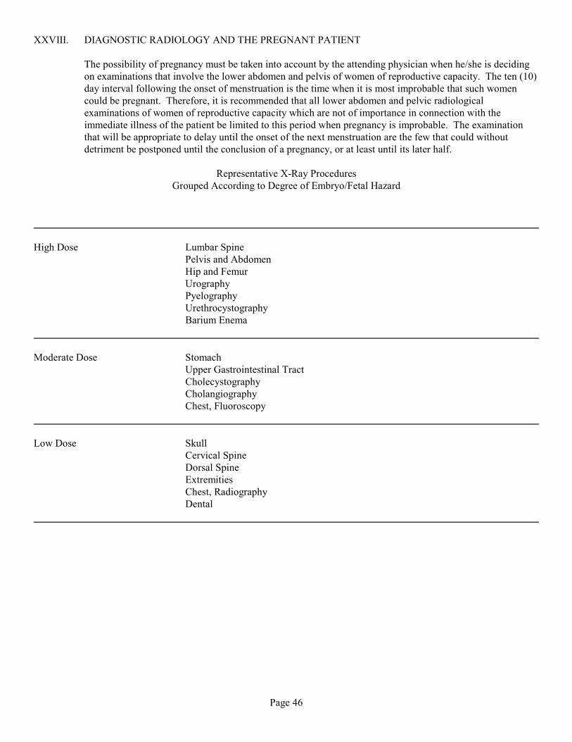
Page 46
XXVIII. DIAGNOSTIC RADIOLOGY AND THE PREGNANT PATIENT
The possibility of pregnancy must be taken into account by the attending physician when he/she is decidingon examinations that involve the lower abdomen and pelvis of women of reproductive capacity. The ten (10)day interval following the onset of menstruation is the time when it is most improbable that such womencould be pregnant. Therefore, it is recommended that all lower abdomen and pelvic radiologicalexaminations of women of reproductive capacity which are not of importance in connection with theimmediate illness of the patient be limited to this period when pregnancy is improbable. The examinationthat will be appropriate to delay until the onset of the next menstruation are the few that could withoutdetriment be postponed until the conclusion of a pregnancy, or at least until its later half.
Representative X-Ray ProceduresGrouped According to Degree of Embryo/Fetal Hazard
High Dose Lumbar SpinePelvis and AbdomenHip and FemurUrographyPyelographyUrethrocystographyBarium Enema
Moderate Dose StomachUpper Gastrointestinal TractCholecystographyCholangiographyChest, Fluoroscopy
Low Dose SkullCervical SpineDorsal SpineExtremitiesChest, RadiographyDental

Page 47
XXIX. PATIENT PREGNANCY SCREENING
Purpose: To assure that all precautions are taken to prevent the exposure of a pregnant female.
Policy: All females in the childbearing years shall pass a pregnancy screen prior to examination.
Procedures: All females in childbearing years will be questioned as to the possibility of pregnancy prior to initiationof a Radiographic/Fluoroscopic and/or Nuclear Medicine Study.
In the event the patient indicates a possibility of pregnancy, the attending physician must be notified forapproval prior to the exam. A verbal order for the performance of a pregnancy test may be obtained andthe examination postponed pending results.

Page 48
XXX. NUCLEAR MEDICINE AND THE PREGNANT PATIENT
The patient who is or thinks she might be pregnant, or who is nursing, should be encouraged to give thisinformation to her attending physician when the examination history is taken.
The nursing woman should suspend breast feeding for an appropriate period of time following a nuclear medicineexamination. The nuclear medicine physician or certified radiological physicist can advise the nursing woman onthe length of time that nursing should be suspended.
The attending physician can use nuclear medicine consultation request forms in non-emergency situations torecord the pregnancy and nursing status of a woman of childbearing age. In addition, the attending physicianshould encourage the patient to provide this information. If there is a medical emergency, if the nuclear medicineexamination will contribute vital information to the diagnosis and if there are no alternative methods forobtaining this information that would result in lower radiation exposure, the examination generally should beperformed regardless of the patient's pregnancy state. Attention must be pain to technical modifications of theprocedure that will minimize radiation exposure.
The nuclear medicine technologist, in the absence of information about the patient's pregnancy or nursing status,should be encouraged to ask the patient if she is or may be pregnant or if she is nursing. If the patient replies thatshe is or may be pregnant or is nursing, the technologist must notify the physician in charge at once.
The nuclear medicine physician should be aware of appropriate alternatives prior to conducting a nuclearmedicine procedure on a pregnant or potentially pregnant woman or one who is nursing. The nuclear medicinephysician should be prepared to consult with the attending physician on possible alternatives which include:
A. Requesting the use of a radionuclide that delivers a lower radiation dose or one that is less likely to cross theplacental barriers than the radionuclide usually used, if the diagnostic objectives can still be met.
B. In the case of a known pregnancy, assessing the possibility of deferring the examination until pregnancy isconcluded.
C. In the case of a possible but unconfirmed pregnancy, deferring the examination that is not immediatelyneeded until the pregnancy is ruled out.
D. Canceling the nonemergency examination once aware that the patient is or may be pregnant.
E. Directing the nursing woman to suspend breast feeding for the period of time that radioactivity is present inthe milk.
F. Ascertaining the advisability of using other clinical modalities to diagnose the patient's condition.

Page 49
XXXI. QUALITY CONTROL PROCEDURES
A. Calibration of dose Calibrator
Test for the following at the indicated frequency. Consider repair, replacement or arithmetic correction if thedose calibrator falls outside the suggested tolerances. (These recommended tolerances are more restrict thanthose in the regulations to ensure that corrective action will be taken before the dose calibrator is outsidepermissible tolerances).
1. Constancy at least once each day prior to assay of patient dosages (±5%).
2. Linearity at installation and quarterly thereafter (±5%).
3. Geometry variation at installation (±10%).
4. Accuracy at installation and at least annually thereafter (±5%).
After repair, adjustment or relocation of the dose calibrator, repeat above test as required.
Constancy means reproducibility in measuring a constant source over a longer period of time. Assay at leastone relatively long-lived source ( Cs, Co, Co or Ra) using a reproducible geometry each day before137 60 57 226
using the calibrator. Consider the use of two (2) or more sources with different photon energies andactivities. Use the following procedure:
1. Assay each reference source using the appropriate dose calibrator setting (use the Cs setting to assay137
Cs).137
2. Measure bkg at the same setting and subtract or confirm the proper operation of the automatic bkg.Subtract circuit if it is used.
3. For each source used, either plot on graph paper or log in a book the bkg level for each setting checkedand the net activity of each constancy source.
4. Using one of the sources used, repeat the above procedure for all commonly used radioisotope settings. Plot or log the results.
5. Establish an action level or tolerance for each recorded measurement at which the individual performingthe test will automatically notify the chief technologist or authorized user of suspected malfunction of thecalibrator.
Inspect the instrument on a quarterly basis to ascertain that the measurement chamber liner is in place andthat the instrument is zeroed according to the manufacturer's instructions.
Linearity means that the calibrator is able to indicate the correct activity over the range of use of thatcalibrator. This test is done using a vial or syringe of Tc whose activity is at least as large as the maximum99m
activity normally assayed in a prepared radiopharmaceutical kit, in a unit dosage syringe or in aradiopharmaceutical therapy, whichever is largest.
Decay Method
Assay the Tc syringe or vial in the dose calibrator, and subtract background to obtain the net activity in99m
mCi. Record the date, time to the nearest minute and net activity on the Dose Calibrator Linearity Test Form. Perform a minimum of two assays per day until the activity is less than 10 ìCi

Page 50
Shield Method
If you decide to use a set of "sleeves" of various thicknesses to test for linearity, it will first be necessary tocalibrate them.
1. Begin the linearity test as described in the decay method above. After making the first assay, the sleevescan be calibrated as follows. Steps 2 through 4 below must be completed in six (6) minutes.
2. Put the base and sleeve one (1) in the dose calibrator with the vial. Record the sleeve number andindicated activity.
3. Remove sleeve one (1) and put in sleeve two (2). Record the sleeve number and indicated activity.
4. Continue for all sleeves.
5. Complete the decay method linearity test steps 2 through 7 above.
6. From the graph made in step d of the decay method, find the decay time associated with the activityindicated with sleeve one (1) in place. This is the "equivalent decay time" for sleeve one (1). Recordthat time with the data recorded in step b.
7. Find the decay time associated with the activity indicated with sleeve two (2) in place. This is the"equivalent decay time" for sleeve two (2). Record that time with the data recorded in step c.
8. Continue for all sleeves.
9. The table of sleeve numbers and equivalent decay time constitutes the calibration of the sleeve set.
The sleeve set may now be used to test linearity of the dose calibrator.
1. Assay the Tc syringe or vial in the dose calibrator and subtract bkg to obtain net activity in mCi. 99m
Record the net activity.
2. Steps 3 through 5 below must be completed within six (6) minutes.
3. Put the base and sleeve one (1) in the dose calibrator with the vial. Record the sleeve number andindicated activity.
4. Remove sleeve one (1) and insert sleeve two (2). Record the sleeve number and indicated activity.
5. Continue for all sleeves.
6. On a sheet of semilog graph, label the logarithmic vertical axis in mCi and label the linear horizontal axisin hours elapsed. At the top of the graph, note the date, manufacturer, model number and serial numberof the dose calibrator.
7. Plot the data using the equivalent decay time associated with each sleeve.
8. Draw a "best fit" straight line through the data points. For the point farthest from the line, calculate itsdeviation from the value on the line (A-observed - A-line)/(A-line) = deviation.

Page 51
9. If the worst deviation is more than ±0.05, the dose calibrator should be repaired or adjusted. If thiscannot be done, it will be necessary to make a correction table or graph that will allow you to convertfrom activity indicated by the dose calibrator to "true activity".
Geometric Variation means that the indicated activity does not change with volume or configuration. Thistest should be done using a syringe and a vial. Vial measurements should be made over a range of usefulvolumes, for example, 1 cc to 20 cc. A geometrical correction for syringe measurements should also bedetermined.
Accuracy, for a given calibrated reference source, means the indicated mCi value is equal to the mCi valuedetermined by the National Institute of Standards and Technology (NIST) or by the supplier who hascompared that source to a source that was calibrated by the NIST. Certified sources are available from theNIST and from radioisotope suppliers. At least two (2) sources with different principal photon energies( Co, Co or Cs) should be used. The regulations require that one source must have a principal photon57 60 137
energy between 100 keV and 500 keV. The regulations also require that if Ra is used, it must be at least226
ten (10) ìCi; other sources must be at least 50 ìCi. Consider using at least one (1) reference source whoseactivity is within the range of activities normally assayed.
1. Assay a calibrated reference source at the appropriate setting (i.e., use the Co setting to assay Co) and57 57
remove the source and measure bkg. Subtract bkg from the indicated activity to obtain net activity. Record this measurement on a Dose Calibrator Geometry and Accuracy Form. Repeat for a total of three(3) determinations.
2. Average the three (3) determinations. The average value should be within five percent (5%) percent ofthe certified activity of the reference source, mathematically corrected for decay.
3. Repeat the procedure for other calibrated reference sources.
4. If the average value does not agree within five percent (5%) of the certified value of the reference source,the dose calibrator may require repair or replacement. Regulations requires repair or replacement if theerror exceeds ten percent (10%).
5. At the same time the accuracy test is performed, assay the source that will be used for the daily constancytest (it need not be certified reference source) on all commonly used radioisotope settings. Record thesettings and indicated mCi values with the accuracy data.
The RSO will review and sign the records of all geometric variation, linearity and accuracy tests.
B. Quality Control, Scintillation Camera
1. Total System Uniformity (Daily)
a. Materials
(1) Flood phantom: fill with water and add 1 to 5 (1-5) mCi of Tc, count rate should be less than99m
20,000 cps, or Co flood source, or Tc point source of 100 to 200 ìCi; count rate of no greater57 99m
than 20,000 cps.
(2) Collimator; low energy, parallel hole.

Page 52
b. Procedure
(1) Place collimator on detector and invert detector head.
(2) Place Co flood source or Tc flood phantom on collimator. If flood phantom is used, protect57 99m
against contamination by interposing plastic-backed absorbent paper between the phantom andthe detector.
(3) Set and visually verify appropriate pulse height analyzer setting with a 20% window.
(4) Set appropriate intensity.
(5) Collect one million uncorrected counts if a camera with a standard field of view is used; collecttwo million uncorrected counts for a camera with a large field of view.
c. Data Treatment
(1) Visually inspect film for nonuniformities.
2. System Spatial Resolution (Weekly)
a. Materials
(1) Flood phantom: fill with water and add 1 to 5 (1-5)mCi of Tc; count rate should be less than99m
20,000 cps, or Co flood source, or Tc point source of 100 to 200 ìCi; count rate of no greater57 99m
than 20,000 cps.
(2) Collimator; low energy, parallel hole.
(3) Bar phantom.
b. Procedure
(1) Attach low-energy parallel-hole collimator and invert detector.
(2) Arrange bar phantom and Tc flood source on face of detector.99m
(3) Set and visually verify appropriate PHA setting using a 20% window.
(4) Set appropriate intensity.
(5) Collect one million uncorrected counts if a camera with a standard field of view is used; collecttwo million uncorrected counts for a camera with a large field of view.
c. Data Treatment
(1) Visually inspect films for degree of resolution.
(2) Are bars straight? Did collimation correct "barreling" or wavy lines noted in intrinsic resolutionstudies (if available)?

Page 53
C. Quality Control of In-House Prepared Radiopharmaceuticals
1. Mo- Tc Generators99 99m
a. Molybdenum breakthrough shall be performed on all generator eluents before administration topatients. Permissible limits: 0.07 ìCi of Mo per mCi of Tc not to exceed 2.5 ìCi of Mo per99 99m 99
dose.
b. Aluminum breakthrough shall be performed on all generator eluents before administration topatients. Permissible limits: 10 ìg of aluminum per ml of eluent.
2. Chromatography
a. Chromatography shall be employed for radiochemical impurities. Determination of percentage Tc99m
tagged or labeled shall be performed on all "kit" prepared radionuclides. Permissible limits: greaterthan 90% Tc bound.99m
3. Radiopharmaceuticals for clinical procedures shall comply with the product label or package insertregarding physical form, route of administration and dosage range.
D. Sealed Sources; Leakage/Contamination
Each sealed source with a half-life greater than thirty days and in any form other than gas, shall be tested forleakage/contamination prior to initial use and at intervals not to exceed six (6) months. Notwithstanding theperiodic leak test required, any sealed source is exempt from such leak tests when the source contains 100ìCi or less of beta and/or gamma emitting material or 10 ìCi or less of alpha emitting material. If at anytime, there is reason to suspect that a sealed source may have been damaged or may be leaking, the sourceshall be tested for leakage before further use. In the absence of a certificate from a transferor indicating thata leak test has been made within six (6) months prior to the transfer, the sealed source shall not be put intouse until tested.
E. Procedures for Calibration of Survey Instruments
1. The source must be approximately a point source.
2. Either the apparent source activity or the exposure rate at a given distance must be traceable bydocumented measurements to a standard certified within 5 percent (5%) accuracy by the NIST.
3. The inverse square law and the radioactive decay law must be used to correct for change in exposure ratedue to changes in distance or source decay.
4. A record must be made of each survey meter calibration.
5. A single point on a survey meter scale may be considered satisfactorily calibrated if the indicatedexposure rate differs from the calculated exposure rate by less than 10 percent (10%).
Readings above 1,000 mR/hr need not be calibrated. However, such scales should be checked for operationand approximately correct response.
At the time of calibration, the apparent exposure rate from a built-in or owner-supplied check source must bedetermined and recorded.
The report of a survey meter calibration should indicate the procedure used and the data obtained.

Page 54
XXXII. PROCEDURES FOR NURSING STAFF AND PATIENT CARE
A. Diagnostic Procedures
Since there is minimal external hazard to others from routine diagnostic doses of radionuclides, there are norestrictions on the patient's activities or his/her contacts with other people. Nursing personnel are notrequired to wear personnel monitoring devices.
The following procedures apply when a patient receives radioactive material for diagnostic purposes:
1. Questions concerning the use of radionuclides for diagnostic nuclear medicine procedures should bepresented to Nuclear Medicine.
2. If radioactive contamination is suspected, nursing personnel should use impermeable disposable glovesto handle items and contact Nuclear Medicine. Particular care should be exercised for handling vomitusduring the first 24 hours following administration of a radionuclide.
3. Special diagnostic procedures will be evaluated on an individual basis and appropriate writteninstructions may be issued.
B. Therapeutic Procedures
1. Patients Receiving Brachytherapy Sources
a. Sealed source therapy will be offered by approved Radiation Therapy Consultants. The RSC willadvise Administration of those radiation therapists that are approved for use of sealed source therapy.
b. The RSC will use in part the following criteria for evaluation of radiation therapists:
(1) All rules and regulations of the Medical Staff relative to record keeping are observed.
(2) Written verification of leak test of sealed sources must be maintained at six (6) month intervals.
(3) Sealed sources that are transported to the Hospital shall be transferred in containers which willlimit the radiation level at one meter (1m) from the center of the container to 2 mR/hr or less.
(4) At least 48 hours notification shall be provided to Radiation Safety so that appropriatepreparation and precautionary measures may be initiated. Notification shall include:
(a) The number, loading and type of sealed source to be transferred
(b) The name and location of the patient to which the material will be transferred
(c) The time of source insertion and removal.
(d) At least 48 hours notification shall also be provided to the Operating Room Supervisor andHead Nurse for sources to be inserted in Surgery.

Page 55
c. The RSO shall be responsible for:
(1) Maintaining a log book in Nuclear Medicine to indicate the number, loading and type of sealedsource that has been transferred as well as the location (name of patient), time of source insertionand removal
(2) Completing the Radioactivity Precaution Tag to be attached to the door of the patient's room andattaching the Caution, Radioactive Materials label to the cover of the patient's chart
(3) Surveying the patient's room and surrounding areas as soon as practical after administration ofthe radionuclide and at conclusion of treatment and completing the Radionuclide Therapy Surveyform.
2. Patients receiving radionuclide therapy as solution, colloid or microsphere
a. Prior to therapy, the form Therapeutic Radionuclide Consultation Form is to be completed andbecomes a part of the patient's chart.
b. The RSO shall be responsible for:
(1) Assuring that the responsible physician has completed the appropriate Appendices to become apart of the patient's chart.
(2) Completing the Radioactivity Precautions Tag to be attached to the foot of the patient's bed or todoor of room and attaching the Caution, Radioactive Materials label to the cover of the patient'schart.
(3) Surveying the patient's room and surrounding areas as soon as practical after administration ofthe radionuclide and at discharge of the patient. In addition, the RSO shall completing the formRadionuclide Therapy Survey.
3. Patients requiring emergency surgery after therapy
a. Consultation shall be made with the Nuclear Physician who shall notify the RSO.
4. Death of patient after therapy
a. Consultation shall be made with the Nuclear Physician who shall notify the RSO.

Page 56
XXXIII. TRAINING PROGRAM: PERSONNEL WORKING IN THE VICINITY OF RADIOACTIVE MATERIAL
A. Personnel to be included in Training
1. Nuclear Medicine Technologists.
2. Any Ancillary Personnel whose duties may require them to work in the vicinity of radioactive material.
B. Frequency of Training
Personnel will be instructed:
1. Before assuming duties with, or in the vicinity of, radioactive materials.
2. During an annual refresher course.
3. Whenever there is a significant change in duties, regulations or the terms of the license.

Page 57
XXXIV. PROCEDURE FOR MONITORING OR CHECKING TRAP EFFLUENT
Charcoal traps can significantly reduce air contamination. They can also become saturated or be spoiled byimproper use, humidity, chemicals or inadequate maintenance.
1. If the trap effluent is monitored by a radiation detector designed to monitor effluent gas, check thedetector according to the manufacturer's instructions. Keep a record of the checks.
2. If you do not monitor the trap effluent, check it on receipt and once each month. Collect the effluentfrom the trap during one patient study in a plastic bag and monitor the activity in the bag by holding thebag against a camera, with the camera adjusted to detect noble gas. Compare the bag’s cpm to bkg cpmwith no other radioactivity in the area. Keep a record of the date, bkg cpm, and bag cpm.
3. The RSO will establish an action level based on cpm or a multiple of bkg cpm. If you measure asignificant increase in the bag cpm, the trap is breaking down and must be replaced.
4. Follow the trap manufacturer's instructions for replacing the trap.

Page 58
APPENDIX A
RADIATION SAFETY COMMITTEE
James J. Balsamo, Jr., MPH, R.T.
Sharon Hafner, R.T., Radiology
Lucien Nedzi, M.D., Radiation Therapy
Rita Preiksaitis, R.N.
Charles F. Reindl, M.S., RSO
Brian Rogers, M.D., Radiology
Brian Rowan, Ph.D.
Robert Sanford, Ph.D., Radiation Therapy
James A. Terry, Ph.D., Chairman
Vicki L. Traina-Dorge, Ph.D.

Page 59
APPENDIX B
TECHNOLOGISTS APPROVED FOR RADIONUCLIDE INJECTIONS
Cheryl Albert, R.T. (N)Courtney Aysenne, R.T. (N)Raymond Qiana, R.T. (N)

Page 60
APPENDIX C
DAILY INCOMING/OUTGOING SHIPMENT INSPECTION LOG
SURVEY INSTRUMENT ______________________________________________ CONVERSION FACTOR _____________________________________________
ACTION LEVELS:10 mR/hr at three feet200 mR/hr at surfaceWipe test of 2,220 dpm or 0.001 microcuries
IF ANY ACTION LEVEL IS EXCEEDED, STOP AND NOTIFY THE RADIATION SAFETY OFFICER
INCOMING OUTGOING
Date Product SupplierExternal Survey
(mR/hr)
Surface 3 Feet
WIPE TEST (dpm)
Bkg Outside Inside
TechInitials
Date WIPE TEST (dpm)
Bkg Outside
ExternalSurvey(mR/hr)Surface
CorrectiveActions
TechInitials

Page 61
APPENDIX D
Radionuclide Distribution Log
Kit Name ______________________________ Lot Number ______________________________ Expiration Date ____________________________________
Date of
Scan
Patient Name Hospital
Number
Social Security
Number
Prescribed
Dose
(mCi)
Measured Dose
(mCi)
Time of
Measurement
Volume Procedure
Performed
Technologist
Performing
Procedure
Eluate mCi/cc ________________________ @ ___________________AM/PM. Kit Assay _______________________ @ ______________ AM/PM.
Total Volume of Eluate _____________________________________________ Prepared by ______________________________________________
Total Activity _____________________________________________________
Total Volume of Vial _______________________________________________

Page 62
APPENDIX E
RADIATION EXPOSURE HISTORY REQUEST FORM
___________________________________ DATE: ________________________________
___________________________________
___________________________________
___________________________________
In order to complete the radiation history on the following individual, a record of their radiation exposure is needed fromyour institution. Attached is an authorization for the release of this information.
PERSONAL DATA
NAME _________________________________________________________________________________________
DATE OF BIRTH ________________________________________________________________________________
SOCIAL SECURITY NO. __________________________________________________________________________
EMPLOYED DATES ______________________________________________________________________________
DEPARTMENT __________________________________________________________________________________
MONITORING PERIOD ___________________________________________________________________________
TOTAL ACCUMULATED DOSE DEEP (mRem) ________________ SHALLOW (mRem) _________________
RESULTS OF BIOASSAY/THYROID MONITORING, IF PERFORMED
TOTAL BODY ESTIMATED BIOASSAYRADIONUCLIDE CRITICAL ORGAN BURDEN (mRem) EXPOSURE (mRem) DATE
Thank you for your assistance.
Very truly yours,
Radiation Safety Officer

Page 63
APPENDIX F
DOSIMETER BADGE MONITORING SERVICE REQUEST
Radiation Regulations require your past radiation exposure in order to initiate dosimeter badge service. Please completethis form, sign, and return to the Radiation Safety Officer.
FULL LEGAL NAME ______________________________________________________________________________Last First Middle/Maiden
SOCIAL SECURITY NO. ___________________________________________________________________________
BIRTH DATE _________________________________________ SEX _______________
DEPARTMENT________________________________________ PHONE _______________________________
PREVIOUS EMPLOYMENT(S) INVOLVING RADIATION EXPOSURE AS MONITORED BY RADIATIONDOSIMETER
1. Indicate NONE, if never monitored: _____________________________
2. Previously monitored locations:
EMPLOYER __________________________________________________________________________________
DEPARTMENT _______________________________________________________________________________
MAILING ADDRESS ___________________________________________________________________________
DATE OF EMPLOYMENT: FROM:_______________________________ TO:_____________________________(Use back of sheet if more than one employer)
3. If previously monitored, estimate your previous calendar quarter exposure: ____________mRem
I HEARBY AUTHORIZE THE RELEASE OF MY RADIATION EXPOSURE HISTORY TO ____________________
_________________________________________________________________________________________________
SIGNATURE __________________________________________
OCCUPATIONAL TITLE ________________________________
DEPARTMENT _________________________________ DATE ___________________

Page 64
APPENDIX G
RADIONUCLIDE DISPOSAL FORM
LOCATION _____________________________________
Radionuclide
Date placed
in Storage
Activity in microcuriesor surface
exposure rate in mR/hr
Date of Disposal
Activity inmicrocuries or
surface exposurerate in mR/hr @
Disposal
Backgroundexposure rate in
mR/hr
Survey meter orassay instrumentused (includingserial number)
Person performingdisposal
(Signature)
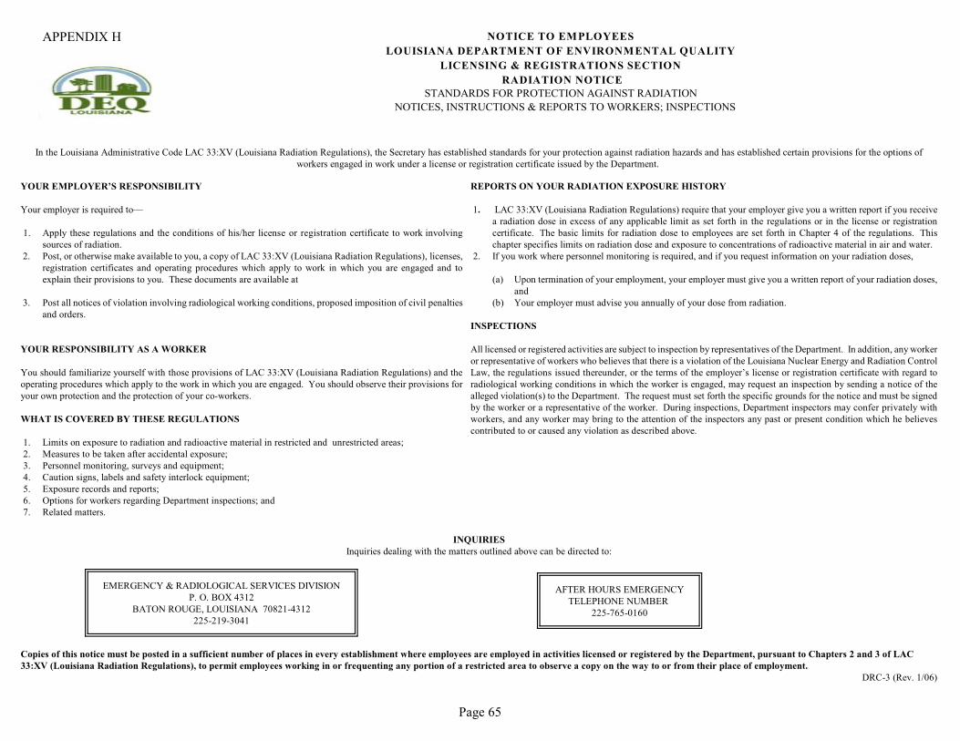
Page 65
APPENDIX H NOTICE TO EMPLOYEES
LOUISIANA DEPARTMENT OF ENVIRONMENTAL QUALITY
LICENSING & REGISTRATIONS SECTION
RADIATION NOTICESTANDARDS FOR PROTECTION AGAINST RADIATION
NOTICES, INSTRUCTIONS & REPORTS TO WORKERS; INSPECTIONS
In the Louisiana Administrative Code LAC 33:XV (Louisiana Radiation Regulations), the Secretary has established standards for your protection against radiation hazards and has established certain provisions for the options ofworkers engaged in work under a license or registration certificate issued by the Department.
YOUR EMPLOYER’S RESPONSIBILITY Your employer is required to— 1. Apply these regulations and the conditions of his/her license or registration certificate to work involving
sources of radiation. 2. Post, or otherwise make available to you, a copy of LAC 33:XV (Louisiana Radiation Regulations), licenses,
registration certificates and operating procedures which apply to work in which you are engaged and toexplain their provisions to you. These documents are available at
3. Post all notices of violation involving radiological working conditions, proposed imposition of civil penaltiesand orders.
YOUR RESPONSIBILITY AS A WORKER
You should familiarize yourself with those provisions of LAC 33:XV (Louisiana Radiation Regulations) and theoperating procedures which apply to the work in which you are engaged. You should observe their provisions foryour own protection and the protection of your co-workers. WHAT IS COVERED BY THESE REGULATIONS 1. Limits on exposure to radiation and radioactive material in restricted and unrestricted areas; 2. Measures to be taken after accidental exposure; 3. Personnel monitoring, surveys and equipment; 4. Caution signs, labels and safety interlock equipment; 5. Exposure records and reports; 6. Options for workers regarding Department inspections; and 7. Related matters.
REPORTS ON YOUR RADIATION EXPOSURE HISTORY 1. LAC 33:XV (Louisiana Radiation Regulations) require that your employer give you a written report if you receive
a radiation dose in excess of any applicable limit as set forth in the regulations or in the license or registrationcertificate. The basic limits for radiation dose to employees are set forth in Chapter 4 of the regulations. Thischapter specifies limits on radiation dose and exposure to concentrations of radioactive material in air and water.
2. If you work where personnel monitoring is required, and if you request information on your radiation doses,
(a) Upon termination of your employment, your employer must give you a written report of your radiation doses,and
(b) Your employer must advise you annually of your dose from radiation. INSPECTIONS All licensed or registered activities are subject to inspection by representatives of the Department. In addition, any workeror representative of workers who believes that there is a violation of the Louisiana Nuclear Energy and Radiation ControlLaw, the regulations issued thereunder, or the terms of the employer’s license or registration certificate with regard toradiological working conditions in which the worker is engaged, may request an inspection by sending a notice of thealleged violation(s) to the Department. The request must set forth the specific grounds for the notice and must be signedby the worker or a representative of the worker. During inspections, Department inspectors may confer privately withworkers, and any worker may bring to the attention of the inspectors any past or present condition which he believescontributed to or caused any violation as described above.
INQUIRIESInquiries dealing with the matters outlined above can be directed to:
Copies of this notice must be posted in a sufficient number of places in every establishment where employees are employed in activities licensed or registered by the Department, pursuant to Chapters 2 and 3 of LAC33:XV (Louisiana Radiation Regulations), to permit employees working in or frequenting any portion of a restricted area to observe a copy on the way to or from their place of employment.
DRC-3 (Rev. 1/06)
EMERGENCY & RADIOLOGICAL SERVICES DIVISIONP. O. BOX 4312
BATON ROUGE, LOUISIANA 70821-4312225-219-3041
AFTER HOURS EMERGENCYTELEPHONE NUMBER
225-765-0160

Page 66
APPENDIX I
DIAGNOSTIC AND/OR THERAPEUTIC PROCEDUREMISADMINISTRATION REPORT
EVENT __________________________________________ REPORT DATE _______________________________
SERVICE PHYSICIAN _____________________________________________________________________________
INVOLVED TECHNOLOGIST ______________________________________________________________________
TYPE OF MISADMINISTRATION: WRONG PATIENT _____WRONG RADIOPHARMACEUTICAL _____WRONG ROUTE _____DOSAGE DIFFERING FROM PRESCRIBED BY 50% _____
INTENDED PROCEDURE __________________________________________________________________________
INTENDED RADIOPHARMACEUTICAL/DOSAGE ____________________________________________________
RADIOPHARMACEUTICAL/DOSAGE _______________________________________________________________
PROCEDURE PERFORMED ________________________________________________________________________
REFERRING PHYSICIAN __________________________________________________________________________
PATIENT______________________________________ SOCIAL SECURITY NUMBER ______________________
RESPONSIBLE TECHNOLOGIST ___________________________________________________________________
CONTRIBUTING FACTOR _________________________________________________________________________
_________________________________________________________________________________________________
_________________________________________________________________________________________________
ACTION TAKEN TO PREVENT RECURRENCE _______________________________________________________
_________________________________________________________________________________________________
_________________________________________________________________________________________________
EFFECTS ON PATIENT ____________________________________________________________________________
_________________________________________________________________________________________________
_________________________________________________________________________________________________
SIGNATURE ___________________________________________________ DATE __________________________RADIATION SAFETY OFFICER
TELEPHONE NUMBER __________________________________________

Page 67
APPENDIX J
THERAPEUTIC RADIONUCLIDE CONSULTATION FORM
PATIENT ________________________________________________ DATE ______________________________
HOSPITAL NUMBER ______________________________________ WARD ____________________________
IS REFERRED TO YOU. OUR FINDINGS IN THE CASE ARE AS FOLLOWS: ______________________________
_________________________________________________________________________________________________
_________________________________________________________________________________________________
REQUEST FOR TREATMENT WITH THERAPEUTIC RADIOISOTOPE:
RADIOISOTOPE FORM AMOUNT METHOD AND DATE OF ADMINISTRATION
CURRENT MEDICATIONS _________________________________________________________________________
IS PATIENT PREGNANT? _____________
DATE OF LAST MENSTRUAL PERIOD ______________________
HAS PATIENT BEEN INSTRUCTED TO AVOID PREGNANCY PRIOR TO TREATMENT AND FOR TWOMONTHS AFTER TREATMENT? _______________
RATIONALE FOR OTHER THAN ROUTINE USE ______________________________________________________
_________________________________________________________________________________________________
PHYSICIAN’S SIGNATURE ____________________________________________________
TO (PHYSICIAN) ____________________________________
THERAPIST ________________________________________
ASSISTANTS _______________________________________
_________________________________________________________________________________________________
DATE TIME RADIONUCLIDE DOSE TECHNIQUE
_________________________________________________________________________________________________
_________________________________________________________________________________________________
REMARKS:

Page 68
APPENDIX K
QUALITY MANAGEMENT PROGRAM
Guidelines for Brachytherapy Applications, Sr Applicator, Radiopharmaceutical Therapy and Administration of Greater90
than 30 ìCi of I or I.125 131
I. Written Directive
The physician will issue a written directive in the patient's Radiation Oncology treatment chart and/or hospital chartprior to the administration of any brachytherapy or radiopharmaceutical. This directive will be signed and dated bythe physician.
II. Patient Identification
Before administration of any dose, the patient's identity will be determined by more than one method by the personadministering the dose. The methods of identification used are noted on the quality management checklist foradministration of brachytherapy treatments or radiopharmaceuticals (see attached forms). This checklist becomespart of the patient's chart.
III. Verification of Dose
Final plans for treatment and/or dose calculation for brachytherapy are signed by the physician as verification ofagreement with the written directive. For administration of radiopharmaceuticals, the quality management checklistis completed to verify the dose ordered in the written directive.
IV. Verification of Administration
Brachytherapy sources are loaded into the applicators or placed interstitially according to the treatment plan/dosecalculation that follows the written directive. On-site brachytherapy sources are located in the storage safe accordingto a posted "map" of the contents of each drawer and are color-coded according to activity. The number and activityof the sources removed from the safe are verified by completing the source utilization log, which accounts for allsources remaining in the safe. Special-order sources are verified upon receipt for agreement with treatment plan andare intended to be patient-specific. The packing list of sources are filed in the source receipt log with a copy of adose-calibrator print-out. Placement of the applicators and after-loaded sources in the patient are determined andverified by radiography using dummy sources. The source loading and its time of insertion and removal aredocumented in the patient's hospital chart. Radioactive seed applications are radiographed after surgery forverification of seed placement and dose calculation. For radiopharmaceutical administration, a copy of the dose-calibrator print-out is attached to the quality management checklist. For Sr the treatment time and treatment dose90
shall be documented in the patient's hospital chart as well as on the Sr Ophthalmic Applicator Therapy Record.90
V. Deviation from Written Directive
Any deviation from the written directive, intended or otherwise, is documented on the treatment plan and noted in thepatient's hospital chart. The physician is consulted as to the appropriate measures to be taken.
VI. Quality Management Program Review (QMPR)
The QMPR will be reviewed annually for completeness and appropriateness by the radiation physicist, oncologistand RSO (see attached review form). Changes will be made as needed and submitted to the division within 30 daysof the modification.

Page 69
APPENDIX L
ANNUAL REVIEW FORM
QUALITY MANAGEMENT PROGRAM
DATE: ________________________________
_____ Written program (policies and procedures)
_____ Program approved by management
_____ Incorporated into license
_____ Modifications submitted within 30 days
_____ Minutes of annual review
_____ Review sample of patient administrations
_____ Review of QM objectives and effectiveness
_____ Review misadministrations
_____ Review recordable events
_____ Event facts written up within 30 days
_____ Event corrective action recommended and evaluated
_____ Record of patient dose
_____ Written directive for brachytherapy dose
_____ Written directive for radiopharmaceutical therapy dose
_____ Record for I and I greater than 30 microcuries125 131
_____ Patient identity verified by two (2) means
_____ Therapy plans by directive
_____ Unintended deviation from plan evaluated
_____ Retention of records for three (3) years
____________________________________Management
_____________________________________RSO

Page 70
APPENDIX M
Quality Management Checklist for Administration of Sodium Iodide ( I, I)125 131
Activities Greater than 30 ìCi
Date: ____________________________________________________________________________________________Patient: __________________________________________________________________________________________Social Security Number: ____________________________________________________________________________Date of Birth: _____________________________________________________________________________________
PATIENT IDENTIFICATION: (completed by person verifying patient identity).
The above patient's identity has been verified by at least two of the following methods: (Circle appropriate number)
1. Asking patient to state and spell his/her name and compare with patient's record/objective.2. Comparing stated date of birth with patient's record/directive.3. Comparing stated SS# with patient's record/directive.4. Comparing stated address with patient's record/directive.5. Comparing photographic identification (ex. driver's license) with patient’s appearance.6. Comparing in-patient identification wrist band with patient record/directive.7. Relative/friend attests patient identity.
Name of attestor: _______________________________________ Relationship: ___________________________Signature of staff verifying the patient's identity: ______________________________________________________
RADIOPHARMACEUTICAL PRESCRIPTION: (completed by person administering radiopharmaceutical).
Procedure: _______________________________________________________________________________________Attending Physician:_______________________________________________________________________________Ordering Physician: ________________________________________________________________________________Radiopharmaceutical Prescribed:______________________________________________________________________Route of Administration: ____________________________________________________________________________Dosage Ordered: ___________________________________________________________________________________Signature of Administrator: __________________________________________________________________________
Radiopharmaceutical Dosage Verification (Completed by person administering radiopharmaceutical):
COMPLICATIONS/COMMENTS/NOTES:
ATTACH DOSE CALIBRATOR SLIP

Page 71
APPENDIX N
INSTRUCTIONS FOR BRACHYTHERAPY
A. Before prescribing a procedure, the authorized user or the physician will personally review the patient's case toestablish that the medical use is indicated for the patient's medical condition.
B. Before administering the radionuclide, the physician will personally make and date a prescription.
C. Before implanting the sealed sources, a qualified person will verify that the radionuclide and source strength of thesources to be used are as prescribed. (NOTE: The licensee may use any appropriate verification method, such aschecking the serial number behind a shield, using a radiation detector or using clearly marked storage spaces for eachtype of sealed source).
D. Any change in the prescription will be recorded in writing in the patient's chart or in another appropriate record andwill be dated and signed by the physician or qualified individual.
E. Radiographs will be obtained and used as the basis for calculating the delivered dose (this may not apply to sourcesused for surface application).
F. After implantation, a qualified person under the supervision of an authorized user will promptly update and sign thepatient's record to reflect the actual loading of the sealed sources and record any change in the prescription.
G. After administering the brachytherapy dose, a qualified person under the supervision of an authorized user will make,date and sign a written record in the patient's chart or in another appropriate record describing the administered dose;and this person will record the agreement, or lack thereof, between the brachytherapy administration and theprescription.
H. Before 50% of the prescribed dose has been administered, a qualified person under the supervision of an authorizeduser (e.g., physicist, physician, dosimetrist or technologist) will check the dose calculations.
1. MANUAL DOSE CALCULATIONS WILL BE CHECKED FOR:
a. Mathematical errors
b. Correct transfer of data from the prescription, tables and graphs
c. Correct use of nomograms when applicable
d. Correct use of all pertinent data in the calculations.
2. Computer-generated dose calculations will be checked by examining the computer printout to ensure that thecorrect inputs for the patient were used in the calculations. Alternatively, the dose may be manually calculated toa key point and the results compared.
3. If the manual calculations are performed using computer outputs or vice versa, the manual portion of thecalculations will be checked as stated in (a) and the computer portion of the calculations will be checked as statedin (b). Emphasis will be placed upon verifying the correct output from one (computer) to be used as an input inanother type of calculation (manual).

Page 72
APPENDIX O
OPERATING ROOM CARE
BRACHYTHERAPHY SOURCES
Method of Implantation:
Temporary:
Usually an intracavitary implantation of Cs or interstitial Ir. Implantation may be delayed if an after-loading137 192
technique is employed. This involves insertion of applicators or catheters before the radioactive sources are inserted.
Permanent:
An interstitial implantation that may be superficial, intraabdominal or intrathoracic. Prostate implants involve use oftemplates with the aid of ultrasound measurements. The radionuclides I or Pd are used. Permanent implants usually125 103
require only a single, simple surgical procedure. Many implants are performed under local anesthesia.
Danger to Personnel:
All radioactive sources emit radiation that is dangerous. Afterloading techniques present less hazard as sources ofradioactivity are kept in shielded containers until applicator is in place. PREGNANT FEMALES ARE NOT TO ASSISTIN THE O. R.
Precautions in the O. R.:
The physician or qualified person will handle the radioactive sources.
Sterilization:
Temporary implant, intracavitary Cs or Ir, (Afterloading Technique) No source sterilization required. Applicators137 192
and catheters will be contained in sterile packaging.
Temporary implant, intracavitary Cs, (Afterloading Technique not employed): Radioactive sources should be soaked a137
minimum of ten (10) minutes in 70% ethanol. This is a disinfecting procedure; sources shall not be steam sterilized.
Temporary implant, interstitial Cs: Radioactive sources should be soaked a minimum of ten (10) hours in two percent137
(2%) glutaraldehyde. Date of activation of glutaraldehyde shall be indicated and solution not used fourteen (14) daysafter activation. Sources shall be carefully removed with sterile technique and adequately rinsed with sterile water toremove all glutaraldehyde residue before implantation. Sources shall not be steam sterilized.
Permanent implant, interstitial I, Ir, or Pd: Sources may be steam sterilized (121 degree C); follow physician's125 192 103
instructions.

Page 73
APPENDIX P
NURSING CARE
BRACHYTHERAPY SEALED SOURCES/RADIOPHARMACEUTICAL THERAPY
Patient _______________________________________________________ Room ______________________________
Hospital Number ______________________________ Physician ________________________________________
Radionuclide _______________ Number of Sources __________ Activity _______________________________
Source Administration __________ / __________ Source Removal __________ /__________ Date Time Date Time
Exposure Rate @ 1 meter __________ mR/hr __________ / __________ By ________________________________Date Time
I. Checked Items Required:
_____ Waste Basket, Plastic Liner_____ Laundry Bag_____ Impermeable Disposable Gloves_____ Paper Towels_____ Disposal Eating Utensils (Isolation Tray)
II. Checked Items Shall Be Observed:
_____ 1. Private room with toilet mandatory.
_____ 2. Patient may not have visitors. Exception: ______________________________________________
_____ 3. No pregnant visitors. Female visitors should be asked if they are pregnant.
_____ 4. No visitors under eighteen (18) years of age.
_____ 5. No adjacent patient or visitor shall be placed within 1.8 meters (six (6) feet) of the patient.
_____ 6. Pregnant nurses and attendants shall NOT be responsible for routine care of this patient.
_____ 7. Wear impermeable disposable gloves when handling urinals, bed pans, emesis basins or other containershaving any material obtained from the body of the patient. Wash impermeable disposable gloves beforeremoving and then wash hands. The impermeable disposable gloves must be left in the patient's room inthe designated waste container. These impermeable disposable gloves need not be sterile.
_____ 8. Nurses or other attendants shall not remain in the immediate proximity, 61 cm (two (2) feet) of thepatient for more than a total of _______________________________.
_____ 9. Housekeeping may not enter the room.
_____ 10. Dietary may not enter the room.
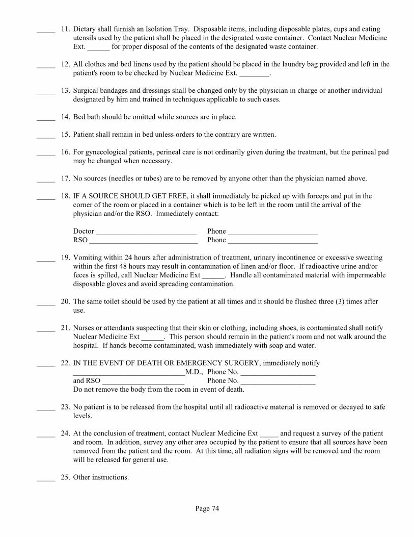
Page 74
_____ 11. Dietary shall furnish an Isolation Tray. Disposable items, including disposable plates, cups and eatingutensils used by the patient shall be placed in the designated waste container. Contact Nuclear MedicineExt. ______ for proper disposal of the contents of the designated waste container.
_____ 12. All clothes and bed linens used by the patient should be placed in the laundry bag provided and left in thepatient's room to be checked by Nuclear Medicine Ext. ________.
_____ 13. Surgical bandages and dressings shall be changed only by the physician in charge or another individualdesignated by him and trained in techniques applicable to such cases.
_____ 14. Bed bath should be omitted while sources are in place.
_____ 15. Patient shall remain in bed unless orders to the contrary are written.
_____ 16. For gynecological patients, perineal care is not ordinarily given during the treatment, but the perineal padmay be changed when necessary.
_____ 17. No sources (needles or tubes) are to be removed by anyone other than the physician named above.
_____ 18. IF A SOURCE SHOULD GET FREE, it shall immediately be picked up with forceps and put in thecorner of the room or placed in a container which is to be left in the room until the arrival of thephysician and/or the RSO. Immediately contact:
Doctor ___________________________ Phone ________________________RSO _____________________________ Phone ________________________
_____ 19. Vomiting within 24 hours after administration of treatment, urinary incontinence or excessive sweatingwithin the first 48 hours may result in contamination of linen and/or floor. If radioactive urine and/orfeces is spilled, call Nuclear Medicine Ext ______. Handle all contaminated material with impermeabledisposable gloves and avoid spreading contamination.
_____ 20. The same toilet should be used by the patient at all times and it should be flushed three (3) times afteruse.
_____ 21. Nurses or attendants suspecting that their skin or clothing, including shoes, is contaminated shall notifyNuclear Medicine Ext ______. This person should remain in the patient's room and not walk around thehospital. If hands become contaminated, wash immediately with soap and water.
_____ 22. IN THE EVENT OF DEATH OR EMERGENCY SURGERY, immediately notify______________________________M.D., Phone No. ____________________and RSO ______________________ Phone No. ____________________Do not remove the body from the room in event of death.
_____ 23. No patient is to be released from the hospital until all radioactive material is removed or decayed to safelevels.
_____ 24. At the conclusion of treatment, contact Nuclear Medicine Ext _____ and request a survey of the patientand room. In addition, survey any other area occupied by the patient to ensure that all sources have beenremoved from the patient and the room. At this time, all radiation signs will be removed and the roomwill be released for general use.
_____ 25. Other instructions.

Page 75
APPENDIX Q
RADIONUCLIDE THERAPY SURVEY
Survey of the patient's room and surrounding areas shall be conducted as soon as practicable after administration ofradionuclide and at conclusion of treatment to ensure that all sources have been removed from the patient and that nosources remain in the patient's room or other area occupied by the patient.
PATIENT __________________________________________________ ROOM ____________________________
HOSPITAL NO. _________________________________ AGE _____________ SEX ________________________
THERAPIST _____________________________________________________________________________________
PERMANENT IMPLANT OR INTERNAL DOSE ____ TEMPORARY IMPLANT _________________________
SOURCE ADMINISTRATION _______________ /______________ SOURCE REMOVAL _________/__________
RADIONUCLIDE ___________ TREATMENT DOSE __________ TECHNIQUE __________________________
INSTRUMENT USED ____________________ MODEL __________ SERIAL __________ BKG ______mR/hr
RADIATION SURVEY OF PATIENT & RADIATION SURVEY OF PATIENT &SURROUNDING AFTER ADMINISTRATION SURROUNDING AT CONCLUSION OF TREATMENT
DATE __________________ DATE _________________TIME ___________________ TIME __________________SURVEYOR ____________________________ SURVEYOR____________________________________
LOCATION mR/hr LOCATION mR/hrMax @ Surface of Patient _____ Max @ Surface of Patient _____Max @ 1 meter from Patient _____ Max @ 1 meter from Patient _____Patient's Bedside, Left _____ Patient's Bedside, Left _____Patient's Bedside, Right _____ Patient's Bedside, Right _____Patient's Bedside, Foot _____ Patient's Bedside, Foot _____Entrance Door _____ Entrance Door _____Adjacent Room _____ Adjacent Room _____Adjacent Room _____ Adjacent Room _____Max in Unrestricted Area _____ Max in Unrestricted Area _____
(Identify) (Identify)
__________________________________________________________________________________________________________________________________________________________________________________________________
SKETCH: (If Applicable)
X indicates location of radiation warning sign, patient bed and chart also posted.

Page 76
APPENDIX R
ACTIVITIES AND DOSE RATES FOR AUTHORIZING PATIENT RELEASE^COLUMN 1 COLUMN 2
ACTIVITIES AT OR BELOW WHICH DOSE RATE AT 1 METER, AT OR BELOWPATIENTS MAY BE RELEASED WHICH PATIENTS MAY BE RELEASED*
RADIONUCLIDE Gbq mCi mSv/hr mrem/hrAg 19 520 0.08 8111
Au 3.5 93 0.21 21198
Cr 4.8 130 0.02 251
Cu 8.4 230 0.27 2764
Cu 14 390 0.22 2267
Ga 8.7 240 0.18 1867
I 6.0 160 0.26 26123
I 0.25 7 0.01 1125
I implant 0.33 9 0.01 1125
I 1.2 33 0.07 7131
In 2.4 64 0.2 20111
Ir implant 0.074 2 0.008 0.8192
P ** ** ** **32
Pd 1.5 40 0.03 3103
Re 28 770 0.15 15186
Re 29 790 0.20 20188
Sc 11 310 0.17 1747
Se 0.089 2 0.005 0.575
Sm 26 700 0.3 30153
Sm 1.1 29 0.04 4117m
Sr ** ** ** **89
Tc 28 760 0.58 5899m
Tl 16 430 0.19 19201
Y ** ** ** **90
Yb 0.37 10 0.02 2169
^The activity values were computed based on 5 millisieverts (0.5 rem) total effective dose equivalent.*If the release is based on the dose rate at 1 meter in Column 2, the licensee must maintain a record as required by10CFR35.75© because the measurement includes shielding by tissue. See Regulatory Position 3.1, Records of Release,for information on records.**Activity and dose rate limits are not applicable in this case because of the minimal exposures to members of the publicresulting from activities normally administered for diagnostic or therapeutic purposes.
NOTES: The mCi values were calculated using Equations 2 or 3 and the physical half-life. The Gbq values werecalculated based on the mCi values and the conversion factor from mCi to Gbq. The dose rate values are calculatedbased on the mCi values and the exposure rate constants.
In general, the values are rounded to two (2) significant figures. However, values less than 0.37 Gbq (10 mCi) or 0.1mSv (10 mrem) per hour are rounded to one significant figure. Details of the calculations are provided in NUREG-1492(Ref. 2).
Although non-byproduct materials are not regulated by the NRC, information on non-byproduct material is included inthis guide for the convenience of the licensee.
Agreement State regulations may vary. Agreement State licensees should check with the State regulations prior to usingthese values.

Page 77
APPENDIX S
ACTIVITIES AND DOSE RATES ABOVE WHICH INSTRUCTIONS SHOULD BEGIVEN WHEN AUTHORIZING PATIENT RELEASE*
COLUMN 1 COLUMN 2ACTIVITY ABOVE WHICH DOSE RATE AT 1 METER ABOVEINSTRUCTIONS ARE REQUIRED WHICH INSTRUCTIONS ARE REQUIRED
RADIONUCLIDE Gbq mCi mSv/hr mrem/hr
Ag 3.8 100 0.02 2111
Au 0.69 19 0.04 4198
Cr 0.96 26 0.004 0.451
Cu 1.7 45 0.05 564
Cu 2.9 77 0.04 467
Ga 1.7 47 0.04 467
I 1.2 33 0.05 5123
I 0.05 1 0.002 0.2125
I implant 0.074 2 0.002 0.2125
I 0.24 7 0.02 2131
In 0.47 13 0.04 4111
Ir implant 0.011 0.3 0.002 0.2192
P ** ** ** **32
Pd 0.3 8 0.007 0.7103
Re 5.7 150 0.03 3186
Re 5.8 160 0.04 4188
Sc 2.3 62 0.03 347
Se 0.018 0.5 0.001 0.175
Sm 5.2 140 0.06 6153
Sm 0.21 6 0.009 0.9117m
Sr ** ** ** **89
Tc 5.6 150 0.12 1299m
Tl 3.1 85 0.04 4201
Y ** ** ** **90
Yb 0.73 2 0.004 0.4169
*The activity values were computed based on 1 millisieverts (0.1 rem) total effective dose equivalent.**Activity and dose rate limits are not applicable in this case because of the minimal exposures to members of the publicresulting from activities normally administered for diagnostic or therapeutic purposes.
NOTES: The mCi values were calculated using Equations 2 or 3 and the physical half-life. The Gbq values werecalculated based on the mCi values and the conversion factor from mCi to Gbq. The dose rate values are calculatedbased on the mCi values and the exposure rate constants.
In general, the values are rounded to two (2) significant figures. However, values less than 0.37 Gbq (10 mCi) or 0.1mSv (10 mrem) per hour are rounded to one significant figure. Details of the calculations are provided in NUREG-1492(Ref. 2).
Although non-byproduct materials are not regulated by the NRC, information on non-byproduct material is included inthis guide for the convenience of the licensee.
Agreement State regulations may vary. Agreement State licensees should check with the State regulations prior to usingthese values.

Page 78
APPENDIX TACTIVITIES OF RADIOPHARMACEUTICALS THAT REQUIRE INSTRUCTIONS AND RECORDS
WHEN ADMINISTERED TO PATIENTS WHO ARE BREAST-FEEDING AN INFANT OR CHILDCOLUMN 1 COLUMN 2 COLUMN 3ACTIVITY ABOVE WHICH ACTIVITY ABOVE WHICH EXAMPLES OF RECOMMENDED
INSTRUCTIONS ARE REQUIRED A RECORD IS REQUIRED DURATION OF INTERRUPTIONOF BREAST-FEEDING*
RADIONUCLIDE MBq mCi MBq mCiI NaI 0.01 0.0004 0.07 0.002 Complete cessation131
(for this infant or child)
I NaI 20 0.5 100 3123
I OIH 100 4 700 20123
I mIBG 70 2 400 10 24 hr for 370 MBq (10 mCi)123
12 hr for 150 MBq (4 mCi)I OIH 3 0.08 10 0.4125
I OIH 10 0.30 60 1.5131
Tc DTPA 1,000 30 6,000 15099m
Tc MAA 50 1.3 200 6.5 12.6 hr for 150 MBq (4 mCi)99m
Tc Pertechnetate 100 3 600 15 24 hr for 1,100 Mbq (30 mCi)99m
12 hr for 440 MBq (12 mCi)Tc DISIDA 1,000 30 6,000 15099n
Tc Glucoheptonate 1,000 30 6,000 17099m
Tc HAM 400 10 2,000 5099m
Tc MIBI 1,000 30 6,000 15099m
Tc MDP 1,000 30 6,000 15099m
Tc PYP 900 25 4,000 12099m
Tc Red Blood Cell 400 10 2,000 50 6 hr for 740 MBq (20 mCi)99m
In Vivo LabelingTc Red Blood Cell 1,000 30 6,000 15099m
In Vitro LabelingTc Sulphur Colloid 300 7 1,000 35 6 hr for 440 MBq (12 mCi)99m
Tc DTPA Aerosol 1,000 30 6,000 15099m
Tc MAG3 1,000 30 6,000 15099m
Tc White Blood 100 4 600 15 24 hr for 1,100 MBq(5 mCi)99m
Cells 12 hr for 440 MBq (2 mCi)Ga Citrate 1 0.04 7 0.2 1 month for 150 MBq (4 mCi)67
2 weeks for 50 MBq (1.3 mCi)1 week for 7 MBq (0.2 mCi)
Cr EDTA 60 1.6 300 851
In White Blood 10 0.2 40 1 1 week fpr 20 MBq (0.5 mCi)111
CellsTl Chloride 40 1 200 5 2 weeks for 110 MBq (3 mCi)201
*The duration of interruption of breast-feeding is selected to reduce the maximum dose to a newborn infant to less than 1 mSv (0.1 rem), althoughthe regulatory limit is 5 mSv (0.5 rem). The actual doses that would be received by most infants would be far below 1 mSv (0.1 rem). Of course, thephysician may use discretion in the recommendation, increasing or decreasing the duration of interruption.
NOTES: Activities are rounded to one significant figure, except when it was considered appropriate to use two significant figures. Details of thecalculations are shown in NUREG-1492, Regulatory Analysis on Criteria for the Release of Patients Administered Radioactive Material (Ref. 2).
If there is no recommendation in Column 3 of this table, the maximum activity normally administered is below the activities that require instructionson interruption or discontinuation on breast-feeding.
Although non-byproduct materials are not regulated by the NRC, information on non-byproduct material is included in this guide for theconvenience of the licensee.
Agreement State regulations may vary. Agreement State licensees should check with the State regulations prior to using these values.

Page 79
APPENDIX U
HOME INSTRUCTION, RADIOPHARMACEUTICAL THERAPY
Patient, _______________________________________________ observe these instructions for ____________days.
1. Sleep alone. If employed, take ______ days off from work.
2. Whenever possible use separate toilet facilities that are not used by other members of the family.
3. Use care so that the area around the toilet is not soiled with urine. Flush the toilet three (3) times after each use.
4. Bed linens and underclothing should be washed separately after the other washing has been completed. Then the tubor washing machine should be rinsed twice.
5. Wash bathtub or shower with soap and cleanser after tub or shower bath.
6. Contact the Nuclear Medicine Department,_________________________, if you have questions.
________________________________________M.D.

Page 80
APPENDIX V
HOME INSTRUCTIONS
Patient: _____________________________________________
A small radioactive source has been placed (implanted) inside your body. The source is actually many small metallicpellets or seeds, which are each about 1/3 to 1/4 of an inch long, similar in size and shape to a grain of rice. To minimizeexposure to radiation to others from the source inside your body, you should do the following for ____________days.
1. Stay a distance of _____ feet from __________________________________________.
2. Maintain separate sleeping arrangements.
3. Minimize time with children and pregnant women.
4. Do not hold or cuddle children.
5. Avoid public transportation.
6. Examine any bandages or linens that come into contact with the implant site for any pellets that may have comeout of the implant site.
7. If you find a seed or pellet that falls out:
DO NOT HANDLE it with you fingers. Use something like a spoon or tweezers to place it in a jar or othercontainer that you may close with a lid.
Place the container with the seed or pellet in a location away from people.
Notify your physician at once.
______________________________________M.D.

Page 81
APPENDIX W
STRONTIUM OPHTHALMIC APPLICATOR THERAPY RECORD90
Patient ___________________________________________ Date: ________________________________
Hospital No. _____________________________ Room ________ Age __________ Sex _______________
Therapist __________________________________________M.D.
DATE TIME SOURCE TREATMENT TREATMENT TECHNIQUE TIME SOURCEREMOVED TIME DOSE RETURNED TOFROM NUCLEAR NUCLEARRADIOLOGY RADIOLOGYLABORATORY LABORATORY
_________________________________________________________________________________________________
_________________________________________________________________________________________________
_________________________________________________________________________________________________
_________________________________________________________________________________________________
_________________________________________________________________________________________________
_________________________________________________________________________________________________

Page 82
APPENDIX X
INSTRUCTIONS FOR FAMILY OF RELEASED PATIENT
Patient Name ______________________________________________________________________________________
For further information contact ________________________________ Phone No. ______________________________
Please show this form to every physician consulted concerning the patient until
(DATE) ___________________________________________________
Patient Name_______________________________________ was treated on __________________________________
with ________________mCi of ________________________ in the form of ___________________________________
NO SPECIAL RADIATION SAFETY PRECAUTIONS ARE REQUIRED AFTER (DATE) ______________________
UNTIL THAT DATE:
Persons under 45 years of age should not remain closer than the following distances from the patient for the timeperiod indicated below:
________________________ to _________________________Date Date
Permissible distance________ feet or more for ________hours/week. At other times, remain farther than six (6) feet.
SPECIAL PRECAUTIONS:
a) Spouse or other individuals caring for patient: _____________________________________________________
b) Children or pregnant women: __________________________________________________________________
______________________________________________________________________________________________
c) Sleeping arrangements: _______________________________________________________________________
______________________________________________________________________________________________
If the patient is to be hospitalized, or if death should occur, notify the following individuals immediately:
______________________________________________________________________________________________
______________________________________________________________________________________________
______________________________________________________________________________________________
(A copy of this form should be kept with the patient's record.)

Page 83
APPENDIX Y
REPORT OF RADIOACTIVITY OF CADAVER
The physician in charge of the case should fill out the following and attach to the patient’s chart and death certificate. The Radiation Safety Officer is available for assistance to the physician.
Hospital: ___________________________________________
Name of Deceased: ___________________________________
Hospital Number: ______________________ Date and Time of Death: _________________________
Diagnosis: __________________________________________________
Radioactivity Survey - Before Autopsy:
Radionuclide: __________________________ Activity (mCi): ____________________
Last Treatment Date: __________ Hour: __________a.m.;p.m.
Survey Date: ______________________________ Hour: _____________a.m./p.m.
Elapsed Time from Last Treatment to Survey in Days: _______________________________________
Maximum Level of Radiation @ Surface of Body (mR/hr): ____________________________________
Instrument Used: _____________________________________________________________________
Model #: ____________________ Serial #: ______________________ Calibration Date: _____________________
Signed: _____________________________________________Radiation Safety Officer
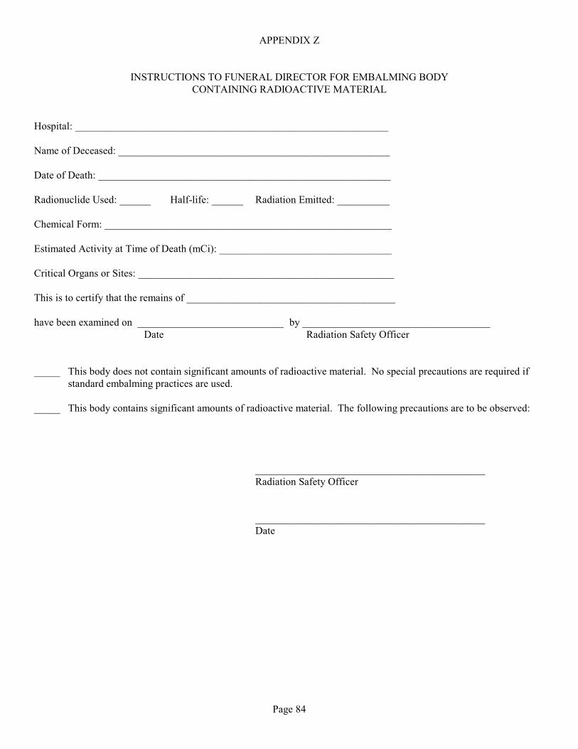
Page 84
APPENDIX Z
INSTRUCTIONS TO FUNERAL DIRECTOR FOR EMBALMING BODYCONTAINING RADIOACTIVE MATERIAL
Hospital: ____________________________________________________________
Name of Deceased: ____________________________________________________
Date of Death: ________________________________________________________
Radionuclide Used: ______ Half-life: ______ Radiation Emitted: __________
Chemical Form: _______________________________________________________
Estimated Activity at Time of Death (mCi): _________________________________
Critical Organs or Sites: _________________________________________________
This is to certify that the remains of ________________________________________
have been examined on ____________________________ by ____________________________________ Date Radiation Safety Officer
_____ This body does not contain significant amounts of radioactive material. No special precautions are required ifstandard embalming practices are used.
_____ This body contains significant amounts of radioactive material. The following precautions are to be observed:
____________________________________________Radiation Safety Officer
____________________________________________Date

Page 85
APPENDIX AA
AUTOPSY OR SURGERY PRECAUTIONS
The following are main precautions required for autopsies or surgery on bodies containing large doses of radionuclides:
A. General
1. Impermeable disposable gloves must always be worn to prevent contamination of skin and nails withmaterial difficult to remove.
2. If the combined beta and gamma dose rate is high enough to deliver more than the permissible dose tohand or whole body, the autopsy or surgery should be performed by a team of physicians working inrelay.
3. Tissue and organs removed should be handled with long handled forceps and scissors. Specimens shouldbe refrigerated in jars or other containers, or fixed, and suitably labeled to indicate when they can safelybe worked on and studied.
B. Special
1. All tissues and body fluids should be surveyed by the RSO and handled according to hisrecommendations. Urine and blood should be removed and stored or disposed.
2. Tissue specimens held three (3) months can be considered inactive.

Page 86
APPENDIX BB
RADIATION SAFETY COMMITTEE
NOTE: THIS FORM SHALL BE LEGIBLE!
APPLICATION FOR AUTHORIZATION TO USE RADIONUCLIDES/RADIATION SOURCES FORIN VIVO HUMAN USE
1. Date: ______New Application ______Amendment ______Renewal
2. Applicant Name: _____________________ Degree: ____________________
Department: _____________________________________ Box Number: _______________________
Telephone Number: _______________________________
3. Description of Radiation Source(s). Describe all the radiation sources that subjects will encounter:
a. Radioactive Material:Compound & Number of
Each Activity Each Method of AdministrationsRadionuclide Administration Administration Each Subject
Where will the material be administered and by whom?
b. X-Ray Radiography:
Type Film mAs/ Number of Entrance Exam Size kVp film Films/Exam Distance Filter mR
Where will radiography occur and who will supervise?
c. Fluoroscopy:
Anatomical Beam Fluoroscop Entrance ExposureArea Size kVp mA Time Rate (mR/mA-minute)
Where will fluoroscopy occur and who will supervise?

Page 87
4. Experimental Limitations:
a. Explain why it is necessary to use radiation rather that alternate procedures:
b. Explain the factors that determine the minimum amount of radiation that is necessary, i.e. limits based ofinstrument sensitivity or physiological parameters:
5. Description of Subjects and Selection Criteria
a. Age Distribution:
b. Sex:
c. Special basis for selection (disease, abnormality, occupation, etc.):
d. Number of special selected subjects:
e. Indicate time frame; how many subjects will be studied each year?
f. How will the subject benefit from the study?
g. Are potential subjects rejected if they are or might be pregnant?How is nonpregnancy assured?
h. Are potential subjects rejected if they:
1) Had volunteered for similar studies within the past year?
2) Had significant medical exposure?
3) Receive significant occupational exposure?
How are these conditions assured?

Page 88
6. Radiation Protection for Subjects:
Describe all methods for protecting the subject from unnecessary radiation, i.e. gonadal shielding, thyroid blockagainst iodine, etc.:
7. Radiation Dosimetry:
Include dosimetry calculations and summary of radiation doses. References should be cited if used:
8. Summary of Risk to Subject:
Attach copy of Consent Form to be employed.
9. The Committee on Use of Human Subjects:
Pending review ___________________________
Approved _______________________________(Date)
10. Experience Form Attached: _____Yes _____ No
Applicant's Signature: _________________________________________ (IN INK SIGNATURE AND DATE)
======================================================================================
11. Review by Radiation Safety Committee:
By:_________________________________________________ Date:____________________
Recommendation: _____Approve _____Disapprove
Comments:

Page 89
RADIATION SAFETY COMMITTEE
EXPERIENCE FORMFOR
IN VIVO HUMAN RADIATION USE
NAME: ______________________________________________ DATE:__________________________
DEPARTMENT: _______________________________________
Formal Courses:
Give names of formal courses taken which would assist in the understanding of radiation protection problems (e.g.nuclear or atomic physics, health physics, radionuclide technology, radiation biology, etc.). Specify when and wheretaken.
Practical Experience:Summarize in 200 words or less. Indicate radionuclides and chemical forms used, amounts, when and where:
_____________________________________Signature

Page 90
APPENDIX CC
RADIATION SAFETY COMMITTEE
NOTE: THIS FORM SHALL BE LEGIBLE!
APPLICATION FOR USE OF RADIOACTIVE MATERIAL/RADIATION SOURCE
1. Date: ______ New Application ______Amendment ______Renewal
2. Applicant Name: ____________________________________ Degree: ________
Department: ________________________________________ Box Number: _____
Telephone Number: __________________________________
3. Check Category for Intended Use:
Animal Use _____
Class Room Use _____
In Vitro Use _____
In Vivo Human Use _____ (Use In Vivo Human Use Form)
4. Radiation Source(s): Chemical/ Maximum mCi Maximum mCi Use
Radionuclide Physical Form Possession (Orders/Month)
Describe other radiation sources (i.e. x-ray unit, x-ray diffraction, etc.):
5. Describe proposed use of each item:

Page 91
6. List individuals who will handle requested material (Name and Title/Position):
7. List Radiation Detection Instruments available (survey meter, beta/gamma counting systems, etc.):Radiation Location
Type Manufacturer Model # Detected (Room Number)
List radiation sources available (x-ray units, diffraction units, sealed sources, etc.):
Type Manufacturer Model # Location (Room Number)
8. Describe Storage of Material (Facilities, Containers, Special Shielding):
9. Describe Handling of Material (Protective Equipment, Special Handling Tools, Hoods):
10. Describe special laboratory radiation safety practices, including monitoring practices:
11. Describe waste disposal procedures and estimate quantities per month of liquid, solid waste. Describe type ofwaste anticipated (i.e. aqueous or nonaqueous, infectious, flammable, etc.):

Page 92
12. Indicate rooms where radioactive materials and/or sources of radiation will be stored, used, and site for sewerdisposal, if appropriate, by drawing a floor plan of the facility:
13. For In Vitro Human Use:
a. Attach Consent Form to be employed.
b. Committee on Use of Human Subjects:
Pending Review: _____
Approved: ________________________________(Date)
14. Experience Form Attached: _____Yes _____ No
15. Applicant's Signature: __________________________________________(In ink signature and date)
16. Review by Radiation Safety Committee:
By: _______________________________ Date: ___________________________
Recommendation: _____Approve _____Disapprove
Comments:

Page 93
RADIATION SAFETY COMMITTEE
EXPERIENCE FORMFOR
USE OF RADIOACTIVE MATERIAL/RADIATION SOURCE
NAME: __________________________________________________ DATE: ________________________________
DEPARTMENT: __________________________________________
Formal Courses:Give names of formal courses taken which would assist in the understanding of radiation protection problems (e.g.nuclear or atomic physics, health physics, radionuclide technology, radiation biology, etc.). Specify when and wheretaken.
Practical Experience:Summarize in 200 words or less. Indicate radionuclides and chemical forms used, amounts, when, and where:
__________________________________Signature

Page 94
APPENDIX DD
RADIATION INCIDENT PLANA. Purpose
To outline a plan of action for meeting the basic requirements of Tulane University's role in an external disastersituation resulting in casualties who are suspected or known to have become contaminated with transferableradioactive material, either by ingestion, deposition on skin or entrance through open wounds.
B. Objectives
1. To handle radiation casualties.
2. To treat radiation casualties using the best medical standards.
3. To avoid or minimize radiation exposure to hospital personnel and patients.
4. To continue the usual hospital functions.
5. To integrate the specialized supplies and experienced radiation safety personnel into hospital routines.
C. Notification of Disaster
1. Any person receiving information of a disaster resulting in radiation casualties should immediately relay suchinformation to the Emergency Department Charge Nurse who will complete the Radiation Incident ReportForm.
2. Upon verification of such an incident, the Emergency Department Charge Nurse shall immediately notify theOffice of Environmental Health and Safety (OEHS) 988-5486 of this emergency situation and request thatthe Radiation Safety Officer (RSO) respond. If the incident occurs after normal work hours, holidays orweekends, the Emergency Department Charge Nurse shall contact Hospital Communications, and requirethem to immediately contact by pager or telephone the OEHS On Call staff member.
Designated responding staff members are listed in the following priority for contact:
a. Radiation Safety Officer (RSO)
b. Medical Physicist (Radiology)
c. Deputy Radiation Safety Officer (DRSO)
d. Hospital Safety Officer (HSO)
The above staff may request that other OEHS members assist in the emergency response activity.
3. The RSO or designee will contact the notifying agency to obtain additional information as necessary andnotify the Radiation Protection Division (504) 756-0160.
4. The Emergency Department Charge Nurse or Physician in conjunction with the RSO will evaluate thesituation and decide whether to implement the Radiation Incident Plan.

Page 95
5. Upon Implementation of this plan, the Emergency Department will contact the telephone operators andadvise them to notify the following of the emergency situation:
Administrator On Call
Nursing Supervisor (Off Shifts)
TUHSC Police
Emergency Department Medical Director
Environmental Services
Public Relations
6. The RSO or designee upon their arrival at the Emergency Department will then coordinate the activation ofthe Radiation Emergency Area (REA). The Emergency Department Charge Nurse will give the completedRadiation Incident Form to the first responding staff member. Prior to arrival of the RSO or designee,Emergency Department personnel will begin to establish the Radiation Emergency Area (REA) by using theappropriate signage and ropes.
D. Planning
1. The Emergency Room Nurses will evaluate the status of the patients present and work with the physicians todischarge or re-locate as many as possible. All pregnant women will be moved to other areas of TUHC.
2. Communications via the Edac radio between the ambulance and Emergency Room staff will establish theextent of trauma injuries and level of contamination. A decision will be made by the RSO, the EmergencyRoom Charge Nurse or the Emergency Room Physician whether to direct patients to the decontamination orto the treatment areas inside the designated Radiation Emergency Area (REA).
3. The Radiation Emergency Area (REA) will be established in the following manner:
a. Room 1074 will be used for Decontamination and rooms 1072 and 1070 will be used for post-decontamination treatment. Drain covers will not be needed to prevent radioactive materials fromentering the sewer system.
b. The RSO or designee will establish the Control Points where a staff member will be positioned with asurvey meter.
c. The REA will be roped off and posted with signs that read, "Caution, Radiation Area." Ropes and signswill be used as provided by the RSO or designee. See REA in Appendix for rope locations.
d. Plastic sheets should be placed on top of the stretcher mattresses.
4. The RSO shall designate person(s) to man Control Points and shall check all radiation monitoring equipmentto insure it is in working order.
5. Office of Environmental Health & Safety (OEHS) and TUHSC Police representatives will be responsible forinsuring all doors are opened in the receiving route of REA, assisting with providing ropes and signs in theREA area and insuring area is free from by-standers and nonessential equipment.

Page 96
6. Public Relations and/or Nursing Supervisor will be responsible for all press information which has beenreviewed for technical content and approved by the physician and RSO prior to release.
E. Radiation/decontamination Team
1. Physicians
a. Emergency Department Physicians shall be in charge of medical care.
b. The RSO or designee shall be in charge of directing decontamination procedures.
c. the RADIATION ACCIDENT PATIENT FORM shall be employed with the assistance of the RSO ordesignee.
2. Emergency Room Personnel will be assigned tasks by the RSO or designee.
a. Two RNs to assist physicians with patients.
b. One Nursing Assistant to be in the REA to receive supplies from outside REA and record treatment.
c. One Nursing Assistant to be outside REA to obtain supplies, as needed.
3. Radiation Safety Personnel (See RADIATION INCIDENT PLAN CRITICAL PERSONNEL)
a. Radiation Safety Officer (RSO) and other responding OEHS personnel.
F. Receiving Procedures
1. Persons suspected of being contaminated with radionuclides will be received at the Emergency Department.
2. Radiation/Decontamination Team (Physicians, Nurses and radiation safety personnel) meeting thetransporting vehicle will be attired as follows:
a. Gowns or other disposable outer clothing to include, universal precaution - personal protective attire, i.e.gloves and goggles as needed.
b. Pocket dosimeters worn at neck region where they can be easily removed by other persons formonitoring. They should be read at intervals during decontamination and reported to the Control Pointattendant.
3. The patient will be met at transporting vehicle with a stretcher having the top surface of the mattress covered.
4. The physician will evaluate the patient to determine if there is any medical problem or associated injuryrequiring care prior to decontamination.
5. If immediate medical care is not needed, then the patient shall be secured in plastic wrap and transported tothe decontamination shower.
6. The RSO or designee shall monitor the patient and log all contaminated areas with location and exposuremeasurement on Radiation Accident Exposure Log. (See RADIATION ACCIDENT EXPOSURE LOG)

Page 97
7. All patient clothing shall be removed and placed in plastic bags labeled "patient's clothing". Valuables shallbe bagged and labeled with patient information. The RSO or designee will be responsible for disposition ofclothing and valuables.
8. The Emergency Room Physician will proceed with required critical patient care and evaluation. The RSO ordesignee will collect swab samples of contaminated areas for radiation safety personnel to monitor. Thesesamples may include samples from ear canals, nostrils, scalp, fingernails, toenails, mouth and contaminatedareas of body, etc. All samples are to be placed in separate containers labeled with patient's name, body areaand time obtained.
9. Physicians shall proceed with decontamination of radioactive areas (OPEN WOUNDS FIRST PRIORITY).
10. The patient shall shower or lightly scrub all contaminated areas with betadine prep without breaking the skin. Medical staff may assist as necessary.
11. The RSO or designee will again monitor following decontamination and log dose and, if necessary, repeat theshower and scrub procedure.
12. After decontamination, the patient may be admitted to a regular hospital bed. If the patient has inhaled oringested radionuclides, he should be placed in a private room. All urine and feces shall be saved formonitoring if there is suspected internal contamination. Radiation signs shall be placed on room door.
13. The transporting vehicle and personnel will be notified by the RSO to REMAIN WITH THE VEHICLE untilmonitored and released or instructed by the RSO concerning decontamination.
14. The RSO or designee shall survey the ambulance and Emergency Medical Service (EMS) personnel prior totheir release to duty. Decontamination procedures will be performed at the direction of the RSO or designee.
15. Area entrances and hallways will be monitored by the RSO or designee after patient is located in RadiationEmergency Area so as to prevent tracking to other hospital areas.
G. Control Point Functions
1. The RSO or designee shall assign OEHS and TUHSC Police staff members to function as Control Pointattendants. One TUHSC Police Officer with the help of Emergency Department staff will secure the REAand then report to the Emergency Department ramp, one TUHSC Police Officer will be posted adjacent to theNursing Station. One OEHS person will install the ropes and signs, and then assist in other radiation safetyfunctions.
2. The attendants shall be responsible for:
a. Restricting access to only personnel authorized by the attending physician, Radiation Safety Officer orNursing Supervisor.
b. Assure that no person or object is allowed to leave REA (after patient admitted) until monitoring hasbeen performed and there is no detectable radionuclide contamination. Permission will be given by theRSO or designee.

Page 98
H. Radiation Emergency Area (REA)
1. The REA will consist of the Emergency Department.
2. The Control Points will be established in corridors outside the REA.
3. Medical personnel attending the patient will wear protective clothing and a pocket dosimeter.
4. Attending personnel will remove outer protective clothing and place in plastic bag labeled radioactive beforeleaving REA.
5. All personnel will be monitored before leaving area.
6. No supplies, clothing, etc., will leave the area until monitored.
7. The REA will remain closed to all personnel until it has been determined to be free of radioactivecontamination by the RSO or designee.
8. The RSO or designee will dictate need for masks, respirators, as well as need to shut off air supply and ventsto avoid airborne contamination by notifying the Maintenance Department.
9. Staff performing decontamination with water should wear plastic or rubber aprons and gloves.
10. Specimens and samples will be collected and obtained according to needs as ascertained by the RSO.
11. Disposal of all radioactive material will be supervised by RSO or designee.
I. Procedures for Transfer of Patient
1. Following decontamination and emergency treatment, the patient may be transferred from the REA to theappropriate section of the hospital.
J. Extreme Medical Emergency Requiring Immediate Surgery
1. In the event of the above circumstances, the patient will be secured in plastic sheet to prevent spread ofcontamination and taken directly to the Operating Room on an uncontaminated stretcher accompanied by theRSO or designee.
2. The OR personnel will be notified of the contaminated patient by the RSO or designee.
3. The Operating Room becomes a part of the REA and no personnel or objects may be removed from the areaunless monitored and found to be free of contamination.
4. Removal of patient following surgery and decontamination will be under supervision of RSO or designee incoordination with attending physician.

Page 99
K. Personnel Clearance
1. Each team member goes to Control Point where the RSO or designee is located and removes protectiveclothing (placing all of it in a plastic bag designated for contaminated linen and trash). Contaminatedmaterial will be transported to the OEHS Hazardous Waste Room in the Medical School.
a. Remove outer gloves first, turning them inside out as they are pulled off.
b. Give dosimeters to Radiation Safety Officer or designee.
c. Remove outer surgical gown, turning it inside out; avoid shaking.
d. Remove head cover.
e. Take shower only at the direction of the RSO.
L. Radiation Safety Officer and Designee Responsibilities
(Several OEHS staff members may be asked to assist; OEHS will be responsible for contacting them andassigning duties.)
1. Supervise radiation protection at the hospital and restrict access to authorized persons only.
2. Install ropes and signs in the REA and insure that Control Points are secure.
3. Distribute dosimeters and record name, dosimeter number and initial reading of each unit. All equipmentwill be checked for proper operation.
4. Monitor patient and decontamination team during care of patient.
5. Monitor transport vehicle and personnel before they leave area.
6. Monitor route from transport vehicle to REA and decontaminate if necessary.
7. Collect all specimens to be analyzed for radiation content.
8. Collect dosimeters, monitor and evaluate personnel throughout the emergency.
9. Assist in the decontamination of patient when treatment of injury permits, as determined by attendingphysician.
10. Collect used protective clothing, wastes, samples and equipment for decontamination, evaluation and/ordisposal.
11. Record radiation survey findings of personnel and property, evaluate and recommend follow-up as necessaryto be completed following the incident.
12. Provide annual training to all staff involved with the radiation incident response.
13. Annually inspect decontamination supplies located in the Emergency Department.

Page 100
M. Emergency Department Physician Responsibilities
1. Evaluate and treat the patient's critical medical problems and then proceed to decontamination.
N. TUHSC Police Responsibilities
1. The TUHSC Officer will remain outside the roped area and not allow anyone to enter the secured locationwithout the approval of the RSO or designee.
2. Allows no person or object to leave REA without approval of the RSO or designee.
3. Makes sure that all doors are opened in the receiving route of REA and elevators are available, if needed.
4. Assists with roping off REA area.
O. Public Relations Responsibilities
1. Provide all press releases.
P. Environmental Services Responsibilities
1. Environmental Services supervising personnel shall be available to obtain additional supplies, whenrequested.
2. Clean REA under direction and supervisory of the RSO or the Deputy RSO.
Q. Emergency Department Charge Nurse Responsibilities
1. Work with physician to discharge as many Emergency Room patients as possible or move them out of theREA.
2. Work with the ED Staff to assist in the REA.
R. Nurse Working Within REA Responsibilities
1. Work directly with the physician in delivering care to the patient.
2. Collects specimen samples, wash contaminated areas, etc.
3. Requests and receives supplies from outside the REA.
4. Records information regarding care for patient’s chart.
S. Emergency Department Personnel Outside REA Responsibilities
1. Obtain any supplies or equipment needed. (See DECONTAMINATION SUPPLIES)
2. Supply medications as needed.
T. Final Survey REA
1. The Radiation Safety Officer and designee shall perform a radiation survey of the REA to confirm all areas,furniture, instruments and supplies are safe and contamination free.

Page 101
RADIATION EMERGENCY AREA PLAN
Contact Charles Reindl at 988-5486 for a copy of the Radiation Emergency Area Plan.

Page 102
RADIATION DISASTER PLAN CRITICAL PERSONNEL
(After Hours Contact Operator 988-5263)
1. Radiation Safety Officer:
Name: Charles F. Reindl, M.S.Work: 988-5486
2. Medical Physicist (Radiology):
Name: James Terry, Ph.D.Work: 988-5486
3. Deputy Radiation Safety Officer
Name: Jim Balsamo Jr., M.P.H.Work: 988-5486
4. Hospital Safety Officer
Name: Skip PeoplesWork: 988-7139
5. Medical Director of Radiology/Nuclear Medicine
Name: Harold NeitzschmanWork: 988-7627
6. Director of Radiology/Nuclear Medicine
Name: Sharon Hafner, R.T.Work: 988-6093
7. Director of Emergency Room Services
Name: Bryan Dean, R.N.Work: 988-1483
8. Security
Name: On DutyWork: 988-5555 or 988-5532
(After normal work hours, telephone the TUHC operator to have the above personnel contacted; the TUHC operator # is(988-5532)

Page 103
REA CONTROL SHEET
Personnel Information:
Name:Social Security No:Date of Birth:
Entering Rea Leaving Rea
DosimeterNumber
DosimeterReading
mRTime
a.m./p.m.
DosimeterReading
mRTime
a.m./p.m.
SurfaceMonitoring
mR/hr. Comments
Instrument Used in Surface Monitoring: Model #: Serial #:
Calibrated:
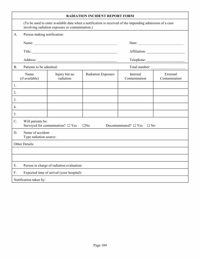
Page 104
RADIATION INCIDENT REPORT FORM
(To be used to enter available data when a notification is received of the impending admission of a caseinvolving radiation exposure or contamination.)
A. Person making notification:
Name: ______________________________________________ Date: _________________________
Title: _______________________________________________ Affiliation: _____________________
Address: ____________________________________________ Telephone: _____________________
B. Patients to be admitted: Total number: ___________________
Name(if available)
Injury but noradiation
Radiation Exposure InternalContamination
ExternalContamination
1.
2.
3.
4.
5.
C. Will patients be:Surveyed for contamination? G Yes GNo Decontaminated? G Yes G No
D. Name of accident:Type radiation source:
Other Details:
E. Person in charge of radiation evaluation:
F. Expected time of arrival (your hospital):
Notification taken by:

Page 105
RADIATION ACCIDENT PATIENT FORM
Full Name:
Social Security Number:
Birth Date: Age: Sex: Race:
Current local address:
Current permanent address:
Name and address of employer:
Father's name: Mother's name:
Women only:Date of last menstrual period: No. of pregnancies or miscarriages:
Could you be pregnant now?Definitely Yes: G Definitely No: G Not sure G
If you are pregnant, estimated date of delivery:
Any problems with the pregnancy?
PAST HISTORY:Any known treatment with x-rays or isotopes?
If so, reason for treatment:
Month/year of treatment:
Place where treatment was given:
Have you ever had any cancer or other malignancy?
If yes, type:
Date of diagnosis:
FAMILY HISTORY:How many children do you have? Are they all healthy?
If not, nature of disease or defect?
Indicate which, if any, of the following malignancies are present in one or more members of your family:Leukemia G Breast G Thyroid G Lung G Stomach G Intestines G Bone G
CURRENT MEDICATIONS:

RADIATION ACCIDENT PATIENT FORM
Page 106
ALLERGIES:
DETAILS OF RADIATION ACCIDENT:Location where accident occurred:
Time and date of exposure:
Type of radiation source:
Location of accident victim:
Distance from source:
Duration of exposure:
Shielding, including buildings, clothing, etc:
Dosimetry:Estimated radiation doses (whole body and organ specific):
Name, title, and address of individuals who estimated doses:
Method of dose estimate: G Historical G Dosimeters Other:
Immediate post-accident medical assessment: Time: __________ Date: __________Location:
Symptoms:
Physical findings:
Laboratory data, including pregnancy test in all women who might be pregnant:
Disposition of patient:
NAME/ADDRESS OF FAMILY PHYSICIAN:
NAME/TITLE OF PERSON COMPLETING THE FORM:
DATE/TITLE OF COMPLETION:

Page 107
DECONTAMINATION SUPPLIES
I. Available in Operating Room:Surgical Scrub SuitsGownsCapsShoe Covers
II. Available in Emergency Room:Patient GownsSheetsTowelsBlanketsPlastic Bags - to be supplied by the RSO or the designee
III. Available in Emergency Room:Labeled containers for fecal and urine specimens Sterile irrigation setsSpecimen bottles Sterile applicationsEmesis basins Sterile suture setsBedpans Mild soapLarge basins Hand Brushes (soft)4 x 4 sponges TapeDisposable gloves Tubes for blood samples
IV. Radiation Emergency Kit kept in the Emergency DepartmentSwabs Rope KitCaution, radioactive signs (1) Roll radioactive tapeRoll of plastic Caution tagsForms: REA Control Sheet Rubber Boots/Plastic Boots
Radiation Accident Exposure Log Forms from Radiation Incident Plan Red ink pens
V. Survey Meters, Pocket Dosimeters (Located in Nuclear Medicine, and OEHS Lab in Room M002 of the MedicalSchool)

Page 108
RADIATION ACCIDENT EXPOSURE LOG
Date:
ANTERIORFigure 1
POSTERIORFigure 2
Patient Name:
Time of Contamination:
Location:
Time of Decontamination:
Location:
Estimated Total Dose: mR/hr
Type of Radiation:
Internal Contamination: GYes GNo
Location:
List Medical Injuries:
Instructions:
1. Label HOT areas with arrow and note exposure inmR/hr.
2. Mark HOT area on chart in RED.
3. Mark decontamination readings in BLACK belowcontamination readings.
NOTES OR DECONTAMINATION PROCEDURES:
X____________________________________________
Signature of Individual Completing Form
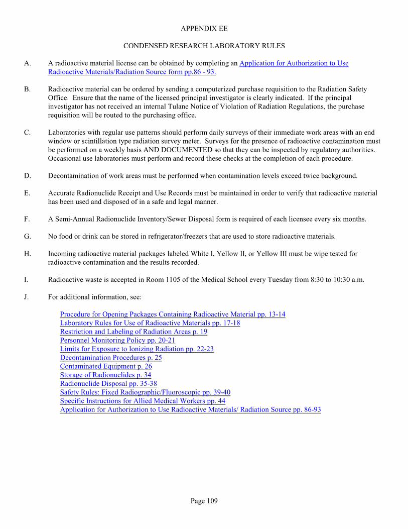
Page 109
APPENDIX EE
CONDENSED RESEARCH LABORATORY RULES
A. A radioactive material license can be obtained by completing an Application for Authorization to UseRadioactive Materials/Radiation Source form pp.86 - 93.
B. Radioactive material can be ordered by sending a computerized purchase requisition to the Radiation SafetyOffice. Ensure that the name of the licensed principal investigator is clearly indicated. If the principalinvestigator has not received an internal Tulane Notice of Violation of Radiation Regulations, the purchaserequisition will be routed to the purchasing office.
C. Laboratories with regular use patterns should perform daily surveys of their immediate work areas with an endwindow or scintillation type radiation survey meter. Surveys for the presence of radioactive contamination mustbe performed on a weekly basis AND DOCUMENTED so that they can be inspected by regulatory authorities. Occasional use laboratories must perform and record these checks at the completion of each procedure.
D. Decontamination of work areas must be performed when contamination levels exceed twice background.
E. Accurate Radionuclide Receipt and Use Records must be maintained in order to verify that radioactive materialhas been used and disposed of in a safe and legal manner.
F. A Semi-Annual Radionuclide Inventory/Sewer Disposal form is required of each licensee every six months.
G. No food or drink can be stored in refrigerator/freezers that are used to store radioactive materials.
H. Incoming radioactive material packages labeled White I, Yellow II, or Yellow III must be wipe tested forradioactive contamination and the results recorded.
I. Radioactive waste is accepted in Room 1105 of the Medical School every Tuesday from 8:30 to 10:30 a.m.
J. For additional information, see:
Procedure for Opening Packages Containing Radioactive Material pp. 13-14Laboratory Rules for Use of Radioactive Materials pp. 17-18Restriction and Labeling of Radiation Areas p. 19Personnel Monitoring Policy pp. 20-21Limits for Exposure to Ionizing Radiation pp. 22-23Decontamination Procedures p. 25Contaminated Equipment p. 26Storage of Radionuclides p. 34Radionuclide Disposal pp. 35-38Safety Rules: Fixed Radiographic/Fluoroscopic pp. 39-40Specific Instructions for Allied Medical Workers pp. 44Application for Authorization to Use Radioactive Materials/ Radiation Source pp. 86-93

Page 110
RADIOACTIVE MATERIAL PACKAGE RECEIPT AT TULANE UNIVERSITY HEALTH SCIENCES CENTER333 S. Liberty St.New Orleans, LA
Radioactive material packages must be logged/wipe tested by the Radiation Safety Office prior to being picked up bydepartment representatives. On most work days this is:
Charles Reindl, Radiation Safety OfficerPhone: 504/988-5486Beep: 504/544-9109
If the package is wet or appears to be damaged, immediately contact the Radiation Safety Office. Ask the carrier toremain at Receiving until it can be determined that neither he/she nor the delivery vehicle is contaminated.
No radioactive material packages will be accepted outside of normal working hours.
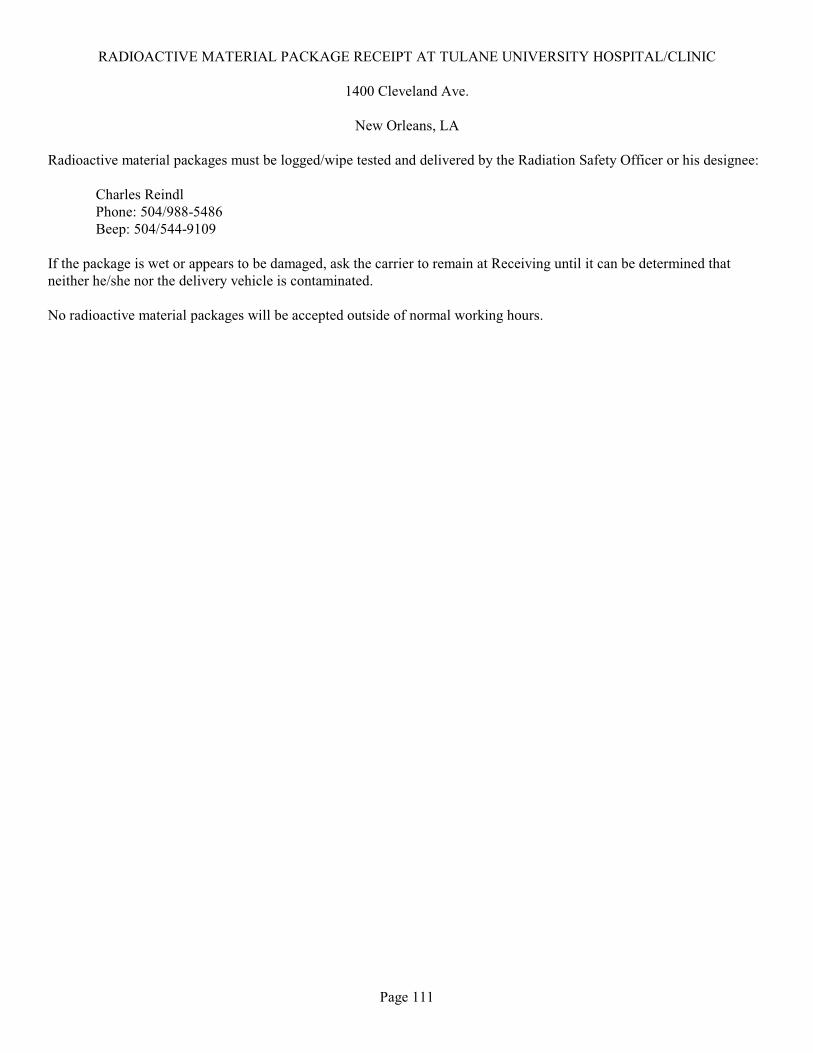
Page 111
RADIOACTIVE MATERIAL PACKAGE RECEIPT AT TULANE UNIVERSITY HOSPITAL/CLINIC
1400 Cleveland Ave.
New Orleans, LA
Radioactive material packages must be logged/wipe tested and delivered by the Radiation Safety Officer or his designee:
Charles ReindlPhone: 504/988-5486Beep: 504/544-9109
If the package is wet or appears to be damaged, ask the carrier to remain at Receiving until it can be determined thatneither he/she nor the delivery vehicle is contaminated.
No radioactive material packages will be accepted outside of normal working hours.

Page 112
RADIOACTIVE MATERIAL PACKAGE RECEIPT AT TULANE UNIVERSITY
8333 Maple St.
New Orleans, LA
Radioactive material packages must be logged/wipe tested by the Radiation Safety Office prior to being delivered to theirrespective departments. In order to achieve this, the outside of the box can be wiped, the wipe sealed in an envelope, andthe envelope stapled to a copy of the package packing slip before being sent to the Radiation Safety Office, TW16.
If the package is wet or appears to be damaged, immediately contact the Radiation Safety Officer:
Charles ReindlPhone: 504/988-5486Beep: 504/544-9109
No radioactive material packages will be accepted outside of normal working hours.

Page 113
RADIOACTIVE MATERIAL RECEIPT AT TULANE NATIONAL PRIMATE RESEARCH CENTER
18703 Three Rivers Rd.
Covington, LA
Radioactive material packages labeled White I, Yellow II, or Yellow III must be monitored for radioactive contamination. In order to do this, wipe the outside of the box with a cotton tipped swab and hold the swab as close as possible to theradiation survey meter probe. Subtract background counts per minute (cpm) from Gross cpm to obtain net cpm. Recordthis number on the corner of the Radionuclide Receipt and Use Record or other suitable form. The cpm to ìCiconversion factor is on the calibration sticker on the side of the instrument. Not that swabs obtained from labeled boxescontaining H, C, or S must be counted on a Liquid Scintillation Counter.3 14 35
If net cpm exceeds twice background cpm or if the package is wet or appears to be damaged, immediately contact theRadiation Safety Officer:
Charles ReindlPhone: 504/988-5486Beep: 504/544-9109
No Radioactive material packages will be accepted outside of normal working hours.