RADIATION PROTECTION IN DIAGNOSTIC AND INTERVENTIONAL RADIOLOGY
Upload
wendy-velezCategory
view
32download
7description

IAEAInternational Atomic Energy Agency
RADIATION PROTECTION INDIAGNOSTIC AND
INTERVENTIONAL RADIOLOGY
Part 16.1: Optimization of protection in fluoroscopy
Practical exercise
IAEA Training Material on Radiation Protection in Diagnostic and Interventional Radiology

IAEA 16.1: Optimization of protection in fluoroscopy 2
Overview
• To become familiar with quality control tests in fluoroscopy.
• To measure the standard entrance dose rate to the patient
• To assess the patient thickness variation effect on scattered radiation

IAEAInternational Atomic Energy Agency
Part 16.1: Optimization of protection in fluoroscopy
Measurement of standard entrance dose rate
IAEA Training Material on Radiation Protection in Diagnostic and Interventional Radiology

IAEA 16.1: Optimization of protection in fluoroscopy 4
Fluoroscopy - Standard Dose Rate
Purpose :
• Measurement of dose rate at the entrance of patient for different thickness
• Effect on scattered radiation
Method :• Use different water equivalent absorber (acrylic, 20 cm
for a standard patient) or copper sheets (2 mm for a standard patient
• Place dosimeter on input (x-ray tube side) of absorber

IAEA 16.2: Optimization of protection in fluoroscopy 5
Set-up for Set-up for measurement of measurement of standard dose standard dose rate.rate.

IAEA 16.2: Optimization of protection in fluoroscopy 6
The ionization chamber should be protected The ionization chamber should be protected pressure and possible malfunctioning. It should pressure and possible malfunctioning. It should be in contact with the acrylic to include be in contact with the acrylic to include backscatter in the measurement. backscatter in the measurement.

IAEA 16.1: Optimization of protection in fluoroscopy 7
UseUse 10 cm10 cm thickness of acrylic to simulate a thin thickness of acrylic to simulate a thin patient. The table to intensifier distance ispatient. The table to intensifier distance is 35 35 cmcm (this distance will be kept constant for the (this distance will be kept constant for the different patient thicknesses)different patient thicknesses)
IAEA 16.1: Optimization of protection in fluoroscopy 8
The chamber is easily centred using the The chamber is easily centred using the fluoroscopic imagefluoroscopic image
IAEA 16.1: Optimization of protection in fluoroscopy 9
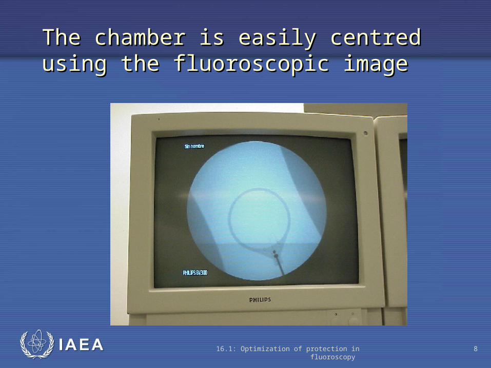
The entrance The entrance dose rate isdose rate is
1.78 mGy/min.1.78 mGy/min.

IAEA 16.1: Optimization of protection in fluoroscopy 10
The phantom The phantom thickness is thickness is nownow 20 cm20 cm..

IAEA 16.1: Optimization of protection in fluoroscopy 11
The chamber now readsThe chamber now reads 8.85 mGy/min.8.85 mGy/min.

IAEA 16.1: Optimization of protection in fluoroscopy 12
If the distance If the distance between the between the input screen of input screen of the intensifier the intensifier and the entrance and the entrance of the patient of the patient isis increased, i.e.,increased, i.e., extra 20 cm.extra 20 cm.The patient The patient entrance dose entrance dose rate will rate will increase.increase.

IAEA 16.1: Optimization of protection in fluoroscopy 13
Note:Note: the chamber looks magnified (the the chamber looks magnified (the intensifier is further away from the patient).intensifier is further away from the patient).

IAEA 16.1: Optimization of protection in fluoroscopy 14
Before (intensifier-table distance: 35 cm)
Now (intensifier-table distance: 55 cm)

IAEA 16.1: Optimization of protection in fluoroscopy 15
The entrance dose The entrance dose rate with the rate with the intensifier at 55 cm intensifier at 55 cm from the table isfrom the table is 17.9 mGy/min17.9 mGy/min (to (to be compared with be compared with the previous value the previous value of 8.85 mGy/min).of 8.85 mGy/min).
IAEA 16.1: Optimization of protection in fluoroscopy 16
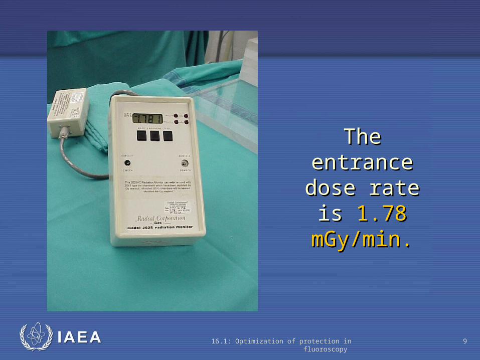
Now the Now the phantom phantom thickness is thickness is increased up toincreased up to 30 cm30 cm of acrylic.of acrylic.

IAEA 16.1: Optimization of protection in fluoroscopy 17
The patient The patient entrance dose entrance dose rate at the rate at the surface of the surface of the phantom phantom increases toincreases to 24.8 mGy/min24.8 mGy/min (the reading was (the reading was 8.85 with 20 cm 8.85 with 20 cm thickness).thickness).

IAEA 16.1: Optimization of protection in fluoroscopy 18
The dose rate due to scatter radiation also The dose rate due to scatter radiation also increases with the patient thickness. Forincreases with the patient thickness. For 30 cm30 cm acrylic, acrylic, 3 mGy/h3 mGy/h is measured close to the is measured close to the phantom.phantom.
IAEA 16.1: Optimization of protection in fluoroscopy 19
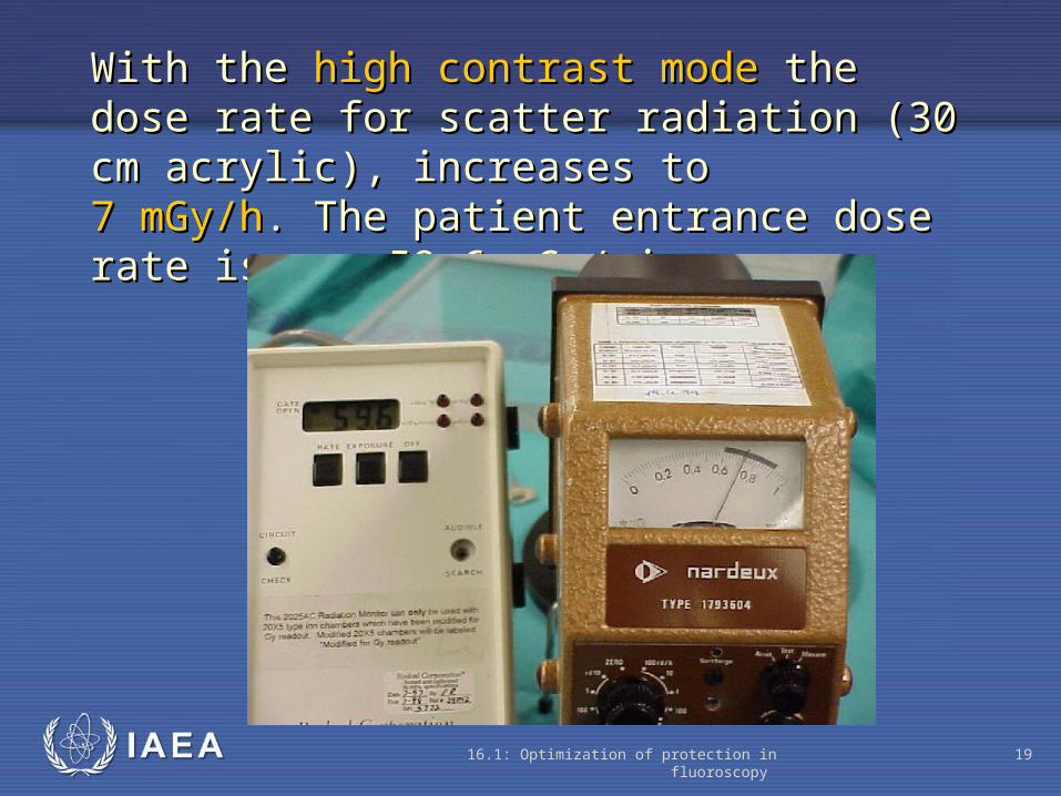
With theWith the high contrast modehigh contrast mode the dose rate for the dose rate for scatter radiation scatter radiation ((30 cm acrylic), increases to30 cm acrylic), increases to 7 mGy/h7 mGy/h.. The patient entrance dose rate is The patient entrance dose rate is now 59.6 mGy/min.now 59.6 mGy/min.

IAEA 16.1: Optimization of protection in fluoroscopy 20
WithWith 10 cm10 cm acrylic the acrylic the dose rate due dose rate due to scattered to scattered radiation isradiation is 0.2 mGy/h. 0.2 mGy/h.
IAEA 16.1: Optimization of protection in fluoroscopy 21
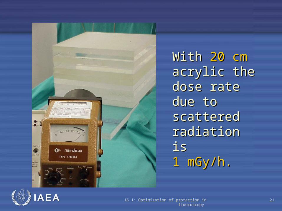
WithWith 20 cm20 cm acrylic the acrylic the dose rate due dose rate due to scattered to scattered radiation isradiation is 1 mGy/h. 1 mGy/h.

IAEA 16.1: Optimization of protection in fluoroscopy 22
WithWith 30 cm30 cm acrylicacrylic the the dose rate due dose rate due to scattered to scattered radiation isradiation is 3 mGy/h. 3 mGy/h.

IAEA 16.1: Optimization of protection in fluoroscopy 23
Note that with the extra thickness the image quality is extremely poor (the border of the chamber is hardly visible)

IAEA 16.1: Optimization of protection in fluoroscopy 24
Fluoroscopy - Standard Dose Rate
Analysis :• should be < 25 mGy/min
Frequency :• acceptance, tube change
• generator repair
• intensifier repair
• 6 monthly

IAEA
Where to Get More Information
15.3: Optimization of protection in radiography 25
Quality Control in Diagnostic Imaging, Gray JE, Winkler NT, Stears J, Frank ED. Available at no cost. http://www.diquad.com/QC%20Book.html