Radiation Protection Education in Fluoroscopy · PDF fileRadiation Protection Education in...
22
511 CE Directed Reading This article is a Directed Reading. Your access to Directed Reading quizzes for continuing education credit is determined by your membership status and CE preference. RADIOLOGIC TECHNOLOGY, May/June 2015, Volume 86, Number 5 Marlene M Johnson, MEd, R.T.(R) Radiation Protection Education in Fluoroscopy After completing this article, the reader should be able to: Summarize the goals of radiation protection and factors affecting those goals. Discuss the history of radiation protection and recent efforts to improve radiation safety. Identify how referring physicians, fluoroscopy operators, and radiographers can contribute to limiting radiation dose to patients. Explain how the education and compliance of personnel performing and assisting during high-dose fluoroscopy can be improved and regulated. Provide examples of how radiographers can improve fluoroscopy education and patient safety by using radiation protection skills. Potential biological damage from radiation received during fluoroscopy procedures is of particular concern because of the high volume and variety of procedures performed and the increasing length of radiation exposure. This article focuses on the effects of low-level radiation, gaps in education and skills among personnel performing and assisting with fluoroscopy, the certification and privileging of fluoroscopy personnel, and compliance with radiation protection practices. T he goals of radiation protection in f luoroscopy for both the patient and the medical person- nel involved are to minimize the possibility of deterministic health effects and help keep the probability of stochastic health effects from ionizing radiation as low as reasonably achiev- able (ALARA). Deterministic effects from radiation exposure are biological changes that occur in the body and manifest after a relatively short latent period of days, months, or years. Latent period refers to the time after exposure during which there are no signs of illness or damage. 2 With deterministic effects, the severity of the biologic response increases as the dose increases. A threshold dose of radiation generally exists, meaning the radiation dose level must be reached before the damage is observed. Deterministic effects include local tis- sue damage, hematological depletion, cytogenic damage, and, in severe cases, acute radiation syndrome. 2 Acute radia- tion syndrome seldom occurs as a result of fluoroscopically guided procedures; however, local tissue damage in the form of skin burns, dermatitis, and epi- lation is possible. 3 Stochastic effects can occur as a result of radiation-induced damage to the DNA of cells, leading to malignant conditions. The threshold dose is inde- pendent of the absorbed dose. The prob- ability of a malignancy is influenced by the person’s age at the time of exposure, sex, and personal susceptibility to can- cer. 2 Health care personnel should have a higher relative concern for possible stochastic effects in pediatric patients who undergo f luoroscopically guided procedures. It is impossible to calculate “The practice of medicine involves not only the science, but also the art of dealing with the prevention, diagnosis, alleviation, and treatment of disease.” 1
Transcript of Radiation Protection Education in Fluoroscopy · PDF fileRadiation Protection Education in...
511
CEDirected Reading
This article is a Directed Reading. Your access to Directed Reading quizzes for continuing education credit is determined by your membership status and CE preference.
RADIOLOGIC TECHNOLOGY, May/June 2015, Volume 86, Number 5
Marlene M Johnson, MEd, R.T.(R)
Radiation Protection Education in Fluoroscopy
After completing this article, the reader should be able to:Summarize the goals of radiation protection and factors affecting those goals.Discuss the history of radiation protection and recent efforts to improve radiation safety.Identify how referring physicians, fluoroscopy operators, and radiographers can
contribute to limiting radiation dose to patients. Explain how the education and compliance of personnel performing and assisting
during high-dose fluoroscopy can be improved and regulated. Provide examples of how radiographers can improve fluoroscopy education and patient
safety by using radiation protection skills.
Potential biological damage from radiation received during fluoroscopy procedures is of particular concern because of the high volume and variety of procedures performed and the increasing length of radiation exposure. This article focuses on the effects of low-level radiation, gaps in education and skills among personnel performing and assisting with fluoroscopy, the certification and privileging of fluoroscopy personnel, and compliance with radiation protection practices. The goals of radiation protection
in f luoroscopy for both the patient and the medical person-nel involved are to minimize
the possibility of deterministic health effects and help keep the probability of stochastic health effects from ionizing radiation as low as reasonably achiev-able (ALARA).
Deterministic effects from radiation exposure are biological changes that occur in the body and manifest after a relatively short latent period of days, months, or years. Latent period refers to the time after exposure during which there are no signs of illness or damage.2
With deterministic effects, the severity of the biologic response increases as the dose increases. A threshold dose of radiation generally exists, meaning the radiation dose level must be reached before the damage is observed.
Deterministic effects include local tis-sue damage, hematological depletion, cytogenic damage, and, in severe cases, acute radiation syndrome.2 Acute radia-tion syndrome seldom occurs as a result of f luoroscopically guided procedures; however, local tissue damage in the form of skin burns, dermatitis, and epi-lation is possible.3
Stochastic effects can occur as a result of radiation-induced damage to the DNA of cells, leading to malignant conditions. The threshold dose is inde-pendent of the absorbed dose. The prob-ability of a malignancy is influenced by the person’s age at the time of exposure, sex, and personal susceptibility to can-cer.2 Health care personnel should have a higher relative concern for possible stochastic effects in pediatric patients who undergo fluoroscopically guided procedures. It is impossible to calculate
“The practice of medicine involves not only the science, but also the art of dealing with the prevention, diagnosis, alleviation, and treatment of disease.”1
512
CEDirected Reading
RADIOLOGIC TECHNOLOGY, May/June 2015, Volume 86, Number 5
Radiation Protection Education in Fluoroscopy
the amount of radiation exposure children might receive in a lifetime, and the potential to live a very long life increases their risk for stochastic effects.3 Standard prac-tice in radiation protection is to be cautious and assume that even small radiation doses can be harmful.2
Factors That Affect Radiation Protection Goals
In clinical practice, a complicated set of issues affects the overall goal of radiation protection. Health care professionals should seek a balance between the dose delivered and the reason for the procedure, including how the procedure is likely to affect the patient out-come. The greatest benefit to the patient should result in the lowest risk of radiation exposure to the patient. In addition, radiation protection practices also affect the amount of radiation exposure to medical personnel dur-ing procedures.1
The physician must assess the technology available and evaluate how the technology affects the outcome of the procedure and the overall radiation dose to the patient, as well as all personnel participating in the pro-cedure. Modern f luoroscopic equipment offers a variety of modes and image quality options and can deliver very high radiation doses for long periods of time. Decisions about mode selection and the type of image quality sufficient to perform the procedure influence the amount of radiation used.1 For example, the ana-tomical area of interest can be magnified, but magnifi-cation results in an increase in dose. Also, images can be recorded at different rates, with various levels of image quality on a variety of recording devices.2
The use of pulsed f luoroscopy or last-image hold can decrease patient dose.4 Exposure to the patient, opera-tor, and personnel assisting with the procedure can be affected greatly by the availability of these options and, more importantly, by whether the operator understands the equipment and uses these options appropriately.1 For example, a cardiologist would never consider plac-ing a stent in a patient using an extremity C-arm, nor would an orthopedic surgeon need to use a f luoroscopic unit that records rapid frames per minute and magnifies the anatomy tenfold.
The use of f luoroscopy for interventional procedures has increased in areas outside of imaging centers.5 It is
now common for cardiologists, orthopedic surgeons, vascular surgeons, pain specialists, and gastroenterolo-gists to perform fluoroscopically guided procedures and medical interventions. The practice of allowing registered radiologist assistants (R.R.A.s) and radiolo-gist practitioner assistants to perform fluoroscopically guided procedures also has been established, and many physician specialists, including radiology residents, are performing f luoroscopically guided procedures very early in their rotations. Some states recognize the use of radiologic technologists and other medical personnel to perform fluoroscopy.5,6
Radiation risks to patients, such as skin injury from highly complex interventional neuroradiology proce-dures are possible.7 However, methods to ensure pro-ficiency among all members of the health care team in performing and assisting during f luoroscopy with opti-mal radiation safety might be lacking.5,6
Historical BackgroundMore than a century has passed since the dangers of
radiation were discovered. The first American fatality resulting from radiation exposure was Clarence Dally, Thomas Edison’s assistant, who was reported to have spent hours experimenting in front of a f luoroscope.8 Early radiologists lost digits from local tissue damage, and early f luoroscopy patients developed skin burns as a result of overexposure.9 Past research focused on the potential biological damage from acute exposure to ionizing radiation based on data on atomic bomb survi-vors, early radiologists, and accidents that involved high acute exposures to radiation.8
Historically, radiation protection concepts, practices, and equipment regulations were developed and revised as new information became available. Early regulations for f luoroscopic equipment focused on equipment safe-guards to prevent overexposure of medical personnel to radiation. Although early work has brought about improvements regarding medical radiation exposure, the amount of radiation exposure from medical procedures in the United States has increased as more procedures are being performed, and the issues related to minimiz-ing radiation exposure during those procedures are more complex. The practice of using fluorosocopy for image guidance is becoming more routine. The variety of
513
CEDirected Reading
RADIOLOGIC TECHNOLOGY, May/June 2015, Volume 86, Number 5
Johnson
medical personnel performing and assisting during fluo-roscopic procedures has increased, and many have limit-ed education in radiation protection. Fluoroscopy equip-ment has become more complicated to operate, with frequent upgrades and options being added as new tech-nology is developed. The options often provide methods of improving image quality with an effect on radiation dose that the operator might or might not understand. Moreover, high patient volumes limit time for personnel training on new and upgraded equipment.9-13
Radiation protection practices were lacking prior to 1950, and as a result occupational radiation expo-sures were higher than they are reported to be today.14,15 Radiographers regularly held patients during exposures to decrease motion, and many radiographers reported holding patients during 50 or more exposures. In addi-tion, radiographers could begin working in the field before the age of 17 years, and fluoroscopic radiation exposure equipment regulations were not in force.15,16 Skin injuries due to overexposure to radiation were com-mon before the 1930s.9 In 1949, the first study was pub-lished demonstrating an increased incidence of cataracts in physicists who worked with cyclotrons, devices capa-ble of increasing charged particles to very high energies.2
Over time, radiation protection concepts changed significantly as the National Council on Radiation Protection and Measurements (NCRP) became active, publishing 19 reports regarding protection practices.8,16 The 5 rem (0.05 Sv) annual dose limit for radiation workers was established in 1959 by the International Commission on Radiological Protection (ICRP) and remained unchanged until 1977.12
After 1977, radiation protection theory and practice focused on the risk vs benefit concept, the practice of ALARA, the methodology of justification, and radia-tion protection practices that would ensure optimiza-tion.12 Fluoroscopic equipment regulations assisted in decreasing radiation exposure during f luoroscopy.10 Reports of radiation-induced skin injuries decreased until the number of interventional procedures began to rise, replacing surgical interventions. Newer examina-tions required longer f luoroscopy times and an increase in the number of images recorded.9
In 1982, the U.S. Radiologic Technologists Study began with a collaborative team that included the
National Cancer Institute (NCI), the University of Minnesota, and the American Registry of Radiologic Technologists (ARRT).15 The team continues to col-lect and analyze information about radiation exposure levels, enabling researchers to investigate the prob-able biological effects of radiation exposure, including malignancies, radiation-induced cataracts, and local tissue damage. Results are periodically released as new data on radiographers’ health are collected. The study continually requests radiographers’ participation on its Web site.15
Recent Developments In 2006, the U.S. population received 7 times more
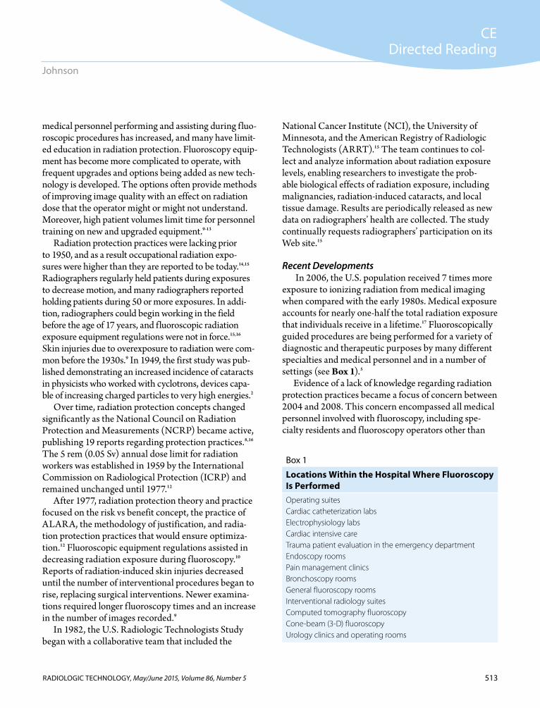
exposure to ionizing radiation from medical imaging when compared with the early 1980s. Medical exposure accounts for nearly one-half the total radiation exposure that individuals receive in a lifetime.17 Fluoroscopically guided procedures are being performed for a variety of diagnostic and therapeutic purposes by many different specialties and medical personnel and in a number of settings (see Box 1).5
Evidence of a lack of knowledge regarding radiation protection practices became a focus of concern between 2004 and 2008. This concern encompassed all medical personnel involved with f luoroscopy, including spe-cialty residents and f luoroscopy operators other than
Box 1
Locations Within the Hospital Where Fluoroscopy Is PerformedOperating suitesCardiac catheterization labsElectrophysiology labs Cardiac intensive careTrauma patient evaluation in the emergency departmentEndoscopy rooms Pain management clinicsBronchoscopy rooms General fluoroscopy roomsInterventional radiology suitesComputed tomography fluoroscopyCone-beam (3-D) fluoroscopy Urology clinics and operating rooms

514
CEDirected Reading
RADIOLOGIC TECHNOLOGY, May/June 2015, Volume 86, Number 5
Radiation Protection Education in Fluoroscopy
radiographers who play a critical role in the manage-ment of medical exposures.5,18-20
In addition to awareness and knowledge of radiation protection practices, compliance with practices became an issue.18-20 Although a medical professional has been trained in radiation protection practices, he or she might not consistently follow them. This might be due to a lack of continuing education as opposed to a lack of initial training and experience. Once personnel are trained, managers should conduct routine evaluation of staff com-pliance and emphasize continuing education to ensure that they continue to use radiation protection practices.18
In the past 14 years, the focus on radiation protec-tion has shifted, with attention directed toward the fol-lowing concerns1,3-6,9,11,13-15,18,19,22:
■ How the educational background of personnel in f luoroscopy might contribute to overexposure of patients and personnel.
■ Quality management of education and equip-ment.
■ Long-term, low-level occupational exposure.■ Radiation-induced cataracts in f luoroscopy opera-
tors.■ Skin injuries acquired during f luoroscopy.Emphasis on reducing exposure is becoming more
holistic, involving all of the medical personnel who care for patients and the patients themselves. Education and compliance with protection standards is accepted as the core for success.
The increase in medical radiation exposure was for-mally recognized by the NCRP in 2009 with the publi-cation of report No. 160, which established specific rec-ommendations for facilities that perform fluoroscopic procedures. The NCRP recommends f luoroscopy training and credentialing for all operators. Training should include the dangers of radiation and proper protection management using features available on the equipment. Physicians should be equipped with the knowledge and skills to make decisions about radiologi-cal examinations based on needs and risks.17
In 2010, the U.S. Food and Drug Administration (FDA) Center for Devices and Radiological Health created an initiative to reduce unnecessary radiation exposure from medical imaging. The FDA emphasized justification and optimization in the protection of
patients and monitoring facilities with quality assurance programs. Patients should be assured they are receiv-ing the right imaging examination at the right time and with the right radiation dose.22
For facilities that participate in the Medicare pro-gram, the Centers for Medicare and Medicaid Services established minimum standards for hospital radiology services and accreditation requirements for free-stand-ing advanced diagnostic imaging facilities. These stan-dards include physicians and nonphysician practitioners who provide the technical component of advanced imaging services.
In response, the NCRP released report No. 168, Radiation Dose Management for Fluoroscopically-Guided Interventional Medical Procedures. This report focuses on the use of f luoroscopic systems as a tool for guiding diagnostic and therapeutic procedures.10 The American College of Radiology (ACR) developed appropriateness guidelines for physicians selecting f luo-roscopy procedures to help them minimize radiation exposure to patients and operators and contain costs.23 The guidelines match patient conditions with appropri-ate imaging examinations.
The FDA also is promoting the use of automated decision support systems. Electronic health records should include complete information on the patient’s imaging history to assist the physician in selecting the appropriate examination. With automated order-ing systems, the physician selects from a list of patient symptoms and medical history and an appropriate examination is suggested, starting with the most basic procedures and continuing with more complex proce-dures. Examinations also are matched to an authoriza-tion code approved by Medicare.10
Occupational Exposure RiskLow-Level Dose Risks
Most occupational exposure of diagnostic imaging personnel occurs during f luoroscopy and mobile radi-ography, especially during interventional and other f lu-oroscopically guided procedures.2 Radiographers who operate f luoroscopy equipment receive higher expo-sures than do other radiographers primarily because of longer exposure times and close proximity to the radia-tion source.2
515
CEDirected Reading
RADIOLOGIC TECHNOLOGY, May/June 2015, Volume 86, Number 5
Johnson
One of the earlier studies on risk estimates for hemato-poietic malignancies in medical radiation workers who had protracted low-to-moderate occupational exposure dem-onstrated an increased risk for nonchronic lymphocytic leukemias decades after the initial radiation exposures.24 This study examined more than 270 000 radiologists and radiologic technologists from various countries. The most reliable finding was increased mortality as a result of leukemia in workers who were employed before 1950.24
Another study on mortality rates in interventional radiographers revealed no difference between radiog-raphers performing other studies and radiographers performing interventional radiology studies.14 The study followed 88 766 U.S. radiologic technologists over the course of approximately 8 years. The authors collected information on work experience, types of procedures, protective measures, medical and family history, as well as lifestyle choices. Over the course of the study, there were 3581 deaths. Of this number, 1209 were from malig-nancies and 979 were from circulatory system diseases. However, the authors did not find a significantly increased mortality risk in radiologic technologists who reported performing a high number of interventional radiography procedures. Nevertheless, they stated the findings should be interpreted “cautiously” because of the short follow-up period. A 2006 study of breast cancer incidence in U.S. radiographers found no direct relevance of the effects of long-term, low-level exposure on breast cancer.25
The most concerning conclusions regarding radiog-rapher practice and number of years worked were for radiographers in the field before 1950 who worked for 5 or more years and repeatedly held patients during exposure for diagnostic studies.15,26 Among those tech-nologists, there was an increase in relative risk for the category that included leukemia, multiple myeloma, and lymphoma. In addition, breast cancer incidence was increased 2.9 times, thyroid cancer had an elevated risk of 1.5 times, nonmelanoma skin cancer was increased 2.2 times, and melanoma risk was increased over all.The breast cancer increase was most prevalent for those who worked before 1935 and significantly elevated for those who worked before age 17. Breast cancer risk was higher for technologists who worked 5 or more years and held 50 patients or more. Genetic and lifestyle fac-tors were not included in the study.15
Radiation-Induced Cataracts Through observations and animal experiments,
researchers determined that the lens of the eye is radiosensitive and the sensitivity is age dependent.2 Sensitivity increases with age, requiring shorter latent times as an individual ages. Latent periods range between 5 and 30 years, with the average time until development being 15 years. The dose-response rela-tionship for radiation-induced cataracts was thought to be a nonlinear threshold, meaning there is a dose of radiation required to reach the point at which cataracts appear and the effect does not become worse as the dose increases.2 The exact threshold amount has been difficult to assess.
Recently, research has suggested that the threshold for an acute exposure is approximately 2 Gy, with 100% of those irradiated developing cataracts after a 10 Gy exposure.2 In the past 5 years, concern has increased regarding eye and extremity exposure of f luoroscopy operators and other personnel who assist during examinations requiring long exposure times. A demon-strated increase in incidence of cataracts among staff involved with interventional procedures, specifically cardiologists, has been attributed to the lack of educa-tion in radiation protection practices among cardiolo-gists.27-33
A study of U.S. radiographers over a period of 20 years suggested that the lowest cumulative ionizing radiation dose to the lens of the eye that can produce a progressive cataract is approximately 2 Gy.27 As a result of epidemiological evidence, the ICRP has reduced the recommended maximum dose to the eye lens by almost eightfold.13 The commission currently recom-mends an equivalent eye dose limit of 20 mSv per year, averaged over 5 years, with no single year exceeding 50 mSv.13
Best practices to protect and minimize dose to the lens of the eye include the use of ceiling-suspended screens, wearing leaded-glass eyewear, positioning the x-ray tube below the table as far away from the patient as possible, and the operator being properly trained in fluo-roscopic technique. For maximum eye protection, oper-ators must position themselves as far as possible from the source of radiation and at a right angle to the area where scatter is most likely to occur.29,30,33 The use of leaded
516
CEDirected Reading
RADIOLOGIC TECHNOLOGY, May/June 2015, Volume 86, Number 5
Radiation Protection Education in Fluoroscopy
glasses has a significant effect on the amount of radia-tion the eye receives, reducing dose by a factor of 5 to 10. Scatter-shielding screens alone can reduce the dose rate by a factor of 5 to 25. Using both of these practices can reduce eye exposure by a factor of 25 or more.31
Fluoroscopy Education Lack of education and training for a variety of health
care professionals can affect f luoroscopy radiation exposure to patients and personnel. Referring physi-cians, medical students, specialty physicians, residents in training, all medical personnel assisting with f luo-roscopy procedures, equipment manufacturers, and medical physicists contribute directly or indirectly to patient and personnel exposures.1,6,34 Routine perfor-mance of f luoroscopy procedures in areas other than medical imaging departments has evolved over time and might have outpaced the standards, licensing, and granting of f luoroscopic privileges.1 As a result, some f luoroscopy operators have not received proper training in radiation protection and the principles of balancing image quality with the examination or pro-cedure being performed.5
If fluoroscopy operators place a higher value on image quality than minimizing radiation dose when selecting or using fluoroscopy equipment, the practice can unnec-essarily increase the amount of radiation exposure to patients and staff involved with the procedure. The per-sonnel operating the equipment might not have had any training in radiation protection practices or could under-estimate the amount of exposure received.6 Radiation exposure levels from the same procedure and with simi-lar patient conditions can differ dramatically between institutions.1 Referring physicians might select the proce-dure that delivers the highest amount of radiation expo-sure without considering other, nonionizing procedures; radiology residents might not be supervised properly or trained on the operation of fluoroscopic equipment as it relates to dose; and other medical personnel who are involved with the procedures might not practice basic radiation protection skills.6
Improving Education in Fluoroscopy The ICRP, FDA, NCRP, NCI, and the American
Association of Physicists in Medicine (AAPM) are
focusing efforts on reducing unnecessary radiation exposure through radiation education, certification, and compliance by establishing initiatives, recommen-dations, standards, and guidelines.5,6,10,35-37
NCRP recommendations for all facilities perform-ing f luoroscopic procedures include ensuring that all10:
■ Operators of f luoroscopy systems are trained and understand the operation of the equipment and the implications of radiation exposure from each mode of operation.
■ Physicians performing procedures using f luoro-scopic equipment are trained and credentialed.
■ Credentialed operators can assess risks and ben-efits for individual patients.
In October 2010 an ICRP committee specifically addressed education and training in radiation protec-tion for diagnostic and interventional procedures. Its recommendations included topics in each area of radia-tion protection, suggested number of teaching hours, and syllabi examples.6,38 The education and training must be planned, clinically relevant, and ensure that optimal radiation protection for patients and medical personnel emphasizes performing procedures compe-tently. This is an invaluable guide for any educational institution, medical school, or department that per-forms f luoroscopic procedures.6
The ACR recently revised its technical standards for the use of radiation in f luoroscopic procedures, estab-lishing qualifications, responsibilities, and credentialing of personnel who perform fluoroscopy. The standards address the issue of necessary education for all medi-cal personnel involved with f luoroscopic procedures including the1:
■ Referring physician and medical students who order procedures.
■ Physicians and other operators of f luoroscopic equipment.
■ Radiographer or other medical personnel assist-ing with the procedure.
■ Physicist who plays a key role in the quality man-agement of equipment and education.
■ Equipment manufacturer personnel who train and educate users during new equipment installa-tions and upgrades.
517
CEDirected Reading
RADIOLOGIC TECHNOLOGY, May/June 2015, Volume 86, Number 5
Johnson
Education and training for medical staff should be promoted by regulatory and health authorities, with programs implemented at universities, health care educational programs, and hospitals.6 The educational requirements for each professional must be determined, and the depth of education should depend on the level of involvement. Education should start at the beginning of the career pathway and continue at regular intervals, building concepts and understanding. A health profes-sional needs a thorough understanding of many radia-tion protection topics that will become a routine part of his or her clinical practice. Methods of educational delivery must be established, and a system of periodic evaluation of radiation protection practice skills is need-ed to ensure competence.6
Because a variety of operators perform fluoroscopy and have varying types and degrees of education,5 a system should be established to identify operators’ cur-rent competency levels and offer educational opportu-nities that are easy to access and require a reasonable time commitment.35 Local credentialing processes should include a review of personnel training records on
f luoroscopic procedures performed in the past and any education that pertains to f luoroscopy.1
Physicians must comply with all applicable state and federal laws in addition to institutional policies for f luoroscopy licensure and certification. The directive for compliance can come from the medical board of the hospital, the state licensing bureau, or other regulatory bodies. The medical board has the responsibility to ensure physicians’ compliance.1 The f luoroscopy cre-dentialing program must specify who is eligible to oper-ate f luoroscopic equipment and who has the respon-sibility to review operators’ credentials. Hospitals and departmental administration should reinforce the need for training and support opportunities for instruction (see Box 2).1,34
Educational Needs of Professionals Involved in Fluoroscopy
Medical Students and Referring PhysiciansThe referring physician and the f luoroscopic opera-
tor have the most control over ensuring that the patient does not receive unnecessary examinations.2 Ordering
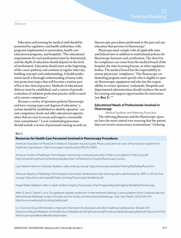
Box 2
Resources for Health Care Personnel Involved in Fluoroscopy Procedures
American Association of Physicists in Medicine. Educators resource guide. Physics education for users of fluoroscopic equipment in healthcare organizations. http://www.aapm.org/education/ERG/FLUORO/.
American Society of Radiologic Technologists. Fluoroscopy continuing education. Protect your patients. Protect yourself. http://www.asrt.org/main/continuing-education/earn-ce/featured-ce-courses/fluoroscopy-courses.
Case Western Reserve University. Radiation safety training manual. https://www.case.edu/does/Training/RadSafety/fluoro.htm.
American Registry of Radiologic Technologists Examination handbook for state licensing exams administered by ARRT in 2015 for fluo-roscopy. https://www.arrt.org/pdfs/State-Licensing/Fluoroscopy-Handbook.pdf.
Image Wisely. Radiation safety in adult medical imaging. Fluoroscopy. http://imagewisely.org/Imaging-Modalities/Fluoroscopy.
Miller D, Vano E, Bartal G. et al. Occupational radiation protection in interventional radiology: a joint guideline of the Cardiovascular and Interventional Radiology Society of Europe and the Society of Internventional Radiology. J Vasc Interv Radiol. 2010;21:607-615. http://www.sirweb.org/clinical/cpg/safety6.pdf.
U.S. Food and Drug Administration. Important information for physicians and other healthcare professionals. Revised 2013. http://www.fda.gov/Radiation-EmittingProducts/RadiationEmittingProductsandProcedures/MedicalImaging/MedicalX-Rays/ucm115354 .htm?source=govdelivery#healthcareproviders.
518
CEDirected Reading
RADIOLOGIC TECHNOLOGY, May/June 2015, Volume 86, Number 5
Radiation Protection Education in Fluoroscopy
the appropriate examination and considering nonioniz-ing options are critical responsibilities of the physician. The physician must be knowledgeable about the pro-cedure and have some understanding of the radiation dose amount. Research has demonstrated that a large number of physicians of various backgrounds need to improve their knowledge of radiation exposure.34,38-41
Cardiologists prescribe the majority of medical imaging examinations, but their radiation protection awareness is low.34 Pediatricians have been found to lack radiation protection awareness and often underestimate relative doses and risks. Many physicians do not real-ize that f luoroscopically guided procedures increase the risk of radiation skin injuries and radiation-induced cancer for both the patient and personnel.34,39,40
In a study by Thomas et al, less than 20% of pediatri-cians surveyed could recall any relevant formal teach-ing during their specialty training, and only 15% were familiar with the ALARA principle. Many medical stu-dents and interns believe that magnetic resonance and ultrasound emit ionizing radiation.40
The radiation protection training of physicians who refer patients for medical imaging procedures has remained largely unaddressed. The challenge of medical education is to identify the information that physicians need to know for everyday practice. Currently, courses in radiation protection are limited. Many medical students become physicians who use fluoroscopy in their practice, order examinations, or answer patients’ questions. A basic course or orientation for medical students on radiation protection that requires a minimum of 5 to 10 hours of instruction could increase their knowledge.6
A general understanding of radiological quantities and units, fundamentals of radiation biology and bio-logical effects of radiation, deterministic effect risk, the principles of optimization, and the national and interna-tional radiation exposure standards could serve medical students who eventually refer patients for diagnostic imaging. Education could include a deeper understand-ing of the types of doses received from procedures, risk of cancer and hereditary disease, and the practice of dose optimization.
The referring physician begins the process of practic-ing justification of examination or procedure selection because of knowledge of the patient’s medical history
and clinical indications. Justification requires weigh-ing the risk vs the benefit to the patient and answering questions the patient might have regarding exposure dose and risk. Even when an imaging study is appro-priate, a comparable examination could yield similar clinical results with no use of ionizing radiation.42 Optimization requires competent performance of the procedure in terms of radiation protection, which is a responsibility of the practitioner performing the exami-nation, but is a helpful concept for an ordering physician to understand.1,6
The ACR has developed appropriateness criteria to help physicians decide which imaging procedure would be best for the patient’s medical condition. The crite-ria are derived from comprehensive, evidence-based guidelines for selecting diagnostic imaging procedures, radiation therapy protocols, and image-guided interven-tional procedures. The guidelines constitute nationally accepted and scientifically based recommendations. Their purpose is to help eliminate inappropriate use of radiologic services and promote the best use of limited health care resources.23
Additional support and education for referring physi-cians is available through the Image Wisely campaign, a joint effort of several radiology organizations that pro-vides general information about radiation safety, how to communicate risk, and links to the ACR’s appropriate-ness criteria, as well as other information for referring physicians and patients.43
The vast amount of information that must be consid-ered and analyzed to keep pace with all of the advances in imaging today can be overwhelming for referring physicians. The ACR advocates the use of medical deci-sion support systems, also called clinical support systems, automated response programs, or simply decision support systems. Such systems also assist in ensuring the proce-dure is paid for under Medicare or Medicaid and thus can be an aid in managing expensive resources.44,45
Medical decision support systems guide the physi-cian to consider a minimally extensive radiologic pro-cedure first instead of a study that is more costly and exposes the patient to more radiation. The systems are generally user friendly, consistent, and educational in that they provide immediate feedback. Decision support systems aim to improve physician clinical
519
CEDirected Reading
RADIOLOGIC TECHNOLOGY, May/June 2015, Volume 86, Number 5
Johnson
decisions and provide quality measures and outcome data that are critical to hospital accreditation standards and requirements of the Affordable Care Act.45
Physicians Operating Fluoroscopy EquipmentEducation and training for the safe use of f luoros-
copy has not kept pace with expanding clinical applica-tions, and some individuals performing f luoroscopy have not received formal training in radiation protec-tion biology and practices.6,35 Some physicians perform fluoroscopy as a basic diagnostic examination, includ-ing upper and lower gastrointestinal studies and anat-omy localization; others use f luoroscopy for complex interventional procedures, needle localization for pain management purposes, and cardiac catheterization. A large number of gastroenterologists use f luoroscopy for endoscopic retrograde cholangiopancreatography, which often requires lengthy f luoroscopy time.38
The ACR standard states that physicians perform-ing the most complex interventional procedures should have the most f luoroscopy-specific radiation protection education.1 According to the ICRP, education ideally should start with basic background knowledge in medi-cal school for all physicians.6 This education should progress to comprehensive radiation protection practice skills for advanced practitioners delivered throughout physicians’ careers by required continuing medical edu-cation.1,6 Implementing radiation protection education requires long-term planning with involvement from regulatory agencies, educational institutions, and medi-cal facilities. The plan must address how the curriculum can be developed, who will teach the courses, and in what format the education will be offered.1,35
According to the ACR, the recommended initial qualifications for physicians performing or supervising f luoroscopically guided procedures should be one of the following1:
■ Board certification in radiology or radiation oncology by the American Board of Radiology, the American Osteopathic Board of Radiology, the Royal College of Physicians and Surgeons of Canada, or the Collège des médecins du Québec. Review of this can occur at time of hire or retro-actively for institutions implementing new guide-lines.
■ Completion of a residency or fellowship pro-gram approved by the Accreditation Council for Graduate Medical Education, the Royal College of Physicians and Surgeons of Canada, the Collège des médecins du Québec, or the American Osteopathic Association, including 6 months of training in f luoroscopic procedures. This should include documentation of successful completion of didactic course lectures and labora-tory instruction in radiation physics, radiation biology, radiation safety, and radiation manage-ment applicable to the use of f luoroscopy, includ-ing passing a written examination in these areas.
■ Privileges granted for specific f luoroscopically guided procedures and completion of continu-ing medical education in radiation dosimetry, radiation protection, and equipment performance related to the use of f luoroscopy.
Recommended credentialing requirements for phy-sicians who have not received education in radiation physics, radiation biology, radiation safety, and radia-tion management who want to obtain privileges to perform complex interventional procedures in vascular, cardiovascular, neurological, and urological practices include1:
■ Documented proof the physician has performed the procedure a minimum of 10 times under the direction of a physician who is credentialed and has been granted f luoroscopy privileges.
■ Documented evidence of completing a minimum of 8 hours of lectures and laboratory instruction in radiation physics, radiation biology, radio-logical safety, and radiation management. This instruction must include f luoroscopic imaging of pediatric patients and pregnant patients, and phy-sicians must satisfactorily pass an examination in these areas.
■ Physicians who want privileges to perform rou-tine f luoroscopic examinations have the same requirements, except that a minimum of 4 hours of instruction is required. The physician should be evaluated annually for competency. Continued privileges should be granted by the medical insti-tution based on the performance of a specified number of f luoroscopic examinations and having
520
CEDirected Reading
RADIOLOGIC TECHNOLOGY, May/June 2015, Volume 86, Number 5
Radiation Protection Education in Fluoroscopy
received instruction and demonstrated competen-cy on all newly installed equipment and upgrades that affect dose.1
Cardiac interventionalists are prominent among practitioners who might not have received radiologi-cal instruction as part of their medical education.39,41 Studies indicate varying knowledge deficiencies among cardiologists regarding equipment standards and dose amounts received during diagnostic imaging proce-dures. According to the literature, other concerns with this group of physicians are failure to use techniques such as collimation to reduce radiation dose and the routine use of techniques such as magnification to produce the highest quality possible images. High f luo-roscopy exposure times and obtaining large numbers of images are common practices among many cardi-ologists.39,41 According to Kuon et al, instruction and guidance on radiation protection and the clinical use of radiation protection techniques should be incorporated into cardiologists’ curriculum base.41
Abatzoglou et al conducted a study aimed at improv-ing radiation protection for interventional cardiology professionals and patients at a hospital in northern Greece. The effort included physician training in radia-tion protection.46 After conducting a seminar on basic x-ray physics and properties, principles of dosimetry, personal dosimetry, radiation protection tools, radiation biology principles and effects, and f luoroscopy parame-ters that contribute to image and dose optimization, the authors assessed effect on patient and cardiologist expo-sure. They reported that the radiation protection semi-nar led to a significant reduction in radiation exposure of the physicians, and once the physicians instructed staff on optimizing parameters, they also noted a reduc-tion in patients’ skin dose and personnel exposure.46
The standards of education and training in radiation effects and physics and the practice of radiation protection for cardiologists should match those for interventional radiologists.6 The recommended radiation protection training requirement for physicians performing complex interventional studies is a minimum of 30 hours to 50 hours of training covering the following topics1,6:
■ Fundamentals of radiation biology and biological effects of radiation.
■ Risk of cancer and hereditary disease.
■ Risk of deterministic effects.■ General principles of radiation protection, includ-
ing optimization.■ Operational radiation protection. ■ Factors affecting patient dose. ■ Factors affecting staff dose. ■ Typical doses from diagnostic and interventional
studies. ■ Risks of fetal exposure.■ Quality control and quality assurance. The educational topics should be specific, thor-
ough, and beyond basic levels so that physicians can adequately educate others.6 The curriculum should include specific examples of radiation protection prac-tices by discussing clinical scenarios relevant to patients and radiographic equipment. Once the physician has mastered the knowledge, he or she should translate that knowledge to the clinical environment. For example, once the physician has learned that the use of the mag-nification mode in f luoroscopy increases patient dose, the knowledge could become more relevant if the physi-cian calculates and compares the amount of exposure that results from procedures with and without magni-fication mode. The physician then might question the routine use of magnification or consider intermittent use of this mode.6
There is a realistic concern regarding radiation-induced skin damage for patients and radiation-induced cataracts for f luoroscopy operators regardless of their specialty practice. Varying degrees of exposure levels have been demonstrated in radiologists and cardiolo-gists.6,33 The aim of optimizing radiation protection during f luoroscopically guided procedures is to adjust imaging parameters and institute protective measures so the image is obtained with the lowest possible radia-tion dose to the patient and medical personnel involved with the procedure. Good technique includes proper patient positioning, field size collimation, use of shield-ing, pulsed f luoroscopy, minimizing exposure time and number of images recorded, and using the appropriate recording medium.1,6
Pediatric interventionalist practice requires the strictest adherence to dose reduction strategies. Equipment purchases should focus on radiation pro-tection; the equipment should include a method for
521
CEDirected Reading
RADIOLOGIC TECHNOLOGY, May/June 2015, Volume 86, Number 5
Johnson
selecting imaging technique based on age, size, and weight, as opposed to standard function selection based on small, medium, and large patients. Major pediatric interventional procedures should be performed only by experienced pediatric operators. Both the ICRP and ACR emphasize a specific mandatory level of training in pediatric radiation protection for all medical person-nel performing and assisting with pediatric interven-tional studies.1,3
The Joint Commission adopted the sentinel event policy in 1996 to help hospitals that report serious adverse effects improve patient safety and learn from the events. A sentinel event is a patient safety event that is not primarily related to the natural course of the patient’s illness or condition. The event is an unexpected occurrence involving serious physical or psychological injury to a patient that results in death, permanent harm, or severe temporary harm requiring an intervention to sustain life. The terms sentinel event and error are not synonymous because not all events occur as a result of error and not all errors result in sentinel events.47
Even though The Joint Commission granted the pre-rogative for medical institutions to decide which types of occurrences are to be considered sentinel events, the ICRP recommends that hospitals categorize patient skin burns from fluoroscopy as sentinel events.11 For a pro-cedure to qualify as a sentinel event, the radiation expo-sure level must reach a predetermined absorbed dose amount or period of time the fluoroscopy unit was being operated. The sentinel event report must be reviewed by a departmental or institutional quality team, which com-pletes a follow-up report that might require additional training for the fluoroscopy operator.47
Registered Radiologist AssistantsIndividuals who are qualified and licensed or certi-
fied under state laws to perform f luoroscopy proce-dures must be supervised by a radiologist or physician who is qualified or has f luoroscopy privileges. These staff members should complete 40 hours of didactic education or the equivalent in digital image acquisi-tion and display, contrast media, f luoroscopic unit operation and safety, image analysis, radiation biology, and radiation protection and have 40 hours of clinical
experience supervised by a radiologist or medical physicist. Required continuing education for these individuals should cover performing f luoroscopy, including equipment details and radiation protection. Ancillary personnel should never perform f luoroscopy of any kind independently, nor should they perform complex interventional studies unless in a formal training situation, such as a medical resident in radiol-ogy or cardiology.1
A registered radiologist assistant is an advanced-level radiographer who is certified and registered by the ARRT. This professional qualifies for the ARRT R.R.A. examination after successfully completing an advanced academic and clinical program. The curriculum for R.R.A.s must be based on one devel-oped through a joint effort between the ACR and the American Society of Radiologic Technologists (ASRT). This curriculum includes an advanced academic pro-gram in patient assessment and management of specific examinations as well as a radiologist-directed clinical preceptorship.1
Under radiologist supervision, specified as either direct or indirect, the R.R.A. performs specific tasks that assist the radiologist in the course of practice. This can involve operating f luoroscopic equipment under the direct supervision of a radiologist. An R.R.A. must comply with all applicable state laws and the joint policy of the ACR and ASRT. R.R.A.s may not interpret images, and the performance of their tasks must be allowed in the state where they practice. R.R.A.s should have received didactic education and training in radiation management and must complete a formal authorization process at the medical facility where they work.1
R.R.A.s are in a position to assess the patient and report to the radiologist information pertaining to patient history and previous radiologic procedures. This can assist in avoiding unnecessary repetition of an examination and ensure that the patient is receiving the correct examination, both of which minimize unneces-sary radiation exposure. R.R.A.s also can contribute to other health professionals’ education by teaching radia-tion practice skills, coaching those in training, volun-teering to lead course lectures, and providing continu-ing education opportunities.1
522
CEDirected Reading
RADIOLOGIC TECHNOLOGY, May/June 2015, Volume 86, Number 5
Radiation Protection Education in Fluoroscopy
Radiologic TechnologistsLack of compliance with established radiation safety
practices could lead a radiographer to contribute to increases in radiation dose.18,19,21,48 Previous research has shown variation in adherence to radiation pro-tection practices among radiographers, along with concern about complacency and apathy toward protec-tion practices.19,20 Research conducted by the ASRT in November 2013 showed that in states that require licensing of radiologic technologists, 4.3% of radiogra-phers surveyed did not know whether their facilities had policies in place to minimize radiation exposure to per-sonnel. However, in states that do not require licensing of technologists, 8% did not know about occupational exposure policies in their facilities.49
When asked to rate their own knowledge of radiation safety, more than 45% of cardiovascular technologists, vascular-interventional technologists, and R.R.A.s considered themselves very knowledgeable, and about 35% rated themselves knowledgeable. More than 40% of radiographers rated themselves as very knowledge-able and 40% as knowledgeable.49 Professional ethics and standards emphasize that radiologic technolo-gists should seriously consider their responsibilities to patients and the medical community, as well as their role in assuring their own personal radiation protection.18,50
Improving radiation safety begins with a personal evaluation of radiographers’ own routine practices.18,20,47 The ARRT is considering different approaches to recertification for radiologic technologists who were certified after 2011 and might address any lack of com-pliance with protection practices through radiography recertification requirements. Research suggests that involving professional associations and health care organizations, along with educational interventions in the form of in-services or incorporating dose feedback systems into radiography practice, can be effective.18,51-54 Radiographers should ensure that they are incorporat-ing best practices into their daily routines, seeking con-tinuing education in radiation protection, and actively participating in the Image Wisely campaign.54
Radiographers have some control over the greatest source of unnecessary patient dose, which is repeated or unnecessary examinations.10,12,17 Radiologic technolo-gists should acquire relevant patient history regarding
previous examinations and radiation exposure history when available. Repeat examinations have been esti-mated to account for as much as 10% of all examina-tions, and most repeat examinations are caused by radiographer errors.2
Radiographers also can contribute to the educational process. They can coach, serve as role models, suggest radiation protection management skills to residents in training, and volunteer to assist in delivering f luoros-copy continuing education.6,55
Accreditation standards for programs that educate entry-level radiographers who assist during f luoroscopy mandate that radiation protection be taught throughout the required cognitive and psychomotor knowledge and skills set.19 The assumption should be that certified radiographers have received instruction in the recom-mended topics, including the following6,56:
■ Physical characteristics of imaging equipment.■ Fundamentals and objectives of radiation protec-
tion, including optimization. ■ Principles and process of justification.■ Potential biological effects of ionizing radiation,
including stochastic and deterministic effects.■ Operational radiation protection.■ Particular aspects of patient and staff radiation
protection, such as applications and limitations of detection devices and patient shielding.
■ Typical doses from diagnostic procedures.■ Risks from fetal exposure.■ Quality control and quality assurance.A certified radiographer also understands topics such
as atomic structure, x-ray production and interaction of x-rays with matter, nuclear structure and activity, radio-logical quantities and units, fundamentals of radiation biology, biological effects of radiation, and national and international standards.6
The major focus of radiographers’ education should be specific to their job and roles.6 Once radiographers expand their role to include interventional f luoroscopy procedures, they should be required to achieve a higher level of understanding of radiation protection practice and be included in the f luoroscopy privileging system.1,6 The ARRT offers secondary certification in interven-tional radiography and cardiology that requires this type of advanced knowledge.57
523
CEDirected Reading
RADIOLOGIC TECHNOLOGY, May/June 2015, Volume 86, Number 5
Johnson
Other Personnel Many health care professionals who are not radio-
logic technologists, such as nurses, might assist with interventional examinations. A basic educational back-ground in radiation protection for this group of profes-sionals is essential. At minimum, nurses and ancillary personnel need a general awareness and understanding of the topics radiographers study.6 Nurses and other staff involved in f luoroscopy should not perform duties related to radiation protection until they have received a solid background in radiation protection principles and practice.6 A review of their past educational experiences should be conducted as part of the privileging process. If a health professional’s knowledge base is not docu-mented, he or she should be provided with or guided to training opportunities to assist in meeting the privileg-ing requirements.6,35
Administrators can optimize the amount of overall dose patients and personnel receive by ensuring they are purchasing equipment that uses the most recent dose-reduction strategies, particularly in pediatric imaging centers. Administrators can promote continu-ing education by sponsoring periodic in-services on dose reduction. They can oversee changes in protocols to ensure safe protection practices and ensure that staff members are properly educated during new installa-tions and upgrades that affect radiation dose. Requiring periodic competency evaluations related to dose reduc-tion can be useful, and administrators should be direct-ly involved in any incidents in which overexposure of patients might have occurred.58 In addition, administra-tors should ensure the development of a privileging pro-cess in f luoroscopy within their department and serve as role models for other departments.59
Physicists
Qualified medical physicists often are employed through the hospital or imaging department, and others are contracted on a part-time basis. The physicist is a critical component of the imaging team, responsible for the quality evaluation and maintenance of radiography equipment. For example, physicists perform radiation measurements, dosimetric calculations, and equipment performance evaluations of f luoroscopic equipment at the time of installation, at regular intervals afterward,
and following equipment upgrades.1 Medical physicists who specialize in radiation protection must obtain edu-cation in this discipline at the highest level and possess the ability to teach other personnel.6
The medical physicist should play a critical role in the education and training of residents and other health care professionals who operate f luoroscopy equipment or who are involved with f luoroscopic procedures.6 Physicists should review radiation exposure records for compliance evaluation and be involved with protocol standardization and other activities related to radiation protection. It is important to involve the physicist when establishing and implementing a f luoroscopy creden-tialing and privileging program and when reviewing documentation to support a privilege request. In addi-tion, physicists can take the lead in developing didactic content and providing training. In 2012, the AAPM developed a comprehensive task report that provides guidance for medical facilities to establish a credential-ing and privileging program for f luoroscopy.35
Equipment ManufacturersRadiation equipment manufacturers play an impor-
tant role in radiation protection by continuing to develop equipment options that decrease radiation dose. Manufacturers have successfully developed equip-ment features required by the FDA and NCRP that allow monitoring and recording of patients’ radiation exposure, a practice that will become more common in the future. Vendors have the responsibility to educate their customers on available dose-reduction options, especially in pediatric and interventional imaging. Radiology equipment manufacturers can be ongoing contributors to the adoption of radiation protection practices and the education and training of equipment operators.6
A well-managed imaging department that values dose reduction strategies should have frequent in-service training with information and reminders about radiation protection practices.2 Advanced clinical application specialists can be valuable contributors to these in-service sessions. New equipment installation education and training provided by clinical applications professionals is essential, and the competency of staff members should be evaluated and documented at the

524
CEDirected Reading
RADIOLOGIC TECHNOLOGY, May/June 2015, Volume 86, Number 5
Radiation Protection Education in Fluoroscopy
completion of training. All users must be aware of any changes with dose implications or new features that affect dose.6
A plan for installation or upgrade education should be established prior to the application specialist’s arriv-al. All personnel responsible for operating the equip-ment, including radiographers and physicians, should be included in the plan. Limited time available for appli-cation training can make it difficult to reach all of the professionals involved with a new system. One possible solution is to train “super-users” during major software upgrades in hospitals. Super-users receive most of the personal instruction from the expert. These users in turn are responsible for training other team members. This type of system requires feedback about who has completed training, and competency evaluation must be required of all members who will operate the new equipment independently.60 In addition, training can be enhanced with online training materials.
PatientsPatients can affect their overall radiation exposure
by ensuring they are providing members of the health care team with information about previous radiological studies and by becoming aware of the potential harm that long-exposure f luoroscopy studies can present (see Box 3).61 A general understanding of radiation and the potential exposure from fluoroscopy examinations and procedures can help patients better participate in their
care and decision making. Information for the public and patients should be balanced and reflect scientific literature and findings.42
In particular, parents of children who undergo these types of procedures must understand f luoroscopy expo-sure because of children’s potential life spans and future additional imaging examinations. Parents also should learn more about complex interventional f luoroscopy procedures so they and their child can cooperate to the best of their ability.61
Fluoroscopy Education MethodsThe 2 most difficult hurdles to implementing a
f luoroscopy education system are the lack of agreement about the training to include and the time require-ments.35,63 However, failing to create educational oppor-tunities leads to longer procedure times and misuse of equipment. The more opportunities to learn, the better, and institutions should not allow flexibility in the f luo-roscopy privileging process.6
Education and training should be provided by a team of radiology professionals. Each team member can contribute by creating a portion of the education based on his or her specific knowledge and background. Physicists are invaluable for contributing to the educa-tion of residents, physicians, radiographers, and oth-ers involved with complex f luoroscopy procedures. However, physicists are not hired as full-time radiation protection faculty and their specific role is ensuring national and regulatory standards of equipment.
Radiologic technologists can make a significant contribution with their educational background and experience. They understand problems with the equip-ment and are generally the most experienced with recent upgrades. Radiographers also can relate to issues of time constraints when dealing with a large number of patients and understand how to deal with difficult patients. Radiologists, clinical applications personnel, equipment manufacturers’ educational divisions, and professional societies are potential contributors.
Lectures can be used for the essential background knowledge and advice on practical skills. Presentations, e-learning, and other types of education must be prac-tical and focus on addressing clinical situations and a wide variety of practice-related issues.6
Box 3
Resources for Patients With Questions About Radiation From Fluoroscopy FDA. U.S. Food and Drug Administration. Fluoroscopy. Information for patients. http://www.fda.gov/Radiation-EmittingProducts/RadiationEmittingProductsandProcedures /MedicalImaging/MedicalX-Rays/ucm115354.htm#patients.
Image Gently. The Alliance for Radiation Safety in Pediatric Imaging. Pause and pulse: image gently in fluoroscopy. http://www.imagegently.org/Procedures/Fluoroscopy.aspx.
Radiology Info.org. The radiology information resource for patients. Procedures. http://www.radiologyinfo.org/en /sitemap/proc-a2z.cfm?alpha=A.

525
CEDirected Reading
RADIOLOGIC TECHNOLOGY, May/June 2015, Volume 86, Number 5
Johnson
Studies have demonstrated that radiation exposure amounts can be decreased with simple educational methods as long as the education is relevant and imme-diate feedback is provided.53,63-66 Education provided to radiology residents before they begin fluoroscopy rotations has been shown to decrease radiation dose. For example, f luoroscopy times for barium enemas, cys-tograms, defecograms, and esophageal procedures were reduced after the incorporation of online training.65 In addition, pediatric dose decreased after residents were instructed about pulsed fluoroscopy and how to obtain the best video frame-captured images using the lowest f luoroscopy pulse.67 Online training, virtual education, and competency evaluation in the form of a check-off list also have proven useful in decreasing dose.64-66 Requiring the use of dose-feedback systems has helped to decrease patient dose as well.66-68 Finally, coaching is useful to decrease dose and increase awareness of exposure.55
All new employees involved with fluoroscopy proce-dures should be trained in the facility’s policies regarding fluoroscopy. Annual patient safety education generally is required by accrediting institutions and could include flu-oroscopy education. Nevertheless, educational materials should be updated whenever a significant change occurs. New information regarding radiation protection becomes available approximately every 18 months from various agencies including the ICRP, NCRP, and the ACR.6
ConclusionThere are many health care professionals, agencies,
and organizations that have an effect on the amount of radiation dose patients receive throughout their life-time, from the medical student who orders diagnostic imaging examinations and interventions, to the equip-ment manufacturer who develops and markets imag-ing equipment. Research has demonstrated a general lack of awareness and knowledge among these groups, and efforts are aimed at improving the educational preparation of health care professionals and raising awareness among equipment manufacturers. Hospitals and departments that are establishing a f luoroscopy privileging process have a challenging task at hand and should refer to the many resources available through the AAPM and agencies that have developed continuing education materials for f luoroscopy operators.69,70
Marlene M Johnson, MEd, R.T.(R), has been a certified radiographer for 40 years, with a career emphasis in teaching the radiologic sciences, directing radiography programs, and serving as a site visitor for the Joint Review Committee on Education in Radiologic Technology for 10 years. In her final position she developed and improved educational programs in nuclear medicine, magnetic resonance imaging, and computed tomography at the University of Utah, where she developed a bachelor of science program. Retired since 2014, she plans to enjoy her leisure time and make periodic contributions to the continuing education of radiographers and other professionals involved with medical imaging.
Reprint requests may be mailed to the American Society of Radiologic Technologists, Communications Department, at 15000 Central Ave SE, Albuquerque, NM 87123-3909, or e-mailed to [email protected].
© 2015 American Society of Radiologic Technologists
References 1. American College of Radiology. ACR-AAPM technical
standard for the management of the use of radiation in fluo-roscopic procedures. http://www.acr.org/~/media/f22c9d1ff 46f43aab001f9ed0466b7e9.pdf. Revised 2013. Accessed December 27, 2014.
2. Bushong SC. Radiologic Science for Technologists: Physics, Biology and Protection. 10th ed. St Louis, MO; Elsevier Mosby: 2013.
3. Khong P-L, Ringertz H, Donoghue V, et al. Radiological pro-tection in paediatric diagnostic and interventional radiology. ICRP Publication 121. Ann ICRP. 2013;42(2). http://www .icrp.org/publication.asp?id=ICRP%20Publication%20121.
4. Cohen MD. Optimizing the use of pulsed fluoroscopy to reduce radiation exposure to children. J Am Coll Radiol. 2008;5(3):205-209. doi:10.1016/j.jacr.2007.09.014.
5. Rehani MM, Ciraj-Bjelac O, Vaño E, et al. Radiological protection in fluoroscopically guided procedures out-side the imaging department. ICRP Publication 117. Ann ICRP. 2010;40(6). http://www.icrp.org/publication .asp?id=ICRP%20Publication%20117.
6. Vaño E, Rosenstein M, Liniecki J, Rehani M, Martin CJ, Vetter RJ. Education and training in radiological protec-tion for diagnostic and interventional procedures. ICRP Publication 113. Ann ICRP. 2009;39(5). http://www.icrp .org/publication.asp?id=ICRP%20Publication%20113.
7. Vano E, Fernandez M, Sanchez RM, et al. Patient radiation dose management in the follow-up of potential skin injuries in

526
CEDirected Reading
RADIOLOGIC TECHNOLOGY, May/June 2015, Volume 86, Number 5
Radiation Protection Education in Fluoroscopy
neuroradiology. AJNR Am J Neuroradiol. 2013;34(2):277-282. doi:10.3174/ajnr.A3211.
8. Brodsky A, Kathren RL. Historical development of radiation safety practices. Radiographics. 1989;9(6):1267-1275.
9. Miller DL, Balter S, Schueler BA, Wagner LK, Strauss KJ, Vañó E. Clinical radiation management for fluoro-scopically guided interventional procedures. Radiology. 2010;257(2):321-332. doi:10.1148/radiol.10091269.
10. National Council on Radiation Protection and Measurements. Report No. 168 – Radiation dose manage-ment for fluoroscopically-guided interventional medical pro-cedures (2010). http://www.ncrponline.org/Publications /Press_Releases/168press.html. Accessed December 17, 2014.
11. International Commission on Radiological Protection. Avoidance of radiation injuries from medical interventional procedures. ICRP Publication 85. Ann ICRP. 2000;30(2). http://www.icrp.org/publication.asp?id=ICRP%20Publication%2085.
12. International Commission on Radiological Protection. Radiological protection in medicine. ICRP Publication 105. Ann ICRP. 2007;37(6). http://www.icrp.org/publication .asp?id=ICRP%20Publication%20105.
13. Stewart FA, Akleyev AV, Hauer-Jensen M, et al. Statement on tissue reactions/early and late effects of radiation in nor-mal tissues and organs–threshold doses for tissue reactions in a radiation protection context. ICRP Publication 118. Ann ICRP. 2012;41(1/2). http://www.icrp.org/publication .asp?id=ICRP%20Publication%20118.
14. Linet MS, Hauptmann M, Freedman DM, et al. Interventional radiography and mortality risks in U.S. radiologic technologists. Pediatr Radiol. 2006; 36(suppl 2):113-120.
15. National Cancer Institute. U.S. radiologic technologists study. http://radtechstudy.nci.nih.gov/. Accessed December 22, 2014.
16. U.S. Food and Drug Administration. CFR code of federal regulations title 21. Fluoroscopic equipment. http://www .accessdata.fda.gov/scripts/cdrh/cfdocs/cfcfr/CFRSearch.cfm?fr=1020.32. Updated September 1, 2014. Accessed January 20, 2015.
17. National Council on Radiation Protection and Measurements. Report No. 160 – Ionizing radiation exposure of the population of the United States. http://www .ncrponline.org/Publications/Press_Releases/160press .html. Accessed January 21, 2015.
18. Johnston J, Killion J, Vealé B, Comello R. U.S. technologists’ radiation exposure perceptions and practices. Radiol Technol. 2011;82(4):311-320.
19. Slechta A, Reagan J. An examination of factors related to radi-ation protection practices. Radiol Technol. 2008;79(4);297-305.
20. Franz KH. Radiation protection in radiologic technol-ogy: apathy versus active involvement. Radiol Technol. 1983;54(2):119-122.
21. Tilson E. Educational and experiential effects on radiog-raphers’ radiation safety behavior. Radiol Technol. 1982;53(4):323-331.
22. Centers for Medicare & Medicaid Services. Advanced diagnostic imaging accreditation. http://www.cms.gov /Medicare/Provider-Enrollment-and-Certification /MedicareProviderSupEnroll/AdvancedDiagnosticImaging Accreditation.html. Accessed December 27, 2014.
23. American College of Radiology. About the ACR appropriate-ness guidelines. http://www.acr.org/Quality-Safety /Appropriateness-Criteria/About-AC. Accessed December 17, 2014.
24. Yoshinaga S, Mabuchi K, Sigurdson AJ, Doody MM, Ron E. Cancer risks among radiologists and radiologic technologists: review of epidemiologic studies. Radiology. 2004;233(2):313-321.
25. Doody MM, Freedman DM, Alexander BH, et al. Breast cancer incidence in U.S. radiologic technologists. Cancer. 2006;106(12):2707-2715.
26. Liu JJ, Freedman DM, Little MP, et al. Work history and mortality risks in 90,268 U.S. radiologic technologists (2014). Occup Environ Med. 2014;71(12):819-835. doi:10.1136 /oemed-2013-101859.
27. Chodick G, Bekiroglu N, Hauptmann M , et al. Risk of cataract after exposure to low doses of ionizing radiation: a 20-year prospective cohort study among U.S. radiologic tech-nologists. Am J Epidemiol. 2008;168;620-631. doi:10.1093 /aje/kwn171.
28. Ciraj-Bjelac O, Rehani MM, Sim KH, Liew HB, Vano E, Kleiman NJ. Risk for radiation induced cataract for staff in interventional cardiology: is there reason for concern? Catheter Cardiovasc Interv. 2010;76:826-834. doi:10.1002 /ccd.22670.
29. Vano E, Gonzalez L, Fernandez JM, Haskal ZJ. Eye lens exposure to radiation in interventional suites: caution is warranted. Radiology. 2008;248:945-953. doi:10.1148/radi ol.2482071800.
30. Dauer LT, Thornton RH, Solomon SB, St Germain J. Unprotected operator eye lens doses in oncologic inter-ventional radiology are clinically significant: estimation from patient kerma-area-product data. J Vasc Interv Radiol. 2010;21:1859-1861. doi:10.1016/j.jvir.2010.08.006.
527
CEDirected Reading
RADIOLOGIC TECHNOLOGY, May/June 2015, Volume 86, Number 5
Johnson
31. Vano E, Kleiman NJ, Duran A, Rehani MM, Echeverri D, Cabrera M. Radiation cataract risk in interventional cardiol-ogy personnel. Radiat Res. 2010;174(4):490-495. doi:10.1667 /RR2207.1.
32. Efstathopoulos, EP, Pantos I, Andreou M, et al. Occupational radiation doses to the extremities and the eyes in interven-tional radiology and cardiology procedures. Br J Radiol. 2011;84:70-77. doi:10.1259/bjr/83222759.
33. Durán A, Hian SK, Miller DL, Le Heron J, Padovani R, Vano E. Recommendations for occupational radiation protec-tion in interventional cardiology. Catheter Cardiovasc Interv. 2013:82(1):29-42. doi:10.1002/ccd.24694.
34. Jacobs L, Vivian G, Steel JR. X-ray dose training: are we exposed to enough? Clin Radiol. 2014;59(10):928-934.
35. American Association of Physicists in Medicine. AAPM Task Group 124. A guide for establishing a credentialing and privileging program for users of f luoroscopic equipment in healthcare organizations. http://www.aapm.org/pubs /reports/RPT_124.pdf. Published December 2012. Accessed December 27, 2014.
36. U S. Food and Drug Administration. Initiative to reduce unnecessary radiation exposure from medical imaging. http://www.fda.gov/Radiation-EmittingProducts /RadiationSafety/RadiationDoseReduction/ucm2007191 .htm. Updated June 5, 2014. Accessed December 20, 2014.
37. Vano E, Cossett JM, Rehani MM. Radiological protec-tion in medicine: work of ICRP Committee 3. Ann ICRP. 2012;41:24.
38. Kim E, McLoughlin M, Lam EC, et al. Retrospective analysis of radiation exposure during endoscopic retrograde cholangi-opancreatography: critical determinants. Can J Gastroenterol. 2011;25(10):555-559.
39. Carpeggiani C, Kraft G, Caramella D, Semelka R, Picano E. Radioprotection (un)awareness in cardiologists, and how to improve it. Int J Cardiovasc Imag. 2012;28(6):1369-1374. doi:10.1007/s10554-011-9937-8.
40. Thomas KE, Parnell-Parmley JE, Haidar S, et al. Assessment of radiation dose awareness among pediatricians. Pediatr Radiol. 2006;36(8):823-832.
41. Kuon E, Robinson DM, Empen K, Dahm JB. Fluoroscopy time–an overestimated factor for patient radiation exposure in invasive cardiology. Rofo. 2005;177(6):812-817.
42. Fazel R, Gerber TC, Balter S, et al. and the American Heart Association Council of Quality of Care and Outcomes Research, Council on Clinical Cardiology, Council on Cardiovascular Radiology and Intervention. Approaches to enhancing radiation safety in cardiovascular imaging: a scientific statement from the American Heart Association. Circulation. 2014;130(19):1730-1748. doi:10.1161 /CIR.0000000000000048.
43. Referring practitioners. Image Wisely Web site. http://www .imagewisely.org/Referring-Practitioners. Accessed February 17, 2015.
44. Strivaros SM, Gledson A, Jackson A, Zeng XJ, Keane J, Jackson A. Decision support systems for clinical radio-logical practice–towards the next generation. Br J Radiol. 2010;83(995):904-914. doi:10.1259/bjr/33620087.
45. American College of Radiology. Best practice guidelines on clinical decision support systems. http://www.acr.org /~/media/ACR/Documents/PDF/Economics/Managed %20Care/Best%20Practices%20Guidelines%20for%20Imaging%20Clinical%20Decision%20Support%20Systems1 .pdf. Accessed December 17, 2014.
46. Abatzoglou I, Koukourakis M, Konstantinides S. Reduction of the radiation dose received by interventional cardiolo-gists following training in radiation protection. Radiat Prot Dosimetry. 2013;155(1):119-121. doi:10.1093/rpd/ncs316.
47. The Joint Commission. Sentinel event. http://www.joint commission.org/sentinel_event.aspx. Accessed January 19, 2015.
48. Reagan JT, Slechta AM. Factors related to radiation safety practices in California. Radiol Technol. 2010;81(6):538-547.
49. Kudlas MK. 2014 Radiation Protection Survey. Presented at: 2014 Healthcare Industry Advisory Council. May 2014; Amelia Island, FL. http://www.asrt.org/docs/librariespro vider3/pdfs/researchreport.pdf?sfvrsn=0. Accessed February 17, 2015.
50. American Society of Radiologic Technologists. The practice standards for medical imaging and radiation therapy: radi-ography practice standards. http://asrt.org/main/standards-regulations/practice-standards/practice-standards. Effective January 16, 2013. Accessed February 17, 2015.
51. Lemley AA, Heil JJ Jr, Griffin EE. A study of radiation safety education practices in acute care Texas hospitals. Radiol Technol. 1987;58(4):323-331.
52. Miglioretti DL, Zhang Y, Johnson E, et al. Personalized technologist dose audit feedback for reducing patient radiation exposure. J Am Coll Radiol. 2014;11(3);300-308. doi:10.1016/j.jacr.2013.10.017.
53. Racadio J, Nachabe R, Carelsen B, et al. Effect of real-time radiation dose feedback on pediatric interventional radiology staff radiation exposure. J Vasc Interv Radiol. 2014;25(1):119-126. doi:10.1016/j.jvir.2013.08.015.
54. Image Wisely: a campaign to increase awareness about adult radiation protection. Radiology. 2010;257(3):601-602.
55. Slegers AS, Gultuna I, Aukes JA, et al. Coaching reduced the radiation dose of pain physicians by half during interven-tional procedures [published online ahead of print October 29, 2014]. Pain Pract. doi:10.1111/papr.12251.
528
CEDirected Reading
RADIOLOGIC TECHNOLOGY, May/June 2015, Volume 86, Number 5
Radiation Protection Education in Fluoroscopy
67. Blackmon KN, Huda W, Lewis MC, Tipnis S, Mah E, Frey DG. A web based foundations of radiological physics for diag-nostic radiology residents. Acad Radiol. 2013;20(3):338-344. doi:10.1016/j.acra.2012.10.002.
68. Willoteaux S, Lions C, Duhamel A, et al. Virtual interven-tional radiology: evaluation of performance as a function of experience. J Radiol. 2009;90(1 Pt 1):37-41.
69. Partners in Radiation Management LLC. Minimizing risks from fluoroscopic x-rays. CMEU approved. http://rmpartner ship.com/. Accessed November 10, 2014.
70. Fluoro Safety. Quality and safety in medical imaging. Online fluoroscopy CME radiation safety courses. http://fluorosafe ty.com/fluoroscopy-cme/. Accessed February 21, 2015.
56. American Society of Radiologic Technologists. Cardiac-interventional and vascular-interventional curriculum. http://www.asrt.org/docs/default-source/educators /ed_curr_civiadptd_020314.pdf?sfvrsn=2. Published 2014. Accessed February 16, 2015.
57. The American Registry of Radiologic Technology. Cardiac-Interventional (CI) Radiography Certification. https://www .arrt.org/Certification/Cardiac-Interventional-Radiography. Accessed January 22, 2015.
58. Orders AB, Wright D. Expanding the scope of practice for radiology managers: radiation safety duties. Radiol Manage. 2003;25(4):40-47.
59. Archer, BR. High-dose fluoroscopy: the adminstrator’s responsibilities. Radiol Manage. 2002;24(2):26-32.
60. Watson L, Odle TG for the ASRT Foundation Health Care Industry Advisory Council Subcommittee on Patient Safety and Quality in Medical Imaging. Patient safety and quality in medical imaging: the radiologic technologist’s role. http://www .asrt.org/docs/librariesprovider3/pdfs/asrt13_white paper .pdf?sfvrsn=0. Published 2013. Accessed February 18, 2015.
61. Steele JR, Jones AK, Ninan EP. Quality initiatives: Establishing an interventional radiology patient radia-tion safety program. Radiographics. 2012;32(1):277-287. doi:10.1148/rg.321115002.
62. Orenstein B. Building a low-dose culture–radiologists, administrators, and technologists share their thoughts on reducing radiation exposure. Radiology Today Web site. http://www.radiologytoday.net/archive/rt0214p18.shtml. Published February 2014. Accessed January 4, 2015.
63. Dickinson RL, Zamora DA, Kanal KM, Stewart BK. Estimated skin dose look-up tables and their effect on dose awareness in the fluoroscopy-guided imaging suite. AJR Am J Roentgenol. 2014;203(3):630-636. doi:10.2214/AJR.13. 10890.
64. Leschied JR, Glazer DI, Bailey JE, Maturen KE. Improving our PRODUCT. A quality and safety improvement project demonstrating the value of a preprocedural checklist for fluo-roscopy [published online ahead of print October 24, 2014]. Acad Radiol. doi:10.1016/j.acra.2014.09.004.
65. Frederick-Dyer KC, Faulkner AR, Chang TT, Heidel RE, Pasciak AS. Online training on the safe use of fluoroscopy can result in a significant decrease in patient dose. Acad Radiol. 2013;20(10):1272-1277. doi:10.1016/j.acra.2013 .07.008.
66. Shah S, Desouches SL, Lowe LH, Kasraie N, Reading B. Implementation of a competency check-off in diagnostic fluoroscopy for radiology residents: impact on reducing radia-tion for three common fluoroscopic exams in children [pub-lished online ahead of print July 24, 2014]. Pediatr Radiol. 2015;45(2):228-234. doi:10.1007/s00247-014-3108-8.
529RADIOLOGIC TECHNOLOGY, May/June 2015, Volume 86, Number 5
Directed Reading Quiz
continued on next page
Radiation Protection Education in Fluoroscopy
1. Which of the following statements is true regarding deterministic effects of radiation dose?a. The effects manifest in future generations.b. The effects are a nonthreshold dose response.c. Severity increases as radiation dose increases.d. The effects require a long latent period.
2. The probability of a malignancy from stochastic effects of radiation is influenced by:
1. age at time of exposure.2. sex.3. personal susceptibility to cancer.
a. 1 and 2b. 1 and 3c. 2 and 3d. 1, 2, and 3
3. Using pulsed fluoroscopy and last-image hold during fluoroscopy increases patient radiation exposure. a. trueb. false
4. Early regulations for fluoroscopic equipment focused on:a. equipment safeguards to protect medical
personnel.b. ensuring excellent image contrast. c. patient safety regarding oral contrast agents.d. using pulsed fluoroscopy to lower patient dose.
5. In 1949, the first study reporting on radiation-induced cataracts concerned which group of professionals?a. physicists b. radiologistsc. radiographersd. optometrists
Read the preceding Directed Reading and choose the answer that is most correct based on the article.
Renewed through Jul. 1, 2020*
To earn continuing education credit: Take this Directed Reading quiz online at asrt.org/drquiz. Or, transfer your responses to the answer sheet on Page 534 and mail to:
Processing Center, 2908 Stewart Creek Blvd., Charlotte, NC 28216.
New and rejoining members are ineligible to take DRs from journal issues published prior to their most recent join date unless they have purchased access to the quiz from the ASRT. To purchase access to other quizzes, go to asrt.org/store.
* Your answer sheet for this Directed Reading must be received in the ASRT office on or before this date. Some quizzes are renewed and the expiration date extended. Check online at asrt.org/drquiz or call Member Services at 800-444-2778.
15803-01 2.0 Category A+ creditsOriginal Expiration Date: Jun. 30, 2017*Approved by MDCB. Check MDCB.org for details.
RADIOLOGIC TECHNOLOGY, May/June 2015, Volume 86, Number 5
Directed Reading Quiz
continued on next page
530
6. After 1977, radiation protection theory and practice focused on:
1. the risk vs benefit concept.2. the practice of the as low as reasonably
achievable, or ALARA, principle.3. practices that would ensure longer
fluoroscopy times.
a. 1 and 2b. 1 and 3c. 2 and 3d. 1, 2, and 3
7. Since 1982, the National Cancer Institute, American Registry of Radiologic Technologists, and the Univer-sity of Minnesota have jointly conducted a study on: a. patients’ exposure to radiation during medical
imaging examinations.b. radiographers’ knowledge of radiation
protection practices.c. radiographers’ health.d. interventional radiologists’ health.
8. In 2006, Americans were exposed to ______ times as much ionizing radiation from medical imaging compared with the early 1980s.a. 5b. 7c. 18d. 20
9. Between 2004 and 2008, the focus of concern for fluoroscopy became:
1. the lack of knowledge regarding radiation protection.
2. medical personnel involved with the examinations.
3. the compliance with radiation safety practices.
a. 1 and 2b. 1 and 3c. 2 and 3d. 1, 2, and 3
10. In which year did the National Council on Radiation Protection (NCRP) formally recognize the increase in medical radiation exposure? a. 2001b. 2009c. 2012d. 2014
11. The U.S. Food and Drug Administration promotes the use of:a. automated decision support systems. b. magnification mode during fluoroscopy. c. registered radiologist assistants. d. fluoroscopy modes that result in the best image
quality.
12. Most occupational exposure of diagnostic imaging personnel occurs during fluoroscopy and mobile radiography, especially during interventional and other fluoroscopically guided procedures. a. trueb. false
13. The International Commission on Radiological Protection (ICRP) recommended an ______ to the lens of the eye to assist in the prevention of radiation-induced cataracts.a. absorbed dose limit of 0.5 Gyb. absorbed dose limit of 5 Gyc. equivalent dose limit of 2 mSv in a year over
5 years d. equivalent dose limit of 20 mSv in a year aver-
aged over 5 years
14. The use of leaded glasses can decrease the dose of radiation to the eye by a factor of ______ to ______.a. 1; 5 b. 5; 10c. 10; 20 d. 20; 25

RADIOLOGIC TECHNOLOGY, May/June 2015, Volume 86, Number 5
Directed Reading Quiz
continued on next page
531
19. According to the ICRP, education and training in fluoroscopy should:a. begin at the level the practitioner is currently
working and catch up later on background knowledge as time permits.
b. be dependent on the practitioner’s level of involvement in fluoroscopy.
c. start with basic background knowledge in medi-cal school for all physicians and progress to very comprehensive radiological protection practice skills for advanced practitioners.
d. be required for all employees in the hospital.
20. Which of the following practices in fluoroscopy optimizes radiation protection during procedures?a. using the magnification mode throughout the
entire procedure b. acquiring large numbers of imagesc. obtaining the best possible quality imagesd. using pulsed fluoroscopy
21. Which of the following groups of physicians requires the strictest adherence to dose reduction strategies? a. pediatric interventionalists b. cardiologistsc. general practice radiologistsd. vascular surgeons
22. Which of the following statements is false about sentinel events? a. The event is an unexpected occurrence involving
serious physical or psychological injury to a patient.
b. These events result in death, permanent harm, or severe temporary harm requiring an interven-tion to sustain life.
c. The terms sentinel event and error are synonymous.
d. A sentinel event is a patient safety event that is not primarily related to the natural course of the patient’s illness or condition.
15. Radiation exposure levels from the same procedure and with similar patient conditions can differ dramatically between institutions because:
1. referring physicians might select the procedure that delivers the highest amount of radiation exposure without considering other, nonionizing procedures.
2. radiology residents might not be supervised properly or trained on the operation of fluoroscopic equipment as it relates to dose.
3. other medical personnel who are involved with the procedures might not practice basic radiation protection skills.
a. 1 and 2b. 1 and 3c. 2 and 3d. 1, 2, and 3
16. The ______ has the responsibility to ensure that physicians comply with all applicable state and federal laws and institutional policies for fluoroscopy licensure and certification.a. radiology departmentb. medical boardc. private group practiced. medical physicist
17. Selecting ultrasonography instead of fluoroscopy when clinically sound to do so is an example of justification.a. trueb. false
18. Which of the following is false regarding computerized decision support systems?a They are difficult to use.b. They improve physicians’ clinical decisions.c. They ensure procedures are covered by
Medicare.d. They can serve as a learning tool for physicians.

RADIOLOGIC TECHNOLOGY, May/June 2015, Volume 86, Number 5
Directed Reading Quiz
532
23. In a recent survey from ASRT, more than ______ % of radiographers rated themselves as either knowledgeable or very knowledgeable about radiation safety.a. 24b. 30c. 60d. 80
24. What is the greatest source of unnecessary patient dose?a. trauma patient evaluationsb. repeated examinationsc. esophageal proceduresd. new equipment installations
25. Which of the following are radiation protection duties of radiology department administrators?
1. purchasing equipment with the most recent dose reduction strategies
2. sponsoring periodic in-services on dose reduction
3. being directly involved in all patient overexposure incidents
a. 1 and 2b. 1 and 3c. 2 and 3d. 1, 2, and 3
26. Which of the following professionals is mainly responsible for performing quality evaluations of radiological equipment? a. radiographersb. radiologistsc. imaging department administratorsd. physicists
27. A well-managed department that values radiation dose strategies should have:
1. frequent in-service training.2. regular reminders about radiation
protection.3. new equipment installation education.
a. 1 and 2b. 1 and 3c. 2 and 3d. 1, 2, and 3
28. Radiographers make good teachers in radiation protection skills because of their expertise in:
1. equipment problems.2. image interpretation.3. dealing with difficult patients.
a. 1 and 2b. 1 and 3c. 2 and 3d. 1, 2, and 3
29. Online training has proven to be effective for decreasing fluoroscopy times for which of the following procedures?
1. esophageal procedures2. barium enemas3. cystograms
a. 1 and 2b. 1 and 3c. 2 and 3d. 1, 2, and 3
30. Approximately how often does new information about radiation protection become available from the ICRP, NCRP, and the ACR? a. monthly b. every 6 monthsc. yearlyd. every 18 months