RADIATION DOSE MONITOR (RDM) - Medsquare€¦ · PIVOT TABLE · Creation of dynamic pivot tables,...
4
PEAK SKIN DOSE · Graphical presentation in 2D or 3D of the skin dose and calculation of the Peak Skin Dose (PSD) from an RDSR modality · Indication of the dose at a particular point on a dose distribution graphical presentation · Visualization of the skin dose evolution during the whole procedure with the aid of a time scale Calculation of the peak skin dose in interventional radiology: a scientific study is being conducted in the following healthcare institutions of the AP-HP (Paris’ Hospitals) group: Jean-Verdier, Kremlin-Bicêtre, Lariboisière, and Necker Enfants-Malades. RDM’s peak skin dose module is being scientifically validated by medical physicists − the first validations will be carried out during 2017. Context 1. Half of the complications in patient radiation safety occur after radiological intervention or diagnosis (see the Report of the National Academy of Medicine, November 2016). 2. In interventional radiology, there are risk-prone procedures that deliver significant doses of X-rays and can result in skin reactions. 3. Most interventional radiology equipment does not report cumulative dose to the skin, and there is no simple correlation between the displayed PSD / Air Kerma and the maximum dose to the skin. Purpose of this study Four hospitals of the AP-HP group are currently conducting a study to validate the new feature of skin dose mapping. The RDM solution will hence be compared with experimental measurements using Gafchromic® films − first performed on anthropomorphic phantom, and then on patients in routine clinical conditions. User’s testimonial 1. Please introduce yourself and your healthcare institution. Jad Farah, PhD. I am currently working as a medical physicist at the University Hospitals of Paris Sud (HUPS) − a European reference center for pulmonary and hepatic diseases. I am working in the Imaging and Nuclear Medicine division, in charge of optimizing patient exposure to ionizing radiation used for diagnostic or therapeutic radiological procedures. Indeed, the HUPS have an innovative technical platform and state-of-the- art equipment: 4 MRIs, 6 CT Scans, 7 Interventional Radiology Tables, 1 PET / CT, 3 SPECT / SPECT-CT, etc. (Siemens, GE Healthcare and Philips). Our hospital group is also known for its interventional neuro-radiology and pediatric radiology departments. The new features of the DACS RADIATION DOSE MONITOR (RDM) Graphical presentation in 2D Graphical presentation in 3D
Transcript of RADIATION DOSE MONITOR (RDM) - Medsquare€¦ · PIVOT TABLE · Creation of dynamic pivot tables,...
PEAK SKIN DOSE
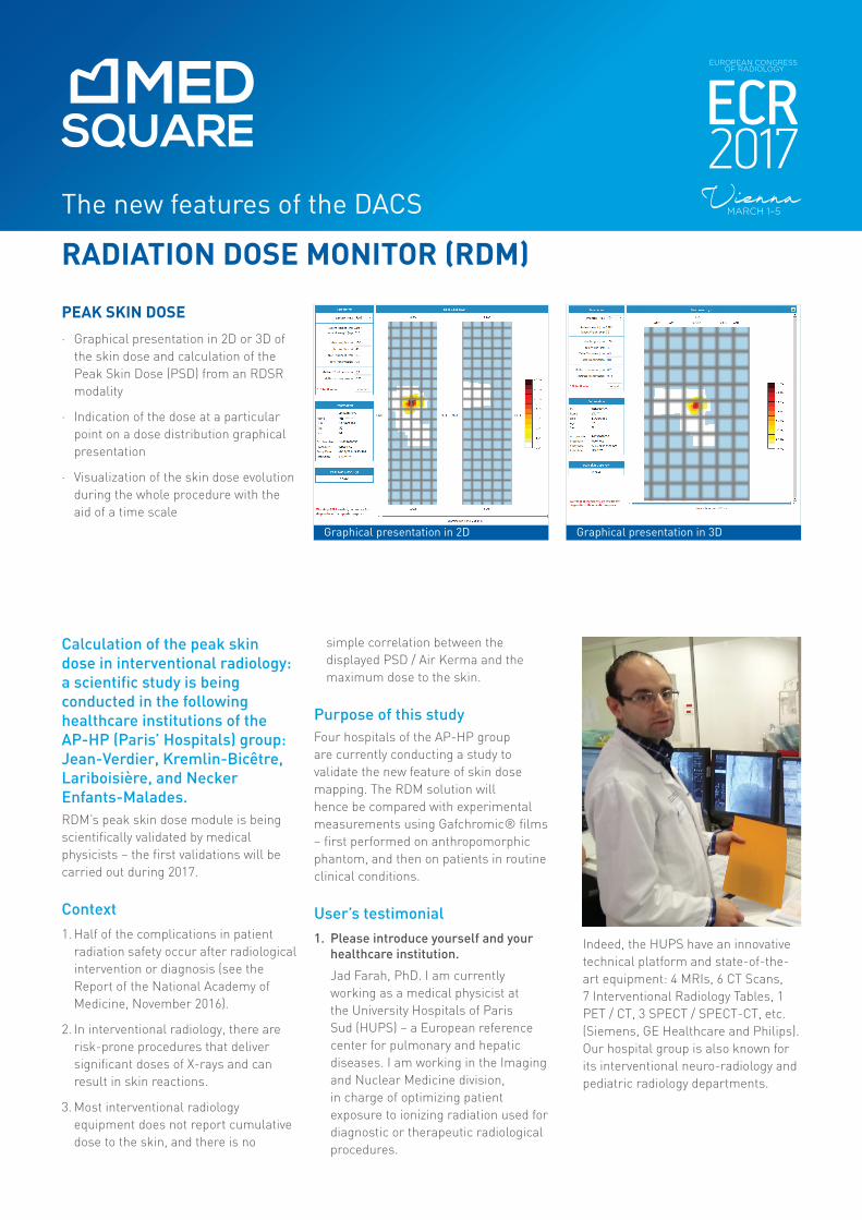
· Graphical presentation in 2D or 3D of the skin dose and calculation of the Peak Skin Dose (PSD) from an RDSR modality
· Indication of the dose at a particular point on a dose distribution graphical presentation
· Visualization of the skin dose evolution during the whole procedure with the aid of a time scale
Calculation of the peak skin dose in interventional radiology: a scientific study is being conducted in the following healthcare institutions of the AP-HP (Paris’ Hospitals) group: Jean-Verdier, Kremlin-Bicêtre, Lariboisière, and Necker Enfants-Malades.RDM’s peak skin dose module is being scientifically validated by medical physicists − the first validations will be carried out during 2017.
Context1. Half of the complications in patient
radiation safety occur after radiological intervention or diagnosis (see the Report of the National Academy of Medicine, November 2016).
2. In interventional radiology, there are risk-prone procedures that deliver significant doses of X-rays and can result in skin reactions.
3. Most interventional radiology equipment does not report cumulative dose to the skin, and there is no
simple correlation between the displayed PSD / Air Kerma and the maximum dose to the skin.
Purpose of this studyFour hospitals of the AP-HP group are currently conducting a study to validate the new feature of skin dose mapping. The RDM solution will hence be compared with experimental measurements using Gafchromic® films − first performed on anthropomorphic phantom, and then on patients in routine clinical conditions.
User’s testimonial1. Please introduce yourself and your
healthcare institution.
Jad Farah, PhD. I am currently working as a medical physicist at the University Hospitals of Paris Sud (HUPS) − a European reference center for pulmonary and hepatic diseases. I am working in the Imaging and Nuclear Medicine division, in charge of optimizing patient exposure to ionizing radiation used for diagnostic or therapeutic radiological procedures.
Indeed, the HUPS have an innovative technical platform and state-of-the-art equipment: 4 MRIs, 6 CT Scans, 7 Interventional Radiology Tables, 1 PET / CT, 3 SPECT / SPECT-CT, etc. (Siemens, GE Healthcare and Philips). Our hospital group is also known for its interventional neuro-radiology and pediatric radiology departments.
The new features of the DACS
RADIATION DOSE MONITOR (RDM)
Graphical presentation in 2D Graphical presentation in 3D
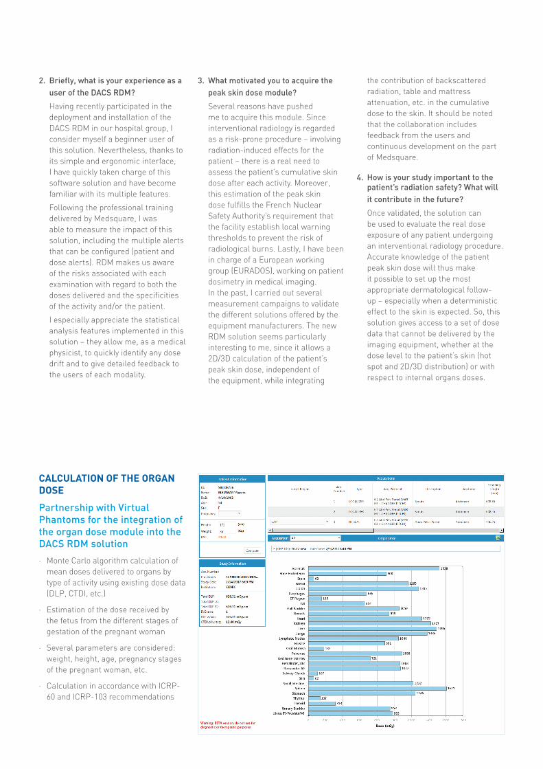
CALCULATION OF THE ORGAN DOSE
Partnership with Virtual Phantoms for the integration of the organ dose module into the DACS RDM solution
· Monte Carlo algorithm calculation of mean doses delivered to organs by type of activity using existing dose data (DLP, CTDI, etc.)
· Estimation of the dose received by the fetus from the different stages of gestation of the pregnant woman
· Several parameters are considered: weight, height, age, pregnancy stages of the pregnant woman, etc.
· Calculation in accordance with ICRP-60 and ICRP-103 recommendations
2. Briefly, what is your experience as a user of the DACS RDM?
Having recently participated in the deployment and installation of the DACS RDM in our hospital group, I consider myself a beginner user of this solution. Nevertheless, thanks to its simple and ergonomic interface, I have quickly taken charge of this software solution and have become familiar with its multiple features.
Following the professional training delivered by Medsquare, I was able to measure the impact of this solution, including the multiple alerts that can be configured (patient and dose alerts). RDM makes us aware of the risks associated with each examination with regard to both the doses delivered and the specificities of the activity and/or the patient.
I especially appreciate the statistical analysis features implemented in this solution − they allow me, as a medical physicist, to quickly identify any dose drift and to give detailed feedback to the users of each modality.
3. What motivated you to acquire the peak skin dose module?
Several reasons have pushed me to acquire this module. Since interventional radiology is regarded as a risk-prone procedure − involving radiation-induced effects for the patient − there is a real need to assess the patient’s cumulative skin dose after each activity. Moreover, this estimation of the peak skin dose fulfills the French Nuclear Safety Authority’s requirement that the facility establish local warning thresholds to prevent the risk of radiological burns. Lastly, I have been in charge of a European working group (EURADOS), working on patient dosimetry in medical imaging. In the past, I carried out several measurement campaigns to validate the different solutions offered by the equipment manufacturers. The new RDM solution seems particularly interesting to me, since it allows a 2D/3D calculation of the patient’s peak skin dose, independent of the equipment, while integrating
the contribution of backscattered radiation, table and mattress attenuation, etc. in the cumulative dose to the skin. It should be noted that the collaboration includes feedback from the users and continuous development on the part of Medsquare.
4. How is your study important to the patient’s radiation safety? What will it contribute in the future?
Once validated, the solution can be used to evaluate the real dose exposure of any patient undergoing an interventional radiology procedure. Accurate knowledge of the patient peak skin dose will thus make it possible to set up the most appropriate dermatological follow-up − especially when a deterministic effect to the skin is expected. So, this solution gives access to a set of dose data that cannot be delivered by the imaging equipment, whether at the dose level to the patient’s skin (hot spot and 2D/3D distribution) or with respect to internal organs doses.
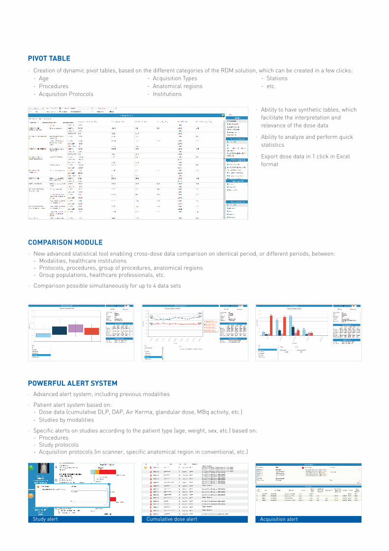
PIVOT TABLE
· Creation of dynamic pivot tables, based on the different categories of the RDM solution, which can be created in a few clicks:- Age - Procedures- Acquisition Protocols
POWERFUL ALERT SYSTEM· Advanced alert system, including previous modalities
· Patient alert system based on:- Dose data (cumulative DLP, DAP, Air Kerma, glandular dose, MBq activity, etc.)- Studies by modalities
· Specific alerts on studies according to the patient type (age, weight, sex, etc.) based on:- Procedures- Study protocols- Acquisition protocols (in scanner, specific anatomical region in conventional, etc.)
COMPARISON MODULE· New advanced statistical tool enabling cross-dose data comparison on identical period, or different periods, between:
- Modalities, healthcare institutions- Protocols, procedures, group of procedures, anatomical regions- Group populations, healthcare professionals, etc.
· Comparison possible simultaneously for up to 4 data sets
- Acquisition Types - Anatomical regions- Institutions
- Stations- etc.
· Ability to have synthetic tables, which facilitate the interpretation and relevance of the dose data
· Ability to analyze and perform quick statistics
· Export dose data in 1 click in Excel format
Study alert Cumulative dose alert Acquisition alert
Medsquare, 17 rue du Jura, 75013 Paris, FRANCEW medsquare.com E [email protected] T + 33 (0)1 55 25 62 50
© 2017 Medsquare SAS - All rights reserved.Medsquare SAS reserves the right to modify the design and specifications described in this document at any time without notice.
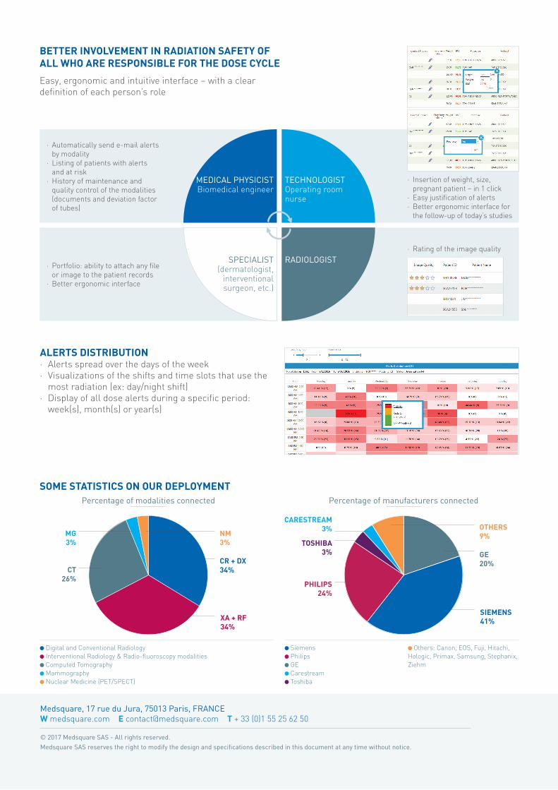
ALERTS DISTRIBUTION· Alerts spread over the days of the week· Visualizations of the shifts and time slots that use the
most radiation (ex: day/night shift)· Display of all dose alerts during a specific period:
week(s), month(s) or year(s)
BETTER INVOLVEMENT IN RADIATION SAFETY OF ALL WHO ARE RESPONSIBLE FOR THE DOSE CYCLE
Easy, ergonomic and intuitive interface − with a clear definition of each person’s role
· Automatically send e-mail alerts by modality
· Listing of patients with alerts and at risk
· History of maintenance and quality control of the modalities (documents and deviation factor of tubes)
· Insertion of weight, size, pregnant patient − in 1 click
· Easy justification of alerts· Better ergonomic interface for
the follow-up of today’s studies
· Portfolio: ability to attach any file or image to the patient records
· Better ergonomic interface
· Rating of the image quality
MEDICAL PHYSICISTBiomedical engineer
SPECIALIST(dermatologist,
interventional surgeon, etc.)
TECHNOLOGIST Operating room nurse
RADIOLOGIST
SOME STATISTICS ON OUR DEPLOYMENTPercentage of modalities connected Percentage of manufacturers connected
Digital and Conventional Radiology Interventional Radiology & Radio-fluoroscopy modalities Computed Tomography Mammography Nuclear Medicine (PET/SPECT)
Siemens Philips GE Carestream Toshiba
CR + DX34%
XA + RF 34%
CT26%
PHILIPS 24%
MG3% TOSHIBA
3%
CARESTREAM 3%
NM3%
Others: Canon, EOS, Fuji, Hitachi, Hologic, Primax, Samsung, Stephanix, Ziehm
SIEMENS 41%
OTHERS 9%
GE 20%